


Abstract
Chronic kidney disease (CKD) affects millions globally and represents a major health burden. This narrative review adopts a life-course perspective to synthesize current evidence on CKD as a consequence of adverse early-life exposures that disrupt nephrogenesis, leading to kidney programming and reduced nephron endowment. The objective of this review is to integrate emerging mechanistic and translational evidence linking developmental programming, gut microbiota, and redox biology within a unified gut–redox axis framework, and to identify potential targets for early-life prevention of CKD. Central to this process is the gut–redox axis, a bidirectional network linking gut microbiota with host redox homeostasis. A balanced axis preserves epithelial integrity, metabolic stability, and immune regulation, whereas dysbiosis and oxidative stress form a self-perpetuating cycle that promotes CKD and related comorbidities. Maternal oxidative stress and impaired microbial transmission exacerbate early-life dysbiosis, persistent epigenetic alterations, and nephron deficits. In adulthood, protein-bound uremic toxins amplify oxidative injury and inflammation, further perturbing microbial composition. Experimental and clinical studies show that early-life interventions—including probiotics, prebiotics, postbiotics, antioxidants, and toxin-lowering strategies—can restore gut–redox balance and improve renal outcomes. These insights highlight opportunities for precision prevention and mechanism-based therapies targeting CKD across the life course.
1. Introduction
Globally, chronic kidney disease (CKD) affects over 700 million individuals—approximately 10% of the adult population—and ranks among the leading causes of death and disability, representing a significant and escalating public health burden [1]. While traditional nephrology has historically focused on the cumulative impact of sequential renal injuries in adulthood, emerging evidence necessitates a paradigm shift toward a life-course approach grounded in the Developmental Origins of Health and Disease (DOHaD) context [2]. This perspective recognizes CKD as a multifactorial disorder rooted in developmental programming, whereby adverse exposures during critical windows of developmental plasticity—particularly the first 1000 days from conception to the child’s second birthday—shape long-term renal structure, function, and disease susceptibility [3]. Adverse maternal conditions—including nutritional imbalance, metabolic disturbances such as gestational diabetes mellitus (GDM), and exposure to environmental toxicants (e.g., heavy metals and phthalates)—can disrupt fetal nephrogenesis, leading to persistent structural and functional alterations via a process referred to as “kidney programming” [4]. A congenitally reduced nephron endowment constitutes the definitive “first hit,” which lowers the threshold for kidney failure when the host is later subjected to environmental or metabolic stressors [5,6].
At the center of this lifelong disease trajectory is the gut–redox axis, a complex, bidirectional regulatory network linking the intestinal microbial ecology with host redox homeostasis [7,8,9]. Redox status—the equilibrium between pro-oxidants, primarily reactive oxygen and nitrogen species (ROS/RNS), and the host’s antioxidant defense systems—is a fundamental modulator of life and kidney health [10,11]. A balanced axis maintains intestinal epithelial barrier integrity, promotes systemic metabolic stability, and regulates immune signaling pathways [12]. In a state of eubiosis, diverse microbial communities produce beneficial metabolites, most notably short-chain fatty acids (SCFAs) like butyrate, which strengthen tight junction proteins (e.g., ZO-1, occludin) and activate the nuclear factor erythroid 2-related factor 2 (Nrf2)/Keap1 pathway, the primary cellular defense against oxidative damage [13]. Conversely, perturbations in this axis trigger a self-perpetuating cycle of gut dysbiosis and oxidative stress [14].
Maternal oxidative stress and dysbiotic microbiota contribute to offspring kidney disease [15,16]. Accumulating human and experimental evidence indicates that the timing of gut–redox disruption critically shapes renal outcomes across the life course. Maternal redox status governs intergenerational microbial transmission. Oxidative stress induced by maternal insults disrupts placental integrity and triggers maternal immune activation, impairing vertical transfer of pioneer microbes [17,18]. Because these early colonizers help establish neonatal redox tone and immune priming, their disruption has been proposed to contribute to early-life dysbiosis and may influence epigenetic regulation and kidney developmental programming, potentially increasing susceptibility to CKD and hypertension later in life [19].
Across the life course, the gut–redox axis evolves from a developmental modulator to a central integrator of aging-related vulnerability. In adulthood—and particularly with advancing age—it functions as both mediator and amplifier of systemic stress signals. In CKD, the accumulation of protein-bound uremic toxins—such as p-cresyl sulfate (pCS), indoxyl sulfate (IS), and trimethylamine N-oxide (TMAO)—has been associated with oxidative injury and may contribute to redox imbalance and disease progression, although direct causal evidence from human interventional studies remains limited [20,21]. These toxins have been shown to be associated with increased oxidative stress through activation of inflammatory signaling pathways, including NF-κB, in a context- and cell-type–dependent manner, and may promote ROS generation via multiple sources such as mitochondrial dysfunction and NADPH oxidase (NOX) activation, thereby contributing to systemic inflammation and renal fibrotic processes. This creates a bidirectional feed-forward loop: progressive kidney impairment reshapes gut microbial ecology toward a more pathogenic profile, which generates further uremic toxins and reactive species, accelerating functional decline [22].
Clinical and preclinical evidence indicates that this axis is modifiable. Microbiota-targeted interventions (e.g., probiotics and prebiotics), redox-modulating antioxidants, and uremic toxin–lowering therapies have demonstrated the potential to attenuate oxidative stress, restore microbial homeostasis, and improve renal outcomes [23,24,25], particularly when implemented early in life through reprogramming strategy [4,19]. By integrating findings from human and experimental research, this narrative review defines the mechanistic framework of gut–redox interactions across the life course and highlights emerging opportunities for precision prevention and mechanism-based interventions. The overall concept is summarized in Figure 1.
Figure 1.
Kidney Programming Trajectory and the Effect of Early-Life Redox–Gut Reprogramming. Upper panel shows the pathological trajectory: renal capacity declines from conception after a prenatal “first hit” reduces nephron endowment. Early loss of SCFA-producing microbiota accelerates deterioration, followed by adult accumulation of uremic toxins (IS, pCS, TMAO) leading to cardiovascular-kidney-metabolic (CKM) syndrome, and later mitochondrial dysfunction, Klotho deficiency, and chronic inflammation. Lower panel shows the therapeutic trajectory: early-life Redox–Gut reprogramming within the first 1000 days shifts the curve toward preserved renal resilience. Microbiota-targeted therapy, antioxidants, and toxin-lowering strategies restore redox balance, maintain kidney function, and delay age-related decline.
2. Materials and Methods
Given the conceptual breadth of the life course framework and the heterogeneity of experimental and clinical methodologies in this field, this review was conducted using a narrative synthesis approach designed to facilitate theory-driven integration rather than quantitative evidence grading. This strategy enabled the consolidation of evolving mechanistic insights across developmental biology, nephrology, microbiome science, redox biology, and aging research within a unified gut–redox–kidney axis framework.
A structured literature search was conducted to identify peer-reviewed articles published in English between January 2000 and December 2025. Major biomedical databases, including MEDLINE, Embase, and the Cochrane Library, were systematically queried. Study selection followed a two-stage screening process: (i) title and abstract screening for relevance, and (ii) full-text assessment for eligibility based on predefined inclusion criteria.
The search strategy was organized around four conceptual modules aligned with the life course model: (1) developmental programming and DOHaD-related kidney vulnerability (e.g., maternal exposures, placental dysfunction, nephrogenesis, early-life insults, epigenetic regulation); (2) gut microbiota–redox signaling interactions (e.g., ROS/RNS pathways, Nrf2/Keap1 signaling, NF-κB activation, gut microbiota composition, microbial metabolites, immune modulation); (3) uremic toxins, inflammaging, and CKD progression (e.g., indoxyl sulfate, TMAO, fibrosis, cellular senescence); and (4) reprogramming and therapeutic modulation strategies targeting microbial ecology or redox balance.
Eligibility criteria included: (i) original experimental or clinical studies, (ii) studies addressing at least one component of the gut–redox–kidney axis, and (iii) articles providing mechanistic, translational, or clinical insight relevant to kidney development, CKD progression, or intervention. Exclusion criteria were: (i) non-peer-reviewed literature, (ii) non-English publications, (iii) studies not directly related to renal outcomes or gut–redox mechanisms, and (iv) purely descriptive reports without mechanistic or clinical relevance.
Both human studies and experimental animal models were included to enable cross-species translational interpretation, with greater interpretative weight given to human data where available, while animal studies were used primarily to support mechanistic insight rather than direct clinical extrapolation, particularly given known interspecies differences in gut microbiota composition and metabolic function. Inclusion priority was given to high-quality mechanistic studies, clinically relevant translational research, and landmark publications that significantly contributed to the DOHaD or gut–kidney axis framework. Reference lists of selected articles were manually screened to ensure comprehensive coverage of seminal and emerging studies. This concept-oriented methodology allowed for a cohesive evaluation of mechanistic continuity from early-life programming to aging-associated renal decline, highlighting modifiable nodes within the gut–redox axis relevant to precision prevention and intervention. During manuscript preparation, the authors used ChatGPT (version GPT-5.4 mini) to improve language clarity and readability and used NotebookLM (Google) and Gemini 3.0 to assist in figure organization. The authors carefully examined and revised all generated content and take full responsibility for the correctness and integrity of the final version.
3. Bidirectional Interactions Between Gut Microbiota and Redox Signaling
Redox status refers to the dynamic balance between the generation of ROS/RNS and the host’s antioxidant defense systems that maintain cellular homeostasis [10]. Within the gastrointestinal ecosystem, this balance is powerfully influenced by the intestinal microbiota, giving rise to the concept of the gut–redox axis—a bidirectional network linking microbial ecology, host oxidative signaling, and immune regulation [14,26]. Through this axis, microbial composition and metabolic activity shape the oxidative environment of the intestine and influence systemic physiological processes (Figure 2).
Figure 2.
Bidirectional Gut–Redox Feedback Loop in Health and Disease. This figure illustrates the dynamic reciprocal interaction between gut microbiota and host redox homeostasis, showing a virtuous cycle in health (top/right) and a vicious cycle in disease (bottom/left).
3.1. Microbiota-to-Redox Signaling
The gut microbiota regulates host redox homeostasis through two principal signaling pathways. Dysbiotic microbial communities are often associated with shifts in community composition, which may include expansion of certain Gram-negative taxa such as members of the Proteobacteria phylum in specific contexts, potentially increasing luminal lipopolysaccharide (LPS) burden. LPS activates the Toll-like receptor 4 (TLR4)/NF-κB signaling cascade, promoting pro-inflammatory cytokine production and NADPH oxidase-mediated ROS generation, thereby amplifying oxidative stress and inflammation [27,28]. In contrast, commensal microbes including Lactobacillus and Bifidobacterium stimulate the Nrf2/Keap1 antioxidant pathway, inducing transcription of protective enzymes such as superoxide dismutase (SOD), catalase (CAT), and glutathione peroxidase (GPx) that restore redox equilibrium [29].
Microbial metabolites are key regulators of the gut–redox network. SCFAs (acetate, propionate, butyrate) inhibit histone deacetylases and activate GPCR signaling, thereby upregulating Nrf2 and enhancing antioxidant enzymes (e.g., SOD, CAT), which reduces oxidative stress [30,31]. In contrast, dysbiosis promotes accumulation of deleterious metabolites such as TMAO and IS, which exacerbate mitochondrial dysfunction, oxidative stress, and tissue injury. TMAO, derived from dietary choline and carnitine, impairs mitochondrial function and activates the NLRP3 inflammasome, promoting renal fibrosis and further perturbing redox homeostasis. These processes collectively accelerate endothelial senescence and vascular aging [32]. IS and pCS, which are protein-bound and poorly cleared by dialysis, enter renal tubular cells via organic anion transporters (OATs) and induce oxidative stress through multiple complementary pathways, including NF-κB activation, AhR signaling, and promotion of tubular cellular senescence, leading to increased ROS production and renal injury [33]. Additionally, tryptophan-derived indole metabolites support epithelial barrier integrity via AhR signaling [34]. Notably, AhR acts as a ligand-dependent redox regulator, whereby uremic toxins promote oxidative stress whereas beneficial microbial metabolites enhance antioxidant defenses, linking gut microbial metabolism to host redox homeostasis [35].
3.2. Redox-Driven Remodeling of the Gut Microbiota
Oxidative stress is a key ecological force shaping the gut microbiota [26]. ROS from mitochondrial leakage, immune activation, or environmental exposures alter the intestinal redox environment and damage microbial components, favoring expansion of facultative anaerobes and pro-inflammatory taxa such as Proteobacteria (e.g., Escherichia coli) [36], while suppressing beneficial anaerobes (Bacteroidetes, Lactobacillus, Bifidobacterium). This shift reduces SCFA production and further disrupts intestinal homeostasis.
Redox signaling pathways mediate these microbiota shifts. ROS activates the NF-κB pathway, promoting pro-inflammatory cytokine release that disrupts microbial balance, whereas impairment of the Nrf2 antioxidant pathway weakens mucosal defenses and allows oxidative conditions that favor pathogenic bacteria [37]. Dysbiosis, in turn, increases LPS-producing Gram-negative bacteria, which stimulate TLR4/MyD88 signaling, further amplifying inflammation and ROS generation [28]. Together, these processes constitute a bidirectional feedback loop rather than a unidirectional cascade. Excess ROS contributes to disruption of epithelial tight junction integrity, including ZO-1, occludin, and claudins, both through direct oxidative modification and indirectly via activation of kinase-dependent pathways such as MLCK-mediated phosphorylation, thereby increasing intestinal permeability and facilitating endotoxin translocation. Together, these processes form a self-reinforcing cycle in which oxidative stress reshapes the microbiota, and dysbiosis amplifies oxidative and inflammatory signaling, linking the gut–redox axis to CKD and related cardiometabolic diseases.
4. Gut–Redox Axis in Pregnancy and Fetal Development
Recent research increasingly highlights the gut–redox axis as an important pathway influencing maternal biological regulation and the programming of fetal development [38,39]. During pregnancy, the maternal gut microbiota undergoes dynamic remodeling to accommodate the metabolic demands of gestation and fetal growth. These microbial shifts interact with hormonal regulation, nutrient metabolism, and immune signaling, collectively shaping the maternal redox environment that influences fetal development and long-term disease susceptibility [40,41].
4.1. Redox Status and Gut Remodeling in Normal Pregnancy
Pregnancy triggers coordinated hormonal, metabolic, and immunological adaptations to support fetal growth and maternal energy balance. Maternal gut microbiota undergoes substantial restructuring [42], and this period is characterized by localized, tightly regulated ROS signaling [43,44], which supports fetal nourishment and energy storage for lactation. Nitric oxide (NO) plays a dual role in redox biology: under physiological conditions, it maintains vascular homeostasis and antioxidant balance, whereas under oxidative stress it reacts with superoxide to form peroxynitrite, a highly reactive species that promotes nitrosative damage to lipids, proteins, and DNA while reducing NO bioavailability [45]. NO also regulates key reproductive processes, including implantation, placental angiogenesis, uterine relaxation, and fetal development [46]. Low levels of asymmetric dimethylarginine (ADMA, an inhibitor of NOS) and enhanced NO bioavailability support early gestational hemodynamic adaptation, whereas rising ADMA levels in late pregnancy modulate uterine relaxation and may contribute to the initiation of labor [47].
Longitudinal studies have demonstrated a progressive reduction in microbial richness (α-diversity) and increased interindividual variability (β-diversity) as pregnancy advances [42]. During the first trimester, the microbial composition resembles that of non-pregnant women and is typically dominated by members of the Firmicutes phylum. By the third trimester, however, there is a notable expansion of Proteobacteria and Actinobacteria, accompanied by a decline in butyrate-producing taxa.
These compositional shifts are partly driven by rising concentrations of pregnancy-associated hormones, particularly progesterone and estrogen. Progesterone has been shown to promote the proliferation of Bifidobacterium species [48]. Although the third-trimester microbiota shares certain features with dysbiotic profiles observed in metabolic syndrome, these changes are generally considered adaptive in healthy pregnancies, facilitating increased energy harvest and maternal fat storage required for lactation [42]. Nevertheless, these physiological adaptations are associated with a mild pro-inflammatory state and increased oxidative stress [49].
4.2. Dysregulated Gut–Redox Axis in Compromised Pregnancy
Emerging evidence identifies the gut–redox axis—the bidirectional interplay between intestinal microbial ecology and host redox signaling—as a central determinant of pregnancy outcomes and offspring programming. When this finely tuned redox balance is disrupted by maternal conditions such as obesity [50], GDM [51], preeclampsia [52], preterm birth [53], preterm premature rupture of membranes [54], or intrauterine growth restriction (IUGR) [55], pregnancy shifts from physiological stress to pathological oxidative damage.
Overweight mothers exhibit pronounced microbial dysbiosis, with decreased Bifidobacterium and Bacteroides and overgrowth of pro-inflammatory taxa such as Staphylococcus and Escherichia coli [56]. This imbalance increases LPS production and reduces beneficial metabolites, notably SCFAs like butyrate; loss of SCFA-producing bacteria such as Faecalibacterium prausnitzii exacerbates insulin resistance and chronic low-grade inflammation. In preeclampsia, gut microbial diversity drops further, with expansion of pathogenic taxa including Clostridium difficile and Fusobacterium [57], correlating with elevated blood pressure (BP) and renal dysfunction. Reduced SCFA production at the uteroplacental interface diminishes propionate and butyrate levels, promoting placental hypoxia and increased ROS generation.
A hallmark of compromised pregnancy is “leaky gut” [58], where pathological oxidative stress damages the intestinal barrier, allowing translocation of endotoxins and uremic precursors into the maternal circulation. Excess maternal oxidative stress may influence the fetal environment through selective placental transfer of certain redox-related molecules (e.g., H2O2) and indirect placental signaling, including inflammatory and endocrine mediators. Although not all ROS or cytokines cross the placenta freely, these pathways may contribute to fetal epigenetic programming and potentially prime offspring for long-term disease susceptibility. Understanding the shift from adaptive remodeling to a dysregulated gut–redox axis provides a mechanistic foundation for precision interventions to protect maternal and fetal health.
4.3. Transplacental Microbial Metabolite Signaling and Fetal Programming
The long-standing paradigm of a sterile fetal environment has been reconsidered with the recognition of a gut–placenta–fetus signaling axis, through which maternal microbial metabolites act as transplacental biochemical messengers. Although the translocation of live bacteria remains controversial [59], accumulating evidence demonstrates that numerous microbiota-derived metabolites enter the maternal circulation and cross the placenta, thereby shaping fetal development before birth [39].
Among these metabolites, SCFAs are the most extensively characterized. Produced by microbial fermentation of dietary fiber in the maternal colon, SCFAs readily reach the fetal compartment. In the fetus, they function as signaling molecules through SCFA receptors GPR41 and GPR43, promoting thymic development, regulatory T-cell differentiation, and intestinal barrier maturation [60].
Other metabolites also contribute to fetal programming. TMAO and related betaines, generated from microbial metabolism of dietary choline and carnitine, can cross the placenta and participate in fetal development. Although elevated TMAO levels are associated with cardiovascular risk in adults [61], physiological levels of maternal-derived TMAO appear to support normal fetal neurodevelopment, including processes such as thalamocortical axonogenesis [62]. In parallel, tryptophan-derived indole metabolites, including indole-3-propionate and indole-3-aldehyde, function as ligands for the AhR. These metabolites support the maturation of fetal innate immune cells and lymphoid structures while also exerting antioxidant and neuroprotective effects [63].
Importantly, the spectrum of metabolites reaching the fetus is highly sensitive to the maternal microbial state. Adverse intrauterine conditions, such as maternal high-fat diets, can induce gut dysbiosis and alter the metabolite profile available for placental transfer, often reducing beneficial SCFAs while increasing potentially harmful metabolites such as TMAO [64]. Conversely, dietary or microbiota-targeted interventions during pregnancy may enhance the production of protective microbial metabolites within the fetoplacental environment, thereby influencing fetal programming and long-term health outcomes [39,65].
5. CKD: A Life Course Perspective
Currently, CKD cannot be fully prevented by adult-focused strategies alone [1,2]. Although KDIGO-recommended interventions such as BP and glycemic control can reduce risk and slow disease progression, they have limited impact on the overall global burden, as they primarily target downstream manifestations rather than upstream developmental and early-life determinants of disease [25]. Nephron endowment is largely fixed before birth, and adverse intrauterine exposures can represent a “first hit” that reduces renal reserve [66]. Early-life risk factors may induce structural and functional alterations in the kidney that embed lifelong vulnerability to renal injury [67]. Therefore, integrating early-life programming into preventive strategies is essential to shift CKD management from reactive treatment toward true risk reduction.
5.1. DOHaD Framework
The DOHaD framework highlights how early-life exposures shape long-term kidney health [68]. Within this paradigm, kidney programming refers to impaired nephrogenesis resulting from adverse intrauterine conditions such as maternal malnutrition [69], metabolic disorders [70,71], environmental toxins [72], or medication exposure [73]. Evidence from epidemiologic studies—including famine cohorts [74]—and experimental models [75] demonstrates that insults during critical developmental windows disrupt kidney formation, redirect developmental pathways, and increase susceptibility to CKD later in life [76]. Key mechanisms implicated in kidney programming include oxidative stress, epigenetic modifications, impaired NO signaling, dysregulation of the renin–angiotensin system (RAS), disturbances in nutrient-sensing pathways, gut microbiota dysbiosis, and inflammation [2,3,34,35].
In humans, full-term neonates typically possess approximately one million nephrons per kidney, although this number can vary more than tenfold [6,77]. Nephrons are generated primarily through branching morphogenesis of the ureteric bud during fetal development. Adverse prenatal or perinatal conditions—such as intrauterine growth restriction (IUGR), preterm birth, maternal or neonatal malnutrition, or exposure to nephrotoxic medications like gentamicin—may disrupt this process and reduce nephron endowment, potentially leading to congenital anomalies of the kidney and urinary tract (CAKUT) [78,79].
Reduced nephron number represents a fundamental “first hit” in kidney programming, limiting renal reserve and rendering remaining nephrons more susceptible to secondary insults throughout life [5,6]. Because direct mechanistic studies in human neonates are constrained by ethical and technical limitations, much of the mechanistic understanding of kidney programming derives from experimental animal models, although a key translational limitation is that rodent nephrogenesis extends into the early postnatal period, whereas in humans it is largely completed in utero, leading to differences in developmental timing and exposure windows.
5.2. Kidney Programming: The Interplay Between Maternal Gut Dysbiosis and Oxidative Stress
5.2.1. Maternal Insults as Triggers of Oxidative Stress-Mediated Kidney Programming
Evidence from experimental models supports oxidative stress as a central mechanism in kidney programming. A range of maternal insults—including nutritional imbalance [80,81], maternal disease [82,83], environmental toxicants [84,85], and medication exposure [86]—can disrupt fetal redox homeostasis during critical windows of nephrogenesis, leading to persistent structural and functional renal alterations and increased lifetime risk of CKD and hypertension.
Among these, maternal nutritional perturbations are the most extensively studied, with both deficiency and excess (e.g., protein restriction, high-fat or high-fructose diets, and micronutrient imbalance) inducing oxidative stress-mediated renal programming in offspring [87]. These conditions disrupt redox balance (ROS/RNS), impair nephron maturation, and permanently alter renal structure and function.
Maternal diseases such as diabetes, hypertension, CKD, and inflammation also promote oxidative stress-mediated programming through systemic endothelial dysfunction and placental impairment [87]. Environmental exposures (e.g., pollutants, heavy metals, endocrine disruptors, and smoking) further exacerbate fetal oxidative stress, contributing to reduced nephron endowment and long-term renal vulnerability [85].
5.2.2. Molecular Mechanisms in Oxidative Stress-Induced Kidney Programming
The developing fetus is highly susceptible to oxidative injury due to immature antioxidant defense systems [44], limiting the capacity to neutralize excess ROS and RNS. Under adverse intrauterine conditions, excessive ROS/RNS can overwhelm redox buffering capacity, leading to damage to proteins, lipids, and DNA.
These mechanisms include upstream processes that increase ROS generation—such as upregulated expression of ROS-producing enzymes and enhanced ROS synthesis—followed by secondary downstream effects, including increased peroxynitrite formation, depletion of antioxidant capacity, elevated ADMA, reduced NO bioavailability, and accumulation of oxidative damage [87]. It also induces epigenetic modifications, including altered DNA methylation, histone modifications, and microRNA regulation [88], which may lead to persistent changes in gene expression affecting RAS signaling, nutrient sensing, mitochondrial function, and inflammation. These mechanisms contribute to long-term structural and functional renal alterations, including reduced nephron endowment, glomerulosclerosis, tubular injury, and albuminuria, increasing susceptibility to hypertension and CKD in adulthood [89].
In rodents, where nephrogenesis extends into the postnatal period, both prenatal and early postnatal exposures influence renal development, providing a broader window to study kidney programming and potential preventive interventions [90].
5.2.3. Maternal Gut Microbiota in the Developmental Origins of Kidney Disease
In parallel with oxidative stress, accumulating evidence indicates that maternal gut microbiota plays an important role in shaping the intrauterine and early-life environments that influence kidney programming [19,20]. The maternal gut microbiome functions as a metabolically active ecosystem that interacts with host physiology through the production of microbial metabolites, modulation of immune responses, and regulation of systemic metabolic pathways. Disruptions in this microbial community—commonly referred to as gut dysbiosis—can therefore have systemic consequences that extend beyond the gastrointestinal tract. During pregnancy, physiological changes in maternal metabolism and immunity are accompanied by shifts in gut microbial composition, and disturbances in this process may influence maternal–fetal interactions.
Maternal gut dysbiosis can affect host metabolism, immune responses, and redox balance through the production of microbial metabolites and inflammatory mediators. Changes in microbial composition can alter the generation of bioactive compounds such as SCFAs, TMAO, bile acid derivatives, and other microbial metabolites that influence host signaling pathways. These metabolites can enter the maternal circulation and potentially affect placental function and fetal development. SCFAs, for instance, are known to regulate energy metabolism, immune responses, and oxidative stress pathways, whereas excessive levels of certain microbial metabolites may contribute to metabolic disturbances and inflammatory activation. Through these mechanisms, maternal gut dysbiosis may amplify oxidative stress and inflammatory signaling within the maternal–placental–fetal axis.
5.2.4. Experimental Evidence Linking Gut Dysbiosis to Kidney Programming
Consistent with this concept, several experimental models of developmental renal disease demonstrate concurrent alterations in maternal or offspring gut microbiota alongside oxidative stress and kidney dysfunction. Models involving maternal high-fructose diets [91], high-fat diets [92], toxin exposure [93], and maternal CKD [94] have all been associated with shifts in gut microbial composition and metabolic profiles. In many of these models, microbial dysbiosis coexists with increased oxidative stress, altered metabolite production, and impaired renal development. These findings suggest that the gut microbiota may function as an intermediary linking environmental exposures to redox imbalance and kidney developmental abnormalities.
The interaction between microbial metabolism and host oxidative pathways therefore represents a critical component of the gut–kidney axis during early life. Microbiota-derived metabolites can influence oxidative signaling pathways, while oxidative stress itself may further alter microbial community composition, creating a bidirectional relationship between host physiology and the gut microbiome. Disruption of this delicate balance during critical developmental periods may have lasting effects on renal structure and function. A deeper understanding of how microbial metabolism interacts with host redox signaling during sensitive developmental windows may provide valuable insights into the developmental origins of kidney disease. Such knowledge may also open new opportunities for early-life interventions—including nutritional modulation, microbiota-targeted therapies, and antioxidant strategies—aimed at preventing CKD and its associated cardiometabolic complications across the life course.
5.3. Childhood: The Foundation of the Gut–Redox Axis
During early childhood, the gut microbiota functions as a critical microbial organ that supports immune maturation and metabolic homeostasis [95]. Initial colonization—primarily through vertical transmission from the mother—establishes pioneer microbial communities essential for long-term health [17]. Beneficial taxa such as Bifidobacterium and Lactobacillus contribute to immune balance and antioxidant defenses. However, disruptions during this plastic period—such as cesarean delivery, early antibiotic exposure, or maternal environmental enteric dysfunction—may lead to persistent dysbiosis.
Such early microbial disturbances may influence epigenetic regulation and immune development, potentially lowering the threshold for systemic inflammation. In childhood, these changes may manifest as subtle alterations in intestinal permeability and reduced production of SCFAs, metabolites that help maintain mitochondrial integrity and suppress renal inflammation [58].
Clinical studies increasingly suggest that disturbances in the gut–redox axis also occur in pediatric CKD [96,97]. Children with early-stage CKD often show reduced microbial diversity and shifts toward dysbiosis, including decreased abundance of SCFA-producing taxa such as Prevotella and increased representation of potentially pro-inflammatory bacteria including members of the Enterobacteriaceae and Enterococcaceae families [96,97,98]. Dysregulated microbial metabolism may alter production of metabolites such as TMAO and protein-bound uremic toxins. In pediatric CKD cohorts, microbial alterations have been associated with reduced urinary TMAO levels but potentially increased circulating concentrations as renal clearance declines [98].
Consistent with these microbial and metabolic disturbances, children with CKD often exhibit elevated oxidative stress biomarkers even at early disease stages [99]. Increased lipid peroxidation markers such as malondialdehyde correlate with disease severity and cardiovascular risk indicators including left ventricular hypertrophy and arterial stiffness [100]. These findings suggest that disruption of the gut–redox balance may contribute to both renal injury and early cardiovascular complications in pediatric CKD.
5.4. Adulthood: Cumulative Stressors and the Gut–Redox Feedback Loop
During adulthood, cumulative environmental, metabolic, and lifestyle exposures continue to shape the gut–kidney axis. Cardiovascular–kidney–metabolic (CKM) syndrome, a systemic clinical condition characterized by intertwined pathological and physiological interactions among metabolic abnormalities, CKD, and CVD, further exemplifies the integrated nature of cardiometabolic and kidney dysfunction leading to multi-organ impairment and increased cardiovascular risk [101]. Within this context, metabolic disorders—including obesity, diabetes, and hypertension—along with dietary habits and environmental toxicants, can disrupt gut microbial composition and sustain oxidative stress [102,103]. As in children, growing evidence shows that adults with CKD exhibit reduced microbial diversity and depletion of beneficial commensal bacteria such as Bifidobacterium, Lactobacillus, and Prevotella, accompanied by expansion of opportunistic taxa including members of the Enterobacteriaceae family [104,105]. This dysbiotic shift favors proteolytic fermentation and increased production of microbial metabolites rather than beneficial SCFAs [16].
Among these metabolites, protein-bound uremic toxins—including IS and pCS—as well as TMAO play key roles in mediating oxidative stress and inflammation [106,107]. These compounds exert distinct but complementary pathogenic effects: IS primarily enters renal tubular cells via OTAs, where it activates NF-κB signaling and promotes oxidative stress and mitochondrial dysfunction, whereas TMAO predominantly targets endothelial cells, contributing to endothelial dysfunction, vascular inflammation, and cardiovascular risk; pCS similarly induces oxidative stress and inflammatory signaling in renal and vascular tissues. Damage to intestinal epithelial tight junctions by oxidative stress increases intestinal permeability, allowing endotoxins and microbial metabolites to enter the systemic circulation. As kidney function declines, elevated urea levels in the intestinal lumen promote the growth of urease-producing bacteria, further aggravating dysbiosis. Through these mechanisms, renal dysfunction and microbial imbalance reinforce each other, forming a bidirectional gut–kidney feedback loop that accelerates CKD progression [108,109].
Oxidative stress and inflammation also damage intestinal tight junction proteins, increasing intestinal permeability and allowing microbial products and endotoxins to enter the circulation. As renal function declines, elevated urea levels in the intestinal lumen further alter the microbial environment by promoting the growth of urease-producing bacteria. Through these interconnected processes, kidney dysfunction and gut dysbiosis reinforce one another, establishing a bidirectional gut–kidney feedback loop that contributes to progressive renal injury and cardiovascular complications during adulthood.
5.5. Aging: Loss of Redox Resilience and Advanced Kidney Vulnerability
Aging is characterized by a progressive decline in the kidney’s capacity to maintain redox and metabolic homeostasis, resulting in increased vulnerability to injury [110,111]. Structural and functional changes accumulate over time, including nephron loss, glomerulosclerosis, tubular atrophy, and interstitial fibrosis, collectively reducing renal reserve. These processes are accompanied by mitochondrial dysfunction and increased ROS production, while age-related impairment of antioxidant defenses further limits the capacity to counteract oxidative damage. Reduced activity of protective pathways, including Klotho signaling and Sirtuin-mediated stress responses, also contributes to diminished cellular resilience and impaired repair [112,113].
Concurrently, aging is associated with altered gut microbial composition, including reduced diversity and depletion of SCFA-producing bacteria [114]. Decreased SCFA availability may impair intestinal barrier integrity, mitochondrial function, and systemic inflammatory control, thereby amplifying oxidative stress. In CKD, these age-related alterations are further exacerbated, with accumulation of gut-derived uremic toxins and persistent inflammation promoting endothelial dysfunction, vascular calcification, and cardiac remodeling.
Overall, aging represents a convergence of lifelong environmental exposures, cumulative oxidative injury, and progressive alterations in renal structure and gut microbiota, increasing susceptibility to kidney failure and cardiovascular complications [115]. Understanding the evolution of the gut–redox axis across the life course may provide a framework for strategies aimed at preserving renal resilience and reducing CKD burden.
6. Early Prevention via Redox–Gut Axis Reprogramming
Antioxidant therapies and gut microbiota-targeted interventions have demonstrated clear benefits in established CKD, as extensively reviewed elsewhere [116,117,118,119]. However, human evidence for their potential to prevent kidney disease by modulating the redox–gut axis during early life remains limited. Importantly, the translational therapeutic window must be interpreted in a species-specific manner: whereas nephrogenesis extends into the postnatal period in rodents, it is largely completed before birth in humans. Therefore, interventions effective in postnatal rodent models likely correspond to prenatal—particularly early to mid-gestational—windows in human development, when nephron induction, branching morphogenesis, and microvascular maturation are most active. Emerging experimental studies indicate that interventions targeting oxidative stress and gut microbial composition during critical developmental windows may offer a promising preventive strategy. In animal models, approaches such as antioxidant supplementation, probiotics, prebiotics, or dietary modifications have been shown to attenuate adverse renal outcomes induced by early-life insults, as illustrated by representative examples in Table 1 [80,85,91,92,94,120,121,122,123,124,125,126,127,128,129,130,131,132,133]. These results support the concept that early-life modulation of the redox–gut axis can reprogram renal development, enhance kidney resilience, and reduce susceptibility to CKD in later life.
Table 1.
Selected examples of early-life redox–gut axis reprogramming therapies for kidney programming-related offspring comorbidities.
6.1. Natural Antioxidants
Several natural antioxidants, including vitamins, amino acids, melatonin, and polyphenol, during pregnancy and lactation have shown benefits to kidney health and prevent kidney programming.
Vitamins C and E are the most commonly used antioxidants: vitamin C, a water-soluble free radical scavenger, and vitamin E, a lipid-soluble inhibitor of oxidative enzymes [134,135]. Vitamins C and E, among others, support kidney health [136], and maternal vitamin C supplementation protects offspring from LPS-induced hypertension by reducing intrarenal oxidative stress and modulating the RAS [120]. However, high doses of vitamins A, β-carotene, and E have been linked to increased mortality [137]; therefore, perinatal vitamin supplementation should be used cautiously, only in cases of deficiency, with attention to contamination risks for the developing fetus [138].
Certain dietary amino acids with antioxidant properties exert therapeutic and protective effects in kidney diseases [139,140]. In light of evidence that NO deficiency contributes to the developmental origins of kidney disease, perinatal supplementation with the NO-related amino acids arginine and citrulline has been investigated as a potential intervention to prevent adult-onset renal pathology in the offspring [80,121,122]. Arginine serves as the substrate for NOS-mediated NO production, whereas citrulline is a precursor of arginine [141,142]. The human kidney can convert citrulline to arginine [143]. Moreover, oral citrulline supplementation bypasses hepatic metabolism, thereby enhancing arginine production and increasing NO bioavailability [143]. Accordingly, citrulline has been more commonly used as a supplement than arginine to enhance NO bioavailability and protect adult rat offspring from kidney programming in oxidative stress-related models, as reviewed elsewhere [87].
In addition, tryptophan, taurine, and cysteine have also been evaluated as reprogramming interventions targeting oxidative stress in maternal CKD-induced kidney programming models [123,124,125]. Despite evidence that glycine and branched-chain amino acids can improve kidney outcomes [144], the role of oxidative stress reduction in mediating these protective effects has not been fully elucidated.
Another natural antioxidant is melatonin [145]. Melatonin, an endogenous tryptophan-derived indolamine, exerts multiple biological functions [146]. It has been clinically applied as an antioxidant therapy in pregnant women, neonates, and children [147,148,149]. Melatonin also plays an important role in pregnancy and fetal development [150]. Melatonin, along with its breakdown products, counteracts ROS and RNS, upregulates antioxidant systems, and enhances nitric oxide availability [151]. Experimental data demonstrate that perinatal melatonin administration may prevent adult-onset conditions, including kidney disorders [152]. As a potent antioxidant, melatonin administration during gestation and lactation has been shown to protect adult rat offspring against maternal caloric restriction-induced kidney programming and hypertension [126]. Its reprogramming effects include reduced renal 8-OHdG expression, restored NO bioavailability, and modulation of the RAS, another important source of oxidative stress in kidney disease. Additionally, maternal melatonin treatment has demonstrated renoprotective effects in several oxidative stress-related kidney programming models, including maternal methyl donor diet, L-NAME exposure, and high fructose intake, as reviewed elsewhere [87]. Although melatonin has a favorable safety profile [153,154], its routine use during pregnancy is not currently recommended [155]. Therefore, the perinatal use of melatonin as a preventive strategy for kidney health, particularly in fetuses and neonates, awaits further clinical translation.
Resveratrol is another natural antioxidant with renoprotective properties [156,157]. It is a naturally occurring nonflavonoid polyphenol belonging to the stilbene family [158]. Resveratrol exerts antioxidant effects by scavenging ROS/RNS, enhancing antioxidant enzyme activity, increasing glutathione levels, and upregulating NOS expression [159]. Perinatal administration of resveratrol has been shown to protect adult rat offspring from maternal CKD-induced kidney programming and hypertension [94]. Beyond its antioxidant activity, resveratrol may also act as a prebiotic by restoring microbial richness, increasing beneficial genera such as Lactobacillus and Bifidobacterium, and modulating microbial metabolite pathways [94]. As reviewed elsewhere [87], beneficial effects of resveratrol on renal outcomes in adult offspring have been demonstrated in several rat models of kidney programming, including maternal high-fructose diet, maternal NO deficiency, bisphenol A exposure, and TCDD exposure. However, a major limitation of the clinical utility of polyphenols is their low bioavailability [160]. Considering the substantial interindividual variability and complexity of resveratrol pharmacokinetics, future studies are needed to improve its therapeutic efficacy. Advanced delivery systems and artificial intelligence–assisted optimization strategies may provide promising approaches to enhance therapeutic precision and scalability [161].
6.2. Synthetic Antioxidants
Along with natural antioxidants, several synthetic antioxidants have also been investigated for their therapeutic potential in kidney diseases [24]. MitoQ, a mitochondria-targeted analogue of coenzyme Q10, reduces oxidative stress by suppressing superoxide production and lipid peroxidation [162]. A previous study demonstrated that perinatal MitoQ treatment prevented hypertension in adult mouse offspring, reduced mitochondrial oxidative stress, and attenuated structural and functional kidney abnormalities in a maternal smoking model [85].
N-acetylcysteine (NAC) is a well-known synthetic antioxidant, although it is derived from the natural amino acid cysteine [163]. NAC serves as a precursor of glutathione and a cysteine analogue that can be utilized for hydrogen sulfide (H2S) synthesis [164]. Perinatal NAC therapy prevented hypertension and oxidative stress in adult rat offspring exposed to maternal suramin administration, which was associated with increased glutathione production, restoration of NO bioavailability, and augmentation of the H2S signaling pathway [127].
Dimethyl fumarate (DMF), a well-established Nrf2 activator [165], provides another example of redox-targeted intervention. In a model combining prenatal dexamethasone exposure with a postnatal high-fat diet, DMF conferred protection by lowering oxidative stress markers—including ADMA and 8-OHdG—while simultaneously increasing NO levels [128].
Synthetic antioxidants, such as SOD mimetics, have also demonstrated potential in managing oxidative stress-related disorders [166]. For instance, gestational administration of the SOD mimetic tempol reduced proteinuria and BP in adult offspring of spontaneously hypertensive rats [167]. Despite these encouraging preclinical results, none of these agents have yet been applied in clinical maternal–fetal or neonatal care.
6.3. Gut Microbiota-Targeted Interventions
Early-life gut microbiota-targeted interventions—including probiotics, prebiotics, postbiotics, and modulation of microbial metabolites—have been investigated in various animal models of kidney programming. In clinical practice, the most commonly used microbiome-modulating approaches are probiotics and prebiotics. Probiotics are defined as live microorganisms that provide health benefits when consumed in sufficient amounts [168], while prebiotics are substrates that selectively stimulate the growth and activity of beneficial gut bacteria [168]. Synbiotics combine both strategies. Although these interventions may reduce uremic toxin production, robust clinical evidence for CKD prevention or treatment in humans remains limited [23,109,169,170].
As summarized in Table 1, preclinical studies consistently demonstrate protective effects of microbiota modulation on BP and renal outcomes in developmental programming models. For example, Lactobacillus casei protects against hypertension induced by maternal high-fructose or high-fat diets, likely via modulation of gut microbiota composition, restoration of SCFA signaling, and regulation of nutrient-sensing pathways [91,92]. Similarly, prebiotic interventions such as inulin reshape microbial composition, increase SCFA production, and are associated with improved BP control and renal protection in experimental models [91,92].
Beyond classical probiotics and prebiotics, dietary and phytochemical interventions (e.g., resveratrol and garlic oil) also exert microbiome-mediated protective effects [171]. Resveratrol restores microbial diversity and suppresses inflammatory signaling (including AhR- and Th17-related pathways) [94], while garlic oil increases SCFA-producing bacteria and enhances hydrogen sulfide (H2S) signaling via upregulation of H2S-generating enzymes [129]. Collectively, these findings highlight H2S as a key mechanistic link between gut microbiota and oxidative stress in kidney programming, acting through antioxidant and Nrf2-related pathways [172,173,174,175]. These interventions prevent hypertension and kidney injury by restoring H2S signaling and reshaping the gut microbiome. For example, maternal NAC increases fecal thiosulfate and Bifidobacterium abundance, mitigating oxidative damage induced by gut dysbiosis during critical developmental windows in spontaneously hypertensive rats [176].
Postbiotics, defined as microbial metabolites or components with biological activity, represent an emerging alternative to live microbial therapy, particularly in vulnerable populations where probiotic safety may be a concern [177,178,179]. However, very limited information is available regarding the use of postbiotics in human CKD [180]. Emerging evidence supports the potential of SCFAs as postbiotic agents for CVD prevention [181]. Among these, short-chain fatty acids (SCFAs; acetate, propionate, and butyrate) have been most extensively studied. In these models, SCFAs exert protective effects primarily through activation of SCFA-mediated signaling pathways, including increased renal expression of SCFA receptors, modulation of gut microbiota–derived metabolites, and receptor-dependent regulation of vascular tone and the RAS [130,131,182]. In particular, butyrate may also enhance nitric oxide bioavailability, supporting its potential antioxidant and vasoprotective effects [183].
Another microbiota-targeted therapy is fecal microbiota transplantation (FMT). Although FMT has been extensively studied in microbiome-related diseases [184,185], its application in human CKD remains limited to preclinical and preliminary clinical studies [186,187]. Only one study has evaluated FMT in a kidney programming model, where maternal FMT from healthy donors attenuated high-fructose-induced programmed hypertension in offspring, accompanied by increased plasma butyrate, upregulation of SCFA receptors (e.g., GPR41, GPR43), reduced oxidative stress, and restoration of gut microbiota–metabolite homeostasis [132].
Other microbiome-modulating approaches include the oral intestinal sorbent AST-120, which reduces microbiota-derived uremic toxins [188] and has shown cardiovascular benefits in adult CKD patients [189], although its effects on gut microbiota composition and kidney programming remain unclear.
In addition, microbial TMAO production can be inhibited by targeting gut microbial choline TMA lyase using 3,3-dimethyl-1-butanol (DMB) or iodomethylcholine (IMC) [190,191]. Maternal inhibition of TMAO formation with DMB or IMC prevents offspring hypertension in maternal CKD and high-fructose diet models, respectively [130,133], through suppression of the TMA–TMAO pathway and remodeling of the gut microbiota.
Collectively, these findings support the concept of developmental reprogramming of the gut–redox axis, whereby early microbiota-targeted interventions may redirect long-term renal and cardiovascular trajectories. However, translation to human CKD prevention remains an important unmet challenge requiring further clinical validation.
7. Conclusions and Future Perspective
The DOHaD framework reframes CKD as a consequence of developmental programming rather than solely the cumulative result of renal insults acquired during adulthood. Although this paradigm has substantially advanced our understanding of early-life determinants of renal vulnerability, key conceptual and methodological challenges remain, particularly regarding the gut–redox axis.
A central unresolved issue is the bidirectional relationship between gut dysbiosis and oxidative stress in CKD, which may function as both cause and consequence of uremia-associated barrier dysfunction, systemic inflammation, and microbiota-derived toxin accumulation [18,97,192]. This ambiguity is compounded by limitations in current microbiome research and model systems, where substantial variability in experimental and clinical conditions hampers causal inference and translational robustness [193].
Future progress will require more physiologically faithful platforms, such as multi-organ microphysiological systems, to better reproduce gut–kidney–immune interactions under controlled conditions [194,195]. In parallel, priority should be given to identifying microbiota-derived metabolites that regulate host redox signaling pathways (e.g., Nrf2, NF-κB, mitochondrial ROS, and inflammasome activation) as potential mechanistic links and therapeutic targets [196].
Another critical gap involves understanding how oxidative stress disrupts intestinal barrier integrity and promotes the “leaky gut–renal inflammation” loop through endotoxin translocation and systemic immune activation. This complexity is further underscored by the “antioxidant paradox,” whereby certain antioxidants may exhibit prooxidant activity depending on cellular redox conditions, acting as modulators rather than uniformly protective agents [197]. Within the DOHaD paradigm, it is also unclear whether early-life perturbations of gut–redox balance can impair nephron development and thereby predispose individuals to CKD later in life. In addition, host genetic variability in antioxidant defense systems and immune signaling pathways may modify susceptibility to microbiota-driven oxidative injury, highlighting the importance of host–microbiome interactions and suggesting that antioxidant effects may be context-dependent across developmental stages.
From a translational perspective, determining whether modulation of the gut–redox axis through microbiota-targeted therapies, dietary interventions, or reduction in intestinal toxin generation can alter CKD trajectory remains an important unresolved question. The identification of reliable gut-derived metabolites and oxidative stress-related markers as predictive biomarkers may further facilitate precision nephrology approaches.
Collectively, future studies must address a central question: how the gut microbiota regulates systemic redox homeostasis and thereby influences the initiation, progression, and potential prevention of CKD across the life course.
Author Contributions
Conceptualization, Y.-L.T. and C.-N.H.; Funding acquisition, Y.-L.T. and C.-N.H.; Writing—original draft, Y.-L.T. and C.-N.H.; data curation, Y.-L.T. and C.-N.H.; Writing—review and editing, Y.-L.T. and C.-N.H. All authors have read and agreed to the published version of the manuscript.
Funding
Financial support for this work was provided by the National Science and Technology Council, Taiwan (Grant Nos. 114-2314-B-182A-048 and 114-2314-B-182A-036-MY3).
Institutional Review Board Statement
Not applicable.
Informed Consent Statement
Not applicable.
Data Availability Statement
No new data were created or analyzed in this study.
Acknowledgments
During the preparation of this work, the authors used ChatGPT, version GPT-5.4 mini to improve language clarity and readability, and NotebookLM (Google) and Gemini 3.0 to assist with figure preparation. The authors carefully reviewed and edited all outputs generated by these tools and take full responsibility for the accuracy and integrity of the final content of the published article.
Conflicts of Interest
The authors declare no conflicts of interest.
References
- GBD 2023 Chronic Kidney Disease Collaborators. Global, regional, and national burden of chronic kidney disease in adults, 1990–2023, and its attributable risk factors: A systematic analysis for the Global Burden of Disease Study 2023. Lancet 2025, 406, 2461–2482. [Google Scholar] [CrossRef] [Scilit]
- Brophy, P.D.; Charlton, J.R.; Bryan Carmody, J.; Reidy, K.J.; Harshman, L.; Segar, J.; Askenazi, D.; Shoham, D.; Bagby, S.P. Chronic Kidney Disease: A Life Course Health Development Perspective. In Handbook of Life Course Health Development [Internet]; Halfon, N., Forrest, C.B., Lerner, R.M., Faustman, E.M., Eds.; Springer: Cham, Switzerland, 2018. [Google Scholar] [PubMed]
- Darling, J.C.; Bamidis, P.D.; Burberry, J.; Rudolf, M.C.J. The First Thousand Days: Early, integrated and evidence-based approaches to improving child health: Coming to a population near you? Arch. Dis. Child. 2020, 105, 837–841. [Google Scholar] [CrossRef] [Scilit]
- Kett, M.M.; Denton, K.M. Renal programming: Cause for concern? Am. J. Physiol.-Regul. Integr. Comp. Physiol. 2011, 300, R791–R803. [Google Scholar] [CrossRef] [Scilit]
- Nenov, V.D.; Taal, M.W.; Sakharova, O.V.; Brenner, B.M. Multi-hit nature of chronic renal disease. Curr. Opin. Nephrol. Hypertens. 2000, 9, 85–97. [Google Scholar] [CrossRef] [Scilit]
- Bertram, J.F.; Douglas-Denton, R.N.; Diouf, B.; Hughson, M.; Hoy, W.E. Human nephron number: Implications for health and disease. Pediatr. Nephrol. 2011, 26, 1529–1533. [Google Scholar] [CrossRef] [Scilit]
- Campbell, E.L.; Colgan, S.P. Control and dysregulation of redox signalling in the gastrointestinal tract. Nat. Rev. Gastroenterol. Hepatol. 2019, 16, 106–120. [Google Scholar] [CrossRef] [Scilit]
- Yun, B.; King, M.; Draz, M.S.; Kline, T.; Rodriguez-Palacios, A. Oxidative reactivity across kingdoms in the gut: Host immunity, stressed microbiota and oxidized foods. Free. Radic. Biol. Med. 2022, 178, 97–110. [Google Scholar] [CrossRef] [Scilit] [PubMed]
- Wu, X.; Mu, B.; Li, G.; Du, R.; Park, S. Gut Microbial Composition, Oxidative Stress, and Immunity in Metabolic Disease: Toward Personalized Interventions. Antioxidants 2026, 15, 175. [Google Scholar] [CrossRef] [Scilit] [PubMed]
- Divvela, S.S.K.; Gallorini, M.; Gellisch, M.; Patel, G.D.; Saso, L.; Brand-Saberi, B. Navigating redox imbalance: The role of oxidative stress in embryonic development and long-term health outcomes. Front. Cell Dev. Biol. 2025, 13, 1521336. [Google Scholar] [CrossRef] [Scilit] [PubMed]
- Kishi, S.; Nagasu, H.; Kidokoro, K.; Kashihara, N. Oxidative stress and the role of redox signalling in chronic kidney disease. Nat. Rev. Nephrol. 2024, 20, 101–119. [Google Scholar] [CrossRef] [Scilit]
- Mann, E.R.; Lam, Y.K.; Uhlig, H.H. Short-chain fatty acids: Linking diet, the microbiome and immunity. Nat. Rev. Immunol. 2024, 24, 577–595. [Google Scholar] [CrossRef] [Scilit]
- Tang, X.; Sun, Y.; Li, Y.; Ma, S.; Zhang, K.; Chen, A.; Lyu, Y.; Yu, R. Sodium butyrate protects against oxidative stress in high-fat-diet-induced obese rats by promoting GSK-3β/Nrf2 signaling pathway and mitochondrial function. J. Food Biochem. 2022, 46, e14334. [Google Scholar] [CrossRef] [Scilit] [PubMed]
- Kurhaluk, N.; Kamiński, P.; Tkaczenko, H. Role of Gut Microbiota in Modulating Oxidative Stress Induced by Environmental Factors. Cell. Physiol. Biochem. 2025, 59, 2–52. [Google Scholar] [CrossRef] [Scilit]
- Lu, P.C.; Tain, Y.L.; Lin, Y.J.; Hsu, C.N. Oxidative Stress in Maternal and Offspring Kidney Disease and Hypertension: A Life-Course Perspective. Antioxidants 2025, 14, 387. [Google Scholar] [CrossRef] [Scilit] [PubMed]
- Bloom, P.P.; Garrett, W.S.; Penniston, K.L.; Winkler, M.H.; Hazen, S.L.; Agudelo, J.; Suryavanshi, M.; Babiker, A.; Dodd, D.; Fischbach, M.A.; et al. Microbiota and kidney disease: The road ahead. Nat. Rev. Nephrol. 2025, 21, 702–716. [Google Scholar] [CrossRef] [Scilit]
- Ferretti, P.; Pasolli, E.; Tett, A.; Asnicar, F.; Gorfer, V.; Fedi, S.; Armanini, F.; Truong, D.T.; Manara, S.; Zolfo, M.; et al. Mother-to-Infant Microbial Transmission from Different Body Sites Shapes the Developing Infant Gut Microbiome. Cell Host Microbe 2018, 24, 133–145.e5. [Google Scholar] [CrossRef] [Scilit]
- McBeth, A.; Miller, E.A.; Thompson, B.; Hanaway, P.; Thexton, A.; Zwickey, H. Balancing Oxidative Stress: How the Gut Microbiome Supports Redox Homeostasis and Mitochondrial Health. J. Restor. Med. 2025, 15, 4–19. [Google Scholar] [CrossRef] [Scilit]
- Hsu, C.N.; Tain, Y.L. Chronic Kidney Disease and Gut Microbiota: What Is Their Connection in Early Life? Int. J. Mol. Sci. 2022, 23, 3954. [Google Scholar] [CrossRef] [Scilit]
- Yang, T.; Richards, E.M.; Pepine, C.J.; Raizada, M.K. The gut microbiota and the brain-gut-kidney axis in hypertension and chronic kidney disease. Nat. Rev. Nephrol. 2018, 14, 442–456. [Google Scholar] [CrossRef] [Scilit] [PubMed]
- Li, X.J.; Shan, Q.Y.; Wu, X.; Miao, H.; Zhao, Y.Y. Gut microbiota regulates oxidative stress and inflammation: A double-edged sword in renal fibrosis. Cell. Mol. Life Sci. 2024, 81, 480. [Google Scholar] [CrossRef] [Scilit] [PubMed]
- Wang, X.; Yang, S.; Li, S.; Zhao, L.; Hao, Y.; Qin, J.; Zhang, L.; Zhang, C.; Bian, W.; Zuo, L.; et al. Aberrant gut microbiota alters host metabolome and impacts renal failure in humans and rodents. Gut 2020, 69, 2131–2142. [Google Scholar] [CrossRef] [Scilit]
- Pei, M.; Wei, L.; Hu, S.; Yang, B.; Si, J.; Yang, H.; Zhai, J. Probiotics, prebiotics and synbiotics for chronic kidney disease: Protocol for a systematic review and meta-analysis. BMJ Open 2018, 8, e020863. [Google Scholar] [CrossRef] [Scilit]
- Jun, M.; Venkataraman, V.; Razavian, M.; Cooper, B.; Zoungas, S.; Ninomiya, T.; Webster, A.C.; Perkovic, V. Antioxidants for chronic kidney disease. Cochrane Database Syst. Rev. 2012, 10, CD008176. [Google Scholar] [CrossRef] [Scilit] [PubMed]
- Kidney Disease: Improving Global Outcomes (KDIGO) CKD Work Group. KDIGO 2024 Clinical Practice Guideline for the Evaluation and Management of Chronic Kidney Disease. Kidney Int. 2024, 105, S117–S314. [Google Scholar] [CrossRef] [Scilit]
- Semenova, N.; Garashchenko, N.; Kolesnikov, S.; Darenskaya, M.; Kolesnikova, L. Gut Microbiome Interactions with Oxidative Stress: Mechanisms and Consequences for Health. Pathophysiology 2024, 31, 309–330. [Google Scholar] [CrossRef] [Scilit]
- Neish, A.S. Redox signaling mediated by the gut microbiota. Free Radic. Res. 2013, 47, 950–957. [Google Scholar] [CrossRef] [Scilit] [PubMed]
- Guo, S.; Al-Sadi, R.; Said, H.M.; Ma, T.Y. Lipopolysaccharide causes an increase in intestinal tight junction permeability in vitro and in vivo by inducing enterocyte membrane expression and localization of TLR-4 and CD14. Am. J. Pathol. 2013, 182, 375–387. [Google Scholar] [CrossRef] [Scilit]
- Rezaie, N.; Aghamohammad, S.; Khiavi, E.H.A.G.; Talebi, M.; Pourshafie, M.R.; Rohani, M. The Analysis and Comparison of Anti-Inflammatory and Antioxidant Characteristics of Postbiotic and Paraprobiotic Derived From Novel Native Probiotic Cocktail in DSS-Induced Colitic Mice. Food Sci. Nutr. 2025, 13, e70034. [Google Scholar] [CrossRef] [Scilit]
- González-Bosch, C.; Boorman, E.; Zunszain, P.A.; Mann, G.E. Short-chain fatty acids as modulators of redox signaling in health and disease. Redox Biol. 2021, 47, 102165. [Google Scholar] [CrossRef] [Scilit]
- Magliocca, G.; Mone, P.; Di Iorio, B.R.; Heidland, A.; Marzocco, S. Short-Chain Fatty Acids in Chronic Kidney Disease: Focus on Inflammation and Oxidative Stress Regulation. Int. J. Mol. Sci. 2022, 23, 5354. [Google Scholar] [CrossRef] [Scilit] [PubMed]
- Kim, S.H.; Yoon, M.Y.; Yoon, S.S. TMAO and the gut microbiome: Implications for the CVD-CKD-IBD axis. Ann. Med. 2025, 57, 2522324. [Google Scholar] [CrossRef] [Scilit]
- Liu, J.R.; Miao, H.; Deng, D.Q.; Vaziri, N.D.; Li, P.; Zhao, Y.Y. Gut microbiota-derived tryptophan metabolism mediates renal fibrosis by aryl hydrocarbon receptor signaling activation. Cell. Mol. Life Sci. 2021, 78, 909–922. [Google Scholar] [CrossRef] [Scilit]
- Tan, Y.Q.; Wang, Y.N.; Feng, H.Y.; Guo, Z.Y.; Li, X.; Nie, X.L.; Zhao, Y.Y. Host/microbiota interactions-derived tryptophan metabolites modulate oxidative stress and inflammation via aryl hydrocarbon receptor signaling. Free Radic. Biol. Med. 2022, 184, 30–41. [Google Scholar] [CrossRef] [Scilit]
- Grishanova, A.Y.; Perepechaeva, M.L. Aryl Hydrocarbon Receptor in Oxidative Stress as a Double Agent and Its Biological and Therapeutic Significance. Int. J. Mol. Sci. 2022, 23, 6719. [Google Scholar] [CrossRef] [Scilit]
- Litvak, Y.; Byndloss, M.X.; Tsolis, R.M.; Bäumler, A.J. Dysbiotic Proteobacteria expansion: A microbial signature of epithelial dysfunction. Curr. Opin. Microbiol. 2017, 39, 1–6. [Google Scholar] [CrossRef] [Scilit]
- Singh, R.; Chandrashekharappa, S.; Bodduluri, S.R.; Baby, B.V.; Hegde, B.; Kotla, N.G.; Hiwale, A.A.; Saiyed, T.; Patel, P.; Vijay-Kumar, M.; et al. Enhancement of the gut barrier integrity by a microbial metabolite through the Nrf2 pathway. Nat. Commun. 2019, 10, 89. [Google Scholar] [CrossRef] [Scilit] [PubMed]
- Grzeszczak, K.; Łanocha-Arendarczyk, N.; Malinowski, W.; Ziętek, P.; Kosik-Bogacka, D. Oxidative Stress in Pregnancy. Biomolecules 2023, 13, 1768. [Google Scholar] [CrossRef] [Scilit]
- Tian, M.; Li, Q.; Zheng, T.; Yang, S.; Chen, F.; Guan, W.; Zhang, S. Maternal microbe-specific modulation of the offspring microbiome and development during pregnancy and lactation. Gut Microbes 2023, 15, 2206505. [Google Scholar] [CrossRef] [Scilit] [PubMed]
- Biagioli, V.; Matera, M.; Ramenghi, L.A.; Falsaperla, R.; Striano, P. Microbiome and Pregnancy Dysbiosis: A Narrative Review on Offspring Health. Nutrients 2025, 17, 1033. [Google Scholar] [CrossRef] [Scilit]
- Sinha, T.; Brushett, S.; Prins, J.; Zhernakova, A. The maternal gut microbiome during pregnancy and its role in maternal and infant health. Curr. Opin. Microbiol. 2023, 74, 102309. [Google Scholar] [CrossRef] [Scilit]
- Fu, Y.; Gou, W.; Wu, P.; Lai, Y.; Liang, X.; Zhang, K.; Shuai, M.; Tang, J.; Miao, Z.; Chen, J.; et al. Landscape of the gut mycobiome dynamics during pregnancy and its relationship with host metabolism and pregnancy health. Gut 2024, 73, 1302–1312. [Google Scholar] [CrossRef] [Scilit]
- Carter, A.M. Placental oxygen consumption. Part I: In vivo studies—A review. Placenta 2000, 21, S31–S37. [Google Scholar] [CrossRef] [Scilit]
- Dennery, P.A. Oxidative stress in development: Nature or nurture? Free Radic. Biol. Med. 2010, 49, 1147–1151. [Google Scholar]
- Prolo, C.; Piacenza, L.; Radi, R. Peroxynitrite: A multifaceted oxidizing and nitrating metabolite. Curr. Opin. Chem. Biol. 2024, 80, 102459. [Google Scholar] [CrossRef] [Scilit]
- Krause, B.J.; Hanson, M.A.; Casanello, P. Role of nitric oxide in placental vascular development and function. Placenta 2011, 32, 797–805. [Google Scholar] [CrossRef] [Scilit]
- Khalil, A.; Hardman, L.; Brien, P.O. The role of arginine, homoarginine and nitric oxide in pregnancy. Amino Acids 2015, 47, 1715–1727. [Google Scholar] [CrossRef] [Scilit]
- Nuriel-Ohayon, M.; Neuman, H.; Ziv, O.; Belogolovski, A.; Barsheshet, Y.; Bloch, N.; Uzan, A.; Lahav, R.; Peretz, A.; Frishman, S.; et al. Progesterone Increases Bifidobacterium Relative Abundance during Late Pregnancy. Cell Rep. 2019, 27, 730–736.e3. [Google Scholar] [CrossRef] [Scilit] [PubMed]
- Hussain, T.; Murtaza, G.; Metwally, E.; Kalhoro, D.H.; Kalhoro, M.S.; Rahu, B.A.; Sahito, R.G.A.; Yin, Y.; Yang, H.; Chughtai, M.I.; et al. The Role of Oxidative Stress and Antioxidant Balance in Pregnancy. Mediat. Inflamm. 2021, 2021, 9962860. [Google Scholar] [CrossRef] [Scilit] [PubMed]
- Negro, S.; Boutsikou, T.; Briana, D.D.; Tataranno, M.L.; Longini, M.; Proietti, F.; Bazzini, F.; Dani, C.; Malamitsi-Puchner, A.; Buonocore, G.; et al. Maternal obesity and perinatal oxidative stress: The strength of the association. J. Biol. Regul. Homeost. Agents 2017, 31, 221–227. [Google Scholar]
- Arya, S.; Ye, C.; Connelly, P.W.; Hanley, A.J.; Sermer, M.; Zinman, B.; Retnakaran, R. Asymmetric dimethylarginine and arginine metabolites in women with and without a history of gestational diabetes. J. Diabetes Complicat. 2017, 31, 964–970. [Google Scholar] [CrossRef] [Scilit]
- Pettersson, A.; Hedner, T.; Milsom, I. Increased circulating concentrations of asymmetric dimethyl arginine (ADMA), an endogenous inhibitor of nitric oxide synthesis, in preeclampsia. Acta Obstet. Gynecol. Scand. 1998, 77, 808–813. [Google Scholar] [CrossRef] [Scilit]
- de Almeida, V.O.; Pereira, R.A.; Amantéa, S.L.; Rhoden, C.R.; Colvero, M.O. Neonatal diseases and oxidative stress in premature infants: An integrative review. J. Pediatr. 2022, 98, 455–462. [Google Scholar] [CrossRef] [Scilit] [PubMed]
- Longini, M.; Perrone, S.; Vezzosi, P.; Marzocchi, B.; Kenanidis, A.; Centini, G.; Rosignoli, L.; Buonocore, G. Association between oxidative stress in pregnancy and preterm premature rupture of membranes. Clin. Biochem. 2007, 40, 793–797. [Google Scholar] [CrossRef] [Scilit]
- Kamath, U.; Rao, G.; Kamath, S.U.; Rai, L. Maternal and fetal indicators of oxidative stress during intrauterine growth retardation (IUGR). Indian J. Clin. Biochem. 2006, 21, 111–115. [Google Scholar] [CrossRef] [Scilit]
- Collado, M.C.; Isolauri, E.; Laitinen, K.; Salminen, S. Distinct composition of gut microbiota during pregnancy in overweight and normal-weight women. Am. J. Clin. Nutr. 2008, 88, 894–899. [Google Scholar] [CrossRef] [Scilit]
- Qing, W.; Shi, Y.; Zhou, H.; Chen, M. Gut microbiota dysbiosis in patients with preeclampsia: A systematic review. Med. Microecol. 2021, 10, 100047. [Google Scholar] [CrossRef] [Scilit]
- Camilleri, M. Leaky gut: Mechanisms, measurement and clinical implications in humans. Gut 2019, 68, 1516–1526. [Google Scholar] [CrossRef] [Scilit] [PubMed]
- Kennedy, K.M.; de Goffau, M.C.; Perez-Muñoz, M.E.; Arrieta, M.C.; Bäckhed, F.; Bork, P.; Braun, T.; Bushman, F.D.; Dore, J.; de Vos, W.M.; et al. Questioning the fetal microbiome illustrates pitfalls of low-biomass microbial studies. Nature 2023, 613, 639–649. [Google Scholar] [CrossRef] [Scilit] [PubMed]
- Ziętek, M.; Celewicz, Z.; Szczuko, M. Short-Chain Fatty Acids, Maternal Microbiota and Metabolism in Pregnancy. Nutrients 2021, 13, 1244. [Google Scholar] [CrossRef] [Scilit]
- Guasti, L.; Galliazzo, S.; Molaro, M.; Visconti, E.; Pennella, B.; Gaudio, G.V.; Lupi, A.; Grandi, A.M.; Squizzato, A. TMAO as a biomarker of cardiovascular events: A systematic review and meta-analysis. Intern. Emerg. Med. 2021, 16, 201–207. [Google Scholar] [CrossRef] [Scilit] [PubMed]
- Husso, A.; Pessa-Morikawa, T.; Koistinen, V.M.; Kärkkäinen, O.; Kwon, H.N.; Lahti, L.; Iivanainen, A.; Hanhineva, K.; Niku, M. Impacts of maternal microbiota and microbial metabolites on fetal intestine, brain, and placenta. BMC Biol. 2023, 21, 207. [Google Scholar] [CrossRef] [Scilit]
- Sugino, K.Y.; Janssen, R.C.; McMahan, R.H.; Zimmerman, C.; Friedman, J.E.; Jonscher, K.R. Vertical Transfer of Maternal Gut Microbes to Offspring of Western Diet-Fed Dams Drives Reduced Levels of Tryptophan Metabolites and Postnatal Innate Immune Response. Nutrients 2024, 16, 1808. [Google Scholar] [CrossRef] [Scilit]
- Zhang, C.; Zhong, W.; Miao, L.; Lin, Z.; Sun, S.; Jiang, M.; Jin, C.; Shi, L. Relationship of maternal high-fat diet to intestinal barrier integrity and disease in offspring. Nutr. Res. 2025, 140, 77–92. [Google Scholar] [CrossRef] [Scilit]
- Harkins, C.P.; Kong, H.H.; Segre, J.A. Manipulating the Human Microbiome to Manage Disease. JAMA 2020, 323, 303–304. [Google Scholar] [CrossRef] [Scilit] [PubMed]
- Luyckx, V.A.; Brenner, B.M. The clinical importance of nephron mass. J. Am. Soc. Nephrol. 2010, 21, 898–910. [Google Scholar] [CrossRef] [Scilit]
- Oulerich, Z.; Sferruzzi-Perri, A.N. Early-life exposures and long-term health: Adverse gestational environments and the programming of offspring renal and vascular disease. Am. J. Physiol. Ren. Physiol. 2024, 327, F21–F36. [Google Scholar] [CrossRef] [Scilit]
- Fleming, T.P.; Velazquez, M.A.; Eckert, J.J. Embryos, DOHaD and David Barker. J. Dev. Orig. Health Dis. 2015, 6, 377–383. [Google Scholar] [CrossRef] [Scilit]
- Luyckx, V.A.; Brenner, B.M. Birth weight, malnutrition and kidney-associated outcomes—A global concern. Nat. Rev. Nephrol. 2015, 11, 135–149. [Google Scholar] [CrossRef] [Scilit]
- Brennan, S.; Kandasamy, Y.; Rudd, D.M.; Schneider, M.E.; Jones, R.E.; Watson, D.L. The effect of diabetes during pregnancy on fetal renal parenchymal growth. J. Nephrol. 2020, 33, 1079–1089. [Google Scholar] [CrossRef] [Scilit] [PubMed]
- Lee, Y.Q.; Collins, C.E.; Gordon, A.; Rae, K.M.; Pringle, K.G. The relationship between maternal obesity and diabetes during pregnancy on offspring kidney structure and function in humans: A systematic review. J. Dev. Orig. Health Dis. 2019, 10, 406–419. [Google Scholar] [CrossRef] [Scilit]
- Hsu, C.N.; Tain, Y.L. Adverse impact of environmental chemicals on developmental origins of kidney disease and hypertension. Front. Endocrinol. 2021, 12, 745716. [Google Scholar] [CrossRef] [Scilit] [PubMed]
- Schreuder, M.F.; Bueters, R.R.; Huigen, M.C.; Russel, F.G.; Masereeuw, R.; van den Heuvel, L.P. Effect of drugs on renal development. Clin. J. Am. Soc. Nephrol. 2011, 6, 212–217. [Google Scholar] [CrossRef] [Scilit] [PubMed]
- Painter, R.C.; Roseboom, T.J.; van Montfrans, G.A.; Bossuyt, P.M.; Krediet, R.T.; Osmond, C.; Barker, D.J.; Bleker, O.P. Microalbuminuria in adults after prenatal exposure to the Dutch famine. J. Am. Soc. Nephrol. 2005, 16, 189–194. [Google Scholar] [CrossRef] [Scilit]
- Nuñez, P.; Fernandez, T.; García-Arévalo, M.; Alonso-Magdalena, P.; Nadal, A.; Perillan, C. Effects of Bisphenol a Treatment During Pregnancy on Kidney Development in Mice: A Stereological and Histopathological Study. J. Dev. Orig. Health Dis. 2018, 9, 208–214. [Google Scholar] [CrossRef] [Scilit] [PubMed]
- Luyckx, V.A.; Bertram, J.F.; Brenner, B.M.; Fall, C.; Hoy, W.E.; Ozanne, S.E.; Vikse, B.E. Effect of fetal and child health on kidney development and long-term risk of hypertension and kidney disease. Lancet 2013, 382, 273–283. [Google Scholar] [CrossRef] [Scilit]
- Nüsken, E.; Dötsch, J.; Weber, L.T.; Nüsken, K.D. Developmental Programming of Renal Function and Re-Programming Approaches. Front. Pediatr. 2018, 6, 36. [Google Scholar] [CrossRef] [Scilit]
- Little, M.H.; McMahon, A.P. Mammalian kidney development: Principles, progress, and projections. Cold Spring Harb. Perspect. Biol. 2012, 4, a008300. [Google Scholar] [CrossRef] [Scilit]
- Murugapoopathy, V.; Gupta, I.R. A Primer on Congenital Anomalies of the Kidneys and Urinary Tracts (CAKUT). Clin. J. Am. Soc. Nephrol. 2020, 15, 723–731. [Google Scholar] [CrossRef] [Scilit]
- Tain, Y.L.; Hsieh, C.S.; Lin, I.C.; Chen, C.C.; Sheen, J.M.; Huang, L.T. Effects of maternal L-citrulline supplementation on renal function and blood pressure in offspring exposed to maternal caloric restriction: The impact of nitric oxide pathway. Nitric Oxide 2010, 23, 34–41. [Google Scholar] [CrossRef] [Scilit]
- Cambonie, G.; Comte, B.; Yzydorczyk, C.; Ntimbane, T.; Germain, N.; Lê, N.L.; Pladys, P.; Gauthier, C.; Lahaie, I.; Abran, D.; et al. Antenatal antioxidant prevents adult hypertension, vascular dysfunction, and microvascular rarefaction associated with in utero exposure to a low-protein diet. Am. J. Physiol.-Regul. Integr. Comp. Physiol. 2007, 292, R1236–R1245. [Google Scholar] [CrossRef] [Scilit]
- Tain, Y.L.; Lee, C.T.; Chan, J.Y.; Hsu, C.N. Maternal melatonin or N-acetylcysteine therapy regulates hydrogen sulfidegenerating pathway and renal transcriptome to prevent prenatal N(G)-Nitro-L-arginine methyl ester (L-NAME)-induced fetal programming of hypertension in adult male offspring. Am. J. Obstet. Gynecol. 2016, 215, 636. [Google Scholar] [CrossRef] [Scilit]
- Martínez Gascón, L.E.; Ortiz, M.C.; Galindo, M.; Sanchez, J.M.; Sancho-Rodriguez, N.; Albaladejo Otón, M.D.; Rodriguez Mulero, M.D.; Rodriguez, F. Role of heme oxygenase in the regulation of the renal hemodynamics in a model of sex dependent programmed hypertension by maternal diabetes. Am. J. Physiol.-Regul. Integr. Comp. Physiol. 2022, 322, R181–R191. [Google Scholar] [CrossRef] [Scilit]
- Zhu, Y.P.; Chen, L.; Wang, X.J.; Jiang, Q.H.; Bei, X.Y.; Sun, W.L.; Xia, S.J.; Jiang, J.T. Maternal exposure to di-n-butyl phthalate (DBP) induces renal fibrosis in adult rat offspring. Oncotarget 2017, 8, 31101–31111. [Google Scholar] [CrossRef] [Scilit]
- Sukjamnong, S.; Chan, Y.L.; Zakarya, R.; Nguyen, L.T.; Anwer, A.G.; Zaky, A.A.; Santiyanont, R.; Oliver, B.G.; Goldys, E.; Pollock, C.A.; et al. MitoQ supplementation prevent long-term impact of maternal smoking on renal development, oxidative stress and mitochondrial density in male mice offspring. Sci. Rep. 2018, 8, 6631. [Google Scholar] [CrossRef] [Scilit]
- Gwathmey, T.M.; Shaltout, H.A.; Rose, J.C.; Diz, D.I.; Chappell, M.C. Glucocorticoid-induced fetal programming alters the functional complement of angiotensin receptor subtypes within the kidney. Hypertension 2011, 57, 620–626. [Google Scholar] [CrossRef] [Scilit]
- Tain, Y.L.; Hsu, C.N. Perinatal Oxidative Stress and Kidney Health: Bridging the Gap between Animal Models and Clinical Reality. Antioxidants 2022, 12, 13. [Google Scholar] [CrossRef] [Scilit]
- Bianco-Miotto, T.; Craig, J.M.; Gasser, Y.P.; van Dijk, S.J.; Ozanne, S.E. Epigenetics and DOHaD: From basics to birth and beyond. J. Dev. Orig. Health Dis. 2017, 8, 513–519. [Google Scholar] [CrossRef] [Scilit]
- Tain, Y.L.; Lee, W.C.; Hsu, C.N.; Lee, W.C.; Huang, L.T.; Lee, C.T.; Lin, C.Y. Asymmetric dimethylarginine is associated with developmental programming of adult kidney disease and hypertension in offspring of streptozotocin-treated mothers. PLoS ONE 2013, 8, e55420. [Google Scholar] [CrossRef] [Scilit]
- Hartman, H.A.; Lai, H.L.; Patterson, L.T. Cessation of renal morphogenesis in mice. Dev. Biol. 2007, 310, 379–387. [Google Scholar] [CrossRef] [Scilit]
- Hsu, C.N.; Lin, Y.J.; Hou, C.Y.; Tain, Y.L. Maternal administration of probiotic or prebiotic prevents male adult rat offspring against developmental programming of hypertension induced by high fructose consumption in pregnancy and lactation. Nutrients 2018, 10, 1229. [Google Scholar] [CrossRef] [Scilit]
- Hsu, C.N.; Hou, C.; Chan, J.Y.H.; Lee, C.T.; Tain, Y.L. Hypertension programmed by perinatal high-fat diet: Effect of maternal gut microbiota-targeted therapy. Nutrients 2019, 11, 2908. [Google Scholar] [CrossRef] [Scilit] [PubMed]
- Hsu, C.N.; Hung, C.H.; Hou, C.Y.; Chang, C.I.; Tain, Y.L. Perinatal Resveratrol Therapy to Dioxin-Exposed Dams Prevents the Programming of Hypertension in Adult Rat Offspring. Antioxidants 2021, 10, 1393. [Google Scholar] [CrossRef] [Scilit]
- Hsu, C.N.; Hou, C.Y.; Chang-Chien, G.P.; Lin, S.; Yang, H.W.; Tain, Y.L. Perinatal Resveratrol Therapy Prevents Hypertension Programmed by Maternal Chronic Kidney Disease in Adult Male Offspring: Implications of the Gut Microbiome and Their Metabolites. Biomedicines 2020, 8, 567. [Google Scholar] [CrossRef] [Scilit]
- Wopereis, H.; Oozeer, R.; Knipping, K.; Belzer, C.; Knol, J. The first thousand days-intestinal microbiology of early life: Establishing a symbiosis. Pediatr. Allergy Immunol. 2014, 25, 428–438. [Google Scholar] [CrossRef] [Scilit]
- Iyengar, A.; Ramadass, B.; Venkatesh, S.; Mak, R.H. Gut microbiota-targeted therapies in pediatric chronic kidney disease: Gaps and opportunities. Pediatr. Nephrol. 2026, 41, 33–43. [Google Scholar] [CrossRef] [Scilit] [PubMed]
- Mocanu, A.; Bogos, R.A.; Lazaruc, T.I.; Trandafir, L.M.; Lupu, V.V.; Ioniuc, I.; Alecsa, M.; Ivanov, A.; Lupu, A.; Starcea, I.M. Exploring a Complex Interplay: Kidney-Gut Axis in Pediatric Chronic Kidney Disease. Nutrients 2023, 15, 3609. [Google Scholar] [CrossRef] [Scilit]
- Hsu, C.N.; Lu, P.C.; Lo, M.H.; Lin, I.C.; Chang-Chien, G.P.; Lin, S.; Tain, Y.L. Gut Microbiota-Dependent Trimethylamine N-Oxide Pathway Associated with Cardiovascular Risk in Children with Early-Stage Chronic Kidney Disease. Int. J. Mol. Sci. 2018, 19, 3699. [Google Scholar] [CrossRef] [Scilit]
- Okamura, D.M.; Himmelfarb, J. Tipping the redox balance of oxidative stress in fibrogenic pathways in chronic kidney disease. Pediatr. Nephrol. 2009, 24, 2309–2319. [Google Scholar] [CrossRef] [Scilit] [PubMed]
- Ece, A.; Gürkan, F.; Kervancioğlu, M.; Kocamaz, H.; Güneş, A.; Atamer, Y.; Selek, S. Oxidative stress, inflammation and early cardiovascular damage in children with chronic renal failure. Pediatr. Nephrol. 2006, 21, 545–552. [Google Scholar] [CrossRef] [Scilit]
- Ndumele, C.E.; Neeland, I.J.; Tuttle, K.R.; Chow, S.L.; Mathew, R.O.; Khan, S.S.; Coresh, J.; Baker-Smith, C.M.; Carnethon, M.R.; Després, J.P.; et al. A Synopsis of the Evidence for the Science and Clinical Management of Cardiovascular-Kidney-Metabolic (CKM) Syndrome: A Scientific Statement from the American Heart Association. Circulation 2023, 148, 1636–1664. [Google Scholar] [CrossRef] [Scilit]
- Jaradat, J.H.; Nashwan, A.J. Cardiovascular-kidney-metabolic syndrome: Understanding the interconnections and the need for holistic intervention. J. Med. Surg. Public Health 2023, 1, 100028. [Google Scholar] [CrossRef] [Scilit]
- Hsu, C.N.; Tain, Y.L. Redox-Driven Precision Medicine for Life-Course Prevention of Cardiovascular-Kidney-Metabolic Syndrome. Antioxidants 2026, 15, 221. [Google Scholar]
- Voroneanu, L.; Burlacu, A.; Brinza, C.; Covic, A.; Balan, G.G.; Nistor, I.; Popa, C.; Hogas, S.; Covic, A. Gut Microbiota in Chronic Kidney Disease: From Composition to Modulation towards Better Outcomes-A Systematic Review. J. Clin. Med. 2023, 12, 1948. [Google Scholar] [PubMed]
- Hobby, G.P.; Karaduta, O.; Dusio, G.F.; Singh, M.; Zybailov, B.L.; Arthur, J.M. Chronic kidney disease and the gut microbiome. Am. J. Physiol. Ren. Physiol. 2019, 316, F1211–F1217. [Google Scholar] [CrossRef] [Scilit]
- Rysz, J.; Franczyk, B.; Ławiński, J.; Olszewski, R.; Ciałkowska-Rysz, A.; Gluba-Brzózka, A. The Impact of CKD on Uremic Toxins and Gut Microbiota. Toxins 2021, 13, 252. [Google Scholar] [CrossRef] [Scilit] [PubMed]
- Yang, C.Y.; Tarng, D.C. Diet, gut microbiome and indoxyl sulphate in chronic kidney disease patients. Nephrology 2018, 23, 16–20. [Google Scholar] [CrossRef] [Scilit]
- Suliman, I.L.; Panculescu, F.G.; Fasie, D.; Cimpineanu, B.; Alexandru, A.; Gafar, N.; Popescu, S.; Nitu, T.S.; Enache, F.D.; Chisnoiu, T.; et al. Gut Microbiome in Patients with Chronic Kidney Disease Stages 4 and 5: A Systematic Literature Review. Int. J. Mol. Sci. 2025, 26, 10706. [Google Scholar] [CrossRef] [Scilit]
- Zhang, W.; Zhang, K.; Xu, W.; Sun, X.; Xue, Y. Microbiota-targeted modulation of the gut-kidney axis in diabetic kidney disease: Therapeutic advances and future perspectives. Biochem. Biophys. Res. Commun. 2026, 800, 153294. [Google Scholar] [CrossRef] [Scilit]
- O’Sullivan, E.D.; Hughes, J.; Ferenbach, D.A. Renal Aging: Causes and Consequences. J. Am. Soc. Nephrol. 2017, 28, 407–420. [Google Scholar] [CrossRef] [Scilit]
- Rex, N.; Melk, A.; Schmitt, R. Cellular senescence and kidney aging. Clin. Sci. 2023, 137, 1805–1821. [Google Scholar] [CrossRef] [Scilit]
- John, G.B.; Cheng, C.Y.; Kuro-o, M. Role of Klotho in aging, phosphate metabolism, and CKD. Am. J. Kidney Dis. 2011, 58, 127–134. [Google Scholar] [CrossRef] [Scilit]
- Longo, V.D.; Kennedy, B.K. Sirtuins in aging and age-related disease. Cell 2002, 126, 257–268. [Google Scholar] [CrossRef] [Scilit]
- Ling, Z.; Liu, X.; Cheng, Y.; Yan, X.; Wu, S. Gut microbiota and aging. Crit. Rev. Food Sci. Nutr. 2022, 62, 3509–3534. [Google Scholar] [CrossRef] [Scilit]
- Bolignano, D.; Mattace-Raso, F.; Sijbrands, E.J.; Zoccali, C. The aging kidney revisited: A systematic review. Ageing Res. Rev. 2014, 14, 65–80. [Google Scholar] [CrossRef] [Scilit] [PubMed]
- Colombijn, J.M.; Hooft, L.; Jun, M.; Webster, A.C.; Bots, M.L.; Verhaar, M.C.; Vernooij, R.W. Antioxidants for adults with chronic kidney disease. Cochrane Database Syst. Rev. 2023, 11, CD008176. [Google Scholar] [PubMed]
- Hasanpour Dehkordi, A.; Ghaderi, M.; Mardani, H.; Joneghani, A.S.; Hasanpour Dehkordi, A. Antioxidants and their role in the prevention and treatment of chronic kidney disease (CKD), perspective of complementary nursing and medicine: A review. Prz. Epidemiol. 2022, 76, 51–57. [Google Scholar] [CrossRef] [Scilit] [PubMed]
- Cooper, T.E.; Khalid, R.; Chan, S.; Craig, J.C.; Hawley, C.M.; Howell, M.; Johnson, D.W.; Jaure, A.; Teixeira-Pinto, A.; Wong, G. Synbiotics, prebiotics and probiotics for people with chronic kidney disease. Cochrane Database Syst. Rev. 2023, 10, CD013631. [Google Scholar]
- Cedillo-Flores, R.; Cuevas-Budhart, M.A.; Cavero-Redondo, I.; Kappes, M.; Ávila-Díaz, M.; Paniagua, R. Impact of Gut Microbiome Modulation on Uremic Toxin Reduction in Chronic Kidney Disease: A Systematic Review and Network Meta-Analysis. Nutrients 2025, 17, 1247. [Google Scholar] [CrossRef] [Scilit]
- Wang, J.; Yin, N.; Deng, Y.; Wei, Y.; Huang, Y.; Pu, X.; Li, L.; Zheng, Y.; Guo, J.; Yu, J.; et al. Ascorbic Acid Protects against Hypertension through Downregulation of ACE1 Gene Expression Mediated by Histone Deacetylation in Prenatal InflammationInduced Offspring. Sci. Rep. 2016, 6, 39469. [Google Scholar] [CrossRef] [Scilit]
- Koeners, M.P.; Racasan, S.; Koomans, H.A.; Joles, J.A.; Braam, B. Nitric oxide, superoxide and renal blood flow autoregulation in SHR after perinatal L-arginine and antioxidants. Acta Physiol. 2007, 190, 329–338. [Google Scholar] [CrossRef] [Scilit] [PubMed]
- Koeners, M.P.; Braam, B.; van der Giezen, D.M.; Goldschmeding, R.; Joles, J.A. Perinatal micronutrient supplements ameliorate hypertension and proteinuria in adult fawn-hooded hypertensive rats. Am. J. Hypertens. 2010, 23, 802–808. [Google Scholar] [CrossRef] [Scilit] [PubMed]
- Hsu, C.N.; Lin, I.C.; Yu, H.R.; Huang, L.T.; Tiao, M.M.; Tain, Y.L. Maternal Tryptophan Supplementation Protects Adult Rat Offspring against Hypertension Programmed by Maternal Chronic Kidney Disease: Implication of Tryptophan-Metabolizing Microbiome and Aryl Hydrocarbon Receptor. Int. J. Mol. Sci. 2020, 21, 4552. [Google Scholar] [CrossRef] [Scilit]
- Tain, Y.L.; Hou, C.Y.; Chang-Chien, G.P.; Lin, S.; Hsu, C.N. Protective Role of Taurine on Rat Offspring Hypertension in the Setting of Maternal Chronic Kidney Disease. Antioxidants 2023, 12, 2059. [Google Scholar] [CrossRef] [Scilit]
- Hsu, C.N.; Hou, C.Y.; Chang-Chien, G.P.; Lin, S.; Tain, Y.L. Dietary Supplementation with Cysteine during Pregnancy Rescues Maternal Chronic Kidney Disease-Induced Hypertension in Male Rat Offspring: The Impact of Hydrogen Sulfide and Microbiota-Derived Tryptophan Metabolites. Antioxidants 2022, 11, 483. [Google Scholar] [CrossRef] [Scilit]
- Tain, Y.L.; Huang, L.T.; Hsu, C.N.; Lee, C.T. Melatonin therapy prevents programmed hypertension and nitric oxide deficiency in offspring exposed to maternal caloric restriction. Oxidative Med. Cell. Longev. 2014, 2014, 283180. [Google Scholar] [CrossRef] [Scilit]
- Tain, Y.L.; Hsu, C.N.; Lee, C.T.; Lin, Y.J.; Tsai, C.C. N-Acetylcysteine prevents programmed hypertension in male rat offspring born to suramin-treated mothers. Biol. Reprod. 2016, 95, 8. [Google Scholar] [CrossRef] [Scilit]
- Hsu, C.N.; Lin, Y.J.; Yu, H.R.; Lin, I.C.; Sheen, J.M.; Huang, L.T.; Tain, Y.L. Protection of Male Rat Offspring against Hypertension Programmed by Prenatal Dexamethasone Administration and Postnatal High-Fat Diet with the Nrf2 Activator Dimethyl Fumarate during Pregnancy. Int. J. Mol. Sci. 2019, 20, 3957. [Google Scholar] [CrossRef] [Scilit]
- Hsu, C.N.; Hou, C.Y.; Chang-Chien, G.P.; Lin, S.; Tain, Y.L. Maternal Garlic Oil Supplementation Prevents High-Fat Diet-Induced Hypertension in Adult Rat Offspring: Implications of H2S-Generating Pathway in the Gut and Kidneys. Mol. Nutr. Food Res. 2021, 65, e2001116. [Google Scholar] [CrossRef] [Scilit]
- Hsu, C.N.; Chang-Chien, G.P.; Lin, S.; Hou, C.Y.; Tain, Y.L. Targeting on gut microbial metabolite trimethylamine-N-Oxide and short-chain fatty acid to prevent maternal high-fructose-diet-induced developmental programming of hypertension in adult male offspring. Mol. Nutr. Food Res. 2019, 63, e1900073. [Google Scholar] [CrossRef] [Scilit] [PubMed]
- Tain, Y.L.; Hou, C.Y.; Chang-Chien, G.P.; Lin, S.; Tzeng, H.T.; Lee, W.C.; Wu, K.L.H.; Yu, H.R.; Chan, J.Y.H.; Hsu, C.N. Reprogramming Effects of Postbiotic Butyrate and Propionate on Maternal High-Fructose Diet-Induced Offspring Hypertension. Nutrients 2023, 15, 1682. [Google Scholar] [CrossRef] [Scilit] [PubMed]
- Hsu, C.N.; Hou, C.Y.; Tzeng, H.T.; Wu, K.L.H.; Lee, W.C.; Chang-Chien, G.P.; Lin, S.F.; Tain, Y.L. Maternal Fecal Microbiota Transplantation Mitigates Hypertension in Offspring Exposed to a High-Fructose Diet. Antioxidants 2025, 14, 1168. [Google Scholar] [CrossRef] [Scilit]
- Tain, Y.L.; Chang-Chien, G.P.; Lin, S.; Hou, C.Y.; Hsu, C.N. Iodomethylcholine Inhibits Trimethylamine-N-Oxide Production and Averts Maternal Chronic Kidney Disease-Programmed Offspring Hypertension. Int. J. Mol. Sci. 2023, 24, 1284. [Google Scholar] [CrossRef] [Scilit]
- Said, H.M.; Nexo, E. Gastrointestinal Handling of Water-Soluble Vitamins. Compr. Physiol. 2018, 8, 1291–1311. [Google Scholar] [CrossRef] [Scilit] [PubMed]
- Niki, E. Role of vitamin E as a lipid-soluble peroxyl radical scavenger: In vitro and in vivo evidence. Free Radic. Biol. Med. 2014, 66, 3–12. [Google Scholar] [CrossRef] [Scilit]
- Rapa, S.F.; Di Iorio, B.R.; Campiglia, P.; Heidland, A.; Marzocco, S. Inflammation and Oxidative Stress in Chronic Kidney Disease-Potential Therapeutic Role of Minerals, Vitamins and Plant-Derived Metabolites. Int. J. Mol. Sci. 2019, 21, 263. [Google Scholar] [CrossRef] [Scilit]
- Bjelakovic, G.; Nikolova, D.; Gluud, L.L.; Simonetti, R.G.; Gluud, C. Antioxidant supplements for prevention of mortality in healthy participants and patients with various diseases. Cochrane Database Syst. Rev. 2012, 3, CD007176. [Google Scholar] [CrossRef] [Scilit]
- Schwalfenberg, G.; Rodushkin, I.; Genuis, S.J. Heavy metal contamination of prenatal vitamins. Toxicol. Rep. 2018, 5, 390–395. [Google Scholar] [CrossRef] [Scilit]
- Li, X.; Zheng, S.; Wu, G. Amino Acid Metabolism in the Kidneys: Nutritional and Physiological Significance. Adv. Exp. Med. Biol. 2020, 1265, 71–95. [Google Scholar]
- Ali, S.S.; Ahsan, H.; Zia, M.K.; Siddiqui, T.; Khan, F.H. Understanding oxidants and antioxidants: Classical team with new players. J. Food Biochem. 2020, 44, e13145. [Google Scholar] [CrossRef] [Scilit]
- Popolo, A.; Adesso, S.; Pinto, A.; Autore, G.; Marzocco, S. L-Arginine and its metabolites in kidney and cardiovascular disease. Amino Acids 2014, 46, 2271–2286. [Google Scholar] [CrossRef] [Scilit]
- Papadia, C.; Osowska, S.; Cynober, L.; Forbes, A. Citrulline in health and disease. Review on human studies. Clin. Nutr. 2018, 37, 1823–1828. [Google Scholar] [CrossRef] [Scilit]
- Cynober, L.; Moinard, C.; De Bandt, J.P. The 2009 ESPEN Sir David Cuthbertson. Citrulline: A new major signaling molecule or just another player in the pharmaconutrition game? Clin. Nutr. 2010, 29, 545–551. [Google Scholar] [CrossRef] [Scilit]
- Song, Y.; Zhou, H.; Ye, L.; Liu, Y.; Guo, F.; Qin, G. Amino acid metabolism modulates chronic kidney disease progression by mediating the aging process: Mechanistic insights and therapeutic interventions. Diabetes Obes. Metab. 2026, 28, 2566–2581. [Google Scholar]
- Reiter, R.J.; Mayo, J.C.; Tan, D.X.; Sainz, R.M.; Alatorre-Jimenez, M.; Qin, L. Melatonin as an antioxidant: Under promises but over delivers. J. Pineal Res. 2016, 61, 253–278. [Google Scholar] [CrossRef] [Scilit]
- Hardeland, R.; Tan, D.X.; Reiter, R.J. Kynuramines, metabolites of melatonin and other indoles: The resurrection of an almost forgotten class of biogenic amines. J. Pineal Res. 2009, 47, 109–126. [Google Scholar] [CrossRef] [Scilit]
- Aversa, S.; Pellegrino, S.; Barberi, I.; Reiter, R.J.; Gitto, E. Potential utility of melatonin as an antioxidant during pregnancy and in the perinatal period. J. Matern.-Fetal Neonatal Med. 2012, 25, 207–221. [Google Scholar] [CrossRef] [Scilit]
- Marseglia, L.; D’Angelo, G.; Manti, S.; Reiter, R.J.; Gitto, E. Potential utility of melatonin in preeclampsia, intrauterine fetal growth retardation, and perinatal asphyxia. Reprod. Sci. 2016, 23, 970–977. [Google Scholar] [CrossRef] [Scilit]
- Kracht, C.L.; Bolamperti, G.; Breeden, R.; Crocker, M.; Christifano, D.N.; Gibler, R.C.; Gyllenborg, G.; Johnson, P.; Lovell, J.; Mathews, L.; et al. Melatonin Use in Young Children: A Systematic Review. JAMA Netw. Open 2026, 9, e2551958. [Google Scholar] [CrossRef] [Scilit]
- Tamura, H.; Nakamura, Y.; Terron, M.P.; Flores, L.J.; Manchester, L.C.; Tan, D.X.; Sugino, N.; Reiter, R.J. Melatonin and pregnancy in the human. Reprod. Toxicol. 2008, 25, 291–303. [Google Scholar] [CrossRef] [Scilit]
- Reiter, R.J.; Tan, D.X.; Terron, M.P.; Flores, L.J.; Czarnocki, Z. Melatonin and its metabolites: New findings regarding their production and their radical scavenging actions. Acta Biochim. Pol. 2007, 54, 1–9. [Google Scholar] [CrossRef] [Scilit]
- Tain, Y.L.; Hsu, C.N. Melatonin Use during Pregnancy and Lactation Complicated by Oxidative Stress: Focus on Offspring’s Cardiovascular-Kidney-Metabolic Health in Animal Models. Antioxidants 2024, 13, 226. [Google Scholar] [CrossRef] [Scilit]
- Andersen, L.P.; Gögenur, I.; Rosenberg, J.; Reiter, R.J. The Safety of Melatonin in Humans. Clin. Drug Investig. 2016, 36, 169–175. [Google Scholar] [CrossRef] [Scilit]
- Vine, T.; Brown, G.M.; Frey, B.N. Melatonin use during pregnancy and lactation: A scoping review of human studies. Braz. J. Psychiatry 2022, 44, 342–348. [Google Scholar] [CrossRef] [Scilit]
- Minari, T.P.; Pisani, L.P. Melatonin supplementation: New insights into health and disease. Sleep Breath 2025, 29, 169. [Google Scholar] [CrossRef] [Scilit]
- Tian, B.; Liu, J. Resveratrol: A review of plant sources, synthesis, stability, modification and food application. J. Sci. Food Agric. 2020, 100, 1392–1404. [Google Scholar] [CrossRef] [Scilit]
- Den Hartogh, D.J.; Tsiani, E. Health Benefits of Resveratrol in Kidney Disease: Evidence from In Vitro and In Vivo Studies. Nutrients 2019, 11, 1624. [Google Scholar] [CrossRef] [Scilit]
- Teka, T.; Zhang, L.; Ge, X.; Li, Y.; Han, L.; Yan, X. Stilbenes: Source plants, chemistry, biosynthesis, pharmacology, application and problems related to their clinical Application-A comprehensive review. Phytochemistry 2022, 197, 113128. [Google Scholar] [CrossRef] [Scilit]
- Truong, V.L.; Jun, M.; Jeong, W.S. Role of resveratrol in regulation of cellular defense systems against oxidative stress. Biofactors 2018, 44, 36–49. [Google Scholar] [CrossRef] [Scilit]
- Walle, T.; Hsieh, F.; DeLegge, M.H.; Oatis, J.E., Jr.; Walle, U.K. High absorption but very low bioavailability of oral resveratrol 957 in humans. Drug Metab. Dispos. 2004, 32, 1377–1382. [Google Scholar] [CrossRef] [Scilit]
- Dikmetas, D.N.; Yenipazar, H.; Can Karaca, A. Recent advances in encapsulation of resveratrol for enhanced delivery. Food Chem. 2024, 460, 140475. [Google Scholar] [CrossRef] [Scilit]
- James, A.M.; Smith, R.A.; Murphy, M.P. Antioxidant and prooxidant properties of mitochondrial Coenzyme Q. Arch. Biochem. Biophys. 2004, 423, 47–56. [Google Scholar] [CrossRef] [Scilit] [PubMed]
- Salamon, S.; Kramar, B.; Marolt, T.P.; Poljšak, B.; Milisav, I. Medical and Dietary Uses of N-Acetylcysteine. Antioxidants 2019, 8, 111. [Google Scholar] [CrossRef] [Scilit] [PubMed]
- Kimura, H. The physiological role of hydrogen sulfide and beyond. Nitric Oxide 2014, 41, 4–10. [Google Scholar] [CrossRef] [Scilit] [PubMed]
- Tintoré, M.; Sastre-Garriga, J. Multiple sclerosis: Dimethyl fumarate is coming of age. Nat. Rev. Neurol. 2016, 12, 436–437. [Google Scholar] [CrossRef] [Scilit]
- Rosa, A.C.; Corsi, D.; Cavi, N.; Bruni, N.; Dosio, F. Superoxide Dismutase Administration: A Review of Proposed Human Uses. Molecules 2021, 26, 1844. [Google Scholar] [CrossRef] [Scilit]
- Koeners, M.P.; Braam, B.; Joles, J.A. Perinatal inhibition of NF-kappaB has long-term antihypertensive effects in spontaneously hypertensive rats. J. Hypertens. 2011, 29, 1160–1166. [Google Scholar] [CrossRef] [Scilit] [PubMed]
- Pandey, K.R.; Naik, S.R.; Vakil, B.V. Probiotics, prebiotics and synbiotics—A review. J. Food Sci. Technol. 2015, 52, 7577–7587. [Google Scholar] [CrossRef] [Scilit] [PubMed]
- Sumida, K.; Lau, W.L.; Kovesdy, C.P.; Kalantar-Zadeh, K.; Kalantar-Zadeh, K. Microbiome modulation as a novel therapeutic approach in chronic kidney disease. Curr. Opin. Nephrol. Hypertens. 2021, 30, 75–84. [Google Scholar] [CrossRef] [Scilit]
- Zheng, H.J.; Guo, J.; Wang, Q.; Wang, L.; Wang, Y.; Zhang, F.; Huang, W.J.; Zhang, W.; Liu, W.J.; Wang, Y. Probiotics, prebiotics, and synbiotics for the improvement of metabolic profiles in patients with chronic kidney disease: A systematic review and meta-analysis of randomized controlled trials. Crit. Rev. Food Sci. Nutr. 2021, 61, 577–598. [Google Scholar] [CrossRef] [Scilit]
- Marco, M.L.; Sanders, M.E.; Gänzle, M.; Arrieta, M.C.; Cotter, P.D.; De Vuyst, L.; Hill, C.; Holzapfel, W.; Lebeer, S.; Merenstein, D.; et al. The International Scientific Association for Probiotics and Prebiotics (ISAPP) consensus statement on fermented foods. Nat. Rev. Gastroenterol. Hepatol. 2021, 18, 196–208. [Google Scholar] [CrossRef] [Scilit]
- Tain, Y.L.; Hou, C.Y.; Chang-Chien, G.P.; Lin, S.; Hsu, C.N. Perinatal Garlic Oil Supplementation Averts Rat Offspring Hypertension Programmed by Maternal Chronic Kidney Disease. Nutrients 2022, 14, 4624. [Google Scholar] [CrossRef] [Scilit]
- Kimura, H. Signaling molecules: Hydrogen sulfide and polysulfide. Antioxid. Redox Signal. 2015, 22, 362–376. [Google Scholar] [CrossRef] [Scilit]
- Linden, D.R. Hydrogen Sulfide Signaling in the Gastrointestinal Tract. Antioxid. Redox Signal. 2014, 20, 818–830. [Google Scholar] [CrossRef] [Scilit]
- Scammahorn, J.J.; Nguyen, I.T.N.; Bos, E.M.; Van Goor, H.; Joles, J.A. Fighting oxidative stress with sulfur: Hydrogen sulfide in the renal and cardiovascular systems. Antioxidants 2021, 10, 373. [Google Scholar] [CrossRef] [Scilit] [PubMed]
- Hsu, C.N.; Hou, C.Y.; Chang-Chien, G.P.; Lin, S.; Tain, Y.L. Maternal N-Acetylcysteine Therapy Prevents Hypertension in Spontaneously Hypertensive Rat Offspring: Implications of Hydrogen Sulfide-Generating Pathway and Gut Microbiota. Antioxidants 2020, 9, 856. [Google Scholar] [CrossRef] [Scilit]
- Zółkiewicz, J.; Marzec, A.; Ruszczyn’ski, M.; Feleszko, W. Postbiotics-A step beyond pre- and probiotics. Nutrients 2020, 12, 2189. [Google Scholar] [CrossRef] [Scilit]
- Boyle, R.J.; Robins-Browne, R.M.; Tang, M.L. Probiotic use in clinical practice: What are the risks? Am. J. Clin. Nutr. 2006, 83, 1256–1264. [Google Scholar] [CrossRef] [Scilit]
- Ma, L.; Tu, H.; Chen, T. Postbiotics in Human Health: A Narrative Review. Nutrients 2023, 15, 291. [Google Scholar] [CrossRef] [Scilit]
- Favero, C.; Giordano, L.; Mihaila, S.M.; Masereeuw, R.; Ortiz, A.; Sanchez-Niño, M.D. Postbiotics and Kidney Disease. Toxins 2022, 14, 623. [Google Scholar] [CrossRef] [Scilit]
- Mousavi Ghahfarrokhi, S.S.; Mohamadzadeh, M.; Samadi, N.; Fazeli, M.R.; Khaki, S.; Khameneh, B.; Khameneh Bagheri, R. Management of Cardiovascular Diseases by Short-Chain Fatty Acid Postbiotics. Curr. Nutr. Rep. 2024, 13, 294–313. [Google Scholar] [CrossRef] [Scilit] [PubMed]
- Pluznick, J.L. Renal and cardiovascular sensory receptors and blood pressure regulation. Am. J. Physiol. Ren. Physiol. 2013, 305, F439–F444. [Google Scholar] [CrossRef] [Scilit]
- Diep, T.N.; Liu, H.; Yan, L.J. Beneficial Effects of Butyrate on Kidney Disease. Nutrients 2025, 17, 772. [Google Scholar] [CrossRef] [Scilit] [PubMed]
- Leshem, A.; Horesh, N.; Elinav, E. Fecal microbial transplantation and its potential application in cardiometabolic syndrome. Front. Immunol. 2019, 10, 1341. [Google Scholar] [CrossRef] [Scilit]
- Zhou, Y.; Xu, H.; Huang, H.; Li, Y.; Chen, H.; He, J.; Du, Y.; Chen, Y.; Zhou, Y.; Nie, Y. Are There Potential Applications of Fecal Microbiota Transplantation beyond Intestinal Disorders? BioMed Res. Int. 2019, 2019, 3469754. [Google Scholar] [CrossRef] [Scilit] [PubMed]
- Zhang, J.; Ren, X.; Li, B.; Zhao, Z.; Li, S.; Zhai, W. Fecal microbiota transplantation is a promising therapy for kidney diseases. Front. Med. 2025, 12, 1628722. [Google Scholar] [CrossRef] [Scilit]
- Zhao, J.; Bai, M.; Yang, X.; Wang, Y.; Li, R.; Sun, S. Alleviation of refractory IgA nephropathy by intensive fecal microbiota transplantation: The first case reports. Ren. Fail. 2021, 43, 928–933. [Google Scholar] [CrossRef] [Scilit]
- Sanaka, T.; Sugino, N.; Teraoka, S.; Ota, K. Therapeutic effects of oral sorbent in undialyzed uremia. Am. J. Kidney Dis. 1988, 12, 97–103. [Google Scholar] [CrossRef] [Scilit] [PubMed]
- Toyoda, S.; Hashimoto, R.; Tezuka, T.; Sakuma, M.; Abe, S.; Ishikawa, T.; Taguchi, I.; Inoue, T. Antioxidative effect of an oral adsorbent, AST-120, and long-term outcomes in chronic kidney disease patients with cardiovascular disease. Hypertens. Res. 2020, 43, 1128–1131. [Google Scholar] [CrossRef] [Scilit]
- Wang, Z.; Roberts, A.B.; Buffa, J.A.; Levison, B.S.; Zhu, W.; Org, E.; Gu, X.; Huang, Y.; Zamanian-Daryoush, M.; Culley, M.K.; et al. Non-lethal Inhibition of Gut Microbial Trimethylamine Production for the Treatment of Atherosclerosis. Cell 2015, 163, 1585–1595. [Google Scholar] [CrossRef] [Scilit]
- Torres, E.R.; Wilcox, J.; Tang, W.H.W. Gut–heart axis: Emerging therapies targeting trimethylamine N-oxide production. Gut Microbes 2026, 18, 2604868. [Google Scholar] [CrossRef] [Scilit] [PubMed]
- Evenepoel, P.; Poesen, R.; Meijers, B. The gut-kidney axis. Pediatr. Nephrol. 2017, 32, 2005–2014. [Google Scholar] [CrossRef] [Scilit]
- Muralitharan, R.R.; Snelson, M.; Meric, G.; Coughlan, M.T.; Marques, F.Z. Guidelines for microbiome studies in renal physiology. Am. J. Physiol.-Ren. Physiol. 2023, 325, F345–F362. [Google Scholar] [CrossRef] [Scilit] [PubMed]
- Musah, S.; Bhattacharya, R.; Himmelfarb, J. Kidney Disease Modeling with Organoids and Organs-on-Chips. Annu. Rev. Biomed. Eng. 2024, 26, 383–414. [Google Scholar] [CrossRef] [Scilit]
- Giordano, L.; Mihaila, S.M.; Eslami Amirabadi, H.; Masereeuw, R. Microphysiological Systems to Recapitulate the Gut-Kidney Axis. Trends Biotechnol. 2021, 39, 811–823. [Google Scholar] [CrossRef] [Scilit]
- Levy, M.; Thaiss, C.A.; Elinav, E. Metabolites: Messengers between the microbiota and the immune system. Genes Dev. 2016, 30, 1589–1597. [Google Scholar] [CrossRef] [Scilit] [PubMed]
- Halliwell, B. The antioxidant paradox. Lancet 2000, 355, 1179–1180. [Google Scholar] [CrossRef] [Scilit]
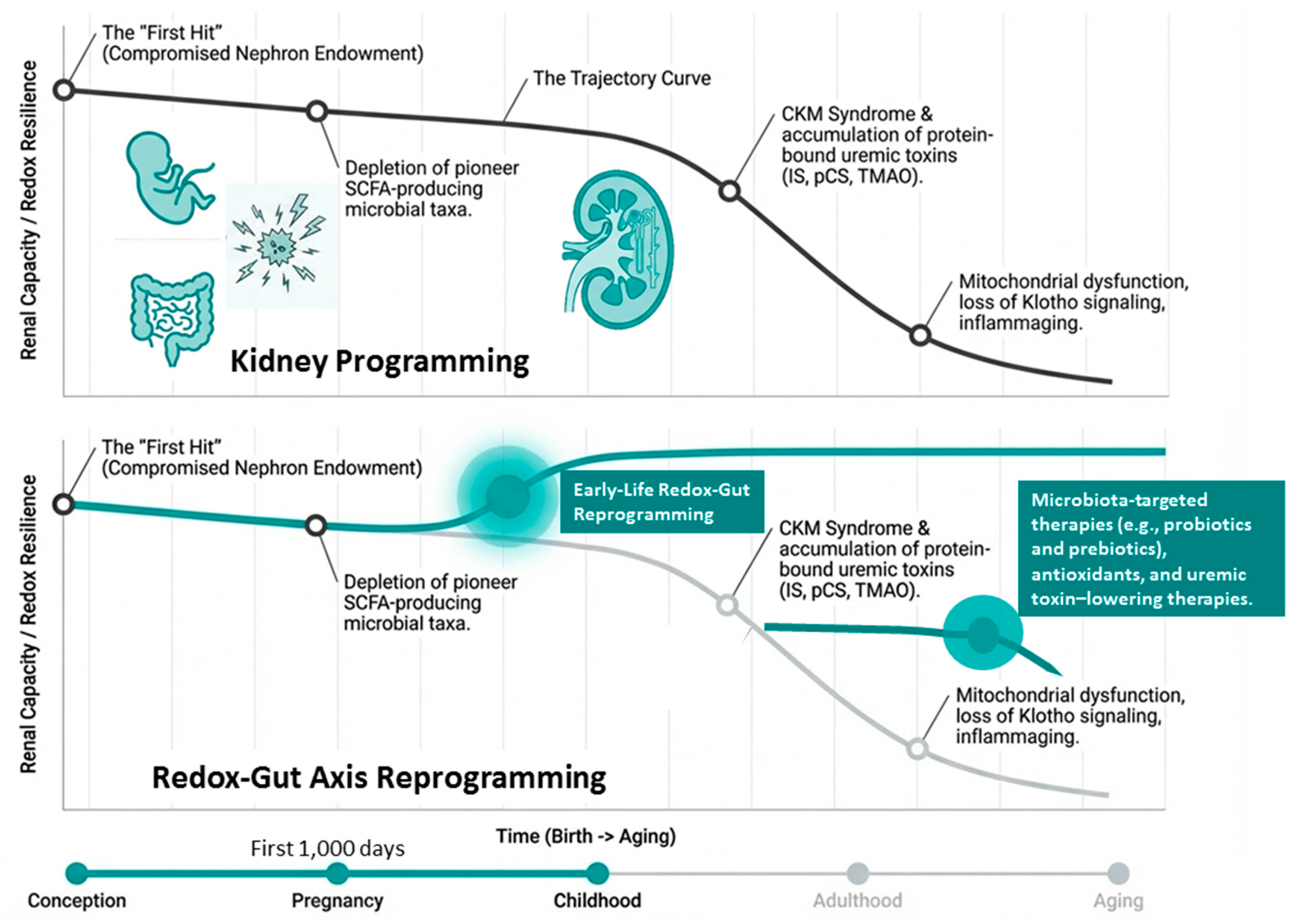
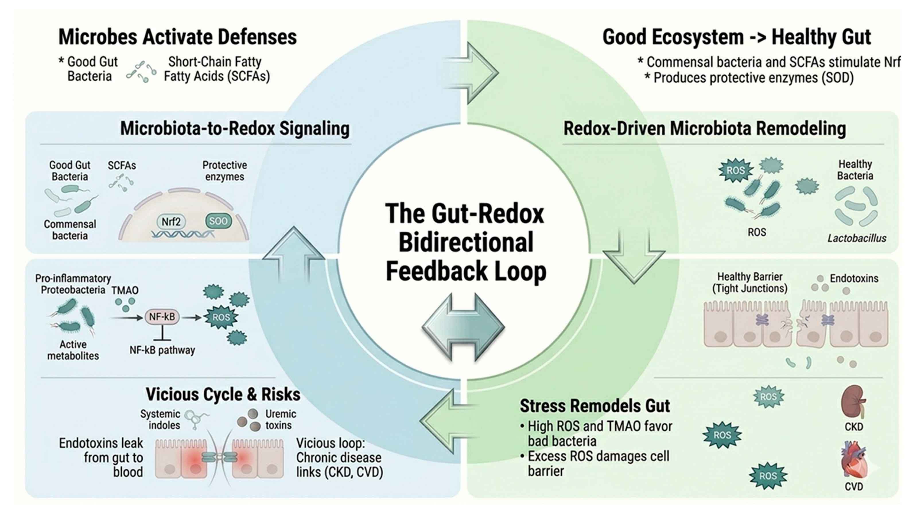

Disclaimer/Publisher’s Note: The statements, opinions and data contained in all publications are solely those of the individual author(s) and contributor(s) and not of MDPI and/or the editor(s). MDPI and/or the editor(s) disclaim responsibility for any injury to people or property resulting from any ideas, methods, instructions or products referred to in the content. |
© 2026 by the authors. Licensee MDPI, Basel, Switzerland. This article is an open access article distributed under the terms and conditions of the Creative Commons Attribution (CC BY) license.