


Abstract
Advances in prenatal diagnosis have improved the perinatal management of congenital heart disease (CHD). However postnatal comorbidities still persist due to multifactorial causes, which limits prenatal prediction of individual outcomes. Oxidative stress (OS), particularly lipid peroxidation, has been suggested to play a role in the development and progression of CHD, with 8-iso-prostaglandin F2α (8-iso-PGF2α) serving as a biomarker of oxidative injury. This prospective case–control study aimed to evaluate OS in fetuses with isolated major CHD by comparing amniotic fluid (AF) 8-iso-PGF2α concentrations with controls. A total of 123 fetuses (83 CHD, 40 controls) were included at a tertiary CHD referral center. CHD cases were subclassified according to anatomical type and expected fetal brain perfusion under placental circulation. Controls were gestational age-matched pregnancies undergoing amniocentesis for indications unlikely to affect OS. All pregnant women underwent standardized fetal biometry, Doppler assessment, and detailed echocardiography. AF samples were obtained by amniocentesis and analyzed for free 8-iso-PGF2α using a competitive ELISA, with values normalized to creatinine. Clinical, obstetric, and Doppler characteristics were comparable between groups. CHD fetuses showed significantly higher AF 8-iso-PGF2α concentrations than controls (2849 ± 1377 vs. 2088 ± 1087 ng/mg Cr, p = < 0.001), and remained significant after adjustment for GA, smoking status, diabetes and maternal age and body mass index (BMI). No consistent differences were observed across anatomical or hemodynamic CHD subgroups. These findings provide the first intrauterine evidence of increased lipid peroxidation in fetuses with CHD as reflected by elevated amniotic fluid 8-iso-PGF2α concentrations.
1. Introduction
Advances in prenatal diagnosis have substantially improved the clinical management of congenital heart disease (CHD), leading to increased survival and reduced early postnatal morbidity [1]. However, prenatal counseling regarding long-term prognosis and potential morbidities, including neurodevelopmental impairment, remains limited. Current risk stratification relies on CHD anatomy and hemodynamic parameters which lack sufficient precision to predict individual outcomes. This limitation has prompted interest in identifying fetal biomarkers to enhance prognostic assessment and better understand CHD pathophysiology in the context of feto-placental circulation.
Oxidative stress (OS), defined as an imbalance between the production of reactive oxygen species and antioxidant defense mechanisms, has emerged as a key contributor to the development and progression of CHD [2,3,4,5]. Experimental and clinical data suggest that sustained OS during fetal life may interfere with cardiac morphogenesis and placental function, thereby impairing fetal perfusion and brain maturation [6]. F2-isoprostanes are formed by the non-enzymatic peroxidation of arachidonic acid, with 8-iso-prostaglandin F2α (8-iso-PGF2α) recognized as one of the best markers of in vivo lipid peroxidation and oxidative injury [7,8,9,10,11,12], which can be accurately quantified in plasma, urine, or cerebrospinal fluid [13].
OS has been proposed to play a role both in the development of CHD and in its impact on cardiovascular function, potentially contributing to associated morbidity. To date, most evidence comes from postnatal and perioperative studies, which have shown that elevated OS, particularly in cyanotic CHD, are associated with ventricular dysfunction, prolonged mechanical ventilation, and neurological complications [11,13,14,15,16,17,18]. Increased urinary and serum 8-iso-PGF2α levels after cardiac surgery, together with delayed biomarker clearance, have been correlated with poorer postoperative recovery and adverse neurodevelopmental outcomes [16,18]. Consistently, a recent meta-analysis reported higher OS levels in cyanotic CHD compared with other CHD types, highlighting its potential as a biomarker of disease severity [2].
Despite this substantial postnatal evidence, data on OS during fetal life are scarce, and its role in the intrauterine environment of CHD is largely unexplored. Preliminary studies using alternative oxidative markers, such as ortho-tyrosine (o-Tyr), suggest increased oxidative activity in the amniotic fluid (AF) of fetuses with CHD, with possible associations between CHD severity and impaired brain growth [19]. However, to date, no prenatal evaluation has evaluated lipid peroxidation biomarkers, particularly 8-iso-PGF2α, across different CHD types.
Notably, 8-iso-PGF2α has also been proposed to evaluate OS in several pregnancy-related disorders involving inflammatory and/or vascular processes such as preeclampsia, fetal growth restriction, gestational diabetes, and premature rupture of fetal membranes [9,19,20,21,22,23,24,25,26,27,28]. In these conditions, elevated AF 8-iso-PGF2α levels have been associated with placental dysfunction and impaired fetal growth [27,28]. Recent advances in liquid chromatography–tandem mass spectrometry (LC-MS/MS) techniques now allow precise and reproducible quantification of OS biomarkers, positioning AF as a promising biological matrix to explore fetal redox homeostasis in placental-related complications and CHD, as a potential novel prognostic tool.
This study aims to (1) evaluate whether fetuses with isolated CHD exhibit increased OS in utero, as reflected by higher 8-iso-PGF2α concentrations in AF, compared to fetuses without major cardiac defects; and, secondarily, to (2) explore associations between AF 8-iso-PGF2α levels and CHD anatomical subtypes, fetal hemodynamics, and neonatal risk categories. We hypothesize that altered fetal perfusion and oxygenation in CHD create a pro-oxidant intrauterine environment that is detectable through this reliable marker of lipid peroxidation. This exploratory study seeks to generate preliminary evidence and establish the basis of future prospective research integrating biochemical, hemodynamic, and neurodevelopmental outcomes.
2. Materials and Methods
2.1. Study Design and Participants
This prospective case–control study was performed at a tertiary referral center for CHD (BCNatal, Fetal Medicine Research Center, Hospital Clínic and Hospital Sant Joan de Déu, University of Barcelona, Spain) and included pregnant women between May 2020 and December 2023. Cases comprised fetuses diagnosed with isolated major CHD whose parents consented to amniocentesis for genetic study. The control group consisted of pregnant women undergoing amniocentesis for clinical indications not expected to influence OS biomarkers. This indication included high-risk screening for aneuploidy with normal molecular karyotype, minor cardiac anomalies without hemodynamic significance, fetal cardiac anomalies not confirmed postnatally, and isolated extracardiac structural anomalies unlikely to influence AF composition. Control cases were matched to the CHD group by gestational age (GA) at amniocentesis. Exclusion criteria included the presence of genetic abnormalities and major structural anomalies known or expected to alter OS or the AF compartment, such as congenital diaphragmatic hernia, hydrops fetalis, neural tube defects and renal anomalies.
Maternal characteristics, as well as perinatal and cardiovascular outcome data, were recorded in all participants. Both cases and controls underwent ultrasonographic examinations at the time of amniocentesis and again at 32–34 weeks of gestation, using a GE Voluson E8 Expert (GE Healthcare, London, UK). Each examination included the estimation of fetal weight, standard feto-placental Doppler evaluation, and comprehensive fetal echocardiography. GA was calculated based on crown-rump length at first-trimester ultrasound [29], and estimated fetal and birth weight centiles were calculated using local reference curves [30]. Feto-placental Doppler evaluation included measurements of the pulsatility index (PI) for the uterine, umbilical, middle cerebral artery and ductus venosus. The cerebroplacental ratio was calculated by dividing the middle cerebral artery PI by the umbilical artery PI [31].
The study protocol was approved by the institutional ethics committee (PIC-124-19) and written informed consent was obtained from all participants.
2.2. CHD Subgroup Classification
To explore the relationship between OS and CHD, cases were subclassified according to anatomical type and expected fetal brain perfusion under placental circulation. Anatomically, fetuses were categorized as right CHD, left CHD, septal CHD, conotruncal CHD, or other types [1]. Fetal brain perfusion was classified according to the expected contribution of well-oxygenated placental blood as low, intermediate, and near-to-normal perfusion, based on previously described classifications [6,32].
The first group, representing low placental contribution, included CHD in which the fetal brain receives a low proportion of placental oxygenated blood. This category comprised mainly severe left ventricular outflow tract obstruction with reversed flow at the aortic isthmus and d-transposition of the great arteries, in which the aorta arises from the right ventricle. The second group, corresponding to intermediate placental contribution, included CHD with mixed brain oxygenation from placental and systemic blood due to abnormal intracardiac shunting. This group encompassed ventricular septal defects and conotruncal anomalies, with the exception of d-transposition of great arteries. The last group, representing near-to-normal placental contribution, mainly comprised right CHD, congenitally corrected transposition of great arteries without additional cardiac defects and those cases not meeting the criteria for the previous groups, such as coarctation of the aorta with anterograde flow in the aortic isthmus.
Additional stratification was performed based on abnormal fetal Doppler patterns, altered intracardiac shunting, and high-risk neonatal outcomes. Doppler abnormalities included obstructed aortic flow, reversed flow in the aortic isthmus, and reversed flow in the ductus arteriosus. Neonatal outcomes were classified as critical CHD, defined by the need for immediate postnatal intervention and/or prostaglandin therapy, and univentricular CHD, defined by single-ventricle physiology.
2.3. Sample Collection and Biomarker Measurement
AF samples were obtained via ultrasound-guided amniocentesis performed by a fetal medicine specialist, following standardized methodology [33]. A mean AF volume of 20 mL was collected per procedure, and 2 mL aliquots were stored at −80 °C until analysis.
AF 8-iso-PGF2α concentrations were measured using a commercially available competitive enzyme-linked immunosorbent assay (ELISA) kit (Cayman Chemical, Ann Arbor, MI, USA; item no. 516351), following the manufacturer’s instructions. This assay has been previously validated for the quantification of 8-iso-PGF2α in biological fluids and is widely used in clinical and translational studies assessing oxidative stress. Analytical performance characteristics provided by the manufacturer include a limit of detection (LOD) of approximately 2 pg/mL and a quantification range of 8–500 pg/mL. Intra-assay and inter-assay coefficients of variation are reported to be <10% and <15%, respectively. To ensure reliability in the AF matrix, all samples were analyzed in duplicate, and measurements were accepted only if the coefficient of variation between duplicates was <15%.
Biomarker concentrations were normalized to AF creatinine and expressed as ng/mg creatinine (ng/mg Cr).
2.4. Statistical Analysis
Data were analyzed using SPSS Statistics v27 (IBM Corp., Armonk, NY, USA).
Given the exploratory nature of the study and the lack of prior data to estimate effect size, no formal a priori sample size calculation was performed. Continuous variables are presented as mean ± standard deviation (SD), and categorical variables as absolute frequency and percentage. Group comparisons were performed using Student’s t-test or Pearson’s χ2 test, as appropriate. Biomarker levels were compared using one-way ANOVA and linear regression models adjusted for GA at amniocentesis. To account for other potential confounding factors, multivariable linear regression analyses were performed based on clinical relevance baseline variables expected to influence the oxidative stress, including smoking status, diabetes, maternal age and body mass index (BMI).
The distribution of 8-iso-PGF2α concentrations was assessed for normality. Given the expected right-skewed distribution of biomarker data, log-transformation was explored. Sensitivity analyses excluding extreme values (defined as values beyond 1.5 times the interquartile range) were performed to evaluate the robustness of the findings. A p-value < 0.05 was considered statistically significant.
3. Results
A total of 123 fetuses were included in the study, comprising 83 CHD and 40 controls. Baseline characteristics were similar between the groups (Table 1), including maternal age, rates of hypothyroidism, pre-gestational diabetes, and preeclampsia, except for smoking habit, which was more frequent in the control group (p < 0.05). GA at amniocentesis and at third-trimester ultrasound did not differ between groups, and fetal growth and Doppler parameters were comparable at both time points (Table 2). Pregnancy complications and perinatal outcomes including GA at delivery, type of delivery and birthweight were also similar. Within the CHD group, postnatal data confirmed a representative spectrum of cardiac defects, with proportions of critical neonatal and univentricular CHD consistent with expected epidemiologic distributions.
Table 1.
Baseline, gestational and perinatal characteristics of the study groups.
Table 2.
Fetal growth and feto-maternal Doppler characteristics in fetuses with congenital heart defects.
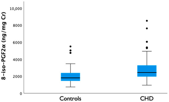
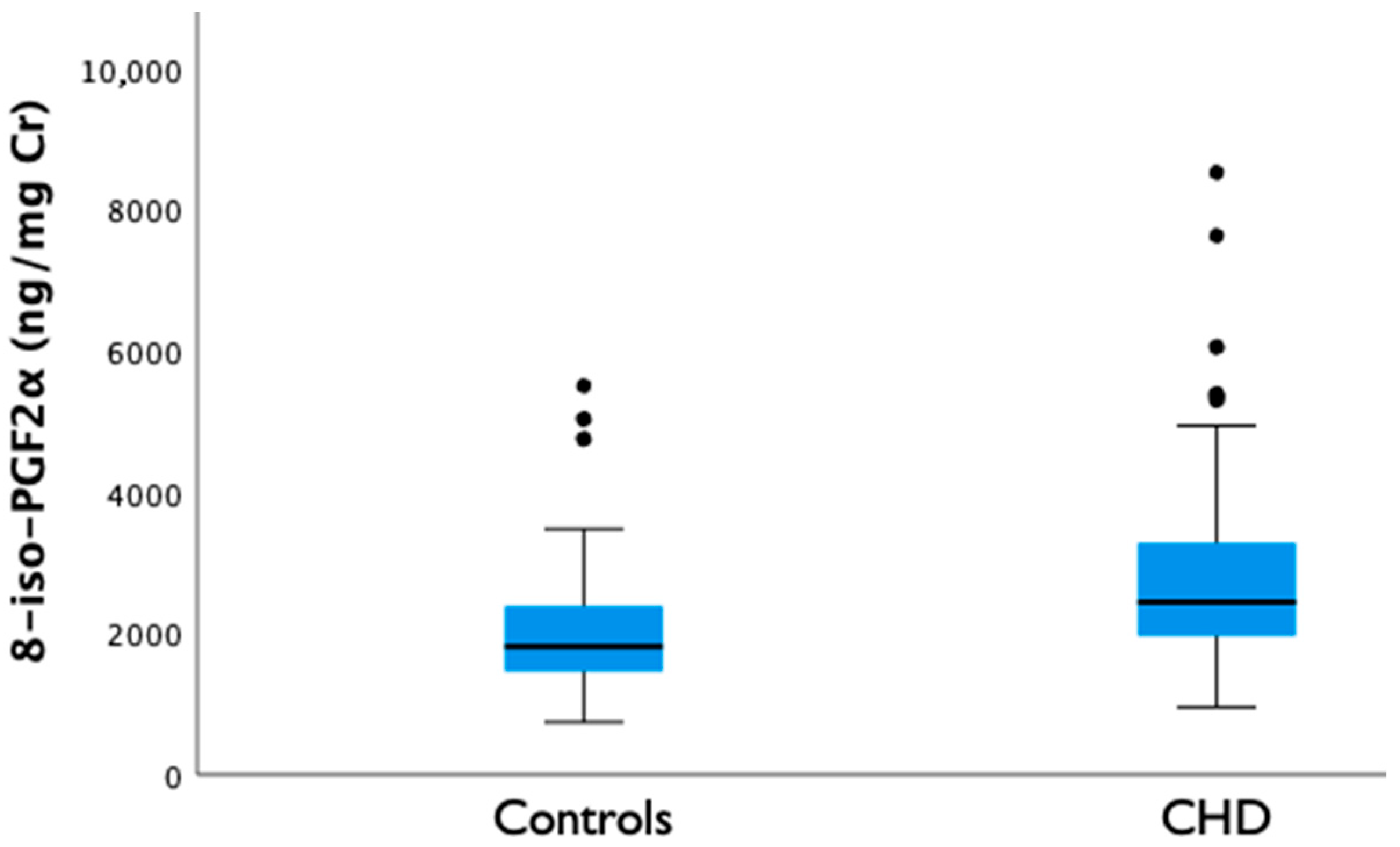
CHD fetuses exhibited higher OS biomarkers in AF compared with controls, as evidenced by the significantly elevated 8-iso-PGF2α concentration (2849 ± 1377 vs. 2088 ± 1087 ng/mg Cr, p ≤ 0.001; mean difference 761 ng/mg Cr (95% CI 270–1253) (Figure 1) after the multivariable linear regression model. Results were robust to log-transformation of 8-iso-PGF2α concentrations and to the exclusion of outliers in sensitivity analyses. A few upper-tail outliers were observed in both groups. Among CHD fetuses, these included one case of truncus arteriosus type I, one fetus with a hypoplastic aortic arch, two cases of d-transposition of the great arteries and two large perimembranous ventricular septal defects. In the control group, outliers consisted of one right aortic arch and two aberrant right subclavian arteries. Aside from the specific lesion, the outlier cases did not share distinctive baseline characteristics. Notably, four CHD cases with outlying values underwent amniocentesis at a later GA (>36 weeks) so the analyses were adjusted by GA as detailed in Methods.
Figure 1.
Amniotic fluid 8-iso-PGF2α concentrations in fetuses with congenital heart defects and controls. Data are expressed as mean. CHD, congenital heart defect.
When analyzed across the predefined study subgroups based on anatomical type, expected placental contribution to brain perfusion, and fetal and postnatal parameters, amniotic 8-iso-PGF2α levels did not differ between groups (Table 3). Thus, no significant differences were detected according to the type of CHD or any of the stratification frameworks applied, although a non-significant trend toward higher levels was noted in fetuses with CHD associated with reversed flow in the ductus arteriosus (p = 0.06). These subgroup analyses were exploratory and hypothesis-generating; accordingly, no adjustment for multiple comparisons was performed.
Table 3.
8-iso-PGF2α levels across congenital heart defect subgroups.
Additional exploratory analyses showed no significant correlations between amniotic fluid 8-iso-PGF2α concentrations and fetal Doppler indices, including z-scores of the umbilical artery, middle cerebral artery, ductus venosus, or cerebroplacental ratio, nor with head circumference (HC) z-scores. Stratification of cases according to different percentiles of 8-iso-PGF2α concentrations did not reveal any clustering of specific congenital heart disease subtypes across percentile categories. Furthermore, subgroup analyses comparing pregnancies complicated by maternal diabetes versus non-diabetic pregnancies, as well as preeclampsia versus pregnancies without preeclampsia, showed no significant differences in AF 8-iso-PGF2α levels (mean 2117.73 vs. 2622.82 ng/mg Cr, p = 0.154 and mean 3386.61 vs. 2561.12 ng/mg Cr, p = 0.533, respectively), and these findings should be interpreted with caution given the limited number of cases with maternal diabetes or preeclampsia in the study population.
4. Discussion
Our results provide the first evidence that fetuses with CHD exhibit significantly higher AF concentrations of 8-iso-PGF2α compared with controls, indicating increased intrauterine OS. No clear differences were identified across anatomical, expected brain placental perfusion or clinical CHD subgroups, although a trend toward higher levels was noted in fetuses with CHD associated with reversed ductus arteriosus flow. These findings suggest that oxidative imbalance is already established prenatally in CHD.
Our findings are consistent with postnatal studies demonstrating increased OS in neonates with CHD, particularly in cyanotic forms or in those requiring early surgical intervention [2,7,15,16]. Increased urinary and serum 8-iso-PGF2α concentrations have been associated with prolonged mechanical ventilation, longer intensive care unit stay, and neurological complications, while delayed postoperative biomarker clearance predicts poorer outcomes [15]. Importantly, our prenatal results indicate that this oxidative imbalance is already present in utero, prior to postnatal circulatory transition or surgical stress. In a previous retrospective case–control study conducted by our group [19], increased o-tyrosine, a marker of protein oxidation, was present in fetuses with CHD, particularly in those with expected lower cerebral oxygenation. However, o-tyrosine provides a more limited view of global OS as it only reflects specific oxidation of amino acids and may be influenced by precursor availability. In contrast, the present study extends this previous work by evaluating 8-iso-PGF2α in a larger prospective cohort and by focusing on a widely accepted and stable end-product of non-enzymatic lipid peroxidation, allowing a more robust assessment of OS status.
Although different CHD phenotypes are associated with distinct fetal circulatory patterns and varying degrees of hypoxia, which could theoretically lead to heterogeneous oxidative responses [34,35,36], stratified analyses based on anatomical, brain placental perfusion, hemodynamic, and clinical classifications did not reveal significant subgroup differences. Differing from our previous study, a non-significant trend toward higher 8-iso-PGF2α levels was observed only in CHD with reversed ductus arteriosus flow, a marker of ductus-dependent circulation associated with postnatal cyanosis. Overall, these findings suggest that OS may represent a shared pathophysiological feature across the CHD spectrum during fetal life, potentially modulated by feto-placental compensatory mechanisms and intrauterine antioxidant capacity [14,28,37,38]. Although limited subgroup sample sizes may have reduced statistical power, the trend observed in cases with reversed ductal flow highlights the possible influence of fetal hemodynamics and supports further investigation into phenotype-specific vulnerability to OS.
To further contextualize these findings, we examined whether the observed increase in OS could be explained by maternal or obstetric factors known to influence oxidative balance in prenatal conditions other than CHD [39]. Rates of diabetes and preeclampsia were comparable between groups, and although smoking exposure, known to increase OS, was more frequent in the control group, fetal growth parameters and Doppler indices were similar across cases and controls. Collectively, these observations reduce the likelihood that obstetric confounders account for the higher AF 8-iso-PGF2α levels observed in fetuses with CHD. Because physiological increases in OS with advancing gestation has been well documented [38,40], cases and controls were matched for GA at sampling, and all analyses were further adjusted for this variable to minimize residual confounding. The persistence of the observed group difference after adjustment supports the presence of an intrinsic oxidative burden associated with CHD rather than a GA-related effect.
Mechanistically, redox imbalance in CHD may result from altered fetal hemodynamics, tissue hypoxia, and impaired antioxidant defenses [13]. Disruption in fetal and placental circulations has been linked to hypoxia–inflammation coupling and increased reactive oxygen species generation [2,36,41,42], providing a plausible biological substrate for intrauterine OS. Beyond CHD, recent evidence indicates that OS is a shared mechanism across pregnancy complications characterized by placental dysfunction, hypoxia, and inflammation, including preeclampsia, fetal growth restriction, gestational diabetes, prematurity, and premature rupture of membranes [14,20,21,23,24,27,39,43,44,45,46,47,48,49,50]. In preeclampsia, elevated 8-iso-PGF2α correlates with blood pressure, inflammatory cytokines, angiogenic imbalance, and disease severity, supporting a direct link with placental pathology [51,52,53,54,55]. Similar patterns have been described in fetal growth restriction, gestational diabetes, and prematurity [45,46,47,48,49,50,56,57,58,59,60].
Within this context, the finding of increased 8-iso-PGF2α in fetuses with CHD extends the relevance of lipid peroxidation beyond placental disorders to conditions primarily defined by fetal cardiovascular maldevelopment. Many forms of CHD are associated with chronic alterations in fetal oxygen delivery, placental structure, and inflammatory signaling, likely converging on oxidative pathways similar to those described in placental-related complications [56,61]. The convergence of clinical and experimental evidence further suggests that OS is not merely a downstream consequence but may actively contribute to abnormal cardiac development. Experimental murine models show that pro-oxidant exposure during organogenesis induces structural cardiac defects, whereas antioxidant interventions mitigate these effects [62], supporting the notion that a proportion of the oxidative burden observed in CHD is environmentally biologically relevant and potentially modifiable [18,63,64].
Although neurodevelopmental outcomes were not assessed in this cohort, previous evidence suggests that OS may contribute to neurological vulnerability in CHD [16,64]. As AF reflects fetal metabolic turnover, elevated 8-iso-PGF2α may also provide indirect insight into fetal oxidative exposure, including potential cerebral involvement, supporting its potential value for future risk assessment [64].
This study has several limitations. First, 8-iso-PGF2α concentrations were measured using an ELISA-based method. While liquid chromatography–tandem mass spectrometry (LC–MS/M) provides higher analytical specificity, ELISA has been reported as a robust, cost-effective, and widely used approach in clinical research [65]. Moreover, previous studies have demonstrated good correlation between ELISA and mass spectrometry methods for OS biomarker quantification in biological samples [66].
Another limitation could be the assessment of a single OS biomarker. Although 8-iso-PGF2α is a well-established and reliable marker of lipid peroxidation, it reflects only one aspect of the redox balance. The absence of complementary oxidative and inflammatory markers limits a more comprehensive characterization of the redox environment. Nevertheless, the use of 8-iso-PGF2α provides a focused and biologically relevant assessment of lipid peroxidation. Future studies incorporating broader biomarker panels are warranted.
Although larger than previous prenatal works, the sample size was limited, and subgroup analyses should therefore be interpreted with caution. Only a single 8-iso-PGF2α measurement per fetus was available. The observational nature of the study precludes establishing a causal relationship, and residual confounding related to individual variability in placental function or genetic susceptibility to oxidative imbalance cannot be completely excluded [5,14]. In addition, a small number of control fetuses presented minor structural anomalies that were not expected to influence OS or AF composition.
Future research should include multicenter longitudinal studies to characterize AF 8-iso-PGF2α dynamics across gestation, investigate redox-based phenotypic subclassifications, and examine associations with neurodevelopmental and perioperative outcomes. Standardized LC-MS/MS assays, assessment of ratio metrics (8-iso-PGF2α/PGF2α), and harmonized sampling windows will be essential to define clinical utility. In addition, whether maternal nutritional, lifestyle, or pharmacologic interventions can modulate fetal oxidative status and improve outcomes in this high-risk population remains to be determined.
Despite these limitations, this study provides the first intrauterine evidence of increased OS in CHD using AF 8-iso-PGF2α. By analyzing AF, the study captures the fetal metabolic environment prior to birth-related circulatory and inflammatory transitions, avoiding major postnatal confounders such as reoxygenation and surgical stress [7,15,16]. Careful cohort selection, exclusion of congenital extracardiac and genetic anomalies, balanced maternal and fetal characteristics between groups, and multivariable adjustment strengthen the robustness of the findings. Importantly, sensitivity analyses excluding outliers and analyses using log-transformed data yielded consistent results, supporting the stability of our results.
Overall, these findings provide novel intrauterine evidence of OS in CHD, supporting its role as an early feature of the disease while warranting further investigation into its mechanistic and clinical implications.
5. Conclusions
This study provides novel intrauterine evidence of increased AF 8-iso-PGF2α concentrations in fetuses with CHD compared with controls. These differences persisted after adjustment for GA and potential maternal and gestational confounders, supporting an association between CHD and increased intrauterine OS. While 8-iso-PGF2α may represent a promising biomarker of fetal oxidative burden, these findings should be interpreted with caution given their exploratory and observational nature. Further multicenter longitudinal studies are needed to confirm these findings and to better define the potential clinical relevance of this biomarker, including its relationship with prenatal brain development, postnatal neurodevelopment, and perioperative outcomes.
Author Contributions
Conceptualization, M.A., O.G. and M.P.-C.; methodology, M.A., M.C.-C., M.C.E.-D., O.G. and M.P.-C.; software, M.A.; validation, M.A., M.C.-C., M.C.E.-D., L.G., O.G. and M.P.-C.; formal analysis, M.A., O.G. and M.P.-C.; investigation, M.A., L.G., L.N., M.B., N.M., F.C., O.G. and M.P.-C.; resources, F.C., M.D.G.-R., O.G., M.P.-C. and M.C.-C.; data curation, M.A., O.G. and M.P.-C.; writing—original draft preparation, M.A. and M.P.-C.; writing—review and editing, M.A., O.G. and M.P.-C.; visualization, M.A.; supervision, O.G. and M.P.-C.; project administration, M.D.G.-R., O.G. and M.P.-C.; funding acquisition, M.D.G.-R., O.G., M.P.-C. and M.C.-C. All authors have read and agreed to the published version of the manuscript.
Funding
This study was funded by the Spanish Network in Maternal, Neonatal, Child and Developmental Health Research (RICORS-SAMID, RD24/0013/0004), Instituto de Salud Carlos III (ISCIII), Madrid, Spain, and by ISCIII projects PI22/00754, PI23/00633, PI24/00653, PI24/00127, PI19/00118, and PI23/00104; Dr. Crispi was funded by ISCIII project INT25/00064. All grants were co-funded by the European Union (European Regional Development Fund, ERDF/FEDER 2021–2027).
Institutional Review Board Statement
The study was conducted in accordance with the Declaration of Helsinki and approved by the institutional ethics committee of Hospital Sant Joan de Déu (PIC-124-19; 25 July 2019).
Informed Consent Statement
Informed consent was obtained from all subjects involved in the study.
Data Availability Statement
The original contributions presented in this study are included in the article. Further inquiries can be directed to the corresponding author.
Acknowledgments
We are indebted to the “Biobanc de l’Hospital Infantil Sant Joan de Déu per a la Investigació” integrated in the Spanish Biobank Network of ISCIII for the sample and data procurement.
Conflicts of Interest
The authors declare no conflicts of interest.
Abbreviations
The following abbreviations are used in this manuscript:
| AF | Amniotic fluid |
| CHD | Congenital heart disease |
| GA | Gestational age |
| HC | Head circumference |
| OS | Oxidative stress |
| 8-iso-PGF2α | 8-iso-prostaglandin F2α |
| o-Tyr | ortho-tyrosine |
| Cr | Creatinine |
| ELISA | Enzyme-linked immunosorbent assay |
| LC-MS/MS | Liquid chromatography–tandem mass spectrometry |
| PI | Pulsatility index |
| SD | Standard deviation |
| CI | Confidence interval |
| ANOVA | Analysis of variance |
| ng/mg | nanograms per milligram |
References
- Donofrio, M.T.; Moon-Grady, A.J.; Hornberger, L.K.; Copel, J.A.; Sklansky, M.S.; Abuhamad, A.; Cuneo, B.F.; Huhta, J.C.; Jonas, R.A.; Krishnan, A.; et al. Diagnosis and Treatment of Fetal Cardiac Disease: A Scientific Statement from the American Heart Association. Circulation 2014, 129, 2183–2242. [Google Scholar] [CrossRef] [Scilit]
- Vanreusel, I.; Taeymans, J.; Van Craenenbroeck, E.; Segers, V.F.M.; Van Berendoncks, A.; Briedé, J.J.; Hens, W. Elevated Oxidative Stress in Patients with Congenital Heart Disease and the Effect of Cyanosis: A Meta-Analysis. Free Radic. Res. 2023, 57, 470–486. [Google Scholar] [CrossRef] [Scilit]
- Wu, Y.; Reece, E.A.; Zhong, J.; Dong, D.; Shen, W.-B.; Harman, C.R.; Yang, P. Type 2 Diabetes Mellitus Induces Congenital Heart Defects in Murine Embryos by Increasing Oxidative Stress, Endoplasmic Reticulum Stress, and Apoptosis. Am. J. Obstet. Gynecol. 2016, 215, 366.e1–366.e10. [Google Scholar] [CrossRef] [Scilit] [PubMed]
- Cardarelli, S.; Biglietto, M.; Orsini, T.; Fustaino, V.; Monaco, L.; de Oliveira do Rêgo, A.G.; Liccardo, F.; Masciarelli, S.; Fazi, F.; Naro, F.; et al. Modulation of CAMP/CGMP Signaling as Prevention of Congenital Heart Defects in Pde2A Deficient Embryos: A Matter of Oxidative Stress. Cell Death Dis. 2024, 15, 169. [Google Scholar] [CrossRef] [Scilit]
- Choudhury, T.Z.; Greskovich, S.C.; Girard, H.B.; Rao, A.S.; Budhathoki, Y.; Cameron, E.M.; Conroy, S.; Li, D.; Zhao, M.-T.; Garg, V. Impact of Genetic Factors on Antioxidant Rescue of Maternal Diabetes-Associated Congenital Heart Disease. JCI Insight 2024, 9, e183516. [Google Scholar] [CrossRef] [Scilit]
- Masoller, N.; Sanz-Cortés, M.; Crispi, F.; Gómez, O.; Bennasar, M.; Egaña-Ugrinovic, G.; Bargalló, N.; Martínez, J.M.; Gratacós, E. Mid-gestation Brain Doppler and Head Biometry in Fetuses with Congenital Heart Disease Predict Abnormal Brain Development at Birth. Ultrasound Obstet. Gynecol. 2016, 47, 65–73. [Google Scholar] [CrossRef] [Scilit]
- Albers, E.; Donahue, B.S.; Milne, G.; Saville, B.R.; Wang, W.; Bichell, D.; McLaughlin, B. Perioperative Plasma F2-Isoprostane Levels Correlate with Markers of Impaired Ventilation in Infants with Single-Ventricle Physiology Undergoing Stage 2 Surgical Palliation on the Cardiopulmonary Bypass. Pediatr. Cardiol. 2012, 33, 562–568. [Google Scholar] [CrossRef] [Scilit]
- Xiao, Y.; Fu, X.; Pattengale, P.; Dien Bard, J.; Xu, Y.-K.; O’Gorman, M.R. A Sensitive LC-MS/MS Method for the Quantification of Urinary 8-Iso-Prostaglandin F2α (8-Iso-PGF2α) Including Pediatric Reference Interval. Clin. Chim. Acta 2016, 460, 128–134. [Google Scholar] [CrossRef] [Scilit] [PubMed]
- van ’t Erve, T.J. Strategies to Decrease Oxidative Stress Biomarker Levels in Human Medical Conditions: A Meta-Analysis on 8-Iso-Prostaglandin F2α. Redox Biol. 2018, 17, 284–296. [Google Scholar] [CrossRef] [Scilit]
- Longini, M.; Perrone, S.; Kenanidis, A.; Vezzosi, P.; Marzocchi, B.; Petraglia, F.; Centini, G.; Buonocore, G. Isoprostanes in Amniotic Fluid: A Predictive Marker for Fetal Growth Restriction in Pregnancy. Free Radic. Biol. Med. 2005, 38, 1537–1541. [Google Scholar] [CrossRef] [Scilit] [PubMed]
- Perrone, S.; Laschi, E.; Buonocore, G. Biomarkers of Oxidative Stress in the Fetus and in the Newborn. Free Radic. Biol. Med. 2019, 142, 23–31. [Google Scholar] [CrossRef] [Scilit]
- van ‘t Erve, T.J.; Lih, F.B.; Kadiiska, M.B.; Deterding, L.J.; Eling, T.E.; Mason, R.P. Reinterpreting the Best Biomarker of Oxidative Stress: The 8-Iso-PGF2α/PGF2α Ratio Distinguishes Chemical from Enzymatic Lipid Peroxidation. Free Radic. Biol. Med. 2015, 83, 245–251. [Google Scholar] [CrossRef] [Scilit]
- Belik, J.; González-Luis, G.E.; Perez-Vizcaino, F.; Villamor, E. Isoprostanes in Fetal and Neonatal Health and Disease. Free Radic. Biol. Med. 2010, 48, 177–188. [Google Scholar] [CrossRef] [Scilit]
- Perrone, S.; Laschi, E.; Buonocore, G. Oxidative Stress Biomarkers in the Perinatal Period: Diagnostic and Prognostic Value. Semin. Fetal Neonatal Med. 2020, 25, 101087. [Google Scholar] [CrossRef] [Scilit] [PubMed]
- Hadley, S.; Vazquez, D.C.; Abad, M.L.; Congiu, S.; Lushchencov, D.; Camprubí, M.C.; Sanchez-De-Toledo, J. Oxidative Stress Response in Children Undergoing Cardiac Surgery: Utility of the Clearance of Isoprostanes. PLoS ONE 2021, 16, e0250124. [Google Scholar] [CrossRef] [Scilit]
- Vázquez, D.C.; Hadley, S.M.; Ordóñez, M.P.; Lopez-Abad, M.; Valls, A.; Vinals, M.L.; Moscoso, B.A.; Benito, S.; Camprubí-Camprubí, M.; Sanchez-de-Toledo, J. Oxidative Stress and Indicators of Brain Damage Following Pediatric Heart Surgery. Antioxidants 2022, 11, 489. [Google Scholar] [CrossRef] [Scilit] [PubMed]
- Dani, C.; Mosca, F.; Gazzolo, D.; Mecacci, F.; Cetin, I.; Buonocore, G. Role of Oxidative Stress in Maternal and Neonatal Diseases. Oxid. Med. Cell. Longev. 2019, 2019, 9742764. [Google Scholar] [CrossRef] [Scilit] [PubMed]
- Doroszko, A.; Dobrowolski, P.; Radziwon-Balicka, A.; Skomro, R. New Insights into the Role of Oxidative Stress in Onset of Cardiovascular Disease. Oxid. Med. Cell. Longev. 2018, 2018, 9563831. [Google Scholar] [CrossRef] [Scilit]
- Escobar-Diaz, M.C.; Pérez-Cruz, M.; Arráez, M.; Cascant-Vilaplana, M.M.; Albiach-Delgado, A.; Kuligowski, J.; Vento, M.; Masoller, N.; Gómez-Roig, M.D.; Gómez, O.; et al. Brain Oxygen Perfusion and Oxidative Stress Biomarkers in Fetuses with Congenital Heart Disease-A Retrospective, Case-Control Pilot Study. Antioxidants 2022, 11, 299. [Google Scholar] [CrossRef] [Scilit]
- Ferguson, K.K.; Kamai, E.M.; Cantonwine, D.E.; Mukherjee, B.; Meeker, J.D.; McElrath, T.F. Associations between Repeated Ultrasound Measures of Fetal Growth and Biomarkers of Maternal Oxidative Stress and Inflammation in Pregnancy. Am. J. Reprod. Immunol. 2018, 80, e13017. [Google Scholar] [CrossRef] [Scilit]
- Toboła-Wróbel, K.; Pietryga, M.; Dydowicz, P.; Napierała, M.; Brązert, J.; Florek, E. Association of Oxidative Stress on Pregnancy. Oxid. Med. Cell. Longev. 2020, 2020, 6398520. [Google Scholar] [CrossRef] [Scilit]
- Ferreira, R.C.; Fragoso, M.B.T.; Bueno, N.B.; Goulart, M.O.F.; de Oliveira, A.C.M. Oxidative Stress Markers in Preeclamptic Placentas: A Systematic Review with Meta-Analysis. Placenta 2020, 99, 89–100. [Google Scholar] [CrossRef] [Scilit] [PubMed]
- Wang, C.N.; Chen, J.Y.S.; Sabu, S.; Chang, Y.L.; Chang, S.D.; Kao, C.C.; Peng, H.H.; Chueh, H.Y.; Chao, A.S.; Cheng, P.J.; et al. Elevated Amniotic Fluid F2-Isoprostane: A Potential Predictive Marker for Preeclampsia. Free Radic. Biol. Med. 2011, 50, 1124–1130. [Google Scholar] [CrossRef] [Scilit]
- Kwiatkowski, S.; Torbé, A.; Dołgowska, B.; Błogowski, W.; Czajka, R.; Chlubek, D.; Rzepka, R. Isoprostanes 8-IPF2α-III: Risk Markers of Premature Rupture of Fetal Membranes? Biomarkers 2009, 14, 406–413. [Google Scholar] [CrossRef] [Scilit]
- Novakovic, T.R.; Dolicanin, Z.C.; Djordjevic, N.Z. Oxidative Stress Biomarkers in Amniotic Fluid of Pregnant Women with Hypothyroidism. J. Matern.-Fetal Neonatal Med. 2019, 32, 1105–1110. [Google Scholar] [CrossRef] [Scilit]
- Cuffe, J.S.; Xu, Z.C.; Perkins, A.V. Biomarkers of Oxidative Stress in Pregnancy Complications. Biomark. Med. 2017, 11, 295–306. [Google Scholar] [CrossRef] [Scilit] [PubMed]
- Escobar, J.; Teramo, K.; Stefanovic, V.; Andersson, S.; Asensi, M.A.; Arduini, A.; Cubells, E.; Sastre, J.; Vxento, M. Amniotic Fluid Oxidative and Nitrosative Stress Biomarkers Correlate with Fetal Chronic Hypoxia in Diabetic Pregnancies. Neonatology 2013, 103, 193–198. [Google Scholar] [CrossRef] [Scilit]
- Kohzadi, M.; Kubow, S.; Koski, K.G. Fetal Growth Is Associated with Amniotic Fluid Antioxidant Capacity, Oxidative Stress, Minerals and Prenatal Supplementation: A Retrospective Study. Antioxidants 2025, 14, 184. [Google Scholar] [CrossRef] [Scilit]
- Robinson, H.P.; Sweet, E.M.; Adam, A.H. The Accuracy of Radiological Estimates of Gestational Age Using Early Fetal Crown-Rump Length Measurements by Ultrasound as a Basis for Comparison. Br. J. Obstet. Gynaecol. 1979, 86, 525–528. [Google Scholar] [CrossRef] [Scilit] [PubMed]
- Figueras, F.; Gratacós, E. Update on the Diagnosis and Classification of Fetal Growth Restriction and Proposal of a Stage-Based Management Protocol. Fetal Diagn. Ther. 2014, 36, 86–98. [Google Scholar] [CrossRef] [Scilit]
- Baschat, A.A.; Gembruch, U. The Cerebroplacental Doppler Ratio Revisited. Ultrasound Obstet. Gynecol. 2003, 21, 124–127. [Google Scholar] [CrossRef] [Scilit]
- Masoller, N.; Sanz-Cortés, M.; Crispi, F.; Gómez, O.; Bennasar, M.; Egaña-Ugrinovic, G.; Bargalló, N.; Martínez, J.M.; Gratacós, E. Severity of Fetal Brain Abnormalities in Congenital Heart Disease in Relation to the Main Expected Pattern of in Utero Brain Blood Supply. Fetal Diagn. Ther. 2016, 39, 269–278. [Google Scholar] [CrossRef] [Scilit]
- Nizard, J. Amniocentesis: Technique and Education. Curr. Opin. Obstet. Gynecol. 2010, 22, 152–154. [Google Scholar] [CrossRef] [Scilit]
- Thompson, L.P.; Al-Hasan, Y. Impact of Oxidative Stress in Fetal Programming. J. Pregnancy 2012, 2012, 582748. [Google Scholar] [CrossRef] [Scilit]
- Rodríguez-Rodríguez, P.; Ramiro-Cortijo, D.; Reyes-Hernández, C.G.; López de Pablo, A.L.; Carmen González, M.; Arribas, S.M. Implication of Oxidative Stress in Fetal Programming of Cardiovascular Disease. Front. Physiol. 2018, 9, 602. [Google Scholar] [CrossRef] [Scilit]
- Impellizzeri, P.; Nascimben, F.; Di Fabrizio, D.; Antonuccio, P.; Antonelli, E.; Peri, F.M.; Calabrese, U.; Arena, S.; Romeo, C. Pathogenesis of Congenital Malformations: Possible Role of Oxidative Stress. Am. J. Perinatol. 2022, 39, 816–823. [Google Scholar] [CrossRef] [Scilit] [PubMed]
- Vanreusel, I.; Vermeulen, D.; Goovaerts, I.; Stoop, T.; Ectors, B.; Cornelis, J.; Hens, W.; de Bliek, E.; Heuten, H.; Van Craenenbroeck, E.M.; et al. Circulating Reactive Oxygen Species in Adults with Congenital Heart Disease. Antioxidants 2022, 11, 2369. [Google Scholar] [CrossRef] [Scilit] [PubMed]
- van ’t Erve, T.J.; Kadiiska, M.B.; London, S.J.; Mason, R.P. Classifying Oxidative Stress by F2-Isoprostane Levels across Human Diseases: A Meta-Analysis. Redox Biol. 2017, 12, 582–599. [Google Scholar] [CrossRef] [Scilit]
- Bogavac, M.; Lakic, N.; Simin, N.; Nikolic, A.; Sudji, J.; Bozin, B. Biomarkers of Oxidative Stress in Amniotic Fluid and Complications in Pregnancy. J. Matern Fetal Neonatal Med. 2012, 25, 104–108. [Google Scholar] [CrossRef] [Scilit] [PubMed]
- Longini, M.; Belvisi, E.; Proietti, F.; Bazzini, F.; Buonocore, G.; Perrone, S. Oxidative Stress Biomarkers: Establishment of Reference Values for Isoprostanes, AOPP, and NPBI in Cord Blood. Mediat. Inflamm. 2017, 2017, 1758432. [Google Scholar] [CrossRef] [Scilit]
- Park, J.Y.; Romero, R.; Lee, J.; Chaemsaithong, P.; Chaiyasit, N.; Yoon, B.H. An Elevated Amniotic Fluid Prostaglandin F 2α Concentration Is Associated with Intra-Amniotic Inflammation/Infection, and Clinical and Histologic Chorioamnionitis, as Well as Impending Preterm Delivery in Patients with Preterm Labor and Intact Membranes. J. Matern.-Fetal Neonatal Med. 2015, 29, 2563–2572. [Google Scholar] [CrossRef] [Scilit] [PubMed]
- Kadiiska, M.; van’t Erve, T.; Mason, R.; Ferguson, K. The Exposure-Dependent Increases in 8-Iso-PGF2α and the Significance of Decoding Its Sources to Identify a Specific Indicator of Oxidative Stress or Inflammation. Free Radic. Biol. Med. 2018, 128, S117–S118. [Google Scholar] [CrossRef] [Scilit]
- McMullan, A.; Zwierzynski, J.B.; Jain, N.; Haneline, L.S.; Shou, W.; Kua, K.L.; Hota, S.K.; Durbin, M.D. Role of Maternal Obesity in Offspring Cardiovascular Development and Congenital Heart Defects. J. Am. Heart Assoc. 2025, 14, e039684. [Google Scholar] [CrossRef] [Scilit]
- Li, L.; Zhou, L.; Li, W.; Shi, F.; Feng, X.; Zhuang, J. Oxidative Stress Biomarkers in Fetal Growth Restriction: A Systematic Review and Meta-Analysis. Arch. Gynecol. Obs. 2025, 312, 1063–1084. [Google Scholar] [CrossRef] [Scilit]
- Li, H.; Yin, Q.; Li, N.; Ouyang, Z.; Zhong, M. Plasma Markers of Oxidative Stress in Patients with Gestational Diabetes Mellitus in the Second and Third Trimester. Obstet. Gynecol. Int. 2016, 2016, 3865454. [Google Scholar] [CrossRef] [Scilit]
- Phoswa, W.N.; Khaliq, O.P. The Role of Oxidative Stress in Hypertensive Disorders of Pregnancy (Preeclampsia, Gestational Hypertension) and Metabolic Disorder of Pregnancy (Gestational Diabetes Mellitus). Oxid. Med. Cell. Longev. 2021, 2021, 5581570. [Google Scholar] [CrossRef] [Scilit]
- Joo, E.H.; Kim, Y.R.; Kim, N.; Jung, J.E.; Han, S.H.; Cho, H.Y. Effect of Endogenic and Exogenic Oxidative Stress Triggers on Adverse Pregnancy Outcomes: Preeclampsia, Fetal Growth Restriction, Gestational Diabetes Mellitus and Preterm Birth. Int. J. Mol. Sci. 2021, 22, 10122. [Google Scholar] [CrossRef] [Scilit] [PubMed]
- Eick, S.M.; Ferguson, K.K.; Milne, G.L.; Rios-McConnell, R.; Vélez-Vega, C.; Rosario, Z.; Alshawabkeh, A.; Cordero, J.F.; Meeker, J.D. Repeated Measures of Urinary Oxidative Stress Biomarkers and Preterm Birth in Puerto Rico. Free Radic. Biol. Med. 2020, 146, 299–305. [Google Scholar] [CrossRef] [Scilit]
- Rosen, E.M.; van ‘t Erve, T.J.; Boss, J.; Sathyanarayana, S.; Barrett, E.S.; Nguyen, R.H.N.; Bush, N.R.; Milne, G.L.; McElrath, T.F.; Swan, S.H.; et al. Urinary Oxidative Stress Biomarkers and Accelerated Time to Spontaneous Delivery. Free Radic. Biol. Med. 2019, 130, 419–425. [Google Scholar] [CrossRef] [Scilit]
- Staff, A.C.; Halvorsen, B.; Ranheim, T.; Henriksen, T. Elevated Level of Free 8-Iso-Prostaglandin F2α in the Decidua Basalis of Women with Preeclampsia. Am. J. Obstet. Gynecol. 1999, 181, 1211–1215. [Google Scholar] [CrossRef] [Scilit] [PubMed]
- Boldeanu, L.; Văduva, C.-C.; Caragea, D.C.; Novac, M.B.; Manasia, M.; Siloși, I.; Manolea, M.M.; Boldeanu, M.V.; Dijmărescu, A.L. Association between Serum 8-Iso-Prostaglandin F2α as an Oxidative Stress Marker and Immunological Markers in a Cohort of Preeclampsia Patients. Life 2023, 13, 2242. [Google Scholar] [CrossRef] [Scilit]
- Dijmărescu, A.L.; Tănase, F.; Novac, M.B.; Siminel, M.A.; Rotaru, I.; Caragea, D.C.; Manolea, M.M.; Văduva, C.-C.; Boldeanu, M.V.; Boldeanu, L. Longitudinal 8-Epi-Prostaglandin F2-Alpha and Angiogenic Profile Mediator Evaluation during Pregnancy in Women with Suspected or Confirmed Pre-Eclampsia. Biomedicines 2024, 12, 433. [Google Scholar] [CrossRef] [Scilit]
- Freire, V.A.F.; de Melo, A.D.; de Lima Santos, H.; Barros-Pinheiro, M. Evaluation of Oxidative Stress Markers in Subtypes of Preeclampsia: A Systematic Review and Meta-Analysis. Placenta 2023, 132, 55–67. [Google Scholar] [CrossRef] [Scilit]
- Staff, A.C.; Ranheim, T.; Henriksen, T.; Halvorsen, B. 8-Iso-Prostaglandin F 2α Reduces Trophoblast Invasion and Matrix Metalloproteinase Activity. Hypertension 2000, 35, 1307–1313. [Google Scholar] [CrossRef] [Scilit]
- Vornic, I.; Buciu, V.; Furau, C.G.; Gaje, P.N.; Ceausu, R.A.; Dumitru, C.-S.; Barb, A.C.; Novacescu, D.; Cumpanas, A.A.; Latcu, S.C.; et al. Oxidative Stress and Placental Pathogenesis: A Contemporary Overview of Potential Biomarkers and Emerging Therapeutics. Int. J. Mol. Sci. 2024, 25, 12195. [Google Scholar] [CrossRef] [Scilit] [PubMed]
- Assani, A.-D.; Boldeanu, L.; Siloși, I.; Boldeanu, M.V.; Dijmărescu, A.L.; Assani, M.-Z.; Manolea, M.-M.; Văduva, C.-C. Pregnancy Under Pressure: Oxidative Stress as a Common Thread in Maternal Disorders. Life 2025, 15, 1348. [Google Scholar] [CrossRef] [Scilit] [PubMed]
- Ibrahim, A.; Khoo, M.I.; Ismail, E.H.E.; Hussain, N.H.N.; Zin, A.A.M.; Noordin, L.; Abdullah, S.; Mahdy, Z.A.; Lah, N.A.Z.N. Oxidative Stress Biomarkers in Pregnancy: A Systematic Review. Reprod. Biol. Endocrinol. 2024, 22, 93. [Google Scholar] [CrossRef] [Scilit] [PubMed]
- Turpin, C.A.; Sakyi, S.A.; Owiredu, W.K.; Ephraim, R.K.D.; Anto, E.O. Association between Adverse Pregnancy Outcome and Imbalance in Angiogenic Regulators and Oxidative Stress Biomarkers in Gestational Hypertension and Preeclampsia. BMC Pregnancy Childbirth 2015, 15, 189. [Google Scholar] [CrossRef] [Scilit]
- Venkatesh, K.K.; Meeker, J.D.; Cantonwine, D.E.; McElrath, T.F.; Ferguson, K.K. Association of Antenatal Depression with Oxidative Stress and Impact on Spontaneous Preterm Birth. J. Perinatol. 2019, 39, 554–562. [Google Scholar] [CrossRef] [Scilit]
- Schoots, M.H.; Gordijn, S.J.; Scherjon, S.A.; van Goor, H.; Hillebrands, J.L. Oxidative Stress in Placental Pathology. Placenta 2018, 69, 153–161. [Google Scholar] [CrossRef] [Scilit]
- Rounioja, S. Intra-Amniotic Lipopolysaccharide Leads to Fetal Cardiac Dysfunction A Mouse Model for Fetal Inflammatory Response. Cardiovasc. Res. 2003, 60, 156–164. [Google Scholar] [CrossRef] [Scilit]
- Saiyin, T.; Engineer, A.; Greco, E.R.; Kim, M.Y.; Lu, X.; Jones, D.L.; Feng, Q. Maternal Voluntary Exercise Mitigates Oxidative Stress and Incidence of Congenital Heart Defects in Pre-Gestational Diabetes. J. Cell. Mol. Med. 2019, 23, 5553–5565. [Google Scholar] [CrossRef] [Scilit]
- Yang, J.; Du, Q.; Xiao, Z.; Guo, R.; Chang, Q.; Li, Y.H. Maternal Oxidative Balance Score during Pregnancy and Congenital Heart Defects. Nutrients 2024, 16, 1825. [Google Scholar] [CrossRef] [Scilit]
- Perrone, S.; Tataranno, L.M.; Stazzoni, G.; Ramenghi, L.; Buonocore, G. Brain Susceptibility to Oxidative Stress in the Perinatal Period. J. Matern.-Fetal Neonatal Med. 2015, 28, 2291–2295. [Google Scholar] [CrossRef] [Scilit] [PubMed]
- Smith, K.A.; Shepherd, J.; Wakil, A.; Kilpatrick, E.S. A Comparison of Methods for the Measurement of 8-IsoPGF2α: A Marker of Oxidative Stress. Ann. Clin. Biochem. Int. J. Lab. Med. 2011, 48, 147–154. [Google Scholar] [CrossRef] [Scilit] [PubMed]
- Mello, L.D. Potential Contribution of ELISA and LFI Assays to Assessment of the Oxidative Stress Condition Based on 8-OxodG Biomarker. Anal. Biochem. 2021, 628, 114215. [Google Scholar] [CrossRef] [Scilit] [PubMed]

Disclaimer/Publisher’s Note: The statements, opinions and data contained in all publications are solely those of the individual author(s) and contributor(s) and not of MDPI and/or the editor(s). MDPI and/or the editor(s) disclaim responsibility for any injury to people or property resulting from any ideas, methods, instructions or products referred to in the content. |
© 2026 by the authors. Licensee MDPI, Basel, Switzerland. This article is an open access article distributed under the terms and conditions of the Creative Commons Attribution (CC BY) license.