


Abstract
The abnormal activation of the Nlrp3 (Nod-like receptor pyrin 3) inflammasome, in response to oxidative stress or impaired antioxidant defense, is linked to aging-related diseases. Previously, we have shown that Peroxiredoxin (Prdx)6 deficiency triggers reactive oxygen species (ROS)-dependent activation of Kruppel-like factor (Klf)9/Nlrp3 inflammasome in aging lens epithelial cells (LECs). Herein, we test the therapeutic efficacy of Prdx6 delivery in abating the oxidative stress-induced aberrant activation of the Klf9/NF-ĸB/Nlrp3 pathway and subsequent pyroptotic cell death in LECs and Prdx6-deficient (Prdx6−/−) LECs. Similar to aged LECs, Prdx6-depleted LECs exhibited activation of Nlrp3 inflammasome components—including ASC, Caspase-1, IL-1β, IL-18, GSDMD—and displayed heightened sensitivity to H2O2/UVB-induced oxidative damage. The delivery of TAT-HA-Prdx6 or the overexpression of Prdx6 in Prdx6−/− mLECs significantly suppressed the aberrant activation of these inflammatory components and restored redox balance by eliminating ROS levels during oxidative stress. Similarly, TAT-HA-Prdx6 effectively internalized into SRA-hLECs and suppressed the H2O2- and/or UVB-induced upregulation of Nlrp3 and its components. Furthermore, the oxidative stress or Prdx6 deficiency led to increased Nlrp3 promoter activity and NF-ĸB activation, accompanied by decreased cytosolic IĸBα and increased phosphorylation of IĸBα; these alterations were reversed by Prdx6 overexpression. The elevated Klf9 transcription observed in aging and Prdx6−/− mLECs or under oxidative stress was also inhibited by Prdx6 delivery. Additionally, Prdx6−/− mLECs and aging LECs displayed increased TXNIP and reduced TRX levels, which were normalized by Prdx6 restoration. Collectively, this study provides the first evidence that the loss of Prdx6 drives aberrant activation of Klf9/NF-ĸB/Nlrp3 inflammasome axis, leading to pyroptotic cell death. Prdx6 delivery represents a promising therapeutic strategy to rescue cells from pyroptosis (oxidative stress-induced inflammatory cell death).
1. Introduction
Aging and oxidative stress share common molecular pathways that drive age-related diseases (ARDs); treating ARDs may enhance resilience against others [1,2,3]. Excessive ROS-driven oxidative stress promotes cell death and underlies numerous pathological conditions/diseases, including neurodegenerative, cardiovascular, and ocular diseases such as age-related cataracts [4,5,6,7,8]. Cataract is the world’s leading cause of blindness [9,10]. Approximately 94 million individuals aged 50 and older worldwide have suffered from visual impairment due to cataractogenesis. This number is expected to increase as the population ages, resulting in a further rise in the number of cataract patients [10,11,12]. In the U.S., more than 100,000 cataract surgeries are performed yearly, at a cost of approximately USD 4 billion annually [13,14]. It is noteworthy that if cataract could be delayed by 10 years, this economic burden might be reduced by 45% (Vision Research: A national Plan, 1983–1987, NIH Publication No. 83-2473, Vol. 2, Part 3 [15]). Although eye care services (by surgery)/intraocular lens (IOL) implantation successfully reduced cataract burden, they could not meet the rising requirement due to the growing aging population, specifically in developing countries, including post-surgical complications [16,17,18]. Thus, there are pressing biomedical and social challenges to identify factor(s) responsible for ARC and to develop therapies based upon those factor(s). To understand oxidative/aging-pathobiology, using the eye lens, one of the best model systems to delineate the oxidative/aging-driven pathobiology, we designed this study to identify factor(s) responsible for the pathobiology of the eye lens, and thereby develop therapeutic molecule(s). Working on this research line, other investigators and our group have shown that ROS can promote survival signaling at physiological levels but drive damage when elevated [19,20,21]. In basal conditions, ROS are tightly regulated by antioxidants, such as Prdx6, which is essential for signaling and survival [19,20]. Prdx6, traditionally known for its peroxidase role in maintaining H2O2 homeostasis, is inactivated by hyperoxidation, leading to H2O2 accumulation and stress signaling [20,22,23,24,25,26,27].
Peroxiredoxins (Prdxs) are small antioxidant proteins having six mammalian isoforms (Prdx1–6) that reduce peroxides via redox-active cysteines and are classified as typical and atypical 2-Cys (Prdx1–5) and 1-Cys (Prdx6) [28,29,30,31,32,33,34,35,36,37,38]. Prdx6 uniquely contains only one conserved cysteine (Cys47) and uses glutathione (GSH) or DTT as electron donors. Trifunctional Prdx6 processes GSH peroxidase, Ca2+-independent phospholipase A2 (aiPLA2), and lysophosphatidylcholine acyltransferase (LPCAT) activities [28,29,33,39,40,41,42,43]. Moreover, as a non-selenoprotein, the role of Prdx6 has recently been shown to have synergistic interactions with melanoproteins like GPx4 [44,45,46]; therefore, it is involved in selenium metabolism and contributes to the expression and activity of selenoproteins. Prdx6 does so by facilitating selenium availability, which is a prerequisite for its expression and activity [47,48,49]. Prdx6 is widely expressed in tissues, including the eye, brain, lungs, liver, and GI tract [28,39,40,42,43,50]. It is enriched in epithelial cells and localized to cytosol, mitochondria, endoplasmic reticulum (ER), lysosomes, and plasma membranes, underscoring its central role in oxidative defense and cell survival across organ systems [51,52,53,54,55,56,57,58,59,60,61]. Nevertheless, several pieces of evidence reveal that Prdx6 occupies a vital position in the regulation of redox-signaling, from cell survival to cell health, as its deficiency leads to oxidative stress-related aging disorders. Age-dependent decline or functional impairment of Prdx6, and, thereby, the increase in oxidative stress, has been found to be related to different types of cell death, like pyroptosis, apoptosis and ferroptosis, leading to cataract formation [8,22,50,58,60,62,63,64,65,66,67,68,69]. These studies indicate that Prdx6 is an essential molecule to maintain cell health and cellular protection against oxidative stress and age-associated oxidative stress-induced pathobiology of the lens [70].
Nlrp3 inflammasome is the most extensively studied inflammasome. It has been demonstrated that Nlrp3 inflammasome activation is a common disease pathway that, once activated, can amplify and sustain inflammation [71]. Recently, several studies have demonstrated direct links between oxidative/aging-evoked activation of the Nlrp3 inflammasome inflammatory pathway and the onset of cataractogenesis [72,73,74,75,76,77]. This includes many activation and inflammatory components that drive the maturation and release of mature interleukin (IL)-1β and IL-18, and induce pyroptosis, playing a critical role in diverse physiological and pathological processes [78]. Inflammation and oxidative stress are interrelated processes that regulate each other and contribute to the pathogenesis of numerous diseases. Evidence indicates that excessive reactive oxygen species (ROS) generation and dysregulated inflammatory responses are strongly associated with metabolic disorders [79]. ROS are key mediators of Nlrp3 inflammasome activation. The inflammasome and pyroptosis have emerged as critical mediators of inflammatory diseases; studies suggest that reducing ROS production—either directly or via activation of antioxidant pathways—can suppress inflammasome activation and pyroptosis, thereby preventing chronic tissue inflammation, oxidative stress, and organ damage [80,81]. Nlrp3 inflammasome-mediated inflammation recruits macrophages and neutrophils, which in turn enhance ROS production, establishing a feedback loop between ROS and Nlrp3 inflammasome activation [82]. ROS are upstream inducers of the inflammasomes [83].
Moreover, transcription factor NF-ĸB plays a central role in cell growth and survival as well as development and proliferation, and is also involved in many pathological conditions, including pyroptosis. NF-ĸB is a key regulator of immune responses, driving pro-inflammatory gene expression, inflammasome activity, and immune cell function [84]. ROS act as key signaling mediators; NF-ĸB-regulated genes help to control intracellular ROS levels, while ROS can either stimulate or inhibit NF-ĸB activity [84]. Dysregulated NF-ĸB activation is implicated in numerous inflammatory diseases [85]. It drives the expression of pro-inflammatory cytokines and chemokines, influences inflammasome activity, and controls the survival, activation, and differentiation of innate immune cells and inflammatory cells. Activation of the Nlrp3 inflammasome in macrophages requires a priming signal in addition to specific activators [86]. Priming stimuli, such as TLRs, NLRs, or cytokine receptors, trigger NF-ĸB activation, leading to the increased expression of Nlrp3 and pro-IL-1β. In resting macrophages, NF-ĸB activity is low, pro-IL-1β is absent, and Nlrp3 activation cannot occur, while the expression of pro-Caspase-1, pro-IL-18, and ASC remains unaffected. Caspases interact with the IKK complex to enhance NF-ĸB transcription activity and nuclear translocation [87,88]. Nlrp3 inflammasome expression is driven by NF-ĸB in cultured hepatocytes. NF-ĸB signaling is a prerequisite for Nlrp3 inflammasome activation in primary hepatocytes. Nlrp3 promoter contains a putative NF-ĸB binding site motif, rendering LPS sensitivity in the reporter gene assay [89]. Kruppel-like factors (KLFs) physiologically regulate the NF-ĸB pathway. Specifically, Klf9 inhibits breast cancer (BC) development by interacting with HDAC1 and NF-ĸB subunits p50/p65, thereby suppressing the promoter activity of matrix metalloproteinase 9 (MMP9) and NF-ĸB target gene, reducing its expression [90,91].
It is noteworthy that Klf9 expression was upregulated in aging LECs/LECs facing oxidative stress, which is directly associated with the reduced expression of antioxidant genes, including Prdx6 [19,20]. Klf9 expression was upregulated by various kinds of oxidative stress, such as H2O2 and paraquat [21]. Cells overexpressed with Klf9 were more susceptible to oxidative stress, while Klf9-deficient cells displayed resistance against oxidative stress-induced cell damage [19,20,21]. Recent reports showed that Klf9 mediated Nlrp3 inflammasome and ROS to mediate pyroptosis in trophoblasts. LPS + ATP stimulates the Klf9 expression in the preeclampsia (PE) cell model. Inhibiting Klf9 expression decreased protein expression of Nlrp3, GSDMD-N, cleaved Caspase-1, cleaved IL-1β, and suppressed the cell death induced by LPS + ATP in HTR-8/SVneo cells [92]. Chronic obstructive pulmonary disease (COPD) is marked by airway inflammation. In COPD mouse lungs, Klf9 was elevated; its inhibition reduced inflammatory cell infiltration and Nlrp3 expression. Klf9 promoted Nlrp3 inflammasome activation and inflammation by binding the miR-494-3p promoter; it repressed PTEN expression, thereby facilitating Nlrp3 inflammasome-mediated Nlrp3 inflammasome and inflammation [93].
TXNIP is a redox-sensitive component; it has been shown that ROS causes TXNIP-mediated Nlrp3 inflammasome activation [83]. TXNIP directly interacts with Nlrp3 after oxidative stress, which is critical for the activation of the inflammasome [94,95,96,97]. TXNIP binds to TRX, which inhibits the reducing activity of TRX in normal conditions [98,99,100,101,102,103]. TRX is an antioxidant enzyme that helps to protect cells from oxidative damage by reducing disulfide bonds in proteins. During oxidative stress conditions, the TXNIP–TRX complex dissociates; TXNIP can directly interact with Nlrp3, facilitating the activation of inflammasome pathways by increasing the TXNIP–Nlrp3 association [83,104,105,106]. TXNIP strongly responds to glucose and works as a central mediator of glucotoxicity in pancreatic β cells. High glucose stimulates the interaction of TXNIP with the inflammasome protein to increase IL-1β secretion by pancreatic β cells [107]. TXNIP, a regulator of insulin resistance, activates the Nlrp3 inflammasome by dissociating from TRX in an ROS-dependent manner and binding Nlrp3. Loss of TXNIP or Nlrp3 reduces IL-1β secretion and improves glucose tolerance, linking TXNIP–Nlrp3 signaling to type 2 diabetes pathogenesis [108].
Herein, we test the hypothesis that aging-associated oxidative stress or oxidative stress is involved in Nlrp3-inflammasome-mediated inflammatory cell death and pyroptosis, which is exaggerated with advancing age due to the decline in antioxidant protein Prdx6. Prdx6 deficiency in aging LECs/LECs facing oxidative stress causes increased ROS accumulation, which is directly associated with Nlrp3 inflammasome activation. In LECs with under- and overexpression of Prdx6, in the presence or absence of oxidants, herein, we demonstrate that normal cellular processes are adversely affected by the activation of Nlrp3 inflammasome signaling pathways evoked by aging/oxidative stress-generated ROS. In addition, we show that Prdx6 delivery reversed adverse aging and oxidative stress-mediated inflammatory signaling.
2. Materials and Methods
2.1. Cell Culture
SRA-hLECs: Human lens epithelial cells (hLECs) cell line (SRA01/04), immortalized with SV40, were originally derived from infant lenses during surgery for retinopathy of prematurity [109] and kindly gifted by the late Dr. Venkat N. Reddy (Eye Research Institute, Oakland University, Rochester, MI, USA). These cells were cultured in Dulbecco’s Modified Eagle Medium (DMEM, Invitrogen, Waltham, MA, USA) with 15–20% fetal bovine serum (FBS, SeraPrime, Fort Collins, CO, USA), 100 µg/mL penicillin and 100 µg/mL streptomycin in 5% CO2 environment at 37 °C, as described previously [62,110].
mLECs: Three types of mouse lens epithelial cells (mLECs) were used: (i) a cell line of wild-type (Prdx6+/+) mLECs; (ii) Prdx6-targeted mutants (Prdx6−/−) mLECs; and (iii) primary mLECs isolated from C57BL/6 mice. All animal procedures complied with the ARVO (Association for Research in Vision and Ophthalmology) “Statement for the Use of Animals in Ophthalmic and Visual Research” and were approved by the Institutional Animal Care and Use Committee (IACUC), University of Nebraska Medical Center (UNMC), Omaha, NE. All animals were maintained under specific pathogen-free conditions in an animal facility.
LECs isolated from Prdx6-targeted mutants (Prdx6−/−) and wild-type (Prdx6+/+) mice were generated and cultured in Dulbecco’s Modified Eagle’s Medium (DMEM; Invitrogen, Carlsbad, CA, USA) supplemented with 10% fetal bovine serum (FBS, SeraPrime, Fort Collins, CO, USA), following our published protocol [51]. Prdx6−/− mutant mice, with a fully inbred C57BL/6 background and wild-type mice of the same sex and age (Prdx6+/+) were used in this study. LECs were isolated from mice of identical age; Western analysis was carried out to confirm the presence of αA-crystalline [51,109], a specific marker of LECs that minimizes the variation due to genetic background. Cells from 3–5 passages were used for the experiments.
Primary mouse lens epithelial cells isolation: Primary mLECs were isolated from C57BL/6 mice of variable ages (Charles River Laboratories, Wilmington, MA, USA). Mice were maintained at 22 ± 2 °C and 55 ± 5% humidity and euthanized by cervical dislocation. Briefly, lenses were extracted and capsules were trimmed and explanted into collagen IV-coated 35 mm culture dishes containing DMEM with 15–20% FBS, with slight modifications of established protocols [51,111,112]. Capsules were spread using forceps, with cell layers oriented upward on the surface of plastic Petri dishes and allowed to attach and grow. Culture explant was trypsinized and re-cultured. Cell cultures reaching 90–100% confluency were used for this study [20,22,51]. αA-crystallin, a specific marker of LEC expression, was confirmed by Western analysis to validate LEC identity [64].
2.2. Lentiviral (LV) Infection
CopGFP (green fluorescence protein) control lentiviral particles (LV ShControl, sc-108084), Prdx6/GFP ShRNA (LV Sh-Prdx6 (human), sc-62896-VS, and LV GFP-ShPrdx6 (mouse), sc-62897-VS) were purchased from Santa Cruz Biotechnology (Dallas, TX, USA). Lentiviral infections were performed in SRA-hLECs and mLECs, following the company’s protocol and our previously published protocol [19,20]. Briefly, SRA-hLECs and mLECs were cultured in a 6-well plate in complete medium; 24 h later, cells were washed and 2 mL fresh medium containing polybrene (5 µg/mL; sc-134220, Santa Cruz Biotechnology) was added to enhance transduction efficiency. LV GFP-ShControl and LV GFP-ShPrdx6 lentiviral particles were added, gently mixed, and incubated overnight; 24 h later, cells were washed, and fresh complete media were added. Infected SRA-hLECs and mLECs were subcultured and treated with puromycin dihydrochloride (sc-108071, Santa Cruz Biotechnology) to select for stable integrants. Puromycin-resistant cells were maintained as stable lines and used for the current study.
For Prdx6 overexpression, Prdx6 (NM_004905) human tagged ORF clone lentiviral particles linked to pLenti-C-mGFP-P2A-Puro vector (LV-GFP-Prdx6, RC207780L4V) and corresponding lentiviral Control particles (LV-GFP-Control, PS100093V) were obtained from OriGene Technologies, Inc. (Rockville, MD, USA). The lentiviral transduction in Prdx6−/− was carried out according to the manufacturer’s instructions. In brief, Prdx6−/− mLECs were seeded in 35 mm plates in complete medium. After 24 h, the cells were washed and incubated with 2 mL of fresh medium containing polybrene (8 µg/mL; sc-134220, Santa Cruz Biotechnology) along with either LV GFP-Control or LV GFP-Prdx6 particles. Following overnight incubation, cells were washed and supplied with fresh medium. Infected Prdx6−/− mLECs were subsequently selected with puromycin dihydrochloride (sc-108071, Santa Cruz Biotechnology) to establish stable cell lines, which were then used for the experiments described in this study.
2.3. Protein Isolation and Western Blotting
Total protein lysate from LECs was extracted using ice-cold radioimmunoprecipitation assay (RIPA) buffer; Western analysis was performed, as described previously [20,22,51,62,113]. The membranes were probed with anti-Prdx6 and anti-TRX1 (LF-PA0187 and LF-PA0011, Ab Frontier, Seoul, Republic of Korea), Nlrp3 (#PA5-79740, ThermoFisher Scientific, Waltham, MA, USA). Caspase-1 (#24232 and #3866), ASC (#13833 and #67824), IL-1β (#122452), IL-18 (#57058 and #54943), Gasdermin D (#39754S), TXNIP (#14715) were purchased from Cell Signaling Technology (Danvers, MA, USA). NF-ĸB (sc-8008), pIĸB (sc-8404) and IĸB (sc-1643) were purchased from Santa Cruz Biotechnology (Dallas, TX, USA). β-actin (A2066, Sigma-Aldrich, St. Loius, MO, USA)/Tubulin (ab44928, Abcam, Waltham, MA, USA) were used as an internal control to monitor protein expressions. After washing, membranes were incubated with appropriate HRP-conjugated secondary antibodies (sc-2354 and sc-2768, Santa Cruz Biotechnology). Specific gene protein bands were visualized using enhanced chemiluminescence Western Blotting Luminol Reagent (sc-2048; Santa Cruz Biotechnology) and imaged with a FUJIFILM-LAS-4000 luminescent image analyzer (FUJIFILM Medical Systems Inc., Hanover Park, IL, USA).
2.4. RNA Isolation and Quantitative mRNA Analysis by RT-qPCR
Total RNA was extracted from human and mouse LECs using TRIzol reagent (Invitrogen, Waltham, MA, USA) based on the single-step guanidine thiocyanate/phenol/chloroform extraction method (Trizol Reagent, Invitrogen). A total RNA (0.5 to 5 µg) was reverse-transcribed into cDNA using Superscript II RNAase H reverse-transcriptase (Invitrogen). Gene expression levels of Prdx6, Nlrp3, ASC, Caspase-1, IL-1β, IL-18, GSDMD, NF-ĸB, TXNIP, TRX and β-actin were quantified using real-time PCR (RT-qPCR) with SYBR Green Master Mix (Roche Diagnostic Corporation, Indianapolis, IN, USA) on a Roche® LC480 Sequence detector system (Roche Diagnostic Corporation). PCR amplification was performed with an initial hot start at 95 °C for 5–10 min, followed by 45–55 cycles of 10 s at 95 °C, 30 s at 60 °C, and 10 s at 72 °C. Primer sequences used for RT-qPCR are shown below, in Table 1.
Table 1.
RT-qPCR primer sequences.
2.5. Cell Viability Assay
Cell viability was measured using the MTS assay to evaluate the effects of H2O2 and/or UVB on LECs. Toxicity was analyzed using the MTS assay. The assay was performed using the CellTiter 96® Aqueous One Solution Proliferation Kit (Promega, Madison, WI, USA) according to the manufacturer’s method and as described previously [51,113,114]. Colorimetric assay measures the reduction of the MTS tetrazolium compound to a soluble formazan product by metabolically active cells. After treatment, MTS reagent was added directly to the culture wells; cells were incubated for 2 h. Absorbance OD was read at 490 nm using a microplate reader (DTX 880, Multimode detector, Molecular Device, San Jose, CA, USA). The data was normalized with the absorbance values of the untreated control(s).
2.6. Quantitation of Intracellular ROS Level by CellROX® Deep Red Reagent
ROS intensity was measured using CellROX® Deep Red Oxidative Stress Reagent (Catalog No. C10422, ThermoFisher Scientific) following the company’s protocol [20,22,113]. Briefly, LV ShControl and LV ShPrdx6 infected SRA-hLECs (5 × 10³) and mLECs were cultured in a 96-well plate; 24 h later, cells were exposed to different concentrations of H2O2 and/or UVB, as indicated in the figure, for 6 h, following incubation with 5 µM of CellROX deep red reagent for 30 min at 37 °C. A total of 3.7% formaldehyde was used to fix the cells for 15 min. Fluorescence intensity was measured at excitation 640 nm and emission 665 nm using Spectra Max Gemini EM (Molecular Devices, San Jose, CA, USA).
2.7. ROS Level by H2-DCF-DA in LECs
Intracellular ROS levels were measured using the fluorescent dye 2′,7′-dichlorofluorescin diacetate (H2-DCF-DA), a cell-permeable, nonpolar compound that is enzymatically deacetylated and oxidized into the fluorescent form, dichlorofluorescein (DCF) [20,22,62,113]. Prdx6+/+ and Prdx6−/− mLECs were trypsinized and seeded in 96-well plates and 12–14 h later, cells were washed and transduced with TAT-HA-Prdx6 in DMEM media containing 0.2% BSA for 3 h and then exposed to H2O2 and/or UVB, as indicated in the figure legends. After 6 h of H2O2 or UVB exposure, media were replaced with Hank’s Balanced Salt Solution (HBSS) containing H2-DCF-DA (10 µM) dye. Cells were incubated for 30 min; intracellular fluorescence was detected at excitation (Ex) at 485 nm and emission (Em) at 530 nm using a microplate reader (Molecular Devices, San Jose, CA, USA).
2.8. Caspase-1 ELISA Assay
The level of Caspase-1 in LECs was quantified using Caspase-1 ELISA kits (Human: Catalog # E4588-100; Mouse: Catalog # E4180-100; BioVision, Milpitas, CA, USA), following the manufacturer’s instructions and our published protocol [60,113]. The standard curve was generated using a series of 2-fold serial dilutions from the highest concentrations of the standards [60,113]. LECs were cultured in serum-free media for 48 h. Cell culture media were removed and the cells were collected after washing twice with 1X PBS. A cellular extract was then prepared using RIPA buffer. Before the assay, all reagents, standards, and samples were equilibrated to room temperature for 30 min. Plates were pre-washed with 1X wash solution, followed by the addition of 100 µL of standards or samples to the well and incubated for 90 min at 37 °C. After removing the standards or samples, 100 µL of Biotin-conjugated detection antibody was added following 60 min of incubation at 37 °C. Wells were washed three times with 1X wash solution before the addition of 100 µL of streptavidin-HRP (SABC) working solution and incubated for 30 min at 37 °C. Wells were washed five times with 1X wash solution and TMB substrate (90 µL) was added to each well and incubated for 15–30 min at 37 °C, protected from light. Upon color development (blue), stop solution (50 µL) was added to each well. Optical density (O.D) was immediately measured at 450 nm using DTX880 multimode detector (Molecular Devices, San Jose, CA, USA).
2.9. IL-1β ELISA Assay
Levels of interleukin-1 (IL-1β) were determined using commercially available ELISA kits (Human; Catalog # ab214025, Abcam, USA) and (Mouse; Catalog # ab197742, Abcam, USA), following the manufacturer’s instructions and our published protocol [60,113]. To generate the standard curve, eight serial dilutions were prepared from the stock standard. The cell culture supernatant was collected and centrifuged at 5000 rpm for 20 min; clear supernatants were aliquoted and stored at −80 °C. Briefly, all solutions were brought to room temperature before the assay. A total of 50 µL of standard and/or sample was added to each well, followed by the antibody cocktail (50 µL). Plates were incubated for 1 h at room temperature on a plate shaker. Wells were washed three times with 1X wash buffer PT; then, 100 µL of TMB Development Solution was added to each well and incubated for 10–15 min in the dark on a plate shaker set to 400 rpm. The reaction was stopped by adding 100 µL of stop solution to each well, followed by mixing on the shaker plate for one minute. The optical density (OD) was examined at 450 nm using DTX880 multimode detector (Molecular Devices, San Jose, CA, USA).
2.10. IL-18 ELISA Assay
IL-18 protein levels were measured in LECs using commercially available ELISA kits: (Human; Catalog # ab215539 and mouse; Catalog # ab216165, Abcam, USA), following the company’s protocol and our published protocol [60,113]. Serial dilutions were prepared from this stock standard solution to generate a standard curve. For sample preparation, cells were cultured, and supernatants were collected at 48 h. Clarified supernatants were transferred to clean tubes after centrifugation for 20 min and immediately stored at −80 °C. All solutions were brought to room temperature before use. Standard or samples (50 µL) were added to the assay plate wells, followed by the addition of 50 µL antibody cocktail. Plate wells were sealed and incubated on a plate shaker at 400 rpm for 1 h at room temperature. Wells were washed three times with 1X wash buffer PT. Excess liquid was removed, and TMB Development Solution (100 µL) was then added. The plate wells were incubated for 10–15 min in the dark on a plate shaker set to 400 rpm. The reaction was stopped with 100 µL of stop solution, mixed for a minute, and absorbance was read at 450 nm using the DTX880 Multimode Detector (Molecular Devices, San Jose, CA, USA).
2.11. Expression and Purification of Recombinant Protein, TAT-HA-Prdx6
Full-length human Prdx6 cDNA was amplified from human LEC cDNA library using Prdx6 gene-specific primers: forward primer (5′-GTCGCCATGGCCGGAGGTCTGCTTC-3′ containing NcoI site); and reverse primer (5′-AATTGGCAGCTGACATCCTCTGGCTC-3′) [114,115,116,117]. The amplified PCR products were purified using QIAEX II Gel Extraction Kit (Cat No. 20021, Qiagen Inc., Valencia, CA, USA). The purified Prdx6 PCR product was ligated into a TA-cloning vector (Invitrogen, Carlsbad, CA, USA) and transformed into competent cells. Plasmids were isolated from selected single colonies; the verified Prdx6 cDNA fragment was subcloned into the pTAT-HA expression vector using NcoI and EcoRI restriction sites (a kind gift from Dr. S. F. Dowdy) [65,66,115,118,119]. TAT-HA-Prdx6 plasmid DNA was transformed into Escherichia coli BL21 (DE3) cells on LB plate supplemented with 100 µg/mL ampicillin. A total of 10 mL of overnight-cultured single colonies was used to inoculate 250 mL of pre-warmed LB–ampicillin medium, which was grown at 37 °C with shaking until OD600 reached ~0.6. Protein expression was induced with 1 mM IPTG; the culture was incubated for an additional 5 h at 37 °C with vigorous shaking. A total of 0.5 mL of the sample was collected before and after induction; the expression was visualized using SDS-PAGE gel. After 5 h of incubation, the remaining culture was centrifuged at 4000× g for 20 min to pellet the cells and then stored at −20 °C overnight. Recombinant proteins were purified after thawing the pellet for 15 min on ice using QIAexpress® Ni-NTA Fast Start kit column (Qiagen Inc., Valencia, CA, USA), as described [51,116,117,120]. This purified protein was quantified and frozen at −80 °C, followed by lyophilization.
2.12. Construction of Nlrp3 Promoter and Promoter Activity
A DNA fragment containing the 5′-flanking region of the mouse Nlrp3 gene (−1866 to +166 bps) was amplified from mouse genomic DNA by using an Advantage® Genomic LA Polymerase Mix (Catalog number 639152, Takara Bio USA, Inc., San Jose, CA, USA). The amplified DNA fragment was cloned into the SacI and XhoI sites of the pGL4.21 reporter vector (Promega) and/or basic pCAT–vector (Promega), generating the Nlrp3 promoter–luciferase construct (pGL4.21–Nlrp3) and/or Nlrp3 promoter–CAT construct (pCAT–mNlrp3), respectively. The recombinant plasmid was amplified in E. coli and confirmed by DNA sequencing. Primers used for isolating the genomic DNA fragment were as follows: forward primer (SacI site): 5′-AAAAGAGCTCCTTCAGCCTTAGCATTTG-3′; and reverse primer (XhoI site) 5′-AAAACTCGAGCTTGATCCAGACGTATGTCC-3′. LECs were transfected with the pCAT-Nlrp3 (−1866 to +166 bps) and/or pGL4.21–Nlrp3 (−1866 to +166 bps) plasmids, along with Renilla, pRL–TK vector (Promega). After 24 h, cells were exposed to H2O2 or lipopolysaccharide (LPS) or UVB, as shown in the figure. Luciferase activity was measured using the Dual-Glo Luc Assay System with a 96-well plate (Promega, Madison, WI, USA) and normalized with Renilla OD. CAT ELISA was performed to determine CAT activity following the manufacturer’s instructions. Absorbance was recorded at 405 nm using a DTX880 Multimode Detector (Molecular Devices, San Jose, CA, USA).
2.13. Extraction of Nuclear and Cytosolic Fractions
Nuclear and cytosolic extracts were prepared, as previously described [20,115,116,121]. Briefly, human and mouse LECs were cultured in 100 mm plates. Cells were gently washed with ice-cold phosphate-buffered saline (pH 7.4), harvested using a cell scraper, and collected by centrifugation in a micro-centrifuge. The cell pellet was resuspended in cytoplasmic extract buffer (10 mM HEPES (pH at 7.9), 0.1 mM EDTA, 10 mM KCl, 0.4% (v/v) Nonidet P-40, 0.5 mM phenylmethylsulfonyl fluoride (PMSF), and 1 mM DTT and Protease Inhibitor Cocktail). After incubation on ice, the suspension was centrifuged (4 °C) at 10,000 rpm for 10 min. The supernatant (cytoplasmic extract) was collected into a fresh tube. The remaining nuclear pellet was resuspended in nuclear extract buffer (20 mM HEPES (pH at 7.9), 1 mM EDTA, 10% (v/v) glycerol, 0.4 M NaCl, 1 mM DTT, 0.5 mM PMSF and Protease Inhibitor Cocktail). The suspension was incubated with continuous vortexing for 2 h at 4 °C. The lysate was centrifuged at 14,000 rpm for 15 min. The supernatant containing the nuclear extract was transferred into fresh tubes, aliquoted, and stored at −80 °C to prevent repeated freeze–thaw cycles. The concentration of the protein was quantified using Bradford reagent and the extract was used for experiments.
2.14. Determination of NF-κB Activation Using HIV–1 LTR–CAT Reporter Assay
To evaluate NF-κB activation in LECs and/or LECs facing oxidative stress and assess the functionality of NF-κB signaling, cells were transfected with HIV–1 LTR–CAT reporter construct, either wild-type or NF-κB-binding site mutant (a generous gift from Dr. Carole Kretz-Remy) [64,122]. The HIV–1 long terminal repeat (LTR) promoter contains multiple transcription factors binding sites, including those for NF-κB, and is known to be highly inducible, upregulated by 12 to 159-fold under various stress conditions, including oxidative stress. HLECs or mLECs were transfected with either the wild-type (pLTR–CAT) or the NF-κB site mutated version (pLTR–CAT Pstl). After 24 h, cells were exposed to H2O2 or LPS or UVB, as shown in the figure. Then, 48 h later, CAT activity was measured using a CAT ELISA kit, according to the company’s protocol. Absorbance was recorded at 405 nm using a DTX880 Multimode Detector (Molecular Devices, San Jose, CA, USA).
2.15. Mouse Klf9 Promoter Linked to Chloramphenicol Acetyltransferase (CAT) Reporter Plasmid
The 5′-region of the mouse Klf9 gene, spanning from −5856 to +71 bp relative to the transcription start sites, was amplified from mouse genomic DNA using an Advantage® Genomic PCR Kit (Cat. No. 639103 and 639104, Clontech Laboratories, Inc, Mountain View, CA, USA, 94043). The amplified DNA fragment was ligated into the basic pCAT reporter plasmid (Promega) using MluI and XhoI restriction enzyme sites to generate the Klf9 promoter–CAT reporter construct. The plasmid was amplified and verified by sequencing, as previously described [20,22]. The primers used for isolating the genomic DNA fragment were as follows: forward primer with MluI site: 5′-AAAAACGCGTGGTCATCGTAGGAAAGATGTGG-3′; and reverse primer containing XhoI site: 5′-AAAACTCGAGCCTACGAGACACTTCTTCCC-3′. LECs were transfected with pCAT–Klf9 promoter; 48 h later, CAT activity was measured using a CAT ELISA kit according to the manufacturer’s instructions and as described previously [19,20]. Absorbance was recorded at 405 nm (DTX880 Multimode Detector, Molecular Devices, San Jose, CA, USA).
2.16. Statistical Analysis
All quantitative data are presented as mean ± standard deviation (SD) from the number of independent experiments indicated. Statistical significance was evaluated using Student’s t-test and/or one-way ANOVA, as appropriate. A p-value of <0.05 and <0.001 was considered statistically significant.
3. Results
3.1. Prdx6-Depleted SRA-hLECs Bore Aberrant Expression of Nlrp3-Inflammasome, Bioactive Caspase-1, IL-1β, IL-18 and GSDMD and Showed More Susceptibility to H2O2- and/or UVB-Induced Cell Damage
A moonlighting protein, Prdx6 is vitally important to maintaining redox homeostasis. We have recently shown that, during aging and oxidative stress conditions, Prdx6 expression declined with increased accumulation of ROS and activation of Nlrp3 inflammasome. To determine whether Prdx6 is essential for Nlrp3 regulation, we stably knocked down the Prdx6 gene expression in SRA-hLECs and validated it by protein and mRNA expression analyses (Figure 1A–C). As hypothesized, we observed significantly increased protein expression of Nlrp3 inflammasome and its components in Prdx6-depleted SRA-hLECs (Figure 1D). It has been reported that a deficiency of the antioxidant gene, Prdx6, makes cells more susceptible to oxidative stress [22,60,110,113]. To confirm this, we performed a functional assay using two different kinds of oxidative stress such as hydrogen peroxide (H2O2) and ultraviolet B (UVB). The cell viability assay indicated that Prdx6-depleted SRA-hLECs showed significantly higher vulnerability to H2O2 (E)- and UVB (G)-induced oxidative stress, resulting in reduced cell viability (Figure 1E,G) and increased accumulation of intracellular ROS generation (Figure 1F,H) in comparison to ShControl. Data indicate that the loss of Prdx6 expression compromises antioxidant defense mechanisms and thereby promotes oxidative stress-induced cytotoxicity and Nlrp3 inflammasome-mediated inflammatory cell death.
Figure 1.
Prdx6-depleted SRA-hLECs were highly vulnerable to H2O2- or UVB-induced cell damage: (A) photomicrograph representing stably infected SRA-hLECs with LV ShControl and LV ShPrdx6; (B,C) expression assays showing the Prdx6-depleted SRA-hLECs. Total protein and RNA were isolated from stably selected SRA-hLECs infected with LV ShControl and LV ShPrdx6. Prdx6 protein and mRNA expression were measured using a specific probe. All histograms are presented as mean ± SD values derived from three separate experiments (n = 3). LV ShControl vs. LV ShPrdx6; * p < 0.001; (D) Prdx6 depletion enhanced the expression of Nlrp3 inflammasome and its target genes in SRA-hLECs. Total protein was isolated from LV ShControl or LV ShPrdx6 infected SRA-hLECs and subjected to Western blot, as indicated in the figure. β-actin/tubulin was used as a loading control; (E–H) depletion of Prdx6 showed reduced cell viability and increased accumulation of ROS under oxidative stress. SRA-hLECs infected with LV ShControl and LV ShPrdx6 were distributed into a 96-well plate, 12–14 h later, these cells were exposed to different concentrations of H2O2 or UVB. 24 h later, cell viability (E,G); and ROS levels were examined at 6 h (F,H). All histograms are presented as mean ± SD values derived from three independent experiments (n = 3). Statistical significance: LV ShControl vs. LV ShPrdx6 with H2O2-treatement, and LV ShControl vs. LV ShPrdx6 with UVB-treatment; # p < 0.05, * p < 0.001.
3.2. Prdx6-Depleted SRA-hLECs Exhibited Heightened Sensitivity to Oxidative Stress Induced by H2O2 or UVB, as Evidenced by Increased Activation of the NLRP3 Inflammasome and Its Downstream Inflammatory Signaling
In response to oxidative stress, Nlrp3 inflammasome-mediated activation leads to an enhancement of the level of cleaved Caspase-1, which subsequently enhances the secretion of pro-inflammatory cytokines and pyroptosis executor GSDMD. Figure 1 shows that Prdx6-depleted LECs enhanced Nlrp3 inflammasome-related gene expression and that these cells displayed higher sensitivity against oxidative stress. Therefore, we were interested in the status of active Caspase-1 and the active form of secreted pro-inflammatory cytokines in response to H2O2- or UVB-induced oxidative stress. We found a significant elevation of Caspase-1 level (Figure 2A,D) in Prdx6-depleted LECs, which were further elevated in response to oxidative stress. Next, we examined the status of pro-inflammatory cytokines in Prdx6-depleted SRA-hLECs and Prdx6-depleted SRA-hLECs facing oxidative stress in cell culture supernatant(s). Our data revealed that secretions of IL-1β (Figure 2B,E) and IL-18 (Figure 2C,F) were higher in LV ShPrdx6-infected SRA-hLECs compared to LV ShControl, and that the levels were markedly increased following the oxidant exposure. Furthermore, we were also interested to know the fate of Nlrp3 inflammasome and its components at the mRNA level with similar oxidative stress conditions, as mentioned above. We found that Prdx6 depletion in SRA-hLECs significantly enhanced the expression of Nlrp3, ASC, Cas-1, IL-1β, IL-18, and GSDMD, which were further enhanced in response to H2O2-induced oxidative stress (Figure 2G), such as protein expression. Data revealed that during oxidative conditions, Prdx6 deficiency supports the activation of Nlrp3 inflammasome-mediated inflammatory pathways.
Figure 2.
Prdx6-depleted SRA-hLECs showed higher sensitivity to H2O2 or UVB-induced oxidative stress and bore Nlrp3 inflammasome and its inflammatory signaling: (A,D) Prdx6-depleted SRA-hLECs and cells facing oxidative stress showed elevated levels of Caspase-1. LV ShControl and LV ShPrdx6 infected SRA-hLECs were cultured and exposed to H2O2 or UVB stress, as indicated in the figure. Forty-eight hours later, a cellular extract was prepared, and an equal amount of cellular extract was used to quantify the levels of Caspase-1; (B,C,E,F) mature IL-1β and IL-18 secretion were increased in Prdx6-depleted SRA-hLECs and the cells facing oxidative stress. LV ShControl and LV ShPrdx6-infected SRA-hLECs were cultured and exposed to H2O2 or UVB stress. After 48 h, the supernatant(s) were collected and IL-1β (B,E) and IL-18 (C,F) levels were measured; and (G) total RNA was isolated from LV ShControl and LV ShPrdx6 SRA-hLECs facing oxidative stress and subjected to RT-qPCR to assess the expression of Nlrp3 inflammasome and its downstream genes, mRNA, using their specific primers, as indicated. Data represent the mean ± SD values calculated from four independent experiments (n = 4). Significant values: 0 vs. 100 µM H2O2 in LV ShControl, 0 vs. 100 µM H2O2 in LV ShPrdx6, and LV ShControl vs. LV ShPrdx6 treated with H2O2; 0 vs. 200 J/m2 UVB in LV ShControl, 0 vs. 200 J/m2 in LV ShPrdx6, and LV ShControl vs. LV ShPrdx6 exposed to UVB; # p < 0.05, * p < 0.001 as indicated in figures.
3.3. Knockdown of Prdx6 in mLECs Resulted in Increased Oxidative Burden and Robust Activation of the Nlrp3 Inflammasome-Mediated Inflammatory Pathway
Prdx6−/− mLECs (a model for aging) and aging LECs bear increased accumulations of ROS [22,60,62]. We also have shown in Figure 1 that Prdx6-deficient SRA-hLECs bore increased ROS accumulation, and these cells were more vulnerable to oxidative stress-induced insults. We were now interested to know whether mLECs, where the Prdx6 gene was knocked down, demonstrate similar results to those we obtained with the SRA-hLECs. Similar SRA-hLECs, mLECs were infected with lentiviral vectors specific to the Prdx6 gene shRNA and made a stable cell line using specific selection markers, as mentioned in Materials and Methods. Prdx6-depleted mLECs exhibited significantly higher ROS (Figure 3B) accumulation with increased levels of active Caspase-1 (Figure 3C), indicating plausible activation of Nlrp3 inflammasome pathway. Thus, we examined Nlrp3 inflammatory components and found that the secreted levels of mature IL-1β and IL-18 (Figure 3D,E) were significantly increased in the cell culture supernatant(s) of Prdx6-knockdown cells. Also, Western blot and mRNA analyses demonstrated the presence of activated Nlrp3 inflammasome with upregulated expression levels of Nlrp3, ASC, Caspase-1, IL-1β, IL-18, and GSDMD in Prdx6-depleted mLECs (Figure 3F,G), suggesting the correlation between inflammasome-mediated inflammatory response and loss of Prdx6.
Figure 3.
Prdx6 knockdown in mLECs led to the increased oxidative load, which was directly linked to increased expression of Nlrp3, ASC, Caspase-1, pro-inflammatory cytokines, and GSDMD: (A) photomicrograph showing stably infected mLECs expressing either LV ShControl or LV ShPrdx6; (B) Prdx6-depleted mLECs demonstrated increased levels of ROS. ROS intensity was observed using CellROX deep red reagent. Data represent the mean ± SD values from three independent experiments (n = 3). LV ShControl vs. LV ShPrdx6; * p < 0.001; (C) Prdx6-depleted mLECs showed elevated levels of Caspase-1. LV ShControl and LV ShPrdx6-infected mLECs were cultured, and 48 h later, cellular extract was prepared. Caspase-1 activity was measured and compared using an equal amount of cellular extract; (D,E) Prdx6-depleted mLECs displayed increased secretion of mature IL-1β and IL-18. LV ShControl and LV ShPrdx6-infected mLECs were cultured, and 48 h later, supernatant was collected to measure IL-1β (D) and IL-18 (E) levels; and (F,G) total protein and RNA were isolated from mLECs stably infected with LV ShControl and LV ShPrdx6 and subjected to Western blot and mRNA analyses to examine the expression of Nlrp3, Caspase-1, ASC, inflammatory cytokines IL-1β and IL-18, and pyroptosis executor GSDMD, using their specific probes, as indicated. The data represent the mean ± SD values derived from four independent experiments (n = 4). LV ShControl vs. LV ShPrdx6; * p < 0.001.
3.4. Delivery of Recombinant Prdx6 (TAT-HA-Prdx6) to Prdx6-Deficient (Prdx6−/−) mLECs Effectively Suppressed Aberrant Nlrp3 Inflammasome Signaling
Prdx6 deficiency causes increased ROS accumulation and Nlrp3 inflammasome activation. Therefore, we wanted to know whether the exogenous delivery of Prdx6 inhibited the increased ROS accumulation and activated the Nlrp3 inflammasome pathway. Using almost all tools/methods noted above, the analyses of protein and mRNA indicated that the elevated expression levels of Nlrp3, ASC, cleaved Caspase-1, IL-1β, IL-18 and cleaved GSDMD found in Prdx6−/− mLECs were markedly reduced by TAT-HA-Prdx6 delivery (Figure 4A,E). Consistent with the suppression of cleaved Caspase-1 protein expression by Prdx6 delivery, the elevated levels of Caspase-1 observed in Prdx6−/− mLECs were also significantly suppressed following exogenous Prdx6 protein delivery, as demonstrated by ELISA analysis (Figure 4B). Further, we found that Prdx6−/− mLECs displayed increased levels of pro-inflammatory cytokines, IL-1β, and IL-18 at protein and mRNA expression, which could also be inhibited by the delivery of Prdx6 protein. Next, we were interested in whether Prdx6 delivery suppressed the secretion of the mature form of IL-1β and IL-18 levels. IL-1β and IL-18 ELISA assay results revealed that the Prdx6 delivery inhibited the secretion of IL-1β and IL-18 (Figure 4C,D).
Figure 4.
Prdx6 delivery subsided the aberrant expression and activation of Nlrp3, bioactive Caspase-1, IL-1β, IL-18, and GSDMD in Prdx6−/− mLECs: (A) total protein was isolated from Prdx6+/+ and Prdx6−/− mLECs with or without TAT-HA-Prdx6 and subjected to Western blot, as shown in the figure; (B) TAT-HA-Prdx6 delivery to Prdx6−/− mLECs displayed reduced levels of Caspase-1. Cellular extract was prepared and Caspase-1 status was measured; (C,D) increased secretion of bioactive IL-1β or IL-18 observed in Prdx6−/− mLECs was inhibited by Prdx6 delivery. Prdx6+/+ and Prdx6−/− mLECs with or without TAT-HA-Prdx6 were cultured. Forty-eight hours later, supernatant(s) were collected and measured for IL-1β (C) and IL-18 (D) levels; and (E) total RNA was isolated from Prdx6+/+ and Prdx6−/− mLECs transduced with or without TAT-HA-Prdx6 and subjected to qPCR, as indicated. The data represent the mean ± SD values of four independent replicates (n = 4). Prdx6+/+ vs. Prdx6−/− and Prdx6−/− vs. Prdx6−/− with TAT-HA-Prdx6; * p < 0.001.
3.5. TAT-HA-Prdx6 Delivery Mitigated Oxidative Stress and Protected the Prdx6−/− mLECs Exposed to H2O2/or UVB
Aging mLECs or Prdx6−/− mLECs (a model for aging) show increased accumulations of ROS levels; these LECs were more susceptible to oxidative stress [51,58,60,121]. Here, we showed that Prdx6−/− mLECs were highly susceptible to UVB-induced oxidative damage and displayed reduced viability with increased oxidative load compared to Prdx6+/+ mLECs (Figure 5A,B). TAT-HA-Prdx6 transduction protects the Prdx6−/− mLECs against UVB- and H2O2-induced oxidative cell death by normalizing the elevated levels of ROS accumulation (Figure 5). These findings collectively suggest that Prdx6 acts as a crucial antioxidant and anti-inflammatory regulator; its supplementation effectively restores cellular defense against oxidative damage and inflammasome activation in Prdx6-deficient mLECs.
Figure 5.
Prdx6 delivery to Prdx6-deficient mLECs suppressed elevated ROS levels and enhanced cell viability in response to UVB- or H2O2-induced cell death: (A,C) TAT-HA-Prdx6 protein delivery resisted against UVB- or H2O2-induced cell death in Prdx6−/− mLECs. Prdx6+/+ and Prdx6−/− mLECs cells were cultured in 96-well plates. Prdx6−/− mLECs were transduced with TAT-HA-Prdx6 (10 μg/mL) protein. 3 h later, these cells were exposed to different concentrations of UVB or H2O2. Twenty-four hours later, cell viability was measured, and (B,D) TAT-HA-Prdx6 protein delivery inhibited the elevated ROS level induced by UVB or H2O2 exposure in Prdx6−/− mLECs. Prdx6+/+ and Prdx6−/− mLECs were cultured in 96-well plates. Prdx6−/− mLECs were transduced with TAT-HA-Prdx6 (10 μg/mL) protein. After 3 h, these cells were exposed to different concentrations of UVB or H2O2 and subjected to ROS measurement at 6 h. The data reflect mean ± SD values derived from four independent experiments (n = 4). 0 vs. 80, 160 and 320 J/m2 UVB treatment, and Prdx6+/+ vs. Prdx6−/− and Prdx6−/− vs. Prdx6−/− supplemented with TAT-HA-Prdx6 exposed to UVB; 0 vs. 25, 50 and 100 µM H2O2 treatment, and Prdx6−/− vs. Prdx6−/− with TAT-HA-Prdx6 under H2O2 treatment; # p < 0.05, * p < 0.001.
3.6. Overexpression of Prdx6 in Prdx6−/− mLECs Significantly Attenuated Nlrp3 Inflammasome Activation and Its Downstream Inflammatory Responses via ROS Suppression
We have previously shown that the delivery of Prdx6 protects against oxidative stress-induced cell damage. The results in Figure 5 reveal that exogenous Prdx6 protein delivery inhibits Nlrp3 inflammasome activation. To validate this, Prdx6−/− mLECs were made to overexpress Prdx6 by stably infecting lentivirus, as described in Materials and Methods. Activated Caspase-1 levels, as well as elevated levels of secreted pro-inflammatory cytokines (IL-1β and IL-18) in Prdx6−/− mLECs, could be blunted by Prdx6 overexpression (Figure 6C–E). Previously, we have also shown that elevated protein expression of Nlrp3 inflammasome and its components in Prdx6−/− mLECs were normalized by Prdx6 overexpression. Similar to protein expression analysis, mRNA analysis further confirmed the suppression of Nlrp3 inflammasome gene expression (Figure 6F), demonstrating that Prdx6 restoration blunts inflammatory gene expression at transcriptional levels in Prdx6-deficient cells.
Figure 6.
Prdx6 attenuated Nlrp3 inflammasome and its inflammatory genes ASC, Caspase-1, IL-1β, and IL-18 secretion by inhibiting ROS accumulation in Prdx6−/− mLECs: (A) photomicrograph representing stably infected Prdx6−/− mLECs with LV GFP-Control or GFP-tagged Prdx6 activation linked with lentiviral (LV GFP-Prdx6); (B) total protein was isolated from LV-GFP-Control or LV GFP-Prdx6 infected Prdx6−/− mLECs and subjected to Western blot, as indicated in the figure; (C) elevated levels of Caspase-1 were inhibited by Prdx6 overexpression in Prdx6−/− mLECs. LV GFP-Control and LV GFP-Prdx6-infected Prdx6−/− mLECs were cultured and 48 h later, cellular extract was prepared. Caspase-1 levels were measured using an equal amount of cellular extract; (D,E) Prdx6 overexpression normalized the increased secretion of mature IL-1β and IL-18 in Prdx6−/− mLECs. LV GFP-Control and LV GFP-Prdx6 infected Prdx6−/− mLECs were cultured, and supernatant was collected after 48 h to evaluate IL-1β (D) and IL-18 (E) levels; and (F) total RNA was isolated from LV GFP-Control and LV GFP-Prdx6 infected Prdx6−/− mLECs and subjected to q-PCR to assess the expression of Nlrp3, Caspase-1, ASC, cytokines, such as IL-1β and IL-18, and pyroptosis executor GSDMD mRNA expression using their specific probes, as indicated. Results represent the mean ± SD values from three independent replicates (n = 3). LV GFP-Control vs. LV GFP-Prdx6 in Prdx6−/− mLECs; * p < 0.001.
3.7. TAT-HA-Prdx6 Protein Delivery to SRA-hLECs Efficiently Suppressed UVB- or H2O2-Induced Expression of Nlrp3 Inflammasome-Associated Genes
Aging and oxidative stress conditions lead to the dysregulation of antioxidant defense mechanisms. We have reported that SRA-hLECs exposed to H2O2 displayed increased expression of Nlrp3, ASC, Caspase-1, pro-inflammatory cytokines and pyroptosis executor GSDMD [60]. Hence, we were interested to know whether TAT-HA-Prdx6 delivery protects the SRA-hLECs against H2O2- or UVB-induced Nlrp3 inflammasome activation. SRA-hLECs were transduced with TAT-HA-Prdx6 protein for 3 h, followed by H2O2 (Figure 7A) or UVB (Figure 7B) exposure, as indicated. Our results revealed that SRA-hLECs exposed to H2O2 or UVB displayed significantly increased expression of Nlrp3 inflammasome and its downstream signaling genes, while in cells supplemented with TAT-HA-Prdx6, the aberrant expression of the above molecules was significantly inhibited, suggesting that Prdx6 cellular abundance is critical to block oxidative stress-evoked Nlrp3 inflammasome-mediated injurious signaling, causing cellular derangements.
Figure 7.
(A,B) Prdx6 delivery abated the H2O2- or UVB-driven oxidative load and increased expression of Nlrp3 inflammasome-related inflammatory components in SRA-hLECs. SRA-hLECs were pretreated with TAT-HA-Prdx6 for 3 h and followed by H2O2 (A) or UVB (B) exposure for 24 h in serum-free DMEM medium and thereafter treated for 3 days. Total RNA was isolated to assess the expression status of Nlrp3, ASC, Caspase-1, IL-1β, IL-18, and GSDMD mRNA expression using qPCR with their specific probes, as indicated in the figure. β-actin was used as a control. The data represent the mean ± SD values calculated from three independent experiments (n = 3). Statistical analyses: 0 vs. 100µM H2O2 treatment, and H2O2 vs. H2O2 plus TAT-HA-Prdx6; 0 vs. 200 J/m2 UVB treatment, and UVB vs. UVB plus TAT-HA-Prdx6; * p < 0.001.
3.8. Prdx6 Negatively Regulated Nlrp3 Gene Transcription
As shown in Figure 1 and Figure 2, Prdx6 deficiency led to the activation of Nlrp3 inflammasome activity at the transcriptional level. We also have shown that Nlrp3 promoter activity was significantly higher in Prdx6−/− mLECs or mLECs facing H2O2-induced oxidative stress [60]. Similar to Prdx6−/−, we found that Prdx6-depleted SRA-hLECs (Figure 8A) and mLECs (Figure 8B) showed a significant increase in Nlrp3 transcription activity. Next, we intended to know whether Nlrp3 transcription is enhanced by UVB exposure, mLECs transfected with the Nlrp3 promoter, and exposed to different concentrations of UVB, as shown. Our results showed a concentration-dependent increase in Nlrp3 transcription in response to UVB exposure in mLECs (Figure 8C). Next, we also examined whether TAT-HA-Prdx6 delivery to Prdx6−/− mLECs suppresses Nlrp3 transcription. As shown in Figure 8D, the delivery of Prdx6 to the cells significantly suppressed elevated Nlrp3 transcription (Figure 8D). Similarly, pretreatment of TAT-HA-Prdx6 before H2O2, LPS, and UVB exposure reduced Nlrp3 promoter activity in wild-type mLECs, as shown in Figure 8E,F. (Note: LPS, which we have used here as a standard reagent, which has been known to activate Nlrp3 inflammasome and proinflammatory cytokines, to validate our findings). Furthermore, CAT reporter assays showed that lentiviral overexpression of Prdx6 (LV GFP-Prdx6) suppressed the enhanced Nlrp3 transcription in Prdx6−/− mLECs (Figure 8G). Furthermore, LV GFP-Control and LV GFP-Prdx6-infected Prdx6−/− mLECs were transfected with pCAT-mNlrp3 promoter following the exposure of H2O2 or LPS or UVB, as indicated. CAT activity was then measured (Figure 8H,I). Data revealed a significant reduction in Nlrp3 promoter activity in LV GFP-Prdx6-infected Prdx6−/− mLECs. Using different model systems, collectively, our results emphasize that Prdx6 inhibits oxidative stress-induced transcriptional activation of the Nlrp3 inflammasome and thereby restrains downstream inflammatory signaling.
Figure 8.
Prdx6 regulation of the transcriptional activity of Nlrp3: (A,B) Prdx6-depleted LECs displayed increased Nlrp3 transcription activity. Redox-active Prdx6-depleted SRA-hLECs (A) or mLECs (B) were transiently transfected with pGL4-mNlrp3 (−1866/+166). Forty-eight hours later, luciferase activity was measured. The data are shown as mean ± SD from three different experiments. LV ShControl vs. LV ShPrdx6; * p < 0.001; (C) UVB-exposed mLECs showed a significant increase in Nlrp3 transcription. mLECs were transiently transfected with pGL4-mNlrp3 (−1866/+166); 24 h later, these cells were exposed to different concentrations of UVB. Luciferase activity was measured after 24 h of UVB exposure. The results are shown as mean ± SD from four independent experiments (n = 4). 0 vs. 80, 160, 320 J/m2 UVB treatment; # p < 0.05, * p < 0.001; (D–F) Prdx6 delivery suppressed the elevated Nlrp3 transcription in redox-active Prdx6−/− mLECs and mLECs facing oxidative stress. Prdx6−/− mLECs were transiently transfected with pGL4-mNlrp3 (−1866/+166). Twenty-four hours later, these transfectants were transduced with TAT-HA-Prdx6 (10 µg/mL) for 24 h and luciferase activity was measured (D). Control vs. TAT-HA-Prdx6; * p < 0.001. mLECs were transiently transfected with pGL4-mNlrp3 (−1866/+166), and 24 h later, these cells were pretreated with TAT-HA-Prdx6 protein for 3 h, followed by H2O2 or LPS (E) or UVB (F) exposure, as indicated in the figure; then, Luciferase activity was measured after 24 h. Results reflect mean ± SD values derived from four separate experiments (n = 4). 0 vs. H2O2, 0 vs. LPS, H2O2 vs. H2O2 + TAT-HA-Prdx6, LPS vs. LPS + TAT-HA-Prdx6; 0 vs. UVB and UVB vs. UVB + TAT-HA-Prdx6; * p < 0.001; (G–I) elevated Nlrp3 transcription was inhibited by Prdx6 overexpression in Prdx6−/− mLECs and mLECs under oxidative stress conditions. Prdx6−/− mLECs and mLECs infected with LV GFP-Control and/or LV GFP-Prdx6 were transfected with pCAT-mNlrp3 (−1866/+166). 24 h later, these transfected mLECs were exposed to H2O2 or LPS (H) or UVB (I). CAT activity was measured at 48 h. Prdx6−/− mLECs (G), and mLECs exposed to 24 h of H2O2 or LPS (H), or UVB (I). All measurements are shown as mean ± SD from four separate experiments (n = 4). LV-GFP-Vector vs. LV-GFP-Prdx6, Prdx6+/+ vs. Prdx6−/− mLECs, 0 vs. H2O2 or LPS and/or UVB, LV-GFP-Vector (H2O2 or LPS or UVB) vs. LV-GFP-Prdx6 (H2O2 or LPS or UVB); * p < 0.001.
3.9. Overexpression of Prdx6 Restored the Altered NF-κB Signaling in Prdx6-Deficient LECs
NF-ĸB is known to regulate the Nlrp3 inflammasome signaling pathways in different cell types. Thus, we were interested to know whether NF-ĸB is involved in Nlrp3 inflammasome signaling in our model system. As shown in Figure 9A,B, NF-ĸB mRNA expression was increased in Prdx6−/− mLECs and Prdx6-depleted SRA-hLECs. In contrast, Prdx6 overexpression reversed these effects by suppressing NF-ĸB mRNA expression in Prdx6−/− mLECs (Figure 9C), suggesting the role of Prdx6 in NF-ĸB transcriptional repression. However, the results revealed that Prdx6 negatively regulates NF-κB signaling under basal and stress conditions. To explore whether Prdx6 depletion contributes to NF-ĸB activation, we examined the nuclear translocation of the NF-ĸB/p65 subunit and degradation of its cytoplasmic inhibitor, I-ĸBα. Immunoblotting of nuclear and cytosolic fractions isolated from Prdx6+/+ and Prdx6−/− mLECs and Prdx6-depleted SRA-hLECs revealed that Prdx6 depletion led to I-ĸBα degradation and increased levels of the phospho-IĸBα, facilitating nuclear translocation of NF-ĸB/p65 (Figure 9D,E). Prdx6 overexpression blocked this translocation of the NF-ĸB levels by preventing I-ĸBα degradation in Prdx6−/− mLECs, indicating that Prdx6 attenuated NF-ĸB activation (Figure 9F). Taken together, the results revealed that Prdx6 negatively regulates NF-κB signaling under basal and stress conditions.
Figure 9.
Prdx6 overexpression resulted in a reduction in NF-ĸB and phosphorylated IĸBα expression in Prdx6-deficient LECs: (A,B,D,E) Prdx6−/− mLECs and/or Prdx6-depleted SRA-hLECs showed increased expression of NF-ĸB and phosphorylated IĸBα. Total RNA and nuclear and cytosol protein extracts were isolated and subjected to RT-qPCR (A,B) and Western analysis (D,E) of NF-ĸB, IĸBα and phospho-IĸBα with specific probes, respectively. Tubulin was used as an internal loading control. Values are expressed as mean ± SD from four separate experiments (n = 4). Prdx6+/+ vs. Prdx6−/− mLECs, LV ShControl vs. LV ShPrdx6; * p < 0.001; (C,F) increased NF-ĸB and phospho-IĸBα in Prdx6−/− mLECs were suppressed by Prdx6 overexpression. Total RNA, as well as nuclear and cytosol protein extracts, were isolated and subjected to RT-qPCR (C) and Western analysis (F) of NF-ĸB and phospho-IĸBα with specific probes, respectively. Tubulin was used as an internal control. Values are expressed as mean ± SD from four independent experiments (n = 4). LV GFP-control vs. LV-GFP-Prdx6; * p < 0.001.
3.10. Redox-Sensitive Transcription Factor NF-κB Was Activated in Prdx6-Deficient and Oxidatively Stressed LECs, Whereas Prdx6 Overexpression Mitigated This Effect
Further, we confirmed NF-κB activity by a transactivation assay using an HIV-1 LTR-CAT reporter containing NF-κB binding sites (Figure 10A). NF-κB expression was significantly increased in Prdx6−/− mLECs or Prdx6-depleted SRA-hLECs, as shown in Figure 9. A transactivation assay using an HIV-1 LTR-CAT construct confirmed the activation of NF-κB transcription in Prdx6-deficient LECs (Figure 10B,C). Previously, we have shown that CoCl2 exposure to HT22 cells significantly increased NF-κB transcriptional activity [123]. HIV-1 LTR-CAT and its mutant (mutated at NF-κB binding sites), and constructed and transfected mLECs were exposed to different oxidants, such as H2O2, LPS and UVB, and CAT activity was determined. The results demonstrated increased NF-κB transcriptional activity in the HIV-1 LTR-CAT transfected construct in response to oxidative stress, while no activity was observed in the NF-κB sites’ mutated construct (Figure 10D). Prdx6 overexpression in Prdx6−/− mLECs via LV GFP-Prdx6 transduction significantly attenuated NF-κB transcriptional activity (Figure 10E), confirming the repressive role of Prdx6 on redox-activated NF-κB signaling.
Figure 10.
Redox-active transcription factor NF-ĸB was activated in Prdx6-deficient LECs and LECs facing oxidative stress but its activity was normalized by Prdx6 overexpression: (A) HIV-1 LTR-CAT and its NF-ĸB mutant construct; (B,C) Prdx6 deficiency displayed an active form of redox-active transcription factor NF-ĸB. Prdx6+/+, Prdx6−/−, and Prdx6-depleted SRA-hLECs were transiently transfected with HIV-1 LTR-CAT; 48 h later, NF-ĸB transcription activity was measured. The data represent the mean ± SD, from four independent repeats (n = 4). Prdx6+/+ vs. Prdx6−/− and LV ShControl vs. LV ShPrdx6; * p < 0.001; (D) SRA-hLECs were transfected with HIV-1 LTR-CAT and its NF-ĸB mutant construct; 24 h after post-transfection, these cells were exposed to H2O2 or LPS or UVB, as indicated in the figure. The transactivation assay was performed after 24 h of oxidative stress exposure and NF-ĸB activity was shown; and (E) LV GFP-Control and LV GFP-Prdx6 infected Prdx6−/− mLECs were transfected with HIV-1 LTR-CAT, and NF-ĸB transcription activity was measured at 48 h. Each value represents the mean ± SD from four independent repeats. Untreated vs. H2O2/LPS/UVB treatment and LV-GFP-Control vs. LV-GFP-Prdx6; * p < 0.001.
3.11. In Silico and Functional Promoter Analyses Revealed That NF-κB Directly Regulates Klf9 Transcription, and This Activity Was Elevated in Aging and Prdx6−/− mLECs
We observed elevated levels of NF-κB and Klf9 in Prdx6−/− mLECs or aging LECs/LECs facing oxidative stress. Therefore, we were interested to know whether there was any correlation between NF-κB and Klf9. We performed in silico motif discovery analysis using MEME Suites. Bioinformatics analysis identified putative NF-κB binding sites/activating elements in the Klf9 promoter, as shown in Figure 11A. We found increased Klf9 promoter activity in aging mLECs and Prdx6−/− mLECs (Figure 11B,C), suggesting that Klf9 may be a direct transcriptional target of NF-κB under oxidative stress conditions, as aging mLECs and Prdx6−/− mLECs bear a higher oxidative load. Overexpression of Prdx6 using LV GFP-Prdx6 in Prdx6−/− mLECs resulted in marked suppression of Klf9 gene transcription (Figure 11D). Similarly, TAT-HA-Prdx6 delivery inhibited age-associated oxidative stress-induced elevated Klf9 transcription in aging LECs (Figure 11E) as well as oxidant-induced (H2O2 and UVB) increased Klf9 transcription activity in mLECs (Figure 11F), suggesting that Prdx6 represses NF-κB-mediated Klf9 transcription, particularly under aging and oxidative stress conditions.
Figure 11.
(A–C) In silico analysis revealed the presence of NF-ĸB binding sites in Klf9 promoter: aged mLECs and Prdx6−/− mLECs, a model for aging, displayed increased Klf9 promoter activity; (A) schematic diagram showing the 5′-construct of mKlf9 promoter, spanning −5856/+71 bps linked to CAT reporter gene, with the position of NF-ĸB binding sequence; (B,C) mLECs were transiently transfected with the Klf9 promoter, along with the GFP vector. Forty-eight hours post transfection, CAT activity was examined. The data show the mean ± SD from four independent experiments (n = 4). 2M vs. 17M-old and Prdx6+/+ vs. Prdx6−/− mLECs; * p < 0.001; (D) Prdx6−/− mLECs infected with LV GFP-Control or LV GFP-Prdx6 were transfected with Klf9 promoter, and CAT activity was measured at 48 h. The results represent the mean ± SD from four separate replicates (n = 4). LV GFP-Control vs. LV-GFP-Prdx6; * p < 0.001; and (E,F) elevated Klf9 transcriptional activity in aging mLECs or mLECs facing oxidative stress was inhibited by TAT-HA-Prdx6. Aging mLECs and mLECs were transiently transfected with Klf9 promoter construct along with GFP plasmid; 24 h later, aging mLECs were transduced with TAT-HA-Prdx6 (10 µg/mL) for 24 h, and mLECs were transduced with TAT-HA-Prdx6 (10 µg/mL) for 3 h, followed by H2O2 and/or UVB exposure for 24 h. CAT activity was measured after 24 h. Transfection efficiency was normalized using GFP O.D, measured at Ex485/Em530 nm. The results represent the mean ± SD from four independent experiments (n = 4). 2M vs. 8M-old and 19M-old, control vs. TAT-HA-Prdx6, untreated control vs. H2O2 vs. H2O2 + TAT-HA-Prdx6, and untreated control vs. UVB vs. UVB + TAT-HA-Prdx6; * p < 0.001.
3.12. Elevated TXNIP Expression and Reduced TRX1 Levels Were Observed in Both Prdx6−/− and Aging mLECs, Implicating a Redox Imbalance Due to Prdx6 Loss
A recent report indicates that TXNIP interacts with Nlrp3 protein and plays an important role in Nlrp3-inflammasome activation by suppressing TRX [97,106,124]. Activated Nlrp3 inflammasome was observed in Prdx6−/− mLECs, a model for aging as well as aging LECs, as shown in Figure 1, Figure 2 and Figure 3. Therefore, we intended to know the expression level of TXNIP and TRX in aging LECs and Prdx6−/− mLECs. Protein and mRNA expression analyses showed a significant increase in TXNIP, with a decrease in TRX expression in Prdx6-deficient (Prdx6−/−) in comparison to Prdx6 wild-type (Prdx6+/+) mLECs (Figure 12A,B), and in aged (19-month-old) vs. young (2-month-old) mLECs (Figure 12C,D). Prdx6 overexpression by LV GFP-Prdx6 (Figure 12E) or delivery by TAT-HA-Prdx6 transduction (Figure 12F) markedly reversed the decreased expression of TXNIP with increased TRX expression in Prdx6−/− mLECs. Data demonstrated that Prdx6 modulates redox balance by regulating the TXNIP–TRX system, facilitating protective cellular microenvironment against age and oxidative stress-associated with oxidative-induced Nlrp3 inflammasome-mediated inflammatory signaling.
Figure 12.
(A–D) Prdx6−/− (a model for aging) and aging mLECs showed increased levels of TXNIP with reduced antioxidant gene TRX. Elevated TXNIP levels in Prdx6−/− and aging mLECs were related to TRX repression. Total protein and RNA were isolated from Prdx6+/+, Prdx6−/− mLECs and aging mLECs and subjected to Western blot and RT-qPCR analysis. Data represent the mean ± SD values of six independent experiments (n = 6). Prdx6+/+ vs. Prdx6−/− mLECs samples; young (2-month-old) vs. Old (19-month-old) mLECs samples; * p < 0.001; (E,F) Prdx6 delivery inhibited the elevated TXNIP expression and enhanced the TRX expression in Prdx6−/− mLECs. Total RNA was isolated from LV GFP-Control and LV GFP-Prdx6 infected Prdx6−/− mLECs (E) and/or Prdx6−/− mLECs transduced with TAT-HA-Prdx6 (10 µg/mL) for 24 h. TXNIP and TRX mRNA expression levels were examined by qPCR. The data show the mean ± SD values derived from six separate experiments (n = 6). Prdx6+/+ vs. Prdx6−/− mLECs and Prdx6−/− control vs. Prdx6−/− with TAT-HA-Prdx6 samples; * p < 0.001.
4. Discussion
Aging is a normal physiological process and is found to be linked to the overaccumulation of oxidative load-induced different types of death pathways, like apoptosis, pyroptosis, ferroptosis, penoptosis and necroptosis, and thereby contributes to various age-related diseases [50,125,126,127,128,129]. Previously, we have shown that during aging and oxidative stress conditions, antioxidant Prdx6 expression and activities decline, which is directly associated with age-related disorders, including cataractogenesis and glaucoma [22,60,62,114]. 1-Cys Prdx6 is a multifunctional antioxidant enzyme with GPx, PLA2, and LPCAT activities. Prdx6 uses glutathione as an electron donor while other members of peroxiredoxins utilize thioredoxin as a physiological reductant [36]. Redox-active Prdx6-deficient mLECs or aging LECs show increased expression and activation of Nlrp3 inflammasome and its inflammatory components with ROS production [60]. Furthermore, the exposure of extrinsic and intrinsic oxidative stressors, like UVB, H2O2, LPS, tunicamycin, glutamate, cobalt chloride, paraquat, etc., to the cells leads to increased ROS generation with a reduction in antioxidant genes, including Prdx6 [20,51,58,63,64,117,120,123,130,131]. It has been shown that aging cells, having a low abundance of Prdx6 as well as Prdx6-deficient cells, are more vulnerable to oxidative stress-induced inflammatory or noninflammatory cell death [20,22,51,58,61,62,121,123,131]. In the present study, Prdx6 knockdown human/mouse LECs displayed increased levels of Nlrp3 inflammasome and its inflammatory component expression and showed higher sensitivity to oxidants such as H2O2, LPS, and UVB-induced oxidative cell death (Figure 1, Figure 2 and Figure 3). It has been reported that Prdx6 silencing enhanced the intracellular ROS level and sensitized senescent cells or normal cells to H2O2-induced cell death [22,62,114,121,132]. H2O2-exposed ARPE-19 cells showed increased ROS production with reduced cell viability and antioxidants [45]. Moreover, it has been demonstrated that epithelial cells play a crucial role in detecting danger signals and initiating the immune response to protect the cells and tissue integrity [60,133]. H2O2 serves a dual role, acting both as a priming and activation signal for the Nlrp3 inflammasome, which leads to the release of bioactive cytokines, such as IL-1β, by activating Caspase-1 [134]. It has been reported that H2O2 exposure remarkably increased the expression and release of TNFα, IL-1β, and IL-6, and the maturation of IL-1β, and that the event was Nlrp3 inflammasome-dependent in tenocytes [135]. Ultraviolet (UV) radiation from sunlight directly affects skin and eye, leading to damage to the intracellular DNA molecules; UVB-induced DNA damage triggers Nlrp3 inflammasome activation [121,136]. The UVB exposure dose dependently increase the Nlrp3 and IL-1β mRNA expression in human keratinocytes [136]. These findings are in agreement with our findings (Figure 2 and Figure 7). UVB and LPS exposure results in mitochondrial damage, ROS production, and LDH release, which further leads to the enhanced expression of Nlrp3, ASC oligomerization, and the activation of Caspase-1. This cascade subsequently facilitates the release of bioactive IL-1β and IL-18, and GSDMD-mediated responses [137]. UVB exposure activated the Nlrp3 inflammasome and promoted the formation of ASC specks and enhanced the activity of Caspase-1, triggering the secretion of IL-1β, IL-18, and LDH in human corneal epithelial (HCE) cells [138]. Oxidative stress induced by UV radiation activates the NF-ĸB, which leads to an increase in Nlrp3 inflammatory components and their pro-inflammatory components. Studies have shown that a polynucleotide and polynucleotide mixture (containing antioxidants such as glutathione and hyaluronic acid) reduced melanogenesis in UVB-irradiated keratinocytes and animal skin by lowering oxidative stress and suppressing NF-ĸB and Nlrp3 inflammasome activation [139], suggesting a prime role of antioxidant molecules. In our study, we found that aging hLECs or Prdx6−/− mLECs or Prdx6 knockdown mLECs had elevated levels of redox-sensitive transcription factors, NF-κB and Klf9, along with Nlrp3 inflammasome and inflammatory components (Figure 8, Figure 10 and Figure 11). Interestingly, Prdx6-deficient mice displayed lens opacity and LECs were more vulnerable to oxidative stress and showed phenotypic changes [58]. However, several recent studies reveal that the oxidative stress-induced Nlrp3 inflammasome-mediated inflammatory pathway is one of the causative factors in the development of cataracts [113,140,141,142,143,144,145,146,147,148].
Moreover, antioxidant delivery, or enhancing the antioxidant gene(s) expression via activating the transcription factors by means of phytochemicals or FDA-approved drugs, protects the cells against oxidative damage [19,20,22,60,62,66,114]. Prdx6 delivery by TAT-HA-Prdx6 protein or by overexpression inhibits the age-associated inflammasome activation by normalizing the ROS production, inhibits the Nlrp3 and its downstream target genes, and thereby protects the cells against UVB or H2O2-induced cell death (Figure 4, Figure 5, Figure 6 and Figure 7). Prdx6 overexpression protects RPE cells (ARPE-19) from oxidative injury by normalizing ROS generation [45]. Glaucomatous TM cells displayed a lower level of Prdx6 with increased expression of ECM proteins and senescence markers, and increased ROS and bioactive TGFβ levels. Prdx6 delivery in glaucomatous cells attenuated the senescence process by reducing DNA damage, ROS and TGFβ levels, and by reducing the expression of ECM proteins and senescence markers [131]. Prdx6−/− LECs showed reduced LEDGF, HSP27, and αB-crystallin transcription and abnormal cell phenotype, which was restored by Prdx6 overexpression [58]. In a previous study, we have shown that an optimum dose of sulforaphane (SFN) enhanced the Prdx6 expression via Nrf2 activation and protected the cells against oxidative damage [20,61]. SFN alleviated Nlrp3 inflammasome-related proteins and cytokines and reduced the ROS generation induced by LPS and Nlrp3 agonists in RAW264.7 cells [149]. The FDA-approved anti-diabetic drug, Metformin, has anti-aging properties and enhances antioxidant gene expression by activating the AMP-activated protein kinase (AMPK)/Bmal1/Nrf2 antioxidant axis, thereby protecting cells. We have recently shown that Metformin treatment of LECs protects the cells against oxidative stress by regulating the antioxidant defense system through activation of the AMPK/Bmal1/Nrf2 Axis [22]. Activated Nlrp3 inflammasome and the increased expression of its target protein Caspase-1, IL-1β, GSDMD, as well as mTOR with reduced expression of pAMPK, was observed in diabetic cardiomyopathy (DCM). Metformin treatment activates AMPK/autophagy and subsequently inhibits the Nlrp3 inflammasome in DCM, indicating that Metformin has cardioprotective and anti-inflammatory properties [150]. In this scenario, we think that activation of the naturally occurring antioxidant system by means of Metformin application or extrinsic supply of Prdx6 to cells and tissues should be a vital approach to block oxidative Nlrp3-induced pathological signaling. LPS is known as an inflammasome inducer, which activates NF-ĸB by inducing phosphorylation of IĸBα and degradation of the IĸBα. It is also known that LPS or nigericin-induced Nlrp3 inflammasome activation increases the production of IL-1β, ASC speck formation, ASC oligomerization, and mitochondrial ROS generation, and that the process was inhibited by vitamin B6 [151]. Thus, in this study, we have used a standard molecule to induce the Nlrp3 inflammasome pathway to validate our results.
Prdx6-depleted LECs and LECs facing oxidative stress demonstrated the increased Nlrp3 transcription that is restored via Prdx6 delivery (Figure 8). In our previous study, we have shown the elevated Nlrp3 transcription in Prdx6−/− mLECs and/or mLECs facing H2O2-induced oxidative stress [60]. Angiotensin II and LPS treatments significantly enhanced the Nlrp3 promoter activity in the human renal tubular epithelial (RTE) cell line and HK-2 cells [152]. We observed increased NF-ĸB mRNA expression and nuclear NF-ĸB with IĸB degradation and elevated pIĸB in cytosol in Prdx6-deficient LECs (Figure 9). Activated NF-ĸB transcription was observed in Prdx6-deficient LECs and LECs facing oxidative stress (Figure 9). In this study, bioinformatics analysis revealed the presence of NF-ĸB and Klf9 binding sites in the Nlrp3 gene promoter (Figure 10A). LPS treatment significantly enhanced the NF-ĸB transcription and nuclear NF-ĸB levels in Raw264.7 cells. LPS administered to adult male Sprague Dawley rats showed markedly enhanced Nlrp3, Caspase-1, IL-1β, IL-18, and NF-ĸB activity and expression [153]. NF-ĸB/p65 directly binds to the Nlrp3 promoter and modulates its transcription. Similarly, our study revealed that NF-κB is a regulator of Nlrp3 transcription in our model system in response to stressors. Furthermore, we observed elevated Klf9 expression in Prdx6-deficient cells as well as in aging cells. We and others have shown that the overaccumulation of Klf9 repressed the Prdx6 expression, leading to increased ROS generation. Klf9 overexpressed cells are more susceptible to oxidative stress, while Klf9 knockdown cells are resistant to oxidative damage [20,22,60]. In COPD mouse models, Klf9 expression was found to be elevated, while depletion reduced the Nlrp3 inflammasome-driven airway inflammation through the miR-494-3p/PTEN axis. Klf9 binds to the miR-494-3p promoter, suppresses PTEN expression, and thereby enhances Nlrp3 inflammasome-mediated inflammatory responses. These findings highlighted Klf9, miR-494-3p, and PTEN as potential diagnostic and therapeutic targets for COPD [93]. We observed that Prdx6−/−, aging LECs, or LECs facing various kinds of oxidative stress showed increased Klf9 promoter activity, which was normalized with the delivery of Prdx6. Interestingly, bioinformatic analysis revealed the presence of multiple functional NF-ĸB sites in the Klf9 gene promoter (Figure 11), suggesting that Klf9 is regulated by NF-κB during the redox cellular microenvironment. TBHQ protects against bronchopulmonary dysplasia (BPD) in hyperoxic mice by enhancing Nrf2-mediated antioxidant defense, suppressing Nlrp3 inflammasome activation and pyroptosis, and promoting alveolar maturation. Thus, Nrf2 activation and Nlrp3 inhibition represent potential therapeutic strategies for BPD [154]. Furthermore, our data revealed that Prdx6−/− mLECs, a model for aging or aging cells, had increased expression of TXNIP, which was directly associated with the reduced expression of TRX (Figure 12). TXNIP inhibits biological function by negatively regulating the bioavailability of TRX in hLECs, which is an important protein that controls oxidative damage in almost all eukaryotic cells [155]. Nitrosative and oxidative stress-induced nuclear translocation of TRX1 is linked to its intracellular compartmentalization and activation of the ERK1/2 MAP kinases, which play a critical role in the negative regulation of TXNIP gene expression [156]. Surprisingly, Prdx6 overexpression or TAT-HA-Prdx6 delivery to Prdx6−/− mLECs suppressed the elevated TXNIP expression, thereby enhancing TRX expression (Figure 12). During oxidative conditions, TRX expression declines with an increase in TXNIP levels. Elevated TXNIP led to activation of the Nlrp3 inflammasome by interacting with Nlrp3, leading to pyroptotic cell death [63,144,157]. TRX1 knockdown aggravated the Nlrp3 activation, while overexpression remarkably inhibited the Nlrp3 activation. TRX1 overexpression inhibited TXNIP and Nlrp3-mediated pyroptosis by weakening the interaction between TXNIP and Nlrp3 in vivo and in vitro in Alzheimer’s disease (AD) [158].
5. Conclusions
Prdx6 plays a critical role in protecting LECs against inflammation induced by aging and oxidative stress. During aging, a progressive loss of antioxidant defense leads to an excessive accumulation of ROS, which triggers activation of the Nlrp3 inflammasome through NF-κB and Klf9 signaling pathways. Our data demonstrated that Prdx6 deficiency in LECs under aging or oxidative stress conditions drives aberrant activation of the Nlrp3 inflammasome-mediated inflammatory pathway. This activation consists in the recruitment of the adaptor protein ASC, which in turn recruits pro-Caspase-1 to assemble the Nlrp3–ASC–proCaspase-1 inflammasome complex. Activated Caspase-1 cleaves the pro-inflammatory cytokines, pro-IL-1β and pro-IL-18, into their mature, biologically active forms, IL-1β and IL-18. Caspase-1 also promotes the activation of GSDMD, leading to pore formation and the release of bioactive cytokines, ultimately causing the pyroptotic cell death. Interestingly, the delivery of Prdx6 suppressed the oxidative-induced aberrant activation of Nlrp3 inflammasome-mediated inflammatory cell death. Further, the elevated Nlrp3 transactivation observed in Prdx6-depleted LECs or LECs facing oxidative stress was inhibited by Prdx6 delivery. As shown in Figure 8 and Figure 11, the Nlrp3 promoter bears NF-ĸB and Klf9 binding sites; NF-ĸB binding sites are present in the Klf9 promoter. Prdx6−/− mLECs or Prdx6-depleted SRA-hLECs showed elevated nuclear NF-ĸB and increased its transcription. Elevated nuclear NF-ĸB and its increased transcription, in response to aging or oxidative stress, were downregulated by the addition of Prdx6. Aging LECs or LECs facing oxidative stress showed increased expression and activation of Klf9. TXNIP expression is needed for Nlrp3 activation; we found the increased expression of TXNIP with reduced expression of TRX in Prdx6−/− and aging LECs. This aberrant process could be reversed by the supply of Prdx6. These data indicate the involvement of NF-ĸB/Klf9 and TXNIP in the abnormal Nlrp3 inflammasome regulation in response to oxidative stress and aging and indicate that the abnormal/pathological pathway can be abated via the supply of an antioxidant, Prdx6 (Figure 13).
Figure 13.
Prdx6’s role in the protection and survival of lens epithelial cells in aging and oxidant-induced oxidative stress. During aging and oxidative stress, the loss of antioxidant defense in LECs leads to excessive ROS accumulation, triggering Nlrp3 inflammasome activation through NF-κB and Klf9 signaling. Activated Klf9 further amplifies oxidative stress by increasing ROS levels and suppressing antioxidant gene expression, while NF-κB directly enhances Klf9 transcription, together promoting sustained inflammasome activation. Aberrantly activated Nlrp3 assembles with ASC and proCaspase-1, resulting in the activation of Caspase-1, maturation of IL-1β/IL-18, cleavage of GSDMD, and induction of pyroptotic cell death. The delivery of Prdx6 suppresses NF-κB and Klf9 activation, reduces ROS accumulation, restores antioxidant defense, and inhibits Nlrp3 inflammasome activation, thereby promoting healthy ocular aging and delaying cataract formation.
Author Contributions
B.C., E.K. and D.P.S. designed and conceived the experiments. B.C., E.K., D.L. and D.P.S. performed the experiments, and B.C. and D.P.S. conducted data analysis. B.C. and D.P.S. wrote the main manuscript, and B.C., D.P.S. and E.K. reviewed and provided comments to make edits. B.C. and D.P.S. supervised the work and provided funding. B.C., E.K. and D.P.S. read and provided inputs on the paper and approved the final version. All authors have read and agreed to the published version of the manuscript.
Funding
This work was supported by funding from UNMC (awarded to B.C. and D.P.S.), the National Eye Institute, NIH (grant EY36404, to D.P.S. and B.C.), and Research to Prevent Blindness (RPB). We gratefully acknowledge support.
Institutional Review Board Statement
The animal experiments were conducted in accordance with the guidelines outlined in the “Statement for the Use of Animals in Ophthalmic and Vision Research” by the Association for Research in Vision and Ophthalmology (ARVO). All animal studies were approved by The University of Nebraska Medical Center (UNMC) Institutional Animal Care and Use Committee (IACUC), under Protocol No. 18-058-05-FC, with approval granted on 9 April 2019.
Informed Consent Statement
Not applicable.
Data Availability Statement
The original contributions presented in this study are included in the article. Further inquiries can be directed to the corresponding authors.
Conflicts of Interest
The authors report no conflicts of interest.
Abbreviations
The abbreviations used throughout this manuscript are as follows:
| Nlrp3 | Nod-Like Receptor Pyrin 3 |
| Prdx6 | Peroxiredoxin 6 |
| ROS | Reactive Oxygen Species |
| Klf9 | Kruppel-like factor 9 |
| LECs | Lens Epithelial Cells |
| Prdx6−/− | Prdx6-deficient |
| ARDs | Age-related diseases |
| Prdxs | Peroxiredoxin |
| GSH | Glutathione |
| GPx | GSH peroxidase |
| aiPLA2 | Ca2+ -independent phospholipase A2 |
| LPCAT | Lysophosphatidylcholine acyltransferase |
| ER | Endoplasmic Reticulum |
| MMP9 | Matrix metalloproteinase 9 |
| PE | Preeclampsia |
| COPD | Chronic obstructive pulmonary disease |
| TXNIP | Thioredoxin-interacting protein |
| TRX | Thioredoxin |
| hLECs | Human lens epithelial cells |
| mLECs | Mouse lens epithelial cells |
| LV | Lentiviral |
| RIPA | Radioimmunoprecipitation assay |
| H2-DCF-DA | Fluorescent dye 2′,7′-dichlorofluorescin diacetate |
| DCF | Dichlorofluorescein |
| HBSS | Hank’s Balanced Salt Solution |
| IL-1β | Interleukin-1 beta |
| IL-18 | Interleukin-18 |
| O.D. | Optical Density |
| PMSF | Phenylmethylsulfonyl fluoride |
| NF-κB | Nuclear factor kappa B |
| CAT | Chloramphenicol Acetyltransferase (CAT) |
| GSDMD | Gasdermin D |
| H2O2 | Hydrogen Peroxide |
| UVB | Ultraviolet B |
References
- Franceschi, C.; Garagnani, P.; Morsiani, C.; Conte, M.; Santoro, A.; Grignolio, A.; Monti, D.; Capri, M.; Salvioli, S. The Continuum of Aging and Age-Related Diseases: Common Mechanisms but Different Rates. Front. Med. 2018, 5, 61. [Google Scholar] [CrossRef] [Scilit]
- Franceschi, C.; Garagnani, P.; Parini, P.; Giuliani, C.; Santoro, A. Inflammaging: A new immune-metabolic viewpoint for age-related diseases. Nat. Rev. Endocrinol. 2018, 14, 576–590. [Google Scholar] [CrossRef] [Scilit]
- Mercken, E.M.; Capri, M.; Carboneau, B.A.; Conte, M.; Heidler, J.; Santoro, A.; Martin-Montalvo, A.; Gonzalez-Freire, M.; Khraiwesh, H.; Gonzalez-Reyes, J.A.; et al. Conserved and species-specific molecular denominators in mammalian skeletal muscle aging. npj Aging Mech. Dis. 2017, 3, 8. [Google Scholar] [CrossRef] [Scilit]
- Valko, M.; Leibfritz, D.; Moncol, J.; Cronin, M.T.; Mazur, M.; Telser, J. Free radicals and antioxidants in normal physiological functions and human disease. Int. J. Biochem. Cell Biol. 2007, 39, 44–84. [Google Scholar] [CrossRef] [Scilit]
- Valko, M.; Rhodes, C.J.; Moncol, J.; Izakovic, M.; Mazur, M. Free radicals, metals and antioxidants in oxidative stress-induced cancer. Chem. Biol. Interact. 2006, 160, 1–40. [Google Scholar] [CrossRef] [Scilit]
- Spector, A. Review: Oxidative stress and disease. J. Ocul. Pharmacol. Ther. 2000, 16, 193–201. [Google Scholar] [CrossRef] [Scilit]
- Franco, R.; Cidlowski, J.A. Apoptosis and glutathione: Beyond an antioxidant. Cell Death Differ. 2009, 16, 1303–1314. [Google Scholar] [CrossRef] [Scilit] [PubMed]
- Shaw, P.X.; Werstuck, G.; Chen, Y. Oxidative stress and aging diseases. Oxid. Med. Cell. Longev. 2014, 2014, 569146. [Google Scholar] [CrossRef] [Scilit] [PubMed]
- Foster, A.; Resnikoff, S. The impact of Vision 2020 on global blindness. Eye 2005, 19, 1133–1135. [Google Scholar] [CrossRef] [Scilit] [PubMed]
- Lin, L.; Liang, Y.; Jiang, G.; Gan, Q.; Yang, T.; Liao, P.; Liang, H. Global, regional, and national burden of cataract: A comprehensive analysis and projections from 1990 to 2021. PLoS ONE 2025, 20, e0326263. [Google Scholar] [CrossRef] [Scilit]
- Cicinelli, M.V.; Buchan, J.C.; Nicholson, M.; Varadaraj, V.; Khanna, R.C. Cataracts. Lancet 2023, 401, 377–389. [Google Scholar] [CrossRef] [Scilit]
- Blindness, G.B.D.; Vision Impairment, C.; Vision Loss Expert Group of the Global Burden of Disease Study. Causes of blindness and vision impairment in 2020 and trends over 30 years, and prevalence of avoidable blindness in relation to VISION 2020: The Right to Sight: An analysis for the Global Burden of Disease Study. Lancet Glob. Health 2021, 9, e144–e160. [Google Scholar] [CrossRef] [Scilit]
- Thylefors, B. Global trachoma control past, present and future approaches. Rev. Int. Trach. Pathol. Ocul. Trop. Subtrop. Sante Publique 1995, 72, 13–80. [Google Scholar] [PubMed]
- Brown, G.C.; Brown, M.M.; Menezes, A.; Busbee, B.G.; Lieske, H.B.; Lieske, P.A. Cataract surgery cost utility revisited in 2012: A new economic paradigm. Ophthalmology 2013, 120, 2367–2376. [Google Scholar] [CrossRef] [Scilit]
- NIH. Vision Research: A National Plan, 1983–1987; NIH Publication No. 83-2473, Vol. 2, Part 3; U.S. Department of Health and Human Services, Public Health Service, National Institutes of Health: Bethesda, MD, USA, 1987. [Google Scholar]
- Shihan, M.H.; Novo, S.G.; Duncan, M.K. Cataract surgeon viewpoints on the need for novel preventative anti-inflammatory and anti-posterior capsular opacification therapies. Curr. Med. Res. Opin. 2019, 35, 1971–1981. [Google Scholar] [CrossRef] [Scilit]
- Lindholm, J.M.; Laine, I.; Tuuminen, R. Intraocular Lens Power, Myopia, and the Risk of Nd:YAG Capsulotomy after 15,375 Cataract Surgeries. J. Clin. Med. 2020, 9, 3071. [Google Scholar] [CrossRef] [Scilit]
- Wu, S.; Tong, N.; Pan, L.; Jiang, X.; Li, Y.; Guo, M.; Li, H. Retrospective Analyses of Potential Risk Factors for Posterior Capsule Opacification after Cataract Surgery. J. Ophthalmol. 2018, 2018, 9089285. [Google Scholar] [CrossRef] [Scilit] [PubMed]
- Chhunchha, B.; Kubo, E.; Singh, D.P. Switching of Redox Signaling by Prdx6 Expression Decides Cellular Fate by Hormetic Phenomena Involving Nrf2 and Reactive Oxygen Species. Cells 2022, 11, 1266. [Google Scholar] [CrossRef] [Scilit]
- Chhunchha, B.; Kubo, E.; Singh, D.P. Sulforaphane-Induced Klf9/Prdx6 Axis Acts as a Molecular Switch to Control Redox Signaling and Determines Fate of Cells. Cells 2019, 8, 1159. [Google Scholar] [CrossRef] [Scilit]
- Zucker, S.N.; Fink, E.E.; Bagati, A.; Mannava, S.; Bianchi-Smiraglia, A.; Bogner, P.N.; Wawrzyniak, J.A.; Foley, C.; Leonova, K.I.; Grimm, M.J.; et al. Nrf2 amplifies oxidative stress via induction of Klf9. Mol. Cell 2014, 53, 916–928. [Google Scholar] [CrossRef] [Scilit]
- Chhunchha, B.; Kubo, E.; Singh, D.P. Obligatory Role of AMPK Activation and Antioxidant Defense Pathway in the Regulatory Effects of Metformin on Cellular Protection and Prevention of Lens Opacity. Cells 2022, 11, 3021. [Google Scholar] [CrossRef] [Scilit]
- Finkel, T.; Holbrook, N.J. Oxidants, oxidative stress and the biology of ageing. Nature 2000, 408, 239–247. [Google Scholar] [CrossRef] [Scilit]
- Finkel, T. Signal transduction by reactive oxygen species. J. Cell Biol. 2011, 194, 7–15. [Google Scholar] [CrossRef] [Scilit]
- Finkel, T. Signal transduction by mitochondrial oxidants. J. Biol. Chem. 2012, 287, 4434–4440. [Google Scholar] [CrossRef] [Scilit]
- Hall, A.; Karplus, P.A.; Poole, L.B. Typical 2-Cys peroxiredoxins–structures, mechanisms and functions. FEBS J. 2009, 276, 2469–2477. [Google Scholar] [CrossRef] [Scilit]
- Arevalo, J.A.; Vazquez-Medina, J.P. The Role of Peroxiredoxin 6 in Cell Signaling. Antioxidants 2018, 7, 172. [Google Scholar] [CrossRef] [Scilit] [PubMed]
- Manevich, Y.; Reddy, K.S.; Shuvaeva, T.; Feinstein, S.I.; Fisher, A.B. Structure and phospholipase function of peroxiredoxin 6: Identification of the catalytic triad and its role in phospholipid substrate binding. J. Lipid Res. 2007, 48, 2306–2318. [Google Scholar] [CrossRef] [Scilit] [PubMed]
- Manevich, Y.; Sweitzer, T.; Pak, J.H.; Feinstein, S.I.; Muzykantov, V.; Fisher, A.B. 1-Cys peroxiredoxin overexpression protects cells against phospholipid peroxidation-mediated membrane damage. Proc. Natl. Acad. Sci. USA 2002, 99, 11599–11604. [Google Scholar] [CrossRef] [Scilit] [PubMed]
- Seo, M.S.; Kang, S.W.; Kim, K.; Baines, I.C.; Lee, T.H.; Rhee, S.G. Identification of a new type of mammalian peroxiredoxin that forms an intramolecular disulfide as a reaction intermediate. J. Biol. Chem. 2000, 275, 20346–20354. [Google Scholar] [CrossRef] [Scilit]
- Wood, Z.A.; Poole, L.B.; Karplus, P.A. Peroxiredoxin evolution and the regulation of hydrogen peroxide signaling. Science 2003, 300, 650–653. [Google Scholar] [CrossRef] [Scilit]
- Wood, Z.A.; Schroder, E.; Robin Harris, J.; Poole, L.B. Structure, mechanism and regulation of peroxiredoxins. Trends Biochem. Sci. 2003, 28, 32–40. [Google Scholar] [CrossRef] [Scilit]
- Wang, X.; Phelan, S.A.; Forsman-Semb, K.; Taylor, E.F.; Petros, C.; Brown, A.; Lerner, C.P.; Paigen, B. Mice with targeted mutation of peroxiredoxin 6 develop normally but are susceptible to oxidative stress. J. Biol. Chem. 2003, 278, 25179–25190. [Google Scholar] [CrossRef] [Scilit]
- Fisher, A.B. Peroxiredoxin 6 in the repair of peroxidized cell membranes and cell signaling. Arch. Biochem. Biophys. 2017, 617, 68–83. [Google Scholar] [CrossRef] [Scilit]
- Fisher, A.B. The phospholipase A(2) activity of peroxiredoxin 6. J. Lipid Res. 2018, 59, 1132–1147. [Google Scholar] [CrossRef] [Scilit] [PubMed]
- Gao, D.; Lv, Y.; Hong, F.; Wu, D.; Wang, T.; Gao, G.; Lin, Z.; Yang, R.; Hu, J.; He, A.; et al. Peroxiredoxin 6 maintains mitochondrial homeostasis and promotes tumor progression through ROS/JNK/p38 MAPK signaling pathway in multiple myeloma. Sci. Rep. 2025, 15, 70. [Google Scholar] [CrossRef] [Scilit]
- Wang, H.; Zhao, Y.; Zhou, F.; Chen, F.; Chen, T.; Wang, J.; Liu, H.; Sun, C.; Zhou, R.; Hu, W.; et al. Peroxiredoxin 6: A Regulatory Target in Cellular Senescence and Age-Related Diseases. Antioxid. Redox Signal. 2025, 43, 400–426. [Google Scholar] [CrossRef] [Scilit] [PubMed]
- Zuo, H.; Huang, L.; Huang, M.; Ma, X.; Zheng, C.; Holman, B.W.B.; Zhang, Y.; Luo, X.; Mao, Y. Peroxiredoxin 6 (Prdx6) and oxidative stress in post-mortem beef tenderization: A proteomic perspective. J. Proteom. 2026, 322, 105527. [Google Scholar] [CrossRef] [Scilit] [PubMed]
- Manevich, Y.; Fisher, A.B. Peroxiredoxin 6, a 1-Cys peroxiredoxin, functions in antioxidant defense and lung phospholipid metabolism. Free Radic. Biol. Med. 2005, 38, 1422–1432. [Google Scholar] [CrossRef] [Scilit]
- Fisher, A.B. Peroxiredoxin 6: A bifunctional enzyme with glutathione peroxidase and phospholipase A(2) activities. Antioxid. Redox Signal. 2011, 15, 831–844. [Google Scholar] [CrossRef] [Scilit]
- Lee, S.P.; Hwang, Y.S.; Kim, Y.J.; Kwon, K.S.; Kim, H.J.; Kim, K.; Chae, H.Z. Cyclophilin a binds to peroxiredoxins and activates its peroxidase activity. J. Biol. Chem. 2001, 276, 29826–29832. [Google Scholar] [CrossRef] [Scilit]
- Manevich, Y.; Shuvaeva, T.; Dodia, C.; Kazi, A.; Feinstein, S.I.; Fisher, A.B. Binding of peroxiredoxin 6 to substrate determines differential phospholipid hydroperoxide peroxidase and phospholipase A(2) activities. Arch. Biochem. Biophys. 2009, 485, 139–149. [Google Scholar] [CrossRef] [Scilit]
- Fisher, A.B.; Dodia, C.; Sorokina, E.M.; Li, H.; Zhou, S.; Raabe, T.; Feinstein, S.I. A novel lysophosphatidylcholine acyl transferase activity is expressed by peroxiredoxin 6. J. Lipid Res. 2016, 57, 587–596. [Google Scholar] [CrossRef] [Scilit] [PubMed]
- Hu, Y.; Li, Z.; Li, M.; Wu, X.; Zhang, S.; Tang, M.; Yu, R.; Yang, M.; Chen, X.; Song, L.; et al. Targeting PRDX6-dependent localization and function of GPX4 enhances ferroptosis-mediated tumor suppression. Mol. Cell 2025, 85, 4602–4620.E9. [Google Scholar] [CrossRef] [Scilit] [PubMed]
- Chen, X.; Tzekov, R.; Su, M.; Zhu, Y.; Han, A.; Li, W. Hydrogen peroxide-induced oxidative damage and protective role of peroxiredoxin 6 protein via EGFR/ERK signaling pathway in RPE cells. Front. Aging Neurosci. 2023, 15, 1169211. [Google Scholar] [CrossRef] [Scilit]
- Torres-Velarde, J.M.; Allen, K.N.; Salvador-Pascual, A.; Leija, R.G.; Luong, D.; Moreno-Santillan, D.D.; Ensminger, D.C.; Vazquez-Medina, J.P. Peroxiredoxin 6 suppresses ferroptosis in lung endothelial cells. Free Radic. Biol. Med. 2024, 218, 82–93. [Google Scholar] [CrossRef] [Scilit]
- Chen, Z.; Inague, A.; Kaushal, K.; Fazeli, G.; Schilling, D.; Xavier da Silva, T.N.; Dos Santos, A.F.; Cheytan, T.; Freitas, F.P.; Yildiz, U.; et al. PRDX6 contributes to selenocysteine metabolism and ferroptosis resistance. Mol. Cell 2024, 84, 4645–4659.E9. [Google Scholar] [CrossRef] [Scilit]
- Fujita, H.; Tanaka, Y.K.; Ogata, S.; Suzuki, N.; Kuno, S.; Barayeu, U.; Akaike, T.; Ogra, Y.; Iwai, K. PRDX6 augments selenium utilization to limit iron toxicity and ferroptosis. Nat. Struct. Mol. Biol. 2024, 31, 1277–1285. [Google Scholar] [CrossRef] [Scilit]
- Ito, J.; Nakamura, T.; Toyama, T.; Chen, D.; Berndt, C.; Poschmann, G.; Mourao, A.S.D.; Doll, S.; Suzuki, M.; Zhang, W.; et al. PRDX6 dictates ferroptosis sensitivity by directing cellular selenium utilization. Mol. Cell 2024, 84, 4629–4644.E9. [Google Scholar] [CrossRef] [Scilit] [PubMed]
- Kubo, E.; Chhunchha, B.; Singh, D.P. Aging, oxidative stress, and cataracts: Therapeutic prospects and translational insights into peroxiredoxin 6. Prog. Retin. Eye Res. 2026, 111, 101444. [Google Scholar] [CrossRef] [Scilit]
- Fatma, N.; Singh, P.; Chhunchha, B.; Kubo, E.; Shinohara, T.; Bhargavan, B.; Singh, D.P. Deficiency of Prdx6 in lens epithelial cells induces ER stress response-mediated impaired homeostasis and apoptosis. Am. J. Physiol. Cell Physiol. 2011, 301, C954–C967. [Google Scholar] [CrossRef] [Scilit]
- Fisher, A.B.; Dodia, C.; Yu, K.; Manevich, Y.; Feinstein, S.I. Lung phospholipid metabolism in transgenic mice overexpressing peroxiredoxin 6. Biochim. Biophys. Acta 2006, 1761, 785–792. [Google Scholar] [CrossRef] [Scilit]
- Gallagher, B.M.; Phelan, S.A. Investigating transcriptional regulation of Prdx6 in mouse liver cells. Free Radic. Biol. Med. 2007, 42, 1270–1277. [Google Scholar] [CrossRef] [Scilit]
- Hasanova, N.; Kubo, E.; Kumamoto, Y.; Takamura, Y.; Akagi, Y. Age-related cataracts and Prdx6: Correlation between severity of lens opacity, age and the level of Prdx 6 expression. Br. J. Ophthalmol. 2009, 93, 1081–1084. [Google Scholar] [CrossRef] [Scilit] [PubMed]
- Kumin, A.; Schafer, M.; Epp, N.; Bugnon, P.; Born-Berclaz, C.; Oxenius, A.; Klippel, A.; Bloch, W.; Werner, S. Peroxiredoxin 6 is required for blood vessel integrity in wounded skin. J. Cell Biol. 2007, 179, 747–760. [Google Scholar] [CrossRef] [Scilit] [PubMed]
- Schroder, E.; Littlechild, J.A.; Lebedev, A.A.; Errington, N.; Vagin, A.A.; Isupov, M.N. Crystal structure of decameric 2-Cys peroxiredoxin from human erythrocytes at 1.7 A resolution. Structure 2000, 8, 605–615. [Google Scholar] [CrossRef] [Scilit] [PubMed]
- Liao, J.; Zhang, Y.; Chen, X.; Zhang, J. The Roles of Peroxiredoxin 6 in Brain Diseases. Mol. Neurobiol. 2021, 58, 4348–4364. [Google Scholar] [CrossRef] [Scilit]
- Fatma, N.; Kubo, E.; Sharma, P.; Beier, D.R.; Singh, D.P. Impaired homeostasis and phenotypic abnormalities in Prdx6-/-mice lens epithelial cells by reactive oxygen species: Increased expression and activation of TGFbeta. Cell Death Differ. 2005, 12, 734–750. [Google Scholar] [CrossRef] [Scilit]
- Bolduc, J.; Koruza, K.; Luo, T.; Malo Pueyo, J.; Vo, T.N.; Ezerina, D.; Messens, J. Peroxiredoxins wear many hats: Factors that fashion their peroxide sensing personalities. Redox Biol. 2021, 42, 101959. [Google Scholar] [CrossRef] [Scilit]
- Chhunchha, B.; Kumar, R.; Kubo, E.; Thakur, P.; Singh, D.P. Prdx6 Regulates Nlrp3 Inflammasome Activation-Driven Inflammatory Response in Lens Epithelial Cells. Int. J. Mol. Sci. 2023, 24, 16276. [Google Scholar] [CrossRef] [Scilit]
- Kubo, E.; Chhunchha, B.; Singh, P.; Sasaki, H.; Singh, D.P. Sulforaphane reactivates cellular antioxidant defense by inducing Nrf2/ARE/Prdx6 activity during aging and oxidative stress. Sci. Rep. 2017, 7, 14130. [Google Scholar] [CrossRef] [Scilit]
- Chhunchha, B.; Kubo, E.; Krueger, R.R.; Singh, D.P. Hydralazine Revives Cellular and Ocular Lens Health-Span by Ameliorating the Aging and Oxidative-Dependent Loss of the Nrf2-Activated Cellular Stress Response. Antioxidants 2023, 12, 140. [Google Scholar] [CrossRef] [Scilit]
- Chhunchha, B.; Kubo, E.; Lehri, D.; Singh, D.P. NLRP3 Inflammasome and Inflammatory Response in Aging Disorders: The Entanglement of Redox Modulation in Different Outcomes. Cells 2025, 14, 994. [Google Scholar] [CrossRef] [Scilit]
- Fatma, N.; Kubo, E.; Takamura, Y.; Ishihara, K.; Garcia, C.; Beebe, D.C.; Singh, D.P. Loss of NF-kappaB control and repression of Prdx6 gene transcription by reactive oxygen species-driven SMAD3-mediated transforming growth factor beta signaling. J. Biol. Chem. 2009, 284, 22758–22772. [Google Scholar] [CrossRef] [Scilit]
- Kubo, E.; Fatma, N.; Akagi, Y.; Beier, D.R.; Singh, S.P.; Singh, D.P. TAT-mediated PRDX6 protein transduction protects against eye lens epithelial cell death and delays lens opacity. Am. J. Physiol. Cell Physiol. 2008, 294, C842–C855. [Google Scholar] [CrossRef] [Scilit]
- Kubo, E.; Singh, D.P.; Fatma, N.; Akagi, Y. TAT-mediated peroxiredoxin 5 and 6 protein transduction protects against high-glucose-induced cytotoxicity in retinal pericytes. Life Sci. 2009, 84, 857–864. [Google Scholar] [CrossRef] [Scilit]
- Huang, S.; Sun, Y.; Yu, X.; Ren, X.; Wang, L.; Sun, Y.; Deng, A. Ferroptosis in ocular diseases: Mechanisms, crosstalk with other cell death pathways, and therapeutic prospects. Front. Med. 2025, 12, 1608975. [Google Scholar] [CrossRef] [Scilit]
- Guo, D.; Du, Y.; Liu, X.; Li, D.; Wei, L.; Zhu, X. Enhanced ferroptosis sensitivity promotes the formation of highly myopic cataract via the DDR2-Hippo pathway. Cell Death Dis. 2025, 16, 64. [Google Scholar] [CrossRef] [Scilit] [PubMed]
- Li, Z.; Lange, M.; Dixon, S.J.; Olzmann, J.A. Lipid Quality Control and Ferroptosis: From Concept to Mechanism. Annu. Rev. Biochem. 2024, 93, 499–528. [Google Scholar] [CrossRef] [Scilit] [PubMed]
- Wahlig, S.; Lovatt, M.; Mehta, J.S. Functional role of peroxiredoxin 6 in the eye. Free Radic. Biol. Med. 2018, 126, 210–220. [Google Scholar] [CrossRef] [Scilit] [PubMed]
- Mugisho, O.O.; Green, C.R. The NLRP3 inflammasome in age-related eye disease: Evidence-based connexin hemichannel therapeutics. Exp. Eye Res. 2022, 215, 108911. [Google Scholar] [CrossRef] [Scilit]
- Geng, W.; Li, P.; Zhang, G.; Zhong, R.; Xu, L.; Kang, L.; Liu, X.; Wu, M.; Ji, M.; Guan, H. Targeted Activation of OGG1 Inhibits Paraptosis in Lens Epithelial Cells of Early Age-Related Cortical Cataract. Investig. Ophthalmol. Vis. Sci. 2025, 66, 29. [Google Scholar] [CrossRef] [Scilit]
- Jin, X.; Jin, H.; Shi, Y.; Guo, Y.; Zhang, H. Pyroptosis, a novel mechanism implicated in cataracts. Mol. Med. Rep. 2018, 18, 2277–2285. [Google Scholar] [CrossRef] [Scilit] [PubMed]
- Liang, Y.; Gan, Q.; Zhong, X.; Lan, T.; Yang, Y.; Lin, L.; Tang, C.; Liang, H. SMP30 Attenuates Lens Epithelial Cells Pyroptosis of Cataract via the Downregulation of p-STAT3. J. Inflamm. Res. 2025, 18, 12361–12377. [Google Scholar] [CrossRef] [Scilit]
- Sun, Y.; Rong, X.; Li, D.; Jiang, Y.; Lu, Y.; Ji, Y. Down-regulation of CRTAC1 attenuates UVB-induced pyroptosis in HLECs through inhibiting ROS production. Biochem. Biophys. Res. Commun. 2020, 532, 159–165. [Google Scholar] [CrossRef] [Scilit]
- Wang, F.; Yang, F.; Yang, G.; Zhou, Q.; Lv, H. Down-regulation of E2F1 attenuates UVB-induced human lens epithelial cell oxidative stress and pyroptosis through inhibiting NLRP3. Hum. Exp. Toxicol. 2025, 44, 9603271241309258. [Google Scholar] [CrossRef] [Scilit]
- Wang, X.; Song, Z.; Li, H.; Liu, K.; Sun, Y.; Liu, X.; Wang, M.; Yang, Y.; Su, S.; Li, Z. Short-wavelength blue light contributes to the pyroptosis of human lens epithelial cells (hLECs). Exp. Eye Res. 2021, 212, 108786. [Google Scholar] [CrossRef] [Scilit]
- Paik, S.; Kim, J.K.; Silwal, P.; Sasakawa, C.; Jo, E.K. An update on the regulatory mechanisms of NLRP3 inflammasome activation. Cell. Mol. Immunol. 2021, 18, 1141–1160. [Google Scholar] [CrossRef] [Scilit]
- Biswas, S.K. Does the Interdependence between Oxidative Stress and Inflammation Explain the Antioxidant Paradox? Oxid. Med. Cell. Longev. 2016, 2016, 5698931. [Google Scholar] [CrossRef] [Scilit] [PubMed]
- Gross, O.; Thomas, C.J.; Guarda, G.; Tschopp, J. The inflammasome: An integrated view. Immunol. Rev. 2011, 243, 136–151. [Google Scholar] [CrossRef] [Scilit] [PubMed]
- Hachim, M.Y.; Khalil, B.A.; Elemam, N.M.; Maghazachi, A.A. Pyroptosis: The missing puzzle among innate and adaptive immunity crosstalk. J. Leukoc. Biol. 2020, 108, 323–338. [Google Scholar] [CrossRef] [Scilit]
- Dominic, A.; Le, N.T.; Takahashi, M. Loop Between NLRP3 Inflammasome and Reactive Oxygen Species. Antioxid. Redox Signal. 2022, 36, 784–796. [Google Scholar] [CrossRef] [Scilit] [PubMed]
- Davis, B.K.; Ting, J.P. NLRP3 has a sweet tooth. Nat. Immunol. 2010, 11, 105–106. [Google Scholar] [CrossRef] [Scilit] [PubMed]
- Morgan, M.J.; Liu, Z.G. Crosstalk of reactive oxygen species and NF-kappaB signaling. Cell Res. 2011, 21, 103–115. [Google Scholar] [CrossRef] [Scilit]
- Liu, T.; Zhang, L.; Joo, D.; Sun, S.C. NF-kappaB signaling in inflammation. Signal Transduct. Target. Ther. 2017, 2, 17023. [Google Scholar] [CrossRef] [Scilit]
- Bauernfeind, F.G.; Horvath, G.; Stutz, A.; Alnemri, E.S.; MacDonald, K.; Speert, D.; Fernandes-Alnemri, T.; Wu, J.; Monks, B.G.; Fitzgerald, K.A.; et al. Cutting edge: NF-kappaB activating pattern recognition and cytokine receptors license NLRP3 inflammasome activation by regulating NLRP3 expression. J. Immunol. 2009, 183, 787–791. [Google Scholar] [CrossRef] [Scilit]
- Allam, R.; Lawlor, K.E.; Yu, E.C.; Mildenhall, A.L.; Moujalled, D.M.; Lewis, R.S.; Ke, F.; Mason, K.D.; White, M.J.; Stacey, K.J.; et al. Mitochondrial apoptosis is dispensable for NLRP3 inflammasome activation but non-apoptotic caspase-8 is required for inflammasome priming. EMBO Rep. 2014, 15, 982–990. [Google Scholar] [CrossRef] [Scilit]
- Lemmers, B.; Salmena, L.; Bidere, N.; Su, H.; Matysiak-Zablocki, E.; Murakami, K.; Ohashi, P.S.; Jurisicova, A.; Lenardo, M.; Hakem, R.; et al. Essential role for caspase-8 in Toll-like receptors and NFkappaB signaling. J. Biol. Chem. 2007, 282, 7416–7423. [Google Scholar] [CrossRef] [Scilit]
- Boaru, S.G.; Borkham-Kamphorst, E.; Van de Leur, E.; Lehnen, E.; Liedtke, C.; Weiskirchen, R. NLRP3 inflammasome expression is driven by NF-kappaB in cultured hepatocytes. Biochem. Biophys. Res. Commun. 2015, 458, 700–706. [Google Scholar] [CrossRef] [Scilit]
- Bai, X.Y.; Li, S.; Wang, M.; Li, X.; Yang, Y.; Xu, Z.; Li, B.; Li, Y.; Xia, K.; Chen, H.; et al. Kruppel-like factor 9 down-regulates matrix metalloproteinase 9 transcription and suppresses human breast cancer invasion. Cancer Lett. 2018, 412, 224–235. [Google Scholar] [CrossRef] [Scilit]
- Zhang, Y.; Yao, C.; Ju, Z.; Jiao, D.; Hu, D.; Qi, L.; Liu, S.; Wu, X.; Zhao, C. Kruppel-like factors in tumors: Key regulators and therapeutic avenues. Front. Oncol. 2023, 13, 1080720. [Google Scholar] [CrossRef] [Scilit] [PubMed]
- Li, H.; Weng, Y.; Lai, L.; Lei, H.; Xu, S.; Zhang, Y.; Li, L. KLF9 regulates PRDX6 expression in hyperglycemia-aggravated bupivacaine neurotoxicity. Mol. Cell. Biochem. 2021, 476, 2125–2134. [Google Scholar] [CrossRef] [Scilit]
- Gu, P.; Wang, Z.; Yu, X.; Wu, N.; Wu, L.; Li, Y.; Hu, X. Mechanism of KLF9 in airway inflammation in chronic obstructive pulmonary. Immun. Inflamm. Dis. 2023, 11, e1043. [Google Scholar] [CrossRef] [Scilit]
- Wang, L.; Zhao, H.; Xu, H.; Liu, X.; Chen, X.; Peng, Q.; Xiao, M. Targeting the TXNIP-NLRP3 interaction with PSSM1443 to suppress inflammation in sepsis-induced myocardial dysfunction. J. Cell. Physiol. 2021, 236, 4625–4639. [Google Scholar] [CrossRef] [Scilit]
- Xi, X.; Zhang, R.; Chi, Y.; Zhu, Z.; Sun, R.; Gong, W. TXNIP Regulates NLRP3 Inflammasome-Induced Pyroptosis Related to Aging via cAMP/PKA and PI3K/Akt Signaling Pathways. Mol. Neurobiol. 2024, 61, 8051–8068. [Google Scholar] [CrossRef] [Scilit]
- Yang, C.; Mo, J.; Liu, Q.; Li, W.; Chen, Y.; Feng, J.; Jia, J.; Liu, L.; Bai, Y.; Zhou, J. TXNIP/NLRP3 aggravates global cerebral ischemia-reperfusion injury-induced cognitive decline in mice. Heliyon 2024, 10, e27423. [Google Scholar] [CrossRef] [Scilit]
- Zhao, Q.; Liu, G.; Ding, Q.; Zheng, F.; Shi, X.; Lin, Z.; Liang, Y. The ROS/TXNIP/NLRP3 pathway mediates LPS-induced microglial inflammatory response. Cytokine 2024, 181, 156677. [Google Scholar] [CrossRef] [Scilit]
- Yoshihara, E.; Matsuo, Y.; Masaki, S.; Chen, Z.; Tian, H.; Masutani, H.; Yamauchi, A.; Hirota, K.; Yodoi, J. Redoxisome Update: TRX and TXNIP/TBP2-Dependent Regulation of NLRP-1/NLRP-3 Inflammasome. Antioxid. Redox Signal. 2024, 40, 595–597. [Google Scholar] [CrossRef] [Scilit] [PubMed]
- Zhang, X.Y.; Liu, C.M.; Ma, Y.H.; Meng, N.; Jiang, J.Y.; Yu, X.H.; Wang, X.L. The TXNIP/Trx-1/GPX4 pathway promotes ferroptosis in hippocampal neurons after hypoxia-ischemia in neonatal rats. Zhongguo Dang Dai Er Ke Za Zhi 2022, 24, 1053–1060. [Google Scholar] [CrossRef] [Scilit] [PubMed]
- Wang, C.Y.; Xu, Y.; Wang, X.; Guo, C.; Wang, T.; Wang, Z.Y. Dl-3-n-Butylphthalide Inhibits NLRP3 Inflammasome and Mitigates Alzheimer’s-Like Pathology via Nrf2-TXNIP-TrX Axis. Antioxid. Redox Signal. 2019, 30, 1411–1431. [Google Scholar] [CrossRef] [Scilit] [PubMed]
- Yoshihara, E.; Masaki, S.; Matsuo, Y.; Chen, Z.; Tian, H.; Yodoi, J. Thioredoxin/Txnip: Redoxisome, as a redox switch for the pathogenesis of diseases. Front. Immunol. 2014, 4, 514. [Google Scholar] [CrossRef] [Scilit]
- Hou, Y.; Wang, Y.; He, Q.; Li, L.; Xie, H.; Zhao, Y.; Zhao, J. Nrf2 inhibits NLRP3 inflammasome activation through regulating Trx1/TXNIP complex in cerebral ischemia reperfusion injury. Behav. Brain Res. 2018, 336, 32–39. [Google Scholar] [CrossRef] [Scilit]
- Yang, Q.; Cai, W.; Jin, H.; Shen, T.; Yu, J. Downregulation of Inflammatory Response via Nrf2/Trx1/TXNIP Axis in Oxidative Stress-Induced ARPE-19 Cells and Mouse Model of AMD. Oxid. Med. Cell. Longev. 2022, 2022, 1497813. [Google Scholar] [CrossRef] [Scilit] [PubMed]
- Abd Elmaaboud, M.A.; Kabel, A.M.; Borg, H.M.; Magdy, A.A.; Kabel, S.M.; Arafa, E.A.; Alsufyani, S.E.; Arab, H.H. Omarigliptin/rosinidin combination ameliorates cyclophosphamide-induced lung toxicity in rats: The interaction between glucagon-like peptide-1, TXNIP/NLRP3 inflammasome signaling, and PI3K/Akt/FoxO1 axis. Biomed. Pharmacother. 2024, 177, 117026. [Google Scholar] [CrossRef] [Scilit]
- Fei, J.; Wang, H.; Han, J.; Zhang, X.; Ma, H.; Qin, X.; Yu, C.; Jiang, J. TXNIP activates NLRP3/IL-1beta and participate in inflammatory response and oxidative stress to promote deep venous thrombosis. Exp. Biol. Med. 2023, 248, 1588–1597. [Google Scholar] [CrossRef] [Scilit]
- Russell-Guzman, J.; Americo-Da Silva, L.; Cadagan, C.; Maturana, M.; Palomero, J.; Estrada, M.; Barrientos, G.; Buvinic, S.; Hidalgo, C.; Llanos, P. Activation of the ROS/TXNIP/NLRP3 pathway disrupts insulin-dependent glucose uptake in skeletal muscle of insulin-resistant obese mice. Free Radic. Biol. Med. 2024, 222, 187–198. [Google Scholar] [CrossRef] [Scilit]
- Filhoulaud, G.; Benhamed, F.; Pagesy, P.; Bonner, C.; Fardini, Y.; Ilias, A.; Movassat, J.; Burnol, A.F.; Guilmeau, S.; Kerr-Conte, J.; et al. O-GlcNacylation Links TxNIP to Inflammasome Activation in Pancreatic beta Cells. Front. Endocrinol. 2019, 10, 291. [Google Scholar] [CrossRef] [Scilit] [PubMed]
- Zhou, R.; Tardivel, A.; Thorens, B.; Choi, I.; Tschopp, J. Thioredoxin-interacting protein links oxidative stress to inflammasome activation. Nat. Immunol. 2010, 11, 136–140. [Google Scholar] [CrossRef] [Scilit] [PubMed]
- Ibaraki, N.; Chen, S.C.; Lin, L.R.; Okamoto, H.; Pipas, J.M.; Reddy, V.N. Human lens epithelial cell line. Exp. Eye Res. 1998, 67, 577–585. [Google Scholar] [CrossRef] [Scilit]
- Chhunchha, B.; Kubo, E.; Fatma, N.; Singh, D.P. Sumoylation-deficient Prdx6 gains protective function by amplifying enzymatic activity and stability and escapes oxidative stress-induced aberrant Sumoylation. Cell Death Dis. 2017, 8, e2525. [Google Scholar] [CrossRef] [Scilit]
- Piatigorsky, J.; Rothschild, S.S. Loss during development of the ability of chick embryonic lens cells to elongate in culture: Inverse relationship between cell division and elongation. Dev. Biol. 1972, 28, 382–389. [Google Scholar] [CrossRef] [Scilit]
- McAvoy, J.W.; Chamberlain, C.G.; de Iongh, R.U.; Hales, A.M.; Lovicu, F.J. Lens development. Eye 1999, 13, 425–437. [Google Scholar] [CrossRef] [Scilit] [PubMed]
- Chhunchha, B.; Kubo, E.; Manoharan, R.R.; Kumar, R.; Singh, D.P. TXNIP-Deficiency and Prdx6 Delivery Inhibit Aging/Oxidative Stress-Driven TXNIP-Nlrp3 Inflammasome Activation and Mitigate Pyroptosis in Lens Epithelial Cells. Antioxidants 2026, 15, 170. [Google Scholar] [CrossRef] [Scilit]
- Chhunchha, B.; Singh, P.; Stamer, W.D.; Singh, D.P. Prdx6 retards senescence and restores trabecular meshwork cell health by regulating reactive oxygen species. Cell Death Discov. 2017, 3, 17060. [Google Scholar] [CrossRef] [Scilit]
- Fatma, N.; Singh, D.P.; Shinohara, T.; Chylack, L.T., Jr. Transcriptional regulation of the antioxidant protein 2 gene, a thiol-specific antioxidant, by lens epithelium-derived growth factor to protect cells from oxidative stress. J. Biol. Chem. 2001, 276, 48899–48907. [Google Scholar] [CrossRef] [Scilit]
- Chhunchha, B.; Kubo, E.; Kompella, U.B.; Singh, D.P. Engineered Sumoylation-Deficient Prdx6 Mutant Protein-Loaded Nanoparticles Provide Increased Cellular Defense and Prevent Lens Opacity. Antioxidants 2021, 10, 1245. [Google Scholar] [CrossRef] [Scilit]
- Chhunchha, B.; Fatma, N.; Kubo, E.; Singh, D.P. Aberrant sumoylation signaling evoked by reactive oxygen species impairs protective function of Prdx6 by destabilization and repression of its transcription. FEBS J. 2014, 281, 3357–3381. [Google Scholar] [CrossRef] [Scilit]
- Nagahara, H.; Vocero-Akbani, A.M.; Snyder, E.L.; Ho, A.; Latham, D.G.; Lissy, N.A.; Becker-Hapak, M.; Ezhevsky, S.A.; Dowdy, S.F. Transduction of full-length TAT fusion proteins into mammalian cells: TAT-p27Kip1 induces cell migration. Nat. Med. 1998, 4, 1449–1452. [Google Scholar] [CrossRef] [Scilit] [PubMed]
- Becker-Hapak, M.; McAllister, S.S.; Dowdy, S.F. TAT-mediated protein transduction into mammalian cells. Methods 2001, 24, 247–256. [Google Scholar] [CrossRef] [Scilit] [PubMed]
- Singh, S.P.; Chhunchha, B.; Fatma, N.; Kubo, E.; Singh, S.P.; Singh, D.P. Delivery of a protein transduction domain-mediated Prdx6 protein ameliorates oxidative stress-induced injury in human and mouse neuronal cells. Am. J. Physiol. Cell Physiol. 2016, 310, C1–C16. [Google Scholar] [CrossRef] [Scilit]
- Chhunchha, B.; Fatma, N.; Bhargavan, B.; Kubo, E.; Kumar, A.; Singh, D.P. Specificity protein, Sp1-mediated increased expression of Prdx6 as a curcumin-induced antioxidant defense in lens epithelial cells against oxidative stress. Cell Death Dis. 2011, 2, e234. [Google Scholar] [CrossRef] [Scilit]
- Kretz-Remy, C.; Bates, E.E.; Arrigo, A.P. Amino acid analogs activate NF-kappaB through redox-dependent IkappaB-alpha degradation by the proteasome without apparent IkappaB-alpha phosphorylation. Consequence on HIV-1 long terminal repeat activation. J. Biol. Chem. 1998, 273, 3180–3191. [Google Scholar] [CrossRef] [Scilit] [PubMed]
- Chhunchha, B.; Fatma, N.; Kubo, E.; Rai, P.; Singh, S.P.; Singh, D.P. Curcumin abates hypoxia-induced oxidative stress based-ER stress-mediated cell death in mouse hippocampal cells (HT22) by controlling Prdx6 and NF-kappaB regulation. Am. J. Physiol. Cell Physiol. 2013, 304, C636–C655. [Google Scholar] [CrossRef] [Scilit]
- Kim, S.K.; Choe, J.Y.; Park, K.Y. TXNIP-mediated nuclear factor-kappaB signaling pathway and intracellular shifting of TXNIP in uric acid-induced NLRP3 inflammasome. Biochem. Biophys. Res. Commun. 2019, 511, 725–731. [Google Scholar] [CrossRef] [Scilit]
- Liguori, I.; Russo, G.; Curcio, F.; Bulli, G.; Aran, L.; Della-Morte, D.; Gargiulo, G.; Testa, G.; Cacciatore, F.; Bonaduce, D.; et al. Oxidative stress, aging, and diseases. Clin. Interv. Aging 2018, 13, 757–772. [Google Scholar] [CrossRef] [Scilit]
- Newton, K.; Strasser, A.; Kayagaki, N.; Dixit, V.M. Cell death. Cell 2024, 187, 235–256. [Google Scholar] [CrossRef] [Scilit] [PubMed]
- Lee, H.M.; Kim, E.J.; Hasan, A.; Kang, T.B. Immune Aging as a Failure of Programmed Cell Death Coordination. Int. J. Mol. Sci. 2026, 27, 1554. [Google Scholar] [CrossRef] [Scilit]
- Yu, Y.; Chen, K. Exercise mitigates Alzheimer’s disease by targeting ferroptosis driven by cellular senescence. Front. Cell Dev. Biol. 2025, 13, 1742209. [Google Scholar] [CrossRef] [Scilit] [PubMed]
- Kourti, M.; Kontoghiorghes, G.J. Targeting Aging and Diseases Associated with Ferroptosis and Senescence Through Modulation of Iron, Oxidative Stress and Lipid Peroxidation. Antioxidants 2025, 15, 15. [Google Scholar] [CrossRef] [Scilit]
- Fatma, N.; Kubo, E.; Sen, M.; Agarwal, N.; Thoreson, W.B.; Camras, C.B.; Singh, D.P. Peroxiredoxin 6 delivery attenuates TNF-alpha-and glutamate-induced retinal ganglion cell death by limiting ROS levels and maintaining Ca2+ homeostasis. Brain Res. 2008, 1233, 63–78. [Google Scholar] [CrossRef] [Scilit]
- Fatma, N.; Kubo, E.; Toris, C.B.; Stamer, W.D.; Camras, C.B.; Singh, D.P. PRDX6 attenuates oxidative stress- and TGFbeta-induced abnormalities of human trabecular meshwork cells. Free Radic. Res. 2009, 43, 783–795. [Google Scholar] [CrossRef] [Scilit]
- Salovska, B.; Kondelova, A.; Pimkova, K.; Liblova, Z.; Pribyl, M.; Fabrik, I.; Bartek, J.; Vajrychova, M.; Hodny, Z. Peroxiredoxin 6 protects irradiated cells from oxidative stress and shapes their senescence-associated cytokine landscape. Redox Biol. 2022, 49, 102212. [Google Scholar] [CrossRef] [Scilit]
- Chhunchha, B.; Manoharan, R.R.; Lehri, D.; Kubo, E.; Singh, D.P. Prdx6 Inhibits Oxidative Stress-Driven Txnip-Nlrp3 Inflammasome Activation And Mitigates Pyroptosis In Iens Epithelial Cells. In Proceedings of the ARVO Annual Meeting, Salt Lake City, UT, USA, 4–8 May 2025. [Google Scholar]
- Surabhi, S.; Jachmann, L.H.; Shumba, P.; Burchhardt, G.; Hammerschmidt, S.; Siemens, N. Hydrogen Peroxide Is Crucial for NLRP3 Inflammasome-Mediated IL-1beta Production and Cell Death in Pneumococcal Infections of Bronchial Epithelial Cells. J. Innate Immun. 2022, 14, 192–206. [Google Scholar] [CrossRef] [Scilit] [PubMed]
- Chen, Q.; Zhou, J.; Zhang, B.; Chen, Z.; Luo, Q.; Song, G. Cyclic Stretching Exacerbates Tendinitis by Enhancing NLRP3 Inflammasome Activity via F-Actin Depolymerization. Inflammation 2018, 41, 1731–1743. [Google Scholar] [CrossRef] [Scilit]
- Hasegawa, T.; Nakashima, M.; Suzuki, Y. Nuclear DNA damage-triggered NLRP3 inflammasome activation promotes UVB-induced inflammatory responses in human keratinocytes. Biochem. Biophys. Res. Commun. 2016, 477, 329–335. [Google Scholar] [CrossRef] [Scilit]
- Zhang, X.J.; Yang, P.Y.; Ding, L.; Wang, J.; Li, X.L.; Xiao, W.L. Isolicoflavonol alleviates UVB-induced photodamage via protecting mitochondria and blocking the activation of NLRP3 inflammasome. Toxicol. Appl. Pharmacol. 2025, 497, 117262. [Google Scholar] [CrossRef] [Scilit] [PubMed]
- Korhonen, E.; Bisevac, J.; Hyttinen, J.M.T.; Piippo, N.; Hytti, M.; Kaarniranta, K.; Petrovski, G.; Kauppinen, A. UV-B-Induced Inflammasome Activation Can Be Prevented by Cis-Urocanic Acid in Human Corneal Epithelial Cells. Investig. Ophthalmol. Vis. Sci. 2020, 61, 7. [Google Scholar] [CrossRef] [Scilit] [PubMed]
- Oh, S.; Jeon, H.D.; Rho, N.K.; Son, K.H.; Byun, K. Polynucleotide Mixture Attenuates Ultraviolet B-Induced Skin Pigmentation. Int. J. Mol. Sci. 2025, 26, 6399. [Google Scholar] [CrossRef] [Scilit]
- Lim, R.R.; Wieser, M.E.; Ganga, R.R.; Barathi, V.A.; Lakshminarayanan, R.; Mohan, R.R.; Hainsworth, D.P.; Chaurasia, S.S. NOD-like Receptors in the Eye: Uncovering Its Role in Diabetic Retinopathy. Int. J. Mol. Sci. 2020, 21, 899. [Google Scholar] [CrossRef] [Scilit]
- Lin, H.L.; Wang, S.; Sato, K.; Zhang, Y.Q.; He, B.T.; Xu, J.; Nakazawa, T.; Qin, Y.J.; Zhang, H.Y. Uric acid-driven NLRP3 inflammasome activation triggers lens epithelial cell senescence and cataract formation. Cell Death Discov. 2024, 10, 126. [Google Scholar] [CrossRef] [Scilit]
- Lledo, V.E.; Alkozi, H.A.; Sanchez-Naves, J.; Fernandez-Torres, M.A.; Guzman-Aranguez, A. Melatonin counteracts oxidative damage in lens by regulation of Nrf2 and NLRP3 inflammasome activity. Exp. Eye Res. 2022, 215, 108912. [Google Scholar] [CrossRef] [Scilit]
- Xiao, X.; Zheng, Y.; Mo, Y.; Wang, W.; Li, X.; Wang, J. Astragaloside IV alleviates oxidative stress-related damage via inhibiting NLRP3 inflammasome in a MAPK signaling dependent pathway in human lens epithelial cells. Drug Dev. Res. 2022, 83, 1016–1023. [Google Scholar] [CrossRef] [Scilit] [PubMed]
- Lian, L.; Le, Z.; Wang, Z.; Chen, Y.A.; Jiao, X.; Qi, H.; Hejtmancik, J.F.; Ma, X.; Zheng, Q.; Ren, Y. SIRT1 Inhibits High Glucose-Induced TXNIP/NLRP3 Inflammasome Activation and Cataract Formation. Investig. Ophthalmol. Vis. Sci. 2023, 64, 16. [Google Scholar] [CrossRef] [Scilit]
- Maran, J.J.; Adesina, M.M.; Green, C.R.; Kwakowsky, A.; Mugisho, O.O. The central role of the NLRP3 inflammasome pathway in the pathogenesis of age-related diseases in the eye and the brain. Ageing Res. Rev. 2023, 88, 101954. [Google Scholar] [CrossRef] [Scilit]
- Xu, H.; Chen, M.; Forrester, J.V.; Lois, N. Cataract surgery induces retinal pro-inflammatory gene expression and protein secretion. Investig. Ophthalmol. Vis. Sci. 2011, 52, 249–255. [Google Scholar] [CrossRef] [Scilit]
- Zhang, Y.; Jiao, Y.; Li, X.; Gao, S.; Zhou, N.; Duan, J.; Zhang, M. Pyroptosis: A New Insight Into Eye Disease Therapy. Front. Pharmacol. 2021, 12, 797110. [Google Scholar] [CrossRef] [Scilit]
- Zou, Y.; Cui, B.; Liang, P.; Tian, X.; Ma, Y.; Zhao, S. Inhibition of NLRP3 Protects Human Lens Epithelial Cells against Oxidative Stress-Induced Apoptosis by NF-kappaB Signaling. Ophthalmic Res. 2020, 63, 174–181. [Google Scholar] [CrossRef] [Scilit]
- Zhou, Z.J.; Dong, J.Y.; Qiu, Y.; Zhang, G.L.; Wei, K.; He, L.H.; Sun, Y.N.; Jiang, H.Z.; Zhang, S.S.; Guo, X.R.; et al. Sulforaphane decreases oxidative stress and inhibits NLRP3 inflammasome activation in a mouse model of ulcerative colitis. Biomed. Pharmacother. 2024, 175, 116706. [Google Scholar] [CrossRef] [Scilit] [PubMed]
- Yang, F.; Qin, Y.; Wang, Y.; Meng, S.; Xian, H.; Che, H.; Lv, J.; Li, Y.; Yu, Y.; Bai, Y.; et al. Metformin Inhibits the NLRP3 Inflammasome via AMPK/mTOR-dependent Effects in Diabetic Cardiomyopathy. Int. J. Biol. Sci. 2019, 15, 1010–1019. [Google Scholar] [CrossRef] [Scilit] [PubMed]
- Zhang, P.; Tsuchiya, K.; Kinoshita, T.; Kushiyama, H.; Suidasari, S.; Hatakeyama, M.; Imura, H.; Kato, N.; Suda, T. Vitamin B6 Prevents IL-1beta Protein Production by Inhibiting NLRP3 Inflammasome Activation. J. Biol. Chem. 2016, 291, 24517–24527. [Google Scholar] [CrossRef] [Scilit]
- Chen, D.; Xiong, X.Q.; Zang, Y.H.; Tong, Y.; Zhou, B.; Chen, Q.; Li, Y.H.; Gao, X.Y.; Kang, Y.M.; Zhu, G.Q. BCL6 attenuates renal inflammation via negative regulation of NLRP3 transcription. Cell Death Dis. 2017, 8, e3156. [Google Scholar] [CrossRef] [Scilit]
- Abd El-Fattah, E.E.; Saber, S.; Mourad, A.A.E.; El-Ahwany, E.; Amin, N.A.; Cavalu, S.; Yahya, G.; Saad, A.S.; Alsharidah, M.; Shata, A.; et al. The dynamic interplay between AMPK/NFkappaB signaling and NLRP3 is a new therapeutic target in inflammation: Emerging role of dapagliflozin in overcoming lipopolysaccharide-mediated lung injury. Biomed. Pharmacother. 2022, 147, 112628. [Google Scholar] [CrossRef] [Scilit] [PubMed]
- Wang, M.; Zhang, F.; Ning, X.; Wu, C.; Zhou, Y.; Gou, Z.; Fan, Y.; Duan, R.; Li, Z.; Shao, C.; et al. Regulating NLRP3 Inflammasome-Induced Pyroptosis via Nrf2: TBHQ Limits Hyperoxia-Induced Lung Injury in a Mouse Model of Bronchopulmonary Dysplasia. Inflammation 2023, 46, 2386–2401. [Google Scholar] [CrossRef] [Scilit] [PubMed]
- Jiang, L.; Zhou, W.; Lu, B.; Yan, Q. ITCH regulates oxidative stress induced by high glucose through thioredoxin interacting protein in cultured human lens epithelial cells. Mol. Med. Rep. 2020, 22, 4307–4319. [Google Scholar] [CrossRef] [Scilit] [PubMed]
- Ogata, F.T.; Batista, W.L.; Sartori, A.; Gesteira, T.F.; Masutani, H.; Arai, R.J.; Yodoi, J.; Stern, A.; Monteiro, H.P. Nitrosative/oxidative stress conditions regulate thioredoxin-interacting protein (TXNIP) expression and thioredoxin-1 (TRX-1) nuclear localization. PLoS ONE 2013, 8, e84588. [Google Scholar] [CrossRef] [Scilit]
- Date, S.; Bhatt, L.K. Exploring Thioredoxin-Interacting Protein (TXNIP) as a Therapeutic Target for Cardiovascular Diseases. Arch. Pharm. 2025, 358, e70082. [Google Scholar] [CrossRef] [Scilit]
- Jia, J.; Sheng, Z.; Zhang, Y.; Guo, L.; Chen, Z.; Zhu, D.; Zeng, X.; Liu, H. Thioredoxin-1 inhibits NLRP3-mediated pyroptosis by regulating TXNIP in models of Alzheimer’s disease. Sci. Rep. 2025, 15, 16551. [Google Scholar] [CrossRef] [Scilit]
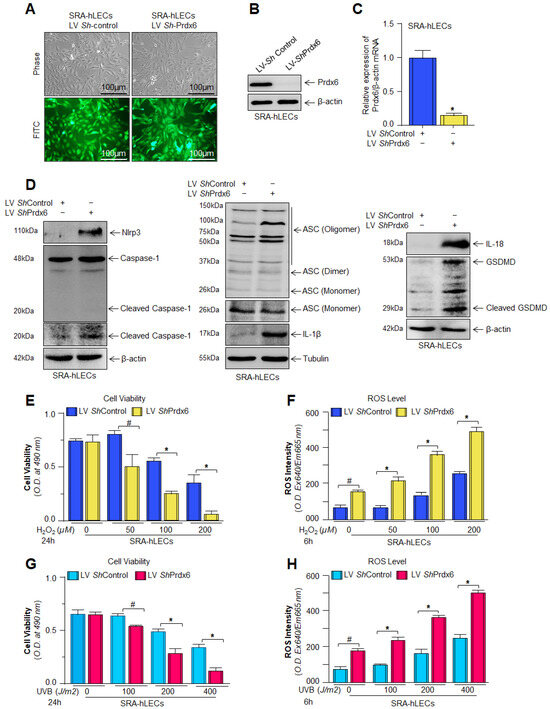
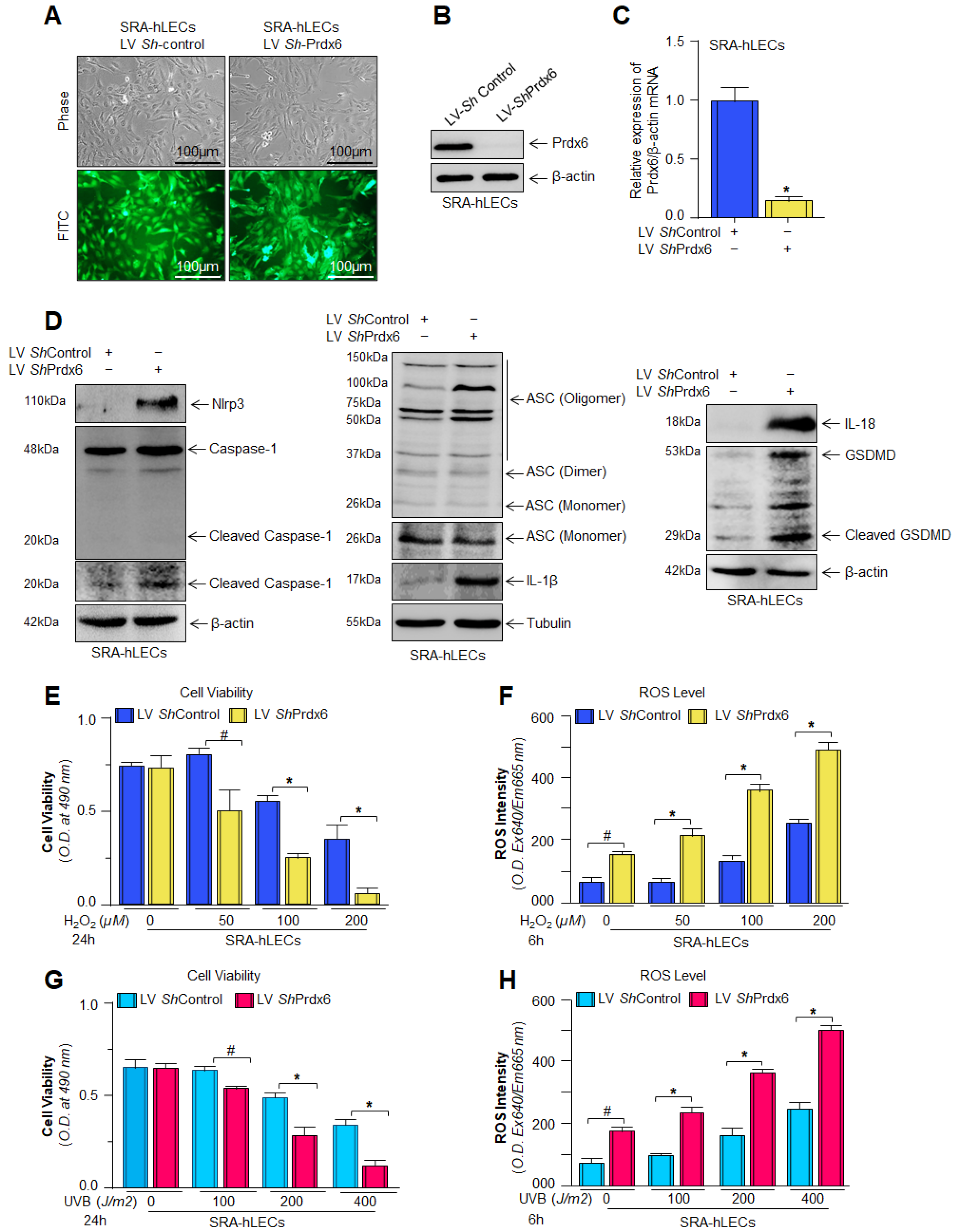
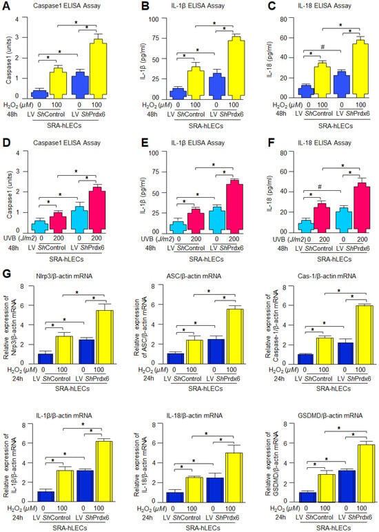
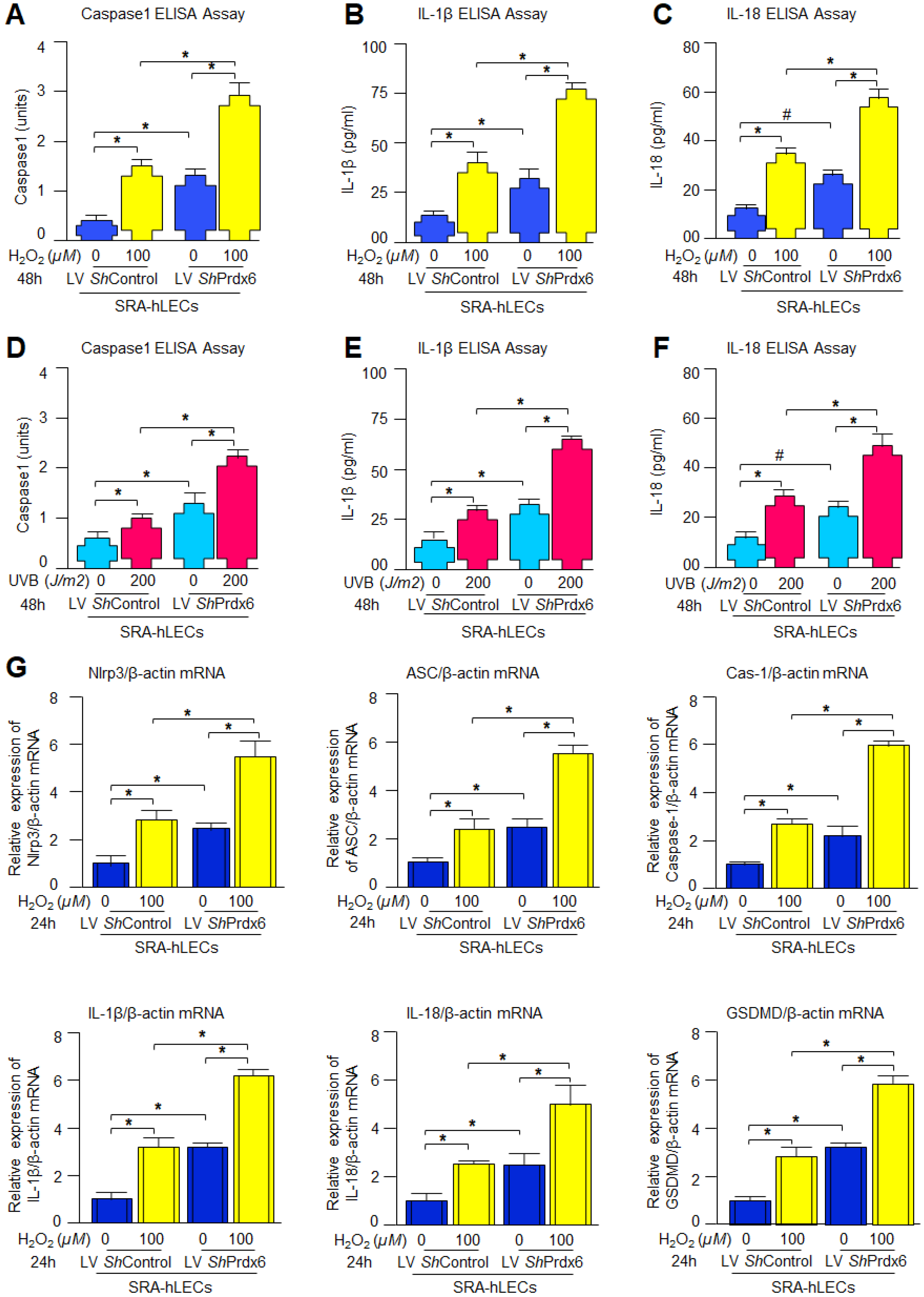
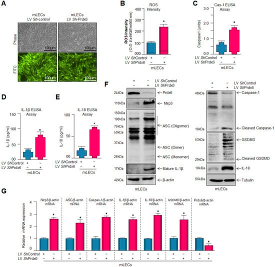
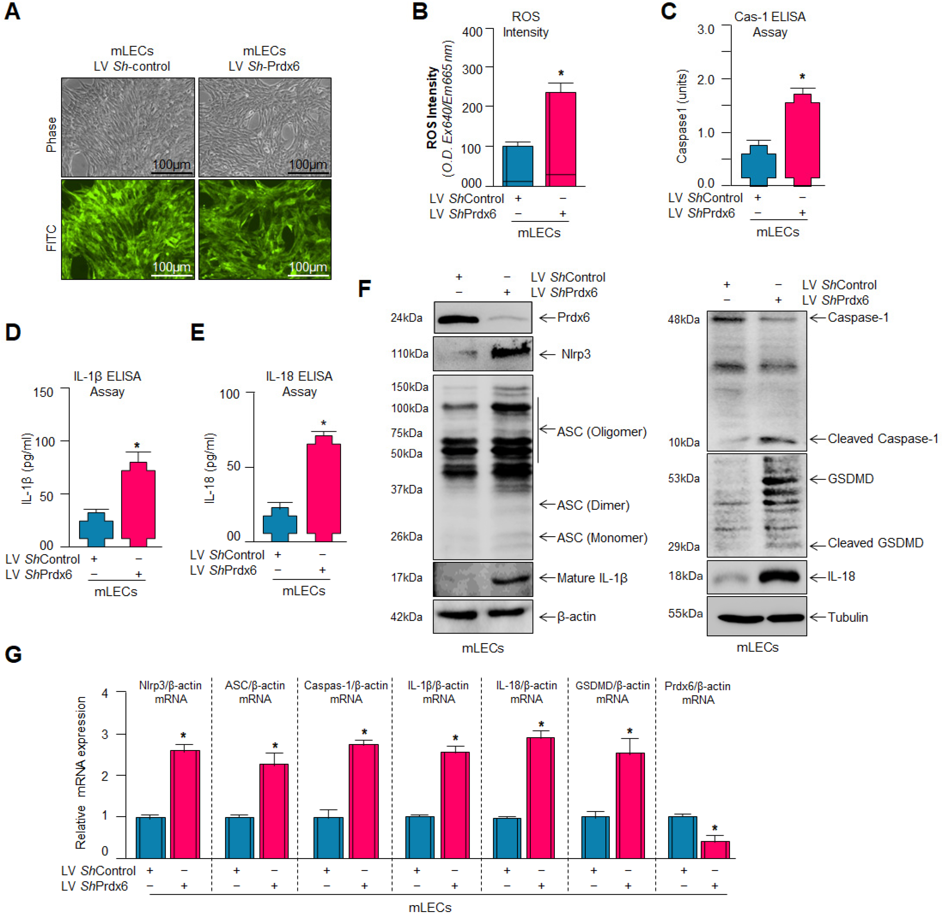
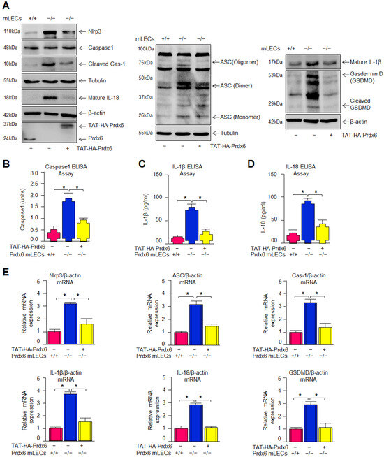
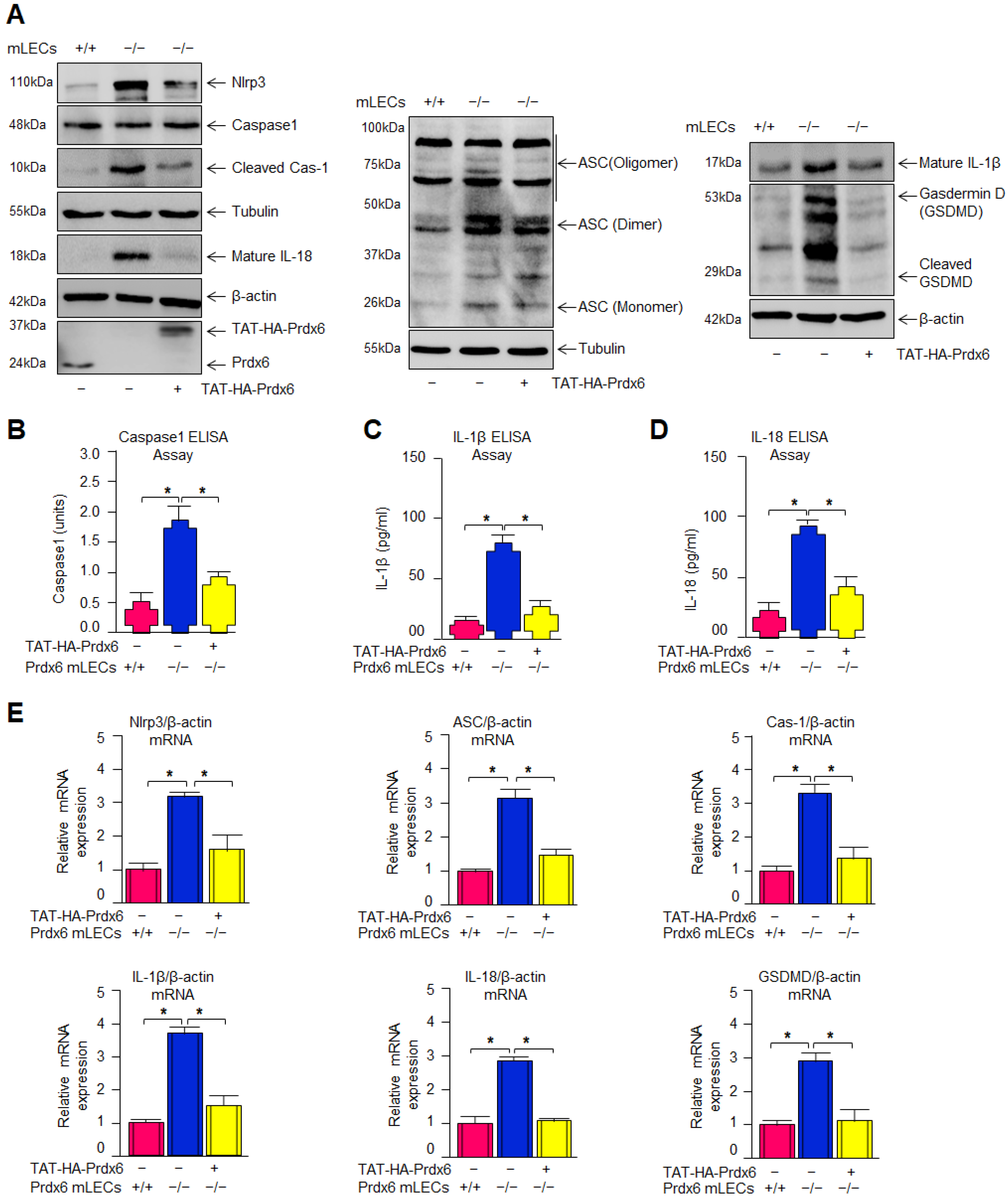
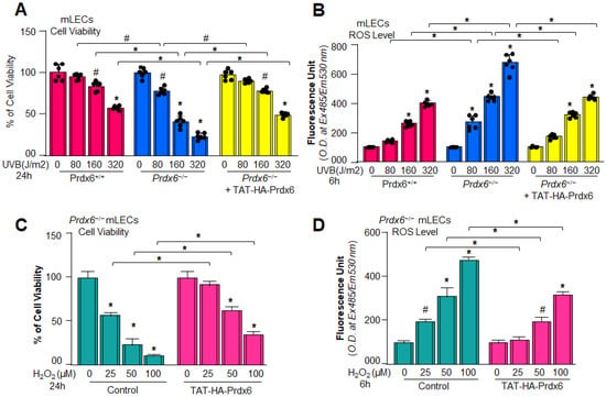
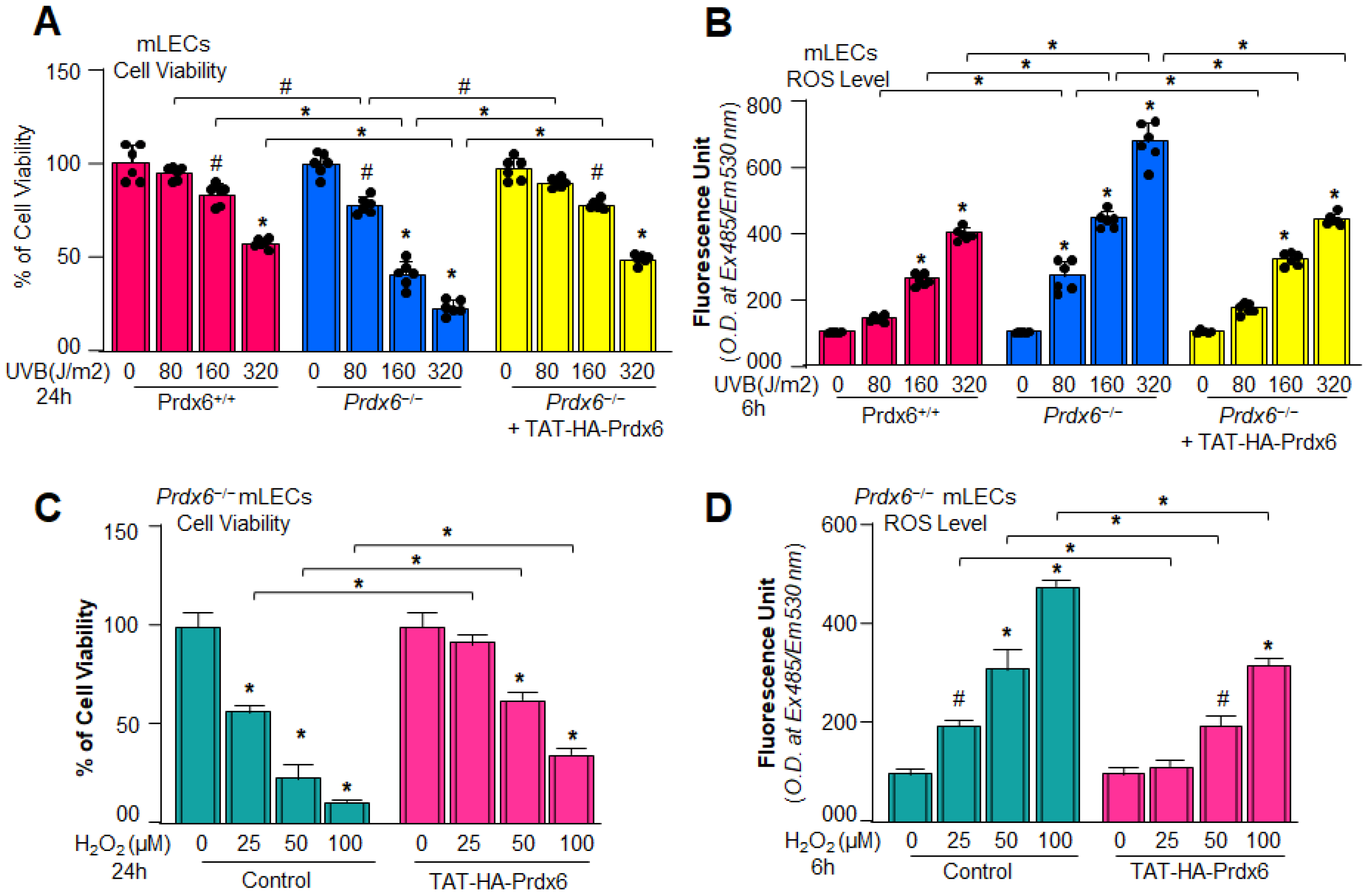
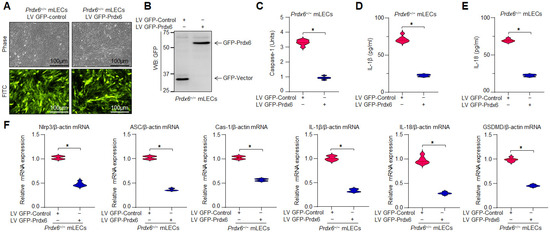
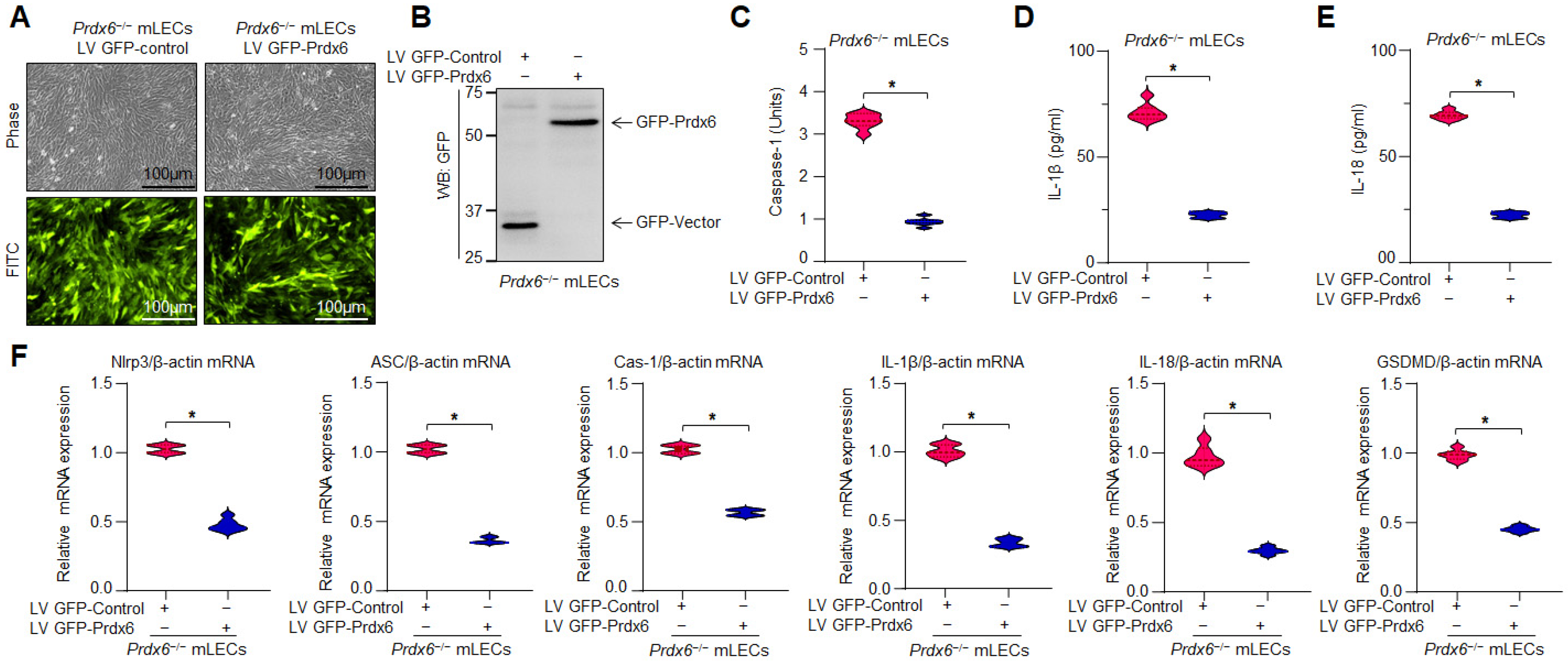
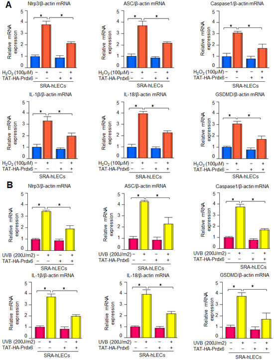
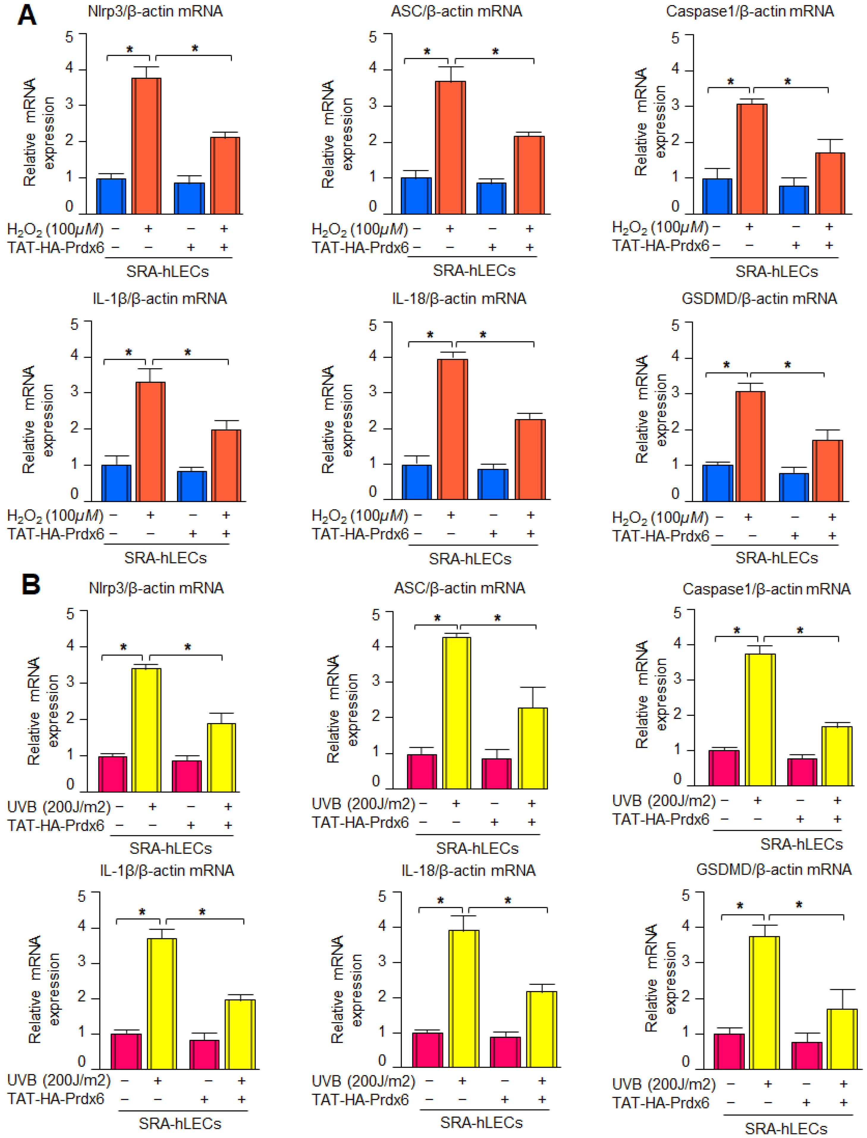
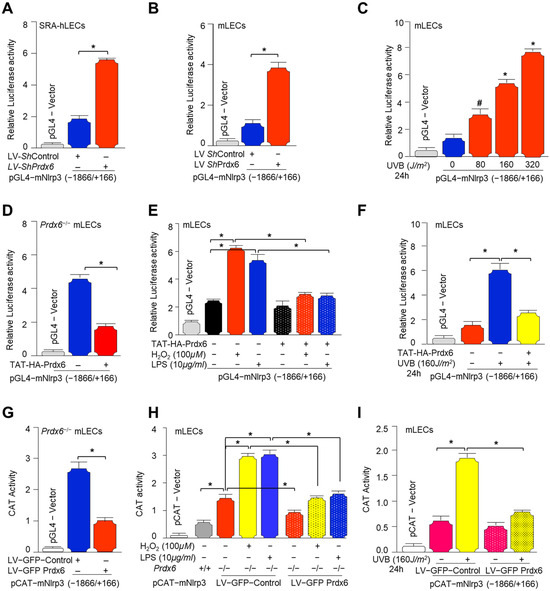
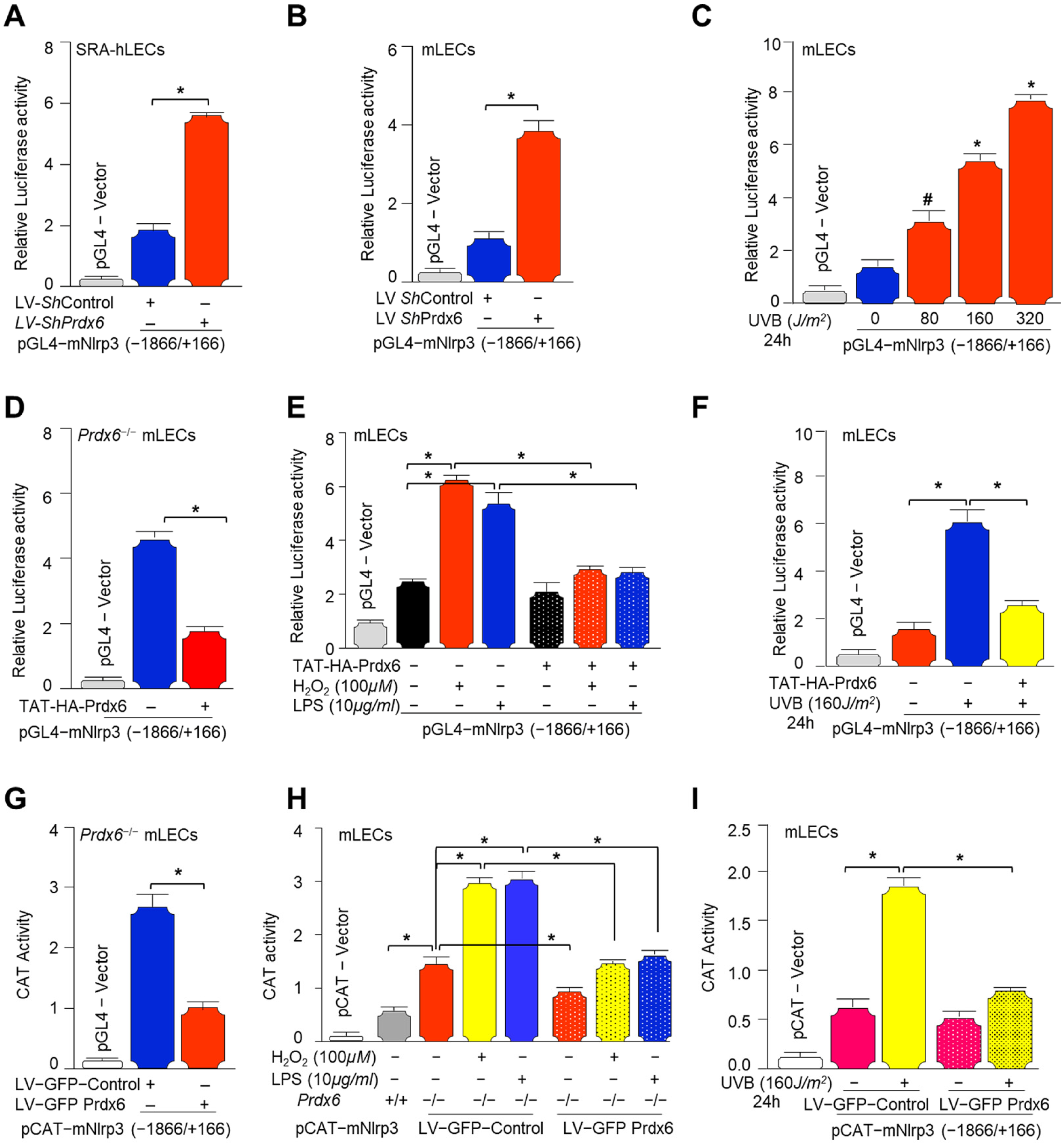
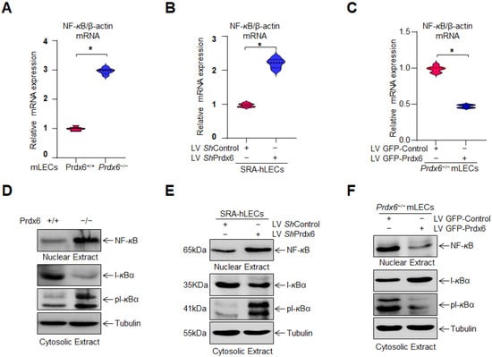
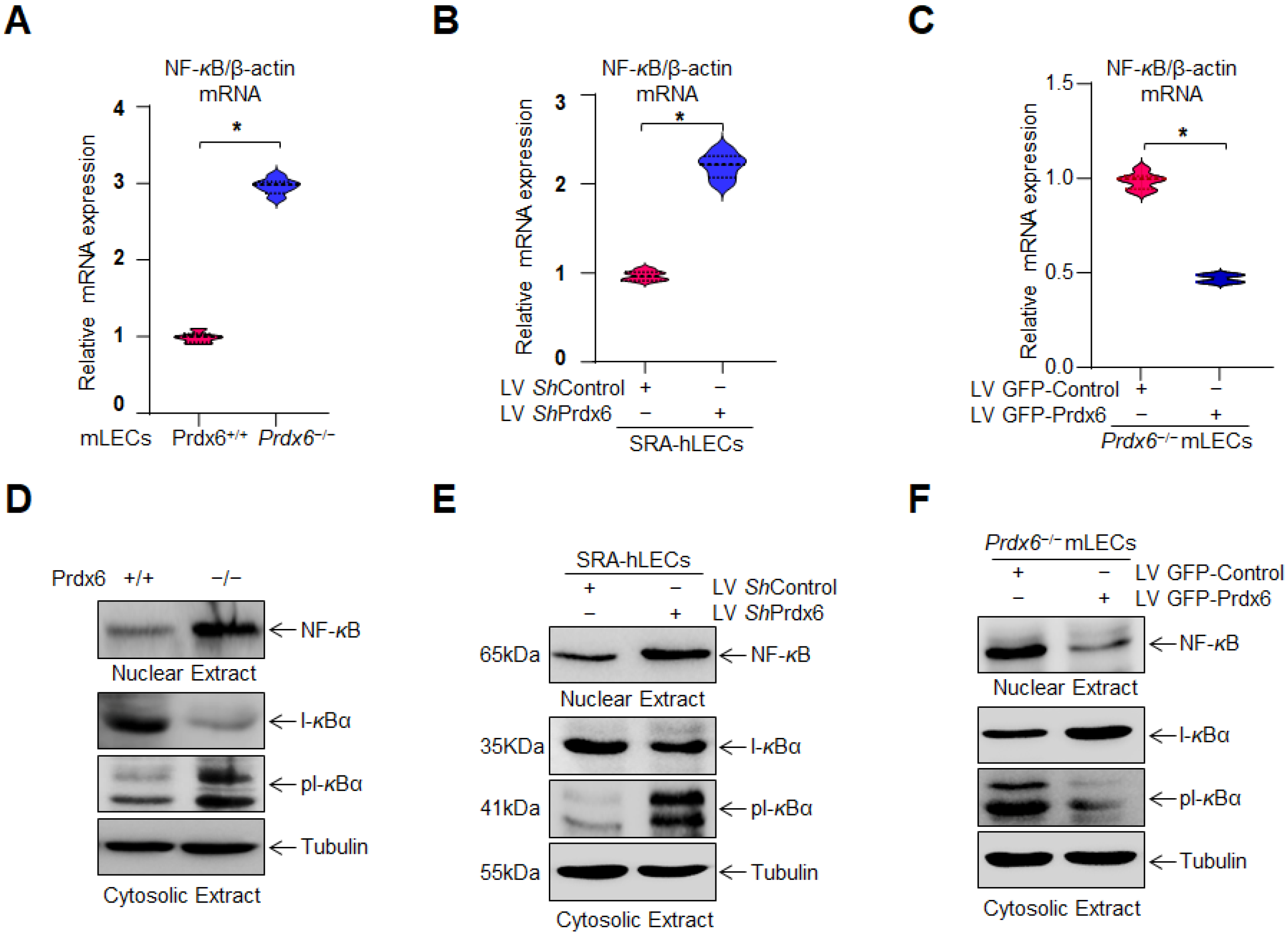
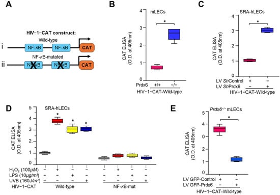
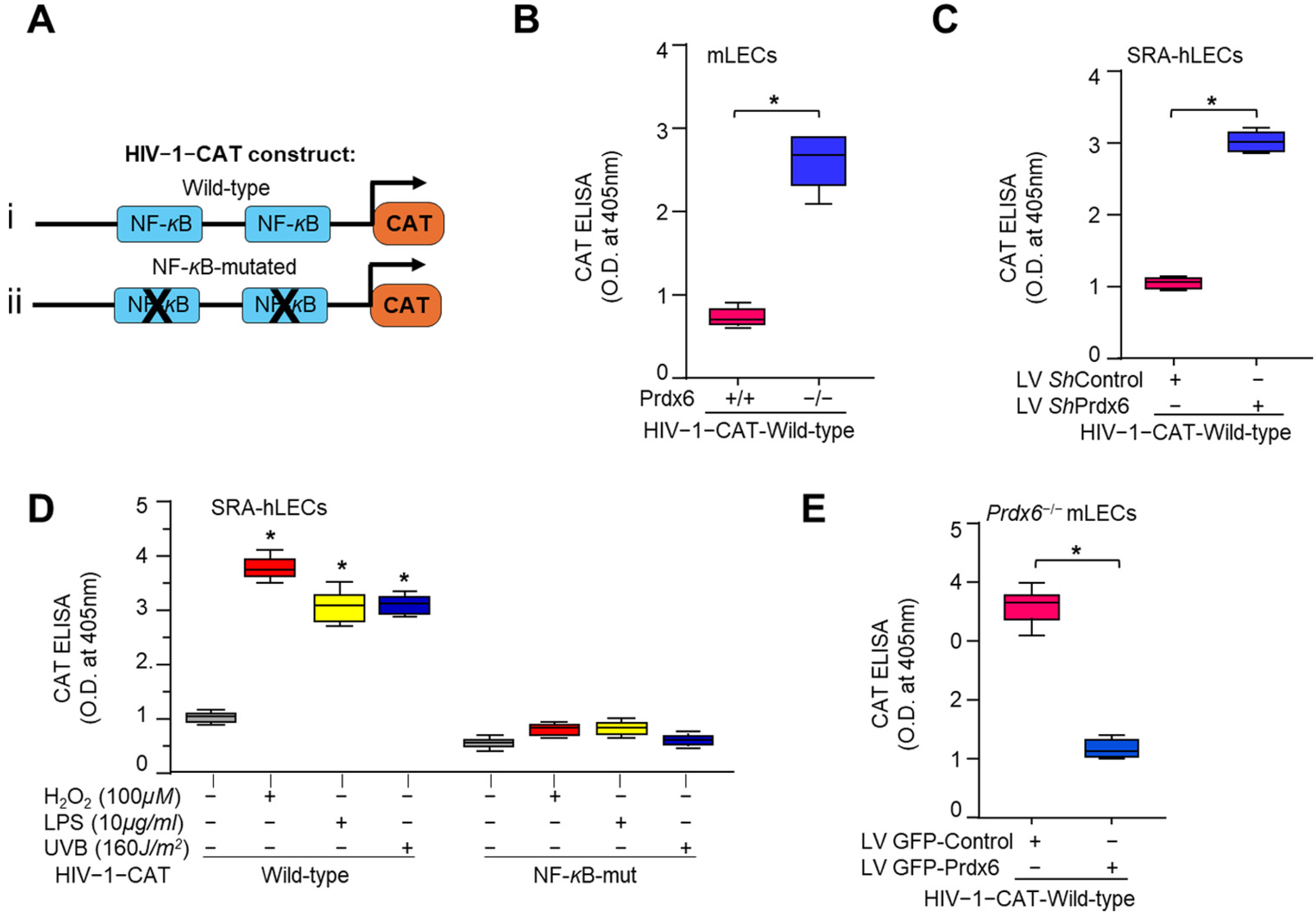
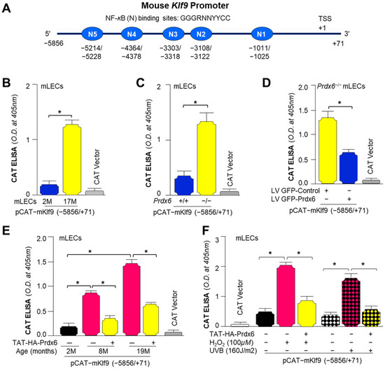
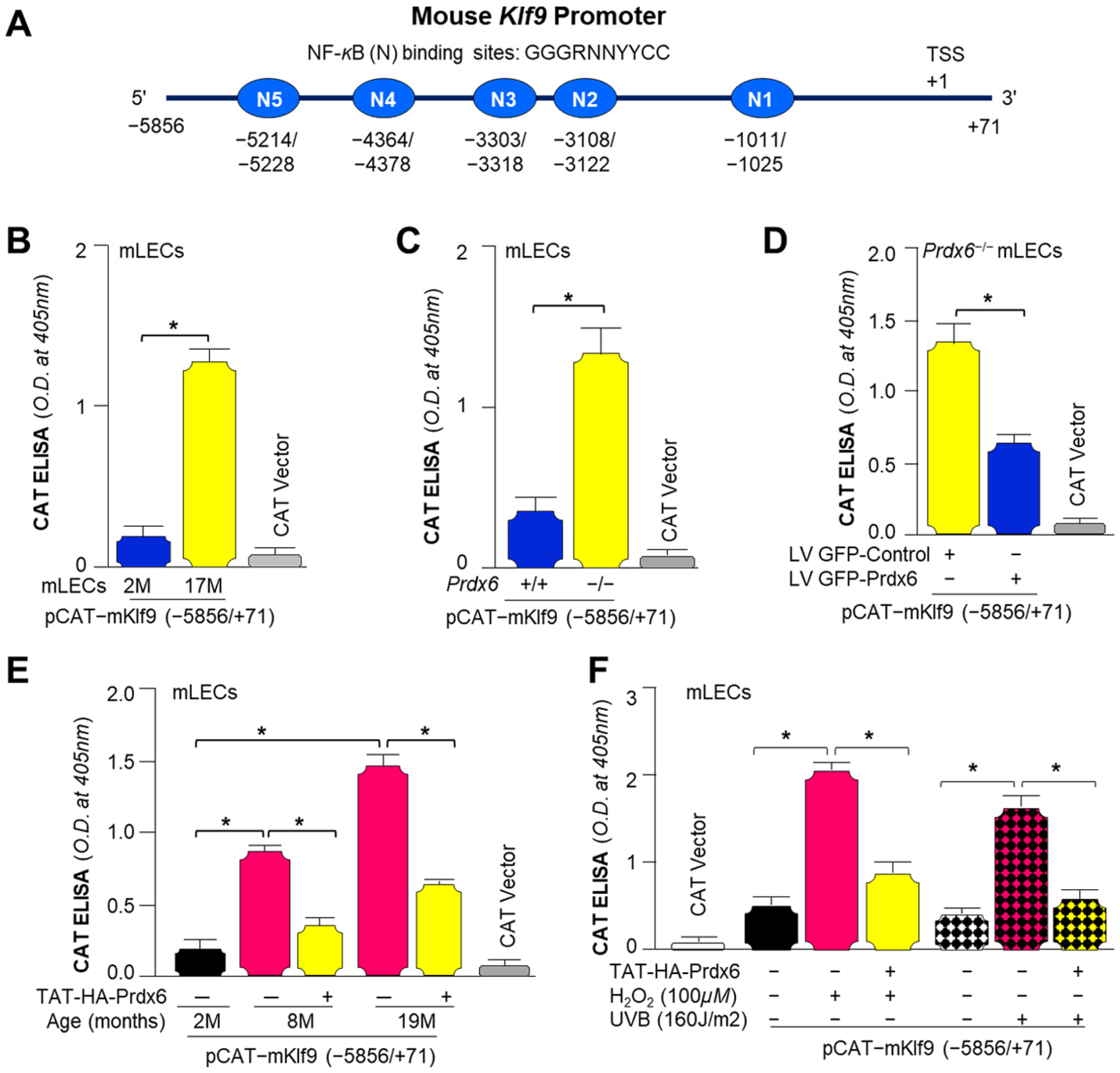
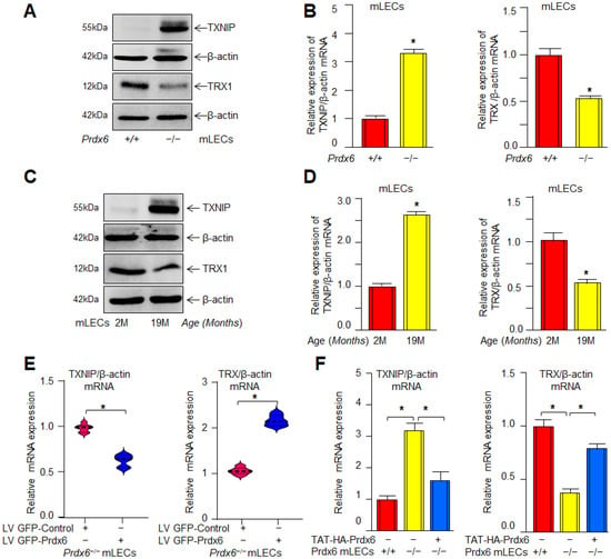
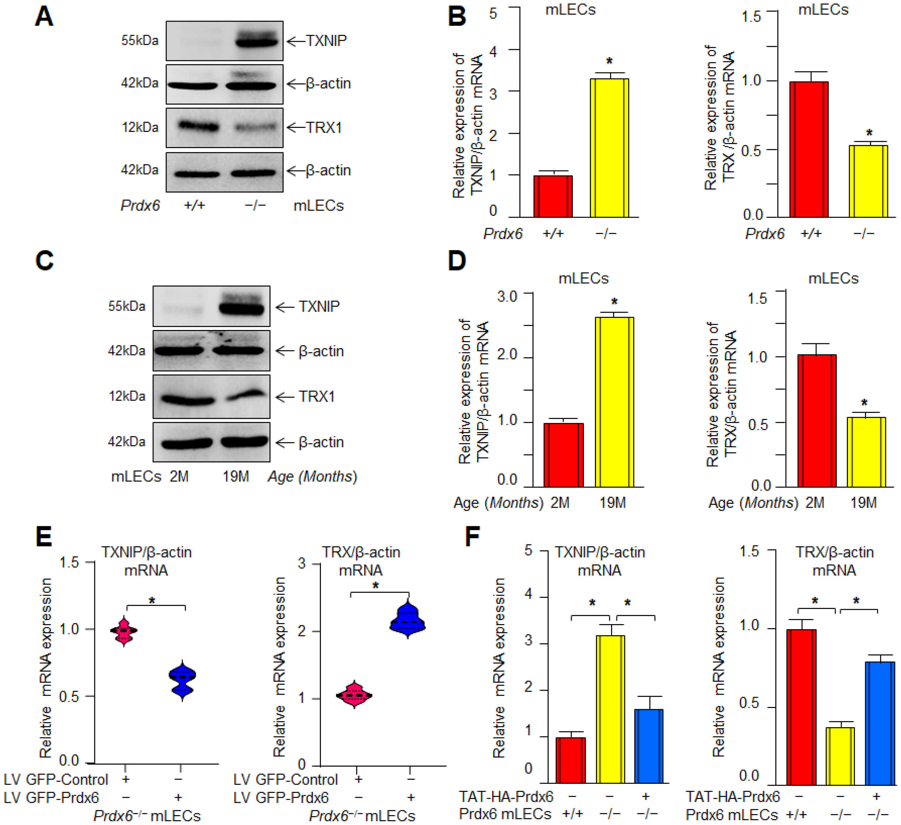
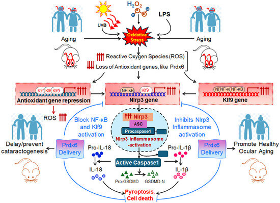
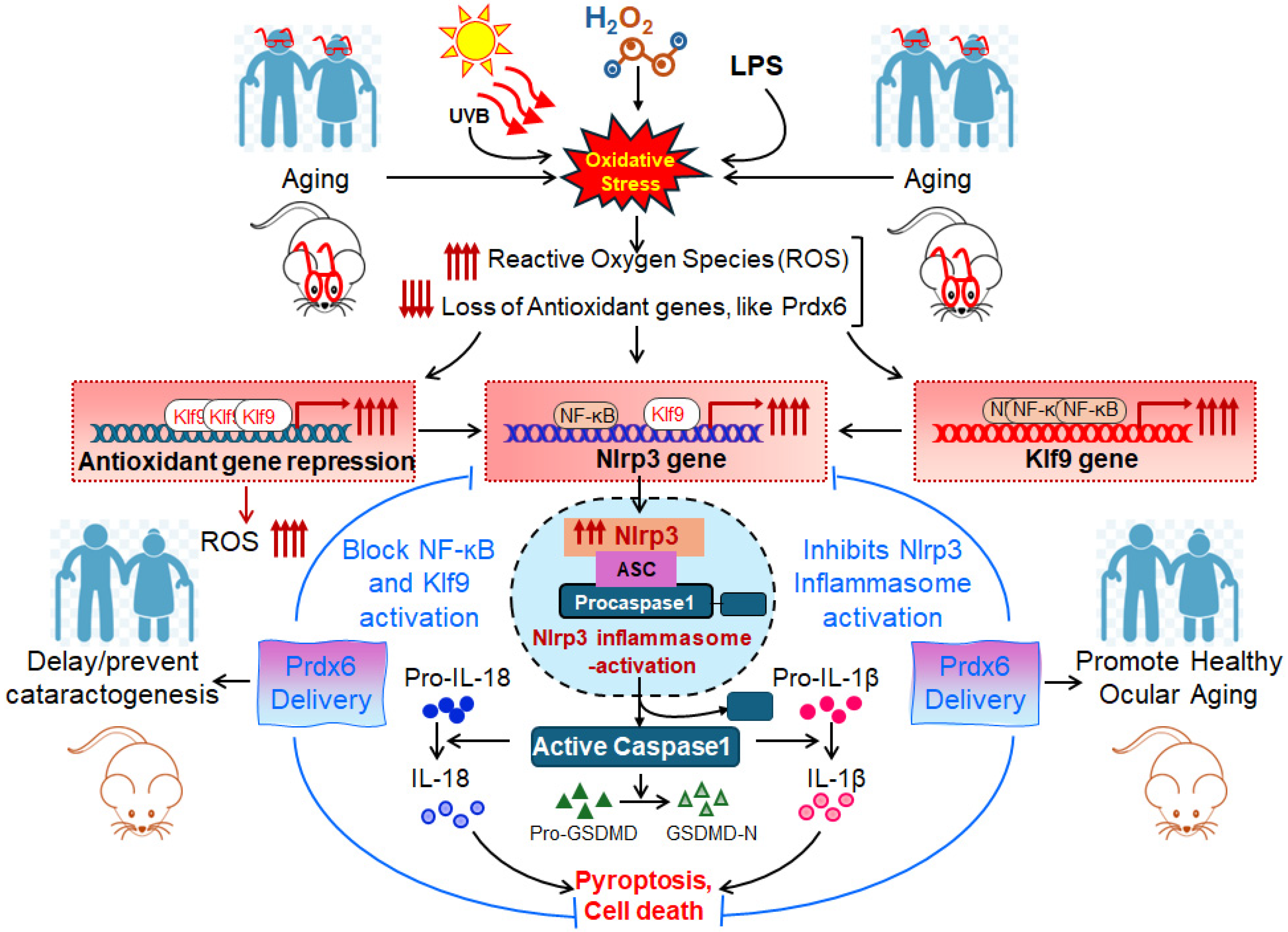

Disclaimer/Publisher’s Note: The statements, opinions and data contained in all publications are solely those of the individual author(s) and contributor(s) and not of MDPI and/or the editor(s). MDPI and/or the editor(s) disclaim responsibility for any injury to people or property resulting from any ideas, methods, instructions or products referred to in the content. |
© 2026 by the authors. Licensee MDPI, Basel, Switzerland. This article is an open access article distributed under the terms and conditions of the Creative Commons Attribution (CC BY) license.