


Abstract
Morin, a natural flavonoid substance found in various plants, exhibits several interesting properties that make it of interest in pharmacological and health research. Morin is known for its potent antioxidant activity. It can help neutralize harmful free radicals in the body, which are implicated in oxidative stress and various chronic diseases. Morin possesses anti-oxidant and anti-inflammatory effects, anticancer properties, neuroprotective effect, cardioprotective effect, anti-diabetic property, nephroprotective and hepatoprotective effects. In order to have a deeper understanding of the numerous and advantageous health benefits of morin, the present article reveals the most recent research findings to report detailed the in vivo biological/pharmacological activities and molecular mechanisms. The articles for this study were screened using the internet database MDPI, Web of Science, Google Scholar and PubMed, with regard to articles published in recent years. The search phrases “morin”, “neuroprotective effects”, “cardioprotective effects”, “hepatoprotective effects”, “nephroprotective effects”, “cancer”, “ anti-diabetic property” and other related keywords along with the terms “free radical scavenging”, “structure”, and “physicochemical properties” were used to find the relevant in vivo studies. It is anticipated that this research will provide some new perspectives and ideas for the investigation of morin, its characteristics, and the scientific underpinnings of its improved use in clinical practice.
1. Introduction
Morin is a naturally occurring flavonoid found in various plant species and has attracted considerable scientific interest because of its diverse biological and pharmacological properties. Its potential biological activities have been investigated primarily in experimental models, with particular attention to its antioxidant, anti-inflammatory, anticancer, and organ-protective effects. Oxidative stress, which results from an imbalance between reactive oxygen species and antioxidant defenses, contributes to cellular damage and is implicated in the pathophysiology of several chronic conditions, including cardiovascular and neurodegenerative diseases [1]. Morin has demonstrated free-radical-scavenging and antioxidant activities in experimental studies, suggesting that it may help mitigate oxidative damage under specific experimental conditions [2]. In addition, its reported anti-inflammatory effects have prompted investigations into its potential influence on inflammatory pathways associated with various pathological processes [2].
Experimental evidence has also suggested that morin may have potential relevance to cardiovascular health by modulating oxidative stress and inflammatory responses, both of which are involved in the development and progression of cardiovascular disorders [3]. Its possible neuroprotective effects have likewise been investigated, particularly in relation to neuronal damage and mechanisms associated with neurodegenerative diseases, including Alzheimer’s disease [4]. In cancer research, morin has been reported to influence cancer-cell proliferation, apoptosis, and other cellular processes in certain experimental models [5]. Furthermore, studies have examined its potential effects on glucose homeostasis and insulin-related pathways, suggesting possible relevance to diabetes research [6]. Hepatoprotective and nephroprotective effects have also been investigated in experimental models of oxidative and inflammatory injury [7]. In addition, research has explored the potential protective effects of morin against pulmonary injury and its possible contribution to the modulation of cellular processes associated with aging [8]. Nevertheless, these findings should be interpreted in the context of the experimental models and conditions employed, as evidence from in vitro and animal studies does not, by itself, establish clinical efficacy or safety in humans.
Several reviews have previously examined the biological and pharmacological properties of morin. Among these, Rajput et al. [9] published a comprehensive review in 2021, which summarized the diverse biological activities and pharmacological potential of morin hydrate. This work provides an important foundation for understanding the compound; however, approximately five years of subsequent research have produced additional findings that merit critical evaluation. The present review therefore aims to complement and extend the previous synthesis by incorporating relevant studies published after the earlier review and available up to 2026. Particular emphasis is placed on recent findings concerning the chemical and physicochemical characteristics of morin, its free-radical-scavenging capacity, its biological activities, the molecular mechanisms potentially underlying these effects, and its reported antioxidant, anticancer, hepatoprotective, nephroprotective, pulmonary-protective, neuroprotective, antidiabetic, and cardiovascular properties. In addition to summarizing reported outcomes, this review critically considers the strength and limitations of the available evidence, distinguishes preliminary experimental findings from clinically established effects, and identifies research gaps that should be addressed in future investigations.
To identify relevant publications, literature searches were conducted using MDPI, Google Scholar, Web of Science and PubMed covering publications from 2001 to 2026. The search strategy included combinations of the following terms: “morin,” “morin hydrate,” “chemical properties,” “physicochemical properties,” “antioxidant activity,” “free-radical scavenging,” “anticancer activity,” “hepatoprotective,” “nephroprotective,” “pulmonary protective,” “neuroprotective,” “antidiabetic,” “cardiovascular,” and “molecular mechanisms.” Search terms were combined using appropriate Boolean operators where applicable. Publications were initially screened by title and abstract for relevance to the scope of the review, followed by full-text assessment when necessary. Studies were considered for inclusion based on their relevance to the chemical characteristics, biological activities, pharmacological mechanisms, and potential applications of morin.
Although previous reviews have provided extensive overviews of morin, the present article addresses a distinct aspect of the literature by adopting a critical, evidence-oriented approach. Rajput et al. [9] comprehensively discussed the biological and pharmacological potential of morin hydrate, whereas Kumar et al. [10] provided a broader overview of its natural sources, chemistry, analytical aspects, nutraceutical prospects, and pharmacological applications. Rather than primarily expanding these descriptive overviews, the present review critically evaluates the strength and limitations of the experimental evidence underlying the reported biological activities of morin. In particular, it distinguishes findings derived from in vitro experiments from those supported by animal models, differentiates direct functional outcomes from indirect changes in biochemical or molecular markers, and examines whether the proposed mechanisms are supported by functional or causal evidence. Where available, experimental doses, administration routes, treatment durations, and model-specific limitations are considered to facilitate comparison across studies. Importantly, the review also distinguishes evidence for direct pharmacological effects from evidence of protection against disease-associated complications, thereby avoiding the interpretation of antioxidant or biomarker changes as definitive therapeutic outcomes. Finally, pharmacokinetic limitations, including aqueous solubility, stability, and oral bioavailability, are considered alongside the translational relevance of the reported findings. Thus, the principal contribution of this review is not simply to catalogue the biological activities of morin, but to assess how convincingly these activities have been demonstrated, identify gaps in the current evidence, and define the experimental requirements for future translational research.
2. Morin
Antioxidants, known as compounds that prevent the oxidation of other molecules in living organisms, are substances that have the property of being hydrogen or electron donors by participating in reactions [11]. Cells, tissues, and organs have complex antioxidant systems that work in coordination to protect against free radical-induced injuries and scavenge ROS/RNS by increasing antioxidant defense system elements, stabilizing mitochondrial function, and hindering the oxidation process through detoxification of free radicals. They are available as natural, enzymatic and non-enzymatic or synthetic compounds [12,13].
Flavonoids are natural, plant-derived dietary bioactive components that have a significant impact on human health, consisting of two outer aromatic rings with three carbon rings. Examples of flavonoids include flavone, flavanol, flavanone, flavanonol, flavonone, flavononol, flavanol, isoflavone and anthocyanidin [14].
Morin hydrate, a bioflavonoid belonging to the class of flavonols and obtained mainly from the fruits, stems, and leaves of plants in the Moraceae family, is a plant component that has beneficial effects when applied against various chronic and life-threatening degenerative diseases. It mainly occurs in the branches of white mulberry, guava, old fustic, Osage orange, onion, apple skin, and almond. It can also be obtained from figs, sweet chestnut, jack fruit, coffee, seaweed, tea, red wine and cereal grains [9]. Morin hydrate shows free radical scavenger, antioxidant, anti-inflammatory, anti-diabetic [15], anti-cancer [16], anti-microbial, anti-arthritis, cardioprotective, neuroprotective, nephroprotective and hepatoprotective effects, acts as a nuclear factor kappa-light chain enhancer of activated B cells (NF-қB) and mitogen-activated protein kinase (MAPK) [8,16,17], exhibits pharmacological activities by modulating various cellular signaling pathways such as kinase (MAPK) and acts as Janus kinases/Signal converter and activator of transcription proteins (JAKs/STATs) [8,18].
2.1. Chemical Structure and Free Radical Scavenging Potential of Morin
Morin (Figure 1) is a naturally occurring bioflavonoid belonging to the flavonol subclass, characterized by a 3-hydroxyl group on the flavone (2-phenyl-1-benzopyran-4-one) backbone, together with additional hydroxyl substituents at positions 2′, 4′, 5, and 7 [8]. Similar to many other flavonoids, morin exhibits limited oral bioavailability, which is partly associated with its poor aqueous solubility, reported to be approximately 28.7 μg/mL in water. Its physicochemical behavior is also influenced by pH and solvent polarity, which can affect its solubility, stability, and potential absorption. Yazdanshenas and Gharib [19] investigated the solubility and thermodynamic properties of morin hydrate in different alcohols, including methanol, ethanol, propan-1-ol, propan-2-ol, and butan-1-ol. Their findings indicated that morin solubility was influenced by solvent polarity and dielectric properties, with an approximately linear relationship observed between solubility and the dielectric constants of the investigated alcohols.
Figure 1.
Chemical structure of morin.
Importantly, the solubility and degradation kinetics of morin hydrate in physiologically relevant aqueous media were systematically investigated by Jangid et al. [20] under different pH, temperature, and light conditions. Their findings demonstrated that morin exhibits pronounced pH-dependent solubility and stability. At 37 °C, its aqueous solubility was reported as 32.69 ± 1.63, 14.88 ± 2.43, 28.72 ± 0.97, 54.86 ± 2.05, and 505.09 ± 14.61 μg/mL at pH 1.2, 5.0, 7.0, 7.4, and 9.0, respectively. These results indicate that morin solubility increases substantially under alkaline conditions, whereas its stability may be compromised depending on the environmental conditions. Therefore, pH-dependent solubility and chemical stability should be considered together when evaluating its pharmaceutical potential. Such physicochemical limitations, together with restricted intestinal permeability and presystemic metabolism, may contribute to the poor oral bioavailability of morin and represent important challenges for its clinical translation.
The two types of antioxidants used in industrial and medicinal applications are natural and synthetic. More research is being carried out on naturally occurring antioxidant molecules found in plants, as the majority of commonly used synthetic antioxidants are thought to be harmful to humans [21,22,23]. The most well-known of these are polyphenolic substances, which have one or more hydroxyl groups in their structure. These substances can be structurally separated into two main classes: flavonoids and phenolic acids, which are essentially hydroxybenzoic and hydroxycinnamic acids [24]. Because of its great antioxidant capacity, morin hydrate stands out among polyphenols. By scavenging free radicals, it guards against oxidation of cellular components. The double bond between the C2-C3 atoms and the presence of the hydroxyl group (-OH), which activates the double bond at the C-3 position, are primarily responsible for morin’s antioxidant potential. Additionally, the presence of two hydroxyl groups (-OH) on the 2′ and 4′ positions of the B ring appears to give morin its anti-lipid peroxidation capability [25]. At the same time, the hydroxyl group at the 4′ position of the B ring is supposed to be mainly responsible for morin’s anti-radical activity. Additionally, the hydroxyl group at position 2′ in the B ring, which is found only in morin, appears to be responsible for its more active anti-radical action. In order to determine the molecular mechanisms associated with morin’s radical-scavenging activity, the relationship between its structure, kinetic, thermochemical, and electronic properties was investigated within the scope of a study. When there was an overabundance of free radicals, morin demonstrated the highest level of radical scavenging activity. Through the use of experimental and computational methods, it was discovered that the main reactive sites are 3-OH, 2′-OH, and 4′-OH, and that the reaction’s first product is the 3-O-2′-O quinone, which tends to dominate in the enol form due to a tautomerism effect and has a structural arrangement that is similar to the 3-O semiquinone [26]. To evaluate the contribution of individual hydroxyl groups to the antioxidant activity of morin, Maciel et al. [27] computationally investigated its reaction with 2,2-diphenyl-1-picrylhydrazyl (DPPH) and reported the corresponding Gibbs free-energy changes (ΔG). According to their findings, the hydroxyl groups at the 2′-ArOH and 3-ArOH positions exhibited negative ΔG values of −7.56 and −4.57 kcal/mol, respectively, indicating thermodynamically favorable reactions at these sites. These values are cited from the original computational study and are not calculations performed in the present work.
2.2. Pharmacological Importance of Morin
Morin is a naturally occurring flavonoid with diverse pharmacological properties, including antioxidant, anti-inflammatory, neuroprotective, hepatoprotective, and cardioprotective activities. Its biological effects are primarily associated with the modulation of oxidative stress, inflammatory responses, and cellular signaling pathways (Figure 2). Experimental evidence suggests that morin exerts these effects through interactions with several interconnected molecular pathways rather than a single specific target.
Figure 2.
Scheme of morin sources and its pharmacological effects.
One of the important mechanisms underlying the antioxidant activity of morin involves the nuclear factor erythroid 2-related factor 2 (Nrf2)/heme oxygenase-1 (HO-1) signaling pathway. Morin has been shown to promote Nrf2 nuclear translocation and HO-1 expression through ERK-dependent signaling, thereby enhancing cellular defense against oxidative stress [3]. In addition, morin suppresses oxidative stress-induced NF-κB activation, reducing the expression of pro-inflammatory mediators such as cyclooxygenase-2 (COX-2) and inducible nitric oxide synthase (iNOS) [9].
The mitogen-activated protein kinase (MAPK) pathway, including ERK, JNK, and p38, is also involved in the pharmacological effects of morin. In lipopolysaccharide (LPS)-stimulated microglial cells, morin inhibited MAPK and NF-κB signaling while enhancing Nrf2/HO-1 activity, resulting in reduced inflammatory mediator production [15]. These findings indicate functional crosstalk between antioxidant and inflammatory signaling pathways. In particular, Nrf2-mediated antioxidant responses may counteract oxidative stress-associated NF-κB activation, whereas MAPK signaling can regulate both inflammatory and cytoprotective cellular responses.
Collectively, the modulation of Nrf2/HO-1, NF-κB, and MAPK signaling provides a plausible molecular basis for the antioxidant and anti-inflammatory properties of morin. These interconnected mechanisms may contribute to the protection of cells against oxidative and inflammatory damage and provide a mechanistic foundation for understanding its neuroprotective potential.
2.3. Neuroprotective Effects of Morin
Neurodegenerative diseases are characterized by progressive neuronal dysfunction and loss, often accompanied by synaptic impairment, protein aggregation, oxidative stress, and neuroinflammation. Although the relative contributions of these processes vary among disorders, excessive reactive oxygen species (ROS) production and disruption of cellular antioxidant defenses have been implicated in neuronal injury and disease progression [28]. Accordingly, compounds capable of modulating oxidative stress and inflammatory signaling have attracted interest as potential neuroprotective agents. Morin, a naturally occurring flavonoid, has been investigated in several experimental models of neurological injury, including neuropathic pain, cerebral ischemia–reperfusion, Parkinson’s disease-like pathology, stress-induced cognitive impairment, seizures, and chemically induced neurotoxicity. However, the diversity of these models necessitates a critical distinction between neuroprotection demonstrated under specific experimental conditions and efficacy against human neurological diseases (Table 1).
Oxidative stress, neuroinflammation, and neuronal survival. Several studies have reported broadly consistent effects of morin on oxidative and inflammatory markers, although the initiating insults and experimental outcomes differ considerably. In rats with vincristine-induced peripheral neuropathy, morin improved paw withdrawal thresholds and body-weight gain, attenuated sciatic nerve dysfunction, and modulated the expression of interleukin-6 (IL-6), nuclear factor kappa B (NF-κB), and phosphorylated NF-κB [29]. Similarly, morin ameliorated oxidative stress, inflammatory responses, and neuronal injury in experimental models of chronic constriction injury, cadmium-induced neurotoxicity, acrylamide exposure, and ifosfamide-induced neurotoxicity [30,31,32,33]. These findings suggest that modulation of oxidative and inflammatory pathways may represent a recurring feature of morin-associated neuroprotection. Nevertheless, differences in the toxic agents, affected tissues, and measured endpoints prevent the assumption that the same mechanism accounts for all observed effects. For example, the reported involvement of poly(ADP-ribose) polymerase (PARP) overactivation in chronic constriction injury provides a more specific mechanistic explanation for peripheral neuropathy than changes in general oxidative stress markers alone [30].
Evidence from cerebral ischemia–reperfusion models further suggests that the effects of morin may extend beyond conventional antioxidant activity. Khamchai et al. [34] reported that morin reduced cerebral infarct size, blood–brain barrier (BBB) leakage, and apoptotic cell death, while promoting angiogenesis through the angiopoietin-1/Tie-2 axis and Wnt/β-catenin signaling pathways. These outcomes indicate that vascular integrity, tissue repair, and neuronal survival may contribute to the observed effects. However, infarct size and BBB permeability are model-specific endpoints, and improvements in these measures do not independently establish sustained neurological recovery. Consequently, studies examining long-term functional outcomes and the persistence of vascular and neuronal protection are needed to determine the broader significance of these findings.
Parkinsonian models and mitochondrial-related mechanisms. Morin has also been investigated in toxin-induced models of Parkinson’s disease. In mice exposed to rotenone, morin attenuated behavioral abnormalities and oxidative and neuroinflammatory responses, with the study also exploring the involvement of the gut–brain axis [35]. In a separate MPTP-induced mouse model, morin reduced dopaminergic neuronal loss in the striatum and substantia nigra, improved motor dysfunction, and attenuated astrocyte activation. Complementary cell experiments showed that morin protected primary neurons against MPP+-induced ROS generation and mitochondrial membrane potential disruption and inhibited NF-κB translocation and astroglial activation [36]. The agreement between behavioral, histological, and cellular findings strengthens the biological plausibility of these observations. Nevertheless, rotenone and MPTP produce experimentally induced parkinsonian features through specific toxic mechanisms and do not reproduce the full, progressive pathology of human Parkinson’s disease. Moreover, evidence obtained from cultured cells cannot establish whether comparable concentrations are achievable in the brain following systemic administration.
Stress-related behavioral changes and cognitive impairment. Studies involving chronic stress, sleep deprivation, and sepsis have examined whether morin can preserve behavioral performance and hippocampal integrity. In chronically stressed mice, morin hydrate improved behavioral outcomes and attenuated oxidative, nitrergic, and inflammatory alterations, including changes in malondialdehyde (MDA), nitrite, glutathione (GSH), and NF-κB-associated responses [37,38]. Morin also ameliorated neurobehavioral impairments and hippocampal neuronal loss following rapid eye movement sleep deprivation [39]. In a murine sepsis model, morin modulated inflammatory responses, reduced microglial activation, and was associated with preservation of synaptic integrity and cognitive function [40]. These findings suggest that the effects of morin may extend to neurological dysfunction associated with stress, sleep disruption, and systemic inflammation. However, these models address distinct biological processes, and behavioral improvements in stress-related paradigms should not automatically be interpreted as evidence of disease-modifying activity in neurodegenerative disorders. Furthermore, behavioral tests may be influenced by locomotor activity, anxiety-related responses, and other nonspecific effects; therefore, behavioral outcomes should be interpreted alongside histological and molecular findings.
Neuroprotection in metabolic disorders and peripheral neuropathy. Experimental evidence also indicates potential benefits of morin in diabetes-associated neurological complications. In streptozotocin-induced diabetic models, morin improved selected measures of oxidative stress, inflammation, neuronal apoptosis, and neurotrophic support. Shyma and Mini [41] associated its effects with the TrkB/Akt signaling pathway, reporting changes in Bcl-2, Bax, caspase-3, and cytochrome c. Other studies reported improvements in sensory nerve conduction, nerve blood flow, and sensorimotor outcomes in experimental diabetic neuropathy [15,42,43]. In addition, AlSharari et al. [42] found that morin treatment altered pain-related behavioral responses and inflammatory and oxidative stress markers in the sciatic nerve. Collectively, these findings support a possible role for morin in modulating metabolic and inflammatory mechanisms associated with experimental diabetic neuropathy. However, the observed effects may partly reflect improvements in systemic glycemic control rather than direct actions on neuronal tissue. Studies designed to distinguish central from peripheral effects and to separate glucose-dependent from glucose-independent mechanisms are therefore warranted.
Seizure-related models and excitability-associated pathways. Morin has demonstrated activity in experimental seizure paradigms, although the findings should be interpreted separately from evidence concerning chronic neurodegeneration. In a kainic acid-induced seizure model, morin reduced granule cell dispersion, suppressed mammalian target of rapamycin complex 1 (mTORC1) activity, and attenuated inflammatory and apoptotic signaling in the hippocampus [44]. In a separate pentylenetetrazol-induced seizure model, morin reduced seizure-related outcomes and modified brain GABA, Na+/K+-ATPase, MDA, nitric oxide, GSH, and superoxide dismutase (SOD) measurements [41]. These studies provide complementary evidence that morin may influence seizure-associated neuronal injury through mechanisms involving oxidative stress, inflammatory signaling, and cellular pathways related to neuronal excitability. Nevertheless, kainic acid and pentylenetetrazol induce seizures through different mechanisms, and outcomes from acute experimental seizure paradigms do not establish efficacy in chronic epilepsy. Further studies should assess dose–response relationships, repeated-dose effects, electroencephalographic outcomes, and long-term neuronal survival.
Critical assessment and research gaps. In conclusion, the available literature shows a recurring association between morin administration and improvements in oxidative stress, inflammatory signaling, apoptosis-related markers, and selected behavioral or functional outcomes. The strongest support comes from studies in which biochemical findings are accompanied by histological, neuronal, or functional measurements. However, several limitations restrict the interpretation of this evidence. First, the studies investigate heterogeneous conditions, ranging from peripheral nerve injury and acute cerebral ischemia to metabolic neuropathy, stress-related behaviors, and toxin-induced parkinsonism. Second, differences in morin formulation, dose, administration route, pretreatment versus post-injury administration, and treatment duration complicate direct comparisons. Third, many reported mechanisms are inferred from changes in protein expression or biochemical markers rather than demonstrated through selective pathway inhibition, genetic manipulation, or other causal experiments. Finally, the predominance of preclinical studies and the limited clinical evidence prevent conclusions about therapeutic efficacy, optimal dosing, brain bioavailability, and long-term safety in humans. Future research should therefore prioritize standardized dose–response studies, pharmacokinetic and blood–brain barrier assessments, mechanistic validation, independent replication, and well-designed clinical investigations where justified by the preclinical evidence.
Dose, administration route, and treatment duration across experimental studies. An important consideration when comparing the neuroprotective effects of morin is the substantial variation in experimental dosing protocols. As summarized in Table 1, morin has been administered using different dose ranges, routes, and treatment schedules depending on the experimental model. For example, doses of 5–20 mg/kg administered intraperitoneally were used in chronic restraint stress and stress-related memory impairment models [37,38], whereas oral administration of 15–30 mg/kg/day was reported in experimental diabetic neuropathy and diabetes-associated brain injury [42,43]. Other studies employed different regimens, including 5–80 mg/kg intraperitoneally for 28 days in a rotenone-induced Parkinsonian model [35] and 100–200 mg/kg orally for two consecutive days in an ifosfamide-induced neurotoxicity model [33]. These differences highlight the importance of considering the experimental model, administration route, treatment duration, and timing of morin exposure when interpreting its biological effects. Where specific methodological details were not available in the accessible publication records, they were not assumed and require confirmation from the original full-text articles. Consequently, direct comparisons between studies should be made cautiously, and further investigations using standardized dosing protocols are warranted.
Table 1.
Critical appraisal of preclinical evidence on the neuroprotective and neurological effects of morin.
2.4. Hepatoprotective Effects of Morin
The liver plays a central role in xenobiotic metabolism, energy homeostasis, and detoxification, making it particularly susceptible to injury induced by drugs, environmental toxicants, and metabolic disturbances. Experimental studies have investigated the hepatoprotective potential of morin in diverse models of liver injury, including drug-induced toxicity, chemical exposure, oxidative stress, inflammation, hepatic fibrosis, ischemia–reperfusion, and diet-associated metabolic dysfunction (Figure 3). Across these models, the most consistent findings involve the modulation of oxidative stress, inflammatory signaling, and apoptosis. Nevertheless, the extent to which these effects are transferable across different etiologies of liver disease remains uncertain [47].
Figure 3.
Schematic diagram shows the possible protective effects of morin by increasing and decreasing of biochemical parameters.
Oxidative stress, inflammation, and apoptosis. Oxidative stress represents a recurring mechanism in several experimental models of morin-associated hepatoprotection. In rats exposed to doxorubicin, morin attenuated hepatorenal toxicity, as reflected by improved antioxidant defense parameters, including glutathione (GSH), superoxide dismutase (SOD), catalase (CAT), and glutathione peroxidase (GPx), together with reduced malondialdehyde (MDA) levels. Changes in TNF-α, IL-1β, and NF-κB signaling, accompanied by modulation of Bax and Bcl-2, further suggested that the protective response involved inflammatory and apoptosis-related pathways [48]. Similar biochemical and molecular changes were reported in acrylamide-induced toxicity, in which morin improved antioxidant status and modulated markers associated with apoptosis, autophagy, and inflammation, including caspase-3, cytochrome c, Beclin-1, LC3A/LC3B, p38 MAPK, and NF-κB-related signaling [49]. These findings suggest convergence on redox-sensitive and cell-survival pathways; however, changes in pathway-associated proteins alone do not establish that any single pathway is responsible for the observed protection.
Evidence from other toxicant models broadly supports this pattern. In ifosfamide-induced liver injury, morin treatment was associated with improved hepatic biochemical parameters and modulation of oxidative stress, inflammatory responses, DNA damage, and apoptosis-related markers [50]. Protective effects have also been reported in ethanol-induced hepatotoxicity, where oral administration of morin at 100 mg/kg for 30 days reduced lipid peroxidation and improved antioxidant and hepatic biochemical parameters [51]. In flutamide-induced hepatotoxicity, morin attenuated increases in serum aminotransferases, bilirubin, and hepatic MDA [52]. Similarly, in carbon tetrachloride (CCl4)-induced acute liver injury, morin reduced inflammatory mediators, including TNF-α, IL-1β, IL-6, and inducible nitric oxide synthase (iNOS) [53]. Although these studies indicate a recurring association between morin treatment and improved biochemical or histological outcomes, differences in toxicant exposure, experimental design, and measured endpoints limit direct comparisons of the magnitude of protection.
The involvement of redox-sensitive inflammatory pathways has also been investigated in lipopolysaccharide (LPS)/D-galactosamine-induced acute liver injury. Tian et al. reported that morin attenuated liver injury through effects associated with inhibition of the Toll-like receptor 4 (TLR4)/NF-κB pathway and activation of the nuclear factor erythroid 2-related factor 2 (Nrf2)/heme oxygenase-1 (HO-1) pathway [54]. In a hepatic ischemia–reperfusion model, morin was likewise associated with enhanced Nrf2/HO-1-related antioxidant responses, reduced MDA, suppression of TLR4/NF-κB-associated inflammatory mediators, and lower expression of apoptosis-related markers, including Bax and caspase-3 [55]. The agreement between these findings supports the hypothesis that modulation of oxidative and inflammatory responses contributes to morin-associated hepatoprotection across distinct acute injury models. Nevertheless, because ischemia–reperfusion and endotoxin-associated injury have different initiating mechanisms, these results should not be interpreted as evidence that morin acts identically in all forms of liver disease.
Hepatic fibrosis and tissue remodeling. Beyond acute hepatotoxicity, morin has been investigated in experimental hepatic fibrosis. In dimethylnitrosamine-induced fibrosis in rats, morin was associated with reductions in collagen type I, transforming growth factor-β1 (TGF-β1), and α-smooth muscle actin (α-SMA), supporting a potential effect on extracellular matrix accumulation and fibrogenic signaling [56]. In a separate CCl4-associated fibrosis model, morin increased hepatic Nrf2 expression and the expression of its downstream antioxidant-response proteins, including HO-1 and NAD(P)H oxidoreductase 1 (NQO1). These changes were accompanied by lower expression of α-SMA, collagen I, and collagen III, together with reduced fibrotic tissue proliferation and inflammatory-cell infiltration [57]. The consistency of these observations suggests that morin may influence both oxidative stress and pathways associated with fibrogenesis. However, reduced expression of fibrosis-related markers in experimental animals does not by itself demonstrate reversal of established fibrosis, prevention of cirrhosis, or clinical antifibrotic efficacy.
Metabolic dysfunction and hepatic injury. Studies addressing metabolic disturbances provide additional evidence for the potential hepatoprotective effects of morin, although these models differ substantially from chemically induced acute injury. In primary rat hepatocytes exposed to high glucose concentrations, morin improved cell survival, reduced reactive oxygen species (ROS), and attenuated apoptosis-associated changes, including DNA fragmentation and chromatin condensation. The findings were also consistent with preservation of mitochondrial integrity and reduced release of mitochondrial apoptotic proteins [58]. In a streptozotocin/nicotinamide-induced type 2 diabetes model, morin was reported to attenuate endoplasmic reticulum (ER) stress-associated signaling through interactions involving protein kinase R-like ER kinase (PERK) and modulation of the PERK–eIF2α–ATF4 pathway [59]. These findings suggest that its effects may extend beyond direct antioxidant activity to the regulation of cellular stress responses. However, in diabetes-associated models, changes in hepatic injury may also be influenced by systemic glucose metabolism; therefore, direct hepatocellular effects should be distinguished from indirect effects resulting from altered metabolic status.
In a high-fat-diet model, morin reduced body-weight gain and triglyceride levels, delayed the development of hepatic steatosis, and improved glucose and insulin tolerance. The study also reported reduced expression of genes downstream of liver X receptor (LXR), identifying LXRα/β antagonism as a potential contributor to the observed metabolic effects [60]. Unlike the toxicant-induced models, this study primarily addresses metabolic dysfunction and steatosis. Its findings therefore suggest a potentially broader role for morin in experimental metabolic liver disease, but they should not be directly equated with evidence of efficacy against established human metabolic dysfunction-associated steatotic liver disease.
Species-specific evidence and related compounds. In an aflatoxin B1-induced injury model, morin alleviated liver and kidney damage in chicks, with findings associated with reduced oxidative stress, inflammatory responses, and heterophil extracellular trap release [61]. This study broadens the experimental evidence beyond rodent models, but species-specific differences in metabolism and immune responses should be considered when interpreting its relevance to human liver injury. In addition, Merwid-Ląd et al. investigated morin-5′-sulfonic acid sodium salt (NaMSA) in cyclophosphamide-induced oxidative disturbances in rat liver and kidney [62]. Although the reported effects on redox-related parameters are relevant to the broader investigation of morin-derived compounds, NaMSA is a chemically modified derivative and should not be treated as interchangeable with parent morin. Its findings therefore provide complementary rather than directly equivalent evidence.
Critical assessment and research gaps. Overall, the available preclinical evidence indicates that morin is associated with improvements in biochemical, histological, and molecular indicators of liver injury across several experimental models. The most recurrent mechanisms involve modulation of oxidative stress, inflammatory signaling, and apoptosis, with additional evidence implicating Nrf2/HO-1, TLR4/NF-κB, PI3K/AKT/mTOR, PERK–eIF2α–ATF4, and LXR-related pathways [48,49,50,54,55,56,57,58,59,60]. However, the evidence base is heterogeneous in terms of the initiating insult, animal species, experimental duration, treatment regimen, and measured outcomes. Furthermore, changes in antioxidant enzymes, cytokines, or signaling proteins are not sufficient on their own to establish pathway-specific causality. Some studies also assess hepatorenal toxicity or systemic metabolic outcomes rather than isolated hepatic effects. Future investigations should therefore prioritize standardized dose–response assessments, pharmacokinetic and bioavailability studies, rigorous evaluation of long-term safety, and experiments designed to distinguish direct hepatic actions from systemic metabolic effects. Independent replication and appropriately designed clinical studies will be required before the hepatoprotective efficacy of morin can be established in humans (Table 2).
Table 2.
Critical appraisal of preclinical evidence on the hepatoprotective, hepatorenal, and metabolic effects of morin.
2.5. Nephroprotective Effects of Morin
The kidneys play a central role in maintaining fluid and electrolyte balance and eliminating metabolic waste products and xenobiotics. Their continuous exposure to circulating drugs, toxic metabolites, and environmental contaminants makes renal tissue susceptible to oxidative stress, inflammation, and cellular injury. Experimental studies have investigated the nephroprotective potential of morin in several models of drug- and toxicant-induced renal damage, including exposure to acrylamide, doxorubicin, imipenem, gentamicin, cisplatin, ifosfamide, aflatoxin B1, and cyclophosphamide, as well as renal ischemia–reperfusion (I/R) injury. Despite differences in the initiating insults, these studies report overlapping effects on antioxidant defenses, inflammatory signaling, apoptosis-related pathways, and renal tissue integrity. However, the extent to which these findings reflect a common protective mechanism remains to be established.
Oxidative stress and preservation of renal tissue integrity. Oxidative stress is one of the most frequently investigated mechanisms in experimental nephrotoxicity. Rodrigo and Bosco [63] reviewed the relationship between oxidative stress and the protective effects of polyphenols in human and rodent kidneys, providing a broader rationale for investigating antioxidant compounds in renal injury. Within this context, morin has been examined in several toxicant-induced models. In acrylamide-induced hepatorenal toxicity, morin improved renal antioxidant parameters, including glutathione (GSH), superoxide dismutase (SOD), catalase (CAT), and glutathione peroxidase (GPx), while reducing lipid peroxidation. These biochemical changes were accompanied by improvements in histopathological findings and alterations in immunohistochemical markers, including p53, epidermal growth factor receptor (EGFR), nephrin, and aquaporin-2 (AQP2) [49]. The simultaneous assessment of antioxidant status, tissue morphology, and renal-associated proteins provides broader evidence than the measurement of oxidative stress markers alone. Nevertheless, because the study addressed hepatorenal toxicity, the renal findings should be interpreted separately from the effects observed in the liver.
A similar protective pattern was reported in doxorubicin-induced hepatorenal toxicity in rats, where morin pretreatment was associated with improved biochemical, histological, inflammatory, and apoptosis-related outcomes [48]. In gentamicin-induced nephrotoxicity, morin treatment also improved antioxidant activity and renal biochemical indicators, including serum creatinine and blood urea nitrogen (BUN), while reducing reactive oxygen species (ROS) and changes in kidney-to-body-weight ratio [64,65,66,67,68,69]. These observations are consistent with the hypothesis that the modulation of oxidative stress contributes to the renal effects of morin. However, antioxidant enzyme activities and circulating renal-function markers provide different types of information: changes in the former indicate modulation of redox status, whereas the latter provide indirect evidence of renal functional impairment. Their combined interpretation is therefore more informative than relying on either endpoint alone.
Drug-induced nephrotoxicity and inflammatory signaling. Morin has also been investigated in models involving antibiotics and anticancer agents. In rabbits exposed to imipenem, morin substantially attenuated the histological damage associated with the antibiotic, particularly proximal tubular necrosis [70]. This finding provides histopathological evidence of a protective effect, although the available information does not establish whether the improvement was accompanied by equivalent changes in quantitative renal-function measures. In ifosfamide-induced nephrotoxicity, morin modulated NF-κB, p53, and Bcl-2 expression, supporting the involvement of inflammatory and apoptosis-associated responses in the observed protection [66]. The agreement between these drug-induced models suggests that morin-associated renal protection is not restricted to a single toxicant. Nevertheless, differences in drug disposition, injury mechanisms, species, and experimental protocols prevent direct comparison of the magnitude of protection across studies.
Cisplatin-induced nephrotoxicity has received particular attention because of the drug’s association with renal oxidative stress, inflammation, and tubular injury. Kaltalioglu and Coskun-Cevher [65] investigated morin and hesperidin, individually and in combination, in cisplatin-treated rats. Their findings indicated that the flavonoids influenced renal oxidative-stress-associated parameters, including catalase activity, nitric oxide metabolites (NOx), and myeloperoxidase (MPO) activity. However, because the study examined both compounds and their combination, the effects attributable specifically to morin should be distinguished from those associated with hesperidin or combined treatment. In a separate study, Wei et al. [71] reported that morin attenuated cisplatin-induced renal injury and reduced changes in TNF-α and IL-1β, together with markers associated with NF-κB, p38 MAPK, and apoptosis, including Bax, phosphorylated p53, and cleaved caspase-3. These results suggest that inflammatory and apoptosis-related signaling may contribute to the observed effects. However, changes in pathway-associated markers do not independently demonstrate that these pathways are necessary mediators of protection.
Mitochondrial dysfunction, endoplasmic reticulum stress, and autophagy. In addition to whole-animal studies, investigations have examined the effects of morin at the subcellular and cellular levels. Ijaz et al. [67] evaluated cisplatin-associated toxicity in isolated rat renal mitochondria and reported that cisplatin increased ROS and thiobarbituric acid-reactive substances (TBARS), whereas morin treatment attenuated these changes. Because isolated mitochondria provide a more controlled experimental system, these findings support the possibility of a direct interaction with mitochondrial oxidative injury. However, an isolated-organelle model cannot reproduce the pharmacokinetics, cellular interactions, or systemic responses that influence renal injury in vivo.
Singh et al. [68] investigated cisplatin-induced endoplasmic reticulum (ER) stress, inflammation, and autophagy in HEK-293 cells and mouse kidneys. Morin hydrate attenuated these responses in association with the regulation of poly(ADP-ribose) polymerase-1 (PARP-1). These findings complement the mitochondrial evidence by suggesting that morin-associated protection may involve multiple cellular stress-response systems. Nevertheless, HEK-293 cells are an established cell line rather than a complete model of renal tissue, and findings from cultured cells should not be considered equivalent to evidence of preserved renal function in animals or humans. In addition, alterations in autophagy-associated markers require careful interpretation because changes in marker abundance alone do not necessarily establish whether autophagic flux has been enhanced or inhibited.
Mycotoxin-associated renal injury and chemically modified morin. Morin has also been investigated in aflatoxin B1-induced toxicity in chicks. In this model, the reported protective effects included modulation of oxidative stress and inflammatory responses and inhibition of heterophil extracellular trap release [61]. These findings extend the evidence beyond conventional rodent models, but species-specific differences in metabolism and immune responses limit direct extrapolation to human aflatoxin-associated renal injury.
A separate study evaluated morin-5′-sulfonic acid sodium salt (NaMSA) in cyclophosphamide-induced oxidative disturbances in rat kidneys. NaMSA was reported to reverse changes in renal redox-related parameters, particularly SOD activity and GSH content [62]. Although these findings are relevant to the broader investigation of morin-derived compounds, NaMSA is chemically distinct from parent morin. Consequently, its effects should be discussed as complementary evidence rather than treated as direct confirmation of the nephroprotective activity of unmodified morin.
Renal ischemia–reperfusion injury. Ischemia–reperfusion injury differs mechanistically from drug-induced nephrotoxicity because the initial insult arises from transient disruption of blood supply followed by reperfusion-associated cellular stress. In a rat model of renal I/R injury, morin was reported to enhance endogenous antioxidant defenses, reduce lipid peroxidation, and improve renal recovery [72]. These findings are consistent with the antioxidant effects reported in toxicant-induced models, suggesting that modulation of redox imbalance may be relevant across different types of renal injury. Nevertheless, the extent of protection may depend on the timing of administration, duration of ischemia, reperfusion period, and severity of the initial insult. Without standardized comparisons, it remains uncertain whether the magnitude of the effects in I/R injury is comparable to that observed in drug-induced models.
Critical assessment and research gaps. Overall, experimental studies suggest that morin may mitigate renal injury through effects associated with oxidative stress, inflammatory signaling, apoptosis, and cellular stress responses. The evidence is supported by multiple experimental approaches, including renal biochemical markers, histopathology, immunohistochemistry, cultured cells, and isolated mitochondria [48,49,50,61,64,65,66,67,68,69,70,71,72]. However, the available studies are heterogeneous in terms of species, injury-inducing agents, treatment schedules, and measured outcomes. Furthermore, some studies investigate combined hepatorenal toxicity, while others focus on isolated renal mitochondria or cell-based systems. These models address complementary but non-equivalent aspects of renal injury.
Several limitations should therefore be considered when interpreting the findings. First, changes in antioxidant enzymes, cytokines, and apoptosis-related proteins do not necessarily establish causal mechanisms. Second, improvements in histopathological appearance or biochemical markers should not automatically be interpreted as complete restoration of renal function. Third, evidence obtained using NaMSA or combined flavonoid treatments cannot be attributed exclusively to parent morin without appropriate experimental comparisons. Finally, the predominantly preclinical nature of the evidence precludes conclusions about clinical efficacy in humans. Future research should incorporate standardized dose–response studies, pharmacokinetic and renal-exposure assessments, functional renal endpoints, long-term safety evaluations, and experiments designed to validate the proposed molecular mechanisms. Well-designed clinical studies will ultimately be required to determine whether the reported nephroprotective effects have translational relevance (Table 3).
Table 3.
Critical appraisal of preclinical evidence on the renoprotective effects of morin.
2.6. Protective Effect of Morin Against Lung Diseases
The lungs are continuously exposed to environmental pollutants, inhaled toxicants, and infectious agents, making them susceptible to oxidative stress, inflammation, and tissue injury. Persistent inflammatory responses and structural alterations in pulmonary tissue contribute to the development and progression of several respiratory disorders, including acute lung injury, pulmonary fibrosis, and asthma [73]. Experimental studies have investigated the protective effects of morin in different models of pulmonary injury, with particular emphasis on its antioxidant, anti-inflammatory, and immunomodulatory properties. Nevertheless, the findings should be interpreted in the context of the specific experimental models, endpoints, and mechanisms investigated.
Oxidative stress and inflammatory signaling in toxicant-induced lung injury. Cigarette smoke exposure is associated with pulmonary inflammation, oxidative stress, and the recruitment of inflammatory cells. Cai et al. [74] reported that morin pretreatment attenuated cigarette smoke-induced lung inflammation, accompanied by reductions in pro-inflammatory cytokines, myeloperoxidase (MPO) activity, malondialdehyde (MDA) levels, the lung wet-to-dry weight ratio, and inflammatory cell infiltration. The authors associated these effects with inhibition of the PI3K/AKT/NF-κB signaling pathway. These findings suggest that morin may influence both inflammatory signaling and oxidative injury. However, because morin was administered before cigarette smoke exposure, the findings primarily support a protective effect under a pretreatment protocol; they do not establish whether morin can reverse established pulmonary injury or prevent chronic respiratory disease.
Acrylamide-induced lung toxicity has also been investigated using biomarkers associated with oxidative stress, inflammation, apoptosis, and autophagy [75]. The inclusion of Bax/Bcl-2, MDA, TNF-α, COX-2, NF-κB, and LC3A/LC3B provides a broader assessment of cellular responses than the measurement of antioxidant or inflammatory markers alone. Nevertheless, changes in these biomarkers should not automatically be interpreted as proof that apoptosis or autophagy is directly regulated by morin. In particular, alterations in LC3 proteins alone are insufficient to establish changes in autophagic flux. Mechanistic conclusions therefore require corroboration using functional assays and complementary molecular approaches.
Inflammasome activity and acute lung injury. In a lipopolysaccharide (LPS)-induced acute lung injury model, morin treatment reduced inflammatory cell numbers in bronchoalveolar lavage fluid (BALF), decreased pulmonary NLRP3 inflammasome-associated protein levels, increased superoxide dismutase (SOD) activity, and inhibited MPO activity [76]. These findings are consistent with an association between morin treatment, reduced inflammatory-cell recruitment, and improved antioxidant status. The NLRP3-related results also suggest a potential role for inflammasome-associated signaling in the observed response. However, reductions in inflammasome protein levels do not, by themselves, establish direct inhibition of NLRP3 activation. Additional measurements of inflammasome assembly, caspase-1 activation, and mature interleukin-1β and interleukin-18 would strengthen this mechanistic interpretation. Furthermore, an LPS-induced acute injury model cannot fully reproduce the complexity of human acute respiratory distress syndrome.
Pulmonary fibrosis and airway remodeling. Bleomycin-induced pulmonary fibrosis provides a model for investigating oxidative injury and fibrotic remodeling. Hemmati et al. [77] reported that morin treatment reduced oxidative stress and attenuated pathological alterations in the lungs of bleomycin-treated mice. These observations suggest that morin may influence processes associated with experimental fibrogenesis. However, reductions in oxidative stress and histopathological abnormalities do not necessarily demonstrate direct inhibition of the fibrotic process. Stronger evidence would require quantitative assessment of collagen accumulation, hydroxyproline content, and profibrotic mediators, together with measurements of pulmonary function. The timing of treatment relative to bleomycin administration is also important because prevention of early injury and treatment of established fibrosis represent different therapeutic questions.
In an ovalbumin-induced chronic asthma model, morin attenuated airway inflammation and remodeling-associated changes, including reductions in TNF-α, MMP-9, and MDA levels, collagen deposition, goblet-cell hyperplasia, and inflammatory-cell accumulation in BALF [78]. These findings indicate that the effects of morin may extend beyond acute inflammatory responses to structural changes associated with experimental allergic airway disease. Nevertheless, ovalbumin-induced asthma is an allergen-driven animal model and does not capture the full heterogeneity of human asthma. Moreover, changes in inflammatory and remodeling-associated biomarkers should be distinguished from direct evidence of improved airway function, reduced airway hyperresponsiveness, or sustained disease control.
Infection-associated pulmonary injury. Wang et al. [79] investigated morin hydrate as an inhibitor of Staphylococcus aureus virulence, focusing on the self-assembly of α-hemolysin. Because this toxin contributes to bacterial pathogenicity and host-cell injury, its inhibition provides a plausible antivirulence mechanism relevant to infection-associated tissue damage. However, the study primarily addresses bacterial virulence rather than establishing a general anti-inflammatory or antioxidant effect of morin in pulmonary disease. Any reported reduction in mortality in an infection model should therefore be interpreted in relation to the specific pathogen, infection protocol, and measured outcomes. The findings do not establish that morin is an effective treatment for human bacterial pneumonia, nor do they demonstrate equivalence to antibiotic therapy.
Critical assessment and future perspectives. Collectively, the available experimental evidence suggests that morin may attenuate pulmonary injury through several interrelated processes, including modulation of inflammatory signaling, oxidative stress, inflammasome-associated responses, and tissue remodeling. However, the studies differ substantially in their injury-inducing agents, experimental designs, treatment schedules, and biological endpoints. Consequently, their findings should not be treated as interchangeable evidence of efficacy across all lung diseases. A further limitation is that improvements in biochemical markers or histological appearance do not necessarily translate into improved pulmonary function or long-term outcomes. Future studies should prioritize dose–response characterization, pharmacokinetic and lung-exposure data, treatment after disease onset, standardized functional and histological endpoints, and systematic safety assessment. Mechanistic claims should be supported by pathway-specific experiments, while studies of infection-associated injury should distinguish direct host-directed effects from antibacterial or antivirulence activity. At present, the evidence supports the investigation of morin as a candidate for further preclinical research rather than a clinically established intervention for pulmonary diseases (Table 4).
Table 4.
Critical appraisal of preclinical evidence on the pulmonary protective and antivirulence effects of morin.
2.7. Protective Effect of Morin Against Cardiovascular Diseases
Cardiovascular diseases encompass a broad spectrum of disorders, including coronary artery disease, myocardial infarction, hypertension, and vascular endothelial dysfunction. Oxidative stress, inflammatory signaling, mitochondrial dysfunction, and dysregulated cell-death pathways contribute to the development and progression of several cardiovascular conditions [80]. In this context, natural polyphenols such as morin have attracted research interest because of their potential to modulate multiple pathways involved in cardiovascular injury. Experimental investigations have reported cardioprotective and vascular effects of morin; however, the strength and clinical relevance of this evidence depend on the disease model, experimental design, and outcomes assessed [81].
Myocardial ischemia–reperfusion injury and mitochondrial protection. Myocardial ischemia–reperfusion (I/R) injury is characterized by oxidative stress, inflammatory responses, mitochondrial dysfunction, and cardiomyocyte death. Liu et al. [82] reported that morin attenuated myocardial I/R injury in rats, with a reduction in the Bax/Bcl-2 ratio, increased Bcl-2 mRNA expression, and decreased Bax mRNA expression. These findings are consistent with a shift toward a less pro-apoptotic molecular profile. Nevertheless, changes in Bax and Bcl-2 expression alone do not establish the extent of cardiomyocyte survival or functional recovery and should be interpreted alongside biochemical, histological, and cardiac-function endpoints.
The potential involvement of mitochondrial protection was further investigated in an isoproterenol-induced myocardial infarction model. Al Numair et al. [83] reported that morin improved biochemical and functional parameters and protected cardiac mitochondrial function against isoproterenol-associated injury. In an isolated rat-heart model, Rameshrad et al. [84] investigated the contribution of mitochondrial ATP-sensitive potassium channels to morin-associated cardioprotection. Morin perfusion reduced MDA accumulation during ischemia and partially improved ischemia-associated hemodynamic dysfunction. Taken together, these studies suggest that mitochondrial integrity and the regulation of oxidative damage may contribute to the observed effects of morin. However, isolated-heart experiments provide mechanistic information under controlled conditions but do not reproduce the systemic influences present in intact organisms.
Additional evidence comes from a ROS-mediated myocardial I/R model in which morin attenuated pathological changes, reduced cardiac injury markers, and modulated inflammatory and apoptosis-associated responses [85]. The reported involvement of the reperfusion injury salvage kinase (RISK) pathway, particularly ERK/eNOS signaling, and the stress-activated protein kinase (SAPK) pathway, including p38/JNK, offers a potential explanation for these effects. Nevertheless, pathway-associated changes do not by themselves establish that these pathways are necessary for morin-mediated protection. Specific pathway inhibition or genetic approaches would provide stronger evidence of causality. The findings of studies examining isoproterenol-induced myocardial injury also support a possible cardioprotective effect of morin, including improvements in myocardial architecture, antioxidant status, hemodynamic parameters, and inflammation- and apoptosis-associated markers [17,86]. However, isoproterenol-induced injury and ischemia–reperfusion injury represent distinct experimental processes and should not be considered equivalent models of human myocardial infarction.
Protection against drug-induced cardiac injury. Doxorubicin is an antineoplastic agent associated with potentially serious cardiac toxicity. Kuzu et al. [87] reported that morin attenuated doxorubicin-induced heart and brain damage, with changes consistent with reduced oxidative stress, inflammation, and apoptosis. This finding broadens the experimental evidence beyond ischemic injury to include drug-associated tissue damage. Nevertheless, antioxidant and anti-inflammatory effects do not establish that morin prevents clinically significant cardiotoxicity. Further investigation should determine whether morin preserves cardiac function and structure over the long term and whether it affects doxorubicin exposure or antitumor efficacy. These considerations are essential before any potential role as an adjunct to chemotherapy can be proposed.
Metabolic disorders and vascular endothelial dysfunction. Cardiovascular risk is closely associated with obesity, dyslipidemia, and endothelial dysfunction. In high-fat diet-fed Wistar albino rats, Madkhali [88] investigated the effects of morin on obesity-associated vascular dysfunction and metabolic parameters. Morin treatment was associated with improvements in body-weight-related measurements and the lipid profile compared with the high-fat diet group. These findings suggest that morin may influence cardiovascular risk-related phenotypes in experimental metabolic disease. However, changes in body weight, lipid concentrations, and body-size indices do not independently establish direct vascular protection. Moreover, it is important to distinguish a direct effect on endothelial function from an indirect effect resulting from changes in body weight or lipid metabolism. Measurements of endothelium-dependent vasodilation, vascular reactivity, and relevant molecular pathways are therefore particularly important when interpreting these results.
A complementary vascular mechanism was investigated by Zhang et al. [89], who reported that morin induced endothelium-dependent relaxation in rat mesenteric arteries through the involvement of transient receptor potential vanilloid 4 (TRPV4) channels and calcium influx. This finding provides a potential mechanistic link between morin and vascular tone regulation. However, the evidence from isolated arteries does not establish that morin lowers systemic blood pressure or treats hypertension in humans. The relationship between TRPV4 activation, endothelial signaling, and the net vascular response also requires evaluation in more complex physiological systems.
Critical assessment and future perspectives. Overall, the available preclinical evidence suggests that morin may influence several processes relevant to cardiovascular injury, including oxidative stress, mitochondrial dysfunction, inflammatory signaling, apoptosis-associated pathways, lipid metabolism, and endothelial function. A strength of this literature is the investigation of morin across different experimental models and biological endpoints. However, the diversity of these models also limits direct comparisons: myocardial I/R injury, isoproterenol-induced injury, doxorubicin-associated cardiotoxicity, diet-induced metabolic dysfunction, and isolated-artery relaxation represent distinct biological settings. Improvements in antioxidant markers or apoptosis-associated proteins should not automatically be equated with preserved cardiac function, reduced infarct size, or prevention of cardiovascular events.
Future studies should establish reproducible dose–response relationships, characterize pharmacokinetics and tissue exposure, and assess the safety of repeated administration. Functional outcomes including cardiac performance, infarct size, vascular reactivity, and blood pressure where appropriate should be evaluated alongside biochemical and molecular endpoints. Mechanistic studies should also distinguish pathway association from causal involvement. Finally, potential interactions with conventional cardiovascular and anticancer drugs warrant investigation. At present, morin should be regarded as a candidate for further preclinical evaluation rather than an established preventive or therapeutic agent for cardiovascular disease (Table 5).
Table 5.
Critical appraisal of preclinical evidence on the cardioprotective and vascular effects of morin.
2.8. Protective Effects of Morin Against Testicular Diseases
Exposure to environmental pollutants, industrial chemicals, nanoparticles, and certain therapeutic agents may adversely affect male reproductive function. Testicular toxicity can involve oxidative stress, inflammation, apoptosis, disruption of the blood–testis barrier, and alterations in spermatogenesis and reproductive hormone homeostasis (Figure 4) [90]. In this context, morin has been investigated in several experimental models of testicular injury. Although the findings generally suggest protective effects, the underlying mechanisms and the extent of reproductive recovery vary according to the toxicant and experimental conditions.
Figure 4.
Schematic diagram shows the possible protective effects of morin on some tissues.
Oxidative stress and signaling pathways in toxicant-induced testicular injury. Acrylamide exposure can impair testicular tissue integrity through oxidative and inflammatory mechanisms. Kucukler et al. [91] reported that morin attenuated acrylamide-induced testicular toxicity in rats, with effects associated with modulation of NF-κB, Bax/Bcl-2, and PI3K/Akt/mTOR signaling pathways. These findings suggest that protective response may involve interactions between inflammatory signaling, apoptosis-associated regulation, and cellular survival pathways. However, changes in pathway-related protein expression do not independently establish causal mechanisms. In addition, improvements in testicular histology or oxidative stress markers should be distinguished from direct evidence of restored fertility, which requires assessment of reproductive performance and, where appropriate, offspring outcomes.
Procarbazine-induced testicular toxicity. A related pattern was observed in a rat model of procarbazine-induced testicular toxicity. Olayinka et al. [92] investigated the protective effects of morin against procarbazine-associated reproductive damage. Morin treatment attenuated testicular oxidative stress and improved sperm-related parameters under the experimental conditions. These findings suggest a potential protective effect of morin against procarbazine-induced reproductive toxicity. However, this interpretation requires particular caution because procarbazine is a chemotherapeutic agent. The study primarily focused on reproductive toxicity and did not establish whether morin alters the antitumor efficacy of procarbazine. Therefore, the potential use of morin as an adjunct during chemotherapy cannot be recommended on the basis of these findings alone. Further studies using tumor-bearing animal models should simultaneously evaluate reproductive outcomes and antitumor efficacy before any clinical implications can be considered.
Nanoparticle-associated reproductive toxicity. Nanomaterials have been investigated as potential reproductive toxicants because their biological effects depend on particle characteristics, exposure route, dose, and tissue distribution. Hussein et al. [93] examined the protective effects of morin and rutin against titanium dioxide nanoparticle (TiO2-NP)-associated reproductive toxicity in rats. The study evaluated the effects of these flavonoids, administered individually and in combination, during nanoparticle exposure. Morin treatment was associated with improvements in oxidative stress-related parameters, sex hormone concentrations, sperm motility and viability, and sperm counts, together with a reduction in sperm abnormalities. Rutin and combined treatment groups were also investigated, indicating that the observed effects should be interpreted according to the individual treatment groups rather than attributed exclusively to morin. Overall, these findings suggest that flavonoid antioxidants may mitigate certain biochemical and functional changes associated with TiO2-NP exposure; however, further studies are needed to establish the specific contribution of each compound and the underlying mechanisms.
Arisha et al. [94] investigated morin in testicular injury induced by photo-extracellularly synthesized silver nanoparticles. The study assessed apoptosis-associated changes, oxidative stress, and alterations in the blood–testis barrier. Morin administration reportedly improved several reproductive and tissue-related parameters. This model extends the investigation beyond conventional toxicants to nanoparticle-associated injury involving the integrity of the testicular microenvironment. Nevertheless, silver and titanium dioxide nanoparticles differ in their physicochemical properties and biological interactions; therefore, findings from these models should not be generalized to nanoparticles as a single toxicological category. Direct evaluation of barrier integrity, particle biodistribution, and long-term reproductive outcomes would strengthen the interpretation of the reported protection.
Drug-induced testicular toxicity and antioxidant defense. Bicalutamide, an androgen receptor antagonist, can affect reproductive physiology through mechanisms that may involve androgen signaling as well as oxidative stress. Olayinka et al. [95] reported that morin attenuated bicalutamide-associated alterations in sperm parameters and testicular antioxidant status. The study evaluated several biochemical indicators, including reduced glutathione (GSH), ascorbic acid, MDA, antioxidant enzymes, and selected tissue enzyme activities. These measurements provide a broad overview of the biochemical response to treatment. However, the protective effects should not be attributed exclusively to antioxidant activity because bicalutamide directly interferes with androgen receptor signaling. Endocrine measurements and functional reproductive outcomes are needed to clarify the relative contributions of oxidative and hormonal mechanisms.
Methotrexate-associated testicular injury has also been investigated in rats. Varışlı et al. [96] evaluated the involvement of Nrf2/HO-1, caspase-3/Bax/Bcl-2, and endoplasmic reticulum (ER) stress-associated signaling pathways, including ATF6, IRE1, PERK, and GRP78. Morin treatment at the investigated doses of 50 and 100 mg/kg was reported to reduce oxidative stress, inflammation, apoptosis-associated responses, and ER stress. The simultaneous examination of antioxidant defense, cell-death pathways, and ER stress provides a more integrated mechanistic perspective than the measurement of a single biomarker class. Nevertheless, the involvement of several signaling pathways does not necessarily demonstrate that all of them are direct molecular targets of morin. Further experiments using pathway-specific interventions would be required to establish causality.
Critical assessment and future perspectives. Overall, the available preclinical studies suggest that morin may attenuate testicular injury induced by acrylamide, nanoparticles, procarbazine, bicalutamide, and methotrexate. Across these models, improvements in oxidative stress markers, inflammation-associated responses, apoptosis-related proteins, sperm parameters, and selected hormonal measurements are recurring observations. However, these endpoints reflect different levels of biological organization and should not be treated as equivalent evidence of reproductive recovery. Improved antioxidant status or sperm motility does not necessarily establish normal spermatogenesis, successful fertilization, or the production of healthy offspring.
The evidence is also limited by differences in toxicants, doses, treatment schedules, and experimental endpoints. In addition, protection against chemically induced testicular injury does not establish that morin is effective against all forms of male infertility. Future research should incorporate standardized dose–response studies, pharmacokinetic characterization, endocrine profiling, quantitative histopathology, sperm DNA integrity, mating and fertility outcomes, and long-term safety assessments. In chemotherapy-associated models, potential interactions with anticancer efficacy should be examined explicitly. At present, the findings support further investigation of morin as a candidate for preclinical reproductive toxicology research rather than an established intervention for male reproductive disorders (Table 6).
Table 6.
Critical appraisal of preclinical evidence on the testicular protective and reproductive effects of morin.
2.9. Antidiabetic Effects of Morin
Diabetes mellitus is a chronic metabolic disorder characterized by impaired glucose regulation and an increased risk of microvascular and macrovascular complications, including diabetic nephropathy, retinopathy, neuropathy, and cardiovascular disease [97]. Persistent hyperglycemia contributes to oxidative stress, inflammation, endothelial dysfunction, and tissue damage. Consequently, experimental research has investigated natural polyphenols such as morin for their potential to modulate glucose homeostasis and mitigate diabetes-associated complications. The available findings suggest that morin may influence several metabolic and vascular pathways; however, its effects differ across experimental models and should not be interpreted as evidence of established clinical efficacy.
Glucose homeostasis and pancreatic β-cell function. Vanitha et al. [98] investigated the effects of morin in streptozotocin-induced diabetic rats and reported reductions in blood glucose levels, increases in serum insulin concentrations, and preservation of insulin-positive pancreatic cells and islet morphology. The study also examined hepatic enzymes involved in carbohydrate metabolism, suggesting that the observed glycemic effects may involve both pancreatic and hepatic mechanisms. These findings support the potential of morin to improve selected metabolic abnormalities in experimental diabetes. Nevertheless, streptozotocin-induced diabetes is primarily associated with pancreatic β-cell injury, and its resemblance to human diabetes depends on the induction protocol. Therefore, improvements in this model cannot automatically be generalized to type 2 diabetes, which involves a complex interaction between insulin resistance, β-cell dysfunction, and other metabolic factors.
A complementary mechanism involving endoplasmic reticulum (ER) stress was investigated by Pandey et al. [59] in streptozotocin/nicotinamide-induced diabetic rats. The study demonstrated that morin attenuated diabetes-associated hepatic injury by modulating the protein kinase RNA-like endoplasmic reticulum kinase (PERK)–eukaryotic initiation factor 2α (eIF2α)–activating transcription factor 4 (ATF4) signaling pathway. Improvements in protein disulfide isomerase (PDI) activity and the ER redox environment were also reported, suggesting that the modulation of ER stress and protein-folding homeostasis may contribute to the hepatoprotective effects of morin under diabetic conditions. Nevertheless, these findings should be interpreted within the specific context of diabetic liver injury. Although they provide mechanistic evidence linking morin to ER stress regulation, they do not independently establish a direct molecular interaction with PERK as the primary cause of the observed effects or demonstrate an improvement in systemic glycemic control. Therefore, the relevance of this study to the broader metabolic effects of morin should be considered in terms of its potential hepatoprotective and ER stress-modulating properties rather than as direct evidence of an antidiabetic effect.
Protection against diabetic nephropathy. Diabetic nephropathy is a major complication associated with chronic hyperglycemia, oxidative stress, and progressive renal tissue injury. Aleisa et al. [99] investigated the nephroprotective effects of morin in experimentally induced diabetic nephropathy. Morin treatment improved renal tissue measurements related to nucleic acids, total glutathione (GSH), and catalase (CAT), while reducing thiobarbituric acid-reactive substances (TBARS). These changes are consistent with improved antioxidant status and reduced lipid peroxidation in diabetic renal tissue. However, biochemical improvements alone do not establish preservation of kidney function or prevention of disease progression. Measurements such as urinary albumin excretion, serum creatinine, estimated filtration-related parameters, and quantitative renal histopathology would provide a more comprehensive assessment of nephroprotection. It is also important to determine whether the renal effects of morin are independent of, or partly mediated by, changes in blood glucose concentrations.
Endothelial function and vascular regulation. Vascular endothelial dysfunction is an important link between diabetes and cardiovascular complications. Taguchi et al. [100] reported that morin improved endothelial function in a diabetic mouse model through a mechanism associated with Akt/eNOS signaling. Morin treatment increased nitric oxide (NO)-related responses and improved endothelium-dependent relaxation in diabetic aortas. These findings suggest that morin may help restore vascular function by influencing signaling involved in endothelial NO production.
In a related investigation, Taguchi et al. [101] examined the effects of morin and quercetin on NO production in the aorta of diabetic mice. The authors reported that the two polyphenols improved NO-related responses through distinct signaling pathways. This comparison is important because it suggests that structurally related flavonoids may not exert identical vascular effects. Nevertheless, results obtained from isolated aortic tissue or experimental vascular responses do not establish that morin reduces blood pressure or prevents cardiovascular events in people with diabetes. Further research should determine the durability of the vascular response, the contribution of changes in glycemic control, and the relevance of the observed effects to systemic cardiovascular function.
Diabetes-associated bone abnormalities. Diabetes can also affect bone metabolism and skeletal integrity. Abuohashish et al. [102] investigated morin in an experimental model of streptozotocin-induced diabetic osteopenia. Diabetic animals exhibited abnormalities in glucose, insulin, oxidative stress, and inflammatory markers, while morin treatment attenuated several of these changes. The study therefore suggests that morin may influence both metabolic disturbances and processes associated with diabetes-related bone loss. However, improvements in glucose and inflammatory markers do not necessarily establish a direct effect on bone remodeling. Distinguishing systemic metabolic effects from local skeletal actions requires assessment of bone mineral density, microarchitecture, bone turnover markers, and relevant histomorphometric endpoints.
Similarly, Abuohashish et al. [103] reported that morin supplementation improved altered bone histomorphometry in hyperglycemic rodents, with effects associated with insulin/insulin-like growth factor-1 (IGF-1) signaling. These findings provide a potential mechanistic connection between metabolic regulation and skeletal outcomes. Nevertheless, because the study addresses bone abnormalities in hyperglycemic animals, it should be interpreted primarily as evidence concerning a diabetes-associated complication rather than as direct proof of a general antidiabetic effect. The extent to which the skeletal changes arise from improved glucose regulation, altered insulin/IGF-1 signaling, or direct actions on bone tissue remains to be established.
Critical assessment and future perspectives. Collectively, experimental studies suggest that morin may influence glucose homeostasis, pancreatic β-cell-associated outcomes, oxidative stress, ER stress, endothelial function, and selected complications of diabetes. A potential strength of this evidence is the involvement of several biological systems, indicating that the effects of morin may extend beyond a single metabolic pathway. However, the studies differ in their animal models, disease-induction protocols, endpoints, and proposed mechanisms. Improvements in antioxidant markers, tissue histology, or vascular relaxation should therefore not be treated as interchangeable evidence of antidiabetic efficacy.
An important distinction is that some studies primarily investigate hyperglycemia or insulin-related responses, whereas others examine complications that develop in the context of experimental diabetes. These outcomes address related but different research questions. Future studies should establish reproducible dose–response relationships, assess pharmacokinetics and safety, and distinguish glucose-lowering effects from direct tissue protection. In models of type 2 diabetes, insulin sensitivity and glucose tolerance should be assessed alongside fasting glucose and insulin. For complication-focused studies, functional renal, vascular, and skeletal endpoints are needed. Ultimately, well-designed clinical studies would be required to determine whether the preclinical findings translate into meaningful benefits in people with diabetes. At present, morin should be considered a candidate for further investigation rather than an established antidiabetic treatment.
The experimental evidence regarding the antidiabetic potential of morin encompasses both direct metabolic effects and protection against diabetes-associated complications (Table 7). However, these two aspects should be distinguished when interpreting its potential antidiabetic activity. Several studies have primarily investigated diabetes-associated complications, including diabetic hepatotoxicity, nephropathy, endothelial dysfunction, and skeletal abnormalities [59,99,100,101,102]. Although improvements in glucose and insulin parameters were also reported in a study of diabetic osteopenia [103], the primary focus of this investigation was diabetes-associated bone injury. In contrast, Vanitha et al. [98] specifically investigated the effects of morin on hyperglycemia, hepatic carbohydrate-metabolizing enzymes, and pancreatic β-cell function in streptozotocin-induced diabetic rats. Therefore, the available evidence suggests that morin may exert protective effects against multiple diabetes-associated complications, while its direct effects on glucose homeostasis and insulin regulation require further investigation. These findings remain limited to experimental models and do not establish clinical antidiabetic efficacy.
Table 7.
Experimental evidence on the metabolic and diabetes-associated protective effects of morin.
2.10. Anti-Cancer Activity of Morin
Cancer is a heterogeneous group of diseases characterized by dysregulated cell proliferation, resistance to cell death, and, in many cases, the capacity for invasion and metastasis. Although surgery, radiotherapy, chemotherapy, targeted therapies, and immunotherapy have improved outcomes for several malignancies, treatment resistance and adverse effects remain important challenges. These limitations have stimulated interest in naturally occurring compounds that may influence cancer-associated signaling pathways or complement established treatments. Plants and other natural products exhibit antioxidant properties due to their bioactive constituents and therefore possess potential anticancer activity [104,105,106]. Morin, a flavonol found in several plant species, has been investigated for its potential anticancer and chemopreventive properties in experimental models. Reported effects include modulation of apoptosis, inflammatory signaling, cell migration, and tumor growth. However, the evidence varies substantially according to cancer type and experimental system, and preclinical findings should not be interpreted as proof of clinical efficacy.
Mammary cancer and metastatic potential. Kumar et al. [107] investigated the effects of morin in a rat model of 7,12-dimethylbenz[a]anthracene (DMBA)-induced mammary carcinogenesis. Morin treatment was associated with modulation of inflammatory mediators, including COX-2, NF-κB, TNF-α, IL-1, and IL-2, as well as changes in apoptosis-associated proteins and mitochondrial apoptotic signaling. The reported findings suggest that morin may influence inflammatory and cell-death pathways during chemically induced mammary carcinogenesis. Nevertheless, changes in apoptosis-associated markers should be interpreted alongside tumor incidence, multiplicity, size, and histopathological outcomes. Furthermore, a carcinogen-induced animal model primarily provides evidence of activity under experimentally induced conditions; it does not establish that morin prevents breast cancer in humans.
A different aspect of breast cancer biology was investigated by Lee et al. [108] using MCF-7 human breast cancer cells exposed to 12-O-tetradecanoylphorbol-13-acetate (TPA). Morin hydrate inhibited TPA-induced cell migration and invasion and reduced the expression or activity of metastasis-associated factors, including MMP-9, MMP-7, urokinase plasminogen activator (uPA), its receptor (uPAR), and fibronectin. The authors also implicated the Akt/GSK-3β/c-Fos signaling pathway in the observed response. These findings suggest that morin may interfere with selected cellular processes involved in tumor invasion. However, migration and invasion assays do not demonstrate inhibition of metastasis in vivo, which involves additional steps such as intravasation, survival in circulation, extravasation, and colonization of distant tissues. Accordingly, these results support further investigation of metastasis-associated mechanisms rather than a conclusion that morin prevents metastatic breast cancer.
Ovarian cancer and potential sensitization to chemotherapy. Bieg et al. [109] examined the individual and combined effects of morin and cisplatin in TOV-21G ovarian cancer cells, described as cisplatin-sensitive, and SK-OV-3 cells, described as cisplatin-resistant. Morin reduced cell viability and proliferation and increased apoptosis-associated responses. In combination experiments, selected morin–cisplatin treatment conditions enhanced the response to cisplatin, while galectin-3 expression decreased at both the mRNA and protein levels. These findings are of particular interest because resistance to platinum-based chemotherapy is an important challenge in ovarian cancer. Nevertheless, combination effects should be classified as synergistic only when supported by an appropriate quantitative drug-interaction analysis, rather than inferred solely from a greater effect of the combination than either compound alone. In addition, reduced galectin-3 expression may be associated with sensitization but does not, by itself, establish that galectin-3 suppression is the causal mechanism. Validation in additional models, including patient-derived systems and in vivo tumors, is necessary before a chemosensitizing role can be proposed for clinical use.
Colorectal cancer: chemoprevention and inflammatory signaling. Several studies have examined morin in experimental models of colorectal carcinogenesis. Sharma et al. [110] investigated its effects in 1,2-dimethylhydrazine (DMH)-induced colon carcinogenesis in rats. Morin treatment was associated with suppression of NF-κB signaling and reductions in downstream inflammatory mediators, including TNF-α, IL-6, COX-2, and prostaglandin E2 (PGE2). The findings suggest that modulation of inflammation may contribute to the reported chemopreventive effects. However, the relationship between inflammatory signaling, cell proliferation, and apoptosis is complex, and changes in individual markers should be interpreted together with tumor-related outcomes.
Vennila and Nalini [111] investigated the effects of morin on aberrant crypt foci (ACF) and bacterial enzyme activities in experimental colon carcinogenesis. ACF are commonly used as early histological or preneoplastic endpoints in colorectal carcinogenesis research, although not all ACF progress to malignant lesions. The reported reduction in ACF formation suggests that morin may influence early events in chemically induced colon carcinogenesis. The accompanying changes in mucosal and fecal bacterial enzyme activities also raise the possibility that interactions between the intestinal environment and carcinogen metabolism contribute to the observed effects. Nevertheless, ACF counts are surrogate endpoints and cannot be equated with a demonstrated reduction in invasive colorectal cancer or colorectal cancer mortality [112,113].
Evidence from oxidative stress-related endpoints provides an additional perspective. Sreedharan et al. [114] reported that morin attenuated disturbances in antioxidant defense and lipid peroxidation in DMH-induced experimental colon carcinogenesis. The study also reported reductions in colon tumor incidence and neoplasm size, supported by histopathological observations. These findings are more informative than antioxidant measurements alone because they include tumor-related outcomes. However, the extent of the effect should be evaluated against the study’s experimental design, dose, treatment schedule, and quantitative tumor data. Moreover, improvements in oxidative stress markers do not independently establish that antioxidant activity is the principal mechanism responsible for the observed anticarcinogenic response.
Colorectal tumor growth and apoptosis. Chen and Zhang [115] investigated morin in colorectal cancer cells and an in vivo tumor model. Their findings implicated the interaction between CCAAT/enhancer-binding protein β (C/EBPβ) and the p65 subunit of NF-κB in the regulation of inflammatory signaling. Morin was reported to inhibit C/EBPβ-associated NF-κB transcriptional activity and inflammatory cytokine production, with a corresponding reduction in colorectal tumor growth. These findings suggest that modulation of transcriptional regulation may contribute to the observed effects. However, the role of C/EBPβ varies with cellular context, and its association with NF-κB does not establish that this interaction is the only or principal mediator of morin activity. Further validation using genetic or pathway-specific approaches would help establish the causal contribution of this mechanism. The relevance of the results to human colorectal cancer also depends on whether comparable effects can be achieved at biologically attainable concentrations.
Hyun et al. [113] examined the apoptotic effects of morin in HCT-116 colorectal cancer cells. Morin treatment was associated with increased Fas receptor expression, activation of caspases-8, -9, and -3, and changes in Bid, Bax, and mitochondrial membrane potential. Cytochrome c release and increased reactive oxygen species (ROS) were also reported. These observations suggest that morin may influence both death-receptor-associated and mitochondria-associated apoptotic pathways. The proposed connection between caspase-8 activation, Bid processing, and mitochondrial signaling provides a plausible explanation for the involvement of interconnected apoptotic mechanisms. Nevertheless, the interpretation of ROS as a mediator requires caution: an increase in ROS may accompany cellular injury without necessarily being required for apoptosis. Mechanistic conclusions are strengthened by targeted interventions and rescue experiments that establish whether a particular pathway is necessary for the observed cell death. The findings also remain limited to the experimental systems investigated and do not establish selective toxicity toward cancer cells over normal tissues.
Critical assessment and future perspectives. Collectively, the available studies indicate that morin has preclinical anticancer activity across several experimental systems. The reported effects involve apoptosis-associated signaling, inflammatory pathways, cellular migration and invasion, early carcinogenesis endpoints, and tumor growth. A potential strength of this literature is the convergence of several biological responses, particularly the recurring involvement of NF-κB-related inflammatory signaling and apoptosis-associated mechanisms. However, the findings cannot be treated as evidence of a single, universal anticancer mechanism because different studies use distinct cancer types, carcinogens, cell lines, treatment schedules, and outcome measures.
A key distinction is that chemoprevention in carcinogen-induced animal models, inhibition of cancer-cell proliferation in vitro, suppression of migration or invasion, and reduction in established tumor growth address different biological questions. None of these outcomes alone establishes that morin prevents or treats cancer in humans. Additional limitations include uncertainty regarding pharmacokinetic exposure, bioavailability, selectivity toward malignant versus normal cells, and the concentrations required for the reported effects. In combination studies, potential interactions with standard anticancer agents should be evaluated in terms of both tumor control and toxicity. Future research should prioritize reproducible dose–response analyses, pharmacokinetic and pharmacodynamic characterization, appropriate normal-cell controls, validated drug-combination analyses, and in vivo models that reflect clinically relevant disease settings. Ultimately, carefully designed clinical studies would be required to establish safety and therapeutic benefit. At present, morin is best regarded as a promising subject for further preclinical investigation rather than an established chemopreventive or anticancer agent (Table 8).
Table 8.
Critical appraisal of preclinical evidence on the anticancer and chemopreventive effects of morin.
3. Discussion
The findings reviewed in the preceding sections indicate that morin has been investigated in a broad range of experimental settings, including neurological, hepatic, renal, pulmonary, cardiovascular, metabolic, reproductive, and cancer-related conditions. Rather than representing independent pharmacological effects, many of the reported outcomes appear to involve interconnected biological processes, particularly oxidative stress, inflammatory signaling, mitochondrial dysfunction, apoptosis, and cellular stress responses. This overlap provides a possible basis for understanding the diverse biological activities attributed to morin. Nevertheless, the involvement of common pathways does not necessarily indicate an identical mechanism across different tissues or disease models. The biological response to morin is likely influenced by the experimental model, dose, exposure duration, route of administration, and the physiological characteristics of the target tissue.
Cancer remains a major global health challenge, highlighting the need to investigate natural compounds as potential complementary approaches to conventional therapies. Morin, a naturally occurring flavonol, has attracted attention due to its antioxidant, anti-inflammatory, antiproliferative, and apoptosis-modulating activities. Experimental findings suggest that its potential anticancer effects may involve the regulation of oxidative stress, inflammatory signaling, mitochondrial function, cell proliferation, and programmed cell death. Moreover, studies using experimental mammary carcinogenesis models indicate that morin may modulate inflammation- and apoptosis-related pathways. However, these effects should be interpreted cautiously, considering their dependence on experimental models, concentrations, exposure duration, and cellular contex [116,117,118,119,120,121,122,123,124].
One of the most frequently reported properties of morin is its antioxidant activity. Its hydroxyl groups and flavonol structure contribute to its capacity to interact with reactive oxygen species and participate in redox-related reactions. Recent experimental findings have further expanded the understanding of this activity. Arias-Santé et al. [125] demonstrated that the oxidation products of morin, myricetin, and fisetin exhibited substantially greater antioxidant activity than their corresponding parent flavonols in ROS-exposed Caco-2 cells. The enhanced activity was associated with the formation of benzofuranone derivatives. This observation is particularly relevant because it suggests that the antioxidant behavior of morin may depend not only on its initial chemical structure but also on its transformation under oxidative conditions. However, these findings should not be interpreted as evidence that oxidized morin metabolites necessarily provide greater antioxidant protection in vivo. Their formation, stability, metabolism, and biological availability require further investigation.
The relationship between oxidative stress and inflammation also provides a common mechanistic theme across several experimental models. Changes in NF-κB-associated signaling, pro-inflammatory cytokines, and antioxidant defense systems have been reported in studies investigating neurological injury, hepatic toxicity, pulmonary inflammation, and metabolic complications. In paracetamol-induced hepatic injury, Kamal et al. [126] reported that morin improved biochemical and histological indicators of liver damage and modulated NF-κB, NOX-2, IL-6, and HO-1-associated responses. Similarly, Lawal et al. [127] investigated morin in type 2 diabetic rats exposed to diesel exhaust particles and reported attenuation of oxidative and inflammatory disturbances. These findings support the possibility that morin may influence the interaction between redox imbalance and inflammatory responses under different pathological conditions. Nevertheless, the studies involve distinct experimental insults, and the observed changes should not be taken as evidence of a single universal anti-inflammatory mechanism.
Evidence from more recent studies also suggests that the biological effects of morin may extend to host–microorganism interactions and formulation-dependent antimicrobial activity. Yaneva et al. [128] investigated conjugated lignin/morin/chitosan systems and demonstrated that antimicrobial activity depended on the composition and complexity of the polymer-based systems. Importantly, the effects of these formulations cannot be attributed exclusively to free morin because lignin and chitosan may also contribute to the observed antimicrobial response. In a separate study, Solera Sales et al. [129] reported that morin exhibited anti-inflammatory and antioxidant effects in oral epithelial and neutrophil models and reduced the viability of selected periodontal pathogens and the biomass of multispecies biofilms. The polymer-based formulation containing morin enhanced several of these effects. These findings broaden the potential applications of morin beyond systemic experimental disease models and suggest possible relevance to local oral and antimicrobial applications. However, the concentrations and exposure conditions used in these experimental systems should be considered when evaluating their potential clinical applicability.
Another important issue emerging from the literature is the distinction between direct tissue-protective effects and improvements that may occur secondary to changes in systemic metabolic or inflammatory status. For example, the findings from diabetic models indicate that morin may influence glucose regulation, pancreatic β-cell-associated responses, endothelial function, and diabetes-related tissue injury. Lawal et al. [127] provide additional evidence that morin may attenuate oxidative and inflammatory disturbances under diabetic conditions, particularly when diabetes is accompanied by environmental toxicant exposure. However, improvements in tissue oxidative stress or inflammatory markers do not necessarily demonstrate a direct glucose-lowering effect. Similarly, improvements in diabetic neuropathy, nephropathy, or skeletal abnormalities may partly reflect changes in systemic glucose metabolism. Future studies should therefore distinguish glucose-dependent effects from direct actions on individual tissues by incorporating appropriate metabolic, functional, and molecular endpoints.
The hepatoprotective and nephroprotective findings also demonstrate the importance of evaluating morin in relation to the specific cause of tissue injury. In addition to the previously discussed experimental studies, the paracetamol-induced liver injury model investigated by Kamal et al. [126] provides further evidence of changes in hepatic antioxidant and inflammatory parameters following morin administration. More recently, Tekin et al. [130] reported that morin attenuated vancomycin-induced nephrotoxicity in rats, with findings associated with Nrf2/HO-1, SIRT1/PGC1α, and AKT/FOXO1A-related signaling. The involvement of antioxidant defense, mitochondrial regulation, and cell-survival pathways provides a mechanistic perspective that complements earlier studies of drug-induced renal injury. Nevertheless, the findings remain specific to the experimental toxicant and animal model. Improvements in biochemical and histological indicators do not independently establish the prevention of chronic kidney disease or demonstrate clinical nephroprotective efficacy.
The physicochemical properties of morin represent another major consideration when interpreting its pharmacological potential. Despite the encouraging results reported in experimental studies, its limited aqueous solubility and restricted oral bioavailability may constrain absorption and tissue exposure. The 2025 review by Vijay and Perumal [124] discussed formulation strategies, including encapsulation, complexation, particle-size reduction, and controlled delivery systems, as possible approaches to overcoming these limitations. The investigation of morin-containing polymer systems [128,129] further illustrates how formulation characteristics may influence biological performance. These approaches are promising, but improved solubility or activity in an experimental formulation does not automatically establish improved systemic bioavailability, tissue distribution, or safety. Therefore, formulation development should be accompanied by pharmacokinetic, pharmacodynamic, stability, and toxicity assessments.
The increasing number of publications on morin also highlights the need to distinguish between primary experimental evidence and review-based interpretations. The 2023 review by Balaga et al. [3] discussed the diverse biological activities of morin, particularly its potential relevance to cancer-related pathways, while the more recent review by Kumar et al. [10] provided a broader assessment of its sources, chemistry, nutraceutical prospects, and pharmacological properties. These reviews complement the experimental literature by identifying recurring mechanisms and research opportunities. However, conclusions drawn from review articles should be interpreted in light of the quality and limitations of the primary studies on which they are based. In particular, the breadth of reported biological activities should not be equated with the strength of evidence supporting therapeutic efficacy.
Despite the growing body of experimental evidence, several limitations remain. First, the available studies are heterogeneous in terms of animal species, cell models, disease-induction protocols, morin doses, administration routes, treatment durations, and measured outcomes. This heterogeneity makes direct comparisons between studies difficult and prevents the identification of a generally applicable dose or treatment regimen. Second, many proposed mechanisms are based on changes in biochemical markers, gene expression, or protein levels. Although these findings provide useful mechanistic clues, they do not always establish direct molecular interactions or demonstrate that a particular signaling pathway is necessary for the observed biological response. Third, the predominance of in vitro and animal studies limits the extrapolation of the findings to human health. The absence of sufficient clinical evidence means that the efficacy, optimal dose, long-term safety, and therapeutic relevance of morin in humans remain uncertain.
Future research should therefore focus on several complementary priorities. Standardized dose–response studies and well-defined treatment schedules are needed to improve comparability across experimental models. Pharmacokinetic investigations should determine the absorption, metabolism, distribution, and elimination of morin, while formulation studies should establish whether improvements in solubility translate into meaningful increases in tissue exposure. Mechanistic studies should incorporate pathway-specific inhibition, genetic approaches, and appropriate rescue experiments to distinguish associations from causal effects. Furthermore, functional and clinically relevant endpoints should accompany biochemical measurements, particularly in studies of neurological, cardiovascular, renal, and metabolic disorders. Independent replication and systematic toxicity assessment are also essential before clinical investigations can be justified.
In conclusion, the available literature suggests that morin is a biologically active flavonol with a broad range of experimentally investigated properties. Its reported effects are frequently associated with the modulation of oxidative stress, inflammatory responses, mitochondrial function, apoptosis, and cellular stress pathways. Recent studies have extended this evidence to oxidation-derived metabolites, diabetes-associated environmental injury, hepatic and renal toxicity, and antimicrobial applications. Nevertheless, the current evidence remains predominantly preclinical, and the diversity of reported activities should be interpreted in relation to the specific experimental conditions and limitations of each study. Morin should therefore be considered a promising candidate for further pharmacological and formulation research rather than an established therapeutic or nutraceutical intervention. Progress toward clinical application will depend on reproducible experimental evidence, improved bioavailability, comprehensive safety characterization, and appropriately designed human studies.
3.1. Pharmacokinetic and Bioavailability Constraints: Implications for Translational Relevance
Despite the broad range of biological activities reported for morin, its pharmacokinetic profile represents a major limitation for translating experimental findings into potential clinical applications. In particular, poor aqueous solubility, limited intestinal permeability, and extensive intestinal first-pass metabolism have been identified as important factors restricting its oral absorption. Li et al. reported extremely low oral bioavailability of morin in rats and demonstrated that both limited solubility and low membrane permeability contribute to its poor absorption. Their pharmacokinetic investigations further indicated that intestinal first-pass metabolism plays a substantial role in limiting systemic exposure [131]. These findings suggest that the biological effects observed following experimental administration should be interpreted in the context of the actual systemic and tissue exposure achieved.
The nonlinear pharmacokinetic behavior of morin introduces an additional consideration when comparing experimental doses. Shia et al. reported that increasing the oral dose from 25 to 50 mg/kg in rats resulted in an approximately 37-fold increase in the area under the concentration–time curve (AUC) of the parent compound [132]. This observation indicates that systemic exposure may not increase proportionally with the administered dose. Consequently, comparisons based solely on administered doses, such as 50 or 100 mg/kg, may not adequately reflect differences in pharmacological exposure between experimental studies. Moreover, the contribution of conjugated metabolites, including glucuronide and sulfate derivatives, should be considered because the concentration of the parent compound may not represent the total circulating morin-related material.
The physicochemical properties of morin further complicate the interpretation of in vitro findings. Jangid et al. reported a water solubility of approximately 28.72 µg/mL at pH 7.0, corresponding to approximately 95 µM when calculated using the molecular weight of morin hydrate (302.24 g/mol). However, solubility was pH-dependent, and a higher value of 54.86 µg/mL was reported in phosphate-buffered saline at pH 7.4 [20]. These measurements indicate that nominal concentrations used in cell-based experiments, particularly those approaching or exceeding 100 µM, should be interpreted with caution. Nevertheless, the measured solubility in aqueous media should not be considered equivalent to the maximum plasma concentration achievable after administration. Solubility measurements, nominal culture-medium concentrations, unbound plasma concentrations, and tissue exposure represent different pharmacological parameters. In addition, protein binding, medium composition, pH, and the presence of organic solvents or formulation excipients may influence the effective concentration available to cells.
A similar exposure-related limitation applies to the animal studies reviewed in this article. Although protective or pharmacological effects have been reported following oral administration of morin at doses ranging from several to hundreds of milligrams per kilogram, corresponding plasma concentration–time profiles and tissue distribution data are generally unavailable in these investigations. Therefore, the administered dose alone cannot establish whether the observed effects are associated with sufficient systemic exposure to the parent compound or its metabolites. This limitation is particularly relevant when comparing studies involving different administration routes, treatment durations, disease models, and formulations. Accordingly, the reported effective doses should be regarded as experimental dose levels rather than directly translatable therapeutic doses.
Formulation strategies have been investigated to overcome these pharmacokinetic limitations. For example, oral administration of morin in a Pluronic F127–Tween 80 mixed-micelle formulation increased its absolute bioavailability in rats from approximately 0.4% to 11.2% [133]. More recent multiparticulate formulations have also demonstrated improvements in plasma Cmax and AUC compared with unformulated morin [134]. Although these findings provide promising formulation-based approaches, improved exposure does not automatically establish enhanced therapeutic efficacy or safety. The relationship between increased systemic exposure, tissue distribution, metabolite formation, and pharmacodynamic responses requires further investigation.
Taken together, the available evidence identifies poor oral bioavailability as a major translational bottleneck for morin. Importantly, the absence of pharmacokinetic measurements in most biological activity studies prevents a reliable determination of whether the concentrations associated with in vitro effects can be achieved in relevant tissues following conventional oral administration. Future studies should therefore integrate pharmacokinetic and pharmacodynamic assessments, including plasma and tissue concentrations of parent morin and its major metabolites, absolute bioavailability, dose proportionality, and exposure–response relationships. Such investigations are essential to determine whether the promising biological activities reported in experimental models can be reproduced at pharmacologically achievable and safe exposure levels. The pharmacokinetic and biopharmaceutical properties of morin and their implications for the interpretation of experimental biological activities are presented in Table 9.
Table 9.
Pharmacokinetic and biopharmaceutical properties of morin and their implications for the interpretation of experimental biological activities.
3.2. Levels of Mechanistic Evidence and Interpretation of Molecular Findings
The mechanistic findings reported in the literature on morin should be interpreted according to the level of experimental evidence supporting the proposed molecular mechanisms. In this review, three distinct levels are considered: correlation, necessity, and direct molecular interaction. Correlational evidence includes changes in the expression, phosphorylation, or activity of proteins associated with specific signaling pathways following morin treatment. For example, altered Nrf2/HO-1, TLR4/NF-κB, or PI3K/Akt/mTOR-associated markers may indicate that these pathways are responsive to morin treatment. However, such observations do not independently establish that the pathway is responsible for the observed biological effect. Evidence of necessity requires functional intervention, such as pharmacological inhibition, gene silencing, or genetic knockout, demonstrating that disruption of the proposed target or pathway attenuates or abolishes the effect of morin. This level provides stronger support for a causal contribution of the pathway to the observed response. Evidence of direct molecular interaction requires experimental demonstration of physical or biochemical interaction between morin and the proposed molecular target, for example, through appropriate binding or target-engagement assays.
These levels should not be considered interchangeable. Changes in pathway-associated biomarkers provide evidence of molecular association, whereas inhibitor- or gene-manipulation experiments can establish whether a pathway is functionally required under the investigated experimental conditions. Similarly, molecular docking, including the computational findings reported in study, can suggest plausible binding modes and generate hypotheses regarding potential molecular targets; however, docking scores and predicted interactions do not independently demonstrate direct binding or target engagement. Accordingly, the mechanistic evidence presented in this review has been classified according to the strongest level experimentally supported by each study. Where only computational predictions are available, these findings are identified separately as in silico predictive evidence. This classification is intended to prevent molecular associations from being interpreted as established causal mechanisms and to distinguish pathway involvement from direct molecular targeting.
4. Conclusions and Future Perspective
Morin is a naturally occurring flavonol that has attracted considerable research interest because of its reported antioxidant, anti-inflammatory, cytoprotective, metabolic, and anticancer activities. The studies reviewed here indicate that morin can modulate several biological processes, including oxidative stress responses, inflammatory signaling, mitochondrial function, cellular proliferation, apoptosis-associated pathways, and metabolic enzyme activity. These findings provide a useful foundation for understanding its biological properties and identifying potential directions for further investigation.
However, the strength of the available evidence varies considerably among research areas. Most reported effects have been investigated using cell-based experiments and animal models, and the experimental conditions, doses, administration routes, exposure periods, and measured endpoints differ substantially among studies. Consequently, the findings cannot be directly compared in every case, and observations obtained from one experimental system should not be assumed to apply to other diseases or biological contexts. In particular, the reported neuroprotective effects relevant to Alzheimer’s disease and the anticancer activities of morin should be interpreted as preclinical findings rather than evidence of established clinical efficacy. Similarly, antioxidant activity alone does not demonstrate that morin prevents disease or provides health benefit in humans.
Several research priorities should be addressed to advance the field. First, future studies should use standardized experimental protocols, clearly defined control and treatment groups, appropriate biological replication, and transparent dose–response analyses. Mechanistic investigations should combine molecular markers with functional endpoints to determine whether proposed signaling pathways directly contribute to the observed effects. In neurodegenerative disease research, behavioral outcomes should be interpreted according to validated experimental paradigms and supported by appropriate biochemical and histological measurements. In cancer research, studies should distinguish effects on cell viability, migration, invasion, early carcinogenesis, and established tumor growth, while assessing selectivity toward malignant and non-malignant cells.
Second, the pharmacokinetic and formulation-related limitations of morin require systematic investigation. Its poor aqueous solubility and limited oral bioavailability may constrain its translational potential. Research into encapsulation, nanoparticle-based delivery, inclusion complexes, and other formulation strategies may help improve solubility, stability, and exposure. Nevertheless, improvements in formulation performance must be supported by comparative pharmacokinetic studies, tissue-distribution assessments, and appropriate safety evaluations before their practical significance can be established.
Third, comprehensive toxicological investigations are needed to characterize acute and chronic toxicity, organ-specific effects, dose-dependent responses, and potential interactions with conventional medicines. These issues are especially relevant when morin is investigated in combination with anticancer agents, because changes in drug activity or toxicity cannot be assumed to be beneficial without direct experimental evidence. Independent replication and the use of disease models with greater translational relevance would further strengthen the reliability of the findings.
Finally, well-designed clinical studies will be necessary to determine whether the biological effects reported in preclinical research translate into meaningful outcomes in humans. Such studies should be preceded by adequate evidence concerning formulation quality, exposure, pharmacokinetics, and safety. Until clinical evidence becomes available, morin should be regarded as an investigational bioactive compound rather than a proven therapeutic agent or an established means of preventing Alzheimer’s disease, cancer, or other chronic conditions.
In conclusion, the principal opportunity for future morin research lies not simply in identifying additional biological activities, but in establishing reproducible mechanisms, clinically relevant exposure levels, acceptable safety profiles, and demonstrable benefits in appropriately designed studies. Addressing these priorities will help clarify the scientific significance of morin and determine whether its promising preclinical properties can ultimately support safe and effective applications in human health.
Author Contributions
C.Ç.: conceptualization, methodology (Equal), writing—review and editing (Equal). A.A.: methodology (Equal), writing—review and editing (Equal). S.T.: methodology (Equal), writing—review and editing (Equal), supervision. M.E.T.: writing—review and editing: F.M.K.: writing—review and editing. E.İ.: conceptualization, methodology (Equal), writing—review and editing (Equal). All authors have read and agreed to the published version of the manuscript.
Funding
This research received no external funding.
Institutional Review Board Statement
Not applicable.
Informed Consent Statement
Not applicable.
Data Availability Statement
No data was used for the research described in the article.
Acknowledgments
During the preparation of this manuscript/study, the author(s) used (ChatGPT, OpenAI, GPT-6) for the purposes of (correcting grammatical errors and improving the clarity, fluency, and overall expression of the manuscript).
Conflicts of Interest
The authors declare no conflict of interest.
Abbreviations
AChE; acetylcholinesterase, ACF; aberrant crypt foci, ACP; acid phosphatase, AKT; protein kinase B, ALP; alkaline phosphatase, ALT; alanine aminotransaminase, AST; aspartate aminotransaminase, AQP2; aquaporin 2, Bax; Bcl-2-associated X protein, Bcl-2; B-cell lymphoma 2, BChE; butyrylcholinesterase, BDNF; brain-derived neurotrophic factor, BMI; body mass index, BUN; blood urea nitrogen, CAT; catalase, CCl4; carbon tetrachloride, COX-2; cyclooxygenase-2, CUS; chronic unpredictable stress, DMBA; 12-dimethyl benz(a)anthracene, DMH; 1,2-dimethylhydrazine, EGFR; epidermal growth factor receptor, EPM; elevated plus-maze, ER; endoplasmic reticulum, GCD; granule cell dispersion, GFAP; glial fibrillary acidic protein, GGT; gamma-glutamyltransferase, GSH; glutathione, GST; glutathione S-transferase, GPx; glutathione peroxidase, GSK-3β; glycogen synthase kinase-3β, GST; glutathione transferase, HFD; high-fat diet, HO-1; heme oxygenase-1, IL-6; interleukin-6, IL-1β; interleukin-1 beta, iNOS; nitric oxide synthase, IGF-1; insulin-like growth factor-1, ISO; isoproterenol, I/R; ischemia reperfusion, JAKs; janus kinases, Keap1; kelch-like ECH-associated protein1, LC3A; light chain 3A, LC3B; light chain 3B, LDH; lactate dehydrogenase, LPO; lipid peroxidation, LPS; lipopolysaccharides, MDA; malonaldehyde, MAPK; mitogen-activated protein kinase, MMPs; matrix metalloproteinases, MPTP; -methyl-4-phenyl-1,2,3,6-tetrahydropyridine, mTOR; mammalian target of rapamycin, MTT; 3-(4,5-dimethylthiazol-2-yl)-2, 5-diphenyltetrazolium bromide, NaMSA; morin-5′-sulfonic acid sodium salt, NF-қB; nuclear factor kappa-light-chain-enhancer of activated B cells, NGF; nerve growth factor, Nrf2; nuclear erythroid-2-related factor, NQO1; NAD(P)H: quinone oxidoreductase, UPR; unfolded protein response, PARP; poly (ADP) ribose polymerase, PDI; protein disulfide isomerase, PGE2; prostaglandin E2, PPAR-γ; peroxisome proliferator activated receptor gamma, PI3K; phosphoinositide 3-kinase, ROS; reactive oxygen species, RNS; reactive nitrogen species, SOD; superoxide dismutase, STATs; signal transducer and activator of transcription proteins, STZ; streptozotocin, TBARS; thiobarbituric acid reactive substances, TGF-β1; transforming growth factor β1, TLR-4; toll-like receptor 4, TNF-α; tumor necrosis factor alpha, TPA; 12-O-tetradecanoylphorbol-13-acetate, TRPV4; transient receptor potential vanilloid type 4.
References
- Ekundayo, B.E.; Ekundayo, M.C.; Alonge, S.A.; Obafemi, B.A.; Obafemi, T.O.; Lebelo, S.L.; Ntwasa, M.M. Neuroprotective effect of morin against chemical-induced neurotoxicities and neurodegenerative disease models. Eur. J. Neurosci. 2026, 64, e70692. [Google Scholar] [CrossRef] [Scilit]
- Khamchai, S.; Chumboatong, W.; Hata, J.; Tocharus, C.; Suksamrarn, A.; Tocharus, J. Morin protects the blood–brain barrier integrity against cerebral ischemia reperfusion through anti-inflammatory actions in rats. Sci. Rep. 2020, 10, 13379. [Google Scholar] [CrossRef] [Scilit]
- Balaga, V.K.R.; Pradhan, A.; Thapa, R.; Patel, N.; Mishra, R.; Singla, N. Morin: A Comprehensive Review on Its Versatile Biological Activity and Associated Therapeutic Potential in Treating Cancers. Pharmacol. Res. Mod. Chin. Med. 2023, 7, 100264. [Google Scholar] [CrossRef] [Scilit]
- Mohammadi, N.; Asle-Rousta, M.; Rahnema, M.; Amini, R. Morin attenuates memory deficits in a rat model of Alzheimer’s disease by ameliorating oxidative stress and neuroinflammation. Eur. J. Pharmacol. 2021, 910, 174506. [Google Scholar] [CrossRef] [Scilit]
- Mottaghi, S.; Abbaszadeh, H. The anticarcinogenic and anticancer effects of the dietary flavonoid, morin: Current status, challenges, and future perspectives. Phytother. Res. 2021, 35, 6843–6861. [Google Scholar] [CrossRef] [Scilit]
- Jiang, B.; Geng, Q.; Li, T.; Firdous, S.M.; Zhou, X. Morin attenuates STZ-induced diabetic retinopathy in experimental animals. Saudi J. Biol. Sci. 2020, 27, 2139–2142. [Google Scholar] [CrossRef] [Scilit]
- El Sayed, N.F.; Abdallah, D.M.; Awad, A.S.; Ahmed, K.A.; El-Abhar, H.S. Novel peripheral role of Nurr-1/GDNF/AKT trajectory in carvedilol and/or morin hydrate hepatoprotective effect in a model of hepatic ischemia/reperfusion. Life Sci. 2021, 273, 119235. [Google Scholar] [CrossRef] [Scilit]
- Ahn, J.; Woo, T.-G.; Kang, S.-M.; Jo, I.; Woo, J.-S.; Park, B.-J.; Ha, N.-C. The flavonoid morin alleviates nuclear deformation in aged cells by disrupting progerin-lamin A/C binding. J. Funct. Foods 2021, 77, 104331. [Google Scholar] [CrossRef] [Scilit]
- Rajput, S.A.; Wang, X.-Q.; Yan, H.-C. Morin hydrate: A comprehensive review on novel natural dietary bioactive compound with versatile biological and pharmacological potential. Biomed. Pharmacother. 2021, 138, 111511. [Google Scholar] [CrossRef] [Scilit]
- Kumar, S.; Raghuvanshi, A.; Kumar, A.; Monga, V. Morin: A Comprehensive Review of Its Sources, Nutraceutical Prospects, Chemistry, and Pharmacological Insights. Phytother. Res. 2026, 40, 2651–2691. [Google Scholar] [CrossRef] [Scilit]
- Ayna, A.; Hakan, O.; Yapıcı, İ.; İzol, E.; Yılmaz, M.A.; Necip, A.; Yapar, Y. Phytochemical Profiling, Biological Activities, and Molecular Docking Analysis of Dorycnium pentaphyllum subsp. haussknechtii. J. Sci. Food Agric. 2026, 107, 138–150. [Google Scholar] [CrossRef] [Scilit]
- İzol, E.; Yılmaz, M.A.; Gülçin, İ. In-Depth Biological Activities, Chemical Profiling by LC-MS/MS of Mersin, Türkiye Bee Products (Pollen, Honey, Propolis, Royal Jelly and Bee Bread). Molecules 2026, 31, 3246. [Google Scholar] [CrossRef] [Scilit]
- İzol, E. Identification of Volatile Compounds and Fatty Acids in Hatay Virgin Olive Oil and Its Antioxidant, Anticholinergic, and Antidiabetic Effects. KSU J. Agric. Nat. 2026, 29, 18–33. [Google Scholar] [CrossRef] [Scilit]
- İzol, E.; Turhan, M.; Yapıcı, İ.; Necip, A.; Yılmaz, M.A.; Zengin, G. Research of Peganum harmala: Phytochemical Content, Mineral Profile, Antioxidant, Antidiabetic, Anticholinergic Properties, and Molecular Docking. Chem. Biodivers. 2025, 22, e202403178. [Google Scholar] [CrossRef] [Scilit]
- Bachewal, P.; Gundu, C.; Yerra, V.G.; Kalvala, A.K.; Areti, A.; Kumar, A. Morin exerts neuroprotection via attenuation of ROS induced oxidative damage and neuroinflammation in experimental diabetic neuropathy. BioFactors 2018, 44, 109–122. [Google Scholar] [CrossRef] [Scilit]
- Cunha, C.; Marinheiro, D.; Ferreira, B.J.M.L.; Oliveira, H.; Daniel-da-Silva, A.L. Morin Hydrate Encapsulation and Release from Mesoporous Silica Nanoparticles for Melanoma Therapy. Molecules 2023, 28, 4776. [Google Scholar] [CrossRef] [Scilit]
- Verma, V.K.; Malik, S.; Narayanan, S.P.; Mutneja, E.; Sahu, A.K.; Bhatia, J.; Arya, D.S. Role of MAPK/NF-κB pathway in cardioprotective effect of Morin in isoproterenol induced myocardial injury in rats. Mol. Biol. Rep. 2019, 46, 1139–1148. [Google Scholar] [CrossRef] [Scilit]
- Chung, S.S.; Oliva, B.; Dwabe, S.; Vadgama, J.V. Combination treatment with flavonoid morin and telomerase inhibitor MST-312 reduces cancer stem cell traits by targeting STAT3 and telomerase. Int. J. Oncol. 2016, 49, 487–498. [Google Scholar] [CrossRef] [Scilit]
- Yazdanshenas, R.; Gharib, F. Solubility and thermodynamic functions measurement of morin hydrate in different alcohols. J. Mol. Liq. 2017, 233, 9–14. [Google Scholar] [CrossRef] [Scilit]
- Jangid, A.K.; Pooja, D.; Kulhari, H. Determination of solubility, stability and degradation kinetics of morin hydrate in physiological solutions. RSC Adv. 2018, 8, 28836–28842. [Google Scholar] [CrossRef] [Scilit]
- Amarowicz, R.; Pegg, R.B. Chapter One—Natural antioxidants of plant origin. Adv. Food Nutr. Res. 2019, 90, 1–81. [Google Scholar]
- Hano, C.; Tungmunnithum, D. Plant polyphenols, more than just simple natural antioxidants: Oxidative stress, aging and age-related diseases. Medicines 2020, 7, 26. [Google Scholar] [CrossRef] [Scilit]
- Pérez-Torres, I.; Castrejón-Téllez, V.; Soto, M.E.; Rubio-Ruiz, M.E.; Manzano-Pech, L.; Guarner-Lans, V. Oxidative stress, plant natural antioxidants, and obesity. Int. J. Mol. Sci. 2021, 22, 1786. [Google Scholar] [CrossRef] [Scilit]
- Flieger, J.; Flieger, W.; Baj, J.; Maciejewski, R. Antioxidants: Classification, natural sources, activity/capacity measurements, and usefulness for the synthesis of nanoparticles. Materials 2021, 14, 4135. [Google Scholar] [CrossRef] [Scilit]
- Ali, H.M.; Ali, I.H. Structure-antioxidant activity relationships, QSAR, DFT calculation, and mechanisms of flavones and flavonols. Med. Chem. Res. 2019, 28, 2262–2269. [Google Scholar] [CrossRef] [Scilit]
- Mendoza-Wilson, A.M.; Santacruz-Ortega, H.; Balandrán-Quintana, R.R. Relationship between structure, properties, and the radical scavenging activity of morin. J. Mol. Struct. 2011, 995, 134–141. [Google Scholar] [CrossRef] [Scilit]
- Maciel, E.N.; Almeida, S.K.C.; da Silva, S.C.; de Souza, G.L.C. Examining the reaction between antioxidant compounds and 2,2-diphenyl-1-picrylhydrazyl (DPPH) through a computational investigation. J. Mol. Model. 2018, 24, 218. [Google Scholar] [CrossRef] [Scilit]
- Chen, J.; Chen, Y.; Zheng, Y.; Zhao, J.; Yu, H.; Zhu, J. Relationship between neuroprotective effects and structure of procyanidins. Molecules 2022, 27, 2308. [Google Scholar] [CrossRef] [Scilit]
- Jiang, K.; Shi, J.; Shi, J. Morin Alleviates Vincristine-Induced Neuropathic Pain via Nerve protective Effect and Inhibition of NF-κB Pathway in Rats. Cell. Mol. Neurobiol. 2019, 39, 799–808. [Google Scholar] [CrossRef] [Scilit]
- Komirishetty, P.; Areti, A.; Sistla, R.; Kumar, A. Morin Mitigates Chronic Constriction Injury (CCI)-Induced Peripheral Neuropathy by Inhibiting Oxidative Stress Induced PARP Over-Activation and Neuroinflammation. Neurochem. Res. 2016, 41, 2029–2042. [Google Scholar] [CrossRef] [Scilit]
- Ma, S.; Zhang, L.L.; Jiang, Q. Protective effect of bioflavonoid morin on Cadmium induced oxidative neuropathy. Biomed. Res. 2017, 28, 1148–1154. [Google Scholar]
- Gur, C.; Kandemir, F.M.; Darendelioglu, E.; Caglayan, C.; Kucukler, S.; Kandemir, O.; Ileriturk, M. Morin protects against acrylamide-induced neurotoxicity in rats: An investigation into different signal pathways. Environ. Sci. Pollut. Res. 2021, 28, 49808–49819. [Google Scholar] [CrossRef] [Scilit]
- Çelik, H.; Kucukler, S.; Çomaklı, S.; Özdemir, S.; Caglayan, C.; Yardım, A.; Kandemir, F.M. Morin attenuates ifosfamide-induced neurotoxicity in rats via suppression of oxidative stress, neuroinflammation and neuronal apoptosis. Neurotoxicology 2020, 76, 126–137. [Google Scholar] [CrossRef] [Scilit]
- Khamchai, S.; Chumboatong, W.; Hata, J.; Tocharus, C.; Suksamrarn, A.; Tocharus, J. Morin Attenuated Cerebral Ischemia/Reperfusion Injury Through Promoting Angiogenesis Mediated by Angiopoietin-1-Tie-2 Axis and Wnt/β-Catenin Pathway. Neurotox. Res. 2022, 40, 14–25. [Google Scholar] [CrossRef] [Scilit]
- Ishola, I.O.; Awogbindin, I.O.; Olubodun-Obadun, T.G.; Oluwafemi, O.A.; Onuelu, J.E.; Adeyemi, O.O. Morin ameliorates rotenone-induced Parkinson disease in mice through antioxidation and anti-neuroinflammation: Gut-brain axis involvement. Brain Res. 2022, 1789, 147958. [Google Scholar] [CrossRef] [Scilit]
- Lee, J.M.; Hong, J.; Moon, G.J.; Jung, U.J.; Won, S.Y.; Kim, S.R. Morin Prevents Granule Cell Dispersion and Neurotoxicity via Suppression of mTORC1 in a Kainic Acid-induced Seizure Model. Exp. Neurobiol. 2018, 27, 226–237. [Google Scholar] [CrossRef] [Scilit]
- Akinluyi, E.; Aderibigbe, A.; Adeoluwa, O.; Adebesin, A.; Adeoluwa, G. Ameliorating Effect of Morin Hydrate on Chronic Restraint Stress-induced Biochemical Disruption, Neuronal, and Behavioral Dysfunctions in BALB/c Mice. BCN 2022, 13, 393–406. [Google Scholar] [CrossRef] [Scilit]
- Elizabeth, A.; Adegbuyi, A.; Olusegun, A.; Benneth, B.-A.; Anthony, E.; Abayomi, A.; Solomon, U. Morin hydrate attenuates chronic stress-induced memory impairment and degeneration of hippocampal subfields in mice: The role of oxidative, nitrergic and neuroinflammatory pathways. Metab. Brain Dis. 2020, 35, 1145–1156. [Google Scholar] [CrossRef] [Scilit]
- Olonode, E.T.; Aderibigbe, A.O.; Adeoluwa, O.A.; Eduviere, A.T.; Ben-Azu, B. Morin hydrate mitigates rapid eye movement sleep deprivation-induced neurobehavioural impairments and loss of viable neurons in the hippocampus of mice. Behav. Brain Res. 2019, 356, 518–525. [Google Scholar] [CrossRef] [Scilit]
- Xu, X.-e.; Li, M.-z.; Yao, E.-s.; Gong, S.; Xie, J.; Gao, W.; Xie, Z.-x.; Li, Z.-f.; Bai, X.-j.; Liu, L.; et al. Morin exerts protective effects on encephalopathy and sepsis-associated cognitive functions in a murine sepsis model. Brain Res. Bull. 2020, 159, 53–60. [Google Scholar] [CrossRef] [Scilit]
- Shyma, R.L.; Mini, S. Neuroprotective effect of Morin via TrkB/Akt pathway against diabetes mediated oxidative stress and apoptosis in neuronal cells. Toxicol. Mech. Methods 2022, 32, 695–704. [Google Scholar] [CrossRef] [Scilit]
- AlSharari, S.D.; Al-Rejaie, S.S.; Abuohashish, H.M.; Aleisa, A.M.; Parmar, M.Y.; Ahmed, M.M. Ameliorative potential of morin in streptozotocin-induced neuropathic pain in rats. Trop. J. Pharm. Res. 2014, 13, 1429–1436. [Google Scholar] [CrossRef] [Scilit]
- Ola, M.S.; Aleisa, A.M.; Al-Rejaie, S.S.; Abuohashish, H.M.; Parmar, M.Y.; Alhomida, A.S.; Ahmed, M.M. Flavonoid, morin inhibits oxidative stress, inflammation and enhances neurotrophic support in the brain of streptozotocin-induced diabetic rats. Neurol. Sci. 2014, 35, 1003–1008. [Google Scholar] [CrossRef] [Scilit]
- Lee, K.M.; Lee, Y.; Chun, H.J.; Kim, A.H.; Kim, J.Y.; Lee, J.Y.; Ishigami, A.; Lee, J. Neuroprotective and anti-inflammatory effects of morin in a murine model of Parkinson’s disease. J. Neurosci. Res. 2016, 94, 865–878. [Google Scholar] [CrossRef] [Scilit]
- Hassan, M.-A.M.; Gad, A.M.; Menze, E.T.; Badary, O.A.; El-Naga, R.N. Protective effects of morin against depressive-like behavior prompted by chronic unpredictable mild stress in rats: Possible role of inflammasome-related pathways. Biochem. Pharmacol. 2020, 180, 114140. [Google Scholar] [CrossRef] [Scilit]
- Kandhare, A.; Mukherjee, A.; Bodhankar, S. Anti-epileptic effect of morin against experimental pentylenetetrazol-induced seizures via modulating brain monoamines and oxidative stress. Asian Pac. J. Trop. Biomed. 2018, 8, 352–359. [Google Scholar] [CrossRef] [Scilit]
- Guo, W.; Xiang, Q.; Mao, B.; Tang, X.; Cui, S.; Li, X.; Zhao, J.; Zhang, H.; Chen, W. Protective Effects of Microbiome-Derived Inosine on Lipopolysaccharide-Induced Acute Liver Damage and Inflammation in Mice via Mediating the TLR4/NF-κB Pathway. J. Agric. Food Chem. 2021, 69, 7619–7628. [Google Scholar] [CrossRef] [Scilit]
- Kuzu, M.; Yıldırım, S.; Kandemir, F.M.; Küçükler, S.; Çağlayan, C.; Türk, E.; Dörtbudak, M.B. Protective effect of morin on doxorubicin-induced hepatorenal toxicity in rats. Chem.-Biol. Interact. 2019, 308, 89–100. [Google Scholar] [CrossRef] [Scilit]
- Kandemir, F.M.; Yıldırım, S.; Kucukler, S.; Caglayan, C.; Darendelioğlu, E.; Dortbudak, M.B. Protective effects of morin against acrylamide-induced hepatotoxicity and nephrotoxicity: A multi-biomarker approach. Food Chem. Toxicol. 2020, 138, 111190. [Google Scholar] [CrossRef] [Scilit]
- Özdemir, S.; Kucukler, S.; Çomaklı, S.; Kandemir, F.M. The protective effect of Morin against ifosfamide-induced acute liver injury in rats associated with the inhibition of DNA damage and apoptosis. Drug Chem. Toxicol. 2022, 45, 1308–1317. [Google Scholar] [CrossRef] [Scilit]
- Shankari, S.G.; Karthikesan, K.; Jalaludeen, A.M.; Ashokkumar, N. Hepatoprotective Effect of Morin on Ethanol-Induced Hepatotoxicity in Rats. J. Basic Clin. Physiol. Pharmacol. 2010, 21, 277–294. [Google Scholar] [CrossRef] [Scilit]
- Mazhar, F.M.; Abd-Ellah, K.S.; Hala, F.; Abd-Ellah, H.F.; Sabry, H.A.; Saleh, A.S. Protective effect of morin against flutamide-induced hepatotoxicity in mal wister rats. Egypt. J. Hosp. Med. 2017, 67, 336–343. [Google Scholar] [CrossRef] [Scilit]
- Lee, H.-S.; Jung, K.H.; Park, I.-S.; Kwon, S.W.; Lee, D.-H.; Hong, S.-S. Protective Effect of Morin on Dimethylnitrosamine-Induced Hepatic Fibrosis in Rats. Dig. Dis. Sci. 2009, 54, 782–788. [Google Scholar] [CrossRef] [Scilit]
- Tian, Y.; Li, Z.; Shen, B.; Zhang, Q.; Feng, H. Protective effects of morin on lipopolysaccharide/d-galactosamine-induced acute liver injury by inhibiting TLR4/NF-κB and activating Nrf2/HO-1 signaling pathways. Int. Immunopharmacol. 2017, 45, 148–155. [Google Scholar] [CrossRef] [Scilit]
- Gendy, A.; Elnagar, M.R.; Soubh, A.; Al-Mokaddem, A.; El-Haddad, A.; El-Sayed, M.K. Morin alleviates hepatic ischemia/reperfusion-induced mischief: In vivo and in silico contribution of Nrf2, TLR4, and NLRP3. Biomed. Pharmacother. 2021, 138, 111539. [Google Scholar] [CrossRef] [Scilit]
- Lee, H.S.; Jung, K.-H.; Hong, S.-W.; Park, I.-S.; Lee, C.; Han, H.-K.; Lee, D.-H.; Hong, S.-S. Morin protects acute liver damage by carbon tetrachloride (CCl4) in rat. Arch. Pharmacal Res. 2008, 31, 1160–1165. [Google Scholar] [CrossRef] [Scilit]
- Sang, L.; Wang, X.M.; Xu, D.Y.; Sang, L.X.; Han, Y.; Jiang, L.Y. Morin enhances hepatic Nrf2 expression in a liver fibrosis rat model. World J. Gastroenterol. 2017, 23, 8334–8344. [Google Scholar] [CrossRef] [Scilit]
- Kapoor, R.; Kakkar, P. Protective role of morin, a flavonoid, against high glucose induced oxidative stress mediated apoptosis in primary rat hepatocytes. PLoS One 2012, 7, e41663. [Google Scholar] [CrossRef] [Scilit]
- Pandey, V.K.; Mathur, A.; Khan, M.F.; Kakkar, P. Activation of PERK-eIF2α-ATF4 pathway contributes to diabetic hepatotoxicity: Attenuation of ER stress by Morin. Cell. Signal. 2019, 59, 41–52. [Google Scholar] [CrossRef] [Scilit]
- Gu, M.; Zhang, Y.; Liu, C.; Wang, D.; Feng, L.; Fan, S.; Yang, B.; Tong, Q.; Ji, G.; Huang, C. Morin, a novel liver X receptor α/β dual antagonist, has potent therapeutic efficacy for nonalcoholic fatty liver diseases. Br. J. Pharmacol. 2017, 174, 3032–3044. [Google Scholar] [CrossRef] [Scilit]
- Gao, X.; Xu, J.; Jiang, L.; Liu, W.; Hong, H.; Qian, Y.; Li, S.; Huang, W.; Zhao, H.; Yang, Z.; et al. Morin alleviates aflatoxin B1-induced liver and kidney injury by inhibiting heterophil extracellular traps release, oxidative stress and inflammatory responses in chicks. Poult. Sci. 2021, 100, 101513. [Google Scholar] [CrossRef] [Scilit]
- Merwid-Ląd, A.; Trocha, M.; Chlebda, E.; Sozański, T.; Magdalan, J.; Ksiądzyna, D.; Kopacz, M.; Kuźniar, A.; Nowak, D.; Pieśniewska, M.; et al. Effects of morin-5′-sulfonic acid sodium salt (NaMSA) on cyclophosphamide-induced changes in oxido-redox state in rat liver and kidney. Hum. Exp. Toxicol. 2012, 31, 812–819. [Google Scholar] [CrossRef] [Scilit]
- Rodrigo, R.; Bosco, C. Oxidative stress and protective effects of polyphenols: Comparative studies in human and rodent kidney. A review. Comp. Biochem. Physiol. Part C Toxicol. Pharmacol. 2006, 142, 317–327. [Google Scholar] [CrossRef] [Scilit]
- Khattab, H.A. Effect of Morin against gentamicin-induced nephrotoxicity in young male rats. Egypt. J. Hosp. Med. 2012, 49, 705–717. [Google Scholar] [CrossRef] [Scilit]
- Kaltalioglu, K.; Coskun-Cevher, S. Potential of morin and hesperidin in the prevention of cisplatin-induced nephrotoxicity. Ren. Fail. 2016, 38, 1291–1299. [Google Scholar] [CrossRef] [Scilit]
- Çomaklı, S.; Kandemir, F.M.; Küçükler, S.; Özdemir, S. Morin mitigates ifosfamide induced nephrotoxicity by regulation of NF-kappaB/p53 and Bcl-2 expression. Biotech. Histochem. 2022, 97, 423–432. [Google Scholar] [CrossRef] [Scilit]
- Ijaz, M.U.; Shahzadi, S.; Zahra, Z.; Samad, A.; Ehsan, N.; Afsar, T.; Almajwal, A.; Al-disi, D.; Abulmeaty, M.; Razak, S. Ameliorative effects of morin on cisplatin-induced toxicity in renal mitochondria isolated from rats. J. King Saud Univ. Sci. 2021, 33, 101655. [Google Scholar] [CrossRef] [Scilit]
- Singh, M.P.; Chauhan, A.K.; Kang, S.C. Morin hydrate ameliorates cisplatin-induced ER stress, inflammation and autophagy in HEK-293 cells and mice kidney via PARP-1 regulation. Int. Immunopharmacol. 2018, 56, 156–167. [Google Scholar] [CrossRef] [Scilit]
- Jonnalagadda, V.G.; Pittala, S.; Lahkar, M.; Pradeep, V. Ameliorative effect of morin hydrate, a flavonoid against gentamicin induced oxidative stress and nephrotoxicity in sprague-dawley rats. Int. J. Pharm. Pharm. Sci. 2013, 6, 852–856. [Google Scholar]
- Lim, S.-C.; Im, Y.-B.; Bae, C.-S.; Han, S.I.; Kim, S.-E.; Han, H.-K. Protective effect of morin on the imipenem-induced nephrotoxicity in rabbits. Arch. Pharmacal Res. 2008, 31, 1060–1065. [Google Scholar] [CrossRef] [Scilit]
- Wei, Z.; He, X.; Kou, J.; Wang, J.; Chen, L.; Yao, M.; Zhou, E.; Fu, Y.; Guo, C.; Yang, Z. Renoprotective mechanisms of morin in cisplatin-induced kidney injury. Int. Immunopharmacol. 2015, 28, 500–506. [Google Scholar] [CrossRef] [Scilit]
- Bartosíková, L.; Necas, J.; Suchý, V.; Jankovská, D.; Janostíková, E.; Bartosík, T.; Klusáková, J.; Jurica, J.; Florian, T.; Frydrych, M.; et al. Morin in the therapy of the ischemia-reperfusion damage model of the rat kidney. Ceska A Slov. Farm. Cas. Ceske Farm. Spol. A Slov. Farm. Spol. 2006, 55, 78–83. [Google Scholar]
- Papaioannou, O.; Karampitsakos, T.; Barbayianni, I.; Chrysikos, S.; Xylourgidis, N.; Tzilas, V.; Bouros, D.; Aidinis, V.; Tzouvelekis, A. Metabolic Disorders in Chronic Lung Diseases. Front. Med. 2018, 4, 246. [Google Scholar] [CrossRef] [Scilit]
- Cai, B.; Gan, X.; He, J.; He, W.; Qiao, Z.; Ma, B.; Han, Y. Morin attenuates cigarette smoke-induced lung inflammation through inhibition of PI3K/AKT/NF-κB signaling pathway. Int. Immunopharmacol. 2018, 63, 198–203. [Google Scholar] [CrossRef] [Scilit]
- Yesildag, K.; Eroz, R.; Genc, A.; Dogan, T.; Satici, E. Evaluation of the protective effects of morin against acrylamide-induced lung toxicity by biomarkers of oxidative stress, inflammation, apoptosis, and autophagy. J. Food Biochem. 2022, 46, e14111. [Google Scholar] [CrossRef] [Scilit]
- Tianzhu, Z.; Shihai, Y.; Juan, D. The Effects of Morin on Lipopolysaccharide-Induced Acute Lung Injury by Suppressing the Lung NLRP3 Inflammasome. Inflammation 2014, 37, 1976–1983. [Google Scholar] [CrossRef] [Scilit]
- Hemmati, A.A.; Pashmforosh, M.; Tabandeh, M.R.; Rezaie, A.; Rajabi Vardanjani, H.; Pipelzadeh, M.H.; Sistani Karampour, N. Protective Effects of Morin Against Bleomycin-Induced Pulmonary Fibrosis in Mice. Jundishapur J. Nat. Pharm. Prod. 2019, 14, e79624. [Google Scholar] [CrossRef] [Scilit]
- Ma, Y.; Ge, A.; Zhu, W.; Liu, Y.-N.; Ji, N.-F.; Zha, W.-J.; Zhang, J.-X.; Zeng, X.-N.; Huang, M. Morin Attenuates Ovalbumin-Induced Airway Inflammation by Modulating Oxidative Stress-Responsive MAPK Signaling. Oxidative Med. Cell. Longev. 2016, 2016, 5843672. [Google Scholar] [CrossRef] [Scilit]
- Wang, J.; Zhou, X.; Liu, S.; Li, G.; Shi, L.; Dong, J.; Li, W.; Deng, X.; Niu, X. Morin hydrate attenuates Staphylococcus aureus virulence by inhibiting the self-assembly of α-hemolysin. J. Appl. Microbiol. 2015, 118, 753–763. [Google Scholar] [CrossRef] [Scilit]
- Walden, R.; Tomlinson, B. Cardiovascular Disease. In Herbal Medicine: Biomolecular and Clinical Aspects; CRC Press: Boca Raton, FL, USA; Taylor & Francis: London, UK, 2011. [Google Scholar]
- Taysi, M.E.; Demirel, M.E.; Cetinkaya, A.; Saylan, A.; Kayis, S.A.; Alisik, M. Investigation of the effect of quercetin on experimental traumatic cardiac injury in rats. Front. Cardiovasc. Med. 2025, 12, 1683944. [Google Scholar] [CrossRef] [Scilit]
- Liu, S.; Wu, N.; Miao, J.; Huang, Z.; Li, X.; Jia, P.; Guo, Y.; Jia, D. Protective effect of morin on myocardial ischemia-reperfusion injury in rats. Int. J. Mol. Med. 2018, 42, 1379–1390. [Google Scholar] [CrossRef] [Scilit]
- Al Numair, K.S.; Chandramohan, G.; Alsaif, M.A.; Baskar, A.A. Protective effect of morin on cardiac mitochondrial function during isoproterenol-induced myocardial infarction in male Wistar rats. Redox Rep. 2012, 17, 14–21. [Google Scholar] [CrossRef] [Scilit]
- Rameshrad, M.; Omidkhoda, S.F.; Razavi, B.M.; Hosseinzadeh, H. Evaluating the possible role of mitochondrial ATP-sensitive potassium channels in the cardioprotective effects of morin in the isolated rat heart. Life Sci. 2021, 264, 118659. [Google Scholar] [CrossRef] [Scilit]
- Verma, V.K.; Malik, S.; Mutneja, E.; Sahu, A.K.; Bhatia, J.; Arya, D.S. Attenuation of ROS-mediated myocardial ischemia–reperfusion injury by morin via regulation of RISK/SAPK pathways. Pharmacol. Rep. 2020, 72, 877–889. [Google Scholar] [CrossRef] [Scilit]
- Pogula, B.K.; Maharajan, M.K.; Oddepalli, D.R.; Boini, L.; Arella, M.; Sabarimuthu, D.Q. Morin protects heart from beta-adrenergic-stimulated myocardial infarction: An electrocardiographic, biochemical, and histological study in rats. J. Physiol. Biochem. 2012, 68, 433–446. [Google Scholar] [CrossRef] [Scilit]
- Kuzu, M.; Kandemir, F.M.; Yildirim, S.; Kucukler, S.; Caglayan, C.; Turk, E. Morin attenuates doxorubicin-induced heart and brain damage by reducing oxidative stress, inflammation and apoptosis. Biomed. Pharmacother. 2018, 106, 443–453. [Google Scholar] [CrossRef] [Scilit]
- Madkhali, H.A. Morin attenuates high-fat diet induced-obesity related vascular endothelial dysfunction in Wistar albino rats. Saudi Pharm. J. 2020, 28, 300–307. [Google Scholar] [CrossRef] [Scilit]
- Zhang, X.; Mao, A.; Xiao, W.; Zhang, P.; Han, X.; Zhou, T.; Chen, Y.; Jin, J.; Ma, X. Morin induces endothelium-dependent relaxation by activating TRPV4 channels in rat mesenteric arteries. Eur. J. Pharmacol. 2019, 859, 172561. [Google Scholar] [CrossRef] [Scilit]
- Bhardwaj, J.K.; Panchal, H.; Saraf, P. Cadmium as a testicular toxicant: A Review. J. Appl. Toxicol. 2021, 41, 105–117. [Google Scholar] [CrossRef] [Scilit]
- Kucukler, S.; Caglayan, C.; Darendelioğlu, E.; Kandemir, F.M. Morin attenuates acrylamide-induced testicular toxicity in rats by regulating the NF-κB, Bax/Bcl-2 and PI3K/Akt/mTOR signaling pathways. Life Sci. 2020, 261, 118301. [Google Scholar] [CrossRef] [Scilit]
- Olayinka, E.T.; Ore, A.; Adeyemo, O.A.; Ola, O.S. The role of flavonoid antioxidant, morin in improving procarbazine-induced oxidative stress on testicular function in rat. Porto Biomed. J. 2019, 4, e28. [Google Scholar] [CrossRef] [Scilit]
- Hussein, M.M.A.; Gad, E.; Ahmed, M.M.; Arisha, A.H.; Mahdy, H.F.; Swelum, A.A.-A.; Tukur, H.A.; Saadeldin, I.M. Amelioration of titanium dioxide nanoparticle reprotoxicity by the antioxidants morin and rutin. Environ. Sci. Pollut. Res. 2019, 26, 29074–29084. [Google Scholar] [CrossRef] [Scilit]
- Arisha, A.H.; Ahmed, M.M.; Kamel, M.A.; Attia, Y.A.; Hussein, M.M.A. Morin ameliorates the testicular apoptosis, oxidative stress, and impact on blood–testis barrier induced by photo-extracellularly synthesized silver nanoparticles. Environ. Sci. Pollut. Res. 2019, 26, 28749–28762. [Google Scholar] [CrossRef] [Scilit]
- Olayinka, E.T.; Ore, A.; Olotu, O.O.; Ogbuji, V.U.; Adeyemo, O.A.; Ola, O.S. Preservation of antioxidant defense system by Morin in bicalutamide-induced rat testicular toxicity. Ife J. Sci. 2021, 23, 115–122. [Google Scholar] [CrossRef] [Scilit]
- Varışlı, B.; Caglayan, C.; Kandemir, F.M.; Gür, C.; Bayav, İ.; Genç, A. The impact of Nrf2/HO-1, caspase-3/Bax/Bcl2 and ATF6/IRE1/PERK/GRP78 signaling pathways in the ameliorative effects of morin against methotrexate-induced testicular toxicity in rats. Mol. Biol. Rep. 2022, 49, 9641–9649. [Google Scholar] [CrossRef] [Scilit]
- Cole, J.B.; Florez, J.C. Genetics of diabetes mellitus and diabetes complications. Nat. Rev. Nephrol. 2020, 16, 377–390. [Google Scholar] [CrossRef] [Scilit]
- Vanitha, P.; Uma, C.; Suganya, N.; Bhakkiyalakshmi, E.; Suriyanarayanan, S.; Gunasekaran, P.; Sivasubramanian, S.; Ramkumar, K.M. Modulatory effects of morin on hyperglycemia by attenuating the hepatic key enzymes of carbohydrate metabolism and β-cell function in streptozotocin-induced diabetic rats. Environ. Toxicol. Pharmacol. 2014, 37, 326–335. [Google Scholar] [CrossRef] [Scilit]
- Aleisa, A.M.; Al-Rejaie, S.S.; Abuohashish, H.M.; Ahmed, M.M.; Parmar, M.Y. Nephro-protective role of morin against experimentally induced diabetic nephropathy. Dig. J. Nanomater. Biostructures 2013, 8, 395–401. [Google Scholar]
- Taguchi, K.; Hida, M.; Hasegawa, M.; Matsumoto, T.; Kobayashi, T. Dietary polyphenol morin rescues endothelial dysfunction in a diabetic mouse model by activating the Akt/eNOS pathway. Mol. Nutr. Food Res. 2016, 60, 580–588. [Google Scholar] [CrossRef] [Scilit]
- Taguchi, K.; Tano, I.; Kaneko, N.; Matsumoto, T.; Kobayashi, T. Plant polyphenols Morin and Quercetin rescue nitric oxide production in diabetic mouse aorta through distinct pathways. Biomed. Pharmacother. 2020, 129, 110463. [Google Scholar] [CrossRef] [Scilit]
- Abuohashish, H.M.; Al-Rejaie, S.S.; Al-Hosaini, K.A.; Parmar, M.Y.; Ahmed, M.M. Alleviating effects of morin against experimentally-induced diabetic osteopenia. Diabetol. Metab. Syndr. 2013, 5, 5. [Google Scholar] [CrossRef] [Scilit]
- Abuohashish, H.M.; AlAsmari, A.F.; Mohany, M.; Ahmed, M.M.; Al-Rejaie, S.S. Supplementation of Morin Restores the Altered Bone Histomorphometry in Hyperglycemic Rodents via Regulation of Insulin/IGF-1 Signaling. Nutrients 2021, 13, 2365. [Google Scholar] [CrossRef] [Scilit]
- Yapıcı, İ.; Kayaoğlu, E.; İzol, E.; Yılmaz, M.A.; Behçet, L.; Cakır, O.; Gülçin, İ. Phytochemical Profile, Antioxidant Capacity, Acetylcholinesterase, and Butyrylcholinesterase Enzyme Inhibition Properties of Myosotis amoena Boiss. Chem. Biodivers. 2025, 22, e202502157. [Google Scholar] [CrossRef] [Scilit]
- İzol, E.; Yerebasan, Ü.; Kayaoğlu, E.; Turhan, M.; Yılmaz, M.A.; Necip, A.; Sağlamtaş, R.; Akıncıoğlu, H.; Gülçin, İ. Molecular Docking, Metabolite Profile, In Vitro Antioxidant, Anticholinergic, Antidiabetic, and Antiglaucoma Properties of Royal Jelly. Metabolites 2026, 16, 729. [Google Scholar] [CrossRef] [Scilit]
- İzol, E.; Beltekin, B.; Turhan, M.; Karakoç, Ş.; Necip, A.; Yılmaz, M.A.; Zengin, G.; Çakır, O. Comprehensive Exploration of Three Pistachio Varieties: Antidiabetic, Anticholinergic, Antioxidant Properties, Phytochemical Profile by LC–MS/MS, and Molecular Docking. Food Sci. Nutr. 2026, 14, e71656. [Google Scholar] [CrossRef] [Scilit]
- Kumar, R.N.; Kumar, K.N.; Salini, K.; Devaraj, S.N. Morin accelerates proliferative inhibition via NF-κB mediated transcriptional regulation of apoptotic events during chemical carcinogen induced mammary cancer in rats. Biomed. Prev. Nutr. 2014, 4, 277–290. [Google Scholar] [CrossRef] [Scilit]
- Lee, K.S.; Nam, G.S.; Baek, J.; Kim, S.; Nam, K.S. Inhibition of TPA-induced metastatic potential by morin hydrate in MCF-7 human breast cancer cells via the Akt/GSK-3β/c-Fos signaling pathway. Int. J. Oncol. 2020, 56, 630–640. [Google Scholar] [CrossRef] [Scilit]
- Bieg, D.; Sypniewski, D.; Nowak, E.; Bednarek, I. Morin decreases galectin-3 expression and sensitizes ovarian cancer cells to cisplatin. Arch. Gynecol. Obstet. 2018, 298, 1181–1194. [Google Scholar] [CrossRef] [Scilit]
- Sharma, S.H.; Kumar, J.S.; Chellappan, D.R.; Nagarajan, S. Molecular chemoprevention by morin–A plant flavonoid that targets nuclear factor kappa B in experimental colon cancer. Biomed. Pharmacother. 2018, 100, 367–373. [Google Scholar] [CrossRef] [Scilit]
- Vennila, S.; Nalini, N. Modifying effects of morin on the development of aberrant crypt foci and bacterial enzymes in experimental colon cancer. Food Chem. Toxicol. 2009, 47, 309–315. [Google Scholar] [CrossRef] [Scilit]
- Jin, J.; Iakova, P.; Jiang, Y.; Lewis, K.; Sullivan, E.; Jawanmardi, N.; Donehower, L.; Timchenko, L.; Timchenko, N.A. Transcriptional and translational regulation of C/EBPβ-HDAC1 protein complexes controls different levels of p53, SIRT1, and PGC1α proteins at the early and late stages of liver cancer. J. Biol. Chem. 2013, 288, 14451–14462. [Google Scholar] [CrossRef] [Scilit]
- Hyun, H.-B.; Lee, W.S.; Go, S.-I.; Nagappan, A.; Park, C.; Han, M.H.; Hong, S.H.; Kim, G.; Kim, G.Y.; Cheong, J. The flavonoid morin from Moraceae induces apoptosis by modulation of Bcl-2 family members and Fas receptor in HCT 116 cells. Int. J. Oncol. 2015, 46, 2670–2678. [Google Scholar] [CrossRef] [Scilit]
- Sreedharan, V.; Venkatachalam, K.K.; Namasivayam, N. Effect of morin on tissue lipid peroxidation and antioxidant status in 1, 2-dimethylhydrazine induced experimental colon carcinogenesis. Investig. New Drugs 2009, 27, 21–30. [Google Scholar] [CrossRef] [Scilit]
- Chen, R.; Zhang, L. Morin inhibits colorectal tumor growth through inhibition of NF-κB signaling pathway. Immunopharmacol. Immunotoxicol. 2019, 41, 622–629. [Google Scholar] [CrossRef] [Scilit]
- Prasad, N.R.; Muthusamy, G.; Shanmugam, M.; Ambudkar, S.V. South Asian Medicinal Compounds as Modulators of Resistance to Chemotherapy and Radiotherapy. Cancers 2016, 8, 32. [Google Scholar] [CrossRef] [Scilit]
- Taysi, S.; Algburi, F.S.; Taysi, M.E.; Caglayan, C. Caffeic acid phenethyl ester: A review on its pharmacological importance, and its association with free radicals, COVID-19, and radiotherapy. Phytother. Res. 2023, 37, 1115–1135. [Google Scholar] [CrossRef] [Scilit]
- İzol, E. In Vitro and In Silico Investigation of Bingöl Honey, the First and Only Turkish Polyfloral Honey with European Union Geographical Indication: Comprehensive Chemical Characteristics and Biological Activities. Eur. Food Res. Technol. 2026, 252, 352. [Google Scholar] [CrossRef] [Scilit]
- Kuo, H.-M.; Chang, L.-S.; Lin, Y.-L.; Lu, H.-F.; Yang, J.-S.; Lee, J.-H.; Chung, J.-G. Morin inhibits the growth of human leukemia HL-60 cells via cell cycle arrest and induction of apoptosis through mitochondria dependent pathway. Anticancer Res. 2007, 27, 395–405. [Google Scholar]
- Iwase, Y.; Takemura, Y.; Ju-ichi, M.; Mukainaka, T.; Ichiishi, E.; Ito, C.; Furukawa, H.; Yano, M.; Tokuda, H.; Nishino, H. Inhibitory effect of flavonoid derivatives on Epstein–Barr virus activation and two-stage carcinogenesis of skin tumors. Cancer Lett. 2001, 173, 105–109. [Google Scholar] [CrossRef] [Scilit]
- Ramadass, N.; Kombiyil, S.; Kasinathan, N.K.; Sivasithamparam, N.D. Attenuation of mitochondrial oxidative stress by morin during chemical carcinogen-mediated mammary carcinogenesis. Biomed. Prev. Nutr. 2012, 2, 9–15. [Google Scholar] [CrossRef] [Scilit]
- Nandhakumar, R.; Salini, K.; Niranjali Devaraj, S. Morin augments anticarcinogenic and antiproliferative efficacy against 7, 12-dimethylbenz (a)-anthracene induced experimental mammary carcinogenesis. Mol. Cell. Biochem. 2012, 364, 79–92. [Google Scholar]
- Goyal, A.; Dubey, N.; Agrawal, A.; Verma, A.; Sharma, R. Therapeutic Potential of Morin: Special Focus on Cognitive Dysfunction, Diabetes Mellitus and Cancer. Pharmacol. Res. Mod. Chin. Med. 2023, 9, 100318. [Google Scholar] [CrossRef] [Scilit]
- Vijay, V.; Perumal, M.K. Morin, a Polypharmacological Marvel: Unveiling Its Versatility and Recent Advances in Bioavailability Enhancement. Food Biosci. 2025, 65, 106104. [Google Scholar] [CrossRef] [Scilit]
- Arias-Santé, M.F.; Fuentes, J.; Ojeda, C.; Aranda, M.; Pastene, E.; Speisky, H. Amplification of the Antioxidant Properties of Myricetin, Fisetin, and Morin Following Their Oxidation. Food Chem. 2024, 435, 137487. [Google Scholar] [CrossRef] [Scilit]
- Kamal, G.F.; Nasr, M.Y.; Hussein, M.A.; Abdel-Aziz, A.; Fayed, A.M. Morin: A Promising Nutraceutical Therapy for Modulation of the NF-κB/NOX-2/IL-6/HO-1 Signaling Pathways in Paracetamol-Induced Liver Toxicity. Biomed. Res. Ther. 2022, 9, 5260–5271. [Google Scholar] [CrossRef] [Scilit]
- Lawal, A.O.; Folorunso, I.M.; Iwaloye, O. Morin Hydrate Protects Type-2-Diabetic Wistar Rats Exposed to Diesel Exhaust Particles from Inflammation and Oxidative Stress. J. Diabetes Metab. Disord. 2022, 21, 805–816. [Google Scholar] [CrossRef] [Scilit]
- Yaneva, Z.; Beev, G.; Rusenova, N.; Ivanova, D.; Tzanova, M.; Stoeva, D.; Toneva, M. Antimicrobial Potential of Conjugated Lignin/Morin/Chitosan Combinations as a Function of System Complexity. Antibiotics 2022, 11, 650. [Google Scholar] [CrossRef] [Scilit]
- Solera Sales, L.; Hewitt, B.; Muchová, M.; Lourenção Brighenti, F.; Kuehne, S.A.; Grant, M.M.; Milward, M.R. Anti-Inflammatory, Antioxidant, and Antimicrobial Evaluation of Morin. Arch. Oral Biol. 2025, 178, 106343. [Google Scholar] [CrossRef] [Scilit]
- Tekin, S.; Bolat, M.; Laçin, B.B.; Karaarslan, T.; Bolat, İ.; Aykurt, F.; Çınar, B.; Warda, M.; Çınar, A. Morin Attenuates Vancomycin-Induced Nephrotoxicity via Nrf-2/HO-1, SIRT1/PGC1α and AKT/FOXO1A Pathways. Mol. Biol. Rep. 2026, 53, 1418. [Google Scholar] [CrossRef] [Scilit]
- Li, J.; Yang, Y.; Ning, E.; Peng, Y.; Zhang, J. Mechanisms of Poor Oral Bioavailability of Flavonoid Morin in Rats: From Physicochemical to Biopharmaceutical Evaluations. Eur. J. Pharm. Sci. 2019, 128, 290–298. [Google Scholar] [CrossRef] [Scilit]
- Shia, C.-S.; Tsai, S.-Y.; Kuo, S.-C.; Hou, Y.-C.; Chao, P.-D.L. Metabolism and Pharmacokinetics of Morin in Rats. J. Pharm. Pharmacol. 2003, 55, 125–130. [Google Scholar] [CrossRef] [Scilit]
- Choi, Y.A.; Yoon, Y.H.; Choi, K.; Kwon, M.; Goo, S.H.; Cha, J.-S.; Choi, M.-K.; Lee, H.S.; Song, I.-S. Enhanced Oral Bioavailability of Morin Administered in Mixed Micelle Formulation with PluronicF127 and Tween80 in Rats. Biol. Pharm. Bull. 2015, 38, 208–217. [Google Scholar] [CrossRef] [Scilit]
- Patel, D.; Wairkar, S. Enhancing Bioavailability of Morin via Multi-Particulate Dosage Forms: Development and Pharmacokinetic Insights. Ann. Pharm. Fr. 2026, 84, 850–860. [Google Scholar] [CrossRef] [Scilit]
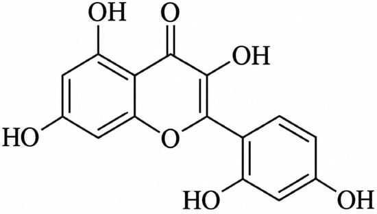
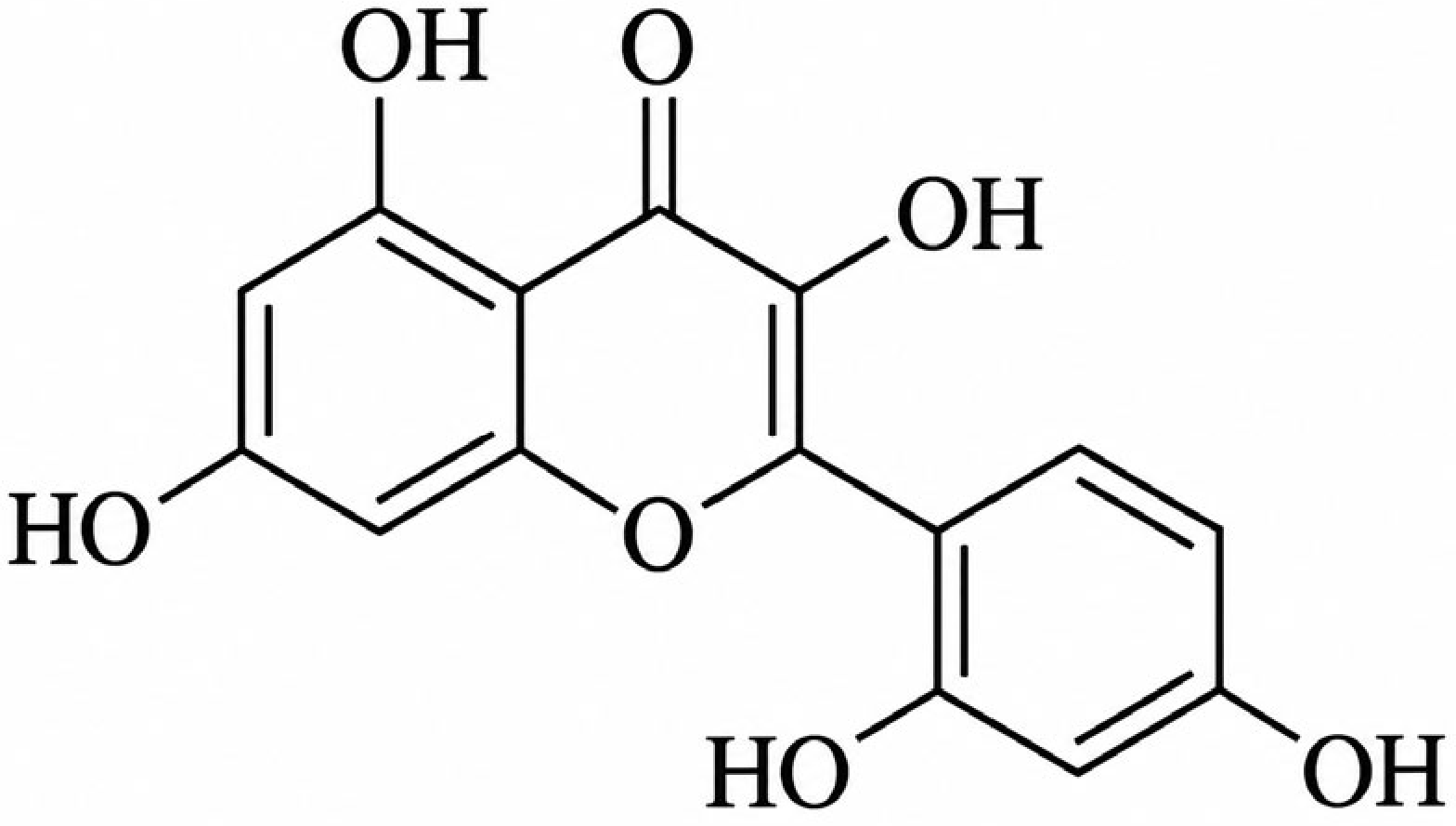
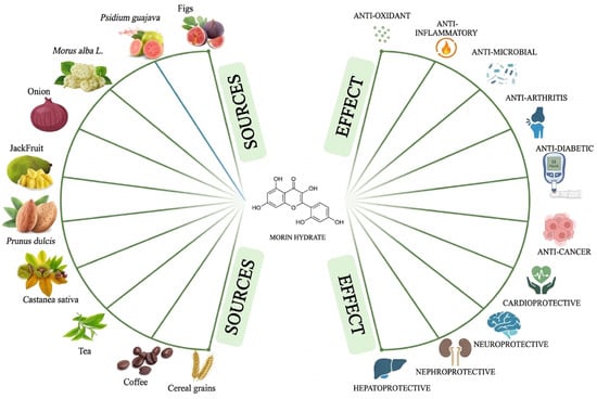
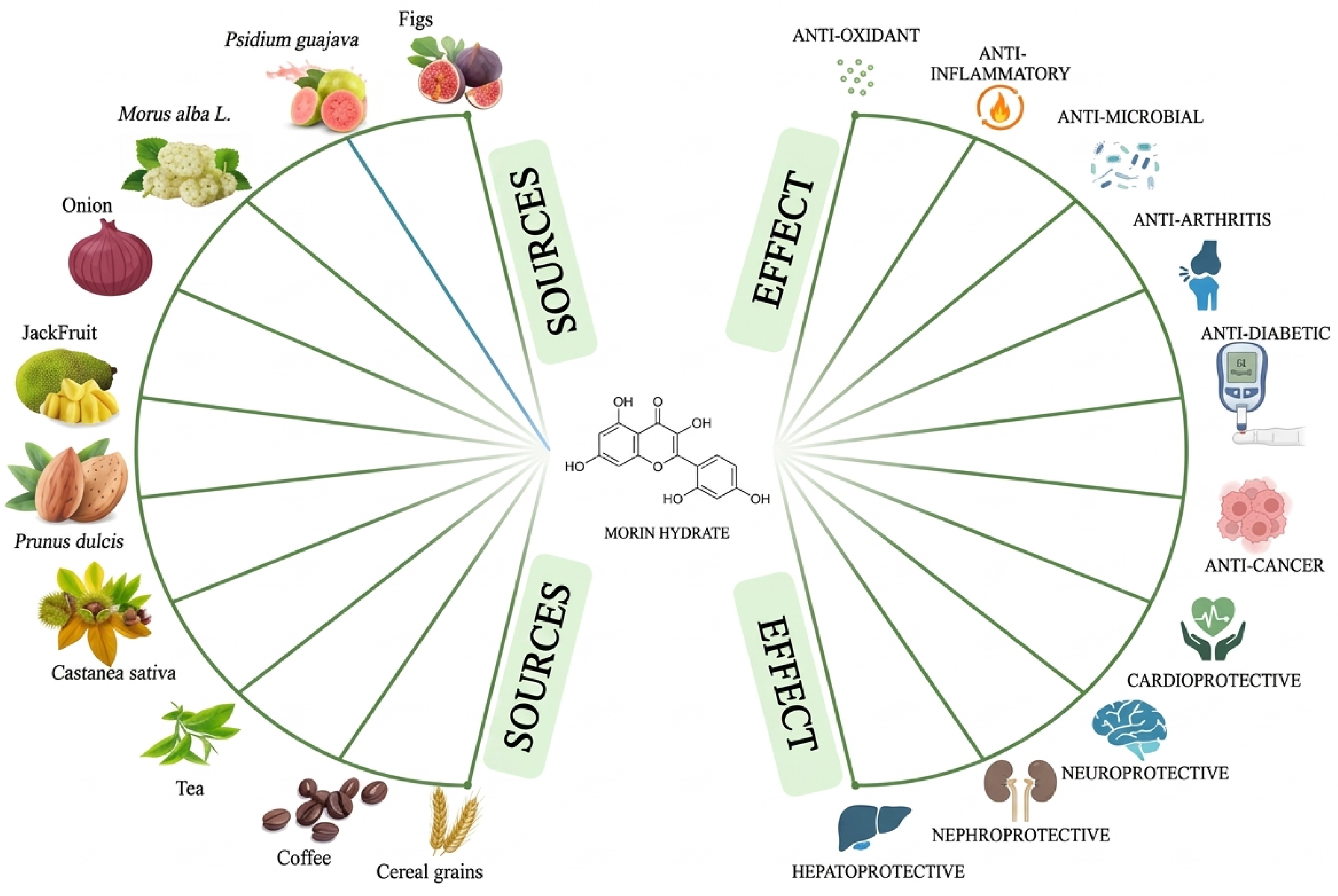
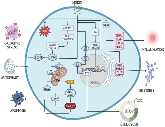
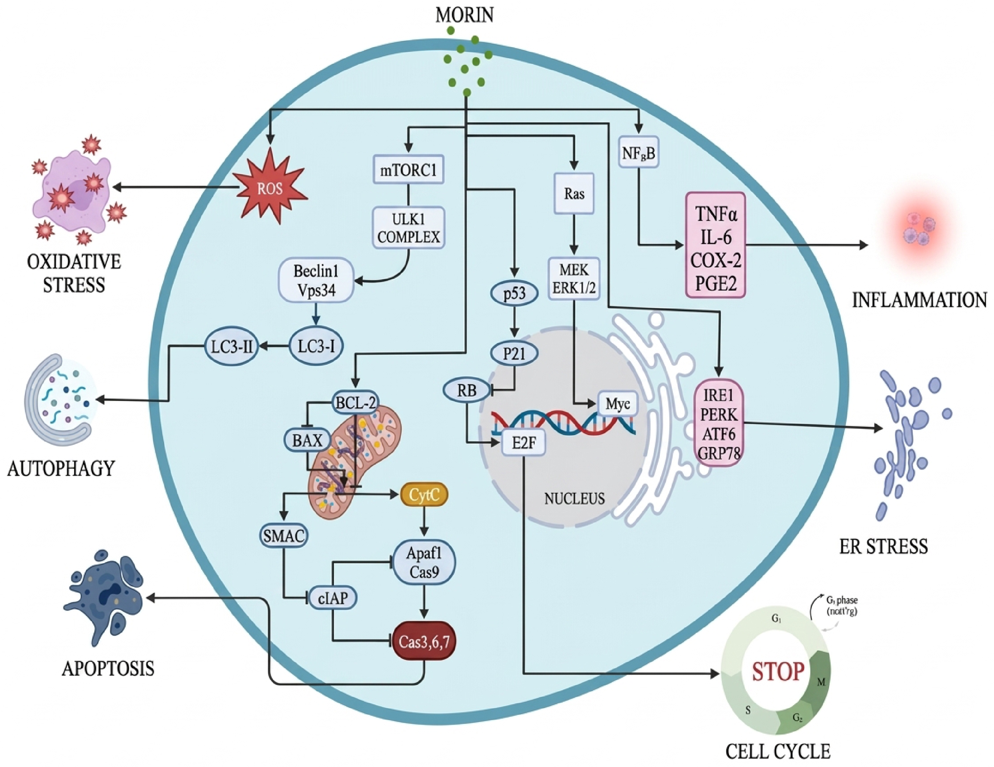
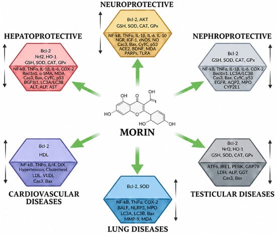
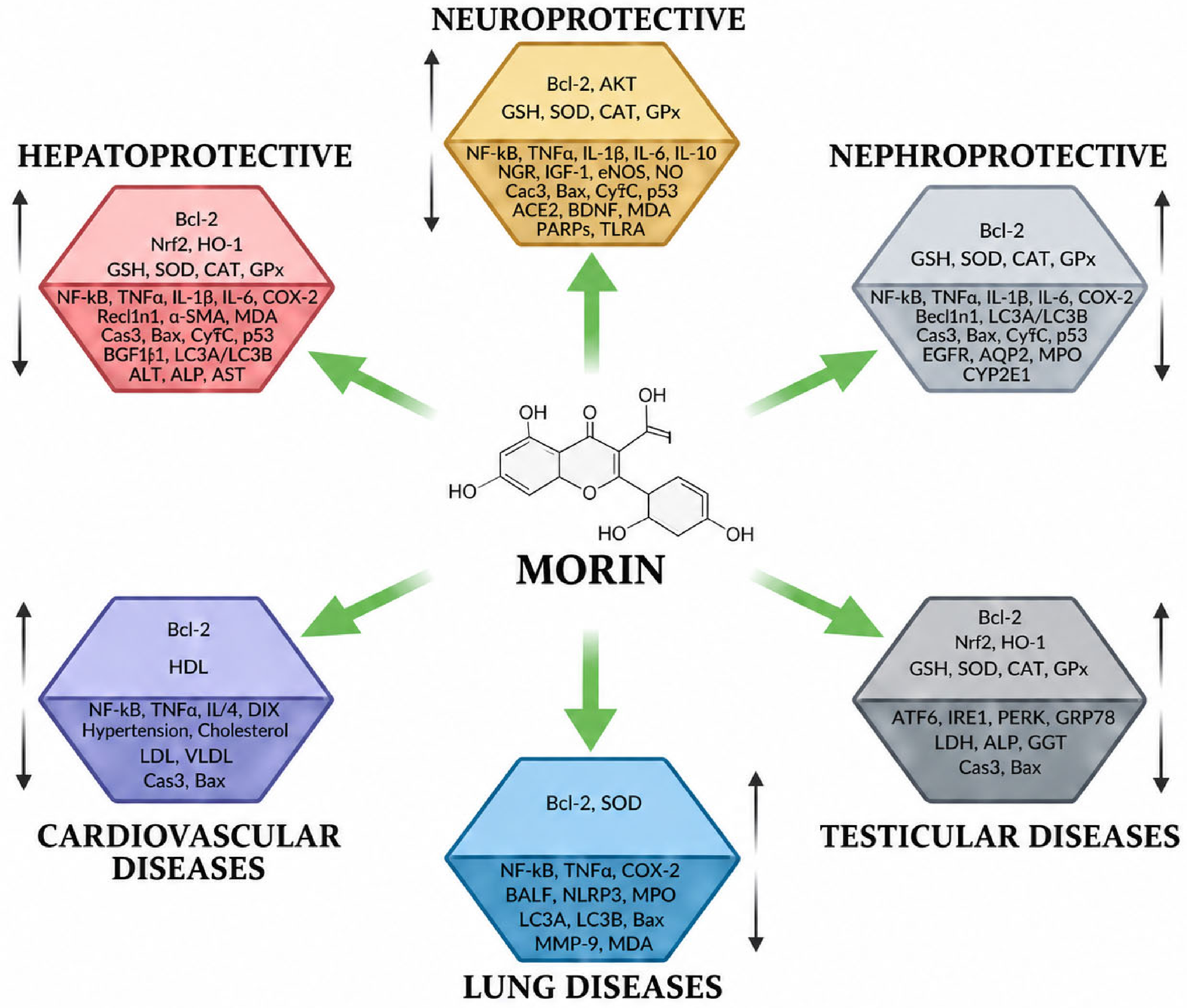

Disclaimer/Publisher’s Note: The statements, opinions and data contained in all publications are solely those of the individual author(s) and contributor(s) and not of MDPI and/or the editor(s). MDPI and/or the editor(s) disclaim responsibility for any injury to people or property resulting from any ideas, methods, instructions or products referred to in the content. |
© 2026 by the authors. Licensee MDPI, Basel, Switzerland. This article is an open access article distributed under the terms and conditions of the Creative Commons Attribution (CC BY) license.