


Abstract
Neurotoxic astrocyte activation is increasingly recognized as an amplifier of neuronal injury, yet the mechanisms by which environmental neurotoxicants engage astrocytic redox machinery remain poorly defined. Here, we investigated whether N-myc downstream-regulated gene 2 (NDRG2) links rotenone (Rot) exposure to neurotoxic astrocyte activation and associated neurotoxicity. In Rot-treated mice, NDRG2 expression increased and was predominantly localized to GFAP-positive astrocytes. Across individual samples, NDRG2 levels correlated inversely with tyrosine hydroxylase and Neu-N expression and positively with astrocytic C3 expression. Consistently, Rot increased NDRG2 expression in C6 astroglial cells, whereas NDRG2 silencing attenuated the induction of C3, H2-D1 and LCN2 and partially restored S100a10 expression. Mechanistically, Rot increased NOX2 and NOX4 expression, NADPH oxidase (NOX) activity, ROS production and NF-κB activation, all of which were attenuated by NDRG2 silencing. Co-immunoprecipitation revealed a selective association between NDRG2 and NOX2, but not NOX4, while molecular docking and dynamics simulations supported the structural feasibility and stability of the NDRG2–NOX2 complex. Rot enhanced the NDRG2–NOX2 association, whereas NDRG2 silencing reduced Rot-induced p47phox–gp91phox complex formation, indicating impaired NOX2 assembly. Inhibition of NOX2 suppressed Rot-induced NOX4 upregulation, and inhibition of either NOX2 or NOX4 attenuated the neurotoxic astrocytic response. Conversely, reactivation of NF-κB largely reversed the protective effects of NDRG2 silencing and restored astrocyte-mediated neuronal toxicity. Together, these findings identify the NDRG2–NOX2 interaction as a mechanistic link between Rot exposure and astrocytic redox-inflammatory signaling. By facilitating NOX2 complex assembly and engaging downstream NOX4/NF-κB signaling, NDRG2 promotes neurotoxic astrocyte activation and the propagation of Rot-induced neuronal injury.
1. Introduction
Rotenone (Rot) is a naturally occurring rotenoid that has been widely used as a pesticide. Epidemiological studies have associated occupational or agricultural Rot exposure with an increased risk of Parkinson’s disease (PD) [1], while systemic Rot administration reproduces key features of nigrostriatal degeneration in experimental rodent models [2]. Owing to its high lipophilicity, Rot readily crosses biological membranes and inhibits mitochondrial complex I, thereby promoting electron leakage, oxidative stress and bioenergetic failure [3,4]. Although mitochondrial complex I inhibition is a well-established initiating event, it does not fully explain the persistence of neuroinflammation, the selective vulnerability of neural circuits, or the progressive nature of neuronal injury after Rot exposure. These observations suggest that secondary, non-neuronal mechanisms amplify the primary mitochondrial insult in response to Rot.
Accumulating evidence supports a major role for glial cells in this amplification process. Rather than acting solely as a neuron-autonomous mitochondrial toxicant, Rot can engage glial inflammatory and oxidative programs that propagate injury within the neural microenvironment. Microglial NADPH oxidase 2 (NOX2), for example, accounts for a substantial component of Rot-induced dopaminergic neurodegeneration [5]. Subsequent studies, including our own, have shown that Rot activates complement receptor 3 (CR3)- and NOX2-dependent inflammatory signaling, promotes glial cross-talk, disrupts the blood–brain barrier (BBB), and facilitates oxidative injury and regulated neuronal death [6,7,8]. Importantly, astrocytes may participate at an early stage of this response. Miyazaki et al. reported that low-dose Rot perturbs midbrain astrocytes before overt microglial activation and that astrocyte-derived signals contribute to region-specific glial dysfunction and dopaminergic neurodegeneration [9]. Thus, a central unresolved question is not whether astrocytes respond to Rot, but how Rot converts astrocytic stress responses into a neurotoxic state capable of amplifying neuronal injury.
Astrocytes are particularly well positioned to amplify toxicant-induced injury because they coordinate neurotransmitter and ion homeostasis, metabolic support, antioxidant buffering, BBB integrity and intercellular inflammatory signaling [10]. Under pathological conditions, these homeostatic functions can be extensively remodeled, generating heterogeneous reactive states with context-dependent effects on neuronal survival [11]. Increased expression of C3, H2-D1 and lipocalin-2 (LCN2) has been associated with neurotoxic inflammatory astrocyte states, but marker expression alone is insufficient to establish functional neurotoxicity [12]. Demonstrating that an altered astrocytic state translates into neuronal injury is therefore essential. Equally important is defining the upstream redox machinery that drives this transition. Despite the central role of oxidative stress in Rot toxicity, the molecular events that couple Rot exposure to neurotoxic astrocyte activation remain incompletely understood.
N-myc downstream-regulated gene 2 (NDRG2) is an astrocyte-enriched, stress-responsive protein that may couple cellular stress to astrocytic remodeling [13]. Notably, the neurological functions of NDRG2 are highly context dependent. Increased astrocytic NDRG2 has been linked to NF-κB-dependent inflammation and glutamate-related secondary injury in ischemic or hemorrhagic brain injury [14], whereas in other settings NDRG2 supports glutamate clearance and confers neuroprotection [15]. This functional plasticity is further illustrated by reports that NDRG2 stabilization limits inflammatory signaling during oxygen–glucose deprivation [16], that NDRG2–PPM1A interaction promotes BBB disruption after subarachnoid hemorrhage [17], and that astrocytic NDRG2 regulates NF-κB/C3 signaling in diabetes-associated cognitive dysfunction [18]. NDRG2 has also been implicated in astrocytic metabolism, ferroptosis, excitotoxicity and cell survival [19,20]. Collectively, these observations suggest that NDRG2 is neither intrinsically protective nor detrimental; rather, its effects may depend on the initiating stressor and on the signaling proteins with which it functionally associates. Whether Rot recruits NDRG2 into a specific redox–regulatory complex in astrocytes has not been established.
NADPH oxidases (NOXs) provide a plausible redox effector system through which NDRG2-dependent signaling could amplify Rot toxicity. In contrast to mitochondrial reactive oxygen species (ROS), which arise as a consequence of electron-transport dysfunction, NOX enzymes are dedicated ROS-generating systems whose activity is tightly controlled [21]. NOX2 is particularly relevant because activation of its catalytic gp91phox-containing membrane complex requires recruitment of cytosolic regulatory components, including p47phox [22,23]. This assembly-dependent architecture creates a mechanistic opportunity for non-enzymatic proteins to modulate oxidase activity by organizing or stabilizing complex formation. NOX4 differs in its regulatory organization and can provide a more sustained source of redox signaling; increased astrocytic NOX4 has recently been implicated in inflammatory and degenerative changes in PD-related models [24]. Our previous work established NOX2 as a major redox effector downstream of microglial receptors during Rot exposure [25], but how NOX activity is controlled in Rot-exposed astrocytes remains unclear. In particular, whether NDRG2 associates with NOX2 to regulate oxidase assembly, and whether NOX2 and NOX4 participate in an ordered astrocytic redox-inflammatory program remain unresolved questions.
We therefore hypothesized that NDRG2 promotes Rot-induced neurotoxic astrocyte activation by facilitating NOX2-dependent redox signaling. To test this hypothesis, we combined a Rot-intoxicated mouse model with NDRG2 loss-of-function studies in astroglial cells, biochemical interaction assays, molecular docking and dynamics simulations, pharmacological dissection of NOX2/NOX4/NF-κB signaling, and an astrocyte-conditioned-medium neurotoxicity assay. Our findings identify an NDRG2–NOX2 interaction that facilitates NOX2 complex assembly and links Rot exposure to a downstream redox-inflammatory program in astrocytes. This mechanism provides a framework for understanding how an initial mitochondrial toxic insult can be converted into sustained glial oxidative signaling and astrocyte-mediated neuronal injury.
2. Materials and Methods
2.1. Chemicals and Reagents
Rotenone (R9975) was obtained from Sigma-Aldrich, Inc. (St. Louis, MO, USA). GSK2795039 (a selective NOX2 inhibitor), GLX351322 (a NOX4 inhibitor), and NF-κB Activator 1 (HY-134476) were purchased from MedChemExpress (Shanghai, China).
2.2. Animals and Rotenone Exposure
Eight-week-old male C57BL/6 mice were obtained from Liaoning Changsheng Biotechnology Co., Ltd. (Benxi, China) were randomly assigned to control (Con) or Rot exposure groups (a total of 40 animals) using the random number table method. For the dose–response experiment, mice were allocated to Con, Rot 0.75 mg/kg, or Rot 1.5 mg/kg groups (n = 10 per group). For the time-course experiment, a separate cohort was exposed to Rot at 1.5 mg/kg and evaluated on days 1, 3, 7, 14, and 21. The Rot dose was selected based on our previous study [6]. Ms. Jianing Liu was aware of the grouping of the animals and handled them. All animal procedures were approved by the Animal Ethics Committee of Dalian Medical University (L2606290463). Mice showing signs of extreme pain, distress, or noticeable behavioral changes during the experiment were removed from the experiment and euthanized.
2.3. C6 Astroglial Cell Culture and Rotenone Treatment
C6 astroglial cells purchased from Wuhan Pricella Biotechnology Co., Ltd. (Wuhan, China) were maintained in DMEM supplemented with 10% fetal bovine serum (FBS) and 1% penicillin–streptomycin at 37 °C in a humidified atmosphere containing 5% CO2. The medium was replaced every other day. For Rot exposure, cells were treated with 0.1, 0.2, or 0.4 μM Rot. These concentrations were selected based on the study by Goswami et al. [26].
2.4. Ndrg2 Knockdown
To minimize the likelihood of sequence-specific off-target effects, two independent siRNAs targeting NDRG2 were used. C6 astroglial cells were seeded in culture plates and transfected at approximately 60–70% confluence with either an NDRG2-specific siRNA or a non-targeting control siRNA (Con-si) using Lipo3000 according to the manufacturer’s instructions. The siRNA sequences were as follows: NDRG2-si-1, 5′-GUAGGACUCAACUAUAAAUTT-3′ (F) and 5′-AUUUAUAGUUGAGUCCUACTT-3′ (R); NDRG2-si-2, 5′-CUGUAUUGGAACAGUUACATT-3′ (F) and 5′-UGUAACUGUUCCAUACAGTT-3′ (R). Konckdown efficacy was assessed after 24 h transfection.
2.5. Western Blotting
Protein samples were collected from the midbrain of mice or C6 astroglial cells after Rot exposure. Protein concentrations were determined using the BCA assay, and equal amounts of protein were separated by SDS-PAGE and transferred to PVDF membranes. Membranes were blocked with non-fat milk for 2 h at room temperature, incubated with primary antibodies overnight at 4 °C, washed, and then incubated with the corresponding secondary antibodies for 1 h. Immunoreactive bands were visualized by enhanced chemiluminescence (ECL) and quantified by densitometric analysis. Target protein levels were normalized to GAPDH or the corresponding total protein, as appropriate. Primary antibodies included NDRG2 (12015-1-AP), p-p65 (Ser468) (82335-1-RR), and p65 (10745-1-AP) from Proteintech (Wuhan, China); NeuN (ab177487), C3 (ab200999), TH (ab137869), GFAP (ab7260), NOX2 (ab310337), NOX4 (ab319055), and LCN2 (ab216462) from Abcam (Waltham, UK); and GAPDH (A19056) and HRP-conjugated secondary antibodies from ABclonal (Wuhan, China).
2.6. Immunofluorescence Staining
Brain sections containing the substantia nigra were washed three times with PBS for 10 min each, followed by antigen retrieval for 5 min using an antigen-retrieval solution for frozen sections. Sections were blocked for 2 h at room temperature and incubated with the corresponding primary antibodies overnight at 4 °C. After three 5-min washes with PBS, sections were incubated with Alexa Fluor™ 594-conjugated goat anti-mouse/rabbit secondary antibodies diluted in PBS (1:1000) for 2 h at room temperature in the dark. Sections were then washed three times with PBS for 5 min each, mounted onto slides, air-dried, and coverslipped with antifade mounting medium containing DAPI. Fluorescence images were acquired by fluorescence microscopy and subsequently analyzed.
2.7. Quantitative Real-Time Pcr (Qrt-Pcr)
Total RNA was extracted from treated cells using TriZol Reagent (Vazyme, Nanjing, China), and cDNA was synthesized using the HiExact RT SuperMix for qPCR kit (Vazyme, China). Transcript levels of genes associated with neurotoxic or protective/homeostatic astrocyte states, including H2-D1, C3, S100a10, and B3gnt5, were quantified using ChamQ Universal SYBR qPCR Master Mix (Vazyme, China). Relative gene expression was calculated using the 2(−ΔΔCt) method after normalization to GAPDH. Primer sequences were as follows: 5′-TGAGGAGGAGGAGGAGAA-3′ (F), 5′-GCAAGGCTGAATGTGGTA-3′ (R) for H2-D1; 5′-CAACCTCAATGTCAACTTCC-3′ (F), 5′-CAATGTCTGCCTTCTCTACT-3′ (R) for C3; 5′-AAGGAGGACCTGAGAGTG-3′ (F), 5′-GCCTACTTCTTCTGCTTCA-3′ (R) for S100a10; 5′-GTCCTACTCCTACAGATACCT-3′ (F), 5′-TCATCATCAGCAGTCATCAG-3′ (R)for B3gnt5; 5′-TTCAACGGCACAGTCAAGGC-3′ (F), 5′-GACTCCACGACATACTCAGCACC-3′ (R) for GAPDH. The corresponding NCBI Gene IDs were as follows: H2-D1 (14964), C3 (12266), S100a10 (20194), and B3gnt5 (108105).
2.8. Intracellular Ros and Nox Activity
Intracellular oxidant production was assessed using dihydroethidium (DHE) staining (Beyotime, Shanghai, China). C6 astroglial cells were seeded in six-well plates, transfected with NDRG2 siRNA, and subsequently treated with Rot for 24 h. Approximately 1 × 10⁵ cells were incubated with 1 μM DHE for 30 min at 37 °C in the dark, washed three times with warm PBS to remove excess probe, and imaged by fluorescence microscopy (Ex/Em = 535/610 nm). DHE fluorescence intensity was used as an index of intracellular ROS/oxidant production.
NADH oxidase (NOX) activity was measured using an NADH Oxidase Activity Assay Kit (Solarbio, Beijing, China). Approximately 1 × 10⁵ C6 astroglial cells from each group were resuspended in 1 mL of extraction buffer supplemented with 10 μL of reagent III, followed by centrifugation at 10,000× g for 10 min at 4 °C after treated with or without NDRG2 siRNA and Rot. The supernatants were collected for NOX activity measurement. 10 μL of supernatant was mixed with 175 μL of reagent IV, 25 μL of reagent V, and 40 μL of reagent VI in a 96-well plate. For the corresponding background control, reagent VI was replaced with an equal volume of distilled water. Absorbance was measured at 600 nm using a microplate reader at 37 °C and recorded as AT1 for the test sample or AC1 for the control. After an additional 1 min incubation at 37 °C, absorbance was measured again and recorded as AT2 or AC2. The absorbance was calculated as ΔA = (AT1 − AT2) − (AC1 − AC2). NOX activity was normalized to the protein concentration of each sample, as determined using the BCA assay, and calculated according to the formula: NOX activity (U/mg protein) = 5000 × ΔA/Cpr, where Cpr represents the sample protein concentration (mg/mL).
2.9. Co-Immunoprecipitation (Co-Ip)
Protein associations were examined by Co-IP, as described previously [7]. C6 astroglial cells were seeded at 1 × 10⁵ cells/well in six-well plate and collected after the indicated treatments. Total protein was extracted from cells in each group, and protein concentrations were determined using the BCA assay. Equal amounts of protein were incubated with antibodies against NDRG2, NOX2, NOX4, or p47phox at 4 °C overnight. Normal IgG was used as a negative immunoprecipitation control. Protein A/G magnetic beads 20 μL were subsequently added, followed by incubation at 4 °C. The immunoprecipitated complexes were collected, washed with lysis buffer, and eluted using SDS sample buffer. The resulting samples were subjected to Western blotting to detect the corresponding interacting proteins. Aliquots of the original cell lysates were retained as input controls. The association between NDRG2 and NOX2/NOX4 and the interaction between p47phox and gp91phox were evaluated by comparing the immunoprecipitated proteins among the indicated treatment groups.
2.10. Molecular Docking and Molecular Dynamics Simulation
Molecular docking was used to compare the predicted interactions of NDRG2 with NOX2 and NOX4. Three-dimensional structures of NDRG2, NOX2, and NOX4 were used for docking, and the resulting complexes were ranked according to predicted binding energy. The top conformations were examined for interfacial residues, short-range contacts, and hydrogen-bond interactions. Based on these results, the top-ranked NDRG2–NOX2 complex was subjected to a 50-ns molecular dynamics simulation to evaluate the stability of the predicted association under dynamic conditions. During the simulation, root-mean-square deviation (RMSD), the number of interfacial hydrogen bonds, the minimum interprotein distance, and the potential and total energy trajectories were monitored.
2.11. Astrocyte-Mediated Neuronal Toxicity Assay
Conditioned medium was collected from astrocytes 24 h after Rot exposure in the presence or absence of NDRG2 silencing, NOX inhibition, or NF-κB activation and subsequently applied to neuronal cells (PC-12, obtained from Wuhan Pricella Biotechnology Co., Ltd.). Neuronal viability was quantified using the CCK-8 assay as a functional readout of astrocyte-mediated neurotoxicity. PC-12 cells were seeded in 96-well plates at a density of 5 × 10³ cells/well and cultured at 37 °C in a humidified atmosphere containing 5% CO2. The culture medium was subsequently replaced with 200 μL of astrocyte-conditioned medium, and the cells were incubated for 24 h. Cell viability was assessed using the CCK-8 assay (Beyotime, Shanghai, China). Briefly, 20 μL of CCK-8 reagent was added to each well, followed by incubation at 37 °C for 2 h. Absorbance was measured at 450 nm using a microplate reader. Wells containing the corresponding culture medium and CCK-8 reagent without cells were used as blank. Cell viability was calculated after background subtraction and expressed as a percentage of the corresponding control group. Detailed procedures were performed as described in our previous study [25].
2.12. Statistical Analysis
Data are presented as the mean ± SEM. Correlations between NDRG2 expression and neuronal or astrocytic markers were analyzed using Pearson’s correlation test. Correlation coefficients (R) and corresponding p-values were reported. Data normality was assessed using the Shapiro–Wilk test, and homogeneity of variance was evaluated using Levene’s test. For normally distributed data with homogeneous variances, differences between two groups were analyzed using Student’s t-test, whereas comparisons among multiple groups were performed using one-way analysis of variance (ANOVA), followed by an appropriate post hoc multiple-comparison test. All experiments were independently repeated at least three times. Statistical analyses and graph generation were performed using GraphPad Prism 10.1, with p < 0.05 considered statistically significant.
3. Results
3.1. Astrocytic Ndrg2 Upregulation Is Associated with Nigral Neurodegeneration in Rot-Intoxicated Mice
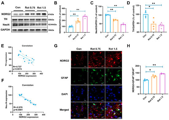
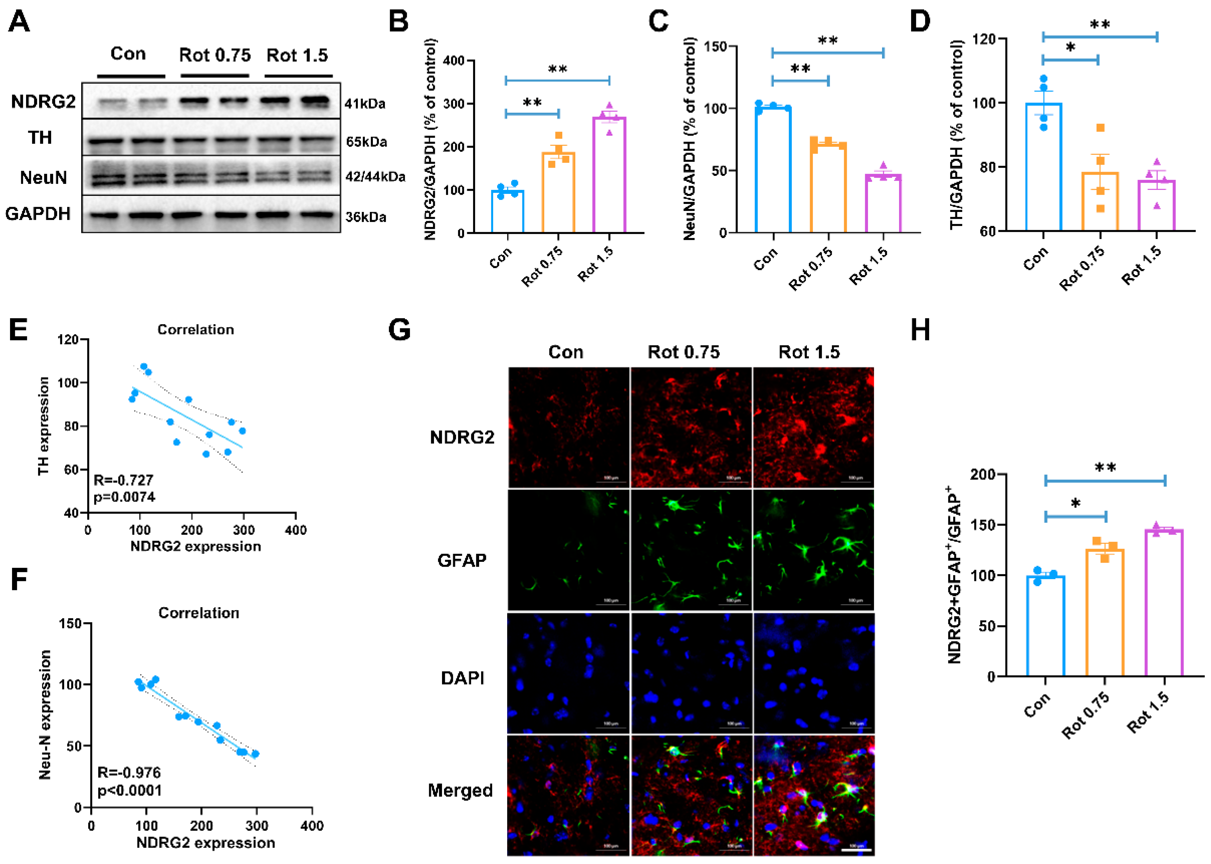
To determine whether NDRG2 responds to Rot in vivo and whether its induction tracks with neuronal injury, we first examined dose-dependent changes in the substantia nigra. Rot exposure progressively increased NDRG2 protein expression, whereas the neuronal markers TH and NeuN showed the opposite pattern (Figure 1A–D). NDRG2 was elevated at both 0.75 and 1.5 mg/kg Rot and reached its highest level at 1.5 mg/kg group, while TH and NeuN were reduced more prominently at the high dose. Across individual samples, NDRG2 expression was inversely correlated with TH (R = −0.727, p = 0.0074; Figure 1E) and NeuN (R = −0.976, p < 0.0001; Figure 1F), linking NDRG2 induction to the severity of neuronal injury. We next examined the cellular localization of the Rot-induced NDRG2 signal. Immunofluorescence showed that the increase in NDRG2 occurred predominantly in GFAP-positive astrocytes in the substantia nigra (Figure 1G), accompanied by an increased proportion of NDRG2-positive/GFAP-positive astrocytes (Figure 1H). Together, these data indicate that Rot induces astrocytic NDRG2 in a dose-dependent manner and that this response closely correlates with nigral neuronal injury, supporting astrocytic NDRG2 as a candidate component of the glial response to Rot.
Figure 1.
Astrocytic NDRG2 upregulation is associated with nigral neurodegeneration in Rot−intoxicated mice. (A–D) Representative immunoblots and densitometric quantification of NDRG2, TH, and NeuN in the substantia nigra of mice exposed to the indicated doses of Rot. (E,F) Correlation analyses of NDRG2 expression with TH (R = −0.727, p = 0.0074) (E) and NeuN (R = −0.976, p < 0.0001) (F). (G) Representative immunofluorescence images of NDRG2 (red), GFAP (green), and DAPI (blue) in the substantia nigra. (H) Quantification of NDRG2-positive/GFAP-positive astrocytes. Scale bar = 100 μm. * p < 0.05, ** p < 0.01. n = 3–4.
3.2. Ndrg2 Induction Precedes and Correlates with Neurotoxic Astrocyte-Associated Changes in Rot-Intoxicated Mice
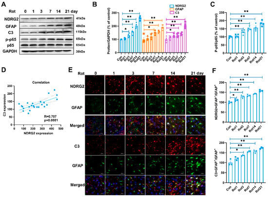
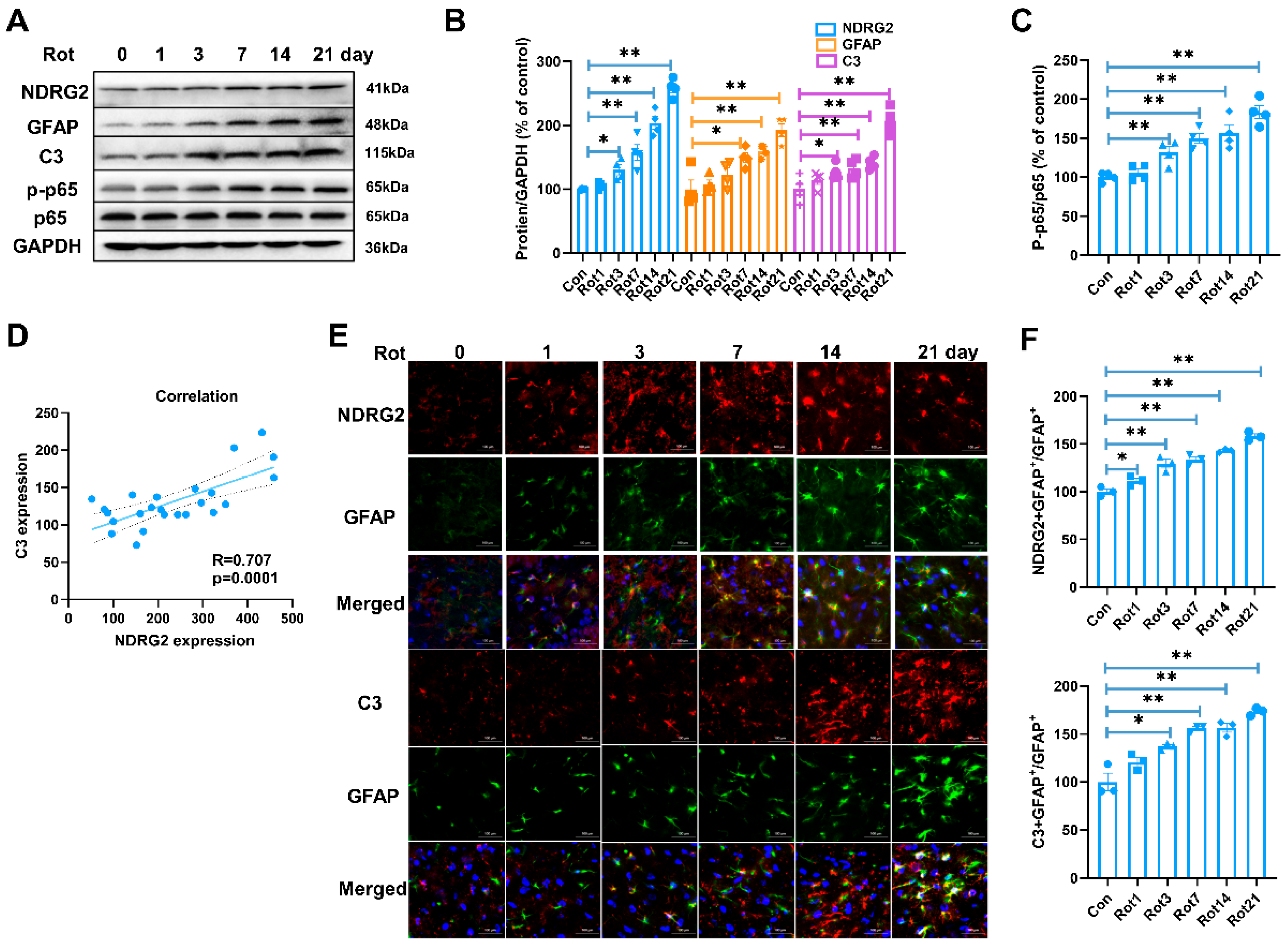
To define the temporal relationship between NDRG2 induction and the emergence of a neurotoxic astrocyte-associated phenotype, we examined NDRG2, GFAP, C3, and NF-κB signaling over 21 days of Rot exposure. NDRG2 expression increased progressively with exposure duration, in parallel with increases in GFAP and C3, while p-p65 also rose over time (Figure 2A–C). Across the time-course samples, NDRG2 expression was positively correlated with C3 expression (R = 0.707, p = 0.0001; Figure 2D). Immunofluorescence further showed progressive increases in both NDRG2-positive/GFAP-positive and C3-positive/GFAP-positive astrocytes (Figure 2E,F). Notably, the increase in astrocytic NDRG2 preceded the rise in astrocytic C3 (Figure 2E,F). This temporal pattern, together with the positive NDRG2–C3 correlation, supports the possibility that NDRG2 induction represents an early astrocytic event associated with progression toward a neurotoxic state during Rot intoxication.
Figure 2.
Astrocytic NDRG2 induction precedes and correlates with neurotoxic astrocyte−associated changes during Rot intoxication. (A) Representative immunoblots of NDRG2, GFAP, C3, p-p65, and p65 in the substantia nigra across the Rot exposure time course. (B) Densitometric quantification of NDRG2, GFAP, and C3. (C) Quantification of p-p65 expression. (D) Correlation between NDRG2 and C3 expression (R = 0.707, p = 0.0001). (E) Representative immunofluorescence images showing NDRG2/GFAP and C3/GFAP colocalization across the exposure period. (F) Quantification of NDRG2-positive/GFAP-positive and C3-positive/GFAP-positive astrocytes. Scale bar = 100 μm. * p < 0.05, ** p < 0.01. n = 3–4.
3.3. Ndrg2 Silencing Suppresses Rot-Induced Neurotoxic Astrocyte-Associated Responses in C6 Astroglial Cells
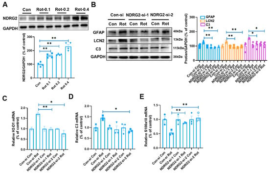
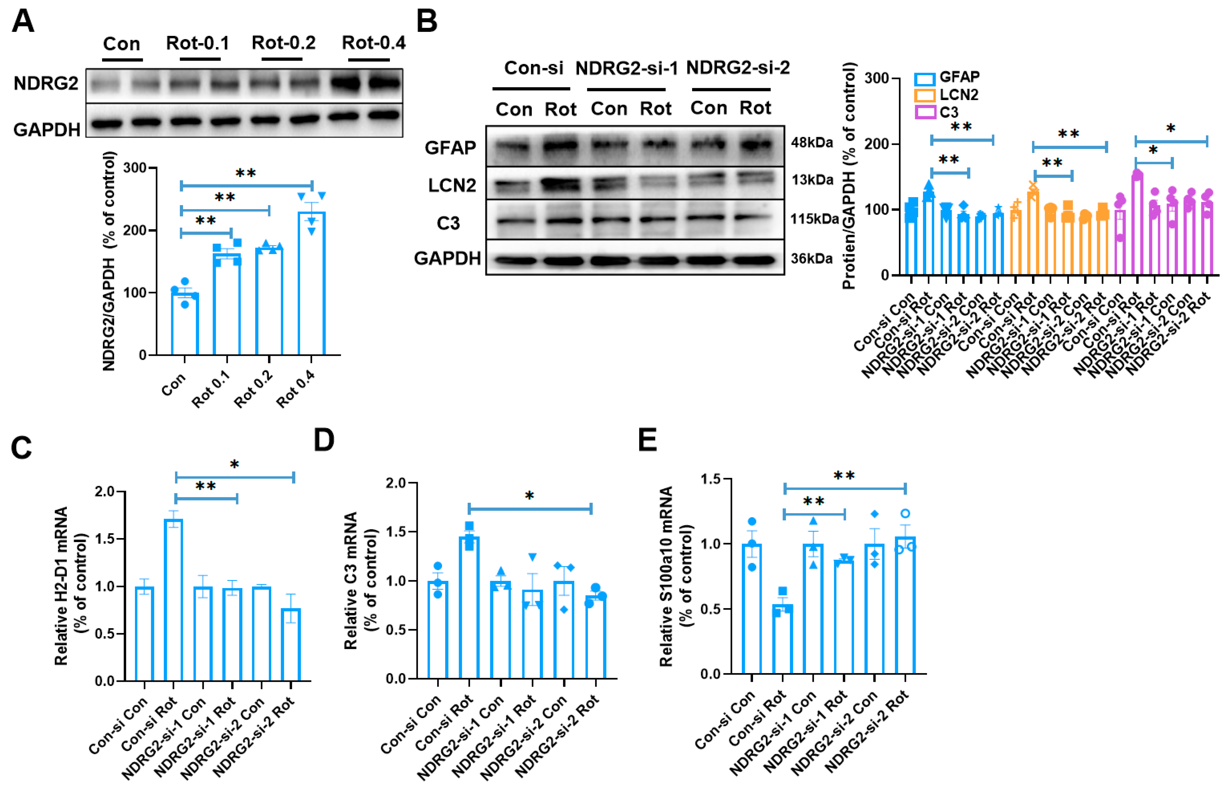
To test whether NDRG2 functionally contributes to the Rot-induced astrocytic response, we first confirmed that Rot increased NDRG2 protein expression in C6 astroglial cells in a dose-dependent manner (Figure 3A). We then reduced NDRG2 expression using two independent siRNAs to minimize sequence-specific off-target effects. In control siRNA-transfected cells, Rot markedly increased GFAP, LCN2, and C3 protein expression, whereas either NDRG2 siRNA substantially attenuated these responses (Figure 3B). A similar pattern was observed at the transcript level. Rot increased the expression of the neurotoxic astrocyte-associated genes H2-D1 and C3, while NDRG2 silencing attenuated their induction. Conversely, NDRG2 silencing partially restored S100a10 expression, which was reduced by Rot (Figure 3C–E). Thus, NDRG2 loss of function shifts the Rot-induced astrocytic response away from a neurotoxic-associated molecular profile, providing functional evidence that NDRG2 contributes to this state transition.
Figure 3.
NDRG2 silencing suppresses Rot−induced neurotoxic astrocyte-associated responses in C6 astroglial cells. (A) Representative immunoblot and quantification of NDRG2 after exposure to increasing Rot concentrations. (B) Effects of two independent NDRG2 siRNAs on Rot-induced GFAP, LCN2, and C3 protein expression. (C–E) Relative mRNA expression of H2-D1 (C), C3 (D), and S100a10 (E) after Rot exposure with or without NDRG2 silencing. * p < 0.05, ** p < 0.01. n = 3–4.
3.4. Ndrg2 Is Required for Full Activation of the Rot-Induced Nox/ros–nf-Κb Response in C6 Astroglial Cells
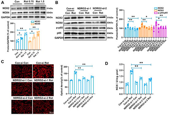
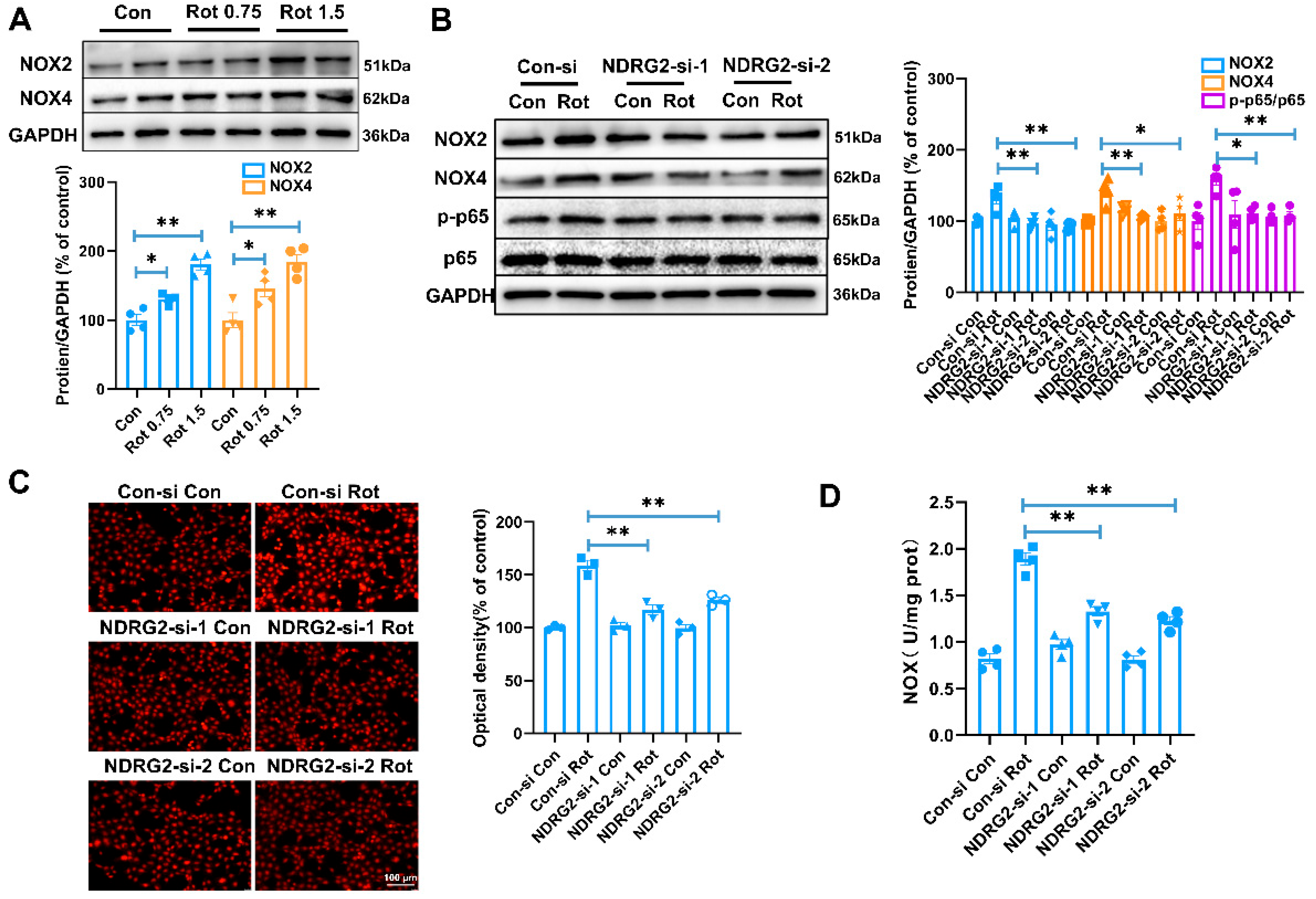
We next asked whether NDRG2 links Rot exposure to astrocytic redox signaling. Rot increased both NOX2 and NOX4 protein expression in the substantia nigra (Figure 4A). In C6 astroglial cells, Rot similarly increased NOX2 and NOX4 expression and elevated the p-p65/p65 ratio, whereas NDRG2 silencing markedly attenuated each of these changes (Figure 4B). We then assessed functional redox responses. Rot increased DHE fluorescence in control siRNA-transfected cells, and this increase was substantially reduced by NDRG2 silencing (Figure 4C). Consistently, Rot increased NOX activity, which was also attenuated after NDRG2 knockdown (Figure 4D). These findings position NDRG2 upstream of a NOX-dependent oxidant response coupled to NF-κB activation. The parallel effects on NOX2 and NOX4 further suggested that NDRG2 may coordinate an ordered NOX-dependent redox pathway rather than regulating a single oxidase in isolation.
Figure 4.
NDRG2 is required for full activation of the Rot-induced NOX/ROS−NF-κB response in astroglial cells. (A) Representative immunoblots and quantification of NOX2 and NOX4 in the substantia nigra of mice exposed to the indicated doses of Rot. (B) Effects of two independent NDRG2 siRNAs on Rot-induced NOX2, NOX4, p-p65, and p65 expression in C6 astroglial cells. (C) Representative DHE fluorescence images and quantification of intracellular oxidant production after Rot exposure with or without NDRG2 silencing. (D) NADPH oxidase activity under the same conditions. Scale bar = 100 μm. * p < 0.05, ** p < 0.01. n = 3–4.
3.5. Ndrg2 Associates with Nox2 and Facilitates Rot-Induced Nox2 Complex Assembly in C6 Astroglial Cells
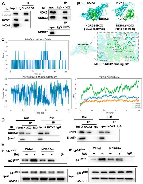
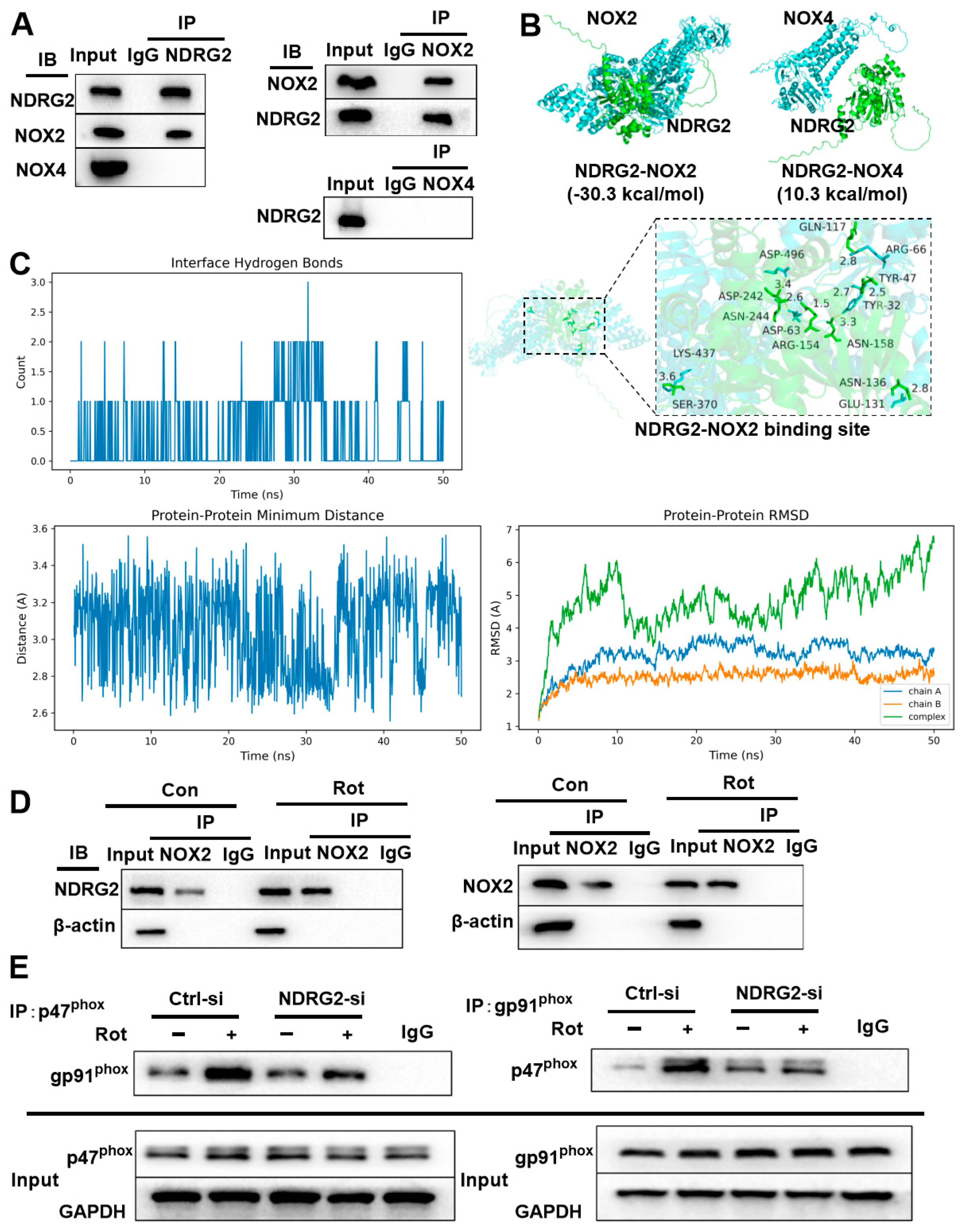
Because NDRG2 silencing reduced NOX2, NOX4, and overall NOX activity, we next asked whether NDRG2 associates preferentially with a specific NOX isoform. Co-IP of NDRG2 recovered NOX2 but not NOX4, and reciprocal immunoprecipitation of NOX2 likewise recovered NDRG2, whereas NOX4 immunoprecipitation did not yield a detectable NDRG2 signal (Figure 5A). These biochemical data indicate a selective NDRG2–NOX2 association. Molecular docking provided complementary structural support: the predicted NDRG2–NOX2 complex showed a favorable binding energy (−30.3 kcal/mol), whereas a favorable NDRG2–NOX4 interaction was not predicted (10.3 kcal/mol) (Figure 5B). The modeled NDRG2–NOX2 interface contained multiple short-range contacts and hydrogen-bond interactions involving several residues.
Figure 5.
NDRG2 associates with NOX2 and facilitates Rot−induced NOX2 complex assembly in C6 astroglial cells. (A) Co-IP analysis showing a selective association of NDRG2 with NOX2 but not NOX4. (B) Molecular docking models of NDRG2−NOX2 and NDRG2–NOX4, with the predicted NDRG2–NOX2 interaction interface shown at higher magnification. (C) Fifty-nanosecond molecular dynamics analysis of the predicted NDRG2–NOX2 complex, including interfacial hydrogen bonds, minimum interprotein distance, and RMSD. (D) Co-IP showing that Rot enhances the NDRG2–NOX2 association. (E) Co-IP showing that NDRG2 silencing attenuates Rot-induced p47phox − gp91phox complex formation. n = 3.
To assess whether the predicted NDRG2–NOX2 complex remained stable under dynamic conditions, we performed a 50-ns molecular dynamics simulation (Figure 5C). Interfacial hydrogen bonds persisted through much of the simulation, and the minimum interprotein distance remained within a relatively narrow range. The individual protein chains showed limited RMSD fluctuations; after an initial conformational adjustment, the overall complex reached a comparatively stable trajectory. Potential and total energy profiles also remained stable. Together, these analyses support the structural feasibility and relative stability of the NDRG2–NOX2 association observed biochemically.
We next determined whether Rot modulates the NDRG2–NOX2 association and whether NDRG2 influences assembly of the NOX2 complex. Rot increased the amount of NDRG2 recovered in NOX2 immunoprecipitates, indicating an enhanced NDRG2–NOX2 association (Figure 5D). Because NOX2 activation depends on the assembly of cytosolic regulatory components with the membrane catalytic complex, we examined the p47phox–gp91phox association. Rot markedly increased p47phox–gp91phox complex formation in control cells, whereas NDRG2 silencing significantly attenuated this response (Figure 5E). Thus, the biochemical, computational, and loss-of-function data converge on a model in which Rot strengthens the NDRG2–NOX2 association and NDRG2 facilitates NOX2 complex assembly, providing a mechanistic basis for the reduced oxidase activity and oxidant production observed after NDRG2 knockdown.
3.6. Nox2–nox4–nf-Κb Signaling Mediates Ndrg2-Dependent Neurotoxic Astrocyte Activation and Astrocyte-Mediated Neurotoxicity
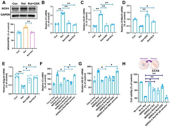
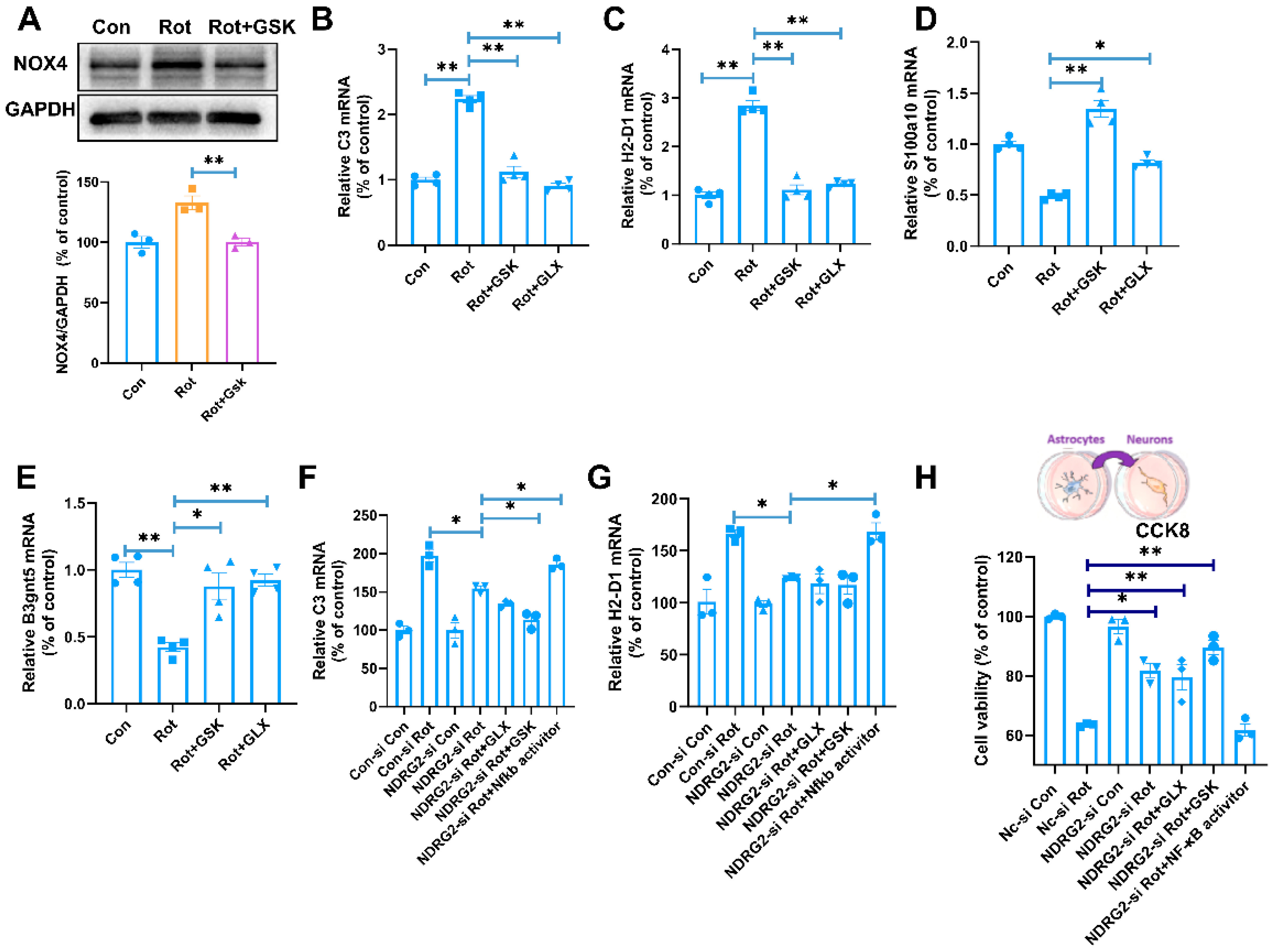
Having placed NDRG2 upstream of NOX activation, we next examined the signaling relationship between NOX2 and NOX4 and asked whether this pathway accounts for the downstream astrocytic phenotype. Pharmacological inhibition of NOX2 with GSK2795039 during Rot exposure largely prevented the Rot-induced increase in NOX4 protein expression (Figure 6A), positioning NOX2 upstream of NOX4 under these experimental conditions. We then tested whether both oxidases contribute to the astrocytic transcriptional response. Rot increased C3 and H2-D1 mRNA expression and decreased S100a10 and B3gnt5, whereas inhibition of either NOX2 with GSK2795039 (25 μM) or NOX4 with GLX351322 (20 μM) attenuated these changes (Figure 6B–E). These results indicate that both NOX2 and NOX4 participate in establishing the Rot-induced neurotoxic astrocyte-associated transcriptional profile.
Figure 6.
NOX2–NOX4–NF-κB signaling mediates NDRG2−dependent neurotoxic astrocyte activation and astrocyte-mediated neurotoxicity. (A) Effect of NOX2 inhibition on Rot-induced NOX4 protein expression. (B–E) Effects of NOX2 or NOX4 inhibition on Rot-induced changes in C3 (B), H2-D1 (C), S100a10 (D), and B3gnt5 (E) mRNA expression. (F,G) Effects of NDRG2 silencing, NOX inhibition, and NF-κB reactivation on C3 (F) and H2-D1 (G) mRNA expression. (H) Neuronal viability after exposure to conditioned medium from astrocytes subjected to the indicated treatments, assessed by CCK-8. * p < 0.05, ** p < 0.01. n = 3–4.
To determine whether NF-κB functions downstream of the NDRG2–NOX pathway, we next performed pathway-rescue experiments. NDRG2 silencing reduced the Rot-induced increases in C3 and H2-D1, and NOX inhibition maintained or further enhanced this suppression (Figure 6F,G). In contrast, pharmacological reactivation of NF-κB in NDRG2-silenced cells restored C3 and H2-D1 expression. These findings place NF-κB downstream of NDRG2-dependent NOX signaling and indicate that reduced NF-κB activity accounts, at least in substantial part, for the suppression of the neurotoxic astrocytic response after NDRG2 loss of function.
Finally, we asked whether this molecular pathway translates into functional astrocyte-mediated neurotoxicity. Conditioned medium from Rot-treated astrocytes markedly reduced PC-12 cell viability in the CCK-8 assay. NDRG2 silencing in astrocytes significantly attenuated this loss of neuronal viability, as did pharmacological inhibition of NOX signaling. Conversely, NF-κB reactivation largely abolished the protection conferred by NDRG2 silencing (Figure 6H). Collectively, these results support a signaling hierarchy in which the NDRG2–NOX2 interaction facilitates NOX2 activation, promotes NOX4-associated redox signaling and downstream NF-κB activation, and thereby contributes to neurotoxic astrocyte activation and the propagation of Rot-induced injury from astrocytes to neurons.
4. Discussion
The present study identifies the NDRG2–NOX2 interaction as a previously unrecognized component of Rot-induced astrocytic redox signaling. Four principal findings support this conclusion. First, Rot increased NDRG2 expression predominantly in GFAP-positive astrocytes in the substantia nigra, and NDRG2 levels were inversely associated with neuronal markers but positively associated with astrocytic C3. Second, NDRG2 silencing attenuated Rot-induced neurotoxic astrocyte-associated molecular changes, reduced ROS production and NOX activity, and limited astrocyte-mediated neuronal toxicity. Third, NDRG2 selectively associated with NOX2, and Rot strengthened this association; NDRG2 silencing, in turn, reduced p47phox–gp91phox complex formation, linking NDRG2 to the assembly-dependent activation of NOX2. Finally, pharmacological experiments placed NOX2 upstream of Rot-induced NOX4 upregulation and supported the involvement of downstream NF-κB signaling in the neurotoxic astrocytic response. Together, these data extend the mechanism of Rot neurotoxicity beyond direct neuronal mitochondrial complex I inhibition and identify astrocytic redox signaling as an active pathway through which the toxic insult can be amplified.
Rot has long been used as an experimental model of PD-related neurodegeneration because its inhibition of mitochondrial complex I produces bioenergetic failure and oxidative stress [27,28]. Recent studies indicate that Rot neurotoxic effects involve a much broader range of cellular processes, including altered neuronal transcriptional programs, mitochondrial redox dysregulation, neuroinflammation and glial dysfunction [29,30]. However, Rot also activates NOX-dependent pathways in multiple neural cell types. In neuronal cells, Rot increases NOX2 and its regulatory subunits p22phox, p40phox, p47phox and p67phox, whereas NOX2 inhibition or knockdown reduces H2O2 generation and apoptosis [31]. Rot-induced Rac1-dependent NOX activation has likewise been linked to oxidative stress, endoplasmic reticulum stress and impaired protein homeostasis [32]. Our finding that Rot increased NOX2 and NOX4 expression, NOX activity and intracellular ROS in astroglial cells therefore places astrocytes within this broader redox response. Importantly, NDRG2 silencing attenuated each of these changes, suggesting that NDRG2 acts upstream of the astrocytic NOX/ROS response rather than simply reflecting a downstream consequence of oxidative injury.
The astrocytic context is particularly relevant because recent studies indicate that astrocytes are direct targets of Rot rather than passive responders to neuronal degeneration. Goswami et al. showed that Rot increased GFAP expression, ROS generation and nitrite levels and impaired mitochondrial activity in C6 astroglial cells, with corresponding astrocytic alterations in several brain regions in vivo [26]. Direct Rot exposure has also been reported to increase intracellular ROS, perturb NF-κB signaling and alter mitochondrial membrane potential in astrocytes [33]. Moreover, NOX-dependent redox signaling has been linked to pathogenic astrocytic responses in other neurological contexts. NOX activation in reactive astrocytes can drive NF-κB-dependent LCN2 expression and neuronal injury [34], whereas NOX4 inhibition reduces astrocytic LCN2 expression and neuroinflammation in an MPTP model [35]. Our data extend these observations by suggesting that NOX2 and NOX4 are not simply parallel sources of ROS during Rot exposure. Pharmacological NOX2 inhibition markedly reduced Rot-induced NOX4 upregulation, while inhibition of either NOX2 or NOX4 attenuated changes in C3, H2-D1, S100a10 and B3gnt5. Under our experimental conditions, these findings are most consistent with a model in which NOX2 activation precedes or facilitates NOX4-associated redox signaling. The accompanying protection of PC-12 cells exposed to astrocyte-conditioned medium that provides functional support for the involvement of this redox pathway in the propagation of toxicity from astrocytes to neuronal cells.
A central mechanistic advance of this study is the identification of NDRG2 as a regulator of NOX2 complex assembly in Rot-exposed astroglial cells. NDRG2 has no established catalytic activity, and several of its biological functions appear to depend on protein–protein interactions. NDRG2 can associate with protein phosphatases and alter the phosphorylation of downstream targets [36]; in primary astrocytes, NDRG2 has also been shown to interact with AKT and the catalytic subunit of PP2A, thereby facilitating PP2A–AKT association and modifying AKT phosphorylation and astrocyte survival [37]. In the present study, Co-IP demonstrated an association of NDRG2 with NOX2 but not NOX4, despite the induction of both oxidases by Rot. The NDRG2–NOX2 association was enhanced by Rot, and molecular docking and molecular dynamics simulations were consistent with a structurally feasible and relatively stable complex. These results support a selective relationship between NDRG2 and NOX2 rather than a nonspecific effect of NDRG2 on multiple NOX isoforms. This selectivity is mechanistically important because NOX2 activation depends on the recruitment of cytosolic regulatory proteins, including p47phox, to the membrane-associated gp91phox/p22phox complex [38]. Rot enhanced p47phox–gp91phox association, whereas NDRG2 silencing markedly reduced this interaction. Thus, NDRG2 may function as a non-enzymatic facilitator of NOX2 assembly, providing a direct conceptual bridge between stress-responsive NDRG2 signaling and oxidase activation. The downstream consequences of NDRG2 silencing—reduced NOX activity, ROS production, NF-κB activation, neurotoxic astrocyte-associated responses and astrocyte-mediated neuronal toxicity—further support the functional relevance of this mechanism. These findings complement previous evidence that gp91phox-dependent NOX2 activation contributes to Rot-induced inflammation and neuronal injury in microglia [39], and suggest that NOX2-dependent redox amplification operates across multiple glial populations through distinct upstream regulatory mechanisms.
Several aspects of the present study warrant cautious interpretation. The in vivo experiments establish temporal and cellular associations between astrocytic NDRG2 induction, neurotoxic astrocyte-associated markers and neuronal loss, whereas mechanistic investigations were performed primarily in C6 cells. Given their glioma origin, C6 cells may differ from normal astrocytes in cellular metabolism and redox signaling, potentially limiting the generalizability of our findings. Furthermore, the astrocyte-mediated neurotoxicity assay was performed using PC-12 cells, a rat pheochromocytoma-derived cell line. As both C6 and PC-12 cells are tumor-derived, further validation using primary astrocytes and neurons is necessary to establish the physiological relevance of these findings. In addition, Co-IP and computational modeling support an NDRG2–NOX2 association but do not by themselves resolve the precise binding interface or establish whether the interaction is direct in intact cells. Mapping the responsible domains and testing interface-disrupting mutants would provide a more stringent assessment of this mechanism. Finally, although the pharmacological data support a NOX2-dependent increase in NOX4 and downstream NF-κB-associated responses, the biochemical steps linking NOX2-derived redox signaling to NOX4 induction remain to be defined. Addressing these questions will help determine whether the NDRG2–NOX2 interface can be exploited selectively to suppress pathological astrocytic redox signaling without broadly inhibiting physiological NOX functions.
5. Conclusions
In summary, our findings support a model in which Rot induces astrocytic NDRG2 and strengthens its association with NOX2, thereby facilitating p47phox–gp91phox complex assembly and NOX2 activation. This response is accompanied by NOX4-associated redox signaling and NF-κB activation, promotes a neurotoxic astrocytic state, and contributes to astrocyte-mediated neuronal injury. By linking a stress-responsive astrocytic protein to the assembly of a dedicated ROS-generating enzyme complex, the NDRG2–NOX2 interaction provides a mechanistic bridge between Rot exposure and glial redox-inflammatory amplification. These findings broaden the current view of Rot neurotoxicity beyond direct mitochondrial injury and nominate the NDRG2–NOX2 interface as a potential target for limiting pathological astrocyte activation in toxicant-associated neurodegeneration.
Author Contributions
Y.M.: writing—original draft. J.L.: Investigation, Methodology and Data curation. C.M.: Formal analysis. J.Z. and J.W.: Investigation. L.H.: Conceptualization, Formal analysis, and Writing—original draft. Q.W.: Conceptualization, Project administration, Supervision, Resources, Funding acquisition and Writing—review & editing. All authors have read and agreed to the published version of the manuscript.
Funding
This research was funded by Basic Research Projects for the Educational Department of Liaoning Province (LJ212610161008) and (LJ212610161024), Dalian Medical University Key Planning for Health and Wellness Interdisciplinary Research Cooperation Project Team Funding (JCHZ2023002).
Institutional Review Board Statement
The animal study protocol was approved by the Ethics Committee of Dalian Medical University (L2606290463, 29 June 2026).
Informed Consent Statement
Not applicable.
Data Availability Statement
The data presented in this study are available on request from the corresponding author due to privacy.
Conflicts of Interest
The authors declare that they have no known competing financial interests or personal relationships that could have appeared to influence the work reported in this paper.
References
- Spivey, A. Rotenone and paraquat linked to Parkinson’s disease: Human exposure study supports years of animal studies. Environ. Health Perspect. 2011, 119, A259. [Google Scholar] [CrossRef] [Scilit] [PubMed]
- Johnson, M.E.; Bobrovskaya, L. An update on the rotenone models of Parkinson’s disease: Their ability to reproduce the features of clinical disease and model gene-environment interactions. Neurotoxicology 2015, 46, 101–116. [Google Scholar] [CrossRef] [Scilit] [PubMed]
- Liu, J.; Liu, Y.; Zhang, Q.; Luo, R.; He, J.; Su, H.; Hou, L.; Wang, Q.; Wang, Q. Rotenone induces ferroptosis and neurotoxicity through inhibition of SIRT1-Nrf2-ferroportin 1/GPX4 pathways in SH-SY5Y cells and mice. Chem.-Biol. Interact. 2025, 421, 111763. [Google Scholar] [CrossRef] [Scilit] [PubMed]
- Tat, J.; Heskett, K.; Boss, G.R. Acute rotenone poisoning: A scoping review. Heliyon 2024, 10, e28334. [Google Scholar] [CrossRef] [Scilit] [PubMed]
- Gao, H.M.; Liu, B.; Hong, J.S. Critical role for microglial NADPH oxidase in rotenone-induced degeneration of dopaminergic neurons. J. Neurosci. 2003, 23, 6181–6187. [Google Scholar] [CrossRef] [Scilit] [PubMed]
- Wang, Q.; Liu, J.; Zhang, Y.; Li, Z.; Zhao, Z.; Jiang, W.; Zhao, J.; Hou, L.; Wang, Q. Microglial CR3 promotes neuron ferroptosis via NOX2-mediated iron deposition in rotenone-induced experimental models of Parkinson’s disease. Redox Biol. 2024, 77, 103369. [Google Scholar] [CrossRef] [Scilit] [PubMed]
- Liu, J.; Hou, L.; Ma, Y.; Tian, L.; Hong, J.S.; Zhao, J.; Wang, Q. The C3-C3aR axis drives rotenone-induced cognitive damage via synaptic engulfment, dark microglia and PANoptosis. Redox Biol. 2026, 90, 104062. [Google Scholar] [CrossRef] [Scilit] [PubMed]
- Ma, Y.; Zhang, X.; Xu, J.; Luo, R.; Li, S.; Su, H.; Wang, Q.; Hou, L. Complement Receptor 3 Regulates Microglial Exosome Release and Related Neurotoxicity via NADPH Oxidase in Neuroinflammation Associated with Parkinson’s Disease. Antioxidants 2025, 14, 963. [Google Scholar] [CrossRef] [Scilit] [PubMed]
- Miyazaki, I.; Isooka, N.; Kikuoka, R.; Imafuku, F.; Masai, K.; Tomimoto, K.; Sakaguchi, M.; Sogawa, C.; Sogawa, N.; Kitamura, Y.; et al. Rotenone targets midbrain astrocytes to produce glial dysfunction-mediated dopaminergic neurodegeneration. Acta Neuropathol. Commun. 2025, 13, 234. [Google Scholar] [CrossRef] [Scilit] [PubMed]
- Patani, R.; Hardingham, G.E.; Liddelow, S.A. Functional roles of reactive astrocytes in neuroinflammation and neurodegeneration. Nat. Rev. Neurol. 2023, 19, 395–409. [Google Scholar] [CrossRef] [Scilit] [PubMed]
- Escartin, C.; Galea, E.; Lakatos, A.; O’Callaghan, J.P.; Petzold, G.C.; Serrano-Pozo, A.; Steinhäuser, C.; Volterra, A.; Carmignoto, G.; Agarwal, A.; et al. Reactive astrocyte nomenclature, definitions, and future directions. Nat. Neurosci. 2021, 24, 312–325. [Google Scholar] [CrossRef] [Scilit] [PubMed]
- Guttenplan, K.A.; Weigel, M.K.; Prakash, P.; Wijewardhane, P.R.; Hasel, P.; Rufen-Blanchette, U.; Münch, A.E.; Blum, J.A.; Fine, J.; Neal, M.C.; et al. Neurotoxic reactive astrocytes induce cell death via saturated lipids. Nature 2021, 599, 102–107. [Google Scholar] [CrossRef] [Scilit] [PubMed]
- Li, X.; Wu, X.; Luo, P.; Xiong, L. Astrocyte-specific NDRG2 gene: Functions in the brain and neurological diseases. Cell Mol. Life Sci. 2020, 77, 2461–2472. [Google Scholar] [CrossRef] [Scilit] [PubMed]
- Zhou, J.; Tao, K.; Guo, K.; Wu, L.; Zhang, Z.; Feng, D.; Gao, G.; Qin, H. Suppression of NDRG2 alleviates brain injury after intracerebral hemorrhage through mitigating astrocyte-drived glutamate neurotoxicity via NF-κB/GLT1 signaling. Brain Res. 2020, 1729, 146600. [Google Scholar] [CrossRef] [Scilit] [PubMed]
- Yin, A.; Guo, H.; Tao, L.; Cai, G.; Wang, Y.; Yao, L.; Xiong, L.; Zhang, J.; Li, Y. NDRG2 Protects the Brain from Excitotoxicity by Facilitating Interstitial Glutamate Uptake. Transl. Stroke Res. 2020, 11, 214–227. [Google Scholar] [CrossRef] [Scilit] [PubMed]
- Xu, J.; Ji, T.; Li, G.; Zhang, H.; Zheng, Y.; Li, M.; Ma, J.; Li, Y.; Chi, G. Lactate attenuates astrocytic inflammation by inhibiting ubiquitination and degradation of NDRG2 under oxygen-glucose deprivation conditions. J. Neuroinflamm. 2022, 19, 314. [Google Scholar] [CrossRef] [Scilit] [PubMed]
- Feng, D.; Zhou, J.; Liu, H.; Wu, X.; Li, F.; Zhao, J.; Zhang, Y.; Wang, L.; Chao, M.; Wang, Q.; et al. Astrocytic NDRG2-PPM1A interaction exacerbates blood-brain barrier disruption after subarachnoid hemorrhage. Sci. Adv. 2022, 8, eabq2423. [Google Scholar] [CrossRef] [Scilit] [PubMed]
- Jiang, T.; Li, Y.; He, S.; Huang, N.; Du, M.; Zhai, Q.; Pu, K.; Wu, M.; Yan, C.; Ma, Z.; et al. Reprogramming astrocytic NDRG2/NF-κB/C3 signaling restores the diabetes-associated cognitive dysfunction. eBioMedicine 2023, 93, 104653. [Google Scholar] [CrossRef] [Scilit] [PubMed]
- Wu, L.; Cheng, Y.; Wang, R.; Sun, S.; Ma, B.; Zhang, Z. NDRG2 regulates glucose metabolism and ferroptosis of OGD/R-treated astrocytes by the Wnt/β-catenin signaling. J. Biochem Mol. Toxicol. 2024, 38, e23827. [Google Scholar] [CrossRef] [Scilit] [PubMed]
- Xu, C.; Gu, T.; Liu, B.; Qu, H.; Liu, Q.; Zhang, L.; Yin, A. Astrocytic N-myc downstream-regulated gene 2 is involved in neural injury induced by sepsis-associated encephalopathy. Exp. Neurol. 2025, 389, 115229. [Google Scholar] [CrossRef] [Scilit] [PubMed]
- Bedard, K.; Krause, K.H. The NOX family of ROS-generating NADPH oxidases: Physiology and pathophysiology. Physiol. Rev. 2007, 87, 245–313. [Google Scholar] [CrossRef] [Scilit] [PubMed]
- Ma, M.W.; Wang, J.; Zhang, Q.; Wang, R.; Dhandapani, K.M.; Vadlamudi, R.K.; Brann, D.W. NADPH oxidase in brain injury and neurodegenerative disorders. Mol. Neurodegener. 2017, 12, 7. [Google Scholar] [CrossRef] [Scilit] [PubMed]
- Vermot, A.; Petit-Härtlein, I.; Smith, S.M.E.; Fieschi, F. NADPH Oxidases (NOX): An Overview from Discovery, Molecular Mechanisms to Physiology and Pathology. Antioxidants 2021, 10, 890. [Google Scholar] [CrossRef] [Scilit] [PubMed]
- Boonpraman, N.; Yoon, S.; Kim, C.Y.; Moon, J.S.; Yi, S.S. NOX4 as a critical effector mediating neuroinflammatory cytokines, myeloperoxidase and osteopontin, specifically in astrocytes in the hippocampus in Parkinson’s disease. Redox Biol. 2023, 62, 102698. [Google Scholar] [CrossRef] [Scilit] [PubMed]
- Li, S.; Guo, Z.; Liu, J.; Ma, Y.; Zhang, X.; Hou, L.; Wang, Q.; Jiang, W.; Wang, Q. CD11b-NOX2 mutual regulation-mediated microglial exosome release contributes to rotenone-induced inflammation and neurotoxicity in BV2 microglia and primary cultures. Free Radic. Biol. Med. 2024, 224, 436–446. [Google Scholar] [CrossRef] [Scilit] [PubMed]
- Goswami, P.; Gupta, S.; Joshi, N.; Sharma, S.; Singh, S. Astrocyte activation and neurotoxicity: A study in different rat brain regions and in rat C6 astroglial cells. Environ. Toxicol. Pharmacol. 2015, 40, 122–139. [Google Scholar] [CrossRef] [Scilit] [PubMed]
- Badruzzaman, M.; Shahjahan, M.; Roy, P.K.; Islam, M.T. Rotenone alters behavior and reproductive functions of freshwater catfish, Mystus cavasius, through deficits of dopaminergic neurons in the brain. Chemosphere 2021, 263, 128355. [Google Scholar] [CrossRef] [Scilit] [PubMed]
- Amaral, L.; Martins, M.; Côrte-Real, M.; Outeiro, T.F.; Chaves, S.R.; Rego, A. The neurotoxicity of pesticides: Implications for Parkinson’s disease. Chemosphere 2025, 377, 144348. [Google Scholar] [CrossRef] [Scilit] [PubMed]
- Saleh, M.A.; Amer-Sarsour, F.; Berant, A.; Pasmanik-Chor, M.; Kobo, H.; Sharabi, Y.; Vatine, G.D.; Ashkenazi, A. Chronic and acute exposure to rotenone reveals distinct Parkinson’s disease-related phenotypes in human iPSC-derived peripheral neurons. Free Radic. Biol. Med. 2024, 213, 164–173. [Google Scholar] [CrossRef] [Scilit] [PubMed]
- Rubio, C.; Serrano-GarcíA, N.; Taddei, E.; CastañEda, E.; Romo, H.; Rubio-Osornio, M. Rotenone-Induced Mitochondrial Dysfunction, Neuroinflammation, Oxidative Stress, and Glial Activation in Parkinson’s and Alzheimer’s Diseases. Biocell 2025, 49, 1391–1412. [Google Scholar] [CrossRef] [Scilit]
- Zhang, R.; Liu, C.; Yang, L.; Ji, T.; Zhang, N.; Dong, X.; Chen, X.; Ma, J.; Gao, W.; Huang, S.; et al. NOX2-derived hydrogen peroxide impedes the AMPK/Akt-mTOR signaling pathway contributing to cell death in neuronal cells. Cell Signal 2022, 94, 110330. [Google Scholar] [CrossRef] [Scilit] [PubMed]
- Pal, R.; Monroe, T.O.; Palmieri, M.; Sardiello, M.; Rodney, G.G. Rotenone induces neurotoxicity through Rac1-dependent activation of NADPH oxidase in SHSY-5Y cells. FEBS Lett. 2014, 588, 472–481. [Google Scholar] [CrossRef] [Scilit] [PubMed]
- de Siqueira, E.A.; Magalhães, E.P.; de Menezes, R.; Sampaio, T.L.; Lima, D.B.; da Silva Martins, C.; Neves, K.R.T.; de Castro Brito, G.A.; Martins, A.M.C.; de Barros Viana, G.S. Vitamin D3 actions on astrocyte cells: A target for therapeutic strategy in Parkinson’s disease? Neurosci. Lett. 2023, 793, 136997. [Google Scholar] [CrossRef] [Scilit] [PubMed]
- Liu, R.; Wang, J.; Chen, Y.; Collier, J.M.; Capuk, O.; Jin, S.; Sun, M.; Mondal, S.K.; Whiteside, T.L.; Stolz, D.B.; et al. NOX activation in reactive astrocytes regulates astrocytic LCN2 expression and neurodegeneration. Cell Death Dis. 2022, 13, 371. [Google Scholar] [CrossRef] [Scilit] [PubMed]
- Lin, Z.; Ying, C.; Si, X.; Xue, N.; Liu, Y.; Zheng, R.; Chen, Y.; Pu, J.; Zhang, B. NOX4 exacerbates Parkinson’s disease pathology by promoting neuronal ferroptosis and neuroinflammation. Neural Regen. Res. 2025, 20, 2038–2052. [Google Scholar] [CrossRef] [Scilit] [PubMed]
- Morishita, K.; Nakahata, S.; Ichikawa, T. Pathophysiological significance of N-myc downstream-regulated gene 2 in cancer development through protein phosphatase 2A phosphorylation regulation. Cancer Sci. 2021, 112, 22–30. [Google Scholar] [CrossRef] [Scilit] [PubMed]
- Wang, Z.; Yang, J.; Zhang, H.; Ke, D.; Zhang, L.; Liu, Y.; Lai, Y.; Zhang, Y.; Qin, X.; Yang, J.; et al. NDRG2 upregulation induces astrocytic apoptosis via AKT inhibition and FoxO3a-Puma activation in depression. Commun. Biol. 2025, 8, 1585. [Google Scholar] [CrossRef] [Scilit] [PubMed]
- Aimeur, S.; Fas, B.A.; Serfaty, X.; Santuz, H.; Sacquin-Mora, S.; Bizouarn, T.; Taly, A.; Baciou, L. Structural profiles of the full phagocyte NADPH oxidase unveiled by combining computational biology and experimental knowledge. J. Biol. Chem. 2024, 300, 107943. [Google Scholar] [CrossRef] [Scilit] [PubMed]
- Tian, L.; Tang, P.; Liu, J.; Liu, Y.; Hou, L.; Zhao, J.; Wang, Q. Microglial gp91phox-mediated neuroinflammation and ferroptosis contributes to learning and memory deficits in rotenone-treated mice. Free Radic. Biol. Med. 2024, 220, 56–66. [Google Scholar] [CrossRef] [Scilit] [PubMed]
Disclaimer/Publisher’s Note: The statements, opinions and data contained in all publications are solely those of the individual author(s) and contributor(s) and not of MDPI and/or the editor(s). MDPI and/or the editor(s) disclaim responsibility for any injury to people or property resulting from any ideas, methods, instructions or products referred to in the content. |
© 2026 by the authors. Licensee MDPI, Basel, Switzerland. This article is an open access article distributed under the terms and conditions of the Creative Commons Attribution (CC BY) license.