


Abstract
This study aimed to identify and characterize an antioxidant peptide from the matrix protein of Hepu pearl (Pinctada fucata) and to elucidate its mechanisms against UVB-induced skin photoaging. Water-soluble pearl proteins were extracted and identified by LC-MS/MS, and a novel 20-residue peptide, Nacrein-LP20 (GENGHKHGCRVKKAKHLSRI), was obtained through in silico screening and chemical synthesis, followed by activity, stability, and biocompatibility evaluation. Within the non-cytotoxic concentration range, Nacrein-LP20 demonstrated significant protective effects at various tested doses. To comprehensively evaluate its dose-dependent mechanisms and full protective capacity, a broad dose range (80, 160, and 320 μg/mL) was further investigated. In UVB-irradiated HaCaT keratinocytes, Nacrein-LP20 reduced intracellular reactive oxygen species (ROS) levels, restored mitochondrial membrane potential and antioxidant enzyme activities, and inhibited apoptosis and senescence. Furthermore, it significantly protected the viability of HSF fibroblasts against UVB-induced damage. DIA proteomics and Western blotting revealed that Nacrein-LP20 restored lamin homeostasis by upregulating ZMPSTE24 and Lamin A/C while reducing Prelamin A accumulation. Topical application in UVB-irradiated mice ameliorated photoaging phenotypes, restored dermal collagen, bidirectionally modulated inflammatory cytokines, and suppressed MMP-1 and MMP-9. These findings provide a modern mechanistic basis for the traditional skincare use of Hepu pearl and support Nacrein-LP20 as a promising functional ingredient for topical anti-photoaging applications.
1. Introduction
Skin aging is a complex biological process driven by both intrinsic genetic factors and extrinsic environmental exposures. Among these, ultraviolet (UV) radiation is the primary extrinsic factor responsible for premature skin aging, inducing photodamage characterized clinically by wrinkle formation, skin laxity, pigmentation abnormalities, and epidermal barrier dysfunction [1]. Global climate change and the progressive depletion of the stratospheric ozone layer have led to a yearly increase in surface UV radiation intensity, exacerbating the public health burden of skin photoaging and associated skin cancers [2]. UVB (280–320 nm) possesses higher energy and primarily acts on the epidermis, directly inducing the formation of photoproducts such as cyclobutane pyrimidine dimers [3]. UVB-induced skin photoaging is synergistically driven by three pathological hallmarks including oxidative stress, inflammatory response, and extracellular matrix degradation [4]. Excessive production of ROS not only directly damages biomacromolecules but also activates inflammatory signaling pathways such as NF-κB, promoting the release of inflammatory cytokines including IL-6 and IL-1β, which in turn upregulate the expression of matrix metalloproteinases (MMPs), leading to progressive degradation of dermal collagen and elastic fibers [5]. Although small-molecule antioxidants such as vitamins C and E have been extensively studied in the field of anti-photoaging, their poor stability, low transdermal absorption, and potential skin irritation severely limit their clinical translation [6,7]. Therefore, the discovery of efficient and safe bioactive molecules with integrated antioxidant, anti-inflammatory, and matrix-protective functions from natural products has become a research hotspot at the interface of ethnopharmacology and skin science. Notably, against the backdrop of increasing international attention to the “food-medicine homology” concept, many traditional Chinese medicine resources also serve as important functional food ingredients, and their beautifying and health-promoting effects via oral or topical application are being re-evaluated and explored by modern food science.
In the treasury of traditional medicines, pearl has long been valued as a precious medicinal ingredient for beauty and skincare. Its skin-benefiting effects are clearly documented in classics such as the Compendium of Materia Medica (Bencao Gangmu), where it is regarded as a classic agent that “moistens and beautifies the complexion” and treats facial pigmentation [8]. According to traditional Chinese medicine theory, pearl is considered “sweet, salty, and cold” in nature, attributing to the heart and liver meridians, and possesses topical functions of “clearing heat, detoxifying, promoting tissue regeneration, and healing sores.” Modern pharmacological studies have also confirmed that pearl extracts exhibit regulatory effects on skin inflammatory responses [9]. Hepu pearl, produced in the Hepu coastal area of Beihai, Guangxi, China, is renowned as “Southern Pearl” for its delicate texture and lustrous appearance, representing a quintessential mineral medicine for topical beautification in the traditional Chinese medicine system. Contemporary research has demonstrated that pearl extracts alleviate UV-induced skin photoaging phenotypes through antioxidant and anti-inflammatory mechanisms [10]. However, the specific molecular entities responsible for the efficacy of pearl powder and the detailed mechanisms underlying their anti-inflammatory actions have long remained elusive. Nacrein, a core matrix protein of the nacreous layer, is primarily known for its involvement in calcium carbonate crystal mineralization [11]. Whether it possesses direct skin-protective functions has not been previously reported.
Based on this, the present study attempted to explore potential active peptide segments from the Hepu pearl matrix protein Nacrein. First, Nacrein was systematically identified as the core matrix protein from the water-soluble proteins of Hepu pearl powder using LC-MS/MS analysis. Subsequently, a multifunctional peptide with a novel sequence, named Nacrein-LP20 (sequence: GENGHKHGCRVKKAKHLSRI), was obtained through computer-assisted virtual screening. This peptide consists of 20 amino acid residues and possesses hydrophilic and cationic properties, suggesting its potential for good physicochemical stability and multi-target bioactivity [12]. Notably, the N-terminal sequence of Nacrein-LP20 contains the GHK motif (Gly-His-Lys). GHK is a naturally occurring tripeptide in human plasma and has been extensively demonstrated to possess antioxidant, anti-inflammatory, and anti-photoaging activities, including free radical scavenging, inhibition of inflammatory cytokine release, and protection of cells against ultraviolet damage [13,14]. Moreover, the presence of characteristic residues such as Cys and Arg in the Nacrein-LP20 sequence may further enhance its bioactivity.
In recent years, naturally derived bioactive peptides have attracted considerable attention due to their high selectivity, low toxicity, and diverse biological functions [15,16]. In the field of skin photoprotection, natural anti-photoaging peptides (APPs) have been shown to alleviate UV-induced skin damage through mechanisms such as inhibiting the release of inflammatory cytokines and downregulating matrix metalloproteinase (MMP) expression, particularly MMP-1 and MMP-9. For example, peptides isolated from the protein hydrolysate of the marine mollusk Tegillarca granosa exhibited significant free radical scavenging capacity [17]; the peptide PT10, identified from collagen of sea bass (Lateolabrax maculatus) scales, was shown to alleviate UVB-induced photoaging damage by inhibiting the MAPK and NF-κB pathways [18]; furthermore, the modified antioxidant peptides WP5, LW5, and YY6 derived from marine organisms were also demonstrated to suppress the expression of inflammatory factors such as IL-6 and IL-1β and downregulate MMP-1 levels in UVB-irradiated cell and animal models [19]. Collectively, these studies indicate that antioxidant and anti-inflammatory activities are the primary mechanisms by which marine-derived active peptides exert their anti-photoaging effects.
However, recent studies have revealed that UV-induced photoaging may also involve deeper nuclear structural damage. Under normal conditions, lamin A is synthesized as its precursor prelamin A, which undergoes C-terminal farnesylation modification followed by cleavage by the zinc metalloprotease ZMPSTE24 to mature into a functional nuclear envelope scaffold protein [20,21]; disruption of this process leads to abnormal accumulation of prelamin A at the nuclear envelope, activation of the cGAS-STING innate immune pathway, and drives cellular senescence [22,23]. Recent studies have found that UVA irradiation induces alternative splicing of the LMNA gene through oxidative stress, generating progerin and disrupting nuclear envelope integrity, which for the first time linked skin photoaging to the molecular pathology of Hutchinson-Gilford progeria syndrome (HGPS) [24]. Notably, nuclear envelope dysfunction has been shown to trigger sustained inflammatory signaling, while inflammatory factors can in turn interfere with DNA damage repair, suggesting a potential vicious cycle involving nuclear envelope damage, inflammation, and cellular senescence [25]. Nevertheless, whether UVB affects nuclear envelope function through similar inflammation-related mechanisms, particularly by interfering with prelamin A processing and maturation, and whether natural active peptides can coordinately regulate lamin homeostasis alongside antioxidant and anti-inflammatory effects to enhance anti-photoaging efficacy, remain unclear.
Based on the above background, the present study focused on Hepu pearl, a traditional Chinese medicine resource that is also edible, aiming to interpret its beautifying efficacy from the perspective of modern functional food ingredients. To this end, we systematically evaluated the anti-UVB photoaging effects of Nacrein-LP20 and explored its underlying molecular mechanisms using DIA quantitative proteomics technology, with particular emphasis on the regulatory role of this peptide in lamin homeostasis.
2. Materials and Methods
2.1. Materials and Chemicals
The Nacrein-LP20 peptide (purity > 95%) was synthesized by Wuhan Baiyixin Biotechnology Co., Ltd. (Wuhan, China) using standard solid-phase chemical synthesis (SPPS), which is a routine, efficient, and relatively cost-effective method for obtaining high-purity research-grade peptides. The peptide was purified by high-performance liquid chromatography (HPLC) and characterized by mass spectrometry as described in Section 2.3. All subsequent experiments in this study were performed using this chemically synthesized peptide. The peptide was dissolved in ultrapure water or PBS as appropriate, aliquoted, and stored at −20 °C until use. 2% porcine red blood cells were purchased from Shanghai Yuchun Biotechnology Co., Ltd. (Shanghai, China). DMEM medium, fetal bovine serum, and penicillin-streptomycin solution were purchased from Gibco (Grand Island, NY, USA). The CCK-8 kit, Calcein AM cell viability assay kit, DCFH-DA probe, JC-1 mitochondrial membrane potential assay kit, SA-β-gal senescence staining kit, RIPA lysis buffer, BCA protein assay kit, and ECL chemiluminescence solution were all purchased from Beyotime Biotechnology (Shanghai, China). The DPPH radical scavenging activity kit and hydroxyl radical assay kit were purchased from Nanjing Jiancheng Bioengineering Institute (Nanjing, China); the ABTS radical scavenging activity assay kit and glutathione were purchased from Beijing Solarbio Science & Technology Co., Ltd. (Beijing, China). The superoxide dismutase, malondialdehyde, catalase, glutathione peroxidase, and lactate dehydrogenase assay kits, as well as Compound 48/80 and neutral red staining solution, were all purchased from Beyotime Biotechnology (Shanghai, China). The Annexin V-FITC/PI double-staining apoptosis detection kit was purchased from Jiangsu Kaiji Biotechnology Co., Ltd. (Nanjing, China). Antibodies against β-actin (AC038), Lamin B1 (AC057), Lamin A/C (A19524), ZMPSTE24 (A18593), p21 (A19094), γ-H2AX (AP0687), Histone H2AX (A11412), and horseradish peroxidase-conjugated secondary antibody (AS014) were all purchased from ABclonal Technology (Wuhan, China); the Prelamin A antibody (MABT858) was purchased from Merck-Millipore (Burlington, MA, USA). Antibodies against MMP-1 (GB115708) and MMP-9 (GB11132), CY3-conjugated goat anti-rabbit IgG secondary antibody (GB21303), and DAPI staining reagent (ready-to-use, G1012) were all purchased from Servicebio (Wuhan, China). Mouse IL-6, IL-1β, IL-18, and IL-10 ELISA kits were purchased from Nanjing Boyan Biotechnology Co., Ltd. (Nanjing, China). Vitamin C was purchased from TCI (Shanghai) Development Co., Ltd. (Shanghai, China). Potassium ferricyanide and trichloroacetic acid were purchased from Shanghai Aladdin Biochemical Technology Co., Ltd. (Shanghai, China). Ultrapure water was prepared using a Direct-S15 plus ultrapure water system (Shanghai Yishuo Scientific Instrument Co., Ltd.) (Shanghai, China). All other reagents used were of analytical grade or higher.
2.2. Extraction of Water-Soluble Proteins from Hepu Pearl Powder and LC-MS/MS Identification
Hepu pearl powder (10 g) was weighed and extracted with 500 mL of ultrapure water under stirring at room temperature for 6 h. The mixture was centrifuged at 3000 rpm for 10 min and the extraction was repeated three times. The combined supernatants were filtered through a 0.22 μm filter membrane and then freeze-dried (−50 °C, 72 h) to obtain protein powder. The lyophilized powder was reconstituted, centrifuged, and the supernatant was collected for protein concentration determination using the BCA method. The protein sample was reduced and alkylated, followed by overnight digestion using Trypsin/Lys-C via the SP3 magnetic bead method. The digested products were desalted and subjected to LC-MS/MS analysis. Liquid chromatography was performed using a C18 reversed-phase column (Thermo Fisher Scientific, Germering, Germany) with a 66-min gradient elution. Mass spectrometry was conducted using an Orbitrap Fusion Lumos high-resolution mass spectrometer (Thermo Fisher Scientific, Waltham, MA, USA) in data-dependent acquisition mode. The raw data were searched against the UniProt protein database of Pinctada fucata (taxonomy ID: 50426, downloaded in January 2026) using PEAKS Studio. The search parameters were set as follows: fixed modification, carbamidomethylation (C); variable modifications, oxidation (M) and protein N-terminal acetylation; digestion mode, Trypsin/Lys-C combined; precursor mass tolerance, ±20 ppm; peptide FDR ≤ 1%; protein identification required at least one unique peptide (≥6 amino acids). The mass spectrometry proteomics data have been deposited to the ProteomeXchange Consortium via the iProX partner repository with the dataset identifier PXD084912.
2.3. Virtual Screening, Chemical Synthesis, Structural Analysis, Activity Validation, and Biocompatibility Evaluation of Candidate Peptides Derived from Hepu Pearl Matrix Protein Nacrein
Based on the LC-MS/MS identification results of water-soluble proteins from Hepu pearl, the core matrix protein Nacrein (UniProt ID: Q27908) was selected as the target. The CAMPR3 platform (https://camp3.bicnirrh.res.in/) (accessed on 14 July 2025) was used to predict theoretical antimicrobial peptide degradation fragments [26], and four candidate peptides (P1–P4) were screened. Although CAMPR3 is primarily applied for antimicrobial peptide prediction, the output candidate peptides were simultaneously used for subsequent antioxidant activity screening. Subsequently, the AnOxPP algorithm (http://www.cqudfbp.net/AnOxPP/index.jsp) (accessed on 15 July 2025) was employed to evaluate the antioxidant potential of each peptide [27]. The antimicrobial activity of each peptide (250 μg/mL) against methicillin-resistant Staphylococcus aureus (MRSA) was assessed using the Oxford cup inhibition zone method, with vancomycin (100 μg/mL) as a positive control. Meanwhile, the total antioxidant capacity was determined using the ABTS method. Based on the comprehensive evaluation of antibacterial and antioxidant activities, the peptide with the optimal activity (P2) was selected and named Nacrein-LP20, with the sequence GENGHKHGCRVKKAKHLSRI.
Physicochemical properties were subsequently analyzed. In silico toxicity, allergenicity, and cell-penetrating potential were predicted using ToxinPred2 (https://webs.iiitd.edu.in/raghava/toxinpred2/) (accessed on 15 July 2025) [28], AllerTOP v2.1 (https://www.ddg-pharmfac.net/allertop_v2/) (accessed on 15 July 2025) [29], and pLM4CPPs (https://ry2acnp6ep.us-east-1.awsapprunner.com/) (accessed on 15 July 2025) [30], respectively, with default parameters. The purity and molecular weight of Nacrein-LP20 were determined using high-performance liquid chromatography (Agilent 1260 Infinity II, Agilent Technologies, Santa Clara, CA, USA) and mass spectrometry (Shimadzu LCMS-2020, Shimadzu Corporation, Kyoto, Japan). For structural analysis, secondary structure was predicted using the DeepPredict algorithm (https://pcrgwd.ucd.ie/wafa/) (accessed on 30 July 2025) [31], and the three-dimensional structure was modeled using AlphaFold 3 (DeepMind, London, UK) [32], followed by validation using circular dichroism spectroscopy (JASCO J-1500, JASCO Corporation, Tokyo, Japan). The peptide sample was dissolved in PBS buffer (0.5 mg/mL) and scanned in the range of 190–250 nm. The obtained CD spectrum was used to calculate the secondary structure composition using CDPro software v3.0 (Colorado State University, Fort Collins, CO, USA). All spectra were acquired at room temperature with a path length of 1 mm and a scan speed of 50 nm/min; three independent scans were averaged.
For biocompatibility evaluation, the hemocompatibility of Nacrein-LP20 was assessed via a hemolysis assay. Different concentrations of the peptide were incubated with a 2% porcine red blood cell suspension, with 1% Triton X-100 as a positive control and PBS as a negative control. The absorbance at 540 nm was measured to calculate the hemolysis rate, following the method previously reported by our group [33]. A hemolysis rate of <5% was considered an acceptable safety threshold, in accordance with ASTM F756 standards. All hemolysis assays were performed in triplicate with three independent replicates.
For the mast cell degranulation assay, P815 cells in the logarithmic growth phase were adjusted to a density of 1 × 10⁶ cells/mL and seeded into 6-well plates (2 mL/well). After 24–48 h of culture until adherence, the culture medium was discarded and the cells were washed with PBS. The cells were then divided into the following treatment groups: (1) normal control group: serum-free basal medium; (2) model group: 4 μg/mL Compound 48/80; (3) positive drug group: 4 μg/mL Compound 48/80 and 320 μg/mL sodium cromoglycate; (4) Nacrein-LP20 low-, medium-, and high-dose groups: 4 μg/mL Compound 48/80 co-treated with 80, 160, or 320 μg/mL Nacrein-LP20, respectively. The cells were incubated at 37 °C with 5% CO2 for 90 min followed by termination of the reaction on ice for 10 min. The cells were then stained with 0.33% neutral red solution on ice in the dark for 15–30 min. After washing with PBS, the cells were observed under an inverted microscope, and the degranulation rate was calculated. The degranulation rate was calculated using the formula: Degranulation rate (%) = (Number of degranulated cells/Total number of cells) × 100%. Degranulated cells were identified by the criteria of blurred cell membrane boundaries and significantly intensified neutral red staining in the cytoplasm [34]. At least five random fields per well were counted, and the assay was performed in triplicate.
2.4. In Vitro Antioxidant and Free Radical Scavenging Assays
Nacrein-LP20 was prepared at concentrations ranging from 20 to 1280 μg/mL, with vitamin C and GSH at equivalent concentrations serving as positive controls. The DPPH radical scavenging activity, hydroxyl radical scavenging activity, and ABTS radical scavenging activity were determined according to the respective kit instructions. Absorbance at the corresponding wavelengths was measured, and the scavenging rates and half-maximal inhibitory concentrations (IC50) were calculated. The ferric reducing power was measured using a modified method [35]. Briefly, 0.1 mL of sample was mixed with 0.25 mL of PBS (0.2 mol/L, pH 6.6) and 0.25 mL of 1% potassium ferricyanide. The mixture was incubated at 50 °C for 20 min, after which 0.25 mL of 10% trichloroacetic acid was added to terminate the reaction. After centrifugation at 3000 rpm for 10 min, 0.25 mL of the supernatant was mixed with 0.25 mL of distilled water and 0.05 mL of 0.1% FeCl3. The mixture was allowed to stand at room temperature for 10 min, and the absorbance was measured at 700 nm. All experiments were performed in triplicate with three independent replicates. Nonlinear regression models were used to fit dose–response curves, and IC50 values were calculated and expressed both as mass concentration (μg/mL) and molar concentration (μM).
2.5. Evaluation of the Antioxidant Stability of Nacrein-LP20
The DPPH radical scavenging method was used to evaluate the antioxidant stability of Nacrein-LP20 (320 μg/mL) under different conditions, with vitamin C and GSH at equivalent concentrations serving as positive controls. The following conditions were examined: temperature (incubation at 25, 37, 60, 80, and 100 °C for 1 h); pH (incubation in buffers at pH 2.0, 4.0, 7.0, 8.0, and 10.0 for 2 h, followed by adjustment to pH 7.0); metal ions (incubation with 0, 50, 100, and 200 μg/mL of Na+, K+, Ca2+, Mg2+, and Cu2+ for 2 h); ultraviolet irradiation (irradiation with a 254 nm UV lamp at a distance of 30 cm for 0, 1, 2, 4, and 8 h); storage (storage at 4 °C and room temperature in the dark for 0, 3, 7, 14, and 30 days); glucose (incubation with 0, 5.5, 11, 16.5, and 25 mM glucose for 2 h); freeze–thaw cycles (freezing at −20 °C until solidification followed by thawing at room temperature, repeated for 0, 5, 10, 15, and 20 cycles); and simulated gastrointestinal digestion (incubation with pepsin at pH 2.0, trypsin at pH 7.4, and proteinase K at pH 7.4, each at 1 mg/mL, for 3 h at 37 °C, followed by termination of the reaction by boiling in a water bath for 5 min). After each treatment, the residual DPPH scavenging activity was measured. The relative scavenging activity was calculated by setting the scavenging activity of the untreated sample as 100%. All experiments were performed in triplicate with three independent replicates.
2.6. Cell Culture
HaCaT cells were cultured in low-glucose DMEM complete medium supplemented with 10% fetal bovine serum and 1% penicillin-streptomycin. The cells were digested for 5 min and centrifuged at 1000 rpm for 3 min. HSF cells were cultured in DMEM/F-12 complete medium supplemented with 10% fetal bovine serum and 1% penicillin-streptomycin. The cells were digested for 1 min and centrifuged at 1000 rpm for 4 min. All cells were maintained in an incubator at 37 °C with 5% CO2. Cell lines were authenticated by short tandem repeat (STR) profiling and tested negative for mycoplasma contamination before use.
2.7. Establishment of the UVB Irradiation Model
After cell seeding and culture for 24 h, the culture medium was aspirated, and the cells were washed with PBS. An appropriate volume of PBS was added to each well, and the cells were placed under a UVB lamp at a distance of 10 cm for irradiation for 30 min with a total irradiation dose of 30 mJ/cm2. The irradiation intensity was calibrated before each experiment using a UV radiometer (UV-B type, wavelength 297 nm, manufactured by Beijing Normal University Photoelectric Instrument Factory, Beijing, China). After irradiation, the PBS was aspirated, and subsequent drug treatments were performed immediately.
2.8. Effect of Nacrein-LP20 on the Viability of HaCaT and HSF Cells
The CCK-8 assay was used to evaluate the cytotoxicity of Nacrein-LP20 and its protective effect against UVB-induced cell damage. Cells in the logarithmic growth phase were adjusted to a density of 1 × 10⁴ cells/mL and seeded into 96-well plates (200 μL/well). After 24 h of culture, the cells were divided into the following treatment groups: for cytotoxicity assessment, cells were treated with various concentrations of Nacrein-LP20 (0–640 μg/mL) for 24 h; for UVB protection assessment, cells were exposed to UVB irradiation (as described in Section 2.7) and then treated with various concentrations of Nacrein-LP20 (0–640 μg/mL) or GSH (320 μg/mL) for 24 h. After treatment, 100 μL of serum-free medium and 10 μL of CCK-8 reagent were added to each well, followed by incubation for 4 h. The absorbance was then measured at 450 nm. All treatments were performed in triplicate with three independent replicates. Based on these preliminary CCK-8 results, while low concentrations (5–20 μg/mL) showed partial efficacy, 320 μg/mL provided the maximal restoration of cell viability without any cytotoxicity. Therefore, 80, 160, and 320 μg/mL were selected for the subsequent mechanistic experiments (Section 2.9, Section 2.10, Section 2.11, Section 2.12, Section 2.13, Section 2.14, Section 2.15, Section 2.16 and Section 2.17) to represent low, medium, and high doses, ensuring a comprehensive evaluation of the peptide’s full protective capacity across a therapeutic window.
2.9. Calcein AM Staining for Cell Viability Assessment
Cells were seeded into 96-well plates at a density of 1 × 10⁴ cells/mL (200 μL/well) and divided into the following groups: normal control group, UVB model group, Nacrein-LP20 treatment groups (80, 160, and 320 μg/mL), and GSH positive control group (320 μg/mL). After UVB irradiation (as described in Section 2.7), the cells were treated with the respective agents and cultured for 24 h. The culture medium was then aspirated, and 100 μL of Calcein AM working solution was added to each well. The plates were incubated at 37 °C for 40 min, and cell viability was observed and photographed under a fluorescence microscope.
2.10. Detection of Intracellular Reactive Oxygen Species Levels
The DCFH-DA probe was used to detect intracellular ROS levels. HaCaT cells in the logarithmic growth phase were seeded into 96-well plates (1 × 10⁴ cells/mL, 200 μL/well) and 6-well plates (5 × 10⁵ cells/mL, 2 mL/well). After 24 h of culture, the cells were divided into the following groups: normal control group, UVB model group, Nacrein-LP20 treatment groups (80, 160, and 320 μg/mL), and GSH positive control group (320 μg/mL). After UVB irradiation (as described in Section 2.7), the cells were treated with the respective agents and cultured for 24 h. Subsequently, DCFH-DA working solution (diluted 1:1000) was added, and the cells were incubated at 37 °C in the dark for 40 min (during which the 6-well plates were gently shaken every 3–5 min). The cells were then washed twice with PBS. For the 96-well plates, cells were observed under a fluorescence microscope. For the 6-well plates, cells were resuspended in 500 μL of PBS, and fluorescence intensity was measured using a flow cytometer (excitation at 488 nm, emission at 525 nm). A total of 10,000 events were acquired per sample, and data were analyzed using FlowJo software v10.8.1 (BD Biosciences, Franklin Lakes, NJ, USA).
2.11. Scratch Wound Healing Assay for Cell Migration Assessment
HaCaT cells in the logarithmic growth phase were adjusted to a density of 5 × 10⁵ cells/mL and seeded into 6-well plates pre-inserted with culture inserts (2 mL/well). The cells were cultured for 24 h until reaching confluence. The culture inserts were then removed, and the cells were washed with PBS. Subsequently, 700 μL of PBS was added to each well, and the cells were subjected to UVB irradiation (as described in Section 2.7). After irradiation, the cells were divided into the following groups: UVB model group (serum-free medium), Nacrein-LP20 treatment groups (80, 160, and 320 μg/mL, diluted in serum-free medium), and GSH positive control group (320 μg/mL, diluted in serum-free medium). Each well received 1.5 mL of the corresponding solution. Photographs were taken under a microscope at 0, 24, 48, and 72 h. The scratch areas were analyzed using ImageJ software v1.54g (National Institutes of Health, Bethesda, MD, USA), and the wound healing rate was calculated as follows: Healing rate (%) = (Initial area − Area at each time point)/Initial area × 100%. To ensure accuracy and minimize bias, the image acquisition and quantification were performed in a blinded manner by an investigator unaware of the group assignments. Three independent replicates were performed, and at least five random fields per well were measured for each time point.
2.12. Detection of Oxidative Stress Markers and LDH Release
Biochemical assay kits were used to determine the levels of SOD, MDA, CAT, GSH-Px, and LDH release. HaCaT cells in the logarithmic growth phase were seeded into 6-well plates (1 × 10⁶ cells/mL, 1.5 mL/well) and 96-well plates (1 × 10⁴ cells/mL, 200 μL/well). After 24 h of culture, the cells were divided into the following groups: normal control group, UVB model group, Nacrein-LP20 treatment groups (80, 160, and 320 μg/mL), and GSH positive control group (320 μg/mL). After UVB irradiation (as described in Section 2.7), the cells were treated with the respective agents and cultured for 24 h. For cells in the 6-well plates, the cells were collected, washed with PBS, and lysed with 400 μL of RIPA lysis buffer. After centrifugation at 1000 rpm for 3 min, the supernatant was collected, and the levels of SOD, MDA, CAT, and GSH-Px were determined according to the kit instructions. For cells in the 96-well plates, the supernatant was collected, and 100 μL of LDH working solution was added to each well. After incubation at 37 °C in the dark for 30 min, 20 μL of stop solution was added, and the absorbance was measured at 450 nm. All measurements were normalized to total protein content, and each assay was performed in triplicate with three independent replicates.
2.13. SA-β-Gal Staining for Cellular Senescence Detection
HaCaT cells in the logarithmic growth phase were adjusted to a density of 5 × 10⁵ cells/mL and seeded into 6-well plates (1.5 mL/well). After 24 h of culture, the cells were divided into the following groups: normal control group, UVB model group, Nacrein-LP20 treatment groups (80, 160, and 320 μg/mL), and GSH positive control group (320 μg/mL). After UVB irradiation (as described in Section 2.7), the cells were treated with the respective agents and cultured for 24 h. The cells were then washed with PBS, and 1 mL of staining fixation solution was added to each well, followed by fixation at room temperature for 15 min. The cells were washed three times with PBS, each for 3 min. Subsequently, 1 mL of SA-β-gal staining working solution (pH 6.0) was added to each well. The plates were sealed with parafilm and incubated overnight at 37 °C in a CO2-free incubator. Blue-green stained cells were observed under a microscope and photographed. The SA-β-gal-positive area was quantified using ImageJ software v1.54g (National Institutes of Health, Bethesda, MD, USA), and at least five random fields per well were analyzed.
2.14. Flow Cytometry Detection of Cell Apoptosis
HaCaT cells in the logarithmic growth phase were adjusted to a density of 5 × 10⁶ cells/mL and seeded into 6-well plates (1.5 mL/well). After 24 h of culture, the cells were divided into the following groups: normal control group, UVB model group, Nacrein-LP20 treatment groups (80, 160, and 320 μg/mL), and GSH positive control group (320 μg/mL). After UVB irradiation (as described in Section 2.7), the cells were treated with the respective agents and cultured for 24 h. The cells were then digested with trypsin, collected, and washed twice with PBS. Approximately 1 × 10⁶ cells were resuspended in 500 μL of Binding Buffer. Subsequently, 5 μL of Annexin V-FITC and 5 μL of PI were added sequentially. The cells were incubated at room temperature in the dark for 5–10 min, and the apoptosis rate was detected by flow cytometry within 1 h. A total of 10,000 events were acquired per sample, and data were analyzed using FlowJo software v10.8.1 (BD Biosciences, Franklin Lakes, NJ, USA).
2.15. JC-1 Staining for Mitochondrial Membrane Potential Detection
HaCaT cells in the logarithmic growth phase were adjusted to a density of 5 × 10⁶ cells/mL and seeded into 6-well plates (1.5 mL/well). After 24 h of culture, the cells were divided into the following groups: normal control group, UVB model group, Nacrein-LP20 treatment groups (80, 160, and 320 μg/mL), and GSH positive control group (320 μg/mL). After UVB irradiation (as described in Section 2.7), the cells were treated with the respective agents and cultured for 24 h. The cells were then washed with PBS, and 1 mL of JC-1 staining working solution and 1 mL of complete medium were added to each well. The plates were incubated at 37 °C in a CO2-free incubator for 25 min. The cells were then washed twice with 1 × JC-1 staining buffer, and 2 mL of complete medium was added to each well. The cells were observed and photographed under a fluorescence microscope. The ratio of red to green fluorescence intensity was quantified using ImageJ software v1.54g (National Institutes of Health, Bethesda, MD, USA), and at least five random fields per well were analyzed.
2.16. DIA Quantitative Proteomics Analysis
To investigate the molecular mechanism by which Nacrein-LP20 protects against UVB-induced photoaging, HaCaT cells from the UVB model group and the Nacrein-LP20 treatment group (320 μg/mL) were collected (three biological replicates per group). Total protein was extracted and digested with trypsin. Data-independent acquisition (DIA) analysis was performed using nano-liquid chromatography coupled with an Orbitrap Astral high-resolution mass spectrometer. The raw data were searched against a protein database to identify peptides and proteins. Differentially expressed proteins were screened using the criteria of fold change ≥ 1.5 and p < 0.05. The mass spectrometry proteomics data have been deposited to the ProteomeXchange Consortium via the iProX partner repository with the dataset identifier PXD084850.
2.17. Western Blot Analysis of Protein Expression
HaCaT cells in the logarithmic growth phase were seeded into 6 cm culture dishes. After 24 h of culture, the cells were divided into the following groups: normal control group, UVB model group, Nacrein-LP20 treatment groups (80, 160, and 320 μg/mL), and GSH positive control group (320 μg/mL). After UVB irradiation (as described in Section 2.7), the cells were treated with the respective agents and cultured for 24 h. The cells were then collected and lysed in RIPA lysis buffer containing protease inhibitors on ice for 15 min. The lysates were centrifuged at 12,000 rpm for 15 min at 4 °C, and the supernatants were collected. Protein concentrations were determined using the BCA method. The protein samples were mixed with loading buffer, boiled at 98 °C for 5 min for denaturation, and then separated by SDS-PAGE gel electrophoresis. The separated proteins were transferred onto PVDF membranes. The membranes were blocked with rapid blocking solution at room temperature for 30 min and then incubated with primary antibodies (β-actin, Lamin B1, Lamin A/C, ZMPSTE24, Prelamin A, γ-H2AX, Histone H2AX, and p21, all at a 1:2000 dilution) overnight at 4 °C. After washing with TBST, the membranes were incubated with secondary antibodies at room temperature for 1.5 h. Protein bands were visualized using ECL chemiluminescence, and the grayscale values of the bands were analyzed. For accurate quantification of proteins with different subcellular localizations, the following normalization strategies were applied: the nuclear envelope structural proteins Lamin A/C and its precursor Prelamin A were normalized to the nuclear internal control Lamin B1; the nuclear envelope metalloprotease ZMPSTE24 and the cell cycle regulatory protein p21 were normalized to the cytoplasmic internal control β-actin; and the DNA damage marker γ-H2AX was normalized to the histone internal control Histone H2AX. Each experiment was performed in triplicate with three independent replicates.
2.18. UVB-Induced Mouse Skin Photoaging Model and Drug Treatment
SPF-grade male BALB/c mice, 8 weeks old, weighing 18–22 g, were purchased from Hunan SJA Laboratory Animal Co., Ltd., Changsha, China (Animal Production License No.: SCXK (Xiang) 2024-0009). The mice were housed in a barrier environment at the Laboratory Animal Center of Guangxi University of Chinese Medicine (Animal Use License No.: SYXK (Gui) 2024-0004). All animal experimental protocols were approved by the Science and Technology Ethics Committee of Guangxi University of Chinese Medicine (Approval No.: GXTCMU-EC 20250930-10) and were strictly conducted in accordance with the Regulations for the Administration of Laboratory Animals and the 3R principles, and the ARRIVE guidelines 2.0 (Animal Research: Reporting of In Vivo Experiments). Only male BALB/c mice were used in this study; sex-based analyses were not performed.
A total of 36 male BALB/c mice (8 weeks old) were acclimated for one week, followed by dorsal hair removal using an electric clipper, with hair shaving repeated twice weekly throughout the irradiation and treatment periods to ensure consistent UVB exposure. These mice were then randomly divided into two batches: 4 mice were assigned to the normal control group (without UVB irradiation), and the remaining 32 mice were subjected to UVB irradiation to establish the photoaging model. UVB irradiation was used to establish the photoaging model, with an irradiation distance of 10 cm, an initial dose of 40 mJ/cm2 (equivalent to 1 minimal erythema dose, MED), irradiation for 30 min per session, three times per week, with a weekly increment of 1 MED for 4 consecutive weeks. After 4 weeks of UVB irradiation, mice were evaluated using a skin photoaging scoring system (0–6 points) assessing erythema, desquamation, and wrinkle severity. Mice with a cumulative score ≥ 4 were considered successfully modeled. Considering the approximately 20% modeling failure rate, 24 mice successfully met the modeling criteria and were selected. These 24 modeled mice were then randomly divided into 5 groups (n = 4 per group) using a random number table, including the UVB model group (treated with ultrapure water), three Nacrein-LP20 treatment groups (80, 160, and 320 μg/mL), and a positive control group (320 μg/mL GSH). Thus, a total of 6 groups (n = 4 per group, total = 24 mice in the treatment phase) were used for the subsequent 8-day topical application and final analysis. Throughout the evaluation process, skin photoaging scores and histopathological assessments were performed by two independent observers blinded to the group assignments. The sample size (n = 4 per group) was determined by G*Power software v3.1.9.7 (Heinrich Heine University, Düsseldorf, Germany) based on preliminary experiments (α = 0.05, power = 0.80, effect size f = 1.2).
The mice in each group received topical application of the corresponding drug or control solution daily at a volume of 200 μL per mouse for 8 consecutive days. After the administration period, the mice were euthanized, and dorsal skin tissues were collected. Each tissue sample was fixed in 4% paraformaldehyde, embedded in paraffin, and serially sectioned. Some sections were subjected to hematoxylin and eosin (H&E) staining to observe histopathological changes, some were subjected to Masson’s trichrome staining to observe collagen fiber distribution, and the remaining sections were used for immunofluorescence detection of MMP-1 and MMP-9 (different sections from each mouse were used to detect the two markers, respectively). Paraffin sections were dewaxed and rehydrated, followed by antigen retrieval. The sections were then blocked with 3% BSA for 30 min, followed by incubation with primary antibodies against MMP-1 (1:300) and MMP-9 (1:300) overnight at 4 °C. After washing with PBS, the sections were incubated with CY3-conjugated goat anti-rabbit IgG secondary antibody (1:300) in the dark for 50 min. Nuclei were counterstained with DAPI for 10 min. After quenching of autofluorescence, the sections were mounted. Images were captured using a fluorescence microscope (DAPI excitation: 330–380 nm/emission: 420 nm; CY3 excitation: 510–560 nm/emission: 590 nm). Five random fields of view were collected from each section, and fluorescence intensity was analyzed using ImageJ software v1.54g (National Institutes of Health, Bethesda, MD, USA). The remaining tissues were stored at −80 °C for later use.
2.19. ELISA Detection of Cytokines in Skin Tissue
Dorsal skin tissues stored at −80 °C were taken, and approximately 50–100 mg of each tissue sample was weighed. Pre-cooled PBS containing protease inhibitors was added at a mass-to-volume ratio of 1:9 (w/v). The tissues were thoroughly homogenized in an ice bath, and the homogenates were centrifuged at 10,000× g for 20 min at 4 °C. The supernatants were collected, and protein concentrations were determined using the BCA method. The supernatants were then aliquoted and stored at −80 °C. The levels of IL-6, IL-1β, IL-10, and IL-18 were measured using double-antibody sandwich ELISA kits. The tissue homogenate supernatants were appropriately diluted and added to 96-well plates pre-coated with capture antibodies. The plates were incubated at 37 °C followed by washing, addition of enzyme-linked detection antibodies, and another washing step. TMB substrate was then added for color development in the dark, and the reaction was terminated with stop solution. The optical density (OD) was measured at 450 nm using a microplate reader. Standard curves were constructed to calculate the cytokine concentration in each sample, which was then normalized to the total protein content. The results were expressed as pg/mg protein. Skin tissue samples from all 4 mice in each group (n = 4) were used for the ELISA assays. All samples were tested in triplicate, and samples from different groups were analyzed on the same plate to minimize inter-plate variation.
2.20. Molecular Docking and Interaction Analysis
The three-dimensional structures of MMP-1 (UniProt: P03956), MMP-9 (UniProt: P14780), ZMPSTE24 (UniProt: O75844), and Nacrein-LP20 were predicted using AlphaFold 3. Protein-peptide docking was performed using the HDOCK server (Huazhong University of Science and Technology, Wuhan, China) (http://hdock.phys.hust.edu.cn/) [36]. The docking conformation with the optimal score was selected as the final model, and the confidence score was calculated according to the formula: Confidence score = 1.0/[1.0 + e0.02×(DockingScore+150)]. Hydrogen bonds (distance ≤ 3.5 Å, angle ≥ 120°) and hydrophobic interactions (carbon distance ≤ 5.0 Å) within the complexes were analyzed using LigPlot+ v2.2.8 (European Bioinformatics Institute, Hinxton, UK) [37] and PyMOL v2.5.0 (Schrödinger, New York, NY, USA) [38] to identify key binding residues.
2.21. Statistical Analysis
All experiments were repeated three times independently, and the data are presented as mean ± standard deviation (SD). Statistical analyses were performed using GraphPad Prism v9.5.1 (GraphPad Software, San Diego, CA, USA). Comparisons among multiple groups were conducted using one-way analysis of variance (ANOVA) followed by Tukey’s post hoc test. A p-value of < 0.05 was considered statistically significant.
3. Results
3.1. LC-MS/MS Identification of Water-Soluble Proteins from Hepu Pearl and Source Analysis of the Nacrein-LP20 Peptide
Water-soluble proteins were extracted from Hepu pearl powder using an aqueous extraction method. After three rounds of extraction, filtration, and freeze-drying, 0.102 g of a light yellow fluffy protein powder was obtained (yield 1.02%, w/w) (Figure 1A). LC-MS/MS analysis showed good peptide separation, with a stable baseline in the TIC chromatogram. The main chromatographic peaks were concentrated in the 12–20 min and 55–65 min intervals, with the highest response values at 12.64 min and 59.93 min (Figure 1A, TIC chromatogram). The peptide mass errors were predominantly distributed within ±5 ppm (Figure 1B). A total of 297 pearl proteins were identified, including high-confidence proteins such as Nacrein (UniProt: Q27908, coverage 10.51%, 6 unique peptides), a Nacrein-derived fragment (UniProt: D1MYY4, coverage 31.58%), and Nacrein-like protein F (UniProt: A0ZSF2, coverage 13.25%) (Figure 1C). The target active peptide Nacrein-LP20 (GENGHKHGCRVKKAKHLSRI) corresponds to residues 320–339 of the Nacrein protein, and the identified downstream peptide ILECAYR further confirmed its sequence (Figures S1 and S2). Nacrein and Nacrein-like protein F shared several unique peptides, including FKPHMEK and QSPINIVSYDAK, indicating that they belong to the same protein family.
Figure 1.
Extraction of water-soluble proteins from Hepu pearl and LC-MS/MS identification. (A) Schematic diagram of the protein extraction process and total ion current (TIC) chromatogram. (B) Distribution of peptide mass errors. (C) High-confidence identified proteins, including Nacrein and Nacrein-like protein family members.
3.2. Computer-Assisted Screening, Physicochemical Properties, Structural Analysis, and Activity and Safety Evaluation of Nacrein-LP20
Based on the LC-MS/MS identification results of water-soluble proteins from Hepu pearl, the core matrix protein Nacrein (UniProt ID: Q27908) was selected as the target. The CAMPR3 platform was used to predict its theoretical antimicrobial peptide degradation fragments, and four candidate peptides (P1-P4, Table 1) were identified, all of which are located on the surface-exposed region of the Nacrein protein (Figure 2A). The AnOxPP algorithm evaluation results showed that the AnOxPP scores of P2 and P4 were 0.9915 and 0.9841, respectively (Table 1). After solid-phase chemical synthesis, the antimicrobial activity of each peptide (250 μg/mL) against MRSA was assessed using the Oxford cup inhibition zone method. The results showed that none of the four candidate peptides formed obvious inhibition zones, whereas the positive control vancomycin (100 μg/mL) exhibited a clear inhibition zone (Figure 2B). The total antioxidant capacity determined by the ABTS method showed that the Trolox equivalent antioxidant capacity of P2 was 1.7881 mM, while that of P4 was 0.1147 mM (Figure 2C). Based on the comprehensive evaluation of these activities, the peptide with the optimal activity (P2) was selected and named Nacrein-LP20, with the sequence GENGHKHGCRVKKAKHLSRI. In silico safety and bioactivity prediction. Nacrein-LP20 was predicted to be non-toxic and non-allergenic, but not a cell-penetrating peptide (Table 1).
Table 1.
Predicted bioactivity of peptides derived from Hepu pearl Nacrein.
Figure 2.
Virtual screening, activity validation, and structural characterization of peptides derived from Hepu pearl Nacrein. (A) Three-dimensional structure surface of the Nacrein protein, with candidate peptides P1 (red), P2 (magenta), P3 (yellow), and P4 (blue) highlighted. (B) Antimicrobial activity of peptides against MRSA detected by the Oxford cup method (Van: vancomycin positive control, 100 μg/mL; P1–P4: candidate peptides, 250 μg/mL). (C) Antioxidant capacity determined by the ABTS method (left: Trolox standard curve; right: comparison of Trolox equivalent antioxidant capacity between P2 and P4 at 250 μg/mL). (D) Three-dimensional structure of Nacrein-LP20 predicted by AlphaFold 3 (DeepMind, London, UK). (E) Circular dichroism spectroscopy analysis (left: CD spectrum measured by JASCO J-1500 in the range of 190–250 nm, 0.5 mg/mL in PBS solution; right: secondary structure composition ratio fitted by CDPro software v3.0 (Colorado State University, Fort Collins, CO, USA).
Physicochemical property analysis indicated that Nacrein-LP20 is a 20-amino acid peptide with a theoretical molecular weight of 2255.63 Da, an isoelectric point (pI) of 10.46, a net charge of +6.5 at pH 7.0, and a grand average of hydropathicity (GRAVY) of –1.320 (Table 2). Structural prediction results showed that the DeepPredict algorithm predicted a fully random coil conformation for the three-state secondary structure of Nacrein-LP20 (CCCCCCCCCCCCCCCCCCCC, Table 3). AlphaFold 3 (DeepMind, London, UK) modeling revealed its three-dimensional folding pattern (Figure 2D). Circular dichroism spectroscopy analysis showed that the peptide exhibited a strong negative absorption peak at ~200 nm and no typical positive peak at ~220 nm (Figure 2E). The secondary structure composition fitted using CDPro software v3.0 (Colorado State University, Fort Collins, CO, USA) was 48.7% random coil, 34.1% β-sheet, 17.2% β-turn, and 0% α-helix.
Table 2.
Physicochemical properties of the Nacrein-LP20.
Table 3.
DeepPredict prediction of the secondary structure of Nacrein-LP20.
Mass spectrometry analysis showed that the experimentally measured molecular weight of Nacrein-LP20 was 2255.60 Da (Figure 3A), which was consistent with the theoretical value. HPLC analysis indicated a purity of 95.45% (Figure 3B). The hemolysis assay demonstrated that the hemolysis rate of Nacrein-LP20 was less than 0.12% across a concentration range of 2.5–1280 μg/mL (Figure 3C,D). The mast cell degranulation assay results showed that in the normal control group, P815 cells exhibited a round morphology with uniformly distributed intracellular granules, and the baseline degranulation rate was 2.0% ± 0.5%. In the model group (Compound 48/80, 4 μg/mL), the degranulation rate increased to 27.5% ± 2.5%, with cells displaying irregular morphology and substantial release of cytoplasmic granules (Figure 3E). Compared with the model group, Nacrein-LP20 treatment groups showed a dose-dependent decrease in degranulation rates (80 μg/mL: 12.5% ± 2.5%; 160 μg/mL: 8.3% ± 1.4%; 320 μg/mL: 6.7% ± 1.4%), with the 160 μg/mL and 320 μg/mL groups exhibiting effects comparable to that of the positive control drug sodium cromoglycate (8.3% ± 1.4%) (Figure 3F).
Figure 3.
Synthesis, identification, hemocompatibility, and anti-allergic activity evaluation of Nacrein-LP20. (A) Mass spectrometry identification. (B) High-performance liquid chromatography (HPLC) analysis. (C) Hemolysis assay phenotypes: well 1, PBS negative control; well 2, 1% Triton X-100 positive control; wells 3–12, Nacrein-LP20 (1280–2.5 μg/mL, serially diluted). (D) Quantitative analysis of hemolysis rates. (E) Mast cell degranulation assay phenotypes. Control: normal control; Compound 48/80: model group; 80–320 μg/mL: Nacrein-LP20 treatment groups; Cromolyn Sodium: positive drug group. (F) Quantitative analysis of degranulation rates. Different letters indicate significant differences between groups (p < 0.05).
3.3. In Vitro Antioxidant Activity of Nacrein-LP20
The in vitro antioxidant activity of Nacrein-LP20 was evaluated using DPPH, ABTS, and hydroxyl radical scavenging assays, as well as the ferric reducing power assay. The DPPH radical scavenging assay showed that the IC50 values of Nacrein-LP20, GSH, and vitamin C were 57.94 μg/mL (25.70 μM), 45.93 μg/mL (149.57 μM), and 4.05 μg/mL (23.00 μM), respectively (Figure 4A,B). The ABTS radical scavenging assay showed that the IC50 values of Nacrein-LP20, vitamin C, and GSH were 118.59 μg/mL (52.62 μM), 15.89 μg/mL (90.23 μM), and 4.29 μg/mL (13.97 μM), respectively (Figure 4C,D). The hydroxyl radical scavenging assay showed that the IC50 values of Nacrein-LP20, GSH, and vitamin C were 82.92 μg/mL (36.79 μM), 211.84 μg/mL (689.97 μM), and 50.24 μg/mL (285.27 μM), respectively (Figure 4E). The ferric reducing power assay indicated that Nacrein-LP20 possessed a certain reducing capacity, comparable to that of GSH but weaker than that of vitamin C (Figure 4F). The color changes observed in these assays are presented in Supplementary Figure S3.
Figure 4.
Quantitative analysis of the in vitro antioxidant activity of Nacrein-LP20. (A,B) DPPH radical scavenging activity. (C,D) ABTS radical scavenging activity. (E) Hydroxyl radical scavenging activity. (F) Ferric reducing power assay.
3.4. Antioxidant Stability Evaluation of Nacrein-LP20
Nacrein-LP20 exhibited good stability under various processing and physiologically relevant stress conditions, including heat, light, glucose, storage, freeze–thaw cycles, pH, and metal ions, and was comparable to GSH and vitamin C under some conditions. As shown in Figure 5A, no significant loss of activity was observed in the temperature range of 25–100 °C (p > 0.05). As shown in Figure 5B, the activity remained stable after 8 h of UV irradiation at 254 nm (p > 0.05), comparable to vitamin C and GSH. As shown in Figure 5C, treatment with 5.5–25 mM glucose had no significant effect (p > 0.05). As shown in Figure 5D,E, more than 80% of the activity was retained after 30 days of storage at 4 °C and room temperature. As shown in Figure 5F, the activity remained stable after 20 freeze–thaw cycles. As shown in Figure 5G, 85–87% of the activity was retained after pepsin treatment. As shown in Figure 5H, Nacrein-LP20 exhibited stable activity in the pH range of 2.0–8.0 (72–82%), but decreased to approximately 35% at pH 10.0 (p < 0.05). As shown in Figure 5I, low concentrations of metal ions (≤100 μg/mL) had no significant effect.
Figure 5.
Antioxidant stability evaluation of Nacrein-LP20. (A) Thermal stability (25–100 °C, 1 h). (B) Light stability (254 nm UV irradiation, 0–8 h). (C) Glycation stability (0–25 mM glucose, 2 h). (D) Storage stability at 4 °C (0–30 d). (E) Storage stability at room temperature (0–30 d). (F) Freeze–thaw stability (0–20 cycles). (G) Digestive enzyme tolerance (pepsin, trypsin, proteinase K, 3 h). (H) pH stability (pH 2.0–10.0, 2 h). (I) Metal ion stability (Na+, K+, Mg2+, Cu2+, Ca2+, 100–200 μg/mL, 2 h). All data are expressed as relative DPPH radical scavenging activity (%), compared with the control group (0 h, 0 d, 0 cycles, or untreated group). ns indicates no significant difference; * p < 0.05.
3.5. Cytotoxicity Evaluation of Nacrein-LP20 on HaCaT Keratinocytes and HSF Fibroblasts and Its Protective Effect Against UVB-Induced Damage
Nacrein-LP20 exhibited no significant cytotoxicity to HaCaT cells within the concentration range of 5–640 μg/mL (p > 0.05, Figure 6A). UVB irradiation dose screening showed that 30 mJ/cm2 reduced cell viability to approximately 50% (Figure 6B); therefore, this dose was selected for subsequent experiments. Both GSH (5–640 μg/mL) and Nacrein-LP20 (20–640 μg/mL) exerted dose-dependent protective effects against UVB-induced cell damage (Figure 6C,D). GSH was effective at 5 μg/mL and restored cell viability to normal levels at 320 μg/mL. Nacrein-LP20 had an effective concentration of 20 μg/mL, and although its sensitivity was lower than that of GSH, it restored cell viability to 80.65 ± 3.10% at 320 μg/mL (p < 0.05). Calcein AM staining showed that Nacrein-LP20 (80–320 μg/mL) increased fluorescence intensity in a dose-dependent manner, and at 320 μg/mL, the fluorescence intensity was not significantly different from that of the GSH group or the normal control group (p > 0.05, Figure 6E,F).
Figure 6.
Cytotoxicity, UVB protective effect, and cell viability assessment of Nacrein-LP20 in HaCaT cells. (A) Cytotoxic effect of Nacrein-LP20 on HaCaT cells (0–640 μg/mL, 24 h). (B) Effect of different doses of UVB irradiation on HaCaT cell viability (0–60 mJ/cm2). (C) Protective effect of GSH on UVB-damaged HaCaT cells (5–640 μg/mL). (D) Protective effect of Nacrein-LP20 on UVB-damaged HaCaT cells (5–640 μg/mL). (E) Calcein AM staining for cell viability observation (scale bar: 100 μm). (F) Quantitative analysis of Calcein AM fluorescence intensity. ns indicates no significant difference; * p < 0.05; different letters indicate significant differences between groups (p < 0.05).
Nacrein-LP20 exhibited no significant cytotoxicity to HSF cells within the concentration range of 5–640 μg/mL (p > 0.05, Figure 7A), with cell viability maintained at 90.15–110.68%. UVB irradiation at 30 mJ/cm2 reduced HSF cell viability to 52.83–58.21% (p < 0.05). At 5 μg/mL, Nacrein-LP20 restored cell viability to 91.45–104.12% (p < 0.05). At concentrations ranging from 10 to 640 μg/mL, cell viability was restored to 95.12–123.99%, with a slight pro-proliferative effect observed at 640 μg/mL (110.23–123.99%, Figure 7B). Calcein AM staining showed that Nacrein-LP20 (80–320 μg/mL) increased fluorescence intensity in a dose-dependent manner. At 320 μg/mL, the fluorescence intensity (1.30–1.43 × 10⁵ RFU) was comparable to that of the normal control and GSH groups (p > 0.05, Figure 7C,D).
Figure 7.
Cytotoxicity, UVB protective effect, and cell viability assessment of Nacrein-LP20 in HSF cells. (A) Cytotoxic effect of Nacrein-LP20 on HSF cells (0–640 μg/mL, 24 h). (B) Protective effect of Nacrein-LP20 on UVB-damaged HSF cells (5–640 μg/mL). (C) Calcein AM staining for cell viability observation (scale bar: 100 μm). (D) Quantitative analysis of Calcein AM fluorescence intensity. ns indicates no significant difference (p > 0.05); different letters indicate significant differences between groups (p < 0.05).
3.6. Inhibitory Effect of Nacrein-LP20 on UVB-Induced ROS Production in HaCaT Cells
The DCFH-DA probe combined with fluorescence microscopy and flow cytometry was used to evaluate the inhibitory effect of Nacrein-LP20 on UVB-induced ROS production in HaCaT cells. Fluorescence microscopy observation (Figure 8A) showed that the green fluorescence intensity increased in the UVB model group, while Nacrein-LP20 (80–320 μg/mL) treatment reduced the fluorescence intensity in a dose-dependent manner. Quantitative fluorescence analysis (Figure 8B) showed that the ROS level in the normal control group was 0.44 ± 0.06 × 10⁵ RFU. The UVB model group exhibited a significantly increased ROS level of 1.33 ± 0.07 × 10⁵ RFU. After treatment with Nacrein-LP20 at 80, 160, and 320 μg/mL, the ROS levels were reduced to 1.16 ± 0.04, 0.90 ± 0.05, and 0.70 ± 0.04 × 10⁵ RFU, respectively. The effect of the 320 μg/mL Nacrein-LP20 group was close to that of the GSH group (0.55 ± 0.05 × 10⁵ RFU). Flow cytometry analysis (Figure 8C,D) further confirmed the above results.
Figure 8.
Inhibitory effect of Nacrein-LP20 on UVB-induced ROS production in HaCaT cells. (A) Representative fluorescence microscopy images of intracellular ROS staining with the DCFH-DA probe. (B) Quantitative analysis of intracellular ROS levels using a fluorescence microplate reader. (C) Flow cytometry histograms of DCF fluorescence intensity. (D) Quantitative analysis of mean fluorescence intensity by flow cytometry. Different letters indicate significant differences between groups (p < 0.05).
3.7. Effect of Nacrein-LP20 on the Migration Ability of UVB-Damaged HaCaT Cells
The scratch wound healing assay was used to evaluate the effect of Nacrein-LP20 on the migration ability of UVB-damaged HaCaT cells. Microscopic observation (Figure 9A) showed that wound closure in the UVB model group was significantly delayed compared with the normal control group, whereas Nacrein-LP20 treatment groups exhibited dose-dependent improvement. Quantitative analysis (Figure 9B) showed that at 24 h, the wound healing rate in the UVB model group (3.56 ± 0.38%) was significantly lower than that in the normal control group (31.84 ± 3.66%). After treatment with Nacrein-LP20 at 80, 160, and 320 μg/mL, the healing rates increased to 11.13 ± 4.42%, 18.90 ± 4.96%, and 27.12 ± 2.47%, respectively, with the 320 μg/mL group and the positive control GSH group (34.51 ± 4.65%) both recovering to normal levels. At 48 h, the healing rate of the Nacrein-LP20 160 μg/mL group (49.13 ± 3.27%) was comparable to that of the GSH group (51.71 ± 4.59%), while the 320 μg/mL group (65.56 ± 1.09%) was significantly better than the GSH group. At 72 h, the healing rates of the Nacrein-LP20 80, 160, and 320 μg/mL groups reached 78.77 ± 1.46%, 88.23 ± 0.69%, and 94.28 ± 0.94%, respectively, with the 320 μg/mL group recovering to the level of the normal control group (95.15 ± 0.41%).
Figure 9.
Promoting effect of Nacrein-LP20 on the migration ability of UVB-damaged HaCaT cells. (A) Representative microscopic images of the scratch wound healing assay at 0, 24, 48, and 72 h. (B) Quantitative analysis of wound healing rates. Different letters indicate significant differences between groups (p < 0.05).
3.8. Effect of Nacrein-LP20 on Oxidative Stress Markers and LDH Release in UVB-Damaged HaCaT Cells
Biochemical assay kits were used to evaluate the effect of Nacrein-LP20 on oxidative stress and cell membrane damage in UVB-damaged HaCaT cells. As shown in Figure 10, UVB irradiation decreased the activities of SOD, CAT, and GSH-Px, while increasing MDA content and LDH release. After treatment with Nacrein-LP20 (80–320 μg/mL), the activities of antioxidant enzymes increased in a dose-dependent manner, while MDA content and LDH release decreased in a dose-dependent manner. The effects were most pronounced in the 160 μg/mL and 320 μg/mL groups, with some indicators returning to levels comparable to those of the normal control group.
Figure 10.
Effect of Nacrein-LP20 on oxidative stress markers and LDH release in UVB-damaged HaCaT cells. (A) SOD activity; (B) CAT activity; (C) GSH-Px activity; (D) MDA content; (E) LDH release. Different letters indicate significant differences between groups (p < 0.05).
3.9. Effect of Nacrein-LP20 on UVB-Induced Apoptosis in HaCaT Cells
The protective effect of Nacrein-LP20 against UVB-induced apoptosis in HaCaT cells was evaluated using Annexin V-FITC/PI double staining followed by flow cytometry. As shown in Figure 11, UVB irradiation significantly increased the rates of early apoptosis, late apoptosis, and total apoptosis. After treatment with Nacrein-LP20 (80–320 μg/mL), all apoptosis indicators were significantly reduced compared with the UVB model group. The early apoptosis rate decreased in a dose-dependent manner, with the 160 μg/mL and 320 μg/mL groups showing lower rates than the GSH group. The late apoptosis rates in the 80 μg/mL and 320 μg/mL groups were comparable to that of the GSH group. The total apoptosis rate in the 320 μg/mL group was lower than that in the GSH group. However, none of the treatment groups returned to the level of the normal control group.
Figure 11.
Protective effect of Nacrein-LP20 against UVB-induced apoptosis in HaCaT cells. (A) Flow cytometry scatter plots. (B) Total apoptosis rate. (C) Early apoptosis rate. (D) Late apoptosis rate. Different letters indicate significant differences between groups (p < 0.05).
3.10. Effect of Nacrein-LP20 on UVB-Induced Senescence in HaCaT Cells
The effect of Nacrein-LP20 on UVB-induced senescence in HaCaT cells was evaluated using SA-β-gal staining. As shown in Figure 12, UVB irradiation increased the number of SA-β-gal-positive cells and staining intensity, accompanied by increased cell volume and blue-green cytoplasmic staining. After treatment with Nacrein-LP20 (80–320 μg/mL), the SA-β-gal-positive area decreased in a dose-dependent manner, and cell morphology tended to normalize. Quantitative analysis showed that the SA-β-gal-positive area in the normal control group was 100.00 ± 4.20%, which increased to 494.75 ± 51.00% in the UVB model group. After treatment with Nacrein-LP20 at 80, 160, and 320 μg/mL, the positive areas were reduced to 188.98 ± 3.60%, 179.03 ± 6.71%, and 147.85 ± 2.15%, respectively. No significant difference was observed between the 160 μg/mL and 320 μg/mL groups, and their effects were comparable to that of the GSH group (145.53 ± 12.07%).
Figure 12.
Protective effect of Nacrein-LP20 against UVB-induced senescence in HaCaT cells. (A) Representative images of SA-β-gal staining. (B) Quantitative analysis of SA-β-gal-positive area. Different letters indicate significant differences between groups (p < 0.05).
3.11. Effect of Nacrein-LP20 on Mitochondrial Membrane Potential in UVB-Damaged HaCaT Cells
The effect of Nacrein-LP20 on mitochondrial membrane potential in UVB-damaged HaCaT cells was evaluated using JC-1 staining. As shown in Figure 13, cells in the normal control group exhibited strong red fluorescence (JC-1 aggregates) and very weak green fluorescence (JC-1 monomers). After UVB irradiation, green fluorescence intensity increased while red fluorescence intensity decreased. Following treatment with Nacrein-LP20 (80–320 μg/mL), red fluorescence intensity increased in a dose-dependent manner, while green fluorescence intensity decreased correspondingly. In the 160 μg/mL and 320 μg/mL groups, the red fluorescence intensity approached that of the normal control group and was comparable to that of the GSH group. Quantitative analysis (Figure 13B) showed that the UVB group exhibited a dramatically decreased Red/Green ratio compared to the control. Nacrein-LP20 dose-dependently elevated the ratio, with the 160 μg/mL and 320 μg/mL groups reaching levels comparable to both the normal control and GSH group. Notably, the 320 μg/mL group showed a significantly higher ratio than the control and GSH groups.
Figure 13.
Protective effect of Nacrein-LP20 on mitochondrial membrane potential in UVB-damaged HaCaT cells. (A) Representative fluorescence images of JC-1 staining (red: JC-1 aggregates; green: JC-1 monomers). Scale bar: 100 μm. (B) Quantitative analysis of relative fluorescence intensity (Red/Green). Different letters indicate significant differences between groups (p < 0.05).
3.12. Proteomic Profiling of Nacrein-LP20-Treated HaCaT Cells
DIA quantitative proteomics analysis was performed to investigate the changes in protein expression in HaCaT cells following treatment with Nacrein-LP20 (320 μg/mL). A total of 8323 protein groups and 111,602 peptides were identified between the UVB model group and the Nacrein-LP20-treated group. SDS-PAGE gel images showed consistent protein extraction quality across all groups (Figure 14A), and PCA analysis confirmed clear separation and good reproducibility between the two groups (Figure 14B). Using a threshold of fold change ≥ 1.5 and p < 0.05, 157 significantly differentially expressed proteins were identified (129 upregulated and 28 downregulated). Volcano plot and hierarchical clustering heatmap revealed distinct expression patterns between the groups (Figure 14C,D). Among these, prelamin A/C (Prelamin A/C, UniProt: Q3BDU5) was the most significantly downregulated protein, with a 3.90-fold decrease in expression following Nacrein-LP20 treatment compared with the UVB model group (p < 0.05) (Figure 14E). Given the established role of ZMPSTE24 in processing Prelamin A to mature Lamin A, these two proteins were selected alongside Lamin A/C for validation.
Figure 14.
DIA quantitative proteomics analysis of the anti-UVB photoaging effect of Nacrein-LP20 in HaCaT cells. (A) SDS-PAGE analysis of total protein extracts. M, protein marker; lanes 1–3, UVB model group; lanes 4–6, Nacrein-LP20-treated group. (B) Principal component analysis (PCA) of protein expression profiles. PC1 and PC2 explained 47.47% and 18.32% of the total variance, respectively. (C) Volcano plot of differentially expressed proteins (DEPs). Red and blue dots represent significantly upregulated and downregulated proteins, respectively (fold change ≥ 1.5, p < 0.05). (D) Hierarchical clustering heatmap of 157 DEPs. Red and blue represent high and low expression levels, respectively. (E) Box plot analysis of the most significantly downregulated protein, prelamin A/C (Q3BDU5). Data are presented as mean ± SD (n = 3). * p < 0.05, compared with the UVB model group.
3.13. Effect of Nacrein-LP20 on UVB-Induced Nuclear Envelope-Related Proteins and DNA Damage in HaCaT Cells
Western blotting was performed to detect changes in the expression of nuclear envelope structural proteins and DNA damage markers. As shown in Figure 15, UVB irradiation (30 mJ/cm2) downregulated the expression of Lamin A/C (0.08 ± 0.01 vs. 1.00 ± 0.14, p < 0.05) and ZMPSTE24 (0.14 ± 0.02 vs. 1.00 ± 0.01, p < 0.05), while increasing Prelamin A expression (1.45 ± 0.14 vs. 1.00 ± 0.09, p < 0.05). Following treatment with Nacrein-LP20 (80–320 μg/mL), Lamin A/C and ZMPSTE24 expression returned to normal levels (p > 0.05). Prelamin A expression decreased in a concentration-dependent manner, returning to normal levels in the 160 μg/mL group (0.72 ± 0.13, p > 0.05) and further decreasing to 0.53 ± 0.01 in the 320 μg/mL group (p < 0.05). UVB irradiation also increased the expression of p21 (1.53 ± 0.12 vs. 1.00 ± 0.12, p < 0.05) and γ-H2AX (2.35 ± 0.43 vs. 1.00 ± 0.14, p < 0.05). After Nacrein-LP20 treatment, p21 expression decreased to 0.58–0.62 (p < 0.05), and γ-H2AX expression decreased to 0.82–1.35 (p < 0.05), returning to normal levels (p > 0.05).
Figure 15.
Effect of Nacrein-LP20 on the expression of nuclear envelope damage- and DNA damage-related proteins in UVB-induced HaCaT cells. (A) Western blot bands; lanes 1–5 represent Control, UVB (30 mJ/cm2), Nacrein-LP20 80, 160, and 320 μg/mL, respectively. (B–F) Quantitative analysis of the relative expression levels of the corresponding proteins. Different letters indicate significant differences between groups (p < 0.05).
3.14. Nacrein-LP20 Ameliorates UVB-Induced Skin Photoaging Phenotype and Histopathological Damage in Mice
To validate the in vivo anti-photoaging activity of Nacrein-LP20, a UVB-induced skin photoaging mouse model was established using an escalating-dose irradiation protocol. The establishment of the UVB-induced skin photoaging model and the validation of the injury phenotype are presented in Figure S4. As shown in Figure 16A, 36 eight-week-old BALB/c mice were acclimated for one week. Four mice were randomly selected as the normal control group without UVB irradiation, and the remaining 32 mice were subjected to UVB irradiation for 4 weeks (starting at 40 mJ/cm2, increasing by 1 MED per week, three times per week). After successful modeling, 24 mice were randomly divided into 5 groups (n = 4): UVB model group, Nacrein-LP20 groups (80, 160, 320 μg/mL), and GSH group (320 μg/mL). During the treatment period, the normal control and UVB model groups received topical PBS daily, while the treatment groups received the corresponding drugs (200 μL per mouse) daily for 8 consecutive days. After treatment, dorsal skin tissues were collected for evaluation of photoaging phenotypes and histopathological changes.
Figure 16.
Protective effect of Nacrein-LP20 against UVB-induced skin photoaging in mice. (A) Schematic diagram of the animal experimental design: 36 BALB/c mice were acclimated for one week, after which 4 mice were randomly selected as the normal control group (without UVB irradiation), and the remaining 32 mice were subjected to UVB irradiation for 4 weeks (starting at 40 mJ/cm2, increasing by 1 MED per week). After successful modeling, 24 mice were randomly divided into 5 groups (n = 4): UVB model group, Nacrein-LP20 groups (80, 160, 320 μg/mL), and GSH group (320 μg/mL). The drugs were topically applied to the dorsal skin at a volume of 200 μL per mouse daily for 8 consecutive days. (B) Macroscopic morphology of the dorsal skin on days 0, 1, 5, and 8 of treatment. (C) Dynamic changes in skin photoaging scores (days 1, 5, and 8). Data are presented as mean ± SD (n = 4). * p < 0.05, compared with the UVB model group. (D) H&E staining showing the full-layer structure of the skin. (E) Masson’s trichrome staining showing collagen fiber distribution. (F,G) Quantitative analysis of stratum corneum thickness and epidermal thickness. (H) Quantitative analysis of collagen volume fraction. (I–L) ELISA detection of IL-6, IL-1β, IL-18, and IL-10 levels in skin tissues. Different letters indicate significant differences between groups (p < 0.05).
As shown in Figure 16B, the macroscopic morphology of the dorsal skin changed dynamically during the treatment period (Days 0–8). The skin of the normal control group remained smooth and intact without erythema or desquamation at all time points. In the UVB model group, severe erythema, desquamation, and fissures were observed on Day 0, and the damage continued to worsen over time, with obvious hyperkeratosis and inflammatory reactions still visible on Day 8. In contrast, all Nacrein-LP20 treatment groups exhibited significant repair effects, with marked erythema resolution by Day 5 and skin essentially restored to a smooth condition close to that of the normal control group by Day 8. The GSH positive control group showed a similar repair kinetics, with comparable effects among all treatment groups.
To objectively quantify the degree of skin damage, a skin photoaging scoring system (score range 0–6) was used for dynamic evaluation. As shown in Figure 16C, on Day 1 of treatment, the baseline scores of all groups were consistent (approximately 4.5–5.0), significantly higher than that of the normal control group (0.13 ± 0.06, p < 0.05), confirming successful modeling and balanced grouping. On Day 5, the score of the UVB model group remained high (4.17 ± 0.15), while the scores of all Nacrein-LP20 and GSH groups were significantly reduced (0.80–1.30, p < 0.05), with no significant differences among the treatment groups (p > 0.05). By Day 8, the score of the UVB model group (3.77 ± 0.15) remained significantly higher than normal levels, whereas the scores of all Nacrein-LP20 and GSH groups dropped to 0.13–0.27, showing no significant difference from the normal control group (p > 0.05), indicating that all treatment groups achieved effective repair of the skin phenotype within the treatment period.
Histologically, H&E staining and Masson’s trichrome staining were used to evaluate the protective effect of Nacrein-LP20 on the full-layer structure of the skin. As shown in Figure 16D, the normal control group exhibited clear epidermal stratification, a thin and flat stratum corneum, and a dense and intact dermal structure. The UVB model group showed typical photoaging pathological changes: significant epidermal thickening with hyperkeratosis, irregular elongation of epidermal rete ridges, disruption of dermal structure with inflammatory cell infiltration, and reduced and atrophied hair follicles. After Nacrein-LP20 treatment, all dose groups showed significant repair effects, with marked alleviation of epidermal thickening, reduced stratum corneum thickness, attenuated dermal inflammation, and dense and organized collagen, with the overall histological phenotype approaching that of the normal control group. The GSH positive control group showed a similar degree of repair.
As shown in Figure 16F,G, the stratum corneum thickness (147.2 ± 8.0 μm) and epidermal thickness (48.1 ± 3.5 μm) in the UVB model group were significantly increased by approximately 10.2-fold and 2.4-fold, respectively, compared with the normal control group (14.4 ± 3.4 μm and 20.2 ± 2.4 μm, p < 0.05). After Nacrein-LP20 treatment, the stratum corneum thickness was significantly reduced (17.5–20.3 μm, p < 0.05), returning to the level of the normal control group (p > 0.05). Epidermal thickness was also significantly reduced (33.4–40.3 μm, p < 0.05) but remained higher than that of the normal control group. No significant differences were observed among the different dose groups (p > 0.05), and the GSH group achieved similar effects.
Dermal collagen matrix is a core component maintaining skin mechanical strength and elasticity, and its degradation is a key pathological event in photoaging. As shown in Figure 16E, Masson’s trichrome staining clearly revealed the distribution of collagen fibers in each group. The normal control group exhibited dense, abundant, and uniformly blue-stained collagen fibers in the dermis. The UVB model group showed fragmented, reduced, and loosely arranged collagen fibers with significantly lighter blue staining. After Nacrein-LP20 treatment, collagen fibers in all dose groups became dense and organized, with deeper blue staining and significant restoration of the collagen architecture. The GSH positive control group showed a similar degree of restoration. Quantitative analysis of collagen volume fraction (Figure 16H) showed that the normal control group had a collagen volume fraction of 98.6 ± 0.8%, which decreased to 75.4 ± 7.6% in the UVB model group (p < 0.05). After Nacrein-LP20 treatment, the collagen volume fraction was restored to 96.8–98.4% (p < 0.05), showing no significant difference from the normal control group (p > 0.05).
ELISA was used to detect the levels of inflammatory cytokines in skin tissues (Figure 16I–L). UVB irradiation increased the levels of IL-6, IL-1β, and IL-18, while decreasing IL-10 levels. After Nacrein-LP20 administration, IL-6 levels decreased in a dose-dependent manner, with the 160 and 320 μg/mL groups (15.5 ± 1.1 and 14.4 ± 0.5 pg/mg protein) returning to normal levels (16.5 ± 0.6 pg/mg protein, p > 0.05). IL-1β levels also decreased in a dose-dependent manner (38.6 ± 2.8, 33.9 ± 1.5, and 31.9 ± 0.9 pg/mg protein vs. 51.5 ± 1.0 pg/mg protein in the model group, p < 0.05). In the 320 μg/mL group, IL-18 levels decreased to 199.9 ± 1.4 pg/mg protein, returning to normal levels (201.2 ± 2.7 pg/mg protein, p > 0.05). IL-10 levels increased in a dose-dependent manner (24.2 ± 0.3, 43.3 ± 2.2, and 56.8 ± 5.3 pg/mg protein versus 15.9 ± 0.4 pg/mg protein in the model group, p < 0.05), with the 160 μg/mL group approaching normal levels (49.8 ± 0.1 pg/mg protein, p > 0.05).
3.15. Nacrein-LP20 Inhibits UVB-Induced MMP-1 and MMP-9 Expression in Mouse Skin
The expression of MMP-1 and MMP-9 in mouse skin tissues was detected by immunofluorescence staining. As shown in Figure 17, the normal control group exhibited weak fluorescence signals for both MMP-1 and MMP-9. After 4 weeks of UVB irradiation, the model group showed diffuse enhancement of MMP-1 fluorescence, with positive cells aggregating into clusters. The increase in MMP-9 fluorescence was even more pronounced (approximately 29.3-fold increase compared with the normal group, p < 0.05; MMP-1 showed a 10.3-fold increase, p < 0.05), and exhibited a unique cord-like arrangement pattern. Following Nacrein-LP20 application, the fluorescence intensities of both MMP-1 and MMP-9 were significantly attenuated in all dose groups, and the cell clustering and cord-like arrangement disappeared. Quantitative analysis showed that the average optical density of the MMP-1-positive area in the Nacrein-LP20 groups was reduced by 76.9–80.5%, and that of MMP-9 was reduced by 80.4–85.6%, both significantly lower than those in the UVB model group (p < 0.05). No significant differences were observed among the different dose groups (p > 0.05), and the low-dose group (80 μg/mL) returned to normal levels. The GSH group exhibited comparable effects.
Figure 17.
Effect of Nacrein-LP20 on UVB-induced MMP-1 and MMP-9 expression in mouse skin. (A) MMP-1 immunofluorescence staining (upper panel) and quantitative analysis of the average optical density of the positive area (right); red (CY3) labels MMP-1, blue (DAPI) labels cell nuclei. (B) MMP-9 immunofluorescence staining (lower panel) and quantitative analysis of the average optical density of the positive area (right); red (CY3) labels MMP-9, blue (DAPI) labels cell nuclei. Scale bar: 20 μm. Different letters indicate significant differences between groups (p < 0.05).
3.16. Molecular Docking and Interaction Analysis of Nacrein-LP20 with MMP-1, MMP-9, and ZMPSTE24
The three-dimensional structures predicted by AlphaFold 3 (DeepMind, London, UK) were subjected to HDOCK server (Huazhong University of Science and Technology, Wuhan, China) docking to computationally predict potential interactions between Nacrein-LP20 and MMP-1, MMP-9, and ZMPSTE24. The results showed that Nacrein-LP20 was predicted to form stable complexes with all three proteins, with docking scores of −208.22, −221.21, and −303.25, and confidence scores of 0.7621, 0.8060, and 0.9554, respectively. Interaction analysis revealed that the MMP-1/Nacrein-LP20 complex contained 13 hydrogen bonds, with key residues including Tyr237, Phe174, Pro173, Asp175, Ser172, Tyr210, Gly178, Gly179, Glu219, and His183 on the protein side, and Arg10, Arg19, Ile20, Lys6, Asn3, His5, and Glu2 on the peptide side (Figure 18A,B). The MMP-9/Nacrein-LP20 complex also contained 13 hydrogen bonds, involving Gly178, Asp185, Asp182, Pro421, His401, Gln402, His405, Phe110, Asp113, and Gln199 on the protein side, and Lys15, Arg10, His7, Lys6, and Arg19 on the peptide side (Figure 18C,D). The ZMPSTE24/Nacrein-LP20 complex contained 21 hydrogen bonds, with extensive interactions between protein-side residues Val332, Glu336, Ala266, Leu435, Leu462, His339, Glu415, Glu34, His459, Phe445, Val447, Gln41, Tyr457, Ser75, Asn265, Glu150, and Lys74, and peptide-side residues Lys13, Lys12, Lys15, Lys6, Arg10, Gly8, His7, Glu2, Asn3, and His5 (Figure 18E,F). Hydrophobic interactions were also present at the interfaces of all three complexes.
Figure 18.
Molecular docking and interaction pattern analysis of proteins with Nacrein-LP20. (A,C,E) 2D interaction diagrams generated by LigPlot+ v2.2.8 (European Bioinformatics Institute, Hinxton, UK), where green dashed lines represent hydrogen bonds and spiky residues indicate hydrophobic interactions; (B,D,F) Overall 3D conformations of the complexes (left) and close-up views of the binding sites (right) rendered by PyMOL v2.5.0 (Schrödinger, New York, NY, USA), with dashed boxes indicating the binding regions and showing the spatial arrangement of key residues; (G–J) Monomeric three-dimensional structures predicted by AlphaFold 3 (DeepMind, London, UK): MMP-1 (cyan), MMP-9 (gold), ZMPSTE24 (gray), and Nacrein-LP20 (purple).
4. Discussion
Pearl has been used as a traditional beautifying medicine for over two thousand years, and its skin-benefiting effect of “moistening and beautifying the complexion” is clearly documented in the Compendium of Materia Medica (Bencao Gangmu). However, the specific molecular entities responsible for the efficacy of pearl powder have long remained elusive. In this study, for the first time, a specific active peptide, Nacrein-LP20, was identified from the matrix protein Nacrein of Hepu pearl. We systematically revealed its multi-mechanism protective effects against UVB-induced photoaging through free radical scavenging, mitochondrial protection, regulation of nuclear envelope-related protein expression, and modulation of inflammation and collagen metabolism. The core innovations of this study are twofold. First, an active peptide with a clear sequence and well-defined mechanism was obtained from the traditional Chinese medicine pearl, providing a modern molecular interpretation of the traditional beautifying efficacy of pearl. Second, we report for the first time that this peptide can regulate the expression of nuclear envelope-related proteins such as ZMPSTE24, Lamin A/C, and Prelamin A, thereby expanding the understanding of anti-photoaging mechanisms from antioxidant and anti-inflammatory effects to the regulation of lamin homeostasis.
However, it should be noted that the current study focused exclusively on the chemically synthesized Nacrein-LP20. Because isolating trace amounts of specific peptides directly from natural pearl powder is technically challenging and yields low quantities, we adopted a rational design strategy combining LC-MS/MS identification and in silico screening, followed by chemical synthesis. Importantly, our LC-MS/MS analysis of the natural Hepu pearl extracts strongly supports its natural occurrence. Specifically, we identified the peptide ILECAYR (residues 339–345), which directly flanks the C-terminus of Nacrein-LP20 (residues 320–339) in the parent Nacrein protein (Figure S1). This mass spectrometry evidence confirms that the sequence region giving rise to Nacrein-LP20 is genuinely present in the natural pearl matrix. Therefore, the current study serves as a proof-of-concept to establish its efficacy and mechanism before proceeding with further translational applications.
Unlike previous studies that randomly screened antioxidant peptides from marine animal hydrolysates [17,39], the present study was grounded in ethnopharmacological knowledge. Starting from Hepu pearl, which is documented in the literature as having beautifying effects, we first obtained its water-soluble proteins by aqueous extraction, identified the core matrix protein Nacrein by LC-MS/MS, then performed computer-assisted virtual screening based on the Nacrein sequence to obtain candidate peptides, and finally locked the target peptide Nacrein-LP20 through activity validation. This strategy provides a clear traditional medicine traceability basis for the discovery of the active peptide. Zhang et al. reviewed the anti-photoaging mechanisms of marine-derived bioactive peptides and pointed out that systematic mining of active peptides from specific marine organisms is a current research hotspot [39]. Guryanova and Ovchinnikova further emphasized in their systematic review that the structural diversity of marine peptides endows them with multiple functions, including antibacterial, anti-inflammatory, anti-aging, and skin-protective activities, but the clear sequence-function relationship remains a core challenge in this field [40]. Nacrein-LP20 obtained from pearl in this study has a well-defined amino acid sequence, and its anti-photoaging mechanism has been preliminarily elucidated, providing a concrete research example to address the aforementioned challenge. From a food science perspective, its sequence origin from a food-grade material (Hepu pearl) provides clear traceability, a key advantage over randomly screened peptides from non-food sources. This traceability, together with the present efficacy data, supports its potential development as a functional food ingredient or cosmeceutical additive.
The sequence characteristics of Nacrein-LP20 are closely related to its multifunctional activities. This peptide contains the GHK motif, a tripeptide naturally present in human plasma. Pickart systematically elucidated the biological functions of GHK, demonstrating its copper-chelating, free radical-scavenging, and wound-healing promoting activities, as well as its ability to block UV-induced damage to keratinocytes and improve the recovery of fibroblasts after X-ray treatment [41]. However, GHK is only a tripeptide, whereas Nacrein-LP20 consists of 20 amino acids, and its activity cannot be simply equated with that of the GHK tripeptide. The GHK motif at the N-terminus of this peptide may contribute to its antioxidant and pro-repair activities, but it is not the sole determining factor. Unlike free GHK, the GHK motif in Nacrein-LP20 is located at the N-terminus, and the downstream cationic amino acid residues (arginine R, lysine K) and hydrophobic residues (valine V, isoleucine I) may enhance its affinity for cell membranes and target proteins by altering spatial conformation or regulating charge distribution. Circular dichroism spectroscopy showed that the peptide predominantly exists in random coil (48.7%) and β-sheet (34.1%) conformations in solution. Furthermore, Fu et al. reported that SLCP extracted from salmon skin collagen, which is also rich in Gly-Pro motifs, exerts anti-UVA photoaging activity via the Nrf2/Keap1 pathway [42]. Unlike the collagen-typical sequence of SLCP, Nacrein-LP20 possesses a unique GHK-Cys-Arg-enriched sequence, suggesting that it may exert its effects through different molecular mechanisms.
Although Nacrein-LP20 carries a net charge of +6.5, its hemolysis rate is extremely low (<0.12%), which stands in stark contrast to many reported cationic antimicrobial peptides that exhibit hemolytic risk due to membrane disruption. The natural antimicrobial peptide Tachyplesin I interacts with cell membranes through its stable β-hairpin structure (stabilized by two disulfide bonds) and exhibits a hemolysis rate of approximately 10% at 100 μg/mL. However, deletion of its cysteine residues disrupts the β-hairpin structure and significantly reduces its hemolytic activity [43]. Nacrein-LP20 contains no disulfide bonds, and its β-sheet component (34.1%) may form a relatively loose folding structure through intermolecular or intramolecular hydrogen bonds, thereby shielding some positively charged residues within hydrophobic microdomains. This flexible folding mode without covalent cross-linking may be the structural basis for its low hemolytic activity. Furthermore, Nacrein-LP20 significantly inhibited Compound 48/80-induced mast cell degranulation, further suggesting its good biocompatibility and potential anti-allergic activity.
Nacrein-LP20 exhibited broad-spectrum free radical scavenging activity in cell-free systems. At the molar concentration level, its hydroxyl radical scavenging capacity (IC50 = 36.79 μM) was significantly superior to that of GSH (689.97 μM) and vitamin C (285.27 μM). This high efficiency may be attributed to the histidine imidazole ring in its GHK motif and the sulfhydryl group of its Cys residue. Furthermore, compared with previously reported marine-derived antioxidant peptides, such as those isolated from Tegillarca granosa [17] and sea bass scales [18], Nacrein-LP20 exhibited a significantly lower DPPH IC50 value (57.94 μg/mL), suggesting superior antioxidant efficiency on a per-unit mass basis.
Nacrein-LP20 maintained over 80% activity under various conditions, including heat treatment (25–100 °C), UV irradiation (254 nm, 8 h), a broad pH range (2.0–8.0), and long-term storage (30 days). This comprehensive stability is superior to that of most reported natural antioxidant peptides. Yang et al. reported that the antioxidant peptides derived from Tegillarca granosa exhibited a significant decrease in activity after heat treatment at temperatures above 80 °C for 30 min [17]. Chen et al. reported that the abalone peptide ATPGDEG retained less than 70% of its activity after simulated gastrointestinal digestion [44]. From a structural biology perspective, the β-sheet conformation (34.1%) may form a stable backbone structure through intermolecular or intramolecular hydrogen bonds, embedding the sulfhydryl group of the Cys residue within hydrophobic microdomains, thereby reducing its sensitivity to oxidation, heat, and extreme pH. This characteristic offers a distinct advantage for the development of topical skin formulations.
At the cellular level, Nacrein-LP20 inhibited UVB-induced reduction in cell viability in both HaCaT and HSF cells, and decreased intracellular ROS levels, which is consistent with findings for other natural anti-photoaging peptides [45,46]. Notably, the effective concentration of Nacrein-LP20 in HSF cells (5 μg/mL) was lower than that in HaCaT cells (20 μg/mL). This differential sensitivity correlates with the intrinsically weaker basal antioxidant defenses of fibroblasts compared to keratinocytes [47,48]. Therefore, HSF cells are more dependent on exogenous antioxidants. Importantly, at equimolar concentrations, Nacrein-LP20 outperformed GSH in restoring HSF cell viability (Figure 7B), suggesting that its protective mechanism extends beyond direct radical scavenging to include mitochondrial protection or migration promotion (Figure 9 and Figure 13).
In addition to its antioxidant protection, Nacrein-LP20 also exhibited the ability to restore the migration capacity of UVB-damaged cells. Scratch wound healing assays showed that Nacrein-LP20 treatment restored the migration capacity of UVB-damaged HaCaT cells in a dose-dependent manner, with effects at 48 h and 72 h superior to those of GSH at the same concentration. Nacrein-LP20 itself possesses free radical-scavenging and antioxidant enzyme activity-restoring functions; however, its pro-migratory effect was superior to that of GSH, which acts primarily through antioxidant mechanisms, suggesting that this peptide activates an independent pro-repair signaling axis beyond its antioxidant activity. The GHK motif has been reported to promote fibroblast and keratinocyte proliferation and accelerate tissue repair by regulating collagen synthesis and extracellular matrix remodeling [41,49]. Liu et al. reported that the PT10 peptide also promotes HaCaT cell migration, a mechanism involving TGF-β pathway activation and MAPK/NF-κB pathway inhibition [18]. These results suggest that Nacrein-LP20 not only alleviates UVB damage through its antioxidant function but also actively repairs the migration capacity of damaged cells, and its dual antioxidant and pro-repair effects provide a unique advantage for its development as an anti-photoaging functional skincare ingredient.
LDH is a well-recognized marker of cell membrane integrity, and its release level is widely used to assess UVB-induced keratinocyte damage [50]. In this study, the reduction in LDH release following Nacrein-LP20 treatment suggests that this peptide effectively alleviates cell membrane damage, consistent with the finding that the frog-derived antioxidant peptide OA-VI12 reduces LDH levels in UVB-irradiated HaCaT cells [51]. Furthermore, Wu et al. reported that antioxidant peptides extracted from P. fucata protein effectively controlled lipid peroxidation and reversed the decline in SOD, GSH-Px, and CAT activities [52]. Similarly, Zeng et al. showed that AAH, an acetylated and aminated hexapeptide from Spirulina platensis, reduced MDA content by 49% and increased SOD, CAT, and GSH-Px activities by 103%, 49%, and 116%, respectively, in UVB-irradiated HaCaT cells [45]. Summart et al. also reported that the cricket-derived tetrapeptides FYDQ and FVEG restored SOD and CAT activities [46]. The restorative effects of Nacrein-LP20 on these indicators within the concentration range of 80–320 μg/mL were highly comparable to those reported for these peptides. These results indicate that the single active peptide Nacrein-LP20 derived from Hepu pearl possesses good antioxidant protective effects, comparable to various known anti-photoaging peptides.
Mechanistically, the restoration of the antioxidant enzyme system by Nacrein-LP20 may result from synergistic effects of directly neutralizing UVB-induced ROS to reduce the oxidative burden on enzyme proteins, and potentially upregulating the transcriptional expression of antioxidant enzymes. The seahorse-derived antioxidant peptide SHP2 has been reported to restore antioxidant enzyme activity through the Keap1/Nrf2/HO-1 signaling pathway [53], suggesting that the Nrf2/ARE pathway is a potential target for Nacrein-LP20, which warrants further investigation. Notably, as the end product of lipid peroxidation, the decrease in MDA content after Nacrein-LP20 treatment directly corroborates the reduction in LDH release and the alleviation of cell membrane damage [54]. Liu et al. similarly reported that collagen and elastin peptides significantly reduced MDA levels in photoaged cells while enhancing SOD, CAT, and GSH-Px activities [55]. Taken together, Nacrein-LP20 exhibits multi-level antioxidant characteristics, synergistically blocking the UVB-induced oxidative damage cascade through direct radical neutralization, enzyme activity restoration, and cell membrane protection.
Mitochondria are not only the main source of ROS production but also the central hub for integrating apoptotic signals. In this study, Nacrein-LP20 treatment restored mitochondrial membrane potential in a dose-dependent manner and significantly reduced the proportions of Annexin V-FITC/PI-positive early and late apoptotic cells. These findings suggest that the peptide exerts cytoprotective effects by blocking the mitochondrial apoptosis pathway. This is in agreement with studies on various natural antioxidant peptides, such as the seahorse-derived SHP2 [53], the golden alga-derived ETT [56], and the cricket-derived FYDQ/FVEG [46], which have been confirmed to exert photoprotective effects by maintaining mitochondrial membrane potential and inhibiting apoptosis.
In terms of cellular senescence, SA-β-gal staining showed that Nacrein-LP20 treatment reduced the SA-β-gal-positive area in a dose-dependent manner, with an inhibitory effect comparable to the positive control GSH. Given that p21 is a key effector molecule of p53-mediated cell cycle arrest and senescence, the observed inhibition of p21 expression by Western blot suggests that the peptide may delay UVB-induced cellular senescence by regulating cell cycle signaling. A review by Wang et al. pointed out that inhibiting key markers of cellular senescence, such as SA-β-gal and p21, is an important pathway for natural anti-photoaging peptides [15,57]. Recent studies have further confirmed that compounds like pterostilbene reduce SA-β-gal activity and p21 levels in UVB-irradiated fibroblasts [58], and the ethyl acetate fraction of Allomyrina dichotoma larvae significantly inhibits UVB-induced elevations of p16INK4a, p21WAF1, and SA-β-gal levels [59]. Our findings align with these observations, suggesting that Nacrein-LP20 mitigates senescence through similar multi-target mechanisms.
A notable finding of this study is the observed association between Nacrein-LP20 treatment and the restoration of ZMPSTE24 expression, reduction in Prelamin A accumulation, and normalization of Lamin A/C homeostasis. The normal maturation of lamin depends on ZMPSTE24-mediated C-terminal farnesylation and cleavage of Prelamin A. Dysregulation of this process leads to abnormal Prelamin A accumulation, which is a core pathological mechanism of progeroid syndromes and is closely associated with skin photoaging [21,60]. Previous studies on photoaging and lamin have mainly focused on the UVA field. Takeuchi and Rünger found that UVA induces alternative splicing of the LMNA gene through oxidative stress, generating progerin and causing nuclear morphological abnormalities, for the first time linking photoaging to the molecular pathology of Hutchinson-Gilford progeria syndrome. Their study clearly demonstrated that UVB does not induce progerin expression [24]. The present study found that UVB irradiation downregulates ZMPSTE24 expression and increases Prelamin A accumulation, while Nacrein-LP20 reverses these changes, suggesting that UVB impairs lamin homeostasis by affecting the post-translational processing and maturation of Prelamin A. UVA and UVB impair lamin homeostasis through distinct upstream pathways: UVA acts on the splicing regulation of LMNA pre-mRNA to generate progerin, whereas UVB affects the post-translational processing and maturation of Prelamin A, leading to its accumulation. Both ultraviolet wavelengths ultimately converge on the disruption of lamin homeostasis and jointly participate in the occurrence and development of skin photoaging, further supporting the concept that photoaging is damage-accelerated intrinsic aging. However, it should be emphasized that these correlative data do not establish that the ZMPSTE24/Prelamin A/Lamin A/C pathway is causally required for the anti-photoaging effects of Nacrein-LP20. Future studies employing ZMPSTE24-specific inhibitors or siRNA-mediated knockdown are needed to determine the causal contribution of this pathway.
The UVB sensitivity of ZMPSTE24 expression warrants further investigation. Quigley et al. resolved the crystal structure of ZMPSTE24, revealing its substrate recognition and catalytic mechanisms [20], and UVB-induced oxidative stress may affect it through direct oxidation of active-center Cys residues or interference with post-translational modifications. Molecular docking suggests that Nacrein-LP20 may form a highly stable complex with ZMPSTE24, raising the hypothesis of a potential direct interaction. Furthermore, mass spectrometry studies have confirmed that small-molecule ligands can directly bind to ZMPSTE24 and confer stability [61], and certain pathogenic mutations in ZMPSTE24 manifest as protein stability defects that can be rescued by exogenous treatment [62]. Based on this indirect evidence, it is possible that Nacrein-LP20 restores ZMPSTE24 protein levels by stabilizing its conformation and protecting it from proteasomal degradation. However, since ZMPSTE24 is a transmembrane metalloprotease whose activity depends on zinc ions and correct membrane localization [63,64], whether Nacrein-LP20 affects its enzymatic activity through direct binding or indirectly restores its expression by scavenging ROS remains to be experimentally distinguished.
Disruption of lamin homeostasis not only affects nuclear envelope structure but can also lead to genomic instability [65]. In this study, UVB irradiation significantly increased γ-H2AX expression, which returned to normal levels after Nacrein-LP20 treatment. γ-H2AX is a sensitive marker of DNA double-strand breaks, and its phosphorylation is an early event in the activation of DNA damage repair pathways [66]. Studies have shown that lamin dysfunction can inhibit 53BP1-mediated non-homologous end joining repair, leading to the accumulation of DNA damage [67]. Therefore, the restoration of lamin homeostasis by Nacrein-LP20 suggests that it may alleviate UVB-induced DNA damage, a hypothesis that warrants further validation.
The pathological effects of Prelamin A accumulation have been extensively documented. Davies et al. systematically elucidated the role of Prelamin A processing dysregulation in Hutchinson-Gilford progeria syndrome, restrictive dermopathy, and atypical progeroid syndromes [21]. In the field of skin photoaging, a review by Markiewicz and Idowu pointed out that the progressive degeneration of nuclear envelope structural proteins is a hallmark of skin aging [68]. The findings of this study advance this body of knowledge by demonstrating for the first time that a natural active peptide can regulate lamin function by rescuing ZMPSTE24 expression and restoring Lamin A/C homeostasis. Additionally, Chen et al. recently reported that activation of the cGAS-STING pathway plays a key role in age-related abnormal uterine receptivity [22], and Tomaskovic et al. demonstrated that DNA damage drives premature aging through cGAS-STING [25]. Disruption of lamin homeostasis has been shown to trigger innate immune pathways, and the restorative effects of Nacrein-LP20 on ZMPSTE24 and Lamin A/C suggest that it may inhibit cGAS-STING pathway activation by maintaining lamin function, a hypothesis that merits further investigation.
The increase in MMP-9 in the UVB model group was significantly higher than that of MMP-1, and MMP-9 exhibited a unique cord-like distribution pattern. The overactivation of MMP-9 and its cord-like distribution pattern suggest that, under chronic UVB irradiation, fibroblasts may undergo directional extracellular matrix remodeling, a phenomenon possibly associated with solar elastosis of elastic fibers in photoaged skin. Compared with previously reported studies, Nacrein-LP20 showed a more pronounced inhibitory effect on MMPs. Huang et al. reported that the modified peptides WP5, LW5, and YY6 inhibited MMP-1 expression at a concentration of 0.25 mg/mL [19]. Fu et al. reported that SLCP inhibited MMP-9 and MMP-12 expression by 39.72–73.02% at 200 μg/mL [42]. In the present study, Nacrein-LP20 at 80 μg/mL restored MMP-1 and MMP-9 to normal levels, with an inhibitory efficiency comparable to or better than those of the aforementioned peptides. Molecular docking computationally predicted that Nacrein-LP20 may form stable complexes with both MMP-1 and MMP-9, raising the hypothesis of potential direct enzyme activity inhibition that requires experimental validation. This is consistent with the molecular docking results of Liu et al. for the PT10 peptide, which also showed that the peptide could dock into the active sites of MMP-1 and MMP-9 [18]. Furthermore, a low dose of Nacrein-LP20 (80 μg/mL) restored MMP-1 and MMP-9 to normal levels, whereas complete recovery of epidermal thickness required a higher dose or longer treatment time. This difference suggests that the regulation of MMP expression may be more sensitive than the reversal of epidermal hyperplasia, or that the repair of epidermal hyperplasia involves more complex multicellular synergistic processes. A similar dose-effect disparity was observed in the animal experiment by Fu et al., where a low dose of SLCP significantly inhibited MMP-9 expression, but restoration of epidermal thickness required a higher dose [42].
In the UVB-irradiated mouse model, topical application of Nacrein-LP20 for 8 consecutive days significantly improved the skin photoaging phenotype. H&E staining showed a significant reduction in stratum corneum thickness, while epidermal thickness remained higher than the normal control, indicating that the 8-day period was insufficient to fully reverse epidermal hyperplasia. Masson’s trichrome staining showed a significant restoration of collagen volume fraction, which was more pronounced than that reported for the modified peptide WP5 [19] and SLCP [42]. Regarding inflammatory regulation, the upregulation of IL-10 is noteworthy. Although marine-derived peptides reported by Huang et al. also inhibited IL-6 and IL-1β [19], the increase in IL-10 suggests that Nacrein-LP20 not only suppresses pro-inflammatory signals but may also actively promote inflammation resolution, consistent with the immunomodulatory functions of marine peptides noted by Guryanova and Ovchinnikova [40].
Regarding the experimental limitations, while topical Nacrein-LP20 showed significant in vivo efficacy, systematic evaluation of transdermal absorption was not performed. Because Nacrein-LP20 (2.26 kDa) exceeds the conventional 500 Da threshold for passive skin permeation [69], its delivery may rely on alternative uptake mechanisms or skin-penetrating peptides [70], and the observed efficacy might be partially attributed to UVB-induced skin barrier disruption. Furthermore, although GSH served as a positive control, its high hydrophilicity and tendency to undergo oxidative inactivation in solution limit its transdermal capacity [71,72]. Therefore, future studies should evaluate transdermal absorption using Franz cell assays and select more suitable topical positive controls (e.g., vitamin C/E derivatives) to objectively evaluate the translational potential of Nacrein-LP20.
Regarding the concentration effect, our results demonstrated that different pathological indicators exhibited distinct sensitivities to Nacrein-LP20. For example, in vitro parameters such as ROS scavenging, apoptosis inhibition, and mitochondrial membrane potential restoration exhibited a clear dose-dependent response, with 320 μg/mL showing the most potent efficacy. Interestingly, in the in vivo animal model, the most pronounced suppression of inflammatory cytokines (IL-6, IL-1β, and IL-10) also required the highest dose of 320 μg/mL (Figure 16I–L). Conversely, for structural restoration, the inhibition of MMP-1/9 expression (Figure 17) and the restoration of collagen volume fraction (Figure 16H) reached near-normal levels even at 80 μg/mL. Furthermore, the attenuation of epidermal hyperplasia was most effective at lower-to-medium doses (80 and 160 μg/mL, Figure 16G). This discrepancy suggests that while higher concentrations are required to suppress the inflammatory cascade and regulate deep cellular mechanisms, lower concentrations are already sufficient to saturate the targets responsible for basic extracellular matrix protection and structural repair. We fully acknowledge that while our current dose gradient successfully demonstrated the multi-target efficacy of the peptide, a more refined and optimized experimental design is indeed necessary. Future studies will utilize a broader concentration gradient (including even lower doses) and extended treatment times to establish the precise dose–response curves and the optimal therapeutic window for different pathological targets.
From a translational and industrial perspective, the current synthesis of Nacrein-LP20 via SPPS is a highly practical approach. As a linear 20-amino-acid peptide without complex post-translational modifications, SPPS ensures structural identity with its presumed natural counterpart. It is worth noting that although the peptide is derived from Hepu pearl, large-scale extraction directly from natural pearls is not economically viable due to the high cost and limited yield of raw materials. Therefore, future efforts should be directed toward two distinct goals. For fundamental research, further high-resolution and targeted mass spectrometry analyses of the natural pearl protein digests will help precisely quantify the native peptide and verify any potential trace modifications. For industrial applications, the SPPS process should be further optimized to reduce costs and achieve scale-up production, thereby fully leveraging its potential as a commercial functional skincare ingredient.
However, some limitations of this study should be noted. The interactions with ZMPSTE24, MMP-1, and MMP-9 are currently based on molecular docking predictions and require biophysical validation using methods such as SPR, ITC, or CETSA. The upstream regulatory mechanisms of lamin homeostasis, including UVB-induced ZMPSTE24 inhibition and the potential involvement of the cGAS-STING pathway, remain to be elucidated. In addition, the contribution of individual residues or motifs (GHK, Cys, Arg/Lys) to the bioactivity of Nacrein-LP20 has not been systematically dissected via alanine scanning or truncation mutagenesis, and morphological evidence for lamin localization at the nuclear envelope is lacking and requires immunofluorescence or electron microscopy validation. Furthermore, skin penetration, distribution, and pharmacokinetics of the peptide have not been evaluated, and its dependence on delivery systems remains unclear. Finally, the relatively short 8-day treatment period does not fully address long-term safety or chronic efficacy, as mentioned above. Future studies addressing these issues will help clarify the translational potential of Nacrein-LP20 as a functional skincare ingredient.
5. Conclusions
In this study, a novel antioxidant peptide, Nacrein-LP20, was derived from the matrix protein Nacrein of Hepu pearl, and its protective effects against UVB-induced skin photoaging were systematically evaluated in vitro and in vivo. The peptide exhibited broad-spectrum free radical scavenging activity, favorable stability under various processing and physiological conditions, and good biocompatibility. In UVB-irradiated HaCaT keratinocytes, Nacrein-LP20 reduced oxidative damage, restored mitochondrial function, and inhibited apoptosis and senescence; in HSF fibroblasts, it significantly protected cell viability. Mechanistically, this is the first report that a food-derived peptide protects against photoaging by restoring lamin homeostasis via upregulation of ZMPSTE24 and reduction in Prelamin A accumulation. Topical application in a UVB-irradiated mouse model ameliorated skin photoaging, restored collagen volume fraction, modulated inflammatory cytokines, and suppressed MMP-1 and MMP-9. Collectively, these findings provide a modern mechanistic basis for the traditional beautifying efficacy of Hepu pearl and support the potential of Nacrein-LP20 as a functional ingredient for topical skincare applications. Further studies are warranted to precisely quantify the native peptide and investigate any potential trace modifications, validate its direct molecular targets, and evaluate its transdermal delivery and long-term safety.
Supplementary Materials
The following supporting information can be downloaded at: https://www.mdpi.com/article/10.3390/antiox15111491/s1, Figure S1: Liquid chromatography-tandem mass spectrometry (LC-MS/MS) spectra of the identified peptides of the Nacrein protein. (A–F) Secondary mass spectra and fragment ion assignments of six high-confidence unique peptides. Major b-ions (blue) and y-ions (red) are annotated. The high completeness of the y-ion series and the reliable sequence coverage provide high-confidence evidence for the accurate identification of the Nacrein protein. Figure S2: Domain composition of the Nacrein protein and analysis of mass spectrometry-identified peptides. (A) Full-length amino acid sequence of Nacrein with the positions of mass spectrometry-identified peptides annotated, including Nacrein-LP20 (highlighted in yellow) and its downstream peptide ILECAYR. (B) Schematic diagram of the Nacrein protein domains (signal peptide, carbonic anhydrase I, GXN repeat region, and carbonic anhydrase II). The target active peptide Nacrein-LP20 is located in the linker region. Figure S3: Colorimetric reaction results of the antioxidant activity evaluation of Nacrein-LP20. (A) DPPH radical scavenging assay; (B) ABTS radical scavenging assay; (C) Hydroxyl radical scavenging assay; (D) Ferric reducing antioxidant power (FRAP) assay. Figure S4: Establishment of the UVB-induced mouse skin photoaging model and validation of the injury phenotype. (A) Determination of the minimal erythema dose (MED). (B) Macroscopic dorsal skin morphology. (C,F) H&E and Masson’s trichrome staining of skin tissues. (D,E,G) Quantitative analysis of stratum corneum thickness, epidermal thickness, and collagen volume fraction.
Author Contributions
Software, investigation, data curation, visualization, writing—original draft preparation, Q.L. and X.M.; Software, investigation, data curation, visualization, J.L. (Jianbing Liu); Investigation, X.L. and D.Z.; Formal analysis, Z.J. and J.L. (Jing Lin); Conceptualization, resources, supervision, project administration, funding acquisition, J.L. (Jiang Lin); Conceptualization, methodology, investigation, resources, visualization, supervision, writing—original draft preparation, writing—review and editing, funding acquisition, P.L. All authors have read and agreed to the published version of the manuscript.
Funding
This research was funded by the Natural Science Foundation of Guangxi (Project Nos. 2026GXNSFAA00640647 (P.L.) and 2025GXNSFDA069048 (J.L.)), the Graduate Education Innovation Program (Grant No. YCSY2025005 (Q.L.)), the University-Level Scientific Research Project of Guangxi University of Chinese Medicine (Grant No. 2025JQ001 (P.L.)), and the National College Students’ Innovation and Entrepreneurship Training Program (Grant Nos. 202610600010 (Z.J.) and S202610600088 (J.Lin)).
Institutional Review Board Statement
The animal study protocol was approved by the Science and Technology Ethics Committee of Guangxi University of Chinese Medicine (Approval No. GXTCMU-EC 20250930-10, date of approval: 30 September 2025). All animal experiments were conducted in accordance with the Regulations for the Administration of Laboratory Animals and the 3R principles, and complied with the ARRIVE guidelines 2.0.
Informed Consent Statement
Not applicable. This study did not involve humans.
Data Availability Statement
The original data presented in the study are openly available in the ProteomeXchange Consortium via the iProX partner repository at https://www.iprox.cn/page/project.html?id=IPX0019841000 (accessed on 23 July 2026) and https://www.iprox.cn/page/project.html?id=IPX0019798000 (accessed on 10 July 2026), reference numbers PXD084912 and PXD084850.
Acknowledgments
During the preparation of the graphical abstract for this manuscript, the authors used Jova.ai (https://jova.ai/text-to-image; 23 July 2026) to generate the initial draft image based on the authors’ conceptual design and prompts. The authors subsequently reviewed, modified, and finalized the image, and take full responsibility for the content of the graphical abstract.
Conflicts of Interest
The authors declare no conflicts of interest.
Abbreviations
The following abbreviations are used in this manuscript:
| ABTS | 2,2′-azino-bis(3-ethylbenzothiazoline-6-sulfonic acid) |
| ANOVA | analysis of variance |
| APP | anti-photoaging peptide |
| ARRIVE | Animal Research: Reporting of In Vivo Experiments |
| BCA | bicinchoninic acid |
| BSA | bovine serum albumin |
| CAT | catalase |
| CCK-8 | cell counting kit-8 |
| CD | circular dichroism |
| CETSA | cellular thermal shift assay |
| cGAS-STING | cyclic GMP-AMP synthase–stimulator of interferon genes |
| CPP | cell-penetrating peptide |
| DAPI | 4′,6-diamidino-2-phenylindole |
| DCFH-DA | 2′,7′-dichlorodihydrofluorescein diacetate |
| DEP | differentially expressed protein |
| DIA | data-independent acquisition |
| DMEM | Dulbecco’s modified Eagle’s medium |
| DPPH | 2,2-diphenyl-1-picrylhydrazyl |
| ECL | enhanced chemiluminescence |
| ELISA | enzyme-linked immunosorbent assay |
| FDR | false discovery rate |
| GRAVY | grand average of hydropathicity |
| GSH | glutathione |
| GSH-Px | glutathione peroxidase |
| H&E | hematoxylin and eosin |
| HGPS | Hutchinson-Gilford progeria syndrome |
| HPLC | high-performance liquid chromatography |
| HSF | human skin fibroblast |
| IC50 | half-maximal inhibitory concentration |
| ITC | isothermal titration calorimetry |
| JC-1 | 5,5′,6,6′-tetrachloro-1,1′,3,3′-tetraethylbenzimidazolylcarbocyanine iodide |
| LDH | lactate dehydrogenase |
| MDA | malondialdehyde |
| MED | minimal erythema dose |
| MMP | matrix metalloproteinase |
| MRM | multiple reaction monitoring |
| MRSA | methicillin-resistant Staphylococcus aureus |
| PBS | phosphate-buffered saline |
| PCA | principal component analysis |
| PRM | parallel reaction monitoring |
| PVDF | polyvinylidene fluoride |
| RFU | relative fluorescence unit |
| RIPA | radioimmunoprecipitation assay |
| ROS | reactive oxygen species |
| SA-β-gal | senescence-associated β-galactosidase |
| SD | standard deviation |
| SDS-PAGE | sodium dodecyl sulfate–polyacrylamide gel electrophoresis |
| SKP | skin-penetrating peptide |
| SOD | superoxide dismutase |
| SPF | specific pathogen-free |
| SPR | surface plasmon resonance |
| STR | short tandem repeat |
| TBST | Tris-buffered saline with Tween 20 |
| TIC | total ion current |
| UV | ultraviolet |
| UVB | ultraviolet B |
| ZMPSTE24 | zinc metalloprotease STE24 |
References
- Pellacani, G.; Lim, H.W.; Stockfleth, E.; Sibaud, V.; Brugués, A.O.; Saint Aroman, M. Photoprotection: Current developments and controversies. J. Eur. Acad. Dermatol. Venereol. 2024, 38, 12–20. [Google Scholar] [CrossRef] [Scilit] [PubMed]
- Wang, L.; Chi, Y.; Li, J.; Yuan, X. The impact of climate change on skin cancer incidence: Mechanisms, vulnerabilities, and mitigation strategies. Front. Public Health 2025, 13, 1674975. [Google Scholar] [CrossRef] [Scilit] [PubMed]
- Boaro, A.; Ramos, L.D.; Bastos, E.L.; Bechara, E.J.H.; Bartoloni, F.H. Comparison of the mechanisms of DNA damage following photoexcitation and chemiexcitation. J. Photochem. Photobiol. B Biol. 2025, 262, 113070. [Google Scholar] [CrossRef] [Scilit] [PubMed]
- Zhang, Z.; Tan, R.; Xiong, Z.; Feng, Y.; Chen, L. Dysregulation of autophagy during photoaging reduce oxidative stress and inflammatory damage caused by UV. Front. Pharmacol. 2025, 16, 1562845. [Google Scholar] [CrossRef] [Scilit] [PubMed]
- Ahn, S.Y.; Lee, S.; Kim, D.; Lee, S. Evaluation of the Antiaging Potential of the Dendropanax morbiferus-Derived Compound Dendropanoxide in TNF-α-Stimulated Human Dermal Fibroblasts. Curr. Issues Mol. Biol. 2025, 47, 188. [Google Scholar] [CrossRef] [Scilit] [PubMed]
- Burke, K.E. Interaction of vitamins C and E as better cosmeceuticals. Dermatol. Ther. 2007, 20, 314–321. [Google Scholar] [CrossRef] [Scilit] [PubMed]
- Romana-Souza, B.; Silva-Xavier, W.; Monte-Alto-Costa, A. Topical application of a commercially available formulation of vitamin C stabilized by vitamin E and ferulic acid reduces tissue viability and protein synthesis in ex vivo human normal skin. J. Cosmet. Dermatol. 2020, 19, 2965–2973. [Google Scholar] [CrossRef] [Scilit] [PubMed]
- Shen, L.; Yao, J. Margarita (Pearl) Extract Alleviates Melasma by Targeting CAMP-Responsive Element Binding Protein 1. J. Cosmet. Dermatol. 2025, 24, e70087. [Google Scholar] [CrossRef] [Scilit] [PubMed]
- Pei, J.; Wang, Y.; Zou, X.; Ruan, H.; Tang, C.; Liao, J.; Si, G.; Sun, P. Extraction, Purification, Bioactivities and Application of Matrix Proteins from Pearl Powder and Nacre Powder: A Review. Front. Bioeng. Biotechnol. 2021, 9, 649665. [Google Scholar] [CrossRef] [Scilit] [PubMed]
- Zhang, Z.; Xu, Y.; Lai, R.; Deng, H.; Zhou, F.; Wang, P.; Pang, X.; Huang, G.; Chen, X.; Lin, H.; et al. Protective Effect of the Pearl Extract from Pinctada fucata martensii Dunker on UV-Induced Photoaging in Mice. Chem. Biodivers. 2022, 19, e202100876. [Google Scholar] [CrossRef] [Scilit] [PubMed]
- Miyamoto, H.; Miyashita, T.; Okushima, M.; Nakano, S.; Morita, T.; Matsushiro, A. A carbonic anhydrase from the nacreous layer in oyster pearls. Proc. Natl. Acad. Sci. USA 1996, 93, 9657–9660. [Google Scholar] [CrossRef] [Scilit] [PubMed]
- He, M.; Liu, C.; Zhang, X.; Chen, X.; Olatunji, O.J.; Suttikhana, I.; Le, T.D.; Ashaolu, T.J. Insights into the physicochemical and functional characteristics of biologically active food-derived peptides. J. Funct. Foods 2025, 131, 106964. [Google Scholar] [CrossRef] [Scilit]
- Pickart, L.; Vasquez-Soltero, J.M.; Margolina, A. GHK Peptide as a Natural Modulator of Multiple Cellular Pathways in Skin Regeneration. BioMed Res. Int. 2015, 2015, 648108. [Google Scholar] [CrossRef] [Scilit] [PubMed]
- Park, J.R.; Lee, H.; Kim, S.I.; Yang, S.R. The tri-peptide GHK-Cu complex ameliorates lipopolysaccharide-induced acute lung injury in mice. Oncotarget 2016, 7, 58405–58417. [Google Scholar] [CrossRef] [Scilit] [PubMed]
- Wang, D.; Li, Y.; Lu, J.; Zhou, Y.; Chang, X.; Liu, J.; Zhang, S.; Yan, W. Natural bioactive peptides in photoaging: Multi-target mechanisms, clinical progress, and future anti-aging applications. Ageing Res. Rev. 2026, 114, 102966. [Google Scholar] [CrossRef] [Scilit] [PubMed]
- Shahidi, F.; Saeid, A. Bioactivity of Marine-Derived Peptides and Proteins: A Review. Mar. Drugs 2025, 23, 157. [Google Scholar] [CrossRef] [Scilit] [PubMed]
- Yang, X.R.; Qiu, Y.T.; Zhao, Y.Q.; Chi, C.F.; Wang, B. Purification and Characterization of Antioxidant Peptides Derived from Protein Hydrolysate of the Marine Bivalve Mollusk Tegillarca granosa. Mar. Drugs 2019, 17, 251. [Google Scholar] [CrossRef] [Scilit] [PubMed]
- Liu, D.; Ren, Y.; Zhong, S.; Xu, B. Isolation and characterization of bioactive collagen peptides from sea bass scales and their anti-photoaging effects. Food Chem. 2025, 496, 146779. [Google Scholar] [CrossRef] [Scilit] [PubMed]
- Huang, Y.; Song, J.; Wang, Z.; Zhu, S.; Hu, Y.; Gan, X.; Luo, S.; He, Q.; Zhang, L.; Wang, Q. Anti-photoaging effects of a group of antioxidant peptides through downregulating matrix metalloproteinases and inflammation factors. Front. Cell Dev. Biol. 2025, 13, 1649391. [Google Scholar] [CrossRef] [Scilit] [PubMed]
- Quigley, A.; Dong, Y.Y.; Pike, A.C.; Dong, L.; Shrestha, L.; Berridge, G.; Stansfeld, P.J.; Sansom, M.S.; Edwards, A.M.; Bountra, C.; et al. The structural basis of ZMPSTE24-dependent laminopathies. Science 2013, 339, 1604–1607. [Google Scholar] [CrossRef] [Scilit] [PubMed]
- Davies, B.S.; Fong, L.G.; Yang, S.H.; Coffiner, C.; Young, S.G. The posttranslational processing of prelamin A and disease. Annu. Rev. Genom. Hum. Genet. 2009, 10, 153–174. [Google Scholar] [CrossRef] [Scilit] [PubMed]
- Chen, S.T.; Shi, W.W.; Ran, F.; Liu, C.K.; Luo, H.N.; Wu, L.J.; Wu, Y.; Zhang, T.T.; Yang, Z.M. The activation of cGAS-STING pathway causes abnormal uterine receptivity in aged mice. Aging Cell 2024, 23, e14303. [Google Scholar] [CrossRef] [Scilit] [PubMed]
- Arnold, R.; Vehns, E.; Randl, H.; Djabali, K. Baricitinib, a JAK-STAT Inhibitor, Reduces the Cellular Toxicity of the Farnesyltransferase Inhibitor Lonafarnib in Progeria Cells. Int. J. Mol. Sci. 2021, 22, 7474. [Google Scholar] [CrossRef] [Scilit] [PubMed]
- Takeuchi, H.; Rünger, T.M. Longwave UV light induces the aging-associated progerin. J. Investig. Dermatol. 2013, 133, 1857–1862. [Google Scholar] [CrossRef] [Scilit] [PubMed]
- Tomaskovic, I.; Prieto-Garcia, C.; Boskovic, M.; Glumac, M.; Tsai, T.L.; Mosler, T.; Kazi, R.; Rathore, R.; Andrade, J.; Hoffmann, M.; et al. DNA-protein cross-links promote cGAS-STING-driven premature aging and embryonic lethality. Science 2026, 391, eadx9445. [Google Scholar] [CrossRef] [Scilit] [PubMed]
- Waghu, F.H.; Idicula-Thomas, S. Collection of antimicrobial peptides database and its derivatives: Applications and beyond. Protein Sci. 2020, 29, 36–42. [Google Scholar] [CrossRef] [Scilit] [PubMed]
- Qin, D.; Jiao, L.; Wang, R.; Zhao, Y.; Hao, Y.; Liang, G. Prediction of antioxidant peptides using a quantitative structure-activity relationship predictor (AnOxPP) based on bidirectional long short-term memory neural network and interpretable amino acid descriptors. Comput. Biol. Med. 2023, 154, 106591. [Google Scholar] [CrossRef] [Scilit] [PubMed]
- Gupta, S.; Kapoor, P.; Chaudhary, K.; Gautam, A.; Kumar, R.; Raghava, G.P. In silico approach for predicting toxicity of peptides and proteins. PLoS ONE 2013, 8, e73957. [Google Scholar] [CrossRef] [Scilit] [PubMed]
- Dimitrov, I.; Bangov, I.; Flower, D.R.; Doytchinova, I. AllerTOP v.2—A server for in silico prediction of allergens. J. Mol. Model. 2014, 20, 2278. [Google Scholar] [CrossRef] [Scilit] [PubMed]
- Kumar, N.; Du, Z.; Li, Y. pLM4CPPs: Protein Language Model-Based Predictor for Cell Penetrating Peptides. J. Chem. Inf. Model. 2025, 65, 1128–1139. [Google Scholar] [CrossRef] [Scilit] [PubMed]
- Alanazi, W.; Meng, D.; Pollastri, G. DeepPredict: A State-of-the-Art Web Server for Protein Secondary Structure and Relative Solvent Accessibility Prediction. Front. Bioinform. 2025, 5, 1607402. [Google Scholar] [CrossRef] [Scilit] [PubMed]
- Abramson, J.; Adler, J.; Dunger, J.; Evans, R.; Green, T.; Pritzel, A.; Ronneberger, O.; Willmore, L.; Ballard, A.J.; Bambrick, J.; et al. Accurate structure prediction of biomolecular interactions with AlphaFold 3. Nature 2024, 630, 493–500. [Google Scholar] [CrossRef] [Scilit] [PubMed]
- Liu, J.; Mo, X.; Li, W.; Li, Q.; Liu, X.; Zhao, Z.; Liao, L.; Lin, J.; Liu, P. A recombinant lectin-like protein (MgLec-1) from Magallana gigas: Antimicrobial, antioxidant, and wound-healing promotion in infected diabetic wounds. Int. J. Biol. Macromol. 2026, 339, 149865. [Google Scholar] [CrossRef] [Scilit] [PubMed]
- Wang, Y.; Zhang, Q.; Liao, H.; Lu, X.; Cong, J.; Liu, Z.; Liu, Q.; Deng, C.; Cheng, Y.; Shu, P.; et al. Anti photoaging mechanism of a novel recombinant human fibronectin peptide (rhFNP) derived from the extracellular matrix. Heliyon 2025, 11, e42730. [Google Scholar] [CrossRef] [Scilit] [PubMed]
- Oyaizu, M. Studies on products of browning reaction antioxidative activities of products of browning reaction prepared from glucosamine. Jpn. J. Nutr. Diet. 1986, 44, 307–315. [Google Scholar] [CrossRef] [Scilit]
- Yan, Y.; Tao, H.; He, J.; Huang, S.Y. The HDOCK server for integrated protein-protein docking. Nat. Protoc. 2020, 15, 1829–1852. [Google Scholar] [CrossRef] [Scilit] [PubMed]
- Laskowski, R.A.; Swindells, M.B. LigPlot+: Multiple ligand-protein interaction diagrams for drug discovery. J. Chem. Inf. Model. 2011, 51, 2778–2786. [Google Scholar] [CrossRef] [Scilit] [PubMed]
- Delano, W.L. The PyMol Molecular Graphics System. Proteins Struct. Funct. Bioinform. 2022, 30, 442–454. [Google Scholar]
- Zhang, X.; Zhuang, H.; Wu, S.; Mao, C.; Dai, Y.; Yan, H. Marine Bioactive Peptides: Anti-Photoaging Mechanisms and Potential Skin Protective Effects. Curr. Issues Mol. Biol. 2024, 46, 990–1009. [Google Scholar] [CrossRef] [Scilit] [PubMed]
- Guryanova, S.V.; Ovchinnikova, T.V. Multifaceted Marine Peptides and Their Therapeutic Potential. Mar. Drugs 2025, 23, 288. [Google Scholar] [CrossRef] [Scilit] [PubMed]
- Pickart, L. The human tri-peptide GHK and tissue remodeling. J. Biomater. Sci. Polym. Ed. 2008, 19, 969–988. [Google Scholar] [CrossRef] [Scilit] [PubMed]
- Fu, B.; Wan, Y.; Ma, X.; Chang, Z.; Yang, X.; Liu, X.; Wu, H.; Cheng, S.; Du, M. Salmon leather collagen peptide alleviates UVA-induced photoaging via Nrf2/Keap1 pathway by enhancing antioxidant defense and preserving skin elasticity. Food Biosci. 2025, 72, 107494. [Google Scholar] [CrossRef] [Scilit]
- Ramamoorthy, A.; Thennarasu, S.; Tan, A.; Gottipati, K.; Sreekumar, S.; Heyl, D.L.; An, F.Y.; Shelburne, C.E. Deletion of all cysteine in tachyplesin I abolishes hemolytic activity and retains antimicrobial activity and lipopolysaccharide selective binding. Biochemistry 2006, 45, 6529–6540. [Google Scholar] [CrossRef] [Scilit] [PubMed]
- Chen, J.; Liang, P.; Xiao, Z.; Chen, M.F.; Gong, F.; Li, C.; Zhou, C.; Hong, P.; Jung, W.K.; Qian, Z.J. Anti-photoaging effect of boiled abalone residual peptide ATPGDEG on UVB-induced keratinocyte HaCaT cells. Food Nutr. Res. 2019, 63, 3508. [Google Scholar] [CrossRef] [Scilit] [PubMed]
- Zeng, Q.; Jiang, J.; Wang, J.; Zhou, Q.; Zhang, X. N-Terminal Acetylation and C-Terminal Amidation of Spirulina platensis-Derived Hexapeptide: Anti-Photoaging Activity and Proteomic Analysis. Mar. Drugs 2019, 17, 520. [Google Scholar] [CrossRef] [Scilit] [PubMed]
- Summart, R.; Udomsil, N.; Imsoonthornruksa, S.; Ketudat-Cairns, M. Antioxidant peptides (FYDQ and FVEG) derived from cricket (Acheta domesticus) protein hydrolysate enhance photoprotection and inhibit apoptosis in UVB-irradiated HaCaT keratinocytes cells. J. Funct. Foods 2025, 131, 106940. [Google Scholar] [CrossRef] [Scilit]
- Leccia, M.T.; Richard, M.J.; Joanny-Crisci, F.; Beani, J.C. UV-A1 cytotoxicity and antioxidant defence in keratinocytes and fibroblasts. Eur. J. Dermatol. 1998, 8, 478–482. [Google Scholar] [PubMed]
- Gegotek, A.; Mucha, M.; Skrzydlewska, E. Skin cells protection against UVA radiation—The comparison of various antioxidants and viability tests. Biomed. Pharmacother. 2024, 181, 117736. [Google Scholar] [CrossRef] [Scilit] [PubMed]
- Choi, H.R.; Kang, Y.A.; Ryoo, S.J.; Shin, J.W.; Na, J.I.; Huh, C.H.; Park, K.C. Stem cell recovering effect of copper-free GHK in skin. J. Pept. Sci. 2012, 18, 685–690. [Google Scholar] [CrossRef] [Scilit] [PubMed]
- Khalil, C.; Shebaby, W. UVB damage onset and progression 24 h post exposure in human-derived skin cells. Toxicol. Rep. 2017, 4, 441–449. [Google Scholar] [CrossRef] [Scilit] [PubMed]
- Yin, S.; Wang, Y.; Liu, N.; Yang, M.; Hu, Y.; Li, X.; Fu, Y.; Luo, M.; Sun, J.; Yang, X. Potential skin protective effects after UVB irradiation afforded by an antioxidant peptide from Odorrana andersonii. Biomed. Pharmacother. 2019, 120, 109535. [Google Scholar] [CrossRef] [Scilit] [PubMed]
- Wu, Y.; Tian, Q.; Li, L.; Khan, M.N.; Yang, X.; Zhang, Z.; Hu, X.; Chen, S. Inhibitory effect of antioxidant peptides derived from Pinctada fucata protein on ultraviolet-induced photoaging in mice. J. Funct. Foods 2013, 5, 527–538. [Google Scholar] [CrossRef] [Scilit]
- Yang, F.; Yang, Y.; Xiao, D.; Kim, P.; Lee, J.; Jeon, Y.J.; Wang, L. Anti-Photoaging Effects of Antioxidant Peptide from Seahorse (Hippocampus abdominalis) in In Vivo and In Vitro Models. Mar. Drugs 2024, 22, 471. [Google Scholar] [CrossRef] [Scilit] [PubMed]
- Chen, Y.; Liu, X.; Lei, X.; Lei, L.; Zhao, J.; Zeng, K.; Ming, J. Premna microphylla Turcz pectin protected UVB-induced skin aging in BALB/c-nu mice via Nrf2 pathway. Int. J. Biol. Macromol. 2022, 215, 12–22. [Google Scholar] [CrossRef] [Scilit] [PubMed]
- Liu, J.; Zhao, L.; Xing, X.; Zhang, Y.; Ji, B.; Zhou, F. The effects and mechanism of collagen peptide and elastin peptide on photoaging of skin cells induced by ultraviolet radiation. Food Biosci. 2025, 68, 106405. [Google Scholar] [CrossRef] [Scilit]
- He, Y.L.; Liu, Y.; Lin, L.; Mo, Y.; Li, H.; Zhou, C.; Hong, P.; Qian, Z.J. Antioxidant peptide ETT from Isochrysis zhanjiangensis attenuate skin aging by maintaining homeostasis and promoting collagen generation. Algal Res. 2024, 82, 103615. [Google Scholar] [CrossRef] [Scilit]
- Muthamil, S.; Kim, H.Y.; Jang, H.J.; Lyu, J.H.; Shin, U.C.; Go, Y.; Park, S.H.; Lee, H.G.; Park, J.H. Biomarkers of Cellular Senescence and Aging: Current State-of-the-Art, Challenges and Future Perspectives. Adv. Biol. 2024, 8, e2400079. [Google Scholar] [CrossRef] [Scilit] [PubMed]
- Zhou, X.; Wang, N.; Wei, J.; Li, G.; Lue, Z.; Liu, C.; Bao, Q.; Feng, Z.; Zhang, M.; Huang, H.; et al. Pterostilbene mitigates the senescence of human dermal fibroblast cells by enhancing mitochondrial quality. Front. Pharmacol. 2025, 16, 1732154. [Google Scholar] [CrossRef] [Scilit] [PubMed]
- Kim, K.; Kim, C.E.; Baek, D.J.; Park, E.Y.; Oh, Y.S. Prevention of UVB-Induced Photoaging by an Ethyl Acetate Fraction from Allomyrina dichotoma Larvae and Its Potential Mechanisms in Human Dermal Fibroblasts. Int. J. Mol. Sci. 2024, 25, 7850. [Google Scholar] [CrossRef] [Scilit] [PubMed]
- Worman, H.J.; Michaelis, S. Prelamin A and ZMPSTE24 in premature and physiological aging. Nucleus 2023, 14, 2270345. [Google Scholar] [CrossRef] [Scilit] [PubMed]
- Mehmood, S.; Marcoux, J.; Gault, J.; Quigley, A.; Michaelis, S.; Young, S.G.; Carpenter, E.P.; Robinson, C.V. Mass spectrometry captures off-target drug binding and provides mechanistic insights into the human metalloprotease ZMPSTE24. Nat. Chem. 2016, 8, 1152–1158. [Google Scholar] [CrossRef] [Scilit] [PubMed]
- Spear, E.D.; Hsu, E.T.; Nie, L.; Carpenter, E.P.; Hrycyna, C.A.; Michaelis, S. ZMPSTE24 missense mutations that cause progeroid diseases decrease prelamin A cleavage activity and/or protein stability. Dis. Model. Mech. 2018, 11, dmm033670. [Google Scholar] [CrossRef] [Scilit] [PubMed]
- Babatz, T.D.; Spear, E.D.; Xu, W.; Sun, O.L.; Nie, L.; Carpenter, E.P.; Michaelis, S. Site specificity determinants for prelamin A cleavage by the zinc metalloprotease ZMPSTE24. J. Biol. Chem. 2021, 296, 100165. [Google Scholar] [CrossRef] [Scilit] [PubMed]
- Goblirsch, B.R.; Pryor, E.E., Jr.; Wiener, M.C. The tripartite architecture of the eukaryotic integral membrane protein zinc metalloprotease Ste24. Proteins 2020, 88, 604–615. [Google Scholar] [CrossRef] [Scilit] [PubMed]
- Schultz, C.W.; Saha, S.; Dhall, A.; Zhang, Y.; Desai, P.; Pongor, L.S.; Scheiblin, D.A.; Magidson, V.; Shuklah, R.P.; Sebastian, R.; et al. Lamin A/C loss promotes R-loop-mediated genomic instability and poor survival in small-cell lung cancer. Proc. Natl. Acad. Sci. USA 2025, 122, e2503387122. [Google Scholar] [CrossRef] [Scilit] [PubMed]
- Rogakou, E.P.; Pilch, D.R.; Orr, A.H.; Ivanova, V.S.; Bonner, W.M. DNA double-stranded breaks induce histone H2AX phosphorylation on serine 139. J. Biol. Chem. 1998, 273, 5858–5868. [Google Scholar] [CrossRef] [Scilit] [PubMed]
- Redwood, A.B.; Perkins, S.M.; Vanderwaal, R.P.; Feng, Z.; Biehl, K.J.; Gonzalez-Suarez, I.; Morgado-Palacin, L.; Shi, W.; Sage, J.; Roti-Roti, J.L.; et al. A dual role for A-type lamins in DNA double-strand break repair. Cell Cycle 2011, 10, 2549–2560. [Google Scholar] [CrossRef] [Scilit] [PubMed]
- Markiewicz, E.; Idowu, O.C. Involvement of the nuclear structural proteins in aging-related responses of human skin to the environmental stress. Clin. Cosmet. Investig. Dermatol. 2018, 11, 297–307. [Google Scholar] [CrossRef] [Scilit] [PubMed]
- Bos, J.D.; Meinardi, M.M. The 500 Dalton rule for the skin penetration of chemical compounds and drugs. Exp. Dermatol. 2000, 9, 165–169. [Google Scholar] [CrossRef] [Scilit] [PubMed]
- Muhammad, A.M.; Ismail, A.; Chong, P.P.; Yap, W.H.; Muhamad, A.; Alitheen, N.B.; Kam, A.; Loo, S.; Lee, K.W. Skin-penetrating peptides (SKPs): Enhancing skin permeation for transdermal delivery of pharmaceuticals and cosmetic compounds. Int. J. Pharm. 2025, 672, 125339. [Google Scholar] [CrossRef] [Scilit] [PubMed]
- Liu, M.; Sharma, M.; Lu, G.; Zhang, Z.; Song, W.; Wen, J. Innovative Solid Lipid Nanoparticle-Enriched Hydrogels for Enhanced Topical Delivery of L-Glutathione: A Novel Approach to Anti-Ageing. Pharmaceutics 2024, 17, 4. [Google Scholar] [CrossRef] [Scilit] [PubMed]
- Hsu-Kim, H. Stability of metal-glutathione complexes during oxidation by hydrogen peroxide and Cu(II)-catalysis. Environ. Sci. Technol. 2007, 41, 2338–2342. [Google Scholar] [CrossRef] [Scilit] [PubMed]
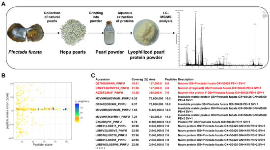
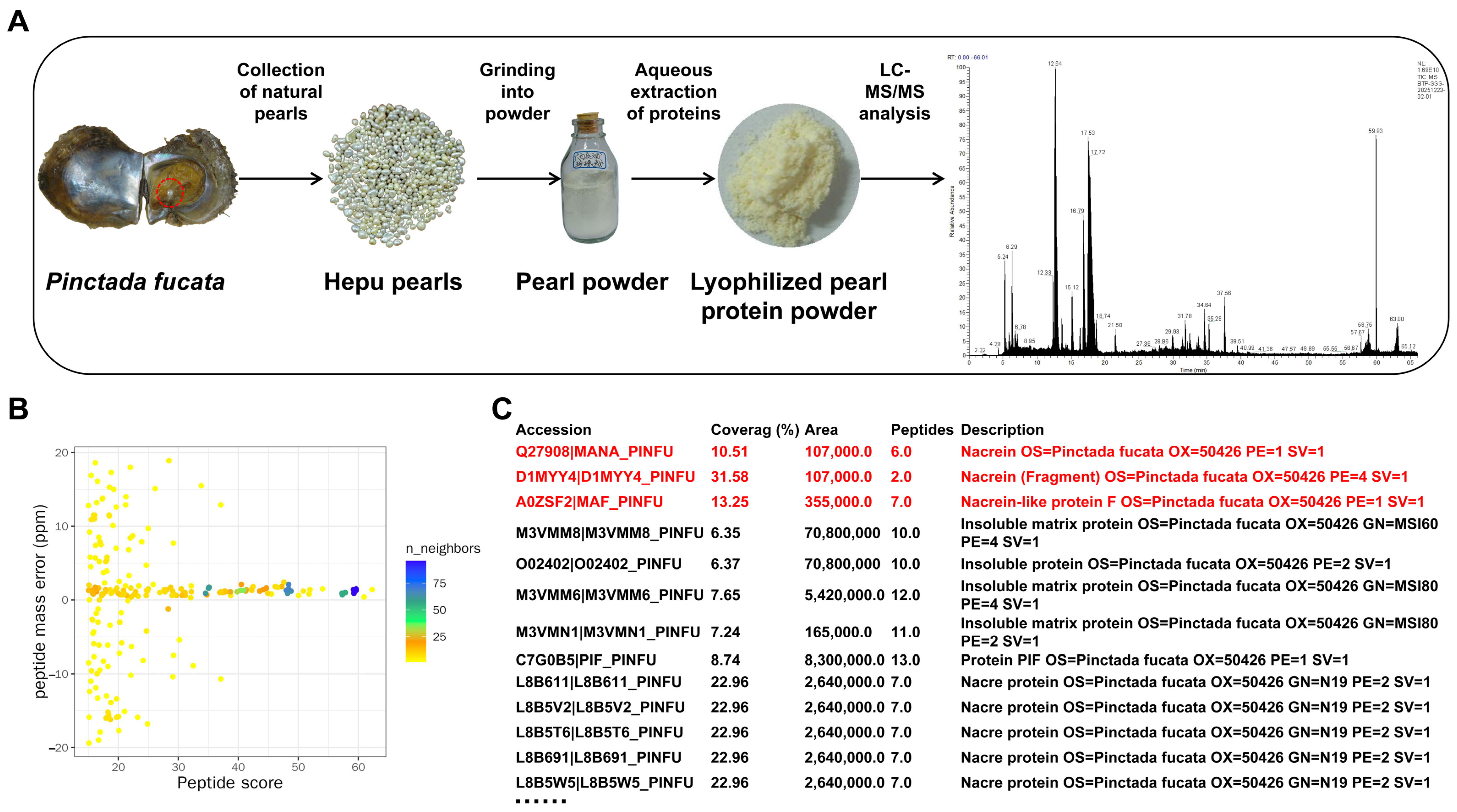
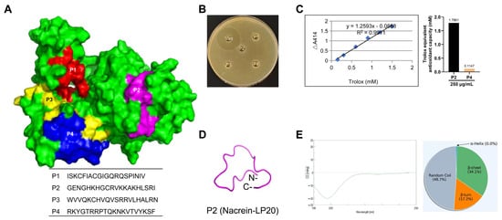
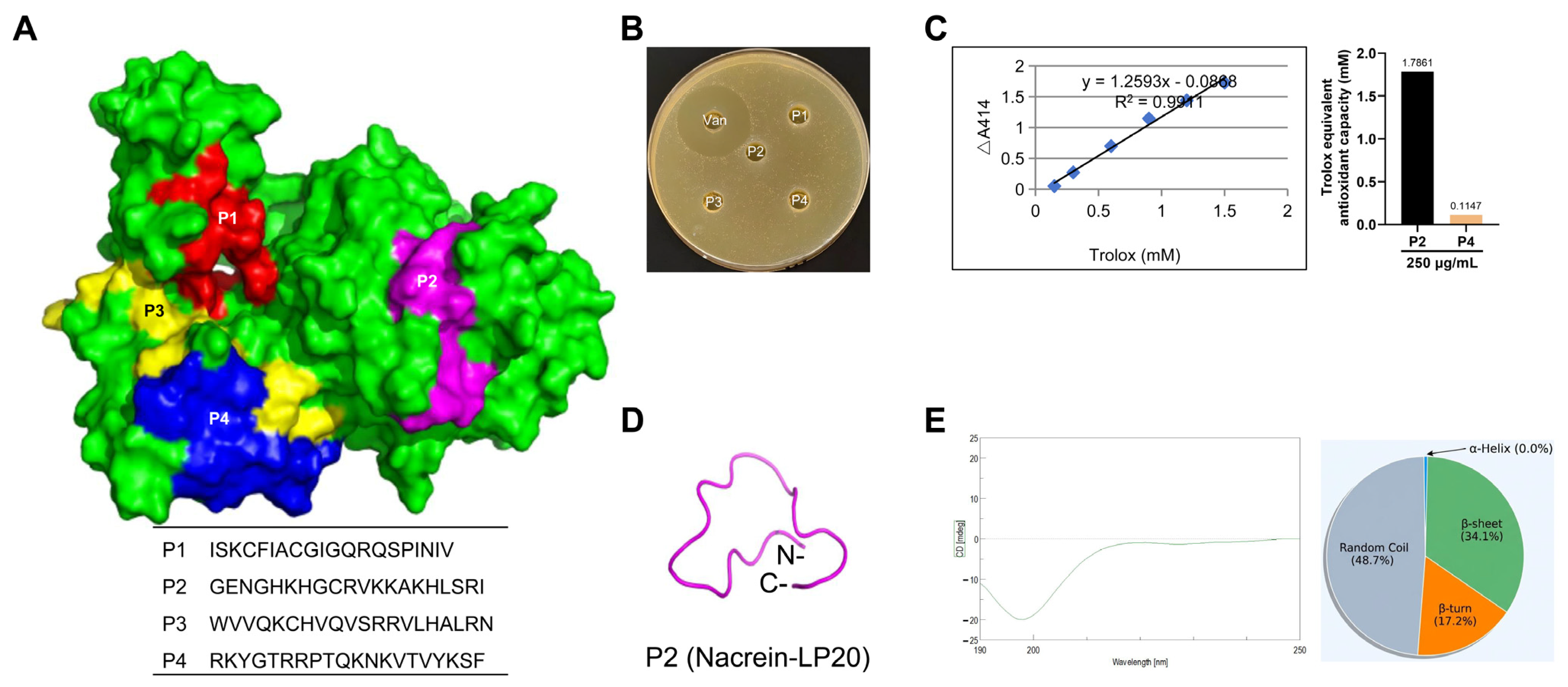
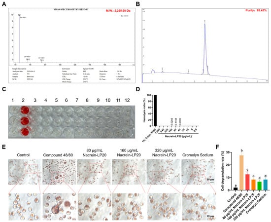
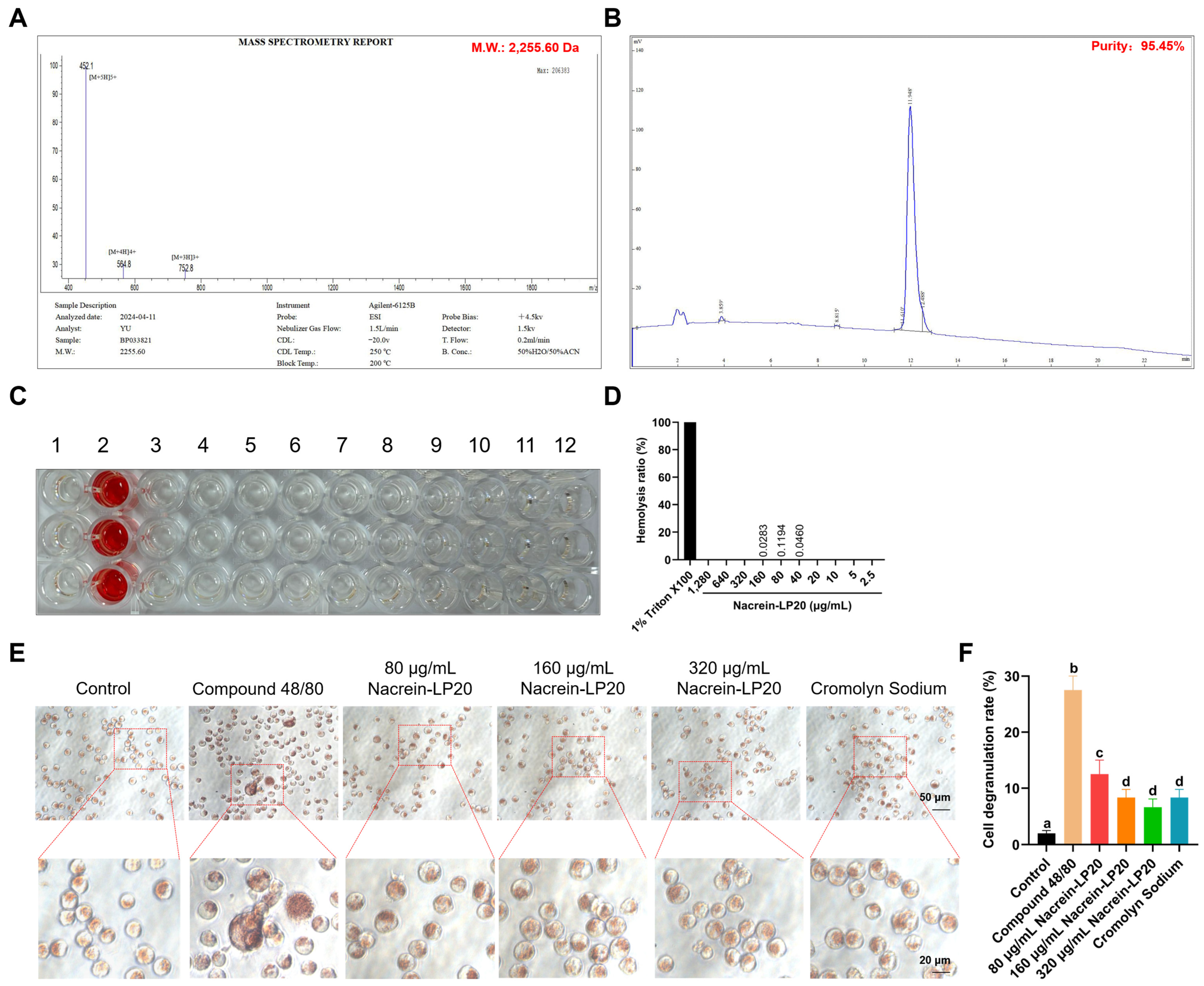
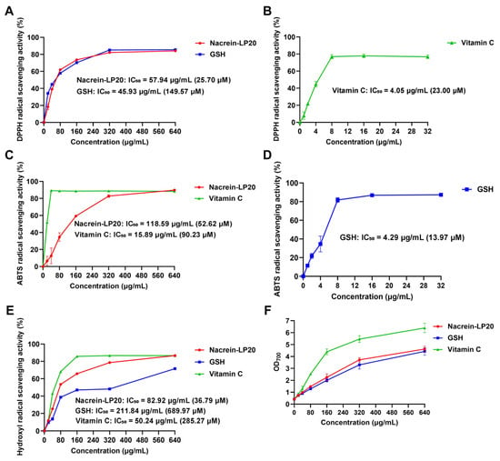
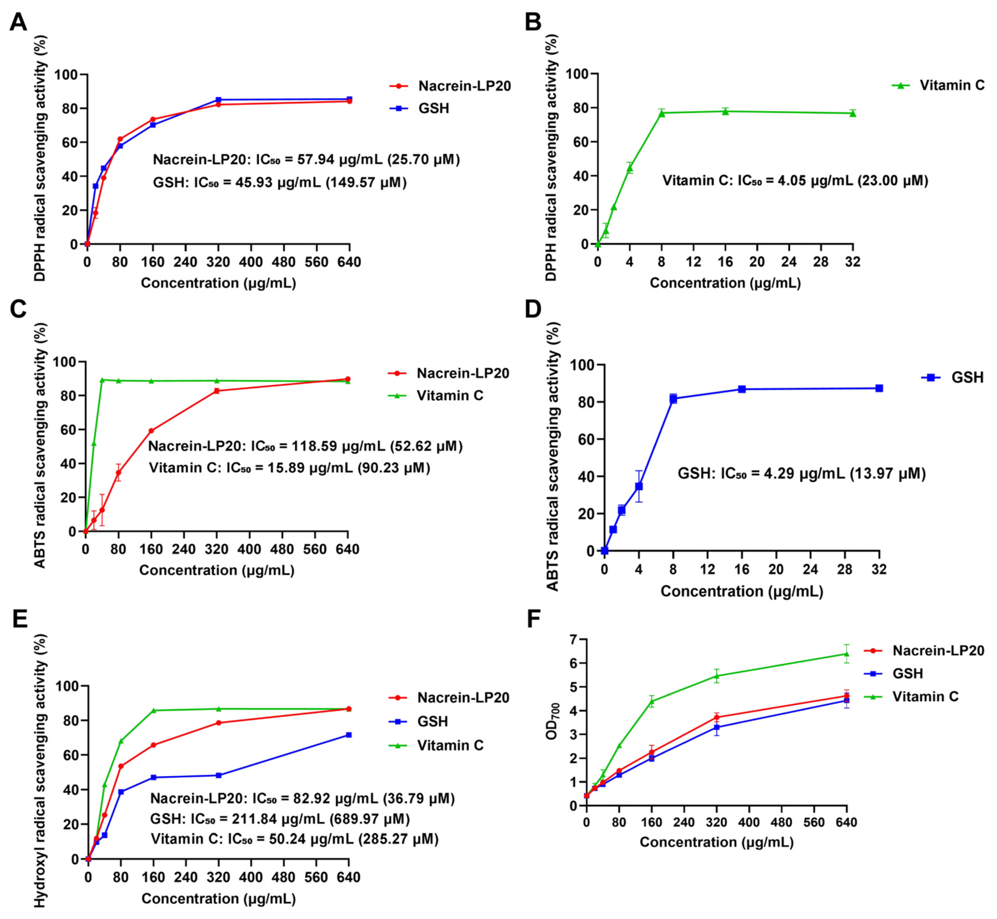
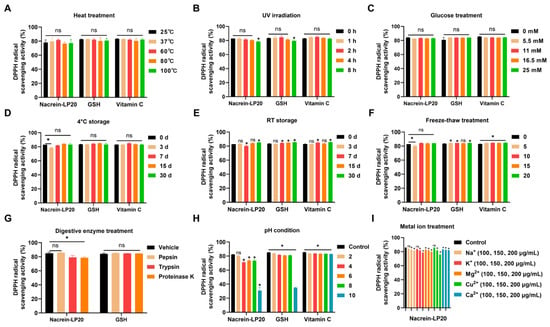
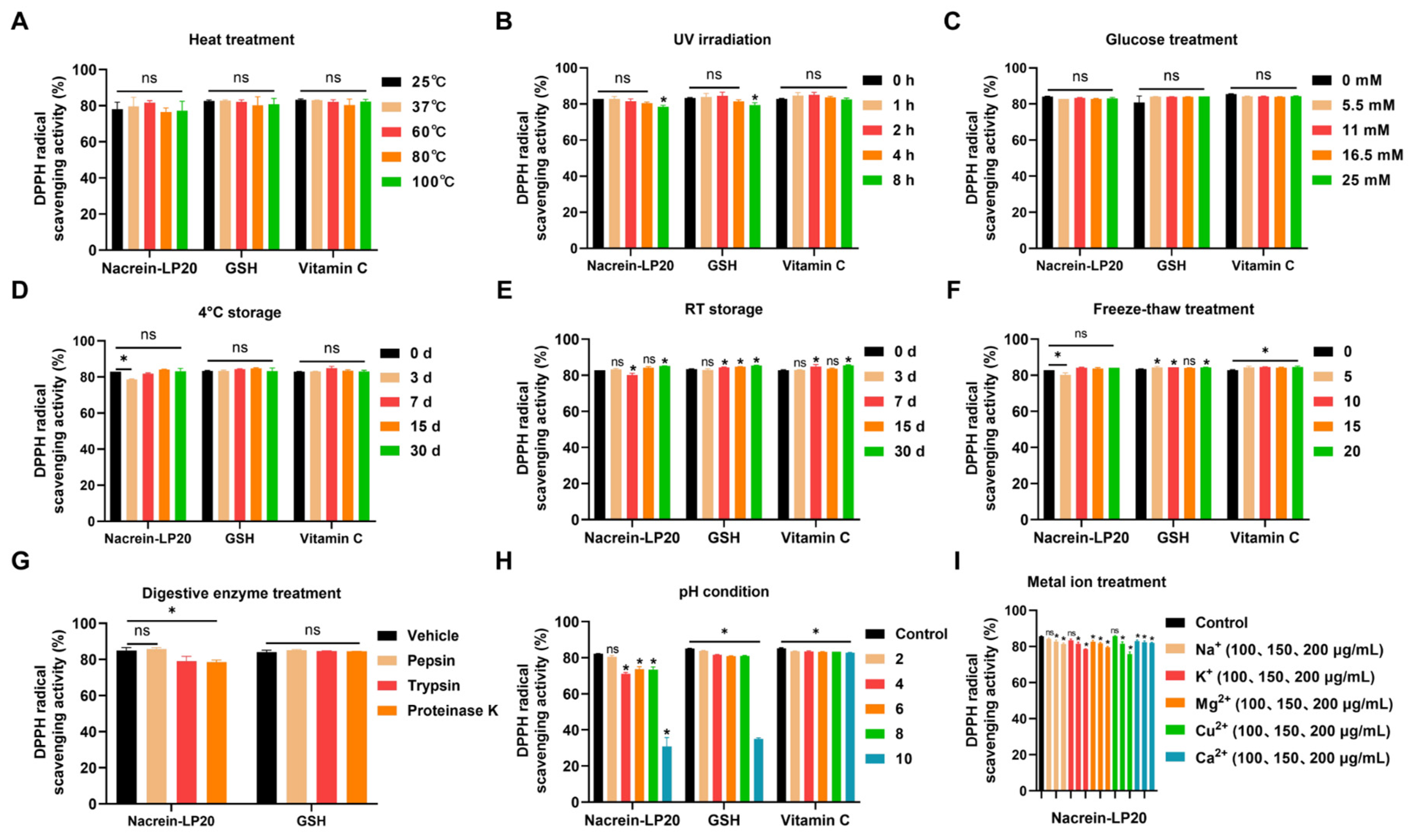
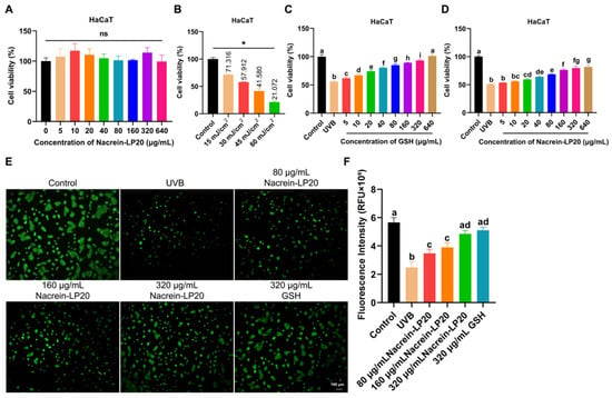
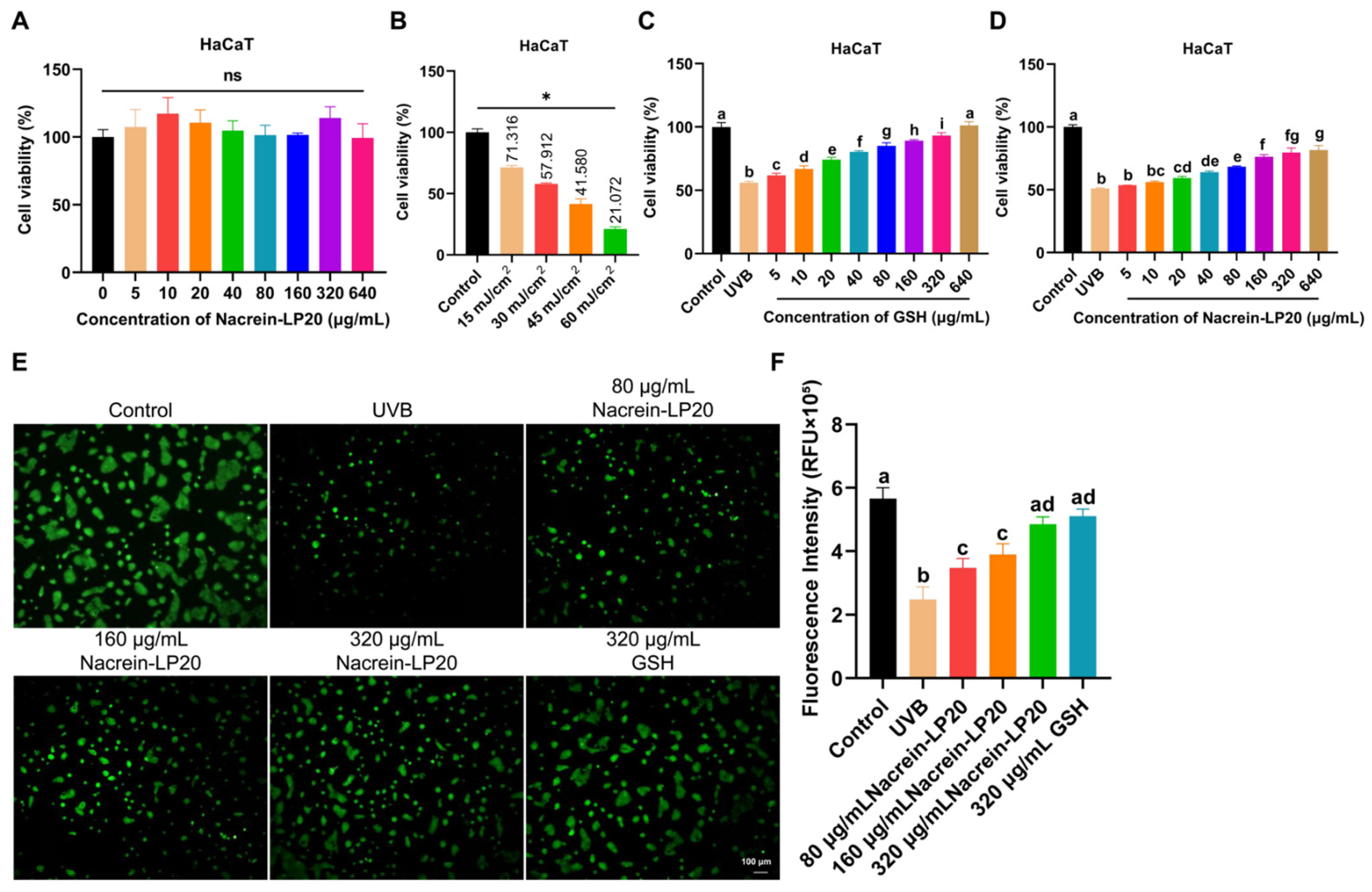
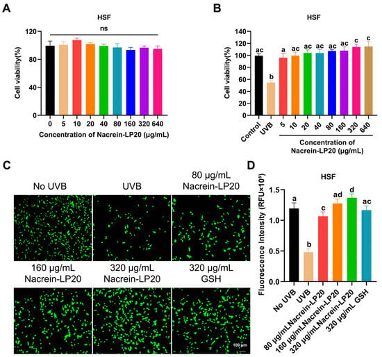
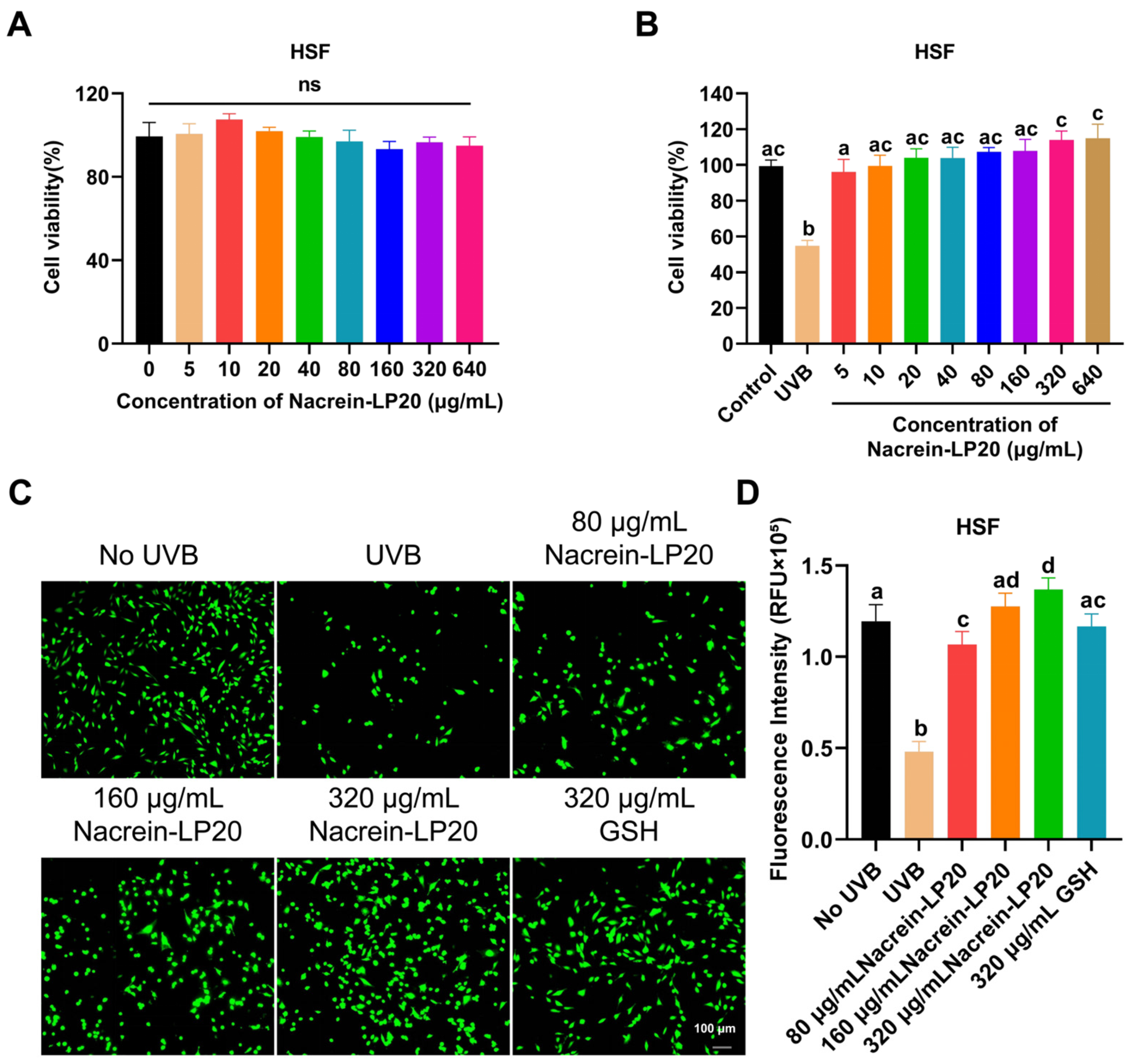
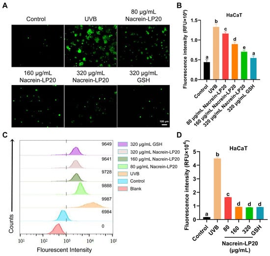
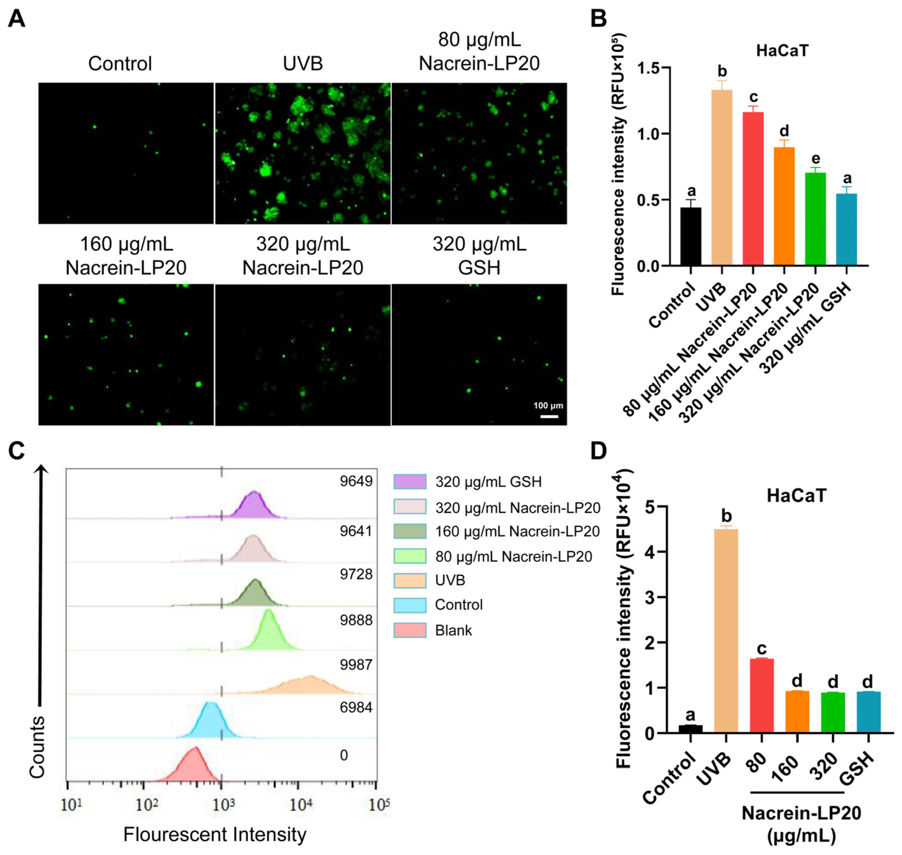
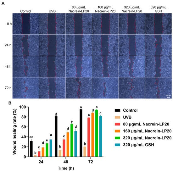
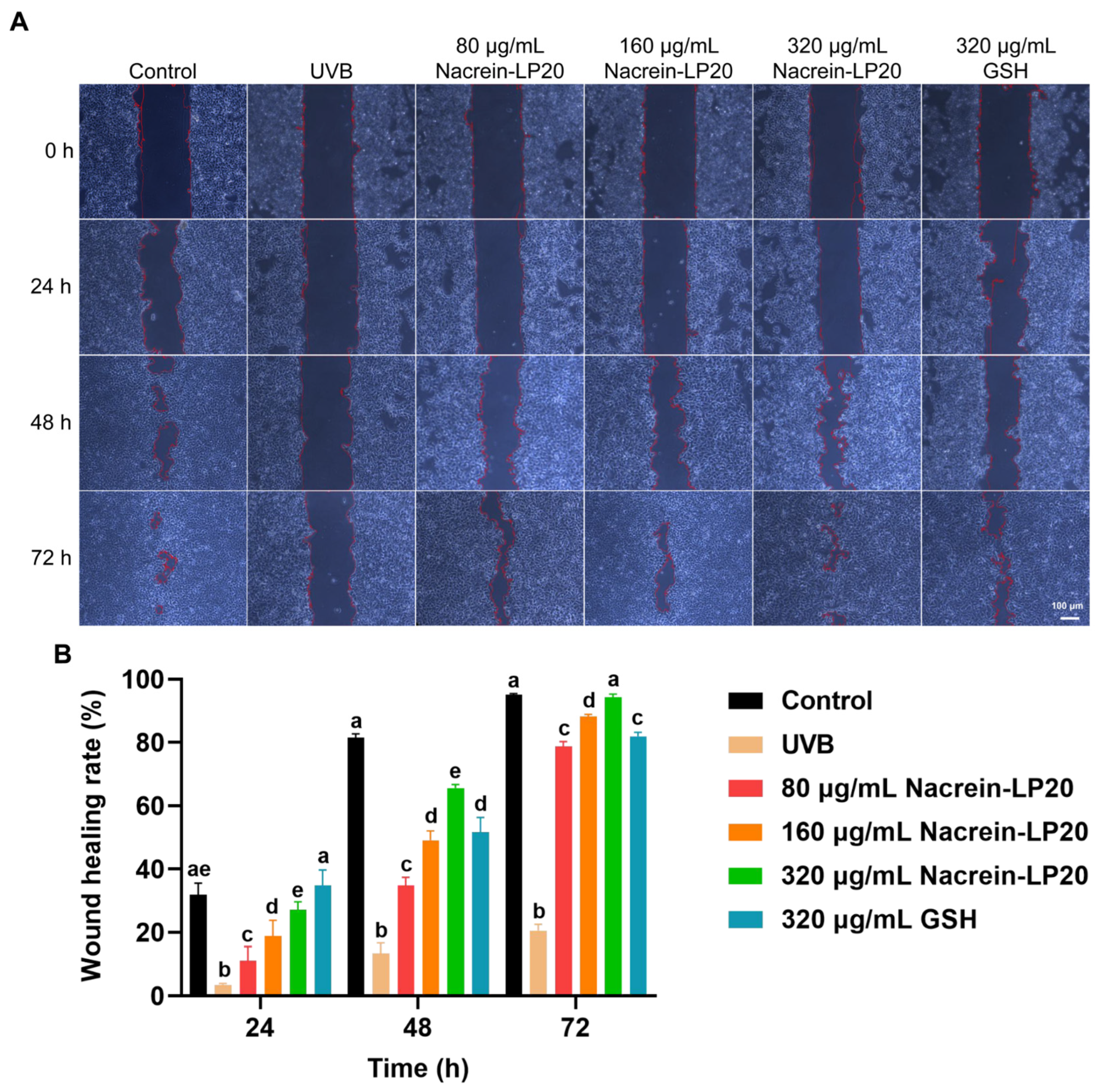
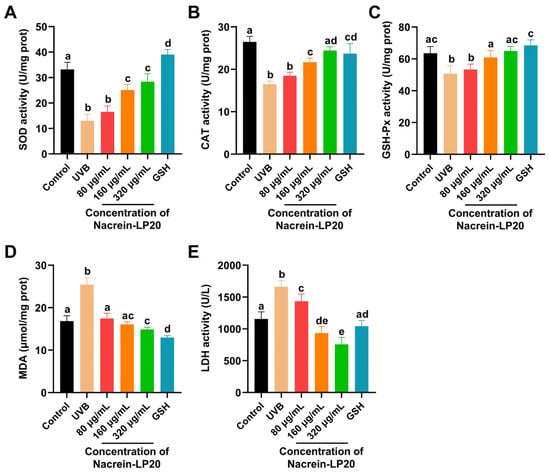
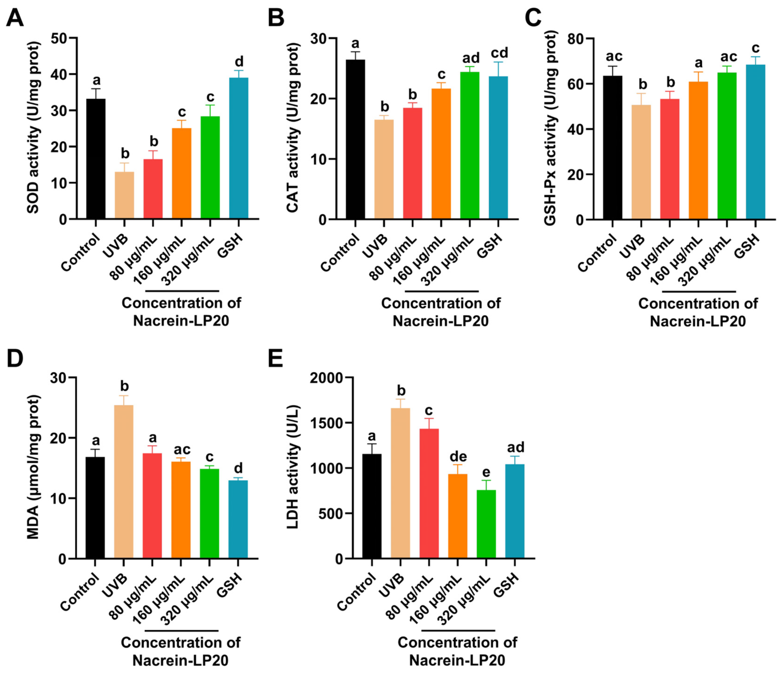
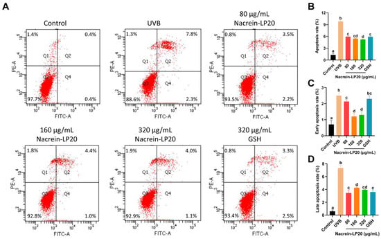
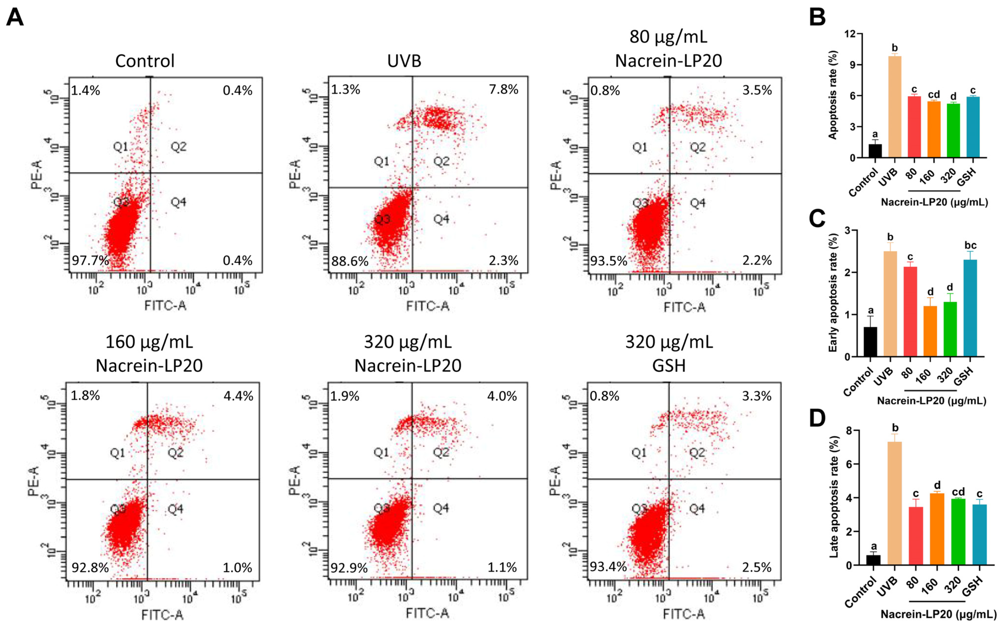
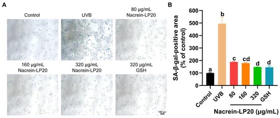
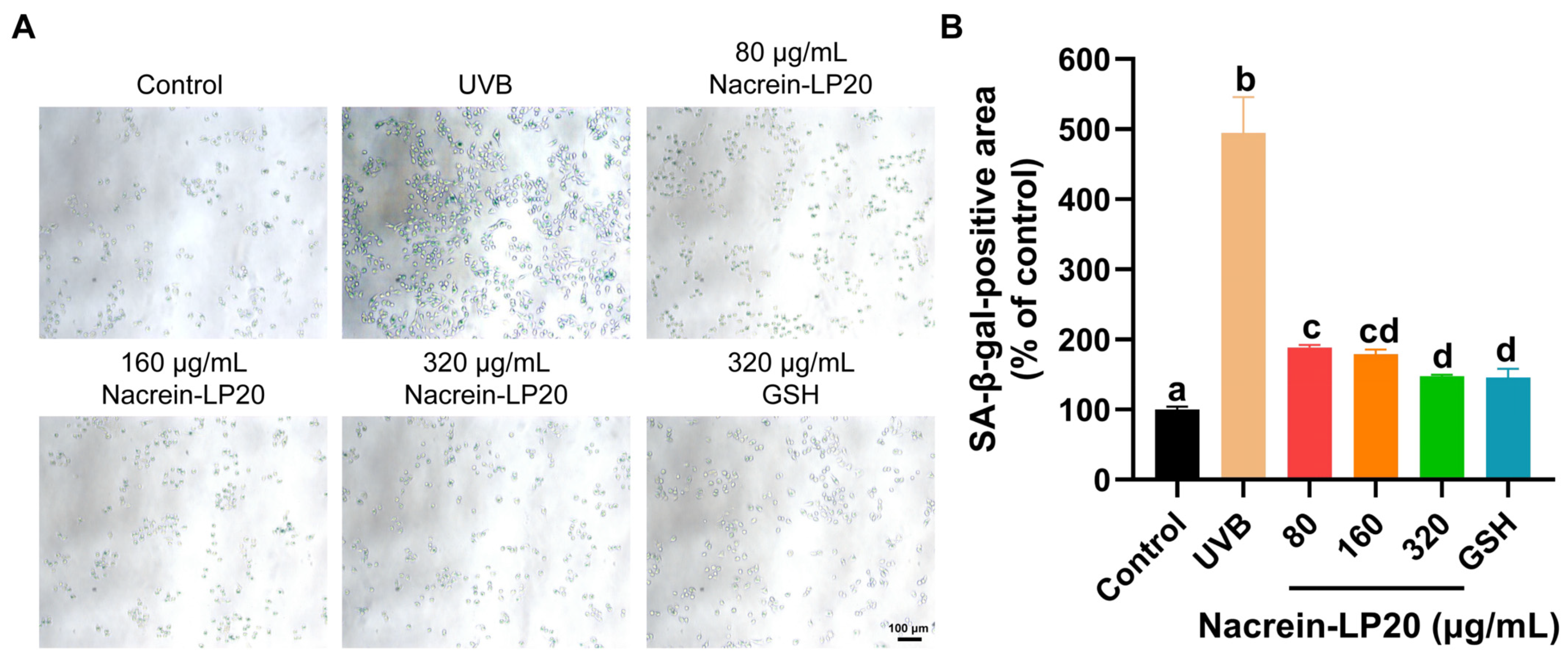
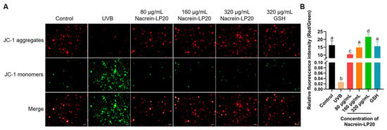
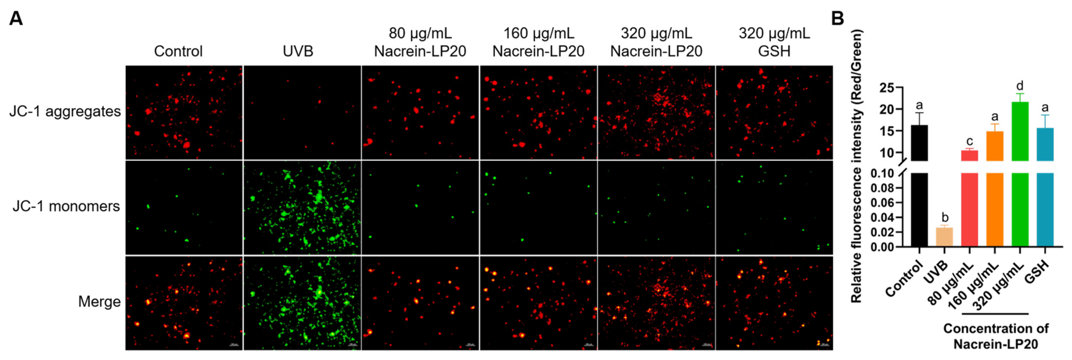
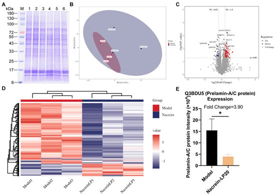
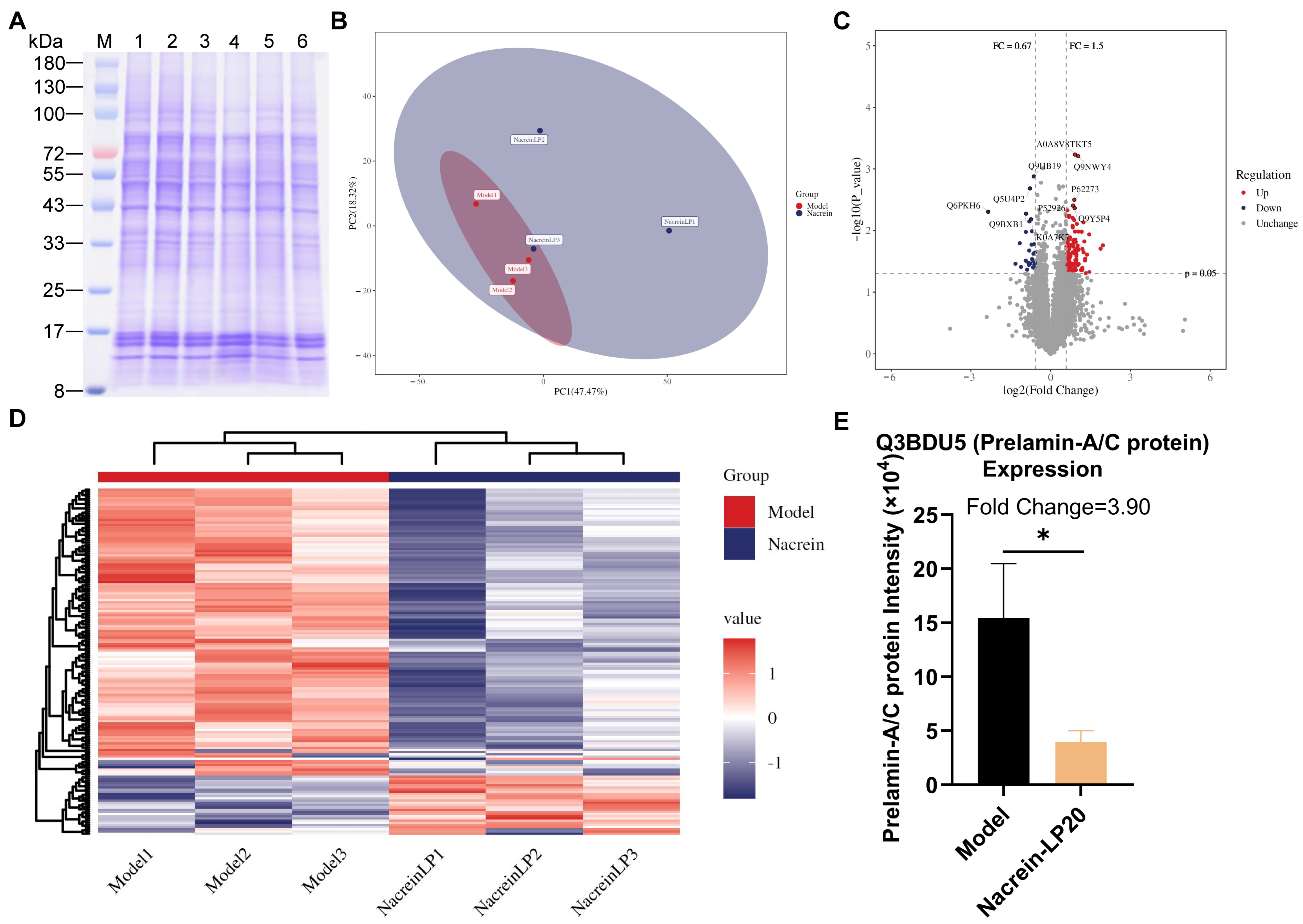
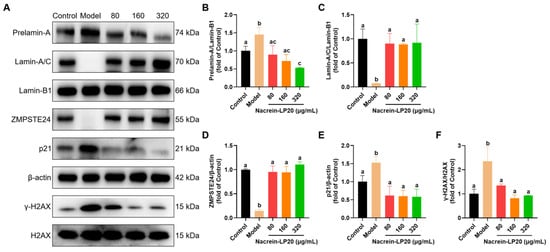
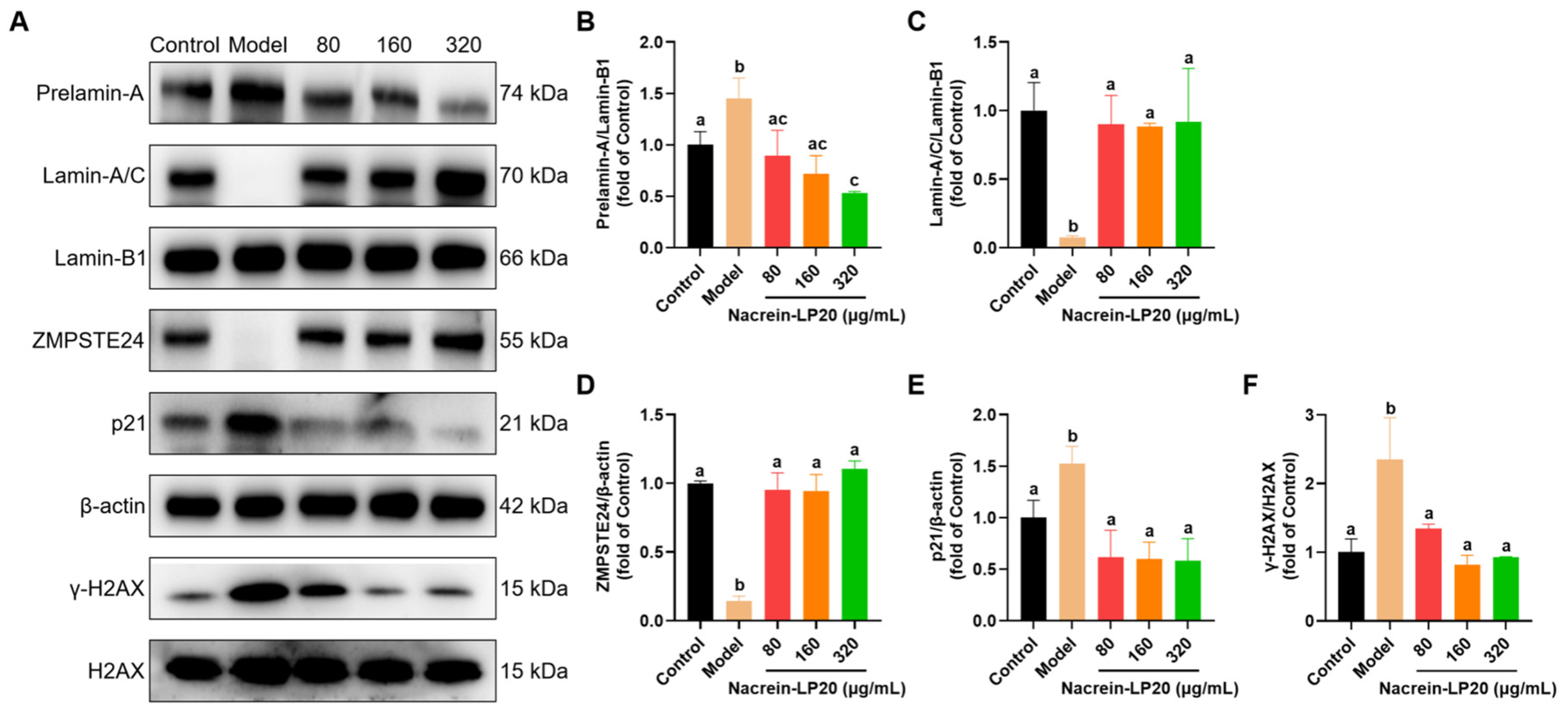
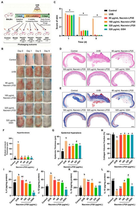
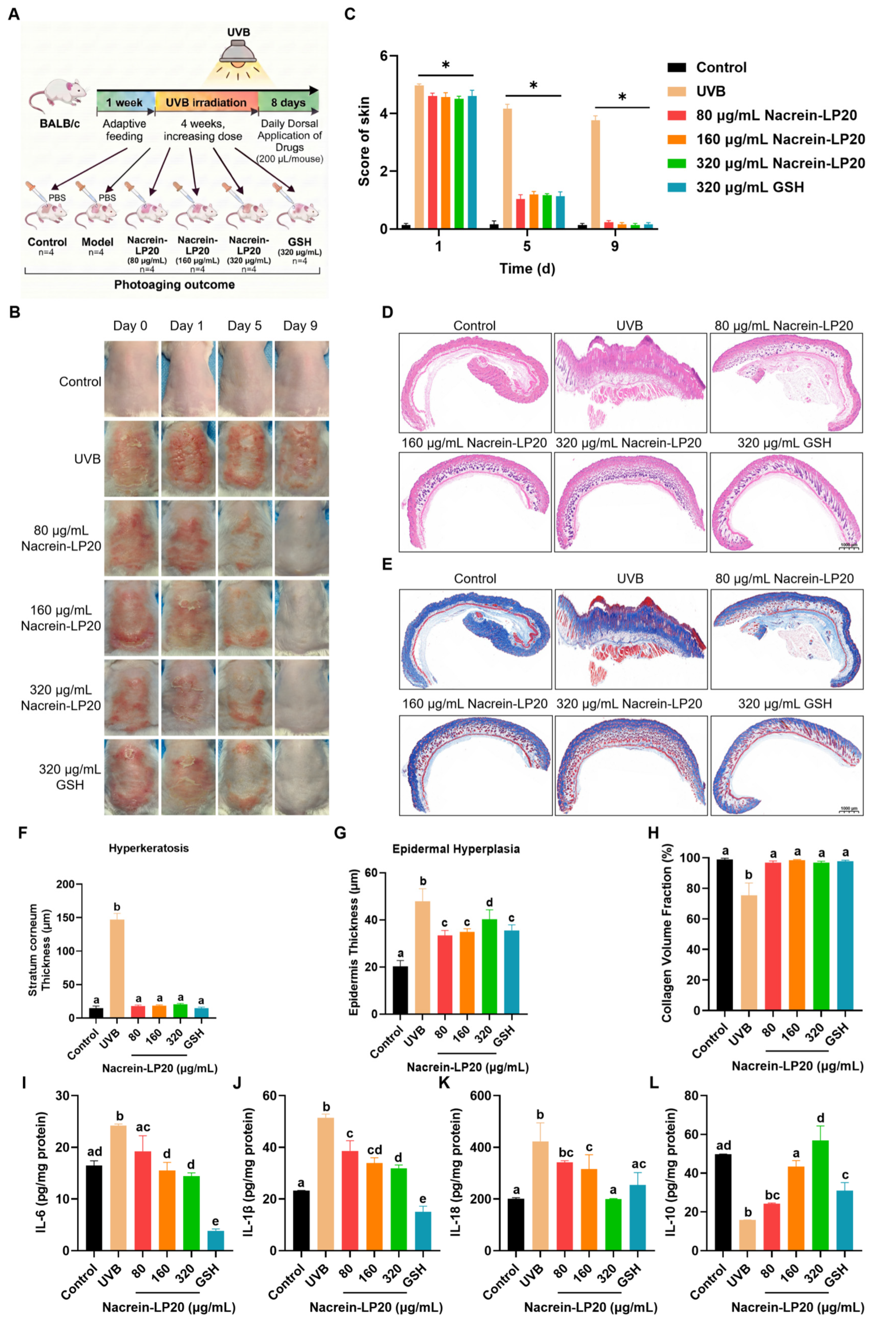
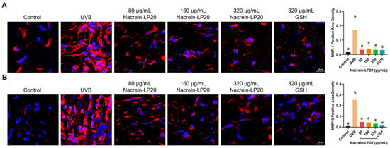
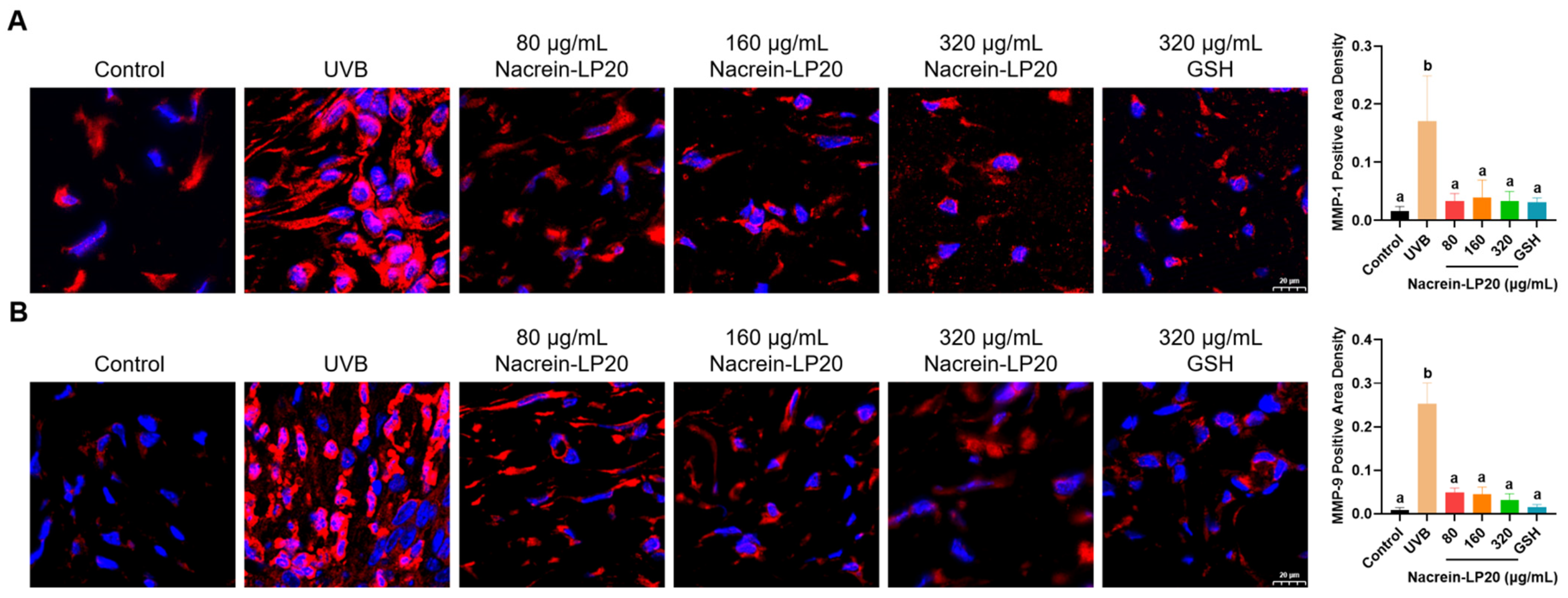
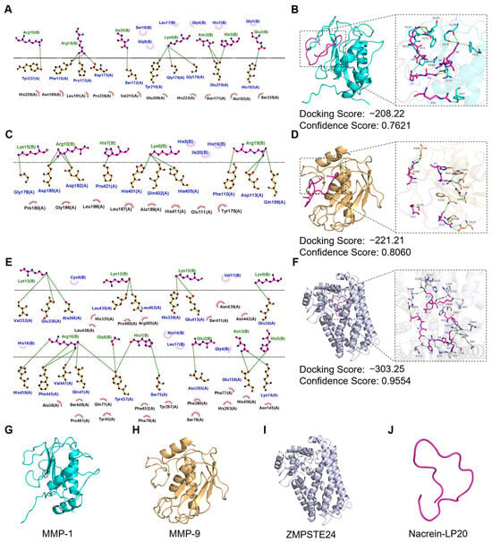
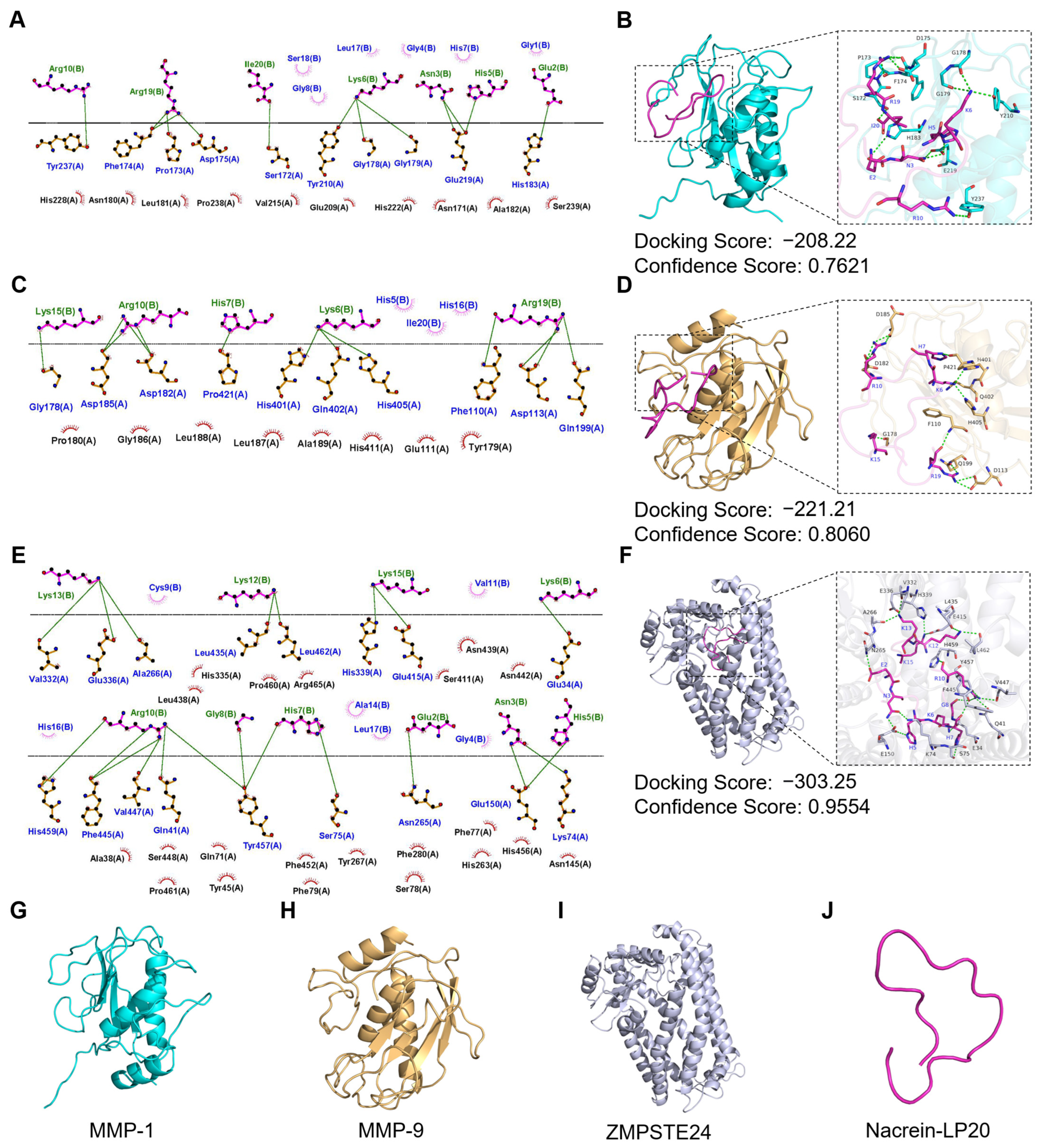

Disclaimer/Publisher’s Note: The statements, opinions and data contained in all publications are solely those of the individual author(s) and contributor(s) and not of MDPI and/or the editor(s). MDPI and/or the editor(s) disclaim responsibility for any injury to people or property resulting from any ideas, methods, instructions or products referred to in the content. |
© 2026 by the authors. Licensee MDPI, Basel, Switzerland. This article is an open access article distributed under the terms and conditions of the Creative Commons Attribution (CC BY) license.