


Abstract
Blue light can induce retinal photo-oxidative stress, whereas the lipofuscin bisretinoid A2E sensitizes retinal pigment epithelium-derived cells to oxidative injury. This study evaluated the antioxidant and retinoprotective effects of puerarin in two complementary models: acute blue light-induced retinal injury in BALB/c mice and A2E-loaded ARPE-19 cells exposed to blue light. Mice received oral puerarin before 430-nm blue light exposure, and retinal structure and function were assessed by optical coherence tomography, electroretinography, histology, and immunofluorescence. In parallel, A2E-loaded ARPE-19 cells were pretreated with puerarin before blue light exposure and analyzed for metabolic activity, membrane injury, reactive oxygen species, mitochondrial membrane potential, glutathione redox status, lipid peroxidation, ferrous iron, and ferroptosis-associated proteins. Oral puerarin preserved retinal and outer nuclear layer thickness and partially restored scotopic electroretinographic responses. These effects were accompanied by increased retinal GPX4 and reduced 4-hydroxynonenal accumulation. In A2E-loaded ARPE-19 cells, puerarin improved metabolic activity and mitochondrial membrane potential, limited membrane injury, reactive oxygen species, lipid peroxidation, and Fe2+ accumulation, and restored the GSH/GSSG ratio and SLC7A11 and GPX4 protein abundance. Erastin attenuated several protective effects of puerarin, supporting the contribution of SLC7A11-dependent glutathione defense. Thus, puerarin attenuates acute photo-oxidative retinal injury in vivo and ferroptosis-associated oxidative damage in A2E-sensitized ARPE-19 cells.
Keywords:
puerarin; blue light; photo-oxidative stress; retinal injury; A2E; ARPE-19; lipid peroxidation; ferroptosis 1. Introduction
Photo-oxidative stress contributes to retinal aging and to retinal disorders in which photoreceptors and the retinal pigment epithelium (RPE) are vulnerable to cumulative oxidative damage. Age-related macular degeneration (AMD), a leading cause of irreversible visual impairment in older adults, is characterized by progressive RPE dysfunction, photoreceptor loss, and, in advanced non-neovascular disease, geographic atrophy [1,2,3]. Complement inhibitors have expanded treatment options for geographic atrophy, but phase 3 trials show that these therapies slow rather than reverse lesion growth and require repeated intravitreal administration [4,5,6]. Strategies that limit oxidative and mitochondrial injury therefore remain relevant to preserving retinal structure and function.
The RPE supports photoreceptor metabolism, maintains the outer blood-retinal barrier, participates in the visual cycle, and phagocytoses shed photoreceptor outer segments [7]. Its high metabolic activity, lifelong light exposure, and accumulation of lipofuscin fluorophores create substantial oxidative pressure. Blue wavelengths can drive retinal photochemical stress under experimental conditions [8,9]. A2E, a major bisretinoid component of RPE lipofuscin, acts as a photosensitizer and, after blue light excitation, promotes reactive oxygen species (ROS) generation, mitochondrial dysfunction, membrane injury, and cell death [10,11,12]. A2E plus blue light has also been reported to increase ferrous iron, deplete glutathione, and promote lipid peroxidation in RPE-derived cells [13]. Among the products of lipid peroxidation, 4-hydroxynonenal (4-HNE), a reactive aldehyde generated from ω-6 polyunsaturated fatty acid oxidation, accumulates when GPX4-dependent detoxification is overwhelmed and serves as both a downstream mediator and a sensitive biomarker of ferroptotic injury. These changes converge on ferroptosis, an iron-dependent form of regulated cell death driven by phospholipid peroxidation and failure of antioxidant defenses [14,15]. SLC7A11 (solute carrier family 7 member 11, also known as system Xc−, a cystine/glutamate antiporter) supports cystine uptake for glutathione (GSH) synthesis, whereas glutathione peroxidase 4 (GPX4) uses GSH to reduce phospholipid hydroperoxides; together, this defense system limits ferroptotic injury [16,17,18].
Puerarin, an isoflavone derived from Pueraria lobata, has antioxidant, anti-inflammatory, and retinal protective activities [19]. In RPE-derived cells, puerarin suppresses ROS-dependent oxidative and endoplasmic reticulum stress [20]. In vivo, it reduces iron overload-induced retinal injury by modulating iron-handling proteins and attenuates retinal ferroptosis-associated changes through an Nrf2-related response [21,22]. The antioxidant action of puerarin is thought to involve two complementary modalities: direct radical scavenging via its phenolic 4′-hydroxyl group, and indirect upregulation of endogenous antioxidant enzymes through Nrf2-dependent signaling [23]. These findings support testing puerarin in retinal photo-oxidative injury, but its effects on retinal function after blue light exposure and on A2E-sensitized cellular injury remain incompletely defined.
We therefore used two complementary, mechanistically distinct experimental systems. An acute blue light-induced mouse model was used to determine whether oral puerarin preserves retinal structure and scotopic function and modulates retinal GPX4 and 4-hydroxynonenal (4-HNE). An A2E-loaded ARPE-19 model was used to examine oxidative stress, mitochondrial membrane potential, Fe2+ accumulation, glutathione redox status, lipid peroxidation, and SLC7A11 and GPX4 abundance. Erastin was used as a pharmacological challenge to assess the contribution of SLC7A11-dependent glutathione defense to puerarin-mediated cellular protection.
2. Materials and Methods
2.1. Reagents and Antibodies
A2E (TargetMol, Boston, MA, USA; distributed by Shanghai Taosu Biotech, Shanghai, China; catalog no. T74051; CAS 173449-96-2) was dissolved in dimethyl sulfoxide (DMSO; Beyotime, Shanghai, China; catalog no. ST038) at 10 mM. Puerarin (MedChemExpress, Monmouth Junction, NJ, USA; catalog no. HY-N0145; CAS 3681-99-0) and erastin (Beyotime; catalog no. Y240017-1 mg; CAS 571203-78-6) were dissolved in DMSO. Stock solutions were aliquoted and stored at −20 °C, and the final DMSO concentration was below 0.1%. The following reagents were used according to the manufacturers’ instructions: Cell Counting Kit-8 (CCK-8; MedChemExpress; HY-K0301-500T), CM-H2DCFDA (Beyotime; S0036S), malondialdehyde (MDA) assay kit (Beyotime; S0131S), lactate dehydrogenase (LDH) assay kit (Beyotime; C0016), JC-1 assay kit (Beyotime; C2006), FerroOrange (Servicebio, Wuhan, China; G727-100T), BODIPY 581/591 C11 (Beyotime; S0043S), GSH/GSSG assay kit (Beyotime; S0053), bicinchoninic acid assay kit (Solarbio, Beijing, China; PC0020), RIPA buffer (Solarbio; R0020), protease inhibitor (Beyotime; P1045-1), phosphatase inhibitor (Beyotime; P1045-2), PVDF membranes (Roche, Basel, Switzerland; 03010040001), and enhanced chemiluminescence reagent (Cytiva, Marlborough, MA, USA; 18391900). Primary antibodies included rabbit anti-SLC7A11 (Proteintech, Rosemont, IL, USA; 26864-1-AP), rabbit anti-GPX4 (Proteintech; 30388-1-AP), HRP-conjugated β-actin (Proteintech; HRP-60008), mouse anti-GPX4 (Proteintech; 67763-1-Ig; 1:250), rabbit anti-RPE65 (Abcam, Cambridge, UK; ab231782; 1:250), and mouse anti-4-HNE (Proteintech; 68538-1-Ig; 1:200). Secondary antibodies were HRP-conjugated goat anti-rabbit IgG (Cell Signaling Technology, Danvers, MA, USA; 7074), Alexa Fluor 488 goat anti-mouse IgG (Abcam; ab150113; 1:500), and Alexa Fluor 594 goat anti-rabbit IgG (Abcam; ab150080; 1:500).
2.2. Blue Light Exposure Conditions
Blue light was delivered using an LED source with a peak wavelength of 430 nm and an emission range of 415–455 nm (Xuzhou Aijia Technology Co., Ltd., Xuzhou, China). At a source-to-sample distance of 20 cm, the irradiance at the exposure plane was approximately 50 mW/cm2 (measured with the UNI-T UT385 radiometer; Uni-Trend Technology Co., Ltd., Dongguan, China). Mice were exposed for 1 h, whereas cell cultures were exposed for 30 min. Dark control samples underwent otherwise identical handling without illumination.
2.3. Cell Culture and Treatment
ARPE-19 cells (ATCC, Manassas, VA, USA) were maintained in DMEM/F12 medium (Gibco, Grand Island, NY, USA; 11320033) supplemented with 10% fetal bovine serum (Gibco; 10099-141) and 1% penicillin-streptomycin (Gibco; 15140-122) at 37 °C in 5% CO2. Experiments were performed at 70–80% confluence. Cells were incubated with 25 μM A2E for 24 h in the dark [13], washed with phosphate-buffered saline, and then treated with 25 μM puerarin in serum-free DMEM/F12 for 12 h in the dark. After another wash, the medium was replaced with phenol red-free DMEM/F12 (Gibco; 21041025), and cells were exposed to blue light for 30 min [13]. Dark controls were wrapped in aluminum foil. All groups were incubated for a further 24 h after exposure. Where indicated, 10 μM erastin was added 2 h before blue light exposure [24]. Vehicle controls received an equivalent concentration of DMSO.
2.4. Cellular Metabolic Activity and Membrane Injury Assays
Cellular metabolic activity was measured using the CCK-8 assay. Cells were seeded at 8 × 10³ cells per well, and 10 μL of CCK-8 reagent was added to 100 μL of culture medium. After incubation for 1.5 h at 37 °C, absorbance was measured at 450 nm using a Tecan Infinite M200 PRO plate reader (Tecan Austria GmbH, Grödig, Austria; Infinite M200 PRO). Relative CCK-8 signal was calculated as (ODexperiment − ODblank)/(ODcontrol − ODblank) × 100%. Membrane injury was evaluated by LDH release. Cell culture supernatant (120 μL) was incubated with 60 μL of LDH reaction reagent for 30 min at room temperature, and absorbance was measured at 490 nm. LDH release was expressed relative to the control group. Each experiment comprised three independent biological replicates, each measured in three technical replicates.
2.5. ROS, Mitochondrial Membrane Potential, Lipid Peroxidation, Fe2+, MDA, and GSH/GSSG Assays
Intracellular ROS were assessed using 5 μM CM-H2DCFDA with Hoechst 33342 counterstaining after incubation for 20–30 min; fluorescence was quantified using ImageJ 2.14.0. Mitochondrial membrane potential was evaluated using JC-1 and expressed as the red-to-green fluorescence ratio. Lipid peroxidation was measured using 2 μM BODIPY 581/591 C11 and expressed as the oxidized-to-reduced fluorescence ratio. Intracellular Fe2+ was detected using 1 μM FerroOrange. MDA was quantified using a thiobarbituric acid-based assay at 532 nm and normalized to total protein. Reduced and oxidized glutathione were measured using a GSH/GSSG assay kit, and the GSH/GSSG ratio was calculated. Fluorescence images were acquired on an OLYMPUS IX53 inverted fluorescence microscope (Olympus Corporation, Tokyo, Japan), using the same microscope settings within each assay and quantified using ImageJ 2.14.0.
2.6. Western Blotting
Cells were lysed in RIPA buffer containing protease and phosphatase inhibitors for 30 min on ice and centrifuged at 12,000× g for 15 min at 4 °C. Protein concentrations were determined using the bicinchoninic acid assay. Equal amounts of protein (30 μg) were separated by 10–12% SDS-PAGE and transferred to PVDF membranes at 300 mA for 90 min. Membranes were blocked for 20 min with Protein-free Fast Blocking Buffer (Cat. No. PS08P, Epizyme Biotechnology, Shanghai, China) and incubated overnight at 4 °C with antibodies against SLC7A11 (1:1000) or GPX4 (1:1000). After incubation with HRP-conjugated goat anti-rabbit IgG (1:2000) for 1 h at room temperature, immunoreactive bands were visualized by enhanced chemiluminescence using a Tanon 4800 system (Tanon, Shanghai, China; 4800). HRP-conjugated β-actin (1:5000) served as the loading control. Band intensities were normalized to β-actin.
2.7. Animals and Blue Light–Induced Retinal Injury
Six-week-old male BALB/c mice (18–22 g) were obtained from Beijing Vital River Laboratory Animal Technology Co., Ltd. (license no. SCXK [Jing] 2021-0006). Animals were housed under specific pathogen-free conditions at 23 ± 2 °C and 50 ± 10% humidity on a 12-h light/dark cycle with food and water available ad libitum. The protocol was approved by the Experimental Animal Ethics Committee of Tianjin Medical University Eye Hospital (approval no. TJYY2025120306) and complied with the ARVO Statement for the Use of Animals in Ophthalmic and Vision Research. Mice were randomized into five groups (n = 6 per group): untreated control, blue light model, and blue light plus puerarin at 10, 50, or 100 mg/kg. Puerarin was formulated in 20% sulfobutylether-beta-cyclodextrin in saline and administered by oral gavage (200 μL) once daily for 7 days; control and model groups received vehicle. On day 7, mice were dark-adapted for 12 h. On day 8, pupils were dilated with 1% tropicamide, and mice were exposed to blue light for 1 h; control animals were handled identically without blue light exposure.
2.8. Electroretinography
Scotopic electroretinography (ERG; Phoenix Research Laboratories, Pleasanton, CA, USA) was performed on day 11 after overnight dark adaptation. Mice were anesthetized with tribromoethanol (250 mg/kg). A corneal electrode, a scalp reference electrode, and a tail ground electrode were used. Responses were recorded at flash intensities of −1.7, −1.1, 0.1, 1.0, and 1.9 log cd·s/m2, and a- and b-wave amplitudes were quantified.
2.9. Optical Coherence Tomography
Optical coherence tomography (OCT; Heidelberg Engineering, Heidelberg, Germany) was performed after ERG on day 11. Anesthetized mice underwent pupil dilation, and circular volume scans were centered on the optic disc. Total retinal and outer nuclear layer (ONL) thicknesses were quantified 500 μm superior and inferior to the optic disc.
2.10. Histology
Mice were euthanized on day 14, and the eyes were enucleated, fixed in 4% paraformaldehyde for 24 h, and embedded in paraffin. Sagittal sections (3 μm) were collected at 100-micrometer intervals and stained with hematoxylin and eosin. ONL and total retinal thicknesses were measured at 250, 500, 750, and 1250 μm on both sides of the optic disc. Measurements from three sections per eye and both eyes were averaged to generate one value per mouse.
2.11. Immunofluorescence Staining
Paraffin sections were deparaffinized, and antigen retrieval was performed in 1x Tris-EDTA buffer by microwave heating. Sections were blocked for 1 h in phosphate-buffered saline containing 0.2% bovine serum albumin, 0.3% Triton X-100, and 5% normal goat serum and incubated overnight at 4 °C with anti-GPX4 plus anti-RPE65 or anti-4-HNE. After incubation with the appropriate Alexa Fluor-conjugated secondary antibodies for 1 h, nuclei were counterstained with DAPI. Confocal images were acquired on a Zeiss LSM 800 confocal laser scanning microscope (Carl Zeiss, Oberkochen, Germany). The excitation/emission (ex/em) settings were: DAPI (Ex 405 nm, Em 410–490 nm), Alexa Fluor 488 (Ex 488 nm, Em 500–550 nm), Alexa Fluor 594 (Ex 561 nm, Em 580–650 nm). Fluorescence images were acquired using identical settings within each staining experiment, and fluorescence intensity was quantified using ImageJ 2.14.0 and averaged to obtain one value per mouse.
2.12. UPLC–MS/MS Analysis
Retinas were collected 2 h after a single oral gavage of puerarin (100 mg/kg) and immediately frozen. Samples were extracted with methanol and analyzed by UPLC-MS/MS (Waters ACQUITY UPLC system coupled to a Waters Xevo TQ-S triple quadrupole mass spectrometer, Waters Corporation, Milford, MA, USA) using an ACQUITY UPLC BEH C18 column. The mobile phases were 0.1% formic acid in water and acetonitrile. A gradient elution program for acetonitrile was applied as follows: 0–1 min, 5% acetonitrile; 1–3 min, 5–50% acetonitrile; 3–4 min, 50–90% acetonitrile; 4–5 min, 90% acetonitrile; 5.1–8 min, 5% acetonitrile. Puerarin was monitored in positive-ion multiple-reaction monitoring mode using m/z 417.0150 to 297.0203 for quantification and m/z 417.0150 to 267.1344 for confirmation. External calibration standards ranged from 0.5 to 100 ng/mL. The reported values represent concentrations measured in the final reconstituted retinal extracts and were not normalized to retinal tissue mass.
2.13. Statistical Analysis
Data are presented as the mean ± standard deviation and were analyzed using GraphPad Prism 9.5. Normality was assessed using the Shapiro–Wilk test. One-way or two-way analysis of variance (ANOVA) was followed by Tukey’s test for all pairwise comparisons or Dunnett’s test for comparisons with a specified control group, as indicated in the figure legends. ERG responses across flash intensities and retinal thickness measurements across locations were analyzed using repeated-measures two-way ANOVA with the Greenhouse–Geisser correction, followed by Tukey’s multiple-comparisons test. An unpaired two-tailed t-test was used for comparisons between two independent groups. A p value below 0.05 was considered statistically significant. Cell experiments comprised three independent biological replicates, each with three technical replicates; the biological replicate was the unit of analysis. Sample sizes are specified in the figure legends.
3. Results
3.1. Puerarin Preserves Retinal Structure and Function After Blue Light Exposure
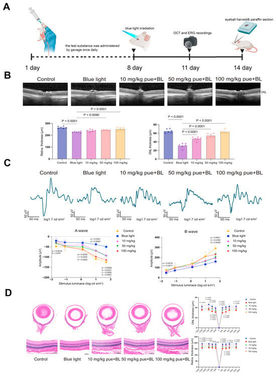
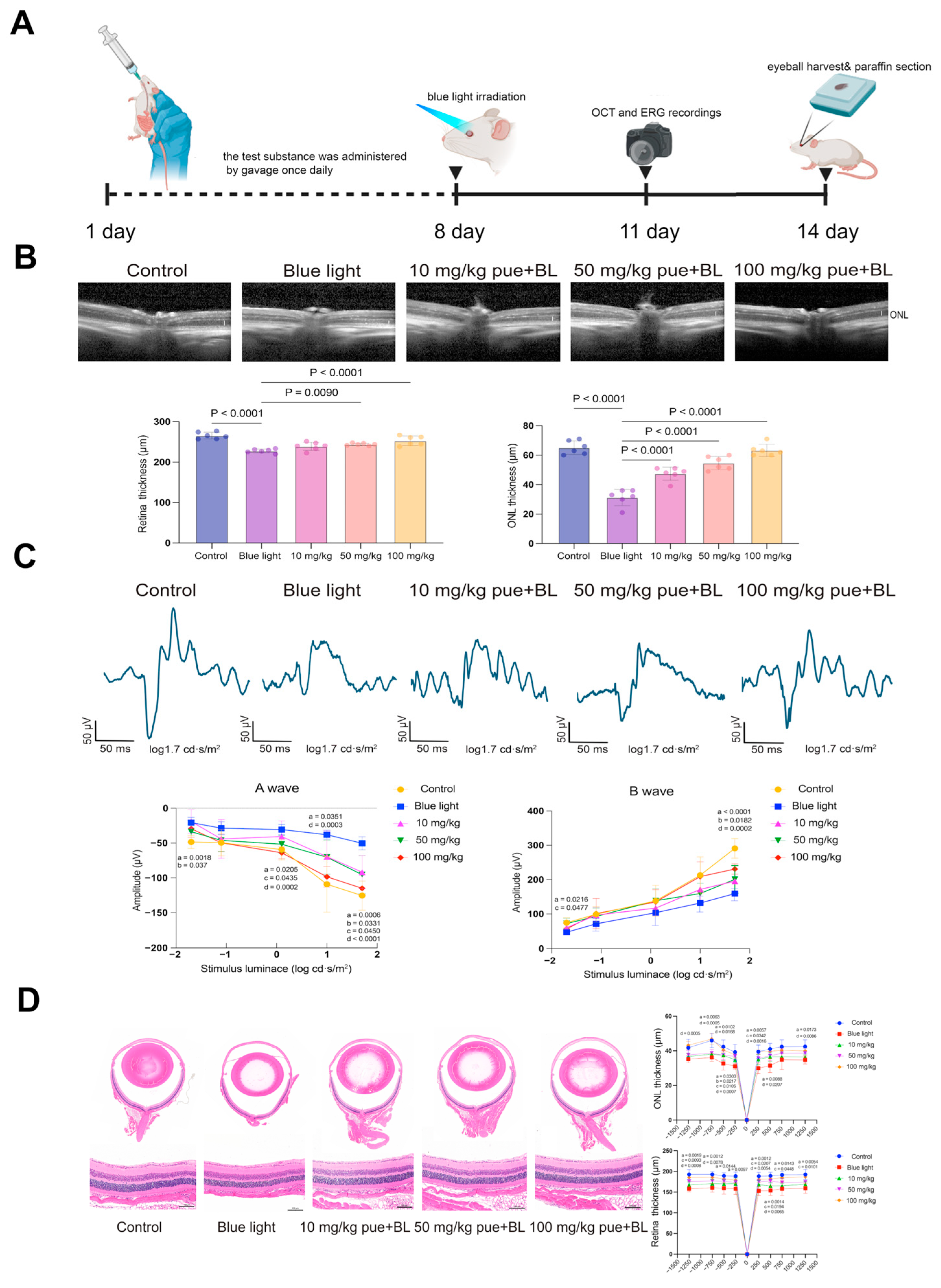
We first evaluated puerarin in an acute blue light-induced retinal injury model. Mice received puerarin (10, 50, or 100 mg/kg) by oral gavage once daily for 7 days and were then exposed to 430-nm blue light. At the exposure plane, the irradiance was approximately 50 mW/cm2 for 1 h. OCT and ERG were performed on day 11, 3 days after exposure, and tissue analyses were performed on day 14 (Figure 1A).
Figure 1.
Puerarin preserves retinal structure, histology, and scotopic function after acute blue light exposure. (A) Experimental timeline. BALB/c mice received oral puerarin (10, 50, or 100 mg/kg) once daily for 7 days and were exposed to 430-nm blue light for 1 h on day 8. OCT and ERG were performed on day 11, and mice were euthanized on day 14 for histological and immunofluorescence analyses. (B) Representative OCT images and quantification of total retinal and ONL thickness. Six eyes, one from each mouse, were analyzed (n = 6 mice). (C) Representative scotopic ERG traces and a- and b-wave amplitudes across flash intensities (−1.7, −1.1, 0.1, 1.0, and 1.9 log cd·s/m2). Six eyes, one from each mouse, were analyzed (n = 6 mice). In the line graphs, a denotes the comparison between the blue light model and control groups; b, c, and d denote comparisons between the model group and the 10, 50, and 100 mg/kg puerarin groups, respectively, at each flash intensity. (D) Representative hematoxylin and eosin staining and quantification of ONL and total retinal thickness. Scale bar, 100 μm. In the graphs, a denotes the comparison between the blue light model and control groups; b, c, and d denote comparisons between the model group and the 10, 50, and 100 mg/kg puerarin groups, respectively, at each retinal location. Data in (B) were analyzed using one-way ANOVA followed by Tukey’s multiple-comparisons test. Data in (C) were analyzed using repeated-measures two-way ANOVA with the Geisser-Greenhouse correction followed by Tukey’s multiple-comparisons test at each flash intensity. Data in (D) were analyzed using repeated-measures two-way ANOVA with the Greenhouse–Geisser correction followed by Tukey’s multiple-comparisons test at each retinal location (n = 6 mice per group). Data are presented as mean ± SD; p < 0.05 was considered statistically significant.
OCT showed that blue light exposure significantly reduced total retinal thickness relative to the control group. Pretreatment with 50 or 100 mg/kg puerarin preserved total retinal thickness relative to the blue light model (p = 0.0090 and p < 0.0001, respectively), whereas 10 mg/kg produced a nonsignificant trend (Figure 1B). ONL thickness was also reduced after blue light exposure, and all three puerarin doses significantly preserved ONL thickness relative to the model group (all p < 0.0001).
Scotopic ERG demonstrated corresponding functional protection. At the highest flash intensity, blue light reduced both a- and b-wave amplitudes relative to controls (p = 0.0006 and p < 0.0001, respectively; Figure 1C). Puerarin produced a dose-dependent recovery, with 100 mg/kg significantly increasing the a-wave (p < 0.0001) and b-wave (p = 0.0002) amplitudes relative to the model group, although responses did not completely return to control values.
Histological findings were consistent with the OCT measurements. Blue light-exposed retinas showed disorganization and thinning of the ONL, with reductions in both ONL and total retinal thickness. Puerarin attenuated these changes in a dose-dependent manner. The 100 mg/kg dose significantly preserved ONL and total retinal thickness relative to the model group across all measured retinal locations (Figure 1D), while the lower doses produced smaller, location-dependent effects.
To determine whether orally administered puerarin could be detected in retinal extracts, retinas were analyzed by UPLC-MS/MS 2 h after a single 100 mg/kg gavage. Puerarin was detected in the final reconstituted extracts from treated mice at a mean concentration of 15.9 ng/mL, equivalent to 0.038 μM in the reconstituted extract (n = 3; p = 0.0211 versus untreated controls; Supplementary Figure S1).
3.2. Puerarin Attenuates Retinal GPX4 Loss and 4-HNE Accumulation After Blue Light Exposure
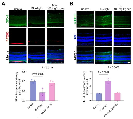
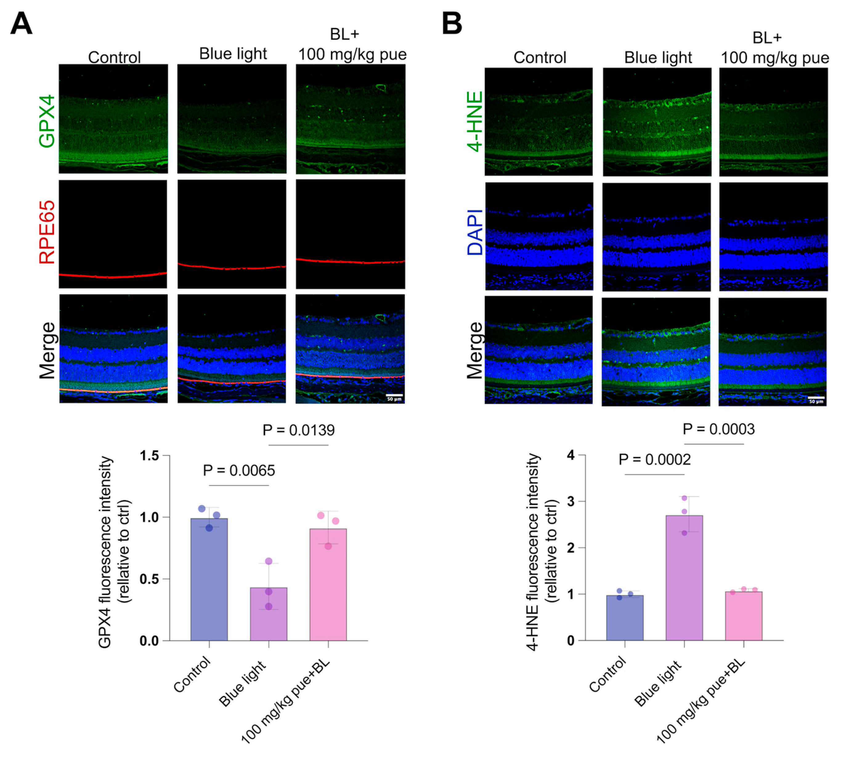
We next determined whether structural and functional protection was accompanied by changes in GPX4, a phospholipid hydroperoxide-detoxifying enzyme, and 4-HNE, a product of lipid peroxidation. Blue light reduced retinal GPX4 fluorescence relative to controls (p = 0.0065), whereas 100 mg/kg puerarin increased GPX4 relative to the model group (p = 0.0139; Figure 2A). Blue light also increased 4-HNE fluorescence across the retina (p = 0.0002 versus control), and puerarin attenuated this increase (p = 0.0003 versus model; Figure 2B). Thus, puerarin-mediated retinal protection was accompanied by preservation of GPX4 and reduction in retinal lipid peroxidation.
Figure 2.
Puerarin attenuates retinal GPX4 loss and 4-HNE accumulation after blue light exposure. Retinal sections were prepared from mice euthanized on day 14. (A) Representative GPX4/RPE65 double immunofluorescence staining and quantification of GPX4 fluorescence in retinal sections. Scale bar, 50 μm. (B) Representative 4-HNE immunofluorescence staining and quantification in retinal sections. Scale bar, 50 μm. Data were analyzed using one-way ANOVA followed by Tukey’s multiple-comparisons test (n = 3 mice per group). Data are presented as mean ± SD; p < 0.05 was considered statistically significant. ONL, outer nuclear layer.
3.3. Puerarin Reduces A2E/Blue Light-Induced Oxidative Injury and Mitochondrial Dysfunction
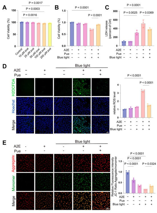
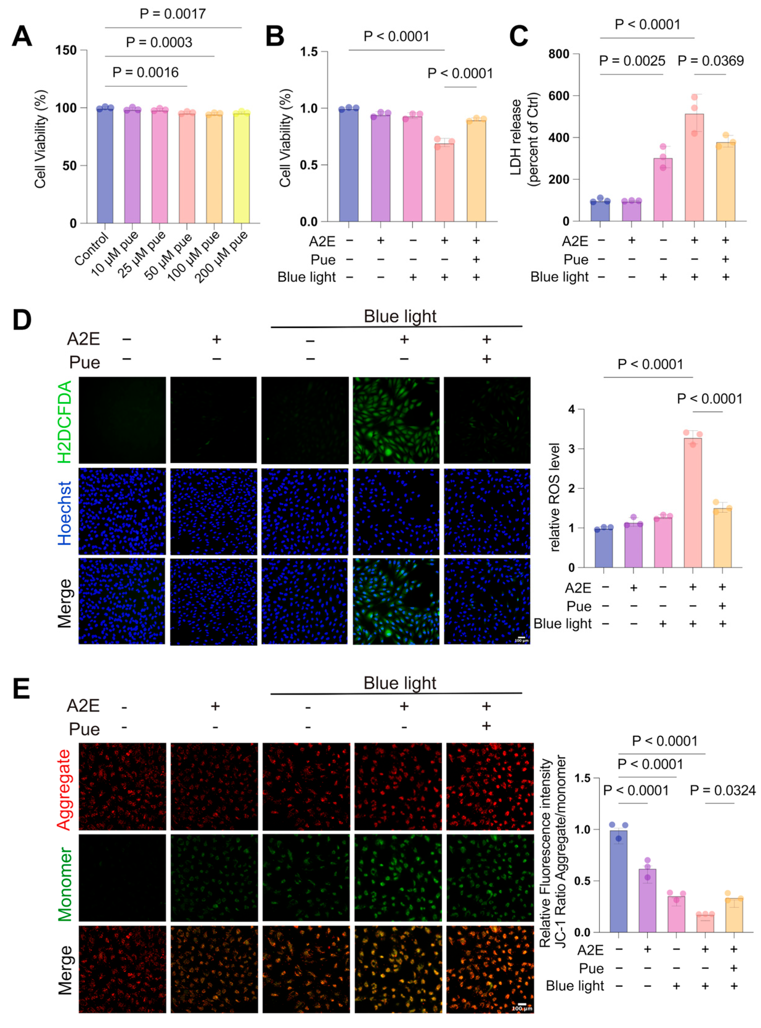
We next used A2E-loaded ARPE-19 cells to examine the cellular response to blue light. Puerarin at 10 or 25 μM did not significantly alter basal CCK-8 metabolic activity, whereas 50–200 μM produced small but significant reductions (Figure 3A). We therefore selected 25 μM, the highest concentration without a detectable reduction in basal CCK-8 signal, for subsequent experiments.
Figure 3.
Puerarin attenuates A2E/blue light-induced injury in ARPE-19 cells. Cells were loaded with A2E (25 μM) for 24 h, pretreated with puerarin (25 μM) for 12 h, and exposed to 430-nm blue light for 30 min. (A) Relative CCK-8 metabolic activity after treatment with puerarin at 10, 25, 50, 100, or 200 μM. (B) Relative CCK-8 metabolic activity after A2E and/or blue light exposure. (C) LDH release. (D) Intracellular ROS detected using CM-H2DCFDA. (E) Mitochondrial membrane potential assessed by the JC-1 red-to-green fluorescence ratio. Data are from three independent biological experiments and are presented as mean ± SD. One-way ANOVA followed by Tukey’s multiple-comparisons test was used; p < 0.05 was considered statistically significant.
Neither A2E nor blue light alone significantly reduced the CCK-8 signal. Combined A2E/blue light exposure markedly reduced metabolic activity (p < 0.0001 versus control), and puerarin significantly attenuated this reduction (p < 0.0001 versus model; Figure 3B). Blue light increased LDH release (p = 0.0025 versus control), and the increase was greater after combined A2E/blue light exposure (p < 0.0001 versus control). Puerarin reduced LDH release relative to the combined model (p = 0.0369; Figure 3C), indicating reduced membrane injury.
Combined A2E/blue light exposure markedly increased intracellular ROS (p < 0.0001 versus control), whereas puerarin suppressed ROS accumulation (p < 0.0001 versus model; Figure 3D). JC-1 staining showed progressive loss of mitochondrial membrane potential after A2E, blue light, or combined A2E/blue light exposure (all p < 0.0001 versus control). Puerarin increased the JC-1 red-to-green fluorescence ratio relative to the combined model (p = 0.0324; Figure 3E). A2E alone reduced mitochondrial membrane potential without significantly changing the CCK-8 signal, LDH release, or ROS, indicating that mitochondrial dysfunction occurred before the other measured indices of cellular injury under these experimental conditions.
3.4. Puerarin Attenuates Ferroptosis-Associated Changes in A2E/Blue Light-Treated ARPE-19 Cells
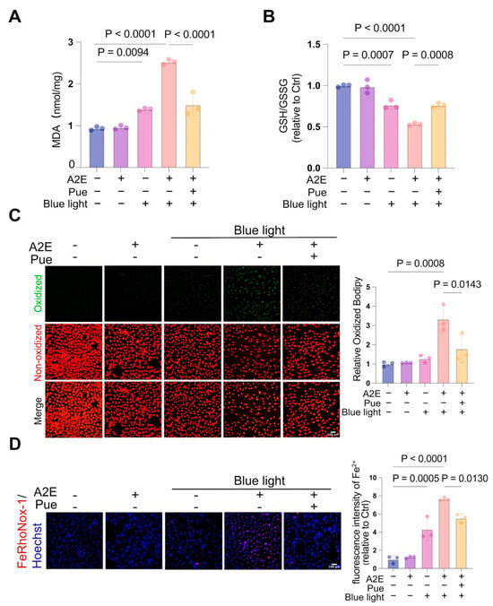
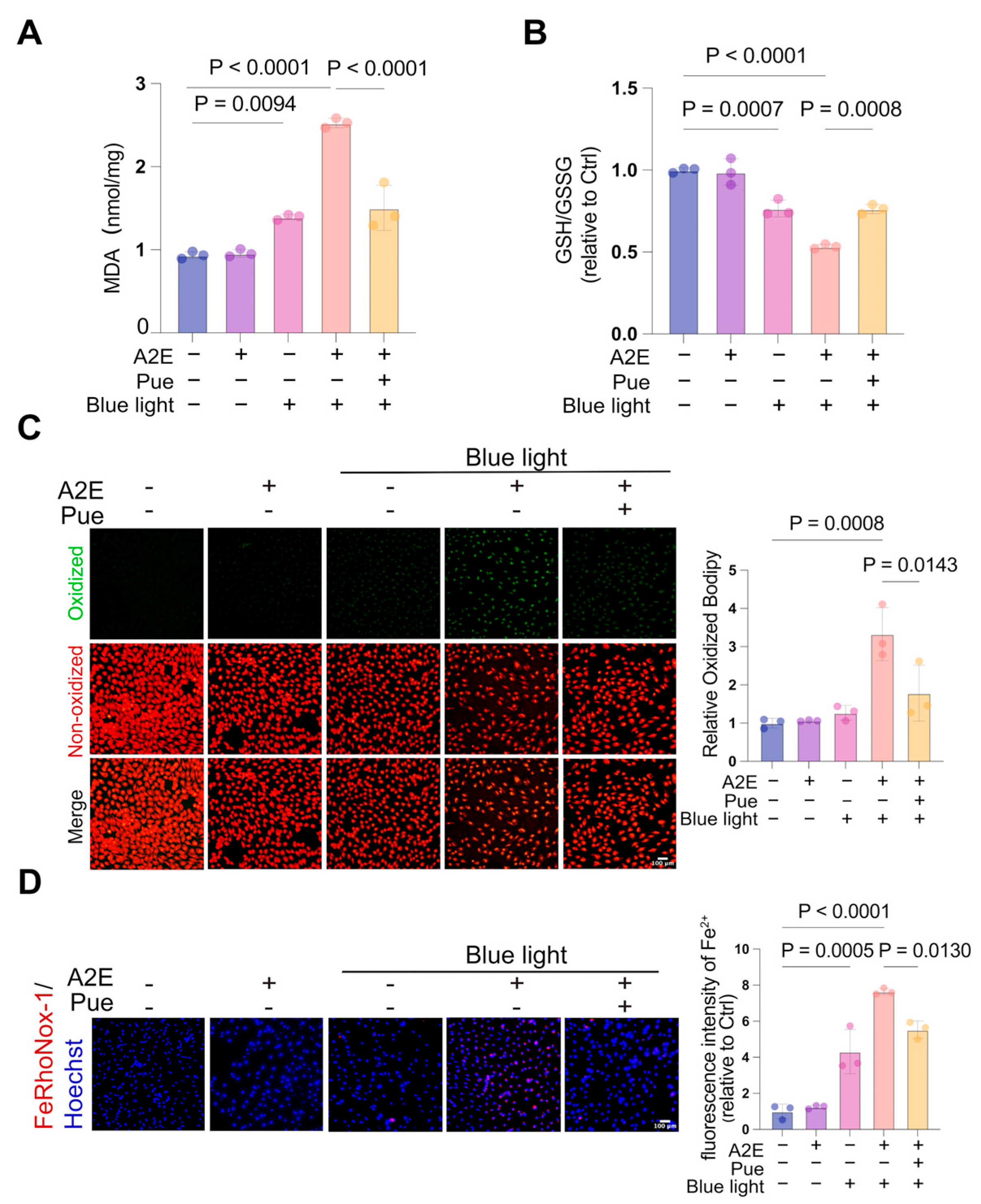
We next assessed iron homeostasis, glutathione redox status, and lipid peroxidation. MDA was increased by blue light alone (p = 0.0094 versus control) and was highest after combined A2E/blue light exposure (p < 0.0001 versus control). Puerarin reduced MDA relative to the combined model (p < 0.0001; Figure 4A). The GSH/GSSG ratio was reduced by blue light alone (p = 0.0007 versus control) and further reduced by A2E/blue light (p < 0.0001 versus control); puerarin partially restored the ratio (p = 0.0008 versus model; Figure 4B).
Figure 4.
Puerarin attenuates ferroptosis-associated changes in A2E/blue light-treated ARPE-19 cells. Cells were loaded with A2E (25 μM) for 24 h, pretreated with puerarin (25 μM) for 12 h, and exposed to 430-nm blue light for 30 min. (A) MDA content. (B) GSH/GSSG ratio. (C) Lipid peroxidation measured using the BODIPY 581/591 C11 oxidized-to-reduced fluorescence ratio. (D) Intracellular Fe2+ detected using FerroOrange. Data are from three independent biological experiments and are presented as mean ± SD. One-way ANOVA followed by Tukey’s multiple-comparisons test was used; p < 0.05 was considered statistically significant.
BODIPY 581/591 C11 imaging showed increased lipid oxidation after combined A2E/blue light exposure (p = 0.0008 versus control), which was attenuated by puerarin (p = 0.0143 versus model; Figure 4C). FerroOrange staining showed that blue light alone increased intracellular Fe2+ (p = 0.0005 versus control), combined A2E/blue light produced the largest increase (p < 0.0001 versus control), and puerarin reduced Fe2+ relative to the combined model (p = 0.0130; Figure 4D). A2E alone did not significantly change these indices. Together, the coordinated changes in ferrous iron, glutathione redox balance, and lipid peroxidation define a ferroptosis-associated oxidative injury phenotype that was attenuated by puerarin.
3.5. Puerarin Restores SLC7A11 and GPX4 Protein Abundance
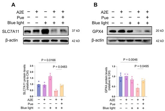
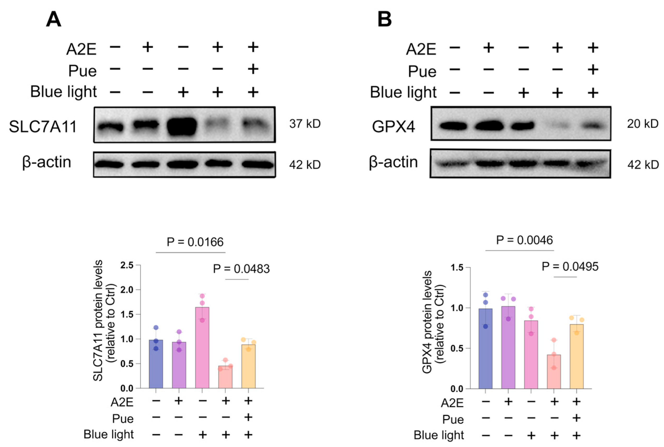
We next examined the SLC7A11-GSH-GPX4 antioxidant defense system. SLC7A11 protein abundance was reduced after combined A2E/blue light exposure relative to controls (p = 0.0166) and increased after puerarin treatment relative to the model group (p = 0.0483; Figure 5A). GPX4 was likewise reduced in the model group (p = 0.0046 versus control) and increased by puerarin (p = 0.0495 versus model; Figure 5B). Blue light alone increased SLC7A11 without a parallel increase in GPX4, consistent with an adaptive response that was insufficient under combined A2E/blue light stress. These results show that puerarin preserves SLC7A11/GPX4-associated antioxidant capacity in this cellular model.
Figure 5.
Puerarin restores SLC7A11 and GPX4 protein abundance in A2E/blue light-treated ARPE-19 cells. Cells were loaded with A2E (25 μM) for 24 h, pretreated with puerarin (25 μM) for 12 h, and exposed to 430-nm blue light for 30 min. (A) Representative immunoblot and quantification of SLC7A11. (B) Representative immunoblot and quantification of GPX4. Beta-actin served as the loading control. Data are from three independent biological experiments and are presented as mean ± SD. One-way ANOVA followed by Dunnett’s multiple-comparisons test against the model group was used; p < 0.05 was considered statistically significant.
3.6. Erastin Attenuates Puerarin-Mediated Cellular Protection
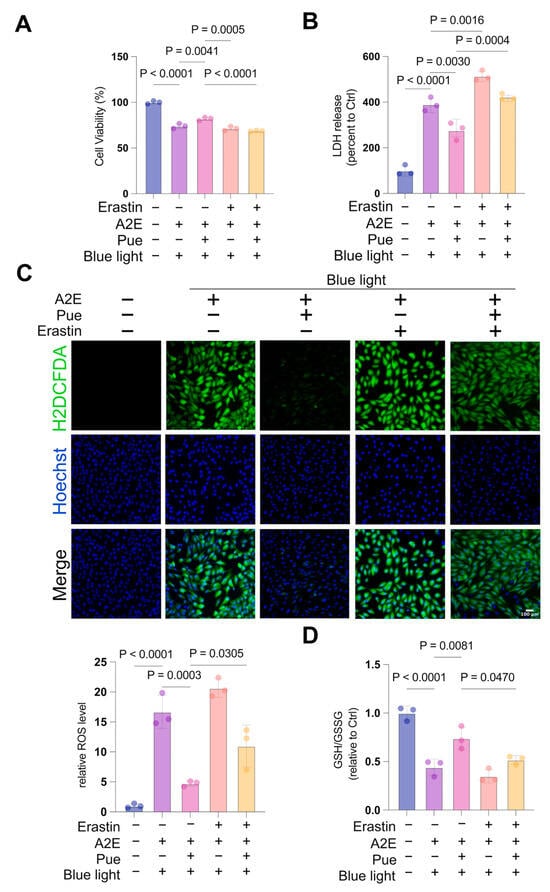
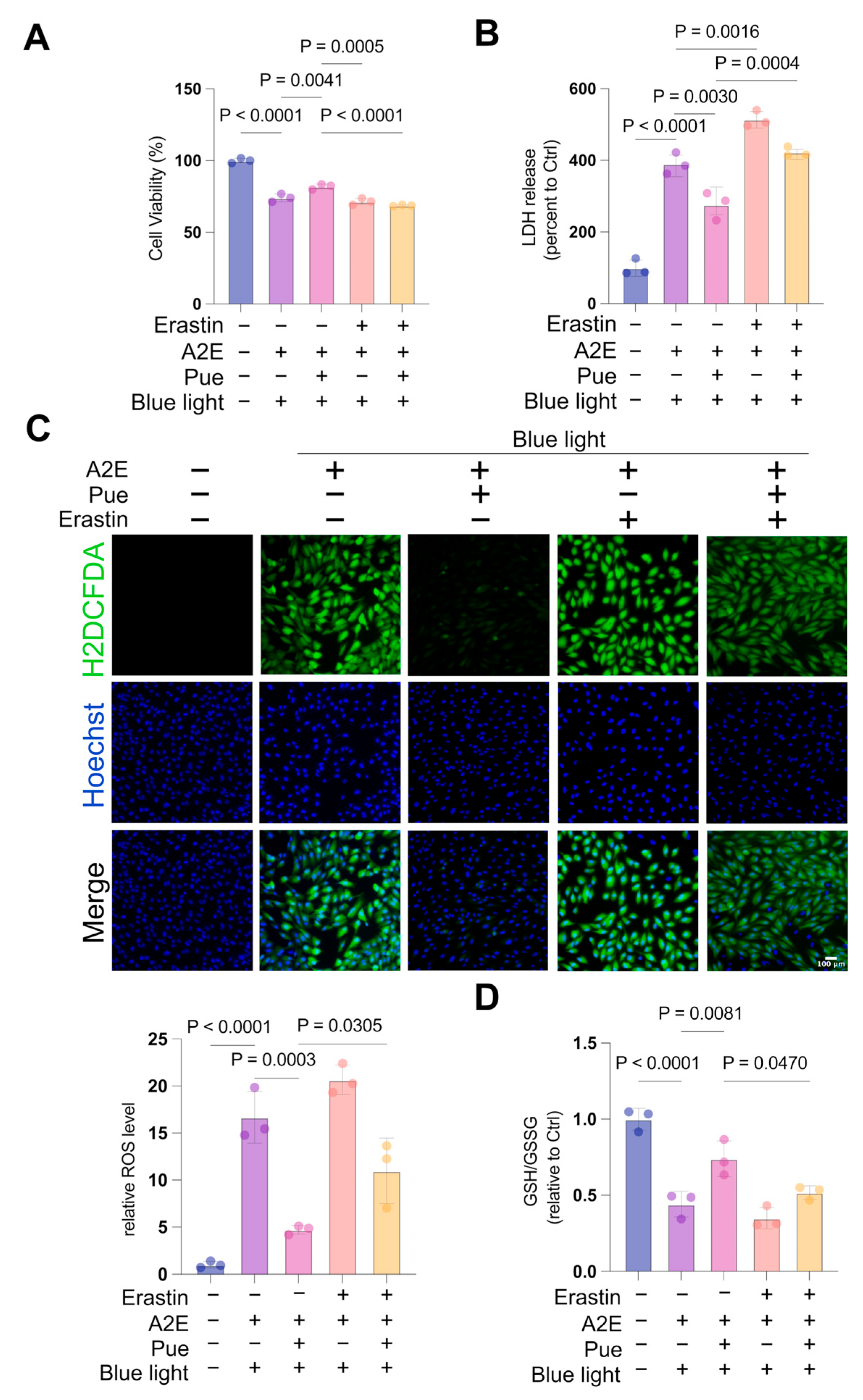
Because puerarin preserved SLC7A11 and GPX4, we tested whether SLC7A11-dependent glutathione defense contributed to its cellular effects. A2E/blue light-exposed ARPE-19 cells were treated with erastin, an inhibitor of SLC7A11-mediated cystine uptake. By limiting intracellular cysteine availability, erastin restricts GSH synthesis and weakens GPX4-dependent protection from phospholipid peroxidation.
The combined A2E/blue light model reduced the CCK-8 metabolic signal relative to controls (p < 0.0001), and puerarin partially restored it (p = 0.0041 versus model). Erastin attenuated the effect of puerarin (p < 0.0001 versus puerarin alone; Figure 6A). LDH release followed the same pattern: puerarin reduced LDH release (p = 0.0030 versus model), whereas erastin increased it relative to puerarin treatment alone (p = 0.0004; Figure 6B).
Figure 6.
Erastin attenuates the cytoprotective effects of puerarin in A2E/blue light-treated ARPE-19 cells. A2E-loaded cells were pretreated with puerarin (25 μM) for 12 h, incubated with erastin (10 μM) for 2 h, and exposed to 430-nm blue light for 30 min. (A) Relative CCK-8 metabolic activity. (B) LDH release. (C) Intracellular ROS detected using CM-H2DCFDA. (D) GSH/GSSG ratio. Data are from three independent biological experiments and are presented as mean ± SD. One-way ANOVA followed by Tukey’s multiple-comparisons test was used; p < 0.05 was considered statistically significant.
Puerarin reduced ROS accumulation in A2E/blue light-treated cells (p = 0.0003 versus model), whereas erastin increased ROS relative to puerarin alone (p = 0.0305; Figure 6C). Puerarin also increased the GSH/GSSG ratio (p = 0.0081 versus model), and erastin attenuated this recovery (p = 0.0470 versus puerarin alone; Figure 6D).
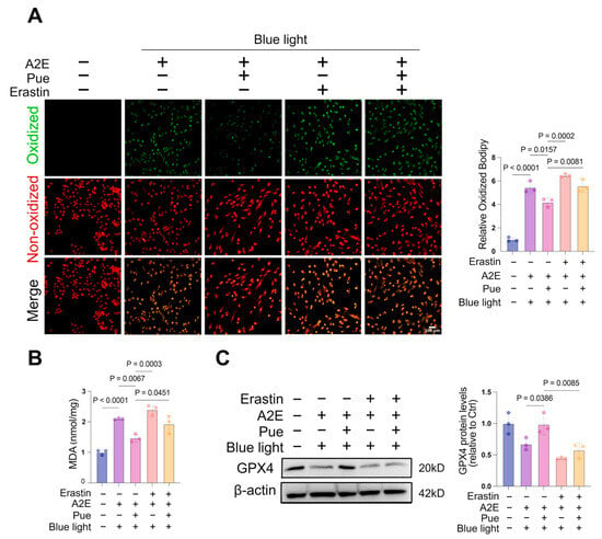
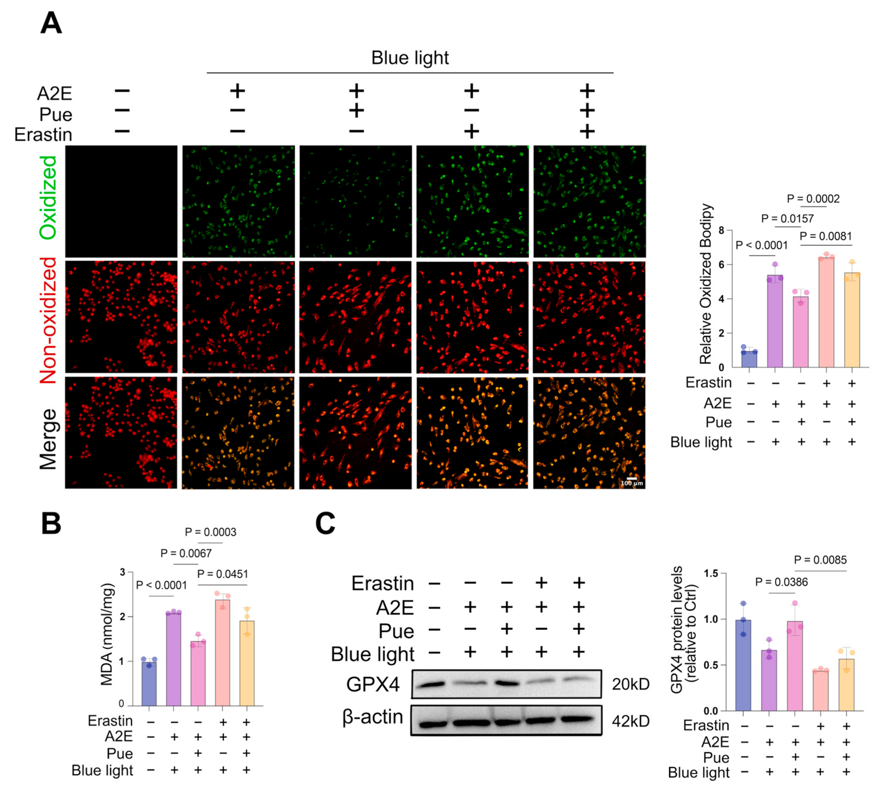
Puerarin reduced the BODIPY oxidized-to-reduced fluorescence ratio and MDA content relative to the model group (p = 0.0157 and p = 0.0067, respectively), whereas erastin attenuated both effects (p = 0.0081 and p = 0.0451 versus puerarin alone, respectively; Figure 7A,B). Puerarin also increased GPX4 protein abundance (p = 0.0386 versus model), while erastin reduced GPX4 relative to puerarin treatment alone (p = 0.0085; Figure 7C).
Figure 7.
Erastin attenuates the anti-lipid-peroxidation effects of puerarin. A2E-loaded ARPE-19 cells were pretreated with puerarin (25 μM) for 12 h, incubated with erastin (10 μM) for 2 h, and exposed to 430-nm blue light for 30 min. (A) Lipid peroxidation measured using the BODIPY 581/591 C11 oxidized-to-reduced fluorescence ratio. (B) MDA content. (C) Representative immunoblot and quantification of GPX4. Beta-actin served as the loading control. Data are from three independent biological experiments and are presented as mean ± SD. Data in (A,B) were analyzed using one-way ANOVA followed by Tukey’s multiple-comparisons test. Data in (C) were analyzed using one-way ANOVA followed by Dunnett’s multiple-comparisons test against the puerarin treatment group. p < 0.05 was considered statistically significant.
Together, these findings show that pharmacological inhibition of SLC7A11-mediated cystine uptake disrupts glutathione redox homeostasis and weakens the cytoprotective and anti-lipid-peroxidation effects of puerarin. They support a functional contribution of SLC7A11-dependent glutathione defense to puerarin-mediated protection in A2E/blue light-treated ARPE-19 cells.
4. Discussion
This study demonstrates that puerarin attenuates acute blue light-induced retinal injury in mice and suppresses a coordinated ferroptosis-associated oxidative injury phenotype in A2E-loaded ARPE-19 cells. Four findings support this conclusion. First, oral puerarin preserved retinal and ONL thickness and improved scotopic ERG responses after blue light exposure. Second, this protection was accompanied by preservation of retinal GPX4 and reduced 4-HNE accumulation. Third, puerarin limited ROS generation, mitochondrial dysfunction, ferrous iron accumulation, glutathione redox imbalance, and lipid peroxidation in A2E-sensitized cells. Fourth, erastin weakened several protective effects, linking puerarin’s cellular activity to SLC7A11-dependent glutathione defense.
Acute blue light exposure produced a reproducible structural and functional retinal injury. The prominent ONL thinning and reduction in a-wave amplitude indicate substantial photoreceptor involvement, while the b-wave change suggests downstream impairment of inner retinal signaling. This pattern is consistent with the high metabolic demand and polyunsaturated lipid content that make photoreceptors vulnerable to photo-oxidative damage [25,26,27]. The agreement among OCT, histology, and ERG strengthens the evidence that oral puerarin provided dose-dependent retinal protection, with the most consistent effect at 100 mg/kg.
The in vivo GPX4 and 4-HNE findings provide a molecular bridge between retinal protection and lipid peroxide control. GPX4 is a principal enzymatic defense against phospholipid hydroperoxides, whereas 4-HNE reflects propagation of lipid peroxidation. Their reciprocal changes after blue light exposure, and normalization by puerarin, indicate that improved lipid peroxide handling accompanied structural and functional preservation. The 4-HNE signal extended through the neural retina rather than being restricted to the RPE; accordingly, these data support a retina-wide photo-oxidative response. Previous protection by ferrostatin-1 in light-induced retinal degeneration further supports the biological relevance of ferroptosis-associated lipid damage in this setting [17].
The A2E-loaded ARPE-19 model addressed a distinct cellular question: how puerarin modifies blue light injury in an A2E-sensitized background. A2E alone reduced mitochondrial membrane potential without altering the other measured injury indices, whereas combined A2E/blue light exposure caused marked ROS accumulation, loss of mitochondrial membrane potential, reduced CCK-8 metabolic activity, and increased LDH release. This sequence is compatible with reports that A2E disrupts lysosomal-mitochondrial signaling and mitochondrial function before overt membrane injury [10,11,12,13,28]. By preserving mitochondrial membrane potential and suppressing ROS, puerarin limited an early amplification step in photo-oxidative injury.
Combined A2E/blue light exposure also produced a coherent ferroptosis-associated signature: increased Fe2+, reduced GSH/GSSG, increased MDA and BODIPY oxidation, and reduced GPX4. Puerarin shifted each of these measures toward control values. The convergence of iron, glutathione, lipid peroxidation, and GPX4 measurements is more informative than any single marker and supports the conclusion that puerarin suppresses ferroptosis-associated oxidative damage in this cellular system [14,15,18,24,29].
SLC7A11 and GPX4 results further define the antioxidant response. Blue light alone increased SLC7A11 without a parallel increase in GPX4, which may reflect a compensatory response to moderate oxidative stress [30]. Under combined A2E/blue light exposure, both SLC7A11 and GPX4 were reduced, and the GSH/GSSG ratio fell, indicating failure of the glutathione-dependent lipid peroxide defense system. Puerarin preserved SLC7A11 and GPX4 abundance and restored glutathione redox balance, providing a mechanistic explanation for its effects on lipid peroxidation.
Erastin provided functional pharmacological support for this interpretation. Inhibition of SLC7A11-mediated cystine uptake reduced the benefits of puerarin on CCK-8 metabolic activity, LDH release, ROS, GSH/GSSG, lipid peroxidation, MDA, and GPX4. These coordinated effects show that intact SLC7A11-dependent glutathione defense contributes materially to puerarin-mediated protection. Because puerarin retained partial activity in the presence of erastin, its antioxidant effect likely also reflects the observed preservation of mitochondrial function and limitation of iron accumulation.
Together, these observations suggest that puerarin engages two complementary antioxidant modalities. On one hand, the phenolic 4′-hydroxyl group of the isoflavone scaffold can directly donate electrons to quench reactive radical species, consistent with the early suppression of intracellular ROS accumulation and preservation of mitochondrial membrane potential [23]. On the other hand, puerarin upregulates the SLC7A11–GSH–GPX4 enzymatic axis, as evidenced by the coordinated recovery of GSH/GSSG, increased SLC7A11 and GPX4 abundance, and reduced downstream lipid peroxidation markers (MDA, BODIPY C11 oxidation, and 4-HNE). The fact that erastin only partially abolished protection indicates that direct radical scavenging and glutathione-dependent enzymatic detoxification operate in parallel rather than being mutually exclusive.
The mouse and cell experiments were used as complementary rather than equivalent disease models. The mouse experiment establishes protection against acute photo-oxidative retinal injury, with the principal structural effect in the ONL. The acute mouse experiment was not intended to reproduce age-associated A2E/lipofuscin accumulation; A2E-specific mechanistic inference derives from the ARPE-19 experiments. This distinction avoids attributing the cellular A2E mechanism to the mouse retina while retaining the cross-model observation that puerarin limits oxidative and lipid-peroxidative injury.
Puerarin also differs mechanistically from macular carotenoids (lutein and zeaxanthin) and β-carotene, which are lipophilic and primarily partition into photoreceptor outer segment membranes to quench singlet oxygen and dissipate excess light energy. Puerarin, by contrast, is a relatively hydrophilic isoflavone C-glycoside that acts in the aqueous cytosolic compartment and additionally upregulates endogenous antioxidant enzyme expression. Notably, puerarin and β-carotene have been reported to act synergistically rather than redundantly, as deprotonated puerarin can regenerate β-carotene from its radical cation and thereby recycle the lipophilic antioxidant within the membrane [23]. This spatial and mechanistic complementarity suggests that puerarin may act adjunctively with macular pigment antioxidants, although head-to-head efficacy comparisons under identical dosing conditions are needed.
UPLC-MS/MS detected puerarin in reconstituted retinal extracts 2 h after oral administration, supporting exposure of retinal samples to the parent compound. Because the result was not normalized to retinal mass and was obtained at a single time point, it represents an analytical detection signal rather than an absolute retinal tissue concentration or a pharmacokinetic profile. It should therefore not be used for quantitative comparison with the 25 μM cell-culture concentration.
In this in vivo study, mice receiving oral puerarin at the highest dose of 100 mg/kg for 7 consecutive days showed no overt gross adverse clinical signs, which is consistent with previous pre-clinical observations in mice receiving repeated oral puerarin at comparable dose ranges [31]. Importantly, dedicated toxicological profiling including serum biochemistry, hematology and organ histopathology was not performed in the present work. By body-surface-area (BSA)-based normalization [32,33], this murine oral dose yields a human equivalent dose of approximately 8.1 mg/kg (~567 mg for a 70 kg adult). This value serves only as a preliminary scaling reference and cannot be interpreted as evidence of human safety or therapeutic efficacy. Inter-species differences in gastrointestinal absorption, hepatic glucuronidation, retinal penetration and drug clearance prevent direct clinical translation of this animal dose. Additionally, the present study was designed to evaluate the protective effects of puerarin against blue-light-induced retinal injury rather than its effects on uninjured retinas; therefore, a puerarin-only group without light insult was not included. Consequently, potential effects of puerarin on uninjured healthy retinas could not be assessed in the present work and warrant dedicated investigation in future studies.
The cellular experiments were designed to interrogate oxidative signaling in proliferative ARPE-19 cells rather than barrier physiology in mature polarized human RPE. Likewise, the mouse and cell exposure paradigms were model-specific and were not intended as a direct radiometric dose comparison; wavelength specificity was outside the scope of this study. Within this defined design, the internally consistent structural, functional, biochemical, and pharmacological results support a retinoprotective antioxidant effect of puerarin and a contribution of ferroptosis-associated pathways in A2E-sensitized ARPE-19 cells.
Overall, puerarin couples preservation of retinal structure and scotopic function in acute photo-oxidative injury with suppression of iron-dependent lipid oxidative stress in A2E-loaded ARPE-19 cells. This integrated antioxidant profile supports puerarin as a candidate for further evaluation in retinal conditions characterized by photo-oxidative stress.
5. Conclusions
Puerarin preserved retinal structure and scotopic function after acute blue light exposure in mice and attenuated ROS generation, mitochondrial dysfunction, ferrous iron accumulation, glutathione redox imbalance, and lipid peroxidation in A2E-loaded ARPE-19 cells. Preservation of SLC7A11 and GPX4, together with attenuation of puerarin’s effects by erastin, supports a functional contribution of SLC7A11-dependent glutathione defense. These results identify puerarin as a candidate antioxidant for photo-oxidative retinal injury while distinguishing the acute mouse model from the A2E-sensitized cellular model.
6. Patents
A patent application related to this study has been filed with the China National Intellectual Property Administration: Puerarin, pharmaceutical composition, method, and its use in the preparation of drugs for treating blue light-induced retinal injury (application no. 202610940942.0; filing date: 27 June 2026).
Supplementary Materials
The following supporting information can be downloaded at: https://www.mdpi.com/article/10.3390/antiox15111483/s1: Figure S1, UPLC-MS/MS analysis of puerarin in mouse retinal extracts. (A) Representative multiple-reaction monitoring chromatogram of a puerarin standard (10 ng/mL); (B) representative chromatogram of the untreated control group; (C) representative chromatogram of the gavage-treated group; and (D) puerarin concentration measured in final reconstituted retinal extracts. Data are presented as mean ± SD.
Author Contributions
Conceptualization, M.Z. and X.L.; methodology, J.Z., Y.S., B.L., Z.L., H.W. and L.Z.; investigation, J.Z., Y.S., B.L., Z.L., H.W., L.H., L.Z., L.S. and Q.L.; formal analysis, B.L., L.S., Q.L. and L.H.; data curation, J.Z., Y.S., B.L. and Z.L.; interpretation of results, C.J.B., J.T.-T., X.L., L.H., L.S., Q.L. and M.Z.; writing—original draft preparation, M.Z. and J.Z.; writing—review and editing, C.J.B., J.T.-T., X.L., L.H., L.S., Q.L. and M.Z.; visualization, J.Z., Y.S. and B.L.; supervision, M.Z. and X.L. All authors have read and agreed to the published version of the manuscript and agree to be accountable for all aspects of the work.
Funding
This research was funded by the National Natural Science Foundation of China (grant no. 82471101, 82671387), Tianjin HealthResearch Project (TJWJ2023QN022), the Natural Science Foundation of Tianjin (grant no. 25JCZDJC00500), the Research Project on Hospital Management Innovation of Tianjin Medical University (grant no. 2026YG94) and the Tianjin Key Medical Discipline Construction Project (grant no. TJYXZDXK-3-004A-2).
Institutional Review Board Statement
The animal study protocol was approved by the Experimental Animal Ethics Committee of Tianjin Medical University Eye Hospital (approval no. TJYY2025120306) and was conducted in accordance with the ARVO Statement for the Use of Animals in Ophthalmic and Vision Research.
Informed Consent Statement
Not applicable.
Data Availability Statement
The data presented in this study are available on request from the corresponding author. The data are not publicly available due to a pending patent application.
Conflicts of Interest
J.T.-T. reports an affiliation with Skyran Biologics Inc. A patent application related to this work has been filed with the China National Intellectual Property Administration (application no. 202610940942.0). The authors declare no other conflicts of interest. The funders had no role in the design of the study; in the collection, analyses, or interpretation of data; in the writing of the manuscript; or in the decision to publish the results.
Abbreviations
The following abbreviations are used in this manuscript:
| A2E | N-retinylidene-N-retinylethanolamine |
| AMD | age-related macular degeneration |
| CCK-8 | cell counting kit-8 |
| DMSO | dimethyl sulfoxide |
| ERG | electroretinography |
| Fe2+ | ferrous iron |
| GPX4 | glutathione peroxidase 4 |
| GSH | reduced glutathione |
| GSSG | oxidized glutathione |
| 4-HNE | 4-hydroxynonenal |
| LDH | lactate dehydrogenase |
| MDA | malondialdehyde |
| OCT | optical coherence tomography |
| ONL | outer nuclear layer |
| PBS | phosphate-buffered saline |
| ROS | reactive oxygen species |
| RPE | retinal pigment epithelium |
| RPE65 | retinal pigment epithelium-specific 65 kDa protein |
| SD | standard deviation |
| SLC7A11 | solute carrier family 7 member 11 |
References
- Mitchell, P.; Liew, G.; Gopinath, B.; Wong, T.Y. Age-related macular degeneration. Lancet 2018, 392, 1147–1159. [Google Scholar] [CrossRef] [Scilit] [PubMed]
- Wong, W.L.; Su, X.; Li, X.; Cheung, C.M.; Klein, R.; Cheng, C.Y.; Wong, T.Y. Global prevalence of age-related macular degeneration and disease burden projection for 2020 and 2040: A systematic review and meta-analysis. Lancet Glob. Health 2014, 2, e106–e116. [Google Scholar] [CrossRef] [Scilit] [PubMed]
- Deng, Y.; Qiao, L.; Du, M.; Qu, C.; Wan, L.; Li, J.; Huang, L. Age-related macular degeneration: Epidemiology, genetics, pathophysiology, diagnosis, and targeted therapy. Genes Dis. 2022, 9, 62–79. [Google Scholar] [CrossRef] [Scilit] [PubMed]
- Bhandari, S.K.; Lee, S.; Kim, H.J. Integrative Landscape of Dry AMD Pathogenesis, Models, and Emerging Therapeutic Strategies. Int. J. Mol. Sci. 2025, 27, 202. [Google Scholar] [CrossRef] [Scilit] [PubMed]
- Heier, J.S.; Lad, E.M.; Holz, F.G.; Rosenfeld, P.J.; Guymer, R.H.; Boyer, D.; Grossi, F.; Baumal, C.R.; Korobelnik, J.F.; Slakter, J.S.; et al. Pegcetacoplan for the treatment of geographic atrophy secondary to age-related macular degeneration (OAKS and DERBY): Two multicentre, randomised, double-masked, sham-controlled, phase 3 trials. Lancet 2023, 402, 1434–1448. [Google Scholar] [CrossRef] [Scilit] [PubMed]
- Khanani, A.M.; Patel, S.S.; Staurenghi, G.; Tadayoni, R.; Danzig, C.J.; Eichenbaum, D.A.; Hsu, J.; Wykoff, C.C.; Heier, J.S.; Lally, D.R.; et al. Efficacy and safety of avacincaptad pegol in patients with geographic atrophy (GATHER2): 12-month results from a randomised, double-masked, phase 3 trial. Lancet 2023, 402, 1449–1458. [Google Scholar] [CrossRef] [Scilit] [PubMed]
- Lakkaraju, A.; Umapathy, A.; Tan, L.X.; Daniele, L.; Philp, N.J.; Boesze-Battaglia, K.; Williams, D.S. The cell biology of the retinal pigment epithelium. Prog. Retin. Eye Res. 2020, 78, 100846. [Google Scholar] [CrossRef] [Scilit] [PubMed]
- Chakravarthy, H.; Georgyev, V.; Wagen, C.; Hosseini, A.; Matsubara, J. Blue light-induced phototoxicity in retinal cells: Implications in age-related macular degeneration. Front. Aging Neurosci. 2024, 16, 1509434. [Google Scholar] [CrossRef] [Scilit] [PubMed]
- Françon, A.; Behar-Cohen, F.; Torriglia, A. The blue light hazard and its use on the evaluation of photochemical risk for domestic lighting. An in vivo study. Environ. Int. 2024, 184, 108471. [Google Scholar] [CrossRef] [Scilit] [PubMed]
- Sparrow, J.R.; Nakanishi, K.; Parish, C.A. The lipofuscin fluorophore A2E mediates blue light-induced damage to retinal pigmented epithelial cells. Investig. Ophthalmol. Vis. Sci. 2000, 41, 1981–1989. [Google Scholar]
- Marie, M.; Bigot, K.; Angebault, C.; Barrau, C.; Gondouin, P.; Pagan, D.; Fouquet, S.; Villette, T.; Sahel, J.A.; Lenaers, G.; et al. Light action spectrum on oxidative stress and mitochondrial damage in A2E-loaded retinal pigment epithelium cells. Cell Death Dis. 2018, 9, 287. [Google Scholar] [CrossRef] [Scilit] [PubMed]
- Alaimo, A.; Liñares, G.G.; Bujjamer, J.M.; Gorojod, R.M.; Alcon, S.P.; Martínez, J.H.; Baldessari, A.; Grecco, H.E.; Kotler, M.L. Toxicity of blue led light and A2E is associated to mitochondrial dynamics impairment in ARPE-19 cells: Implications for age-related macular degeneration. Arch. Toxicol. 2019, 93, 1401–1415. [Google Scholar] [CrossRef] [Scilit] [PubMed]
- Yang, B.; Yang, K.; Chen, Y.; Li, Q.; Chen, J.; Li, S.; Wu, Y. Exposure of A2E to blue light promotes ferroptosis in the retinal pigment epithelium. Cell Mol. Biol. Lett. 2025, 30, 22. [Google Scholar] [CrossRef] [Scilit] [PubMed]
- Dixon, S.J.; Lemberg, K.M.; Lamprecht, M.R.; Skouta, R.; Zaitsev, E.M.; Gleason, C.E.; Patel, D.N.; Bauer, A.J.; Cantley, A.M.; Yang, W.S.; et al. Ferroptosis: An iron-dependent form of nonapoptotic cell death. Cell 2012, 149, 1060–1072. [Google Scholar] [CrossRef] [Scilit] [PubMed]
- Li, J.; Cao, F.; Yin, H.L.; Huang, Z.J.; Lin, Z.T.; Mao, N.; Sun, B.; Wang, G. Ferroptosis: Past, present and future. Cell Death Dis. 2020, 11, 88. [Google Scholar] [CrossRef] [Scilit] [PubMed]
- Totsuka, K.; Ueta, T.; Uchida, T.; Roggia, M.F.; Nakagawa, S.; Vavvas, D.G.; Honjo, M.; Aihara, M. Oxidative stress induces ferroptotic cell death in retinal pigment epithelial cells. Exp. Eye Res. 2019, 181, 316–324. [Google Scholar] [CrossRef] [Scilit] [PubMed]
- Tang, W.; Guo, J.; Liu, W.; Ma, J.; Xu, G. Ferrostatin-1 attenuates ferroptosis and protects the retina against light-induced retinal degeneration. Biochem. Biophys. Res. Commun. 2021, 548, 27–34. [Google Scholar] [CrossRef] [Scilit] [PubMed]
- Koppula, P.; Zhuang, L.; Gan, B. Cystine transporter SLC7A11/xCT in cancer: Ferroptosis, nutrient dependency, and cancer therapy. Protein Cell 2021, 12, 599–620. [Google Scholar] [CrossRef] [Scilit] [PubMed]
- Meng, F.; Guo, B.; Ma, Y.Q.; Li, K.W.; Niu, F.J. Puerarin: A review of its mechanisms of action and clinical studies in ophthalmology. Phytomedicine 2022, 107, 154465. [Google Scholar] [CrossRef] [Scilit] [PubMed]
- Wang, K.; Zhu, X.; Zhang, K.; Yao, Y.; Zhuang, M.; Tan, C.; Zhou, F.; Zhu, L. Puerarin inhibits amyloid β-induced NLRP3 inflammasome activation in retinal pigment epithelial cells via suppressing ROS-dependent oxidative and endoplasmic reticulum stresses. Exp. Cell Res. 2017, 357, 335–340. [Google Scholar] [CrossRef] [Scilit] [PubMed]
- Song, Q.; Zhao, Y.; Li, Q.; Han, X.; Duan, J. Puerarin protects against iron overload-induced retinal injury through regulation of iron-handling proteins. Biomed. Pharmacother. 2020, 122, 109690. [Google Scholar] [CrossRef] [Scilit] [PubMed]
- Song, Q.; Jian, W.; Zhang, Y.; Li, Q.; Zhao, Y.; Liu, R.; Zeng, Y.; Zhang, F.; Duan, J. Puerarin Attenuates Iron Overload-Induced Ferroptosis in Retina through a Nrf2-Mediated Mechanism. Mol. Nutr. Food Res. 2024, 68, e2300123. [Google Scholar] [CrossRef] [Scilit] [PubMed]
- Han, R.-M.; Tian, Y.-X.; Becker, E.M.; Andersen, M.L.; Zhang, J.-P.; Skibsted, L.H. Puerarin and conjugate bases as radical scavengers and antioxidants: Molecular mechanism and synergism with beta-carotene. J. Agric. Food Chem. 2007, 55, 2384–2391. [Google Scholar] [CrossRef] [Scilit] [PubMed]
- Sun, Y.; Zheng, Y.; Wang, C.; Liu, Y. Glutathione depletion induces ferroptosis, autophagy, and premature cell senescence in retinal pigment epithelial cells. Cell Death Dis. 2018, 9, 753. [Google Scholar] [CrossRef] [Scilit] [PubMed]
- Kuse, Y.; Ogawa, K.; Tsuruma, K.; Shimazawa, M.; Hara, H. Damage of photoreceptor-derived cells in culture induced by light emitting diode-derived blue light. Sci. Rep. 2014, 4, 5223. [Google Scholar] [CrossRef] [Scilit] [PubMed]
- Lee, B.L.; Kang, J.H.; Kim, H.M.; Jeong, S.H.; Jang, D.S.; Jang, Y.P.; Choung, S.Y. Polyphenol-enriched Vaccinium uliginosum L. fractions reduce retinal damage induced by blue light in A2E-laden ARPE19 cell cultures and mice. Nutr. Res. 2016, 36, 1402–1414. [Google Scholar] [CrossRef] [Scilit] [PubMed]
- Kim, G.H.; Kim, H.I.; Paik, S.S.; Jung, S.W.; Kang, S.; Kim, I.B. Functional and morphological evaluation of blue light-emitting diode-induced retinal degeneration in mice. Graefe’s Arch. Clin. Exp. Ophthalmol. 2016, 254, 705–716. [Google Scholar] [CrossRef] [Scilit] [PubMed]
- Luo, M.M.; Chen, L.; Wang, S.; Zeng, C.; Li, D.Z.; Bi, Y.; Liu, L.Q.; Cai, S.J. The Effect of A2E on the Uptake and Release of Calcium in the Lysosomes and Mitochondria of Human RPE Cells Exposed to Blue Light. J. Ophthalmol. 2021, 2021, 5586659. [Google Scholar] [CrossRef] [Scilit] [PubMed]
- Gao, M.; Yi, J.; Zhu, J.; Minikes, A.M.; Monian, P.; Thompson, C.B.; Jiang, X. Role of Mitochondria in Ferroptosis. Mol. Cell 2019, 73, 354–363.e353. [Google Scholar] [CrossRef] [Scilit] [PubMed]
- Ji, C.; Wang, Y.; Ju, Y.; Liu, S.; Chen, X.; Wang, J.; Sun, N.; Tang, Z.; Gu, P.; Ji, J. The role of HMOX1-mediated ferroptosis in blue light-induced damage to retinal pigment epithelium. Sci. Rep. 2025, 15, 18949. [Google Scholar] [CrossRef] [Scilit] [PubMed]
- Zeng, M.-S.; Yu, W.-D.; Wang, H.-X.; Xu, P.-P.; Liu, J.-Y. Puerarin reduces impairment of intestinal and adipose immune responses to influenza virus infection in mice. Arch. Virol. 2021, 166, 2387–2397. [Google Scholar] [CrossRef] [Scilit] [PubMed]
- Reagan-Shaw, S.; Nihal, M.; Ahmad, N. Dose translation from animal to human studies revisited. FASEB J. 2008, 22, 659–661. [Google Scholar] [CrossRef] [Scilit] [PubMed]
- Nair, A.B.; Jacob, S. A simple practice guide for dose conversion between animals and human. J. Basic Clin. Pharm. 2016, 7, 27–31. [Google Scholar] [CrossRef] [Scilit] [PubMed]
Disclaimer/Publisher’s Note: The statements, opinions and data contained in all publications are solely those of the individual author(s) and contributor(s) and not of MDPI and/or the editor(s). MDPI and/or the editor(s) disclaim responsibility for any injury to people or property resulting from any ideas, methods, instructions or products referred to in the content. |
© 2026 by the authors. Licensee MDPI, Basel, Switzerland. This article is an open access article distributed under the terms and conditions of the Creative Commons Attribution (CC BY) license.