


Abstract
In a previous study, we isolated a water-soluble heteropolysaccharide (CRWP-S) from the edible lichen Cladina rangiferina (L.) Nyl., which exhibited potent free radical-scavenging capacities. However, its protective efficacy against ultraviolet B (UVB)-induced skin photoaging and the underlying molecular mechanisms remain to be fully elucidated. In this study, we established a UVB-induced photoaging model in BALB/c mice and cell senescence model in HaCaT keratinocytes to evaluate the photoprotective effects and mechanisms of CRWP-S. Microneedle-assisted topical application of CRWP-S effectively attenuated UVB-induced macroscopic photoaging phenotypes, while mitigating dermal collagen degradation and restored NRF2 expression in vivo. CRWP-S treatment enhanced HaCaT cell viability, reduced intracellular reactive oxygen species (ROS) and malondialdehyde (MDA) levels, and restored the activities of superoxide dismutase (SOD) and catalase (CAT). Mechanistically, CRWP-S inhibited senescence-associated β-galactosidase (SA-β-Gal) activity and downregulated the expression of senescence markers p53 and p21. Furthermore, CRWP-S promoted NRF2 nuclear translocation and upregulated downstream heme oxygenase-1 (HO-1) while downregulating KEAP1 expression. Pharmacological inhibition of NRF2 using ML385 largely abolished the ROS-scavenging and cytoprotective effects of CRWP-S. Taken together, these findings demonstrate that CRWP-S protects against UVB-induced skin photoaging via activation of the KEAP1–NRF2 signaling pathway, highlighting its strong potential as a functional bioactive ingredient for anti-photoaging formulations.
1. Introduction
Skin photoaging represents the predominant extrinsic skin aging process driven primarily by chronic solar ultraviolet (UV) exposure. Clinically, it manifests as deep wrinkles, laxity, hyperpigmentation, and dermal elastosis [1], which may progress to precancerous lesions such as actinic keratosis in severe cases [2,3]. As the primary physiological barrier, the skin is exceptionally vulnerable to UV radiation, with cumulative exposure accounting for up to 80% of extrinsic aging [4]. Wavelength-dependent UV radiation comprises UVA (320–400 nm), UVB (290–320 nm), and UVC (200–290 nm), with each waveband exhibiting distinct penetration depths and biological impacts [5]. UVC is completely absorbed by the stratospheric ozone layer. UVA penetrates deeply into the dermis, where it generates abundant ROS mainly through endogenous photosensitizers, leading to increased MMP expression and cellular senescence and ultimately to the degradation and denaturation of collagen and elastic fibers [6,7]. Despite its smaller proportion in terrestrial UV radiation, the biological impact of UVB on human skin is generally far greater than that of UVA because its higher-energy photons elicit more severe biological perturbations in epidermal cells [8,9]. Consequently, counteracting UVB-induced damage remains a vital strategy for skin health maintenance and functional cosmeceutical development.
The molecular pathogenesis of UVB-induced photoaging primarily involves direct DNA damage and indirect oxidative stress driven by the excessive accumulation of reactive oxygen species (ROS) [10]. UVB is largely absorbed by the epidermis, where its high-energy photons directly cause DNA damage by forming photoproducts such as cyclobutane pyrimidine dimers (CPDs). Simultaneously, UVB promotes intracellular ROS production through NADPH oxidase (NOX) activation and disruption of the mitochondrial electron transport chain. Excessive ROS production induces persistent oxidative stress, catalyzing lipid peroxidation, protein oxidation, and DNA damage in keratinocytes [11,12,13]. Furthermore, ROS act as critical second messengers driving stress-induced premature senescence (SIPS) and the secretion of senescence-associated secretory phenotype (SASP) factors [14,15,16]. In the epidermis, this cascade impairs keratinocyte proliferation and compromises barrier integrity; in the dermis, it triggers downstream signaling that promotes extracellular matrix (ECM) degradation by fibroblasts through the paracrine manner [17].
To counteract ROS-driven oxidative damage, cells rely on endogenous antioxidant defense systems, among which the KEAP1–NRF2 pathway serves as the master regulator [18]. Under basal conditions, nuclear factor erythroid 2-related factor 2 (NRF2) is bound by Kelch-like ECH-associated protein 1 (KEAP1) in the cytoplasm and targeted for degradation. Upon oxidative stress, NRF2 dissociates from KEAP1 or KEAP1 undergoes degradation, allowing NRF2 to translocate into the nucleus and upregulate downstream phase II detoxifying enzymes, including heme oxygenase-1 (HO-1), superoxide dismutase (SOD), and catalase (CAT) [19,20]. Therefore, reinforcing the KEAP1–NRF2 antioxidant defense represents a promising strategy for preventing and ameliorating skin photoaging.
Natural polysaccharides are widely recognized for cutaneous photoprotection because of their high biocompatibility, low immunogenicity, and broad pharmacological activities. Cladina rangiferina (L.) Nyl., commonly termed reindeer lichen, is an alpine species with remarkable resilience to environmental extremes and centuries of traditional medicinal use [21]. While previous phytochemical studies predominantly focused on small secondary metabolites such as lichen acids, polysaccharides actually account for more than 50% of the thallus dry weigh [22]. In our previous work, we prepared a water-soluble heteropolysaccharide fraction (CRWP-S) containing both alpha- and beta-anomeric configurations. Due to its high density of active hydroxyl groups, CRWP-S effectively scavenged reactive oxygen species and restored endogenous antioxidant enzyme activities in cells under oxidative challenge. Nevertheless, its therapeutic potential against UVB-induced skin photoaging and the underlying molecular targets remain unexplored. Therefore, the present study investigated the photoprotective effects of CRWP-S using both in vitro (HaCaT cells) and in vivo (BALB/c mice) models, with a specific focus on its regulation of the KEAP1-NRF2 signaling pathway.
2. Materials and Methods
2.1. Preparation and Formulation of CRWP-S
Cladina rangiferina (L.) Nyl. was collected from the Qilian Mountains (Qinghai Province, China) and authenticated by Professor L. Mei (No. 20120703). The water-soluble polysaccharide fraction, CRWP-S, was prepared following our established protocol [21].
Briefly, the dried thalli powder was refluxed with 95% ethanol for 12 h to remove lipophilic impurities, followed by hot-water extraction (90 °C, 1:20 w/v, 3 times). The concentrated extract was precipitated with 80% (v/v) ethanol at 4 °C overnight. The resulting precipitate was redissolved in deionized water, deproteinized using the Sevag method, and subjected to repeated freeze–thaw cycles to yield refined CRWP-S. For experimental use, the CRWP-S powder was reconstituted in PBS (Servicebio, Wuhan, China), sterilized through a 0.22 μm membrane filter, and stored at −20 °C in the dark.
The chemical identity and structural characteristics of CRWP-S were thoroughly established in our previous work [21]. Briefly, CRWP-S is a heteropolysaccharide fraction (molecular weight: 15–200 kDa; total carbohydrate content: 92.41%; protein content: 6.13%) predominantly composed of glucose, mannose, and galactose at a molar ratio of 11.23:1.27:1.00, featuring typical pyranose ring configurations with both α- and β-glycosidic linkages. The CRWP-S sample evaluated in the present study was from the exact same batch characterized previously.
2.2. Establishment of the UVB-Induced Skin Photoaging Mouse Model
All animal procedures were approved by the Animal Care and Use Committee of Jilin University (Approval No. 2025376) and performed in strict accordance with the Guide for the Care and Use of Laboratory Animals and the ARRIVE guidelines. Female BALB/c mice (7 weeks old, 23 ± 2 g) were purchased from Changchun Yisi Experimental Animal Technology Co., Ltd. (License No. SCXK 2025-0001) and were acclimated for one week under standard laboratory conditions: a temperature-controlled environment (22 ± 2 °C) with a 12 h light/dark cycle and ad libitum access to food and water before formal modeling.
Following acclimation, mice were randomly divided into four groups (n = 6): (1) Control group; (2) UVB model group; (3) UVB + Low-dose CRWP-S (0.05 mg/mL); and (4) UVB + High-dose CRWP-S (0.1 mg/mL). The sample size of n = 6 per group was determined based on the effect size of epidermal thickness difference from preliminary experiments, calculated via power analysis (α = 0.05, power = 0.80) [23]. One day before the first irradiation, the dorsal hair was removed with an electric clipper and a gentle depilatory cream; only animals with no visible erythema or damage 24 h later entered the study.
UVB was delivered using a narrow-band UVB lamp (YIXIAN, G15T8E, peak 313 nm). Prior to the formal experiment, mice were randomly divided into four groups (n = 3/group) and irradiated at 50, 100, 200, and 300 mJ·cm−2 to explore the MED. The standard definition of MED is the minimal UVB dose that induces visible erythema. After 24 h, dorsal erythema was visually assessed by two independent observers. 200 mJ·cm−2 was determined as 1 MED.
Microneedle treatment was applied uniformly to all experimental groups including the blank control group and the UVB model group. The 4-week induction protocol used escalating doses: 1 MED (first week), 1.5 MED (second week), 2 MED (third week), and 4 MED (fourth week), three times per week [24]. Thirty minutes after each UVB session, 200 μL of CRWP-S solution was applied to the dorsal area and massaged gently with a microneedle roller (needle length 200 μm) in perpendicular directions (horizontally and vertically) to promote transdermal penetration.
Mice were euthanized 24 h after the final UVB exposure. Dorsal skin tissues were immediately harvested for subsequent analyses.
2.3. Assessment of Skin Hydration
The skin hydration levels of the dorsal area were measured with a skin capacitance meter (Corneometer® CM 825, Courage + Khazaka Electronic GmbH, Cologne, Germany). All measurements were performed on live animals under controlled ambient conditions (22 ± 2 °C, 50 ± 10% humidity). At 24 h after the final UVB irradiation, skin hydration level was measured at marked dorsal regions of the mice. For each mouse, four fixed areas were selected on the dorsal skin. The final value for each mouse was the average of these four areas and presented as the raw device output in arbitrary units (a.u., range 0–120).
2.4. Histopathological Analysis and Immunofluorescence Analysis
2.4.1. Tissue Fixation and Embedding
The dorsal skin tissues were fixed in 4% paraformaldehyde (Servicebio, Wuhan, China) (pH 7.4, 4 °C, 48 h). Following fixation, the specimens were dehydrated through a graded ethanol series, cleared in xylene, and embedded in paraffin wax. Serial sections (5 μm thick) were cut on a rotary microtome (Leica Biosystems, Nussloch, Germany) and mounted onto poly-L-lysine-coated glass slides. Before any staining procedure, all paraffin-embedded sections were deparaffinized in xylene (20 min) and rehydrated through a descending ethanol series (100%, 95%, 80%, and 70% for 3 min each).
2.4.2. Hematoxylin and Eosin (H&E) Staining
Following deparaffinization and rehydration as described in Section 2.4.1, the sections were rinsed with distilled water and stained with Harris hematoxylin (Sigma-Aldrich, St. Louis, MO, USA) for 5 min, rinsed, and counterstained with 0.5% Eosin Y in 95% ethanol (Sigma-Aldrich) for 3 min. Images were captured using an optical microscope.
2.4.3. Masson’s Trichrome Staining
Masson’s trichrome staining was performed on the pretreated sections using a commercial kit (Solarbio, Beijing, China) according to the manufacturer’s instructions. Briefly, sections were mordanted in Bouin’s fixative (56 °C, 1 h), stained with Weigert’s iron hematoxylin (10 min), followed by Biebrich scarlet-acid fuchsin (10 min) and aniline blue (5 min), differentiated in 1% acetic acid, dehydrated, cleared, and mounted.
2.4.4. Picrosirius Red Staining
Following deparaffinization and rehydration, the sections were incubated with Picrosirius Red staining solution (Wuhan Boerfu Biotechnology, Wuhan, China) for 1 h at room temperature. The excess dye was removed by washing twice with 0.5% glacial acetic acid (5 s per wash) to minimize background interference. The sections were then dehydrated through a graded ethanol series (70%, 80%, 90%, 95%, and 100% for 30 s each), cleared in xylene (Sinopharm Chemical Reagent, Shanghai, China), and mounted with neutral resin (Wuhan Boerfu Biotechnology, Wuhan, China). The stained sections were examined under a polarized light microscope (Olympus BX53 equipped with a polarizing attachment). ImageJ (version 1.54r) was used for threshold segmentation to identify orange-red (mature type I collagen) areas and total tissue areas. CVF was calculated as collagen-positive area/total tissue area × 100%.
2.4.5. Immunofluorescence Staining for NRF2
Following deparaffinization and rehydration as described in Section 2.4.1, the sections were subjected to microwave antigen retrieval in sodium citrate buffer (pH 6.0). After blocking with 3% BSA, sections were incubated overnight at 4 °C with anti-NRF2 antibody (GB113808, Servicebio, Wuhan, China). Following washing, sections were incubated with CY3-conjugated secondary antibody in the dark for 1 h. Nuclei were counterstained with DAPI (Beyotime, Shanghai, China). Images were captured using a fluorescence microscope.
All histological sections were evaluated by two independent observers. The observers were blinded to the group assignments.
2.5. Cell Culture
HaCaT cells were cultured in Dulbecco’s Modified Eagle Medium (DMEM) (Servicebio, Wuhan, China) supplemented with 10% FBS (Procell, Wuhan, China) and 1% penicillin-streptomycin at 37 °C in an incubator of 5% CO2. Cells were passaged at 80–90% confluence, and only cultures in the logarithmic growth phase with high viability were used.
2.6. Establishment of the UVB-Induced Cell Senescence Model
HaCaT cells were seeded into culture plates and allowed to adhere for 24 h. Prior to experiments, cells were subjected to gradient doses of UVB to determine the condition by evaluating senescence markers (SA-β-gal and p53/p21). The dose of 340 mJ·cm−2 was selected for subsequent experiments because it stably induced cellular senescence while preserving 70% cell viability.
Before irradiation, the culture medium was replaced with an equal volume of sterile PBS. Cells were exposed to irradiation at a distance of 10 cm for 10 min, delivering a total radiant dose of 340 mJ·cm−2. After irradiation, the PBS was removed and replaced with either complete medium or medium containing the specified concentrations of CRWP-S. The cells were then incubated for an additional 24 h [20].
2.7. Cell Viability and Cytotoxicity Assay
Cell viability was quantified using the Cell Counting Kit-8 (CCK-8) assay (Beyotime, Shanghai, China) to evaluate both the potential cytotoxicity of CRWP-S and its protective effect against UVB-induced damage. HaCaT cells were seeded into 96-well plates (5 × 10³ cells/well) and left to attach for 24 h. For cytotoxicity testing, we incubated the cells with a range of CRWP-S concentrations for 24 h. For photoprotection experiments, the culture medium was replaced with PBS, cells were exposed to 340 mJ·cm−2 UVB, and then we added 100 μL of CRWP-S at the indicated concentrations and incubated for another 24 h. CCK-8 reagent (10 μL/well) was added, and after 1 h at 37 °C, absorbance was read at 450 nm.
2.8. Cell Apoptosis Analysis
Apoptosis was detected using an Annexin V-FITC/PI Apoptosis Detection Kit (Absin, Shanghai, China). HaCaT cells were seeded in 6-well plates (1 × 10⁴ cells/well) and allowed to adhere. After replacing the medium with 1 mL of sterile PBS, cells were exposed to UVB irradiation. Following exposure, cells were incubated with specified doses of CRWP-S for 24 h. The cells were then harvested using EDTA-free trypsin, washed, and resuspended. According to the manufacturer’s instructions, the cells were sequentially stained with 5 μL of Annexin V-FITC and 5 μL of Propidium Iodide (PI) in the dark.
2.9. Senescence-Associated β-Galactosidase (SA-β-gal) Staining
The activity of SA-β-gal was assessed using a commercial staining kit (Beyotime, Shanghai, China) to evaluate cellular senescence. Following UVB exposure and subsequent 24 h treatment with CRWP-S in 6-well plates, the culture medium was aspirated. The cells were washed with PBS and fixed with 1 mL of β-galactosidase staining fixative for 15 min at room temperature. After additional washing, each well was incubated overnight at 37 °C with 1 mL of a freshly prepared staining working solution (comprising staining solutions A, B, C, and X-Gal in a ratio of 10:10:930:50, μL). The percentage of SA-β-gal-positive cells was quantified using ImageJ software as the ratio of blue-stained cells to total cells.
2.10. Intracellular ROS Measurement
Intracellular ROS levels were determined using the 2′,7′-dichlorodihydrofluorescein diacetate (DCFH-DA) fluorescent probe (Biosharp, Hefei, China). HaCaT cells were seeded in 6-well plates at a density of 1 × 10⁵ cells per well. Following the specified UVB irradiation and CRWP-S treatments, the culture medium was replaced with 1 mL of DCFH-DA solution (diluted 1:1000 in serum-free medium to a final concentration of 10 μM). The cells were incubated in the dark at 37 °C for 20 min and subsequently washed three times with sterile PBS to remove extracellular probes. The fluorescence intensity was visualized and captured using an inverted fluorescence microscope (IX71, Olympus, Tokyo, Japan).
2.11. Biochemical Assays
The activities of superoxide dismutase (SOD) and catalase (CAT), along with the content of malondialdehyde (MDA), were measured in cell lysates using commercial kits strictly according to the manufacturer’s instructions (MLBIO, Shanghai, China). The results are expressed as U/mg protein for SOD and CAT and nmol/mg protein for MDA.
2.12. Immunofluorescence Staining for NRF2 Nuclear Translocation
HaCaT cells were cultured on coverslips in 24-well plates and subjected to the designated experimental treatments. Upon termination of the culture, the cells were fixed with 4% paraformaldehyde for 15 min and permeabilized with 0.1% Triton X-100 for 10 min. After blocking with 3% bovine serum albumin (BSA) for 1 h to minimize non-specific binding, the cells were incubated overnight at 4 °C with a rabbit anti-NRF2 primary antibody (80593-1-RR, Proteintech). Subsequently, the cells were incubated with a CY3-conjugated goat anti-rabbit secondary antibody (Proteintech, Wuhan, China) in the dark for 1 h. The coverslips were mounted using an anti-fluorescence quenching mounting medium containing DAPI (Beyotime, Shanghai, China) for nuclear counterstaining. Fluorescence images captured using fluorescence microscopes (Axio Imager Z2, Zeiss, Germany; IX71, Olympus, Japan).
2.13. NRF2 Inhibition Treatment
To confirm whether the photoprotective effect of CRWP-S is mediated via the KEAP1–NRF2 pathway, the specific NRF2 inhibitor ML385 (MCE, Monmouth Junction, NJ, USA) was employed. HaCaT cells were seeded in 6-well plates and exposed to UVB irradiation upon reaching 80% confluence, as described in Section 2.6. Post-irradiation, cells were divided into four groups: (1) Control; (2) UVB model; (3) UVB + CRWP-S (400 μg/mL); and (4) UVB + CRWP-S (400 μg/mL) + ML385 (10 μM). CRWP-S at 400 μg/mL was selected because it exerted the most pronounced protective effect. After a 24 h incubation, cells were harvested. Intracellular ROS levels were quantified by flow cytometry, while antioxidant marker activities (SOD, CAT, and MDA), NRF2 nuclear translocation, and NRF2 pathway-related protein expressions (NRF2, KEAP1, and HO-1) were evaluated according to the respective assays detailed below.
2.14. Western Blot Analysis
HaCaT cells were seeded in 6-well plates at a density of 1 × 10⁵ cells per well and allowed to adhere. Following UVB irradiation and subsequent 24 h treatment with specified concentrations of CRWP-S, cells were washed three times with ice-cold PBS. Total proteins were extracted using RIPA lysis buffer supplemented with protease and phosphatase inhibitors on ice (Beyotime, Shanghai, China). Protein concentrations were quantified using a BCA protein assay kit (Beyotime, Shanghai, China) according to the manufacturer’s instructions. Equal amounts of protein samples were separated by SDS-PAGE and subsequently transferred onto PVDF membranes (Millipore, MA, USA). The membranes were blocked with protein-free blocking buffer for 1 h at room temperature with gentle agitation. The membranes were then incubated overnight at 4 °C with the following primary antibodies: p53 (40060F, Abmart, Shanghai, China), p21 (10355-1-AP, Proteintech, Wuhan, China), NRF2 (80593-1-RR, Proteintech, Wuhan, China), KEAP1 (80744-1-RR, Proteintech, Wuhan, China), HO-1 (10701-1-AP, Proteintech, Wuhan, China), and β-actin (66009-1-Ig, Proteintech, Wuhan, China). After washing three times with TBST, the membranes were incubated with HRP-conjugated secondary antibodies (SA00001-2, Proteintech, Wuhan, China) for 2 h at room temperature. Western blot bands were quantified using ImageJ and normalized to β-actin.
2.15. Statistical Analysis
All cell-based experiments were performed with three fully independent biological replicates, and each sample was tested in 3–6 technical replicate wells. Data are presented as the mean ± standard deviation (Mean ± SD). Statistical analyses were conducted using GraphPad Prism 8 software. Prior to group comparisons, data were subjected to normality and homogeneity of variance tests. For multiple-group comparisons, one-way analysis of variance (ANOVA) was employed, followed by Tukey’s post hoc test.
3. Results
3.1. CRWP-S Ameliorates UVB-Induced Skin Photoaging and Improves Skin Hydration in Mice
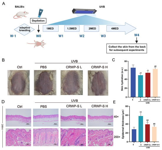
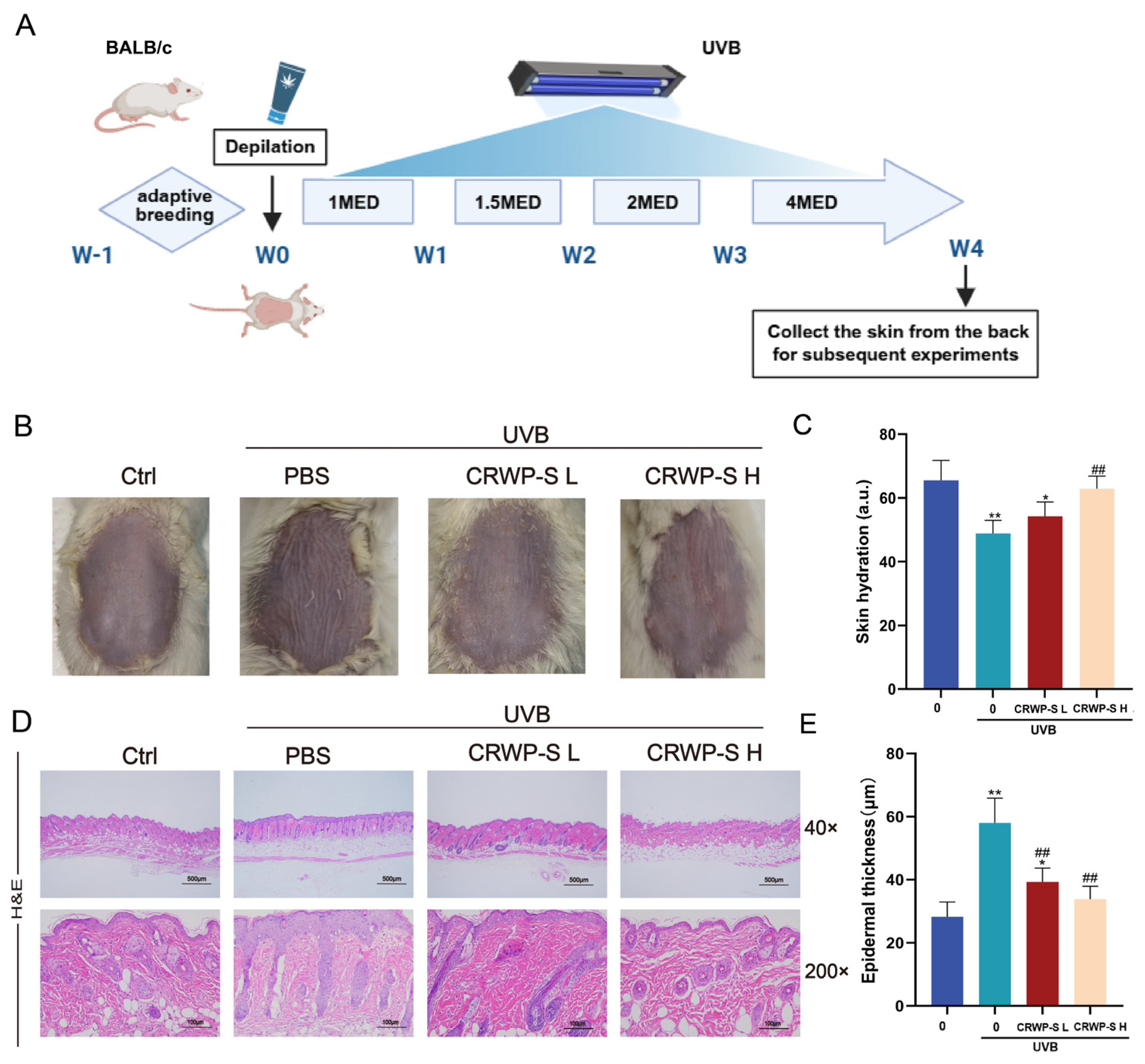
Initially, we evaluated the protective effect of CRWP-S against photoaging in BALB/c mice. To bypass the stratum corneum barrier limiting the absorption of high molecular weight, a microneedle roller was utilized to facilitate transdermal penetration. The schematic diagram of the in vivo experimental design is illustrated in Figure 1A. Under UVB irradiation, the dorsal skin of model mice displayed characteristic macroscopic features of photoaging: visible wrinkles, roughness, dryness, and patches of desquamation potentially reflecting epidermal barrier impairment (Figure 1B). Topical CRWP-S treatment attenuated these changes with more pronounced effects observed at the higher dose.
Figure 1.
Protective effects of CRWP-S on UVB-induced skin photoaging and skin hydration levels in mice. (A) Schematic illustration of the experimental design and treatment protocol. (B) Representative macroscopic images of dorsal skin after 4 weeks of UVB irradiation, showing changes in wrinkles and skin texture. (C) Dorsal skin hydration levels measured by a corneometer. (D) Representative images of H&E-stained transverse sections of dorsal skin. (E) Quantitative analysis of epidermal thickness based on H&E staining. Data are presented as mean ± SD (n = 6). ns, not significant; * p < 0.05, ** p < 0.01 vs. Control group; ## p < 0.01 vs. UVB-irradiated model group. Ctrl, the control group; PBS, the model group treated with PBS; CRWP-S L, low-dose CRWP-S (0.05 mg/mL); CRWP-S H, high-dose CRWP-S (0.1 mg/mL).
Dorsal skin hydration was assessed using a corneometer. In the control group (Figure 1C), the skin hydration level was maintained at 65.48 ± 6.24 a.u. Upon UVB irradiation, the hydration was markedly reduced to 48.88 ± 4.06 a.u. (p < 0.05), indicating a decline in hydration levels. The low-dose group showed significant improvement (p < 0.05), although hydration levels remained below baseline. In the high-dose CRWP-S group, skin hydration recovered to 59.85 ± 3.98 a.u. (p < 0.01), showing no significant difference compared to the control group (p > 0.05). These results demonstrated that high-dose CRWP-S effectively restores skin hydration.
Hematoxylin and eosin staining (H&E) was performed to evaluate the impact of CRWP-S on skin architecture (Figure 1D). The model group exhibited marked epidermal hyperplasia. The epidermal thickness increased from 28.24 ± 4.63 μm in the control group to 57.97 ± 7.84 μm following UVB irradiation, with particularly pronounced hyperplasia of the stratum spinosum (Figure 1E). This was accompanied by cellular disorganization, loss of polarity, and nuclear abnormalities (pyknosis, hyperchromasia, and fragmentation). An indistinct dermal-epidermal junction was also observed, suggesting liquefactive degeneration of the basal layer. In the dermis, collagen fibers appeared fragmented, stained weakly, and lost their normal compact organization. CRWP-S treatment mitigated these pathological changes. Specifically, the epidermal thickness decreased significantly (Figure 1E). The layered architecture of the epidermis was restored, and the dermal-epidermal junction became sharply defined again. In the high-dose group, epidermal thickness averaged 33.77 ± 4.11 μm, approaching the control value.
3.2. CRWP-S Mitigates UVB-Induced Dermal Collagen Degradation and Structural Disorganization
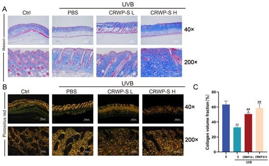
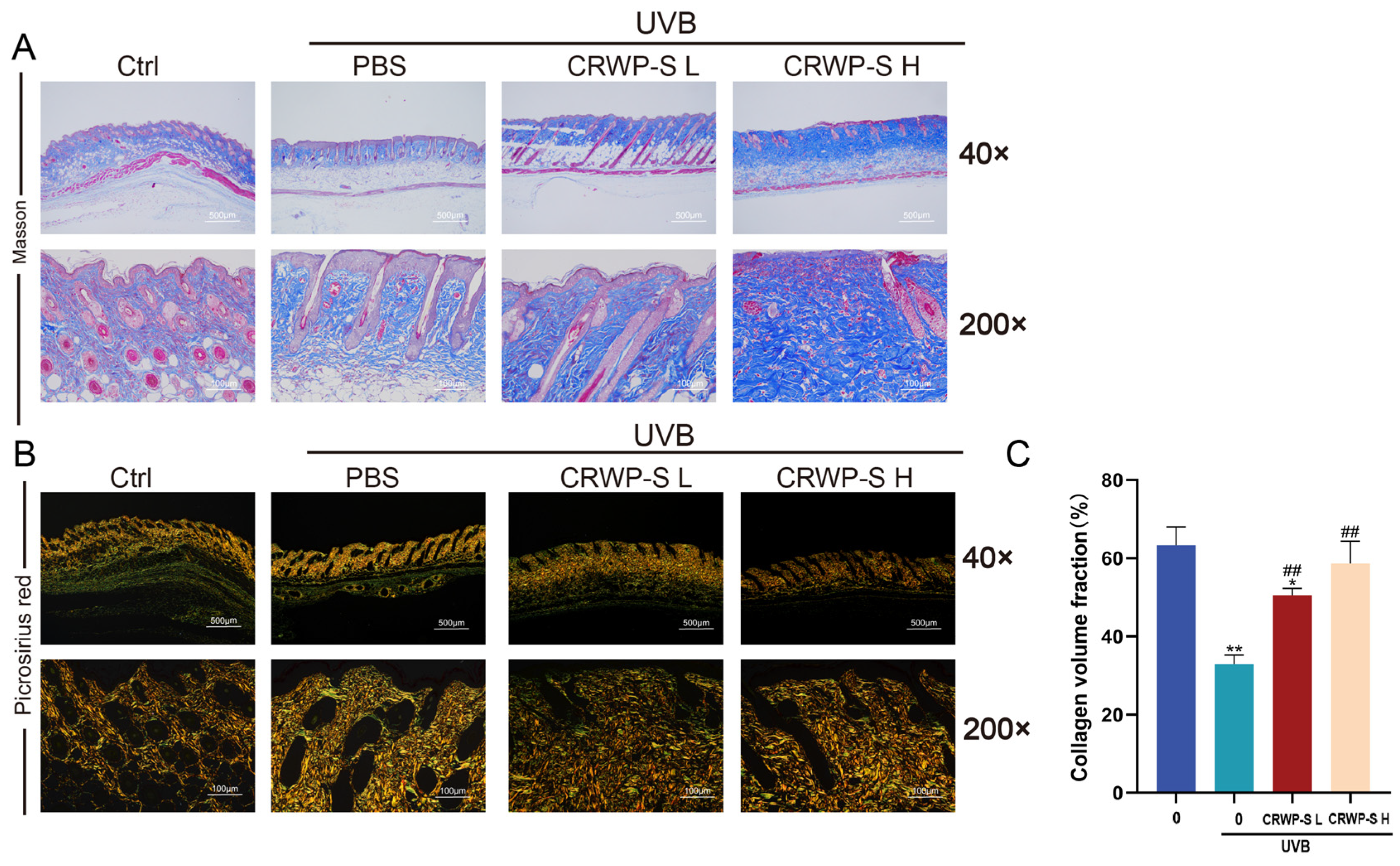
Masson’s trichrome staining revealed dense and uniformly woven collagen bundles in the control group. The papillary dermis was characterized by tightly packed collagen bundles forming a compact meshwork with clearly defined bundle margins (Figure 2A). UVB exposure severely compromised this structural integrity, as evidenced by faded blue staining as well as fragmented and disorganized fiber bundles. CRWP-S treatment effectively mitigated these pathological alterations, restoring the bundled morphology and regular alignment of fibers, particularly in the high-dose group.
Figure 2.
Effects of CRWP-S on dermal collagen and structure in UVB-irradiated mice. (A) Representative images of Masson’s trichrome staining. Collagen fibers are stained blue, while muscle and cytoplasm are stained red. (B) Representative images of Picrosirius red staining observed under polarized light microscopy. Orange-red birefringence indicates mature Type I collagen, while greenish birefringence indicates immature Type III collagen. (C) Quantitative analysis of the Collagen Volume Fraction (CVF). Data are presented as mean ± SD (n = 6). * p < 0.05, ** p < 0.01 vs. Control group; ## p < 0.01, vs. UVB-irradiated model group. Ctrl, the control group; PBS, the model group treated with PBS; CRWP-S L, low-dose CRWP-S (0.05 mg/mL); CRWP-S H, high-dose CRWP-S (0.1 mg/mL).
To further evaluate collagen deposition and maturity, Picrosirius red staining was analyzed under polarized light microscopy (Figure 2B,C). Dermis from the untreated control group exhibited intense orange-red birefringence, a classic signature of tightly packed, mature Type I collagen, with a collagen volume fraction (CVF) of 63.33 ± 2.71%. Following UVB irradiation, CVF in the model group dropped to 32.84 ± 1.03%. CRWP-S treatment effectively alleviated these alterations and restored the total CVF to 57.35 ± 4.39% (p < 0.05). These histological metrics demonstrate that topically applied CRWP-S successfully counteracts the morphological alterations associated with skin photoaging.
3.3. CRWP-S Modulates NRF2 Expression in UVB-Induced Photoaging Mouse Skin
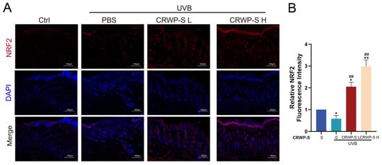
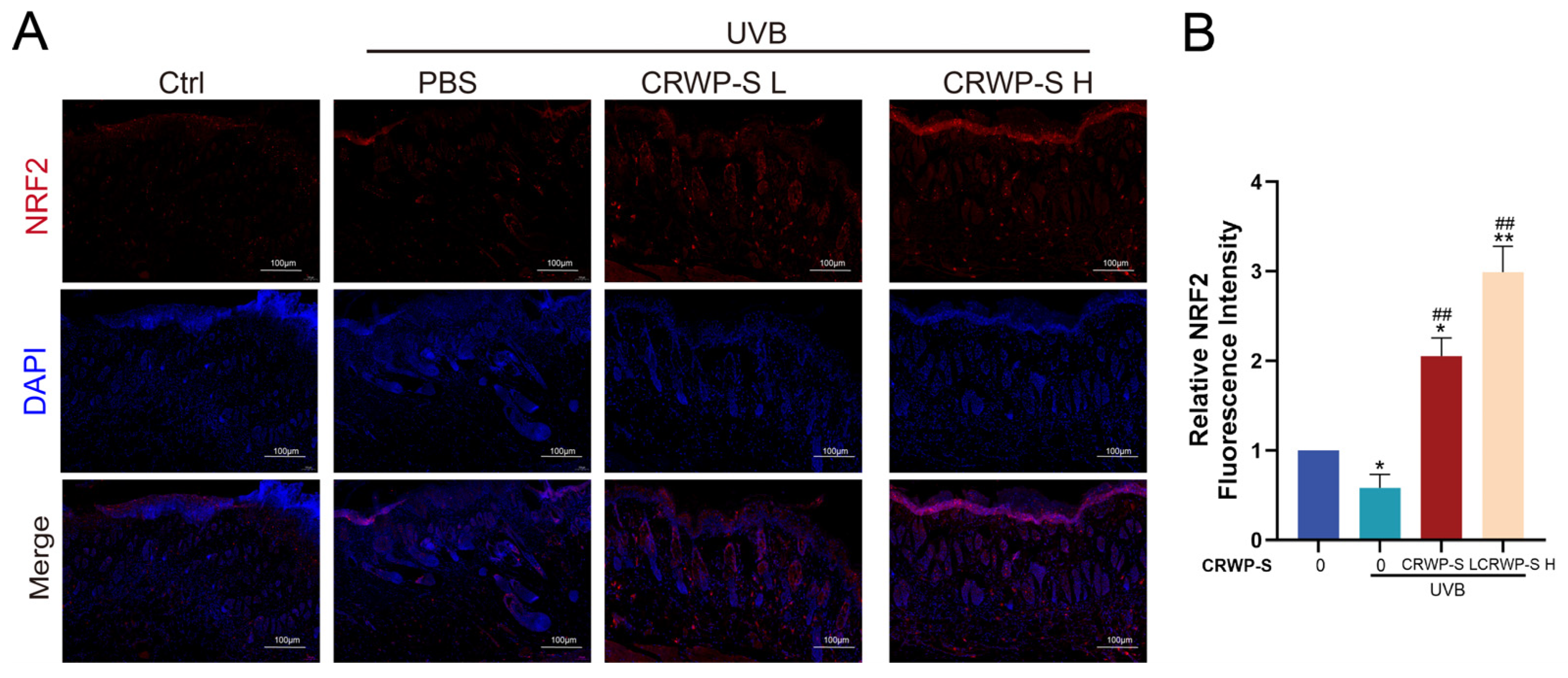
To explore the potential mechanisms underlying the photoprotective effect of CRWP-S in vivo, we evaluated NRF2 expression and spatial distribution in mouse dorsal skin sections via immunofluorescence staining (Figure 3). Control skin displayed baseline NRF2 fluorescence (Figure 3B), whereas UVB irradiation led to a significant reduction in fluorescence intensity (p < 0.05). Notably, CRWP-S intervention effectively upregulated the fluorescence intensity level of NRF2. Furthermore, merged images revealed an increased overlap of NRF2 (red) with DAPI-stained nuclei (blue) in the high-dose CRWP-S group, suggesting enhanced nuclear localized signals (Figure 3A). The overlapping signals were predominantly observed in the thin epidermal layer, which is mainly enriched with keratinocytes. These results indicate that CRWP-S can promote the expression and activation of NRF2 in vivo.
Figure 3.
Effect of CRWP-S on NRF2 expression in UVB-irradiated mouse skin. (A) Representative immunofluorescence images of NRF2 in mouse skin tissues of each group. (B) Quantitative analysis of relative fluorescence intensity of NRF2. * p < 0.05, ** p < 0.01 vs. Control group; ## p < 0.01 vs. UVB-irradiated model group. Ctrl, the control group; PBS, the model group treated with PBS; CRWP-S L, low-dose CRWP-S (0.05 mg/mL); CRWP-S H, high-dose CRWP-S (0.1 mg/mL).
3.4. CRWP-S Can Alleviate UVB-Induced Cell Viability Decline and Apoptosis Increase
Based on the protective effect of CRWP-S against UVB-induced skin photoaging in vivo, an in vitro model was established to investigate the underlying mechanisms. Since UVB radiation (290–320 nm) primarily targets epidermal keratinocytes as the primary biological barrier, HaCaT cells were employed for these mechanistic studies.
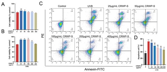
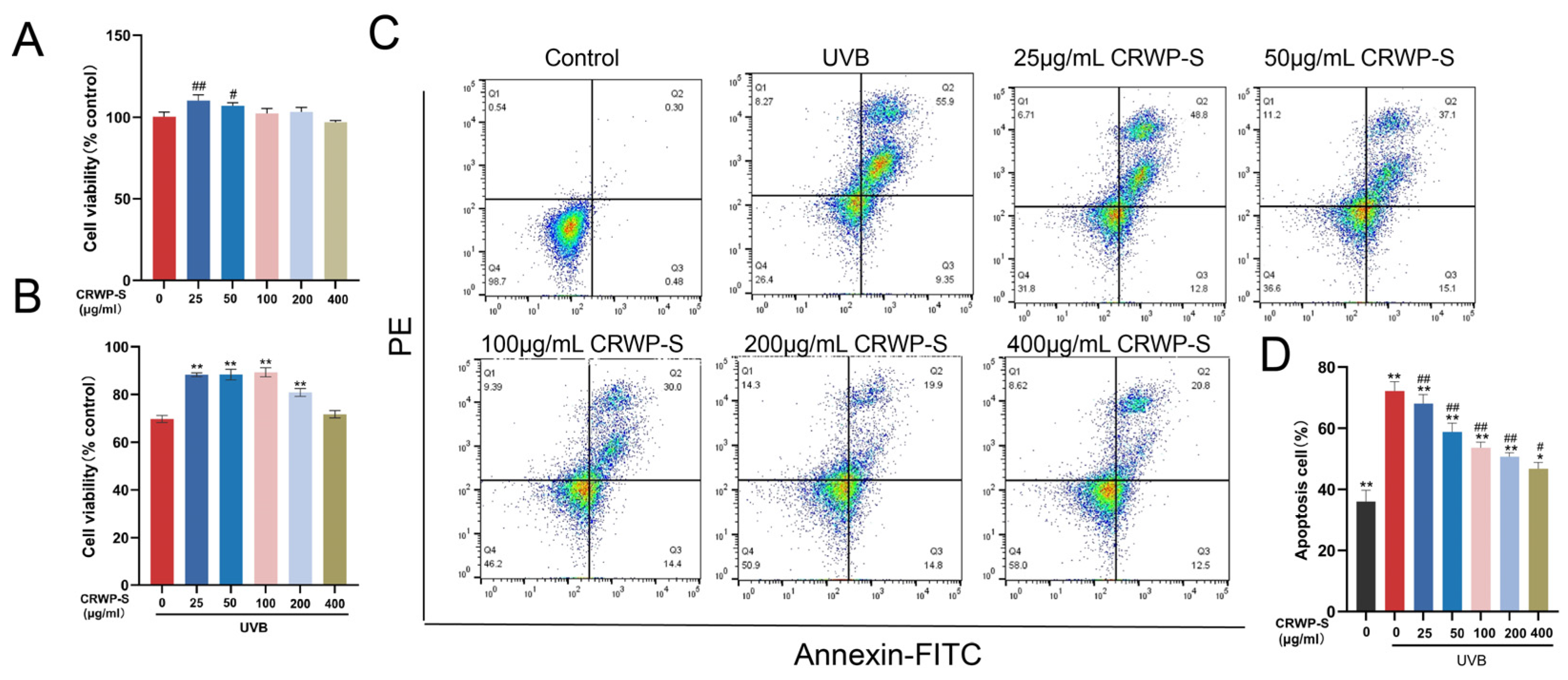
The potential cytotoxicity of CRWP-S was first evaluated using the CCK-8 assay. As illustrated in Figure 4A, CRWP-S treatment for 24 h at concentrations of 25 and 50 μg/mL promoted cell viability compared to the untreated control, suggesting a potential proliferative effect. At higher concentrations ranging from 100 to 400 μg/mL, cell viability remained stable with no significant deviation from the control group (p > 0.05). Consequently, CRWP-S was determined to be non-toxic within the range of 25–400 μg/mL, and these concentrations were selected for subsequent experiments.
Figure 4.
Effects of CRWP-S on UVB-induced cytotoxicity and apoptosis. (A) Cell viability of HaCaT cells treated with various concentrations of CRWP-S (25–400 μg/mL) for 24 h. (B) Cell viability of UVB-irradiated HaCaT cells treated with various concentrations of CRWP-S (25–400 μg/mL). (C) Representative flow cytometry scatter plots of Annexin V-FITC/PI double staining. The pseudocolor indicates cell density. (D) Quantitative analysis of the apoptotic rate. Results are presented as mean ± SD. ns, not significant; * p < 0.05, ** p < 0.01 vs. the control group; # p < 0.05, ## p < 0.01 vs. UVB-irradiated model group.
To evaluate the photoprotective capacity of CRWP-S, a UVB-induced injury model was established. Treatment with CRWP-S (25–400 μg/mL) significantly rescued HaCaT cells from UVB-induced cytotoxicity. Furthermore, higher doses led to a more pronounced recovery of cell viability (Figure 4B).
To further characterize this protective effect, apoptosis was quantified via Annexin V-FITC/PI double staining and flow cytometry (Figure 4C,D). The control group exhibited a negligible proportion of apoptotic cells, with an overall apoptotic rate of approximately 4.82 ± 1.20%. However, UVB irradiation induced a marked increase in both early and late apoptosis, with the total apoptotic rate rising to 68.18 ± 3.15%. CRWP-S treatment significantly attenuated this pro-apoptotic effect, leading to a dose-dependent reduction in the apoptotic rate. These findings demonstrate that CRWP-S effectively preserves cell survival and mitigates apoptosis in keratinocytes under UVB stress.
3.5. CRWP-S Attenuates UVB-Induced Cellular Senescence in HaCaT Cells
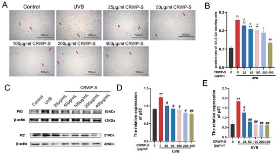
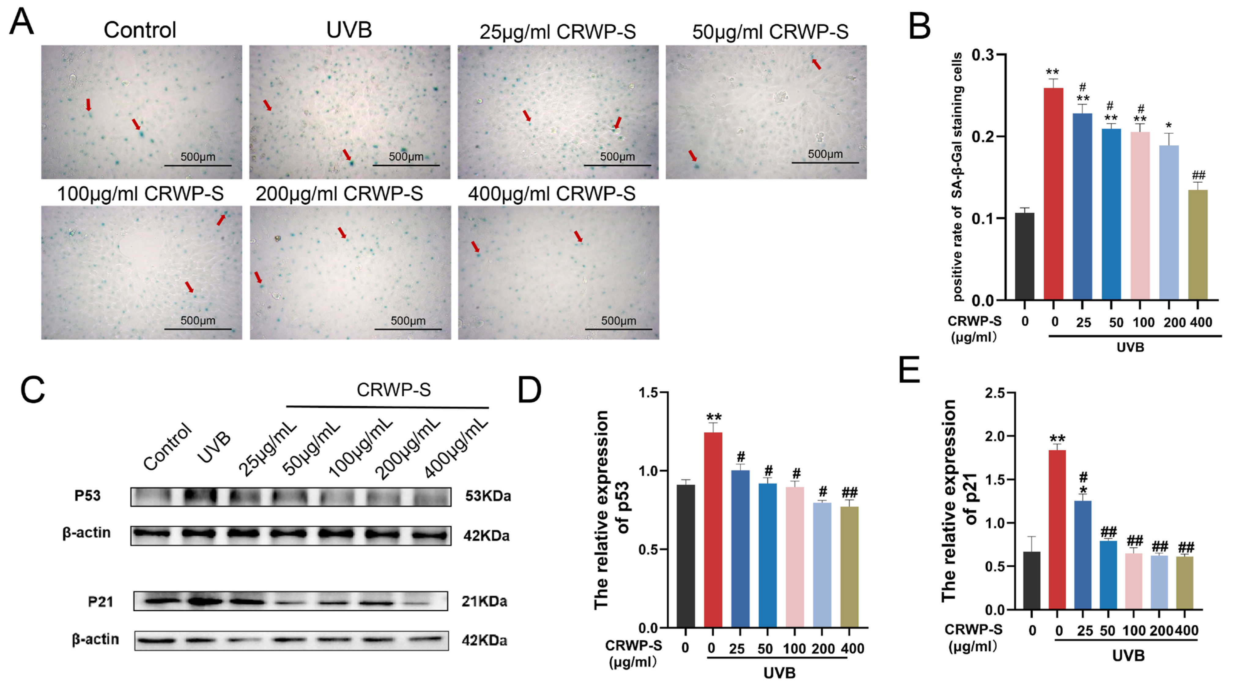
UVB-induced genotoxic stress often triggers stress-induced premature senescence (SIPS), a state of irreversible cell cycle arrest that impairs skin regeneration. We first evaluated the anti-senescence efficacy of CRWP-S using the SA-β-gal assay (Figure 5A,B). While the control group exhibited a minimal baseline of SA-β-gal-positive cells, UVB irradiation led to a significant accumulation of blue-stained senescent cells (p < 0.05), confirming the successful establishment of the cellular senescence model. CRWP-S treatment reduced the proportion of SA-β-gal positive cells.
Figure 5.
Effects of CRWP-S on UVB-induced cellular senescence in HaCaT cells. (A) Representative images of SA-β-gal staining. Arrows indicate SA-β-gal-positive (senescent) cells. (B) Quantitative analysis of SA-β-gal-positive cells. (C) Representative Western blot bands of p53 and p21 protein expression, with β-actin as the loading control. (D) Quantitative analysis of p53 expression. (E) Quantitative analysis of p21 expression. Results are presented as mean ± SD. ns, not significant; * p < 0.05, ** p < 0.01 vs. the Control group; # p < 0.05, ## p < 0.01 vs. the UVB-irradiated model group.
To further validate this anti-senescent effect at the molecular level, we examined the expression of senescence-related markers, specifically p53 and its downstream effector p21. These proteins are critical mediators of cell cycle arrest in response to DNA damage. Western blot analysis revealed that UVB irradiation markedly upregulated the protein levels of both p53 and p21 (Figure 5C–E). However, CRWP-S treatment effectively suppressed their expression, which was consistent with the observed reduction in SA-β-gal staining. These results demonstrate that CRWP-S effectively prevents keratinocytes from entering a senescent state following UVB exposure.
3.6. CRWP-S Is Associated with the Attenuation of Oxidative Stress and the Modulation of the KEAP1/NRF2 Signaling Pathway in HaCaT Cells
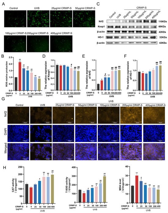
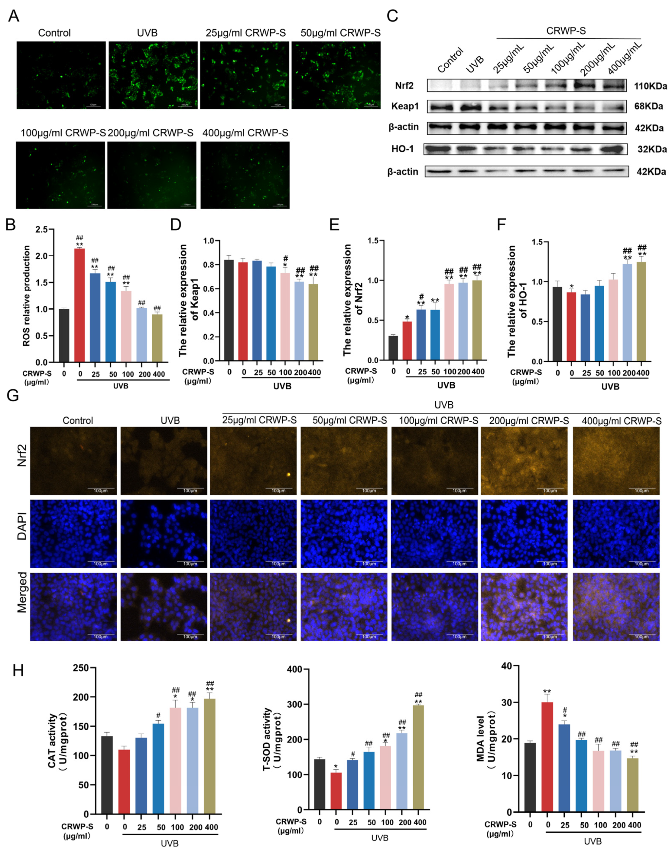
Oxidative stress is a primary driver of cellular senescence and apoptosis in photoaged skin. To evaluate the antioxidant capacity of CRWP-S, intracellular reactive oxygen species (ROS) levels were first monitored using the DCFH-DA fluorescent probe. As shown in Figure 6A, UVB irradiation triggered a marked increase in ROS levels in HaCaT cells (2.1-fold compared to the untreated cells), whereas CRWP-S treatment significantly reduced the fluorescence intensity, indicating potent ROS-scavenging activity (Figure 6B).
Figure 6.
Effects of CRWP-S on oxidative stress and the NRF2 signaling pathway in UVB-irradiated HaCaT cells. (A) Representative fluorescence images of intracellular ROS detected by DCFH-DA staining. (B) Quantitative analysis of ROS immunofluorescence intensity. (C–F) Representative Western blot bands and quantitative analysis of NRF2, HO-1, and KEAP1 protein expression. β-actin was used as a loading control. (G) Representative immunofluorescence images showing the subcellular localization of NRF2 (yellow fluorescence); nuclei were counterstained with DAPI (blue). (H) Quantitative analysis of SOD activity, CAT activity, and MDA levels. Results are presented as mean ± SD from at least three independent experiments. ns, not significant; * p < 0.05, ** p < 0.01 vs. the Control group; # p < 0.05, ## p < 0.01 vs. the UVB-irradiated model group.
To further explore whether CRWP-S modulates NRF2-associated signaling in vitro, Western blot analysis was performed to detect the expression of KEAP1, NRF2, and HO-1 (Figure 6C–F). While UVB exposure induced a compensatory increase in NRF2, HO-1 expression was significantly inhibited (p < 0.05), and KEAP1 expression remained largely unchanged. However, CRWP-S treatment downregulated KEAP1 protein expression, accompanied by an upregulation of NRF2 and HO-1 protein expression. These findings were further supported by immunofluorescence staining (Figure 6G). In the resting state, NRF2 was largely sequestered in the cytoplasm, whereas CRWP-S treatment facilitated its nuclear translocation compared with the UVB model group.
Additionally, we assessed the activities of key endogenous antioxidant enzymes and the extent of lipid peroxidation (Figure 6H). UVB exposure led to a significant decline in SOD and CAT activities alongside a marked elevation in MDA levels. However, CRWP-S treatment effectively reversed these changes. Taken together, these results suggest that CRWP-S can activate NRF2 to reinforce the cellular antioxidant defense, thereby effectively neutralizing UVB-induced oxidative stress.
3.7. Pharmacological Inhibition of NRF2 Function Partially Abolishes the Anti-Photoaging Effects of CRWP-S
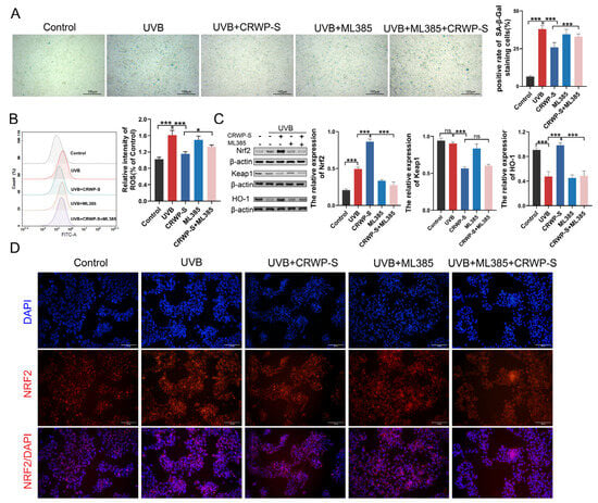
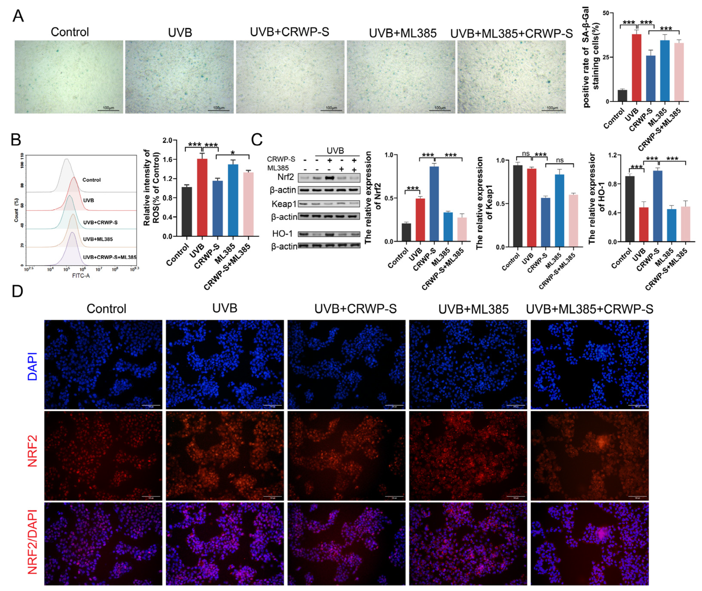
To confirm whether the protective effects of CRWP-S against photoaging and oxidative stress are directly mediated by the NRF2 pathway, HaCaT cells were co-treated with the specific NRF2 inhibitor ML385 (10 μM) during UVB exposure and CRWP-S (400 μg/mL) intervention. The results demonstrated that inhibition of NRF2 by ML385 resulted in a high percentage of SA-β-gal–positive cells comparable to that observed in the UVB model group (Figure 7A), confirming that NRF2 plays a pivotal role in defending against photoaging. Notably, compared with the CRWP-S treatment group, co-treatment with ML385 significantly increased the proportion of senescent cells (p < 0.05), indicating that NRF2 blockade attenuated the anti-senescent action of CRWP-S.
Figure 7.
The protective effects of CRWP-S against oxidative stress and photoaging depend on NRF2 activation. Cells were treated with CRWP-S (400 μg/mL) and the NRF2 inhibitor ML385 (10 μM) under UVB irradiation. (A) Representative images of SA-β-gal staining. (B) Flow cytometric analysis of intracellular ROS levels. (C) Representative Western blot images and quantitative analysis of NRF2, KEAP1, and HO-1 protein expression. (D) Immunofluorescence staining showing the subcellular distribution of NRF2. Data are presented as mean ± SD from three independent experiments. * p < 0.05, *** p < 0.001; ns, not significant.
We next investigated whether CRWP-S could still enhance the endogenous antioxidant system after NRF2 inhibition. Flow cytometry demonstrated that ML385 co-treatment sustained intracellular ROS fluorescence at high levels, and the levels were not reduced even after incubation with CRWP-S (Figure 7B). Western blot analysis results revealed that the intervention of ML385 alone downregulated the expression of NRF2 and HO-1 (Figure 7C). In agreement with the Western blot trends, immunofluorescence staining showed ML385 intervention caused punctate NRF2 accumulation at the cell periphery with a corresponding reduction in nuclear NRF2 fluorescence (Figure 7D). These results indicated that ML385 effectively inhibited NRF2 function. Notably, incubation with CRWP-S failed to significantly promote the expression of HO-1 and NRF2 accumulation in the nucleus. The above findings provide compelling evidence that CRWP-S may exert its antioxidant and photoprotective actions primarily through the activation of NRF2 signaling.
4. Discussion
Chronic ultraviolet (UV) radiation triggers skin photoaging, a predominant extrinsic aging process clinically characterized by coarsening, wrinkling, dyspigmentation, and loss of elasticity. Underlying this structural deterioration is a complex interplay of persistent oxidative stress, genomic damage, inflammatory infiltration, and enzymatic extracellular matrix (ECM) breakdown [25].
A major challenge for the topical efficacy of CRWP-S is its high molecular weight (15–200 kDa), which exceeds the conventional 500-Dalton rule of thumb for passive transdermal permeation [26]. To bypass the stratum corneum barrier, we utilized microneedle-mediated physical microchannels to facilitate the delivery of the macromolecules directly into the epidermal compartment [27,28]. This approach not only enhanced the bioavailability of macromolecular natural products but also aligned with current translational techniques for functional cosmeceutical delivery [29,30]. Crucially, the 200 μm needle length selectively breaches the stratum corneum barrier without penetrating into deeper dermal layers or triggering wound-repair signaling [27]. Histological evidence showed no obvious tissue disruption or inflammatory cell infiltration. While reparative responses cannot be completely observed and detected, previous studies have confirmed that microneedling at this depth causes minimal skin perturbation [28]. However, the uniform application of microneedling across all groups minimizes its potential confounding effects. Therefore, the observed photoprotective outcomes are primarily attributed to the microneedle-delivered CRWP-S.
In our study, a UVB-induced murine photoaging model was successfully established, faithfully recapitulating clinical photoaging phenotypes such as severe wrinkling and epidermal xerosis. The regimen of gradually increasing the dose to 4 MED aims to simulate cumulative UVB exposure and gradually achieve the cumulative dose required to induce photoaging, while avoiding acute skin injury caused by high doses. Only female mice were employed to ensure a homogeneous endocrine background, as estrogen deficiency is well documented to markedly aggravate UVB-triggered skin photoaging [31]. The epidermal thickening observed in this model reflects a hyperproliferative response of keratinocytes to UVB-induced injury, a process that compromises barrier function and accelerates dehydration [32]. Although UVB does not entirely penetrate the dermis, pro-inflammatory cytokines and matrix metalloproteinases (MMPs) released from damaged keratinocytes can propagate to the dermis via paracrine signaling. This biochemical cascade accelerates ECM degradation and collagen fragmentation [33,34]. Our histopathological data confirmed that microneedle-assisted CRWP-S intervention effectively mitigated these pathological changes, supporting its potential application in lichen-derived polysaccharides in skin photoprotection.
At the cellular level, we utilized HaCaT cells to investigate the mechanistic basis of CRWP-S action, as keratinocytes absorb approximately 90% of incident UVB energy [35]. It should be noted that the single high-dose UVB exposure in HaCaT cells primarily models premature senescence induced by cellular stress. Our results showed that CRWP-S significantly reduced UVB-induced apoptosis and decreased senescence-associated β-galactosidase (SA-β-gal) activity. From a structure-activity perspective, the high density of hydroxyl groups in CRWP-S enables it to function as a hydrogen or electron donor to directly quench reactive species [36]. This is consistent with other bioactive polysaccharides, such as those from Dendrobium officinale polysaccharides (DOPs), where specific glycosidic linkages and monomeric compositions are critical determinants of radical-scavenging capacity [37]. Furthermore, consistent with our previous findings in Pb2+-injured A549 cells [21], CRWP-S also significantly restored the UVB-suppressed activities of SOD and CAT in HaCaT cells in the present study. This suggests that CRWP-S protects against oxidative stress not only by directly scavenging free radicals, but also by preserving the endogenous antioxidant enzyme system.
Excessive intracellular ROS generation induced by UVB irradiation represents a major cause of cellular senescence and death. Under basal physiological conditions, the expression of NRF2 in the cytoplasm is maintained at a low level because KEAP1 binds to NRF2 through its Kelch domain and promotes NRF2 degradation [38]. Although UVB irradiation partially activates the KEAP1-NRF2 pathway as an endogenous response [38], severe oxidative stress overwhelms the cellular scavenging capacity. Targeted activation of the KEAP1-NRF2 signaling has therefore emerged as a premier strategy for oxidative stress-related diseases and age-related systemic disorders. Certain small molecules and natural products modulate this signaling axis through different modalities. Agents like BZBS (Bazi Bushen capsule) directly compete with NRF2 for the KEAP1 binding site (Arg483, Arginine 483 residue in KEAP1) without disrupting KEAP1 [39], thereby releasing NRF2 to translocate into the nucleus and increasing the expression of downstream antioxidant enzymes. Other active components, such as betulinic acid [40] and Atonp2 (a non-bactericidal cathelicidin mature peptide derived from Nanorana ventripunctata skin) [41], induce KEAP1 ubiquitination and degradation, thereby resulting in NRF2 release and nuclear translocation. In our investigation, continuous UVB irradiation ultimately depleted NRF2 expression in mouse skin, although minor nuclear NRF2 accumulation remained detectable. This suggests that the skin’s endogenous antioxidant system is insufficient to handle continuous oxidative damage. In the in vitro experiments, acute UVB exposure initiates a compensatory defense response. NRF2 protein was upregulated but mainly retained in the cytoplasm and failed to effectively initiate downstream antioxidant enzyme expression. CRWP-S intervention effectively restored NRF2 expression and facilitated its nuclear accumulation in vivo. Mechanistically, in vitro investigations revealed that CRWP-S downregulated KEAP1 protein expression, driving NRF2 nuclear translocation, and subsequently upregulated downstream HO-1 expression, thereby fortifying the endogenous antioxidant defense system. Crucially, pharmacological inhibition of NRF2 with ML385 suppressed the ROS-scavenging capacity of CRWP-S and increased the proportion of senescent cells (Figure 7). These findings provide compelling evidence that the photoprotective and anti-aging efficacy of CRWP-S is partly mediated by the activation of the KEAP1/NRF2 signaling pathway.
Additionally, our investigation has certain limitations. Compared with human skin, depilated mouse skin has a higher hair follicle density, which may result in an overestimation of topical absorption. Therefore, a gap remains when translating these preclinical findings (both in vitro and animal models) to actual human skin bioavailability. To bridge this gap, future research will focus on evaluating human skin penetration, long-term safety, and formulation optimization. Furthermore, future studies incorporating genetic approaches (e.g., siRNA knockdown) will further extend the mechanistic insights of this study. Nonetheless, these findings highlight CRWP-S as a promising natural candidate for functional photoprotective cosmeceuticals.
5. Conclusions
This study systematically demonstrated the photoprotective effects and underlying molecular mechanisms of CRWP-S against UVB-induced skin damage. Through both in vivo and in vitro models, our findings confirm that microneedle-assisted delivery effectively overcomes the transdermal permeability limitations imposed by the macromolecular nature of CRWP-S. This strategy effectively mitigates UVB-induced photoaging phenotypes, such as wrinkling and skin dehydration, and preserves skin structural integrity by inhibiting epidermal hyperplasia and dermal collagen degradation. Mechanistically, CRWP-S modulates the expression of aging-related proteins and simultaneously reinforces antioxidant defenses. Through these pathways, it maintains keratinocyte viability and attenuates stress-induced premature senescence under UVB exposure. These findings suggest that CRWP-S, as a lichen-derived polysaccharide, engages endogenous protective programs rather than merely acting as a passive radical scavenger, rendering it a promising agent for anti-photoaging applications.
Author Contributions
Investigation, writing—original draft, B.L.; validation, methodology, X.Y., R.Q.; software, data curation, M.Z., J.D.; isolation and characterization, H.B.; validation, data curation, H.Y.; supervision, conceptualization, writing—review & editing, W.N. All authors have read and agreed to the published version of the manuscript.
Funding
This research was funded by the Department of Science and Technology of Jilin Province, grant number 20260204093YY.
Institutional Review Board Statement
The animal study protocol was approved by the Jilin University Institutional Animal Care and Use Committee (IACUC permit No. 2025376; date: 20 June 2025).
Informed Consent Statement
Not applicable.
Data Availability Statement
Data are contained within the article.
Acknowledgments
During the preparation of this manuscript, the authors used [Gemini, version 3] and [GPT, version 5.2] for the purposes of polishing language. The authors have reviewed and edited the output and take full responsibility for the content of this publication.
Conflicts of Interest
The authors declare no conflicts of interest.
Abbreviations
The following abbreviations are used in this manuscript:
| UVB | Ultraviolet B |
| NRF2 | Nuclear factor erythroid 2-related factor 2 |
| ROS | Reactive oxygen species |
| SASP | Senescence-associated secretory phenotype |
| ECM | Extracellular matrix |
| KEAP1 | Kelch-like ECH-associated protein 1 |
| MED | Minimal erythema dose |
| H&E | Hematoxylin and eosin |
| BSA | Bovine serum albumin |
| HaCaT | Human keratinocyte cell line |
| CCK-8 | Cell Counting Kit-8 |
| SA-β-gal | Senescence-associated β-galactosidase |
| DCFH-DA | 2′,7′-dichlorofluorescin diacetate |
| FITC | Fluorescein isothiocyanate |
| PI | Propidium iodide |
| SOD | Superoxide dismutase |
| CAT | Catalase |
| MDA | Malondialdehyde |
| MMPs | Matrix metalloproteinases |
| Arg483 | Arginine 483 |
| BZBS | Bazi Bushen capsule |
References
- Kaltchenko, M.V.; Chien, A.L. Photoaging: Current Concepts on Molecular Mechanisms, Prevention, and Treatment. Am. J. Clin. Dermatol. 2025, 26, 321–344. [Google Scholar] [CrossRef] [Scilit] [PubMed]
- Eisen, D.B.; Asgari, M.M.; Bennett, D.D.; Connolly, S.M.; Dellavalle, R.P.; Freeman, E.E.; Goldenberg, G.; Leffell, D.J.; Peschin, S.; Sligh, J.E.; et al. Guidelines of care for the management of actinic keratosis: Executive summary. J. Am. Acad. Dermatol. 2021, 85, 945–955. [Google Scholar] [CrossRef] [Scilit] [PubMed]
- Thamm, J.R.; Schuh, S.; Welzel, J. Epidemiology and Risk Factors of Actinic Keratosis. What is New for The Management for Sun-Damaged Skin. Dermatol. Pract. Concept. 2024, 14, e2024146S. [Google Scholar] [CrossRef] [Scilit] [PubMed]
- Agin, P. Photoaging/photodamage and photoprotection. J. Am. Acad. Dermatol. 1991, 24, 315–317. [Google Scholar] [CrossRef] [Scilit] [PubMed]
- Lim, H.W.; Schalka, S.; Trautinger, F.; Baptista, M.S.; Chien, A.L.; Cole, C.; Gilaberte, Y.; González, S.; Hamzavi, I.; Krutmann, J.; et al. International modified Delphi consensus statement on visible light photoprotection: Effects, measurement, and recommendations. J. Investig. Dermatol. 2026, 146, 2094–2104.e2. [Google Scholar] [PubMed]
- McKay, R.L.; Wu, S. Blue light and ultraviolet radiation: Comparative biophysical properties and their roles in skin carcinogenesis and photoaging. J. Derm. Oncol. 2026, 2, 2595383. [Google Scholar] [CrossRef] [Scilit] [PubMed]
- Wang, D.; Li, Y.; Lu, J.; Zhou, Y.; Chang, X.; Liu, J.; Zhang, S.; Yan, W. Natural bioactive peptides in photoaging: Multi-target mechanisms, clinical progress, and future anti-aging applications. Ageing Res. Rev. 2026, 114, 102966. [Google Scholar] [CrossRef] [Scilit] [PubMed]
- Bajgar, R.; Moukova, A.; Chalupnikova, N.; Kolarova, H. Differences in the Effects of Broad-Band UVA and Narrow-Band UVB on Epidermal Keratinocytes. Int. J. Environ. Res. Public Health 2021, 18, 12480. [Google Scholar] [CrossRef] [Scilit] [PubMed]
- Al-Sadek, T.; Yusuf, N. Ultraviolet Radiation Biological and Medical Implications. Curr. Issues Mol. Biol. 2024, 46, 1924–1942. [Google Scholar] [CrossRef] [Scilit] [PubMed]
- Bosch, R.; Philips, N.; Suárez-Pérez, J.A.; Juarranz, A.; Devmurari, A.; Chalensouk-Khaosaat, J.; González, S. Mechanisms of Photoaging and Cutaneous Photocarcinogenesis, and Photoprotective Strategies with Phytochemicals. Antioxidants 2015, 4, 248–268. [Google Scholar] [CrossRef] [Scilit] [PubMed]
- Masaki, H. Role of antioxidants in the skin: Anti-aging effects. J. Dermatol. Sci. 2010, 58, 85–90. [Google Scholar] [CrossRef] [Scilit] [PubMed]
- Wölfle, U.; Esser, P.R.; Simon-Haarhaus, B.; Martin, S.F.; Lademann, J.; Schempp, C.M. UVB-induced DNA damage, generation of reactive oxygen species, and inflammation are effectively attenuated by the flavonoid luteolin in vitro and in vivo. Free Radic. Biol. Med. 2011, 50, 1081–1093. [Google Scholar] [CrossRef] [Scilit] [PubMed]
- Panich, U.; Sittithumcharee, G.; Rathviboon, N.; Jirawatnotai, S. Ultraviolet Radiation-Induced Skin Aging: The Role of DNA Damage and Oxidative Stress in Epidermal Stem Cell Damage Mediated Skin Aging. Stem Cells Int. 2016, 2016, 7370642. [Google Scholar] [CrossRef] [Scilit] [PubMed]
- Kammeyer, A.; Luiten, R.M. Oxidation events and skin aging. Ageing Res. Rev. 2015, 21, 16–29. [Google Scholar] [CrossRef] [Scilit] [PubMed]
- Kwon, K.R.; Alam, M.B.; Park, J.H.; Kim, T.H.; Lee, S.H. Attenuation of UVB-Induced Photo-Aging by Polyphenolic-Rich Spatholobus Suberectus Stem Extract Via Modulation of MAPK/AP-1/MMPs Signaling in Human Keratinocytes. Nutrients 2019, 11, 1341. [Google Scholar] [CrossRef] [Scilit] [PubMed]
- Lee, J.E.; Oh, J.; Song, D.; Lee, M.; Hahn, D.; Boo, Y.C.; Kang, N.J. Acetylated Resveratrol and Oxyresveratrol Suppress UVB-Induced MMP-1 Expression in Human Dermal Fibroblasts. Antioxidants 2021, 10, 1252. [Google Scholar] [CrossRef] [Scilit] [PubMed]
- Cadet, J.; Grand, A.; Douki, T. Solar UV radiation-induced DNA Bipyrimidine photoproducts: Formation and mechanistic insights. Top. Curr. Chem. 2015, 356, 249–275. [Google Scholar] [CrossRef] [Scilit] [PubMed]
- Wang, T.; Jian, Z.; Baskys, A.; Yang, J.; Li, J.; Guo, H.; Hei, Y.; Xian, P.; He, Z.; Li, Z.; et al. MSC-derived exosomes protect against oxidative stress-induced skin injury via adaptive regulation of the NRF2 defense system. Biomaterials 2020, 257, 120264. [Google Scholar] [CrossRef] [Scilit] [PubMed]
- Kerns, M.L.; Miller, R.J.; Mazhar, M.; Byrd, A.S.; Archer, N.K.; Pinkser, B.L.; Lew, L.; Dillen, C.A.; Wang, R.; Miller, L.S.; et al. Pathogenic and therapeutic role for NRF2 signaling in ultraviolet light-induced skin pigmentation. JCI Insight 2020, 5, e139342. [Google Scholar] [CrossRef] [Scilit] [PubMed]
- Sun, J.M.; Liu, Y.X.; Liu, Y.D.; Ho, C.K.; Tsai, Y.T.; Wen, D.S.; Huang, L.; Zheng, D.N.; Gao, Y.; Zhang, Y.F.; et al. Salvianolic acid B protects against UVB-induced skin aging via activation of NRF2. Phytomedicine 2024, 130, 155676. [Google Scholar] [CrossRef] [Scilit] [PubMed]
- Huang, X.; Ma, J.; Wei, L.; Song, J.; Li, C.; Yang, H.; Du, Y.; Gao, T.; Bi, H. An antioxidant α-glucan from Cladina rangiferina (L.) Nyl. and its protective effect on alveolar epithelial cells from Pb(2+)-induced oxidative damage. Int. J. Biol. Macromol. 2018, 112, 101–109. [Google Scholar] [CrossRef] [Scilit] [PubMed]
- Olafsdottir, E.S.; Ingólfsdottir, K. Polysaccharides from lichens: Structural characteristics and biological activity. Planta Med. 2001, 67, 199–208. [Google Scholar] [CrossRef] [Scilit] [PubMed]
- Chen, Z.; Zhong, Y.; Chen, L.; Liu, W.; Lin, C.; Chen, Y.; Wang, X. HGF Aggravated Periodontitis-Associated Gut Barrier and Microbial Dysfunction: Implications for Oral-Gut Axis Regulation. Biology 2025, 14, 496. [Google Scholar] [CrossRef] [Scilit] [PubMed]
- Kim, C.W.; Alam, M.B.; Song, B.R.; Lee, C.H.; Kim, S.L.; Lee, S.H. γ-Mangosteen, an autophagy enhancer, prevents skin-aging via activating KEAP1/NRF2 signaling and downregulating MAPKs/AP-1/NF-κB-mediated MMPs. Phytomedicine 2024, 132, 155815. [Google Scholar] [CrossRef] [Scilit] [PubMed]
- Gęgotek, A.; Bielawska, K.; Biernacki, M.; Dobrzyńska, I.; Skrzydlewska, E. Time-dependent effect of rutin on skin fibroblasts membrane disruption following UV radiation. Redox Biol. 2017, 12, 733–744. [Google Scholar] [CrossRef] [Scilit] [PubMed]
- Goh, A.P.; Goh, S.M.; Tow, W.K.; Toh, K.M.; Palanisamy, U.D.; Sundralingam, U. Exploring the Role of Herbal Compounds in Skin Aging: A Systematic Review of Topical Approaches. Phytother. Res. 2025, 39, 315–363. [Google Scholar] [CrossRef] [Scilit] [PubMed]
- Alkilani, A.Z.; McCrudden, M.T.; Donnelly, R.F. Transdermal Drug Delivery: Innovative Pharmaceutical Developments Based on Disruption of the Barrier Properties of the stratum corneum. Pharmaceutics 2015, 7, 438–470. [Google Scholar] [CrossRef] [Scilit] [PubMed]
- Sun, Z.; Zheng, Y.; Wang, T.; Zhang, J.; Li, J.; Wu, Z.; Zhang, F.; Gao, T.; Yu, L.; Xu, X.; et al. Aloe Vera Gel and Rind-Derived Nanoparticles Mitigate Skin Photoaging via Activation of Nrf2/ARE Pathway. Int. J. Nanomed. 2025, 20, 4051–4067. [Google Scholar] [CrossRef] [Scilit] [PubMed]
- Luo, H.; Zan, F.; Cui, J. Effect of microneedle roller on promoting transdermal absorption of crossbow-medicine liquid via transdermal administration of Traditional Chinese Medicine and the safety of crossbow-medicine needle therapy: An experimental study. J. Ethnopharmacol. 2023, 317, 116751. [Google Scholar] [CrossRef] [Scilit] [PubMed]
- Zhang, J.; Li, H.; Albakr, L.; Zhang, Y.; Lu, A.; Chen, W.; Shao, T.; Zhu, L.; Yuan, H.; Yang, G.; et al. Microneedle-enabled therapeutics delivery and biosensing in clinical trials. J. Control Release 2023, 360, 687–704. [Google Scholar] [CrossRef] [Scilit] [PubMed]
- Li, D.; Wang, Y.; Zhao, W.; Li, L.; Zhang, P. Gold@Mesoporous Polydopamine Nanocomposite Hydrogel Loaded with Estrogen for the Treatment of Skin Photoaging. Int. J. Nanomed. 2025, 20, 4571–4587. [Google Scholar] [CrossRef] [Scilit] [PubMed]
- Fernando, I.P.S.; Dias, M.; Madusanka, D.M.D.; Han, E.J.; Kim, M.J.; Jeon, Y.J.; Lee, K.; Cheong, S.H.; Han, Y.S.; Park, S.R.; et al. Human Keratinocyte UVB-Protective Effects of a Low Molecular Weight Fucoidan from Sargassum horneri Purified by Step Gradient Ethanol Precipitation. Antioxidants 2020, 9, 340. [Google Scholar] [CrossRef] [Scilit] [PubMed]
- Ke, Y.; Wang, X.J. TGFβ Signaling in Photoaging and UV-Induced Skin Cancer. J. Investig. Dermatol. 2021, 141, 1104–1110. [Google Scholar] [CrossRef] [Scilit] [PubMed]
- Baldisserotto, A.; Barbari, R.; Tupini, C.; Buzzi, R.; Durini, E.; Lampronti, I.; Manfredini, S.; Baldini, E.; Vertuani, S. Multifunctional Profiling of Moringa oleifera Leaf Extracts for Topical Application: A Comparative Study of Different Collection Time. Antioxidants 2023, 12, 411. [Google Scholar] [CrossRef] [Scilit] [PubMed]
- Yu, F.; Zhou, J.; Chen, M.; Liu, Y.; Qiu, J.; Chen, J.; Teng, Y.; Gou, K.; Tang, Y.; Xu, Y.; et al. Exosome-Mediated Rewiring of Oxidative Stress-Inflammation-ECM Remodeling Axis Mitigates UVB-Triggered Skin Photoaging. Int. J. Nanomed. 2026, 21, 600909. [Google Scholar] [CrossRef] [Scilit] [PubMed]
- Glady, A.; Tanaka, M.; Moniaga, C.S.; Yasui, M.; Hara-Chikuma, M. Involvement of NADPH oxidase 1 in UVB-induced cell signaling and cytotoxicity in human keratinocytes. Biochem. Biophys. Rep. 2018, 14, 7–15. [Google Scholar] [CrossRef] [Scilit] [PubMed]
- Zeng, Q.; Zhou, F.; Lei, L.; Chen, J.; Lu, J.; Zhou, J.; Cao, K.; Gao, L.; Xia, F.; Ding, S.; et al. Ganoderma lucidum polysaccharides protect fibroblasts against UVB-induced photoaging. Mol. Med. Rep. 2017, 15, 111–116. [Google Scholar] [CrossRef] [Scilit] [PubMed]
- Kim, H.J.; Hong, J.H. Emerging Therapeutic Strategies for Nrf2-Associated Skin Disorders: From Photoaging to Autoimmunity. Antioxidants 2026, 15, 69. [Google Scholar] [CrossRef] [Scilit] [PubMed]
- Jiang, Y.; Li, M.; Li, H.; Li, Y.; Guo, Z.; Xing, X.; Niu, K.; Zhang, R.; Lu, X.; Zhao, Z.; et al. Bazi Bushen capsule alleviates skin photoaging by targeting the KEAP1-NRF2 pathway. Phytomedicine 2025, 148, 157360. [Google Scholar] [CrossRef] [Scilit] [PubMed]
- Song, B.R.; Kim, S.; Lee, S.H. Betulinic Acid-Enriched Dillenia indica L. Bark Extract Attenuates UVB-Induced Skin Aging via KEAP1-Mediated Antioxidant Pathways. Antioxidants 2025, 14, 1144. [Google Scholar] [CrossRef] [Scilit] [PubMed]
- Feng, G.; Chen, Q.; Liu, J.; Li, J.; Li, X.; Ye, Z.; Wu, J.; Yang, H.; Mu, L. A non-bactericidal cathelicidin with antioxidant properties ameliorates UVB-induced mouse skin photoaging via intracellular ROS scavenging and Keap1/Nrf2 pathway activation. Free Radic. Biol. Med. 2024, 224, 144–161. [Google Scholar] [CrossRef] [Scilit] [PubMed]
Disclaimer/Publisher’s Note: The statements, opinions and data contained in all publications are solely those of the individual author(s) and contributor(s) and not of MDPI and/or the editor(s). MDPI and/or the editor(s) disclaim responsibility for any injury to people or property resulting from any ideas, methods, instructions or products referred to in the content. |
© 2026 by the authors. Licensee MDPI, Basel, Switzerland. This article is an open access article distributed under the terms and conditions of the Creative Commons Attribution (CC BY) license.