


Abstract
Extracellular vesicles (EVs) released by cells carry a diverse molecular cargo, including proteins, lipids, nucleic acids, and mitochondria. In addition, EVs carry antioxidants and free radical-producing enzymes like NADPH oxidases or other oxidases. Growing evidence suggests that EV cargo plays a critical role in intercellular communication and disease pathogenesis. In pulmonary hypertension (PH), circulating EV levels are elevated; however, whether EVs transmit mitochondria and the composition of their protein cargo remain poorly understood. To address this knowledge gap, we characterized the cargo of EVs in a rat model of Sugen5416 (SU; 20 mg/kg, S.C.)- and hypoxia (10% O2)-induced PH. EVs were isolated from blood collected from the left ventricles of SU/hypoxia (SU/Hx) and SU/normoxia (SU/Nx) rats and analyzed using quantitative mass spectrometry-based proteomics and flow cytometry. Proteomic profiling revealed that EVs contain proteins involved in coagulation, complement, and immune signaling, metabolism, receptor signaling, and cytoskeletal organization. EVs from SU/Hx rats contained specific proteins involved in cytoskeletal structure, metabolism, immune signaling, and cell adhesion that were preferentially enriched and contained post-translational modifications. These molecular changes are possibly linked to underlying PH progression. Importantly, EVs from SU/Hx rats contained a significantly greater abundance of mitochondrial protein components, detected by mass spectrometry and a more hyperpolarized mitochondrial membrane potential as determined by the MitoTracker Red-to-MitoTracker Green signal ratio, while exhibiting lower levels of mitochondrial superoxide anions, as determined by MitoSOX fluorescence. Together, these findings demonstrate, for the first time, the presence of mitochondrial content and protein cargo in circulating EV-enriched preparations from an experimental PH model.
Keywords:
proteomics; lung; disease; flow cytometry; blood coagulation; fibrosis; angiogenesis; redox; reactive oxygen species; MitoSOX 1. Introduction
Extracellular vesicles (EVs) are cargo involved in cell-to-cell communication [1,2]. EVs are released through exocytosis, direct plasma membrane budding, and apoptotic body formation [1,2,3]. These vesicles transport a variety of biological materials, including mitochondria, RNA, DNA, lipids, and proteins. In addition, EVs carry antioxidants, reactive oxygen species (ROS), including superoxide and other free radicals, and/or free radical-producing enzymes like NADPH oxidases or other oxidases [4]. Under disease conditions, they deliver biological and signaling molecules from diseased donor cells to healthy recipient cells, where they can trigger inflammation, alter cellular phenotype, reprogram metabolism, and affect numerous cellular functions. In pulmonary hypertension (PH), EVs derived from both lung and non-lung cells have been shown to play pathological roles by transmitting biomolecules that promote disease initiation and progression [5].
EVs originate from several pulmonary cell types, including pulmonary artery endothelial cells, pulmonary venous endothelial cells, pulmonary artery smooth muscle cells, and platelets [5]. Pulmonary microvascular endothelial cells release EVs enriched with cAMP, forming an additional compartment for cAMP signaling [6]. Endothelial-derived EVs from scleroderma-associated PH patients increase endothelial inflammation [7]. Our laboratory previously demonstrated that EVs from scleroderma- and idiopathic-PH patients carry DNA methyltransferases (DNMTs) and ten-eleven translocation demethylases (TETs), and that inflammatory cytokine expression negatively correlates with reduced TET3 levels [8]. Additionally, EVs from late-stage PH (Sugen5416/Hypoxia/Normoxia) rats induce ICAM-1 expression, an important mediator of immune activation and inflammation, in pulmonary artery endothelial cells [9]. EVs expressing CD62P, CD144, and CD235a from idiopathic and chronic thromboembolic PH patients have also been shown to transmit P-selectin glycoprotein ligand-1 and activate transcription and translation of VEGF-A and FGF in human pulmonary artery endothelial cells [10]. Furthermore, EVs derived from human mesenchymal stromal cells (MSCs) have been shown to prevent and even reverse monocrotaline-induced PH in mice, improve mitochondrial function, and repair hypoxia- and Sugen5416/hypoxia-induced injury in pulmonary vessels and pulmonary artery smooth muscle cells in rodent PH models [11,12]. Despite this growing body of research, our understanding of the proteins and organelles carried specifically by EVs in PH remains incomplete.
Given the importance of EVs in transmitting biomolecules from diseased donor cells to recipient cells, we performed unbiased quantitative mass spectrometry-based proteomic profiling and flow cytometry to characterize the study of EV proteins and quantify mitochondrial cargo in EVs isolated from blood drained from the left ventricle, in control and Sugen5416/Hypoxia/Normoxia PH (SU/Hx) rats. Intriguingly, our findings reveal that EVs from PH rats carry a higher abundance of mitochondrial cargo, as well as both unmodified and post-translationally modified (PTM) cytoskeletal, signaling, cell adhesion, complement, immune proteins, and metabolic enzymes with modifications potentially associated with redox signaling mechanisms contributing to PH development.
2. Materials and Methods
Animal: All animal procedures were approved by the New York Medical College Institutional Animal Care and Use Committee (Approval #20300; Approved on 14 September 2023) and conducted in accordance with the National Institutes of Health Guide for the Care and Use of Laboratory Animals. Age- and weight-matched Sprague–Dawley (SD) rats (n = 5 per group) were randomly assigned to receive a subcutaneous injection of Sugen5416 (SU: 20 mg/kg in DMSO). Rats were then maintained either in normoxia (Nx: 21% O2; ambient conditions) for 8 weeks (control group) or in hypoxia (Hx: 10% O2) for 3 weeks, followed by normoxia for an additional 5 weeks, as previously described [13]. At the end of the exposure period, rats were euthanized, and blood was collected from the left ventricle for proteomic and flow cytometry analyses.
Extracellular Vesicles Extraction: The EVs were extracted from the plasma using a kit (Cat# 4484450; Thermo Fisher Scientific; https://documents.thermofisher.com/TFS-Assets/CMD/posters/Exosome-poster-ISEV-2013-Boston.pdf, accessed on 31 August 2026). Briefly, rat plasma samples were collected and immediately placed on ice. Samples were centrifuged at 2000× g for 20 min at 4 °C, and the resulting supernatant (clarified plasma) was carefully transferred to fresh tubes without disturbing the pellet and kept on ice. Clarified plasma was then transferred to a new microcentrifuge tube and mixed with 0.5 volumes of 1× PBS, followed by thorough vortexing to ensure homogeneity. Next, 0.2 volumes of Precipitation Reagent for plasma were added based on the total plasma–PBS volume (e.g., 100 µL plasma + 50 µL PBS + 30 µL Precipitation Reagent). The mixture was vortexed or gently inverted until a cloudy suspension formed, then incubated at room temperature for 10 min and centrifuged at 10,000× g for 5 min at room temperature. After centrifugation, the supernatant was discarded without disturbing the EV-containing pellet. The pellet was resuspended in PBS, with the resuspension volume adjusted according to the initial plasma volume. EVs were used immediately for downstream applications or stored at −20 °C for later use. In addition, from some samples, EVs were isolated by ultracentrifugation methods, as previously published [8] and confirmed by flow cytometry.
Flow Cytometry: To evaluate the alterations in mitochondrial membrane potential and matrix superoxide levels reported in the study, mitochondrial membrane potential and mitochondrial superoxide production were quantified using MitoTracker dyes and MitoSOX, respectively, and analyzed by flow cytometry as previously described [14]. Extracted EVs resuspended in PBS were incubated for 15–30 min at 4 °C with either MitoTracker Green (FITC) (ThermoFisher, Cat# M7514), MitoTracker Red FM (APC) (ThermoFisher, Cat# M22425), MitoSOX (ThermoFisher, Cat# M360007), PE-CD63 antibody (Miltenyi Biotec, Charlestown, MA, USA; Cat# 130-129-816), or PE-CD63 antibody and MitoTracker Green (FITC) together in the dark, according to the manufacturers’ recommendations. After staining, samples were washed twice with 2 mL of FCS buffer at 400× g for 10 min at 4 °C. The resulting pellets were resuspended in 300 µL of FCS buffer and loaded onto the flow cytometer (Attune NxT, ThermoFisher Scientific, Waltham, MA, USA) with BL1-BL3 (Ex: 488 nm and Em: 530/590/695 nm and YL1-YL4 (Ex: 561 nm and Em: 585/620/695/780 nm) channels. The instrument was calibrated using fluorescent reference beads. PMT voltages were optimized using an initial voltration step with fully stained experimental cells to maximize the stain index and keep positive populations within the linear detection range. The primary threshold was set on Forward Scatter (FSC-H) at a value of 10,000 to exclude cell debris and instrument noise. An unstained control was run first to establish system parameters, followed by compensation controls and stained samples for each group. The resulting compensation matrix was applied to all samples. Samples were acquired at 25–100 µL/min with a minimum of 10,000 events, and FSC/SSC parameters were optimized per run. Flow Cytometry Standard (.fcs) files were exported and analyzed using FlowJo (version 11, Becton Dickinson and Company, Franklin Lakes, NJ, USA). EVs were gated based on relative compensation, and analytes were reported when signal intensities met the predefined gating criteria compared with the unstained and dye control.
Proteomics: Proteins from resuspended EV preparations (300 µg/mL total protein) were analyzed by mass spectrometry-based proteomics at the University of Colorado. Proteomic workflows followed previously published methods [15,16].
In total, 10 μg of samples were mixed with 5% SDS in 100 mM triethylammonium bicarbonate (TEAB), pH 7.5, lysis buffer. Protein digestion was performed using a 96-well S-Trap plate (Protifi, Huntington, NY, USA) following the manufacturer’s protocol. Samples were reduced with 10 mM DTT at 55 °C for 30 min, cooled to room temperature, and alkylated with 25 mM iodoacetamide in the dark for 30 min. A final concentration of 1.2% phosphoric acid was then added, followed by six volumes of binding buffer (90% methanol, 100 mM TEAB, pH 7.1). After gentle mixing, the protein mixtures were loaded onto the 96-well S-Trap plate, centrifuged at 1500× g for 2 min, and the flow-through was collected and reloaded. This loading step was repeated three times. The plate was then washed three times with 300 μL of binding buffer.
For digestion, 1 μg of sequencing-grade trypsin (Promega, Madison, WI, USA) and 125 μL of digestion buffer (50 mM TEAB) were added to each well, and samples were incubated at 37 °C for 6 h. Peptides were eluted sequentially using three buffers—50 mM TEAB, 0.2% formic acid in water, and 50% acetonitrile with 0.2% formic acid—each applied in 100 μL volumes and repeated once. All eluates were pooled, lyophilized, and resuspended in 200 μL of 0.1% formic acid. In total, 20 μL of each sample was loaded onto individual Evotips for desalting and then washed with 20 μL of 0.1% FA, followed by the addition of 100 μL of storage solvent (0.1% FA) to keep the Evotips wet until analysis. The Evosep One system (Evosep, Odense, Denmark) was used to separate peptides on a Pepsep column (150 µm inner diameter, 15 cm) packed with ReproSil C18 1.9 µm, 120A resin. The system was coupled to the timsTOF Pro mass spectrometer (Bruker Daltonics, Bremen, Germany) via the nano-electrospray ion source (Captive Spray, Bruker Daltonics). The mass spectrometer was operated in parallel accumulation–serial fragmentation (PASEF) mode with a ramp time of 100 ms, acquiring 10 PASEF MS/MS scans per topN acquisition cycle. MS/MS spectra were recorded over an m/z range of 100–1700, with ion mobility scanned from 0.7 to 1.50 Vs/cm2. Precursors were isolated within ±1 Th and fragmented using ion mobility–dependent collision energies linearly ramped from 20 to 59 eV in positive ion mode. Low-abundance precursor ions with intensities >500 counts but below a target intensity of 20,000 counts were rescheduled for repeated fragmentation; all other ions were dynamically excluded for 0.4 min.
Raw data files were converted to MGF peak lists, and all downstream identification, validation, filtering, and quantification steps were performed using FragPipe version 13.0. Database searching was carried out with MSFragger version 3.0 against a rat protein database supplemented with decoys and common contaminants. Search parameters were set as follows: enzyme specificity—trypsin, fully specific, allowing up to two missed cleavages; precursor isotope error tolerance up to two isotopic peaks; mass tolerances—15.0 ppm for MS1 and 0.04 Da for MS2. Carbamidomethylation of cysteine (+57.021464 Da) was included as a fixed modification. Variable modifications included oxidation of methionine (+15.994915 Da), hydroxylation of proline (+15.994915 Da), protein N-terminal acetylation (+42.010565 Da), and pyroglutamate formation from N-terminal glutamine or cysteine (−17.026549 Da).
Modified protein ratios were determined by comparing modified protein levels between the SU/Hx and SU/Nx groups and normalizing the values to the corresponding unmodified protein levels within each group.
Statistical analysis: Power analysis (Power & Precision 9), assuming a 50% difference between groups (α = 0.05, power = 95%), indicates a sample size of n = 5 rats/group. All statistical analyses and generated figures were created using GraphPad Prism 5.0 Software. Technical replicates were used in all assays and averaged together. All data underwent outlier tests, and outliers were removed as necessary (Robust regression, Outlier removal Q = 1%). All values in the figures are the mean ± SD. N is the number of rats per group.
3. Results
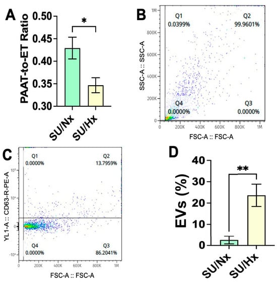
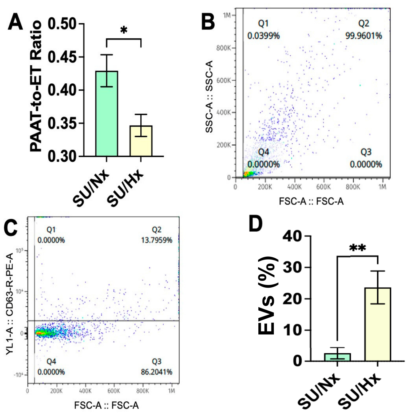
Proportion of CD63+ events in blood is increased in pulmonary hypertension: As expected, SU/Hx significantly reduced the pulmonary artery acceleration time-to-ejection time (PAAT-to-ET) ratio compared with SU/Nx controls (Figure 1A). A decreased PAAT-to-ET ratio is associated with increased pulmonary arterial pressure [17]. Consistent with these hemodynamic changes, SU/Hx rats also exhibited cardiac remodeling (Table 1). The combination of increased interventricular septal thickness (IVS) during both diastole and systole together with the reduced PAAT/ET ratio confirms the development of SU/Hx-induced PH. CD63+ is highly enriched on the surface of EVs and is widely used as a marker for EVs. Forward- and side-scatter flow-cytometry analyses demonstrated a significantly increased proportion of CD63+ events in blood collected from the left ventricle of SU/Hx rats compared with SU/Nx rats (Figure 1B–D).
Figure 1.
SU/Hx increases the proportion of EVs in blood. This figure compares circulating EVs isolated from rats with Sugen-induced pulmonary hypertension maintained under normoxic (SU/Nx) or hypoxic (SU/Hx) conditions. (A) Pulmonary artery acceleration time–to–ejection time (PAAT/ET) ratio, a Doppler-based measure of pulmonary hemodynamics, is significantly reduced in SU/Hx rats compared with SU/Nx controls (* p < 0.05; n = 5), consistent with severe pulmonary hypertension. (B) Forward scatter-area (FSC-A) versus side scatter-area (SSC-A) flow-cytometry plots display the size and granularity of isolated EVs. (C) CD63 fluorescence (YL1-A::CD63-R-PE-A) plotted against FSC-A identifies CD63+ EVs. (D) Quantification of CD63+ EVs shows a significantly higher proportion of CD63+ particles in the SU/Hx group compared with SU/Nx (** p < 0.01; n = 5).
Table 1.
Echocardiographic assessment of left ventricular geometry and function in SU/Nx and SU/Hx groups.
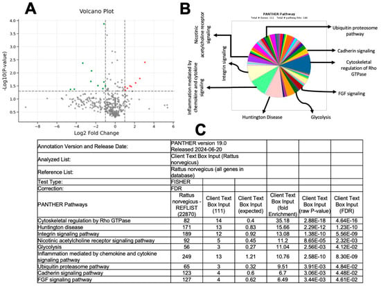
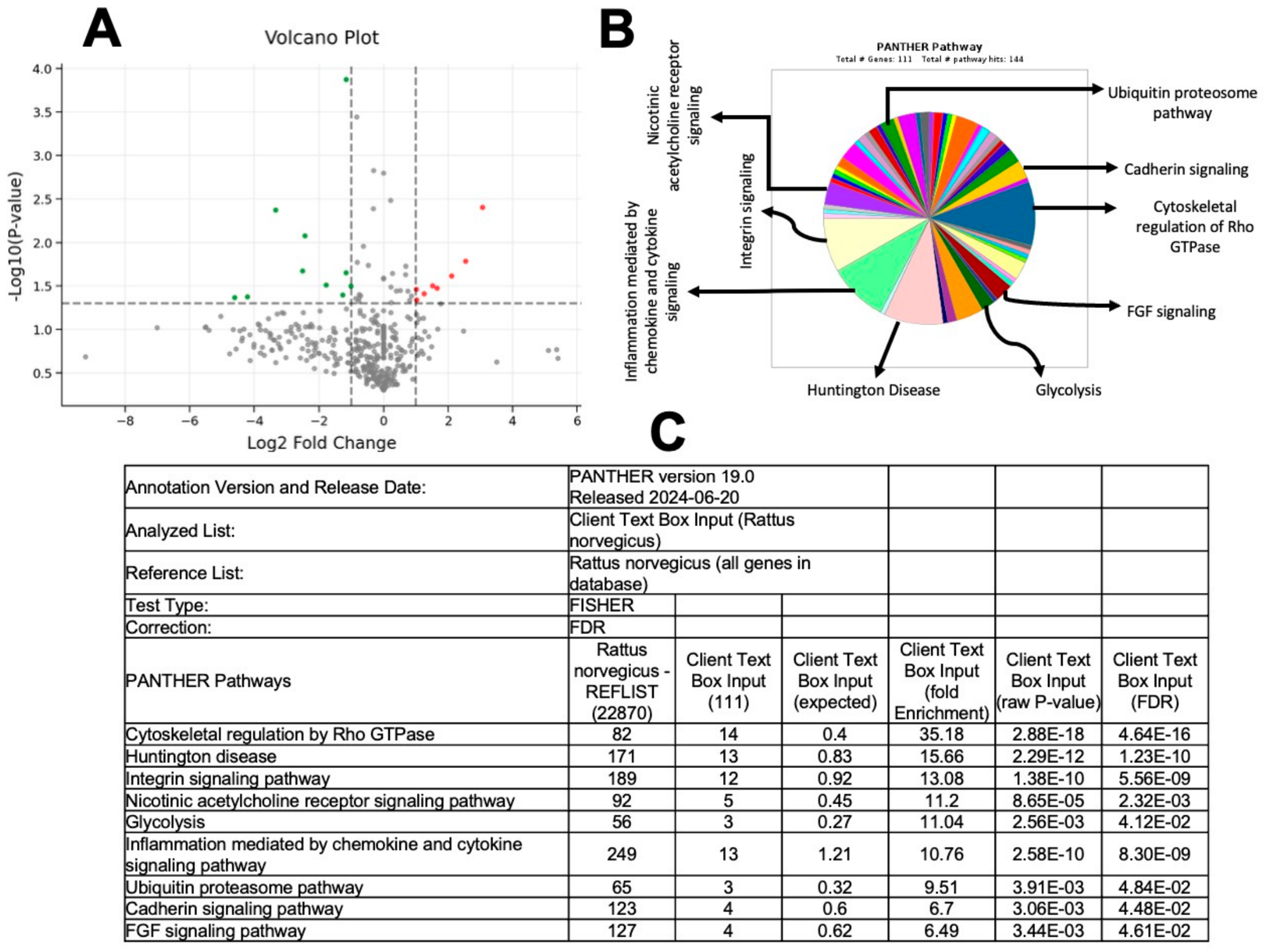
Characteristics of proteins identified in EV-enriched plasma preparation by proteomics: To characterize the protein cargo of EVs, we performed unbiased proteomic analysis on EV-enriched plasma preparations isolated from the blood of SU/Hx and SU/Nx rats. The volcano plot demonstrated clear differential protein abundance between the two groups (Figure 2A). In silico analysis, using the Panther Classification System, identified the biological functions associated with the up- and down-regulated proteins. Overall, proteins from 144 distinct pathways were detected (Figure 2B,C). These enriched pathways included cytoskeletal regulation via Rho GTPase, cadherin signaling, FGF signaling, nicotinic acetylcholine receptor signaling, chemokine- and cytokine-mediated inflammation, integrin signaling, ubiquitin-proteasome processes, and glycolysis.
Figure 2.
Proteomic profiling and pathway enrichment analysis of EV cargo. (A) Volcano plot depicting differentially abundant EV proteins. The x-axis represents log2 fold change (SU/Hx vs. SU/Nx; n = 5), and the y-axis represents −log10(p-value). Proteins significantly upregulated in SU/Hx rats are shown on the right (red), while significantly downregulated proteins are shown on the left (green). Proteins that do not meet significance thresholds are shown in gray. p-values were adjusted using the Benjamini–Hochberg (BH) false discovery rate (FDR) method. (B,C) PANTHER pathway enrichment analysis (version 19.0; Rattus norvegicus database) of differentially expressed EV proteins. Enriched pathways include cytoskeletal regulation by Rho GTPase, Huntington disease, integrin signaling, chemokine/cytokine-mediated inflammation, cadherin signaling, glycolysis, nicotinic acetylcholine receptor signaling, ubiquitin–proteasome pathways, and FGF signaling. Ranked pathway enrichment analysis was performed using Fisher’s exact test with false discovery rate (FDR) correction, highlighting the most significantly altered functional pathways in SU/Hx-derived EVs. Collectively, these findings demonstrate that SU/Hx profoundly alters signaling, metabolic, and structural pathways within EV protein cargo.
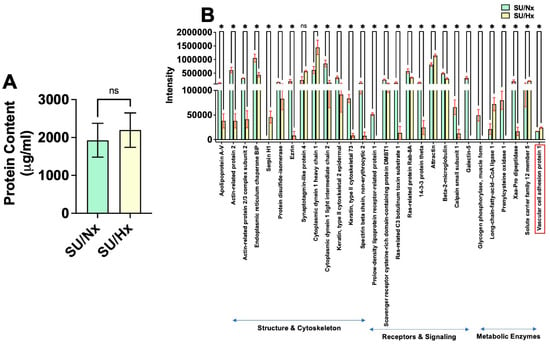
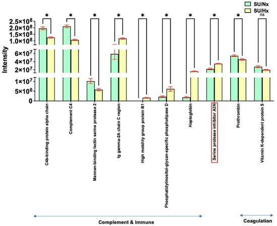
Expression levels and biological functions of differentially regulated proteins in the EVs of rats with pulmonary hypertension: While total EV protein content did not differ between the two groups (Figure 3A), proteins, including those involved in blood coagulation, were reduced in EVs from SU/Hx rats compared with SU/Nx rats; several key proteins were notably increased. These included cytoplasmic dynein 1 heavy chain 1, a cytoskeletal motor protein involved in mitosis, intracellular trafficking, and retrograde transport; attractin, a signaling protein associated with inflammatory responses and energy homeostasis; long-chain fatty acid–CoA ligase 1, a metabolic enzyme essential for fatty acid metabolism and β-oxidation; vascular cell adhesion protein 1 (VCAM1), a cell-adhesion molecule that mediates leukocyte binding to vascular endothelial cells; Ig-gamma-2A chain C region, a complement- and immune-related protein; high-mobility group protein B1 (HMGB1), a DNA-binding and chromatin-modifying protein that also functions extracellularly as a potent pro-inflammatory mediator; phosphatidylinositol-glycan-specific phospholipase D (GPI-PLD), an enzyme that cleaves GPI anchors involved in immune activation; haptoglobin, which binds and removes free hemoglobin from the circulation, preventing iron loss and oxidative tissue injury while modulating immune responses; and serine protease inhibitor A3N (Serpin A3N), a neutrophil elastase inhibitor that regulates inflammation and limits tissue damage. All these proteins were elevated in EVs isolated from SU/Hx rats compared with SU/Nx rats (Figure 3B and Figure 4).
Figure 3.
Differential expression of EV proteins grouped by functional category. (A) Total EV protein content (µg/mL) does not differ between SU/Nx and SU/Hx groups. (B) The figure displays the abundance of selected EV proteins in SU/Nx (green bars) versus SU/Hx (yellow bars) rats (n = 5 per group). Structure and Cytoskeleton: Most cytoskeletal and structural proteins, including Apolipoprotein A-V and actin-related protein 2/3 complex subunit 2, are reduced in SU/Hx EV. Exceptions include Serpin H1 and cytoplasmic dynein 1 heavy chain 1, which are elevated. Receptors and Signaling: Signaling proteins such as Prolow-density lipoprotein receptor-related protein 1 and Rab-8A are decreased, whereas Attractin is increased. Metabolic Enzymes: Several metabolic enzymes, including long-chain fatty acid–CoA ligase 1, solute carrier family 12 member 5, and vascular cell adhesion protein 1 (VCAM1; in red box), are significantly upregulated in SU/Hx EVs. Collectively, these patterns indicate that SU/Hx reshapes the EV proteome, selectively enhancing metabolic, adhesion-related, and stress-responsive protein cargo. * p < 0.05 and ns = not significant.
Figure 4.
Biological functions of EV proteins altered in pulmonary hypertension. This figure illustrates differences in EV proteins associated with immune pathways, complement activation, and the coagulation cascade between SU/Nx (green bars) and SU/Hx (yellow bars) groups (n = 5 rats/group). Complement and Immune Proteins: C4b-binding protein α-chain, Complement C4, and Mannan-binding lectin serine protease 2 are significantly reduced in SU/Hx EVs. Conversely, Ig-gamma-2A chain C region, phosphatidylinositol-glycan-specific phospholipase D (GPI-PLD), and haptoglobin are elevated (* p < 0.05). Coagulation-Related Proteins: Serine protease inhibitor A3N (Serpin A3N), a regulator of neutrophil elastase and inflammation, is significantly increased, whereas prothrombin is decreased in SU/Hx EVs (* p < 0.05). Vitamin K-dependent protein S is unchanged. These results indicate that SU/Hx EVs carry altered immune, complement, and coagulation protein signatures that may contribute to pulmonary hypertension pathobiology. ns = not significant.
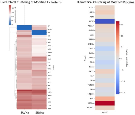
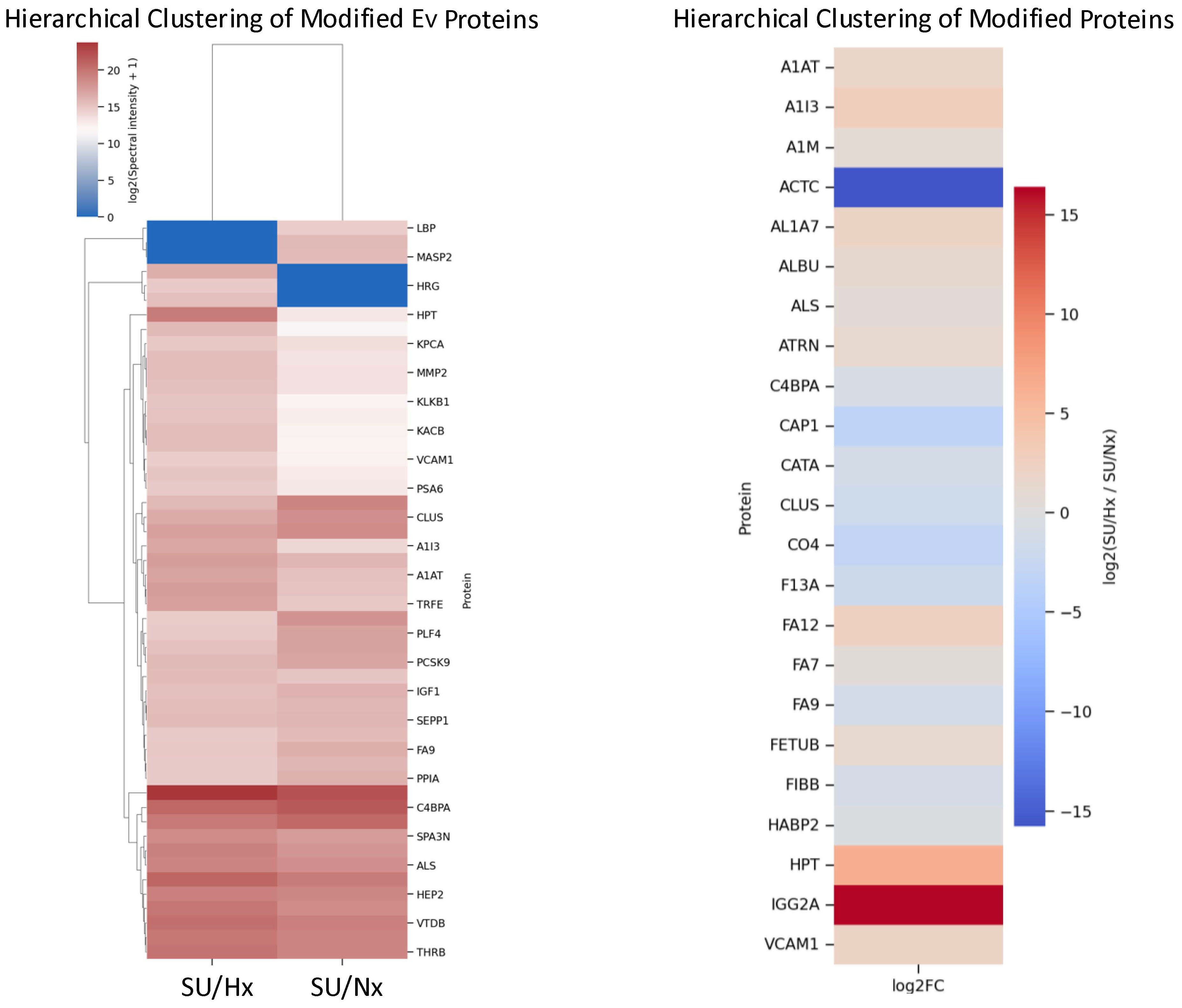
Determination of post-translationally modified proteins in EVs of rats with pulmonary hypertension: To characterize these post-translational modifications (PTMs) within EV cargo, we performed PTM-specific proteomic analysis on EVs isolated from SU/Hx rats. A comprehensive list of the detected modified proteins is provided in Table 2. Notably, oxidation of M (15.9949), N-terminal acetylation (42.0106), P oxidation (15.9949), N-terminal pyroglutamate (−17.0265), and Cysteine carbamidomethylation (57.0215) modifications were observed. Expression levels of these modified proteins in SU/Hx versus SU/Nx groups are presented in Figure 5 and Table 2, while their corresponding biological functions are summarized in Table 3.
Table 2.
List of modified proteins.
Figure 5.
Hierarchical clustering and differential enrichment of modified EV proteins in pulmonary hypertension. (Left panel) Hierarchical clustering heat map of post-translationally modified proteins identified in EV-enriched plasma preparations isolated from blood collected from the left ventricle of SU/Hx and SU/Nx rats. Protein abundance is displayed as log2-transformed spectral intensity (+1), with red indicating higher and blue indicating lower relative abundance. (Right panel) Heat map illustrating the log2 fold-change (SU/Hx vs. SU/Nx) of selected differentially modified EV proteins. Proteins such as HPT, IGG2A, VCAM1, and FETUB are markedly enriched in SU/Hx rats, whereas others (e.g., ACTC) are reduced, emphasizing condition-specific remodeling of EV protein cargo in experimental pulmonary hypertension.
Table 3.
Functions of Modified Proteins.
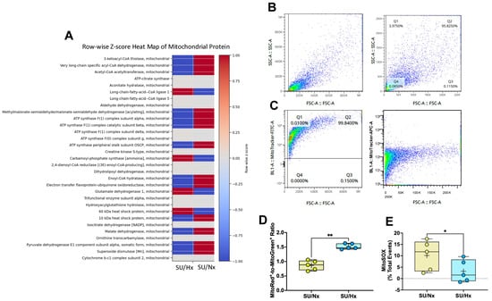
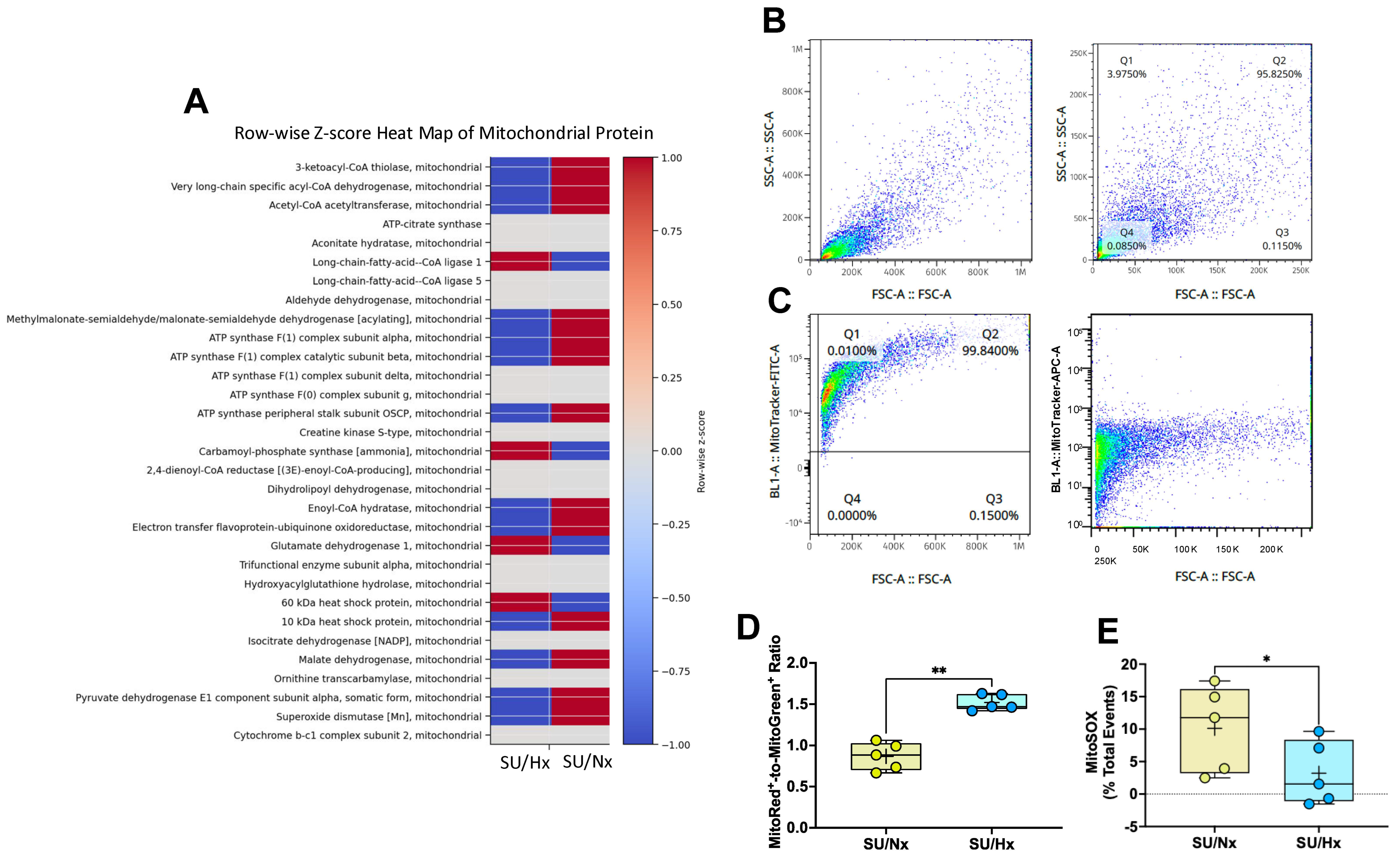
Mitochondrial content is increased in EVs from rats with pulmonary hypertension: Recent studies indicate that EV-mediated transfer of mitochondrial components can alter metabolic and inflammatory responses in recipient cells [18,19]. Deletion of the small GTPase Rab7 has been shown to increase secretion of EVs containing ubiquitinated cargo, including intact mitochondria [20]. Interestingly, in our study, Rab8A—an important regulator of intracellular vesicle trafficking and extracellular vesicle release—was decreased in EVs from SU/Hx rats, whereas several mitochondrial proteins, including long-chain fatty acid–CoA ligase 1, carbamoyl-phosphate synthase, glutamate dehydrogenase 1, and the 60 kDa heat shock protein, were increased compared with SU/Nx controls (Figure 6A). These findings prompted us to assess other aspects of mitochondrial content within the EVs. Representative forward and side scatter plots (Figure 6B), along with MitoGreen+ and MitoRed+ mitochondrial staining patterns (Figure 6C), are shown. Unexpectedly, EVs from SU/Hx rats exhibited an elevated MitoRed+/MitoGreen+ ratio, indicating the presence of a greater abundance of more hyperpolarized mitochondrial components with negative membrane potential in EVs from SU/Hx rats (Figure 6D). However, EVs from SU/Hx rats paradoxically exhibited a reduced MitoSOX signal (Figure 6E).
Figure 6.
EV mitochondrial protein enrichment and mitochondrial content are increased in SU/Hx rats. (A) Row-wise z-score heat map of mitochondrial proteins identified by proteomic analysis in EV-enriched plasma preparations isolated from blood collected from the left ventricle of SU/Hx and SU/Nx rats. Protein abundance is normalized across rows to highlight relative enrichment or depletion between conditions, with red indicating higher and blue indicating lower relative abundance. Multiple mitochondrial enzymes and chaperones are enriched in EVs from SU/Hx rats. (B) Representative flow-cytometry forward scatter (FSC-A) versus side scatter (SSC-A) density plots showing the size and granularity of isolated EVs prior to mitochondrial staining. (C) Representative flow-cytometry plots of EVs stained with MitoTracker Green (total mitochondrial mass) and MitoTracker Red (mitochondrial membrane potential), gated on FSC-A versus fluorescence intensity. (D) Quantification of MitoGreen+ and MitoRed+ EVs expressed as a percentage of total EV events in SU/Nx and SU/Hx groups. Ratio of MitoRed+ to MitoGreen+ EVs, indicating an increased proportion of negatively charged mitochondrial components within EVs from SU/Hx rats. (E) Quantification of MitoSOX+ EVs expressed as a percentage of total EV events in the SU/Nx and SU/Hx groups. The proportion of MitoSOX+ EVs was significantly reduced in SU/Hx rats, indicating lower mitochondrial superoxide levels within EVs. Data are shown as individual data points with box-and-whisker plots. Statistical significance was determined using unpaired t-tests; * p < 0.05 and ** p < 0.01.
4. Discussion
The findings of this study demonstrate that the proportion of EV-enriched plasma preparation levels in blood collected from the left ventricle is significantly higher in PH rats than in non-PH rats; EVs from PH rats carry both unmodified and post-translationally modified proteins and are enriched in mitochondrial components, which appear to be more hyperpolarized, while paradoxically exhibiting reduced mitochondrial superoxide anion levels, suggesting altered mitochondrial redox regulation.
PH is a multifactorial and multicellular disease [21]. Several animal models, including monocrotaline, hypoxia, and SU/Hx, have been used to investigate its pathogenesis [22]. Among these, the SU/Hx rat model is widely utilized because the pattern of pulmonary artery remodeling closely resembles that observed in human PH [22]. By isolating EVs from blood collected from the left ventricle and performing unbiased proteomic analyses to identify both unmodified and post-translationally modified proteins, we determined that EVs from SU/Hx rats carry increased selective protein cargo (See Table 2 and Table 3). Proteomic analysis revealed that EVs carried proteins with oxidation-related post-translational modifications, including oxidation of methionine and proline residues, and pyroglutamate formation and carbamidomethylation of cysteines. The +15.9949 Da mass shift commonly reflects the addition of a single oxygen atom and is most frequently attributed to oxidation of methionine (Met, M) and proline (Pro, P). Proline oxidation (+15.9949 Da) results in hydroxyproline formation, a modification commonly observed in collagen and frequently associated with fibrotic processes, which are known contributors to PH pathogenesis. Notably, an increased oxidation of the methionine (2.7:1.0) and proline (2.0:1.0) ratio, determined by normalizing the modified-peptide signals to the corresponding unmodified protein or peptide and to the SU/Nx control, indicates a higher abundance of oxidized methionine- and proline-containing proteins in EVs from SU/Hx rats compared to SU/Nx rats. The Q (−17.0265 Da) modification, often corresponding to N-terminal pyroglutamate formation, is a well-recognized PTM event that can substantially alter protein function by affecting charge, hydrogen bonding, and susceptibility to proteolytic degradation. N-terminal pyroglutamate formation removes the primary amine, protecting proteins from aminopeptidase-mediated degradation and playing a critical role in the maturation and stability of several hormones, cytokines, and neuropeptides, including neurotensin. This modification may also influence protein folding and intermolecular interactions by eliminating a hydrogen bond donor. The higher glutamine modification ratio (3.0:1.0) observed in SU/Hx rats indicates increased accumulation of pyroglutamate residues in EV proteins. Collectively, modifications of methionine, proline, and glutamine suggest that SU/Hx exposure likely alters the molecular composition and potential functionality of EV proteins in rats. Although cysteine carbamidomethylation (+57.0215 Da modification) is a standard alkylation of cysteine residues typically introduced during sample preparation using iodoacetamide to block free thiols and prevent artificial disulfide bond formation, it is noteworthy that the reduced cysteine residues are highly reactive and particularly susceptible to redox-dependent modifications [23].
Our findings demonstrate that EV cargo from SU/Hx rats is enriched in a distinct cluster of both lung- and non-lung-associated proteins, including both unmodified proteins and proteins bearing post-translational modifications (e.g., HPT, IGG2A, VCAM1, and FETUB). Notably, HMGB1, a chromatin-associated DNA-binding protein that can also function extracellularly as a potent pro-inflammatory damage-associated molecular pattern (DAMP), was significantly enriched in SU/Hx EVs despite the absence of detectable PTMs. In addition to proteins linked to pulmonary vascular pathology, EVs contained numerous proteins, including coagulation factors and proteins involved in basement membrane maintenance, extracellular matrix organization, cell–matrix and matrix–matrix interactions, heme binding, growth hormone signaling, antibody-dependent cellular cytotoxicity, phagocytosis, complement activation, lectin pathway activation, protease inhibition, and pathways associated with non-small-cell lung cancer. In contrast, a reciprocal cluster of proteins, including CO4, CAP1, CLUS, and PLF4, was markedly reduced in SU/Hx EVs but abundant in SU/Nx controls, suggesting either diminished expression, impaired packaging, or selective exclusion of these proteins under pulmonary hypertensive conditions. A separate cluster comprising highly abundant circulating proteins, such as MUG1, VTDB, and THRB, formed an independent branch within the clustering analysis, indicating that their segregation was primarily driven by overall abundance rather than disease-specific regulation. Functional annotation revealed that EV-associated proteins collectively participate in biological pathways central to pulmonary hypertension pathogenesis, including inflammation, cell adhesion, vascular smooth muscle cell phenotypic switching, angiogenesis, coagulation, cardiac remodeling, innate immune activation, cell-cycle regulation, apoptosis, PAR and Rho/ROCK signaling, and metabolic homeostasis [24,25,26]. These findings support the concept that SU/Hx-induced PH is associated with extensive remodeling of the circulating EV proteome. The presence of protein cargo suggests that EVs may serve as integrative mediators of interorgan communication, facilitating vascular remodeling, inflammatory signaling, immune dysregulation, and systemic pathophysiological responses that contribute to disease progression. These results further support growing evidence that PH is a systemic disorder characterized by widespread molecular and vascular abnormalities extending beyond the pulmonary circulation [27].
EVs released from diseased donor cells or organs can transmit biological materials to healthy recipient cells and organs [2]. Therefore, it is reasonable to propose that EVs carrying both unmodified and post-translationally modified proteins potentially contribute to the inflammation and cardiac remodeling observed in SU/Hx-exposed rats. In addition, EVs transporting proteins that promote inflammation, vascular smooth muscle cell dedifferentiation, and hypoxia-induced intra-pulmonary fibrosis [10,28,29] are likely to trigger remodeling of the vasculature and potentially other organs. Because proteins associated with coagulation and complement activation were elevated in SU/Hx rats, these EVs may also promote blood clot formation and activate immune pathways that drive systemic inflammation. Although the mechanism by which proteins (primarily liver-associated) enter circulating EVs remains unclear, PH is well known to impair liver function through venous congestion, which reduces oxygen delivery and disrupts hepatic synthetic activity [30]. Collectively, these findings suggest that both lung-associated and non-lung-associated EV cargo can potentially influence organ function, including vascular function, beyond the lungs.
Next, using flow cytometry, we found that circulating EVs contain mitochondrial components, as detected by labeling with mitochondria-specific dyes. MitoTracker Green labels total mitochondrial mass independently of membrane potential, whereas MitoTracker Red selectively stains mitochondria with an intact negative membrane potential. Interestingly, the increased ratio of MitoTracker Red+ to MitoTracker Green+ mitochondria in SU/Hx rats suggests a relative enrichment of mitochondrial content with strongly negative membrane potentials. Such hyperpolarized mitochondria are typically associated with enhanced electron retention and increased electron leak from Complex I and Complex III of the electron transport chain, processes that can promote reactive oxygen species generation. Paradoxically, EVs from SU/Hx rats exhibited reduced MitoSOX fluorescence despite evidence of deeply negative mitochondrial membrane potential. This observation suggests that hyperpolarized mitochondrial content is not necessarily accompanied by increased mitochondrial superoxide accumulation within EVs and may instead reflect reduced localized superoxide production or changes in MitoSOX probe accessibility, uptake, or retention. Nevertheless, because other ROS-generating pathways implicated in PH, such as NADPH oxidases and xanthine oxidase, were not evaluated, increased ROS production from non-mitochondrial sources cannot be ruled out. Supporting the presence of metabolically active and redox-sensitive cargo within EVs, proteomic analyses identified mitochondrial metabolic enzymes as well as proteins associated with the endoplasmic reticulum, nucleus, and peroxisomes, suggesting that EVs may serve as vehicles for inter-organellar signaling and redox regulation during PH progression. Although EVs are known to transport mitochondrial material [19,20,31], and EV-mediated mitochondrial trafficking may represent an additional mechanism for mitochondrial exchange distinct from direct cell–cell pore formation via connexin-43, as observed between hematopoietic stem cells and bone marrow fibroblasts [32,33], it has remained unclear whether EVs carry intact mitochondria or mitochondrial contents in the context of PH. To the best of our knowledge, this study provides the first evidence that EVs in PH may contain, at least intact, mitochondrial components influencing membrane potential. However, whether the content of these mitochondria originates from the lungs, the liver, or both remains to be determined. Furthermore, the effects of mitochondrial content on organ-level physiology, whole-body metabolism, and immune responses remain to be fully elucidated. Because mitochondria are evolutionary descendants of bacteria under the endosymbiotic theory, the presence of mitochondrial content, including nucleic acids, in the circulating EVs could potentially activate cGAS—STING-interferon signaling, promoting inflammation and the synthesis of immunomodulatory metabolites such as kynurenine—a pathway implicated in PH and SARS-CoV-2-associated pulmonary complications [34,35,36,37,38,39,40]. Consequently, mitochondria released as a result of PH may contribute to increased systemic oxidative stress, thereby elevating the risk of inflammation and damage in peripheral tissues and organs; this needs to be investigated in the future.
In conclusion, we demonstrate that there is an increased proportion of circulating EVs in blood from the experimental model of PH. In addition, using an unbiased proteomic approach, we demonstrate that these EVs carry proteins that may contribute to immune activation, inflammation, coagulation, and cardiac remodeling. Finally, our results revealed that EVs from PH rats contain a greater abundance of mitochondrial components, which appear to be more hyperpolarized.
Limitations: Although our study demonstrates increased levels of lung- and liver-associated proteins and mitochondrial content in circulating EVs, the biological function and the precise cellular and tissue origins of these cargo components remain unclear and warrant further investigation; this is an important direction for future work. Other limitations include the limitation of flow cytometry; incomplete characterization of EV purity and heterogeneity; precipitation-related co-isolation; absence of direct EV-transfer experiments; and the potential for some detected protein modifications to arise during sample preparation.
Author Contributions
M.D. (Melinee D’Silva) performed the experiments, analyzed the data, and contributed to writing of the manuscript; C.S. performed the experiments; M.D. (Monika Dzieciatkowska) performed the proteomic experiments and contributed to the data analysis; A.D. interpreted the proteomic results; M.S.W. interpreted the MitoTracker and MitoSOX results; S.A.G. conceived the experiments, procured the funding, interpreted the results, and wrote the manuscript. All authors have read and agreed to the published version of the manuscript.
Funding
This study was funded by NHLBI (R01HL166546) and the intramural Dean’s Translational Science Initiative Award.
Institutional Review Board Statement
Not applicable.
Informed Consent Statement
Not applicable.
Data Availability Statement
All data is presented in the paper and will be available upon request. Mass-spectrometry/proteomic data is deposited in MassIVE (submission ID is MassIVE MSV000098403).
Acknowledgments
All animal procedures were approved by the New York Medical College Institutional Animal Care and Use Committee (Approval #20300; Approved on 14 September 2023) and conducted in accordance with the National Institutes of Health Guide for the Care and Use of Laboratory Animals.
Conflicts of Interest
The authors declare no conflicts of interest.
References
- Kumar, M.A.; Baba, S.K.; Sadida, H.Q.; Marzooqi, S.A.; Jerobin, J.; Altemani, F.H.; Algehainy, N.; Alanazi, M.A.; Abou-Samra, A.B.; Kumar, R.; et al. Extracellular vesicles as tools and targets in therapy for diseases. Signal Transduct. Target. Ther. 2024, 9, 27. [Google Scholar] [CrossRef] [Scilit] [PubMed]
- Tkach, M.; Thery, C. Communication by Extracellular Vesicles: Where We Are and Where We Need to Go. Cell 2016, 164, 1226–1232. [Google Scholar] [CrossRef] [Scilit] [PubMed]
- Witwer, K.W.; Thery, C. Extracellular vesicles or exosomes? On primacy, precision, and popularity influencing a choice of nomenclature. J. Extracell. Vesicles 2019, 8, 1648167. [Google Scholar] [CrossRef] [Scilit] [PubMed]
- Chiaradia, E.; Tancini, B.; Emiliani, C.; Delo, F.; Pellegrino, R.M.; Tognoloni, A.; Urbanelli, L.; Buratta, S. Extracellular Vesicles under Oxidative Stress Conditions: Biological Properties and Physiological Roles. Cells 2021, 10, 1763. [Google Scholar] [CrossRef] [Scilit] [PubMed]
- Conti, M.; Minniti, M.; Tine, M.; De Francesco, M.; Gaeta, R.; Nieri, D.; Semenzato, U.; Biondini, D.; Camera, M.; Cosio, M.G.; et al. Extracellular Vesicles in Pulmonary Hypertension: A Dangerous Liaison? Biology 2023, 12, 1099. [Google Scholar] [CrossRef] [Scilit] [PubMed]
- Sayner, S.L.; Choi, C.S.; Maulucci, M.E.; Ramila, K.C.; Zhou, C.; Scruggs, A.K.; Yarbrough, T.; Blair, L.A.; King, J.A.; Seifert, R.; et al. Extracellular vesicles: Another compartment for the second messenger, cyclic adenosine monophosphate. Am. J. Physiol. Lung Cell. Mol. Physiol. 2019, 316, L691–L700. [Google Scholar] [CrossRef] [Scilit] [PubMed]
- Lammi, M.R.; Saketkoo, L.A.; Okpechi, S.C.; Ghonim, M.A.; Wyczechowska, D.; Bauer, N.; Pyakurel, K.; Saito, S.; deBoisblanc, B.P.; Boulares, A.H. Microparticles in systemic sclerosis: Potential pro-inflammatory mediators and pulmonary hypertension biomarkers. Respirology 2019, 24, 675–683. [Google Scholar] [CrossRef] [Scilit] [PubMed]
- D’Addario, C.A.; Lanier, G.M.; Jacob, C.; Bauer, N.; Hewes, J.L.; Bhadra, A.; Gupte, S.A. Differences in the expression of DNA methyltransferases and demethylases in leukocytes and the severity of pulmonary arterial hypertension between ethnic groups. Physiol. Rep. 2022, 10, e15282. [Google Scholar] [CrossRef] [Scilit] [PubMed]
- Blair, L.A.; Haven, A.K.; Bauer, N.N. Circulating microparticles in severe pulmonary arterial hypertension increase intercellular adhesion molecule-1 expression selectively in pulmonary artery endothelium. Respir. Res. 2016, 17, 133. [Google Scholar] [CrossRef] [Scilit] [PubMed]
- Khandagale, A.; Aberg, M.; Wikstrom, G.; Bergstrom Lind, S.; Shevchenko, G.; Bjorklund, E.; Siegbahn, A.; Christersson, C. Role of Extracellular Vesicles in Pulmonary Arterial Hypertension: Modulation of Pulmonary Endothelial Function and Angiogenesis. Arterioscler. Thromb. Vasc. Biol. 2020, 40, 2293–2309. [Google Scholar] [CrossRef] [Scilit] [PubMed]
- Hogan, S.E.; Rodriguez Salazar, M.P.; Cheadle, J.; Glenn, R.; Medrano, C.; Petersen, T.H.; Ilagan, R.M. Mesenchymal stromal cell-derived exosomes improve mitochondrial health in pulmonary arterial hypertension. Am. J. Physiol. Lung Cell. Mol. Physiol. 2019, 316, L723–L737. [Google Scholar] [CrossRef] [Scilit] [PubMed]
- Hewes, J.L.; Bauer, N.N. Extracellular vesicles in pulmonary hypertension: Lessons from mesenchymal stromal cell-derived exosomes. Am. J. Physiol. Lung Cell. Mol. Physiol. 2019, 316, L720–L722. [Google Scholar] [CrossRef] [Scilit] [PubMed]
- Abe, K.; Toba, M.; Alzoubi, A.; Ito, M.; Fagan, K.A.; Cool, C.D.; Voelkel, N.F.; McMurtry, I.F.; Oka, M. Formation of plexiform lesions in experimental severe pulmonary arterial hypertension. Circulation 2010, 121, 2747–2754. [Google Scholar] [CrossRef] [Scilit] [PubMed]
- de Brito Monteiro, L.; Davanzo, G.G.; de Aguiar, C.F.; Correa da Silva, F.; Andrade, J.R.; Campos Codo, A.; Silva Pereira, J.A.D.; Freitas, L.P.; Moraes-Vieira, P.M. M-CSF- and L929-derived macrophages present distinct metabolic profiles with similar inflammatory outcomes. Immunobiology 2020, 225, 151935. [Google Scholar] [CrossRef] [Scilit] [PubMed]
- Jacob, C.; Kitagawa, A.; Signoretti, C.; Dzieciatkowska, M.; D’Alessandro, A.; Gupte, A.; Hossain, S.; D’Addario, C.A.; Gupte, R.; Gupte, S.A. Mediterranean G6PD variant mitigates expression of DNA methyltransferases and right heart pressure in experimental model of pulmonary hypertension. J. Biol. Chem. 2022, 298, 102691. [Google Scholar] [CrossRef] [Scilit] [PubMed]
- Dzieciatkowska, M.; Hill, R.; Hansen, K.C. GeLC-MS/MS analysis of complex protein mixtures. Methods Mol. Biol. 2014, 1156, 53–66. [Google Scholar] [CrossRef] [Scilit] [PubMed]
- Joshi, S.R.; Kitagawa, A.; Jacob, C.; Hashimoto, R.; Dhagia, V.; Ramesh, A.; Zheng, C.; Zhang, H.; Jordan, A.; Waddell, I.; et al. Hypoxic activation of glucose-6-phosphate dehydrogenase controls the expression of genes involved in the pathogenesis of pulmonary hypertension through the regulation of DNA methylation. Am. J. Physiol. Lung Cell. Mol. Physiol. 2020, 318, L773–L786. [Google Scholar] [CrossRef] [Scilit] [PubMed]
- Tabima, D.M.; Frizzell, S.; Gladwin, M.T. Reactive oxygen and nitrogen species in pulmonary hypertension. Free Radic. Biol. Med. 2012, 52, 1970–1986. [Google Scholar] [CrossRef] [Scilit] [PubMed]
- Amari, L.; Germain, M. Mitochondrial Extracellular Vesicles—Origins and Roles. Front. Mol. Neurosci. 2021, 14, 767219. [Google Scholar] [CrossRef] [Scilit] [PubMed]
- Liang, W.; Sagar, S.; Ravindran, R.; Najor, R.H.; Quiles, J.M.; Chi, L.; Diao, R.Y.; Woodall, B.P.; Leon, L.J.; Zumaya, E.; et al. Mitochondria are secreted in extracellular vesicles when lysosomal function is impaired. Nat. Commun. 2023, 14, 5031. [Google Scholar] [CrossRef] [Scilit] [PubMed]
- Wilkins, M.R. Pulmonary hypertension: The science behind the disease spectrum. Eur. Respir. Rev. 2012, 21, 19–26. [Google Scholar] [CrossRef] [Scilit] [PubMed]
- Stenmark, K.R.; Meyrick, B.; Galie, N.; Mooi, W.J.; McMurtry, I.F. Animal models of pulmonary arterial hypertension: The hope for etiological discovery and pharmacological cure. Am. J. Physiol. Lung Cell. Mol. Physiol. 2009, 297, L1013–L1032. [Google Scholar] [CrossRef] [Scilit] [PubMed]
- Marino, S.M.; Gladyshev, V.N. Analysis and functional prediction of reactive cysteine residues. J. Biol. Chem. 2012, 287, 4419–4425. [Google Scholar] [CrossRef] [Scilit] [PubMed]
- Humbert, M.; Morrell, N.W.; Archer, S.L.; Stenmark, K.R.; MacLean, M.R.; Lang, I.M.; Christman, B.W.; Weir, E.K.; Eickelberg, O.; Voelkel, N.F.; et al. Cellular and molecular pathobiology of pulmonary arterial hypertension. J. Am. Coll. Cardiol. 2004, 43, 13S–24S. [Google Scholar] [CrossRef] [Scilit] [PubMed]
- D’Alessandro, A.; El Kasmi, K.C.; Plecita-Hlavata, L.; Jezek, P.; Li, M.; Zhang, H.; Gupte, S.A.; Stenmark, K.R. Hallmarks of Pulmonary Hypertension: Mesenchymal and Inflammatory Cell Metabolic Reprogramming. Antioxid. Redox Signal. 2018, 28, 230–250. [Google Scholar] [CrossRef] [Scilit] [PubMed]
- Stenmark, K.R.; Fagan, K.A.; Frid, M.G. Hypoxia-induced pulmonary vascular remodeling: Cellular and molecular mechanisms. Circ. Res. 2006, 99, 675–691. [Google Scholar] [CrossRef] [Scilit] [PubMed]
- Nickel, N.P.; Yuan, K.; Dorfmuller, P.; Provencher, S.; Lai, Y.C.; Bonnet, S.; Austin, E.D.; Koch, C.D.; Morris, A.; Perros, F.; et al. Beyond the Lungs: Systemic Manifestations of Pulmonary Arterial Hypertension. Am. J. Respir. Crit. Care Med. 2020, 201, 148–157. [Google Scholar] [CrossRef] [Scilit] [PubMed]
- Alvarado-Vasquez, N.; Rangel-Escareno, C.; de Jesus Ramos-Abundis, J.; Becerril, C.; Negrete-Garcia, M.C. The possible role of hypoxia-induced exosomes on the fibroblast metabolism in idiopathic pulmonary fibrosis. Biomed. Pharmacother. 2024, 181, 117680. [Google Scholar] [CrossRef] [Scilit] [PubMed]
- Su, S.A.; Xie, Y.; Fu, Z.; Wang, Y.; Wang, J.A.; Xiang, M. Emerging role of exosome-mediated intercellular communication in vascular remodeling. Oncotarget 2017, 8, 25700–25712. [Google Scholar] [CrossRef] [Scilit] [PubMed]
- Nickel, N.P.; Galura, G.M.; Zuckerman, M.J.; Hakim, M.N.; Alkhateeb, H.; Mukherjee, D.; Austin, E.D.; Heresi, G.A. Liver abnormalities in pulmonary arterial hypertension. Pulm. Circ. 2021, 11, 20458940211054304. [Google Scholar] [CrossRef] [Scilit] [PubMed]
- Corssac, G.B.; Bonetto, J.P.; Campos-Carraro, C.; Cechinel, L.R.; Zimmer, A.; Parmeggiani, B.; Grings, M.; Carregal, V.M.; Massensini, A.R.; Siqueira, I.; et al. Pulmonary arterial hypertension induces the release of circulating extracellular vesicles with oxidative content and alters redox and mitochondrial homeostasis in the brains of rats. Hypertens. Res. 2021, 44, 918–931. [Google Scholar] [CrossRef] [Scilit] [PubMed]
- Taniguchi Ishikawa, E.; Gonzalez-Nieto, D.; Ghiaur, G.; Dunn, S.K.; Ficker, A.M.; Murali, B.; Madhu, M.; Gutstein, D.E.; Fishman, G.I.; Barrio, L.C.; et al. Connexin-43 prevents hematopoietic stem cell senescence through transfer of reactive oxygen species to bone marrow stromal cells. Proc. Natl. Acad. Sci. USA 2012, 109, 9071–9076, Erratum in Proc. Natl. Acad. Sci. USA 2012, 109, 12834–12835. [Google Scholar] [CrossRef] [Scilit] [PubMed]
- Peruzzotti-Jametti, L.; Bernstock, J.D.; Willis, C.M.; Manferrari, G.; Rogall, R.; Fernandez-Vizarra, E.; Williamson, J.C.; Braga, A.; van den Bosch, A.; Leonardi, T.; et al. Neural stem cells traffic functional mitochondria via extracellular vesicles. PLoS Biol. 2021, 19, e3001166. [Google Scholar] [CrossRef] [Scilit] [PubMed]
- Murphy, M.P.; O’Neill, L.A.J. A break in mitochondrial endosymbiosis as a basis for inflammatory diseases. Nature 2024, 626, 271–279. [Google Scholar] [CrossRef] [Scilit] [PubMed]
- Kim, J.; Kim, H.S.; Chung, J.H. Molecular mechanisms of mitochondrial DNA release and activation of the cGAS-STING pathway. Exp. Mol. Med. 2023, 55, 510–519. [Google Scholar] [CrossRef] [Scilit] [PubMed]
- Nemkov, T.; Stephenson, D.; Erickson, C.; Dzieciatkowska, M.; Key, A.; Moore, A.; Earley, E.J.; Page, G.P.; Lacroix, I.S.; Stone, M.; et al. Regulation of kynurenine metabolism by blood donor genetics and biology impacts red cell hemolysis in vitro and in vivo. Blood 2024, 143, 456–472. [Google Scholar] [CrossRef] [Scilit] [PubMed]
- Nagy, B.M.; Nagaraj, C.; Meinitzer, A.; Sharma, N.; Papp, R.; Foris, V.; Ghanim, B.; Kwapiszewska, G.; Kovacs, G.; Klepetko, W.; et al. Importance of kynurenine in pulmonary hypertension. Am. J. Physiol. Lung Cell. Mol. Physiol. 2017, 313, L741–L751. [Google Scholar] [CrossRef] [Scilit] [PubMed]
- Cai, Z.; Tian, S.; Klein, T.; Tu, L.; Geenen, L.W.; Koudstaal, T.; van den Bosch, A.E.; de Rijke, Y.B.; Reiss, I.K.M.; Boersma, E.; et al. Kynurenine metabolites predict survival in pulmonary arterial hypertension: A role for IL-6/IL-6Ralpha. Sci. Rep. 2022, 12, 12326. [Google Scholar] [CrossRef] [Scilit] [PubMed]
- Thomas, T.; Stefanoni, D.; Reisz, J.A.; Nemkov, T.; Bertolone, L.; Francis, R.O.; Hudson, K.E.; Zimring, J.C.; Hansen, K.C.; Hod, E.A.; et al. COVID-19 infection alters kynurenine and fatty acid metabolism, correlating with IL-6 levels and renal status. JCI Insight 2020, 5, e140327. [Google Scholar] [CrossRef] [Scilit] [PubMed]
- D’Alessandro, A.; Thomas, T.; Akpan, I.J.; Reisz, J.A.; Cendali, F.I.; Gamboni, F.; Nemkov, T.; Thangaraju, K.; Katneni, U.; Tanaka, K.; et al. Biological and Clinical Factors Contributing to the Metabolic Heterogeneity of Hospitalized Patients with and without COVID-19. Cells 2021, 10, 2293. [Google Scholar] [CrossRef] [Scilit] [PubMed]


Disclaimer/Publisher’s Note: The statements, opinions and data contained in all publications are solely those of the individual author(s) and contributor(s) and not of MDPI and/or the editor(s). MDPI and/or the editor(s) disclaim responsibility for any injury to people or property resulting from any ideas, methods, instructions or products referred to in the content. |
© 2026 by the authors. Licensee MDPI, Basel, Switzerland. This article is an open access article distributed under the terms and conditions of the Creative Commons Attribution (CC BY) license.