


Abstract
Obstructive sleep apnea (OSA) and heart failure (HF) share pathways involving intermittent hypoxia and oxidative stress. This randomized clinical trial with blinded endpoint assessment evaluated systemic oxidative balance during sequential treatment with empagliflozin and continuous positive airway pressure (CPAP). Seventy adults with HF and predominantly obstructive OSA were randomized to empagliflozin 10 mg daily plus background HF therapy (n = 37) or background therapy alone (n = 33). Assessments were performed at baseline, month 3, and month 6; CPAP was initiated in both groups after month 3. The primary endpoint was the between-group difference in AHI change from baseline to month 3, estimated using a baseline-adjusted linear mixed-effects model. TOS, TAS, and OSI were exploratory outcomes. Median AHI (IQR) in the study and control groups was 58.9 (28.5–70.3) and 55.0 (29.9–67.8) at baseline, and 44.2 (22.4–59.3) and 52.2 (30.8–70.2) at month 3, respectively. The baseline-adjusted between-group difference in AHI change from baseline to month 3 was −11.04 (95% CI −16.72 to −5.35; p < 0.001). The exploratory mixed-effects models showed lower TOS and OSI and higher TAS in the study group at both follow-up visits (all p < 0.001). Empagliflozin initiation was associated with early reductions in AHI and improvements in exploratory markers of systemic oxidative balance before CPAP initiation. Although the shared CPAP phase did not allow separation of drug and CPAP effects, these findings support further investigation of the respiratory and redox effects of empagliflozin in patients with coexisting HF and OSA.
1. Introduction
Heart failure (HF) is a heterogeneous clinical syndrome that results from structural and/or functional cardiac abnormalities impairing ventricular filling or ejection, leading to elevated filling pressures and inadequate tissue perfusion [1,2]. Contemporary classification according to left ventricular ejection fraction (LVEF) distinguishes HF with preserved, mildly reduced, and reduced ejection fraction (≥50%, 41–49%, and ≤40%, respectively), phenotypes that differ in pathophysiology, prognosis, and therapeutic response [3]. Neurohormonal activation—sustained stimulation of the sympathetic nervous system and the renin–angiotensin–aldosterone system—together with systemic inflammation, oxidative stress, and adverse ventricular remodeling, plays a central role in HF progression. Despite evidence-based pharmacotherapy, HF remains a major global health burden affecting more than 64 million people worldwide [4].
Obstructive sleep apnea (OSA) is a highly prevalent sleep-related breathing disorder characterized by recurrent partial or complete upper-airway collapse during sleep, producing intermittent hypoxia, sleep fragmentation, and large intrathoracic pressure swings [5,6]. Although the apnea–hypopnea index (AHI) remains the conventional metric of severity, the clinical consequences of OSA extend beyond event frequency and are shaped by the depth and duration of nocturnal hypoxemia, symptom burden, and cardiovascular comorbidity [7]. The recurrent hypoxia–reoxygenation cycles that characterize OSA behave analogously to ischemia–reperfusion, promoting excessive generation of reactive oxygen species, depletion of antioxidant defenses, endothelial dysfunction, and sympathetic overactivity—pathways strongly linked to the development and progression of cardiovascular disease [8,9,10].
Oxidative stress reflects an imbalance between oxidant production and antioxidant defense. It can be captured at the systemic level by the total oxidant status (TOS), the total antioxidant status (TAS), and their ratio, the oxidative stress index (OSI), which together provide an integrated measure of redox balance. In OSA, intermittent hypoxia is a principal driver of this imbalance, whereas in HF systemic inflammation and neurohormonal activation add a further oxidative load [8,9,11]. Because both conditions converge on the same downstream redox pathways, the HF–OSA overlap may represent a state of amplified oxidative injury.
HF and OSA commonly coexist and interact through bidirectional, self-reinforcing mechanisms. Sleep-disordered breathing affects approximately 40% to 60% of patients with symptomatic HF, with OSA accounting for about one third of cases, and untreated OSA increases the risk of developing HF [12,13]. Recurrent obstruction and intermittent hypoxemia may worsen HF through sympathetic activation, afterload stress, oxidative and inflammatory pathways, and adverse myocardial remodeling; conversely, HF may aggravate OSA through fluid retention, elevated venous pressures, and nocturnal rostral fluid shift that promote peripharyngeal edema and upper-airway collapsibility [14,15].
Continuous positive airway pressure (CPAP) is the standard treatment for OSA; it prevents upper-airway collapse, reduces nocturnal hypoxemia, and, in patients with coexisting HF, may favorably influence hemodynamics and ventricular function [16,17]. However, treatment response is variable and residual risk persists, motivating interest in adjunctive strategies that target both HF pathophysiology and mechanisms contributing to OSA severity. Sodium–glucose cotransporter-2 inhibitors (SGLT2is) have become a cornerstone of guideline-directed HF therapy irrespective of diabetes status [1,3]. Beyond natriuresis and preload/afterload reduction, SGLT2is improve myocardial energetics and have been reported to attenuate inflammation and oxidative stress; by reducing fluid overload and rostral fluid shift, they may also lessen upper-airway narrowing [18,19,20]. Exploratory analyses of cardiovascular outcome trials and small dedicated studies suggest a favorable signal on OSA [21,22,23].
Despite this biological plausibility, the systemic oxidative consequences of the HF–OSA overlap, and how they respond to pharmacological and mechanical therapy, remain insufficiently characterized. In the present randomized controlled trial, we assessed TOS, TAS, and OSI as exploratory measures of systemic oxidative balance alongside OSA severity, nocturnal oxygenation, and cardiac status in patients with coexisting HF and OSA. Because empagliflozin was initiated before CPAP, the first 3 months provided a randomized comparison of the two pharmacological strategies before mechanical OSA treatment. During months 3–6, both groups received CPAP; therefore, changes during this period could be described temporally but could not be attributed exclusively to CPAP in the absence of a concurrent no-CPAP group.
2. Materials and Methods
2.1. Study Design and Population
This single-center, randomized, controlled, open-label trial with blinded endpoint assessment prospectively enrolled and followed participants at the Department of Otolaryngology, University Clinical Hospital in Bialystok, Poland, between 5 June 2023 and 9 May 2025. Participants were recruited from the Department and Outpatient Clinic of Otolaryngology, the Outpatient Clinic of Cardiology of the University Clinical Hospital in Bialystok, and the Department of Internal Medicine with a cardiology profile of the Independent City Hospital in Bialystok. The trial was designed in two phases: an initial pre-CPAP phase (0–3 months) to assess the role of empagliflozin on OSA with HF, and a second phase (3–6 months) in which CPAP was added to both groups.
Eligible participants were adults aged ≥18 years with diagnosed HF, not previously treated with any SGLT2 inhibitor, and with mild, moderate, or severe OSA confirmed by an overnight sleep study, not previously treated with CPAP; a body mass index (BMI) < 40 kg/m2; and clinical eligibility for OSA treatment according to current recommendations. Participants with and without type 2 diabetes were eligible, and diabetes was not required for enrollment. Clinical stability was defined as the absence of HF decompensation, HF-related hospitalization, or modification of background HF pharmacotherapy during the 6 months preceding enrollment. Exclusion criteria were age < 18 years, BMI ≥ 40 kg/m2, active malignancy, severe renal impairment (estimated glomerular filtration rate < 25 mL/min/1.73 m2), advanced hepatic insufficiency, symptomatic hypotension, clinically significant volume depletion or dehydration, hypersensitivity to any component of the study medication, and regular use of antioxidant or vitamin supplements. HF diagnosis had been verified at enrollment in accordance with the 2021 ESC guidelines, based on current or previous symptoms and/or signs of HF together with objective evidence of a structural or functional cardiac abnormality [2]. Medical records, echocardiographic findings, and NT-proBNP measurements were reviewed to confirm the diagnosis. HF phenotypes were classified as HFrEF (LVEF ≤ 40%), HFmrEF (LVEF 41–49%), or HFpEF (LVEF ≥ 50%), and functional status at enrollment was assessed according to the NYHA classification; enrolled participants were in NYHA classes I–III.
A total of 135 patients with HF at high risk of sleep-disordered breathing (STOP-Bang score > 4) underwent a sleep study; 87 (64.4%) were diagnosed with OSA. Ten eligible patients declined participation before randomization. An additional seven patients were not randomized because they did not attend the randomization visit (n = 3), experienced a change in clinical status before randomization (n = 2), or had an incomplete baseline assessment (n = 2). The remaining 70 participants were randomized and constituted the intention-to-treat population; all randomized participants completed the 6-month follow-up and were included in the final analyses. No participants withdrew or were excluded after randomization (Figure 1). At enrollment, all participants were SGLT2 inhibitor-naïve and clinically stable, with no HF decompensation, HF-related hospitalization, or modification of HF pharmacotherapy during the preceding 6 months. All patients were receiving stable background HF therapy, including a β-blocker, an angiotensin-converting enzyme inhibitor or angiotensin receptor blocker, a mineralocorticoid receptor antagonist, and low maintenance doses of loop diuretics (≤10 mg torasemide or ≤40 mg furosemide daily). Patients and members of the public were not involved in the design, conduct, reporting, or dissemination planning of the study. No important changes to the study procedures or prespecified redox assessments were introduced after participant enrolment commenced.
Figure 1.
Study flow diagram. Of 135 patients with heart failure assessed for eligibility, 87 were diagnosed with obstructive sleep apnea (OSA), and 70 were randomized and completed the 6-month study protocol (study group, n = 37; control group, n = 33). CPAP was initiated in both groups after the 3-month assessment.
2.2. Randomization and Interventions
Participants were randomized in a 1:1 ratio using a computer-generated stratified permuted-block randomization sequence with randomly varying block sizes of four and six within each stratum. Stratification was based on baseline AHI (5.0–14.9, 15.0–29.9, or ≥30 events/h) and LVEF (≤40%, 41–49%, or ≥50%), resulting in nine strata (Supplementary Table S3). Recruitment was concluded when the prespecified overall sample size of 70 participants had been reached. The allocation sequence was retained and administered by an independent investigator. The principal investigator (A.P.), who was responsible for participant enrollment, had no access to the sequence and remained unaware of the upcoming allocation until assignment.
All participants were SGLT2 inhibitor-naïve and CPAP-naïve at enrollment; therefore, the study did not require the withdrawal of any previously established SGLT2 inhibitor or CPAP therapy. Before randomization, the study physician individually assessed each participant and confirmed that temporary deferral of SGLT2 inhibitor initiation would be clinically acceptable if the participant was assigned to the control group. Participants in both groups continued their existing background HF therapy, which could be modified whenever clinically indicated. Participants assigned to the study group (n = 37) additionally initiated empagliflozin 10 mg once daily at baseline and continued treatment throughout the 6-month follow-up, whereas those assigned to the control group (n = 33) did not initiate an SGLT2 inhibitor as part of the study intervention. All participants underwent clinical reassessment at each follow-up visit. Clinical care took precedence over the assigned study strategy: if SGLT2 inhibitor therapy became clinically necessary, its initiation was permitted at the discretion of the treating physician and was to be documented as a modification of concomitant therapy. After completion of the 6-month follow-up, participants in the control group were re-evaluated for SGLT2 inhibitor therapy, which was initiated in the absence of contraindications.
Study assessments were performed at baseline and at the 3- and 6-month visits. The predefined 3-month pre-CPAP phase was intended to distinguish changes associated with empagliflozin initiation from those attributable to the mechanical treatment of upper-airway obstruction. Simultaneous initiation of empagliflozin and CPAP would have prevented the attribution of early changes in sleep-related, oxygenation, cardiac, and oxidative-stress outcomes to either intervention. Following the 3-month assessment, all participants were clinically re-evaluated, and CPAP therapy was initiated.
The trial was open-label for participants and treating physicians, who knew treatment allocation from randomization. Sleep-study scoring personnel, the echocardiographer, and laboratory personnel were blinded to randomized treatment allocation during outcome assessment. The same blinded echocardiographer performed both image acquisition and interpretation at baseline, month 3, and month 6. Blinding of sleep-study scoring personnel was maintained at all three assessments and concerned assignment to empagliflozin or control. Sleep recordings and laboratory samples were identified using participant codes assigned sequentially at enrollment according to a predetermined scheme independent of treatment allocation. An independent technician held the coding key. Outcome assessors did not have access to medication lists or clinical records revealing treatment allocation and did not participate in treatment consultations. Treatment allocation remained coded in the statistical dataset until data collection and cleaning had been completed and the database had been locked. The treatment-allocation code was then released to the statistician before the final analyses. The statistician therefore knew group identities during the final analyses and the additional analyses performed during manuscript revision.
Safety was assessed at each follow-up visit through patient interview and clinical evaluation, supplemented by periodic laboratory testing. Monitoring included HF symptoms, blood pressure, body weight, renal function, electrolytes, and adverse events. Telephone contact with the study physician was available between scheduled visits, including for urgent concerns. Decisions regarding earlier CPAP initiation, initiation of an SGLT2 inhibitor in the control group, or modification of background HF therapy were based on the treating physician’s assessment of symptoms, patient-reported changes in clinical status, and laboratory findings. Clinically indicated treatment took precedence over the assigned study strategy.
2.3. Assessments
2.3.1. Sleep Study
All participants underwent type III home sleep apnea testing using the NOX T3s system (Nox Medical, Reykjavik, Iceland) at baseline, month 3, and month 6. Baseline and month-3 recordings included nasal pressure, thermistor airflow, thoracic and abdominal respiratory inductance plethysmography (RIP), oxygen saturation, pulse rate, body position, and audio/snoring. Month-6 testing was performed during CPAP, recording mask pressure through a mask connection and RIP-derived flow. Month-6 AHI was obtained from NOX recordings, not CPAP device downloads. Recordings were analyzed in Noxturnal 7.2 and required ≥4 h of technically adequate airflow and oximetry signals. Repeat testing was necessary for twelve, nine, and seven studies at baseline, month 3, and month 6, respectively, because of battery depletion or missing nasal-pressure or oximetry signals, including connection failure or sensor removal. Valid recordings were ultimately available for all participants at each assessment.
The same American Academy of Sleep Medicine scoring criteria and manual review by personnel blinded to randomized allocation were used throughout. Apnea was defined as a ≥90% airflow reduction lasting ≥10 s and classified as obstructive, central, or mixed according to preserved, absent, or initially absent and subsequently resumed respiratory effort, respectively. Hypopnea required a ≥30% airflow reduction lasting ≥10 s with ≥3% desaturation. Obstructive hypopneas showed snoring, increased inspiratory flow flattening, or new thoracoabdominal paradox; central hypopneas lacked these features. AHI was calculated per hour of estimated sleep time. Additional measures included mean oxygen saturation (MOS), lowest oxygen saturation (LOS), and time with oxygen saturation < 90% (T < 90%).
OSA severity was classified according to the AHI as mild (≥5 to 14.9), moderate (15.0–29.9), or severe (≥30). Eligibility for CPAP therapy was determined on the basis of OSA severity, symptoms, and the overall clinical assessment. Participants with moderate or severe OSA were considered eligible for CPAP following clinical evaluation. In participants with mild OSA, eligibility required clinically relevant OSA-related symptoms, such as excessive daytime sleepiness, impaired sleep-related quality of life, or insomnia, and/or a cardiovascular comorbidity warranting active treatment. All participants with mild OSA enrolled in the study had coexisting HF and were individually confirmed to be eligible for OSA treatment. Because patients with HF may exhibit both obstructive and central respiratory events, all recordings were additionally reviewed for the distribution of apnea types. Patients with predominant central sleep apnea, defined by a central apnea index ≥ 5 events/h together with central apneas accounting for >50% of all apneas, were not classified as having predominant OSA and were excluded from the present analysis. The presence of occasional or non-predominant central events did not preclude the diagnosis of OSA when obstructive respiratory events remained predominant.
2.3.2. Echocardiography
Transthoracic echocardiography was performed at baseline and after 3 and 6 months using a Philips EPIQ CVx v7.0 system (Philips Healthcare, Bothell, WA, USA) with an X5-1c 3D transducer and 3D Live xMatrix software (VM13.0). Both two-dimensional and three-dimensional LVEF were assessed in accordance with the recommendations of the Polish Cardiac Society [24].
2.3.3. Biochemical and Oxidative Stress Analyses
Fasting venous blood was collected between 07:00 and 09:00 a.m. at baseline and after 3 and 6 months. Serum was allowed to clot for 30 min at room temperature, centrifuged at 2000× g for 10 min at 4 °C, and stored at −80 °C until analysis. N-terminal pro–B-type natriuretic peptide (NT-proBNP) was measured with the Elecsys proBNP II assay (Roche Diagnostics GmbH, Mannheim, Germany). Total oxidant status and total antioxidant status were determined in duplicate in serum using commercially available photometric microplate assays from Immundiagnostik AG (Bensheim, Germany), according to the manufacturer’s instructions. TOS was measured with the PerOx (TOS) kit, based on peroxidase-mediated determination of total lipid peroxides, and TAS with the ImAnOx (TAS) kit, based on the antioxidant-dependent elimination of exogenously added hydrogen peroxide. The oxidative stress index was calculated as the TOS-to-TAS ratio (dimensionless; both TOS and TAS expressed in µmol/L). Samples were assayed in duplicate, and assay quality was verified using the control samples supplied with each kit; the values obtained agreed with the certified values. The intra-assay coefficients of variation were <3% (TOS) and <7% (TAS), and the inter-assay coefficients of variation were <3% (TOS) and <2% (TAS). TOS, TAS, and OSI were prospectively prespecified as study assessments in the study documentation approved by the institutional Bioethics Committee before participant enrolment. The present redox-focused analysis included the complete randomized cohort (n = 70), with no missing outcome data.
2.3.4. Heart Failure Questionnaire
HF symptom burden was assessed using a study-specific symptom score comprising dyspnea, palpitations, chest pain, lower-extremity edema, and nocturia. The total symptom score ranged from 5 to 20 points, with higher scores indicating greater HF symptom burden (Table S2, Supplementary Material).
2.4. Statistical Analysis
The sample size calculation was based on the primary endpoint, defined as the between-group difference in the change in AHI from baseline to month 3. Based on the 2022 DAHOS trial protocol and previous interventional studies, a clinically relevant between-group difference of 7 events/h and a common standard deviation of 8 events/h were assumed [25,26,27,28]. With a two-sided significance level of 0.05, 90% power, and a 1:1 allocation ratio, 29 evaluable participants were required per group. After allowing for an anticipated 15% loss of primary outcome data, the target sample size was increased to 35 participants per group (70 participants in total).
The primary endpoint was the between-group difference in AHI change from baseline to month 3. This endpoint was evaluated using a linear mixed-effects model of post-baseline AHI values adjusted for baseline AHI. The endpoint definition and the original statistical analysis plan were finalized before examination of the outcome data; however, the mixed-effects model was introduced during manuscript revision. The original plan specified nonparametric within-group and between-group comparisons using the Wilcoxon signed-rank and Mann–Whitney U tests, respectively. The exploratory Spearman correlation analyses, Benjamini–Hochberg correction within the correlation families, and ΔBMI-adjusted partial Spearman correlations were already included in the initial manuscript submission. These analyses are reported as exploratory and are not presented as prospectively prespecified analyses. The revision-stage additions comprised the baseline-adjusted linear mixed-effects models, exploratory AHI responder analyses using thresholds, and extension of Benjamini–Hochberg correction to the nine supportive between-group comparisons of changes in TOS, TAS, and OSI across the three study intervals. These additions were not part of the original statistical analysis plan.
Data distributions were assessed using histograms and the Shapiro–Wilk test. Continuous variables are presented as the median and interquartile range (IQR), and categorical variables as numbers and percentages. Within-group comparisons were performed using the Wilcoxon signed-rank test, between-group comparisons using the Mann–Whitney U test, and categorical comparisons using Pearson’s chi-square or Fisher’s exact test, as appropriate. Changes (Δ) were calculated as the later value minus the earlier value for the 0–3-, 3–6-, and 0–6-month intervals. Between-group differences in changes were accompanied by the Mann–Whitney effect size (r = Z/√N) and Hodges–Lehmann estimates with 95% percentile confidence intervals obtained from 10,000 bootstrap resamples. These interval-specific comparisons were considered supportive analyses.
The AHI change endpoint and exploratory redox outcomes (TOS, TAS, and OSI) were analyzed longitudinally using linear mixed-effects models. Measurements obtained at months 3 and 6 were modeled as repeated outcomes, with the corresponding baseline value, treatment group, visit, and treatment group × visit interaction included as fixed effects and participant included as a random intercept. Visit was modeled as a categorical variable, and the models were estimated using restricted maximum likelihood. Model-derived contrasts estimated adjusted between-group differences at months 3 and 6 with corresponding 95% confidence intervals. For AHI, the month-3 contrast estimated the baseline-adjusted between-group difference in AHI change from baseline to month 3 (study minus control). The model used post-baseline AHI values as the dependent variable and included baseline AHI as a covariate. Under this model specification, the treatment contrast at month 3 is identical to that obtained when modeling change from baseline with the same baseline adjustment. It is not the unadjusted difference in observed change scores. The treatment group × visit interaction tested whether the adjusted between-group difference changed from month 3 to month 6, rather than the baseline-to-month-3 treatment contrast. Model assumptions were evaluated using residual diagnostics.
Associations were assessed using Spearman’s rank correlation coefficient (ρ). For each treatment group and reference outcome (AHI, LVEF, T < 90%, or HF symptom score), nine correlations were evaluated: three redox markers (TOS, TAS, and OSI) across three intervals (0–3, 3–6, and 0–6 months). These constituted eight separate families of nine tests, with Benjamini–Hochberg correction applied independently within each family to control the false discovery rate at 5%. A separate family comprised the nine supportive between-group Mann–Whitney U comparisons of changes in TOS, TAS, and OSI across the same three intervals. Benjamini–Hochberg correction was applied jointly to these nine tests. Within-group Wilcoxon tests were not included in this family. p-values from the linear mixed-effects models, AHI responder analyses, and ΔBMI-adjusted partial Spearman correlations were nominal and were not adjusted for multiplicity. Partial correlations were considered exploratory sensitivity analyses. FDR control was applied within the specified families rather than across all study analyses. Exploratory AHI responder analyses were performed using two clinically interpretable criteria: (1) a reduction in AHI of at least 50% from baseline and (2) achievement of an AHI < 15. The proportions of participants meeting each criterion at months 3 and 6 were compared between groups using Fisher’s exact test. No responder analyses were performed for TOS, TAS, OSI, or the study-specific HF symptom score because validated clinically meaningful response thresholds were not available for these outcomes.
All tests were two-sided, with p < 0.05 or FDR-adjusted q < 0.05 considered statistically significant, as appropriate. Analyses followed the intention-to-treat principle. As all randomized participants completed follow-up without missing outcome data, no imputation was required. All analyses were performed using Statistica version 13.3 (TIBCO Software Inc., Palo Alto, CA, USA). The trial is reported according to CONSORT 2025.
2.5. Ethics
The protocol was approved on 30 March 2023 by the Bioethics Committee of the Medical University of Bialystok (approval no. APK.002.526.2022) and conducted in accordance with the Declaration of Helsinki and Good Clinical Practice. All participants provided written informed consent before enrolment. The ethics approval explicitly covered the sequential treatment schedule, including initiation of CPAP after the month-3 assessment in both groups and deferral of SGLT2 inhibitor initiation in the control group during the 6-month study period. This schedule and the potential risks associated with delayed treatment initiation were explained and discussed with participants during the informed consent process. The trial was retrospectively registered at ClinicalTrials.gov on 29 April 2026 (NCT07556094). Prospective registration was inadvertently omitted at study initiation because of an administrative oversight. The study design, eligibility criteria, interventions, scheduled assessments, and study outcomes had been defined in the protocol approved by the Bioethics Committee before the enrolment of the first participant. The timing and status of the original analyses and the analyses introduced during manuscript revision are explicitly described in Section 2.4. Nevertheless, retrospective registration is acknowledged as a reporting limitation because it does not provide the same protection against selective outcome reporting as prospective registration.
3. Results
3.1. Baseline Characteristics
In the overall cohort, the median age was 62 years (IQR 58–68), and 41 of 70 participants (58.6%) were men; the population was ethnically homogeneous (all White, of Polish nationality). At baseline, the two groups were well balanced with respect to demographic, anthropometric, sleep-related, cardiac, oxidative, and patient-reported characteristics, with no statistically significant between-group differences, including for the oxidative stress markers TOS, TAS, and OSI (Table 1). All randomized participants completed the planned treatment and follow-up. Overall, at least one adverse event was reported by 18/37 (48.6%) participants in the empagliflozin group and 22/33 (66.7%) in the control group. All reported adverse events were mild CPAP-related symptoms. Dry mouth occurred in 5/37 (13.5%) and 10/33 (30.3%), watery eyes in 9/37 (24.3%) and 8/33 (24.2%), and mask-related skin marks or irritation in 12/37 (32.4%) and 17/33 (51.5%), respectively. Participants could report more than one symptom. These symptoms did not lead to treatment discontinuation. Apart from these mild CPAP-related symptoms, no other adverse events were reported. No adverse events were considered attributable to empagliflozin.
Table 1.
Baseline characteristics of the study population. Continuous variables are presented as median (interquartile range, IQR), and categorical variables as n (%). Between-group comparisons were performed using the Mann–Whitney U test for continuous variables and the χ2 test for categorical variables.
During the 6-month follow-up, no serious adverse events, HF-related hospitalizations, or major modifications of background HF therapy occurred in either group (0/37 in the study group and 0/33 in controls for each outcome). No participant required CPAP initiation before the scheduled month-3 assessment (0/37 and 0/33, respectively), and no control participant required rescue initiation of an SGLT2 inhibitor before completion of follow-up (0/33). Treatment adherence was high throughout the study. Adherence to empagliflozin, assessed by pill count, was 95%. CPAP adherence assessment was prespecified in the original statistical analysis plan. Adherence was assessed objectively using data downloaded from the devices’ memory cards. Mean nightly use was calculated across all nights during the CPAP follow-up period (months 3–6), including nights without CPAP use. During Phase II, CPAP use was comparable between groups, with a mean nightly use of 5.6 ± 1.0 h in the study group and 5.4 ± 1.1 h in the control group. The proportion of participants using CPAP for ≥ 4 h/night was similarly high (89.2% vs. 87.9%, respectively). Mean therapeutic CPAP was also comparable between groups (15.4 ± 2.4 vs. 15.2 ± 2.6 cm H2O, respectively).
3.2. Longitudinal Analyses of AHI and Oxidative Stress Markers
Median AHI (IQR) in the study and control groups was 58.9 (28.5–70.3) and 55.0 (29.9–67.8) at baseline, and 44.2 (22.4–59.3) and 52.2 (30.8–70.2) at month 3, respectively (Supplementary Table S1). The median individual change from baseline to month 3 was −9.7 (−13.8 to −5.3) in the study group and +1.7 (+0.9 to +2.7) in the control group. The baseline-adjusted between-group difference in AHI change from baseline to month 3 (study minus control) was −11.04 (95% CI −16.72 to −5.35; p < 0.001).
At month 6, during the shared CPAP phase, median AHI (IQR) was 4.6 (3.2–5.5) in the study group and 6.6 (4.5–11.2) in controls (Supplementary Table S1). The median individual change from baseline to month 6 was −53.9 (−63.7 to −27.2) and −47.6 (−59.8 to −22.5), respectively.
Exploratory redox outcomes (TOS, TAS, and OSI) were analyzed using baseline-adjusted linear mixed-effects models (Table 2). At month 3, the study group showed lower TOS, higher TAS, and lower OSI compared with the control group (all p < 0.001). Corresponding between-group differences remained evident at month 6 for TOS, TAS, and OSI (p < 0.001). The group × visit interactions were not significant for TOS (p = 0.705) or OSI (p = 0.404), whereas the interaction for TAS was significant (p = 0.005).
Table 2.
Baseline-adjusted longitudinal between-group differences in oxidative stress markers derived from linear mixed-effects models.
Supportive interval-specific analyses are presented in Table 3 and Figure 2. During Phase I, before CPAP initiation, reductions in TOS and OSI and the increase in TAS were greater in the study group than in the control group (all p < 0.001). During Phase II, further within-group improvements were observed in both groups. In the supportive change-score analyses, no significant between-group differences were observed for TOS or TAS, whereas the reduction in OSI was greater in the control group. This isolated segment-specific finding was not supported by a significant group × visit interaction for OSI in the longitudinal model. Across the full 6-month follow-up, supportive analyses showed greater cumulative improvements in TOS, TAS, and OSI in the study group (all p < 0.001).
Figure 2.
Systemic oxidative balance over 6 months. Median values (whiskers, interquartile range) for total oxidant status (TOS; (A)), total antioxidant status (TAS; (B)), and the oxidative stress index (OSI; (C)) in the study (n = 37) and control (n = 33) groups at baseline, 3, and 6 months. The vertical dotted line at the 3-month visit indicates the initiation of CPAP therapy; the blue solid line represents the study group, the orange dashed line represents the control group; p-values indicate between-group comparisons of changes (Δ) during Phase I (0–3 months) and Phase II (3–6 months) by the Mann–Whitney U test.
Table 3.
Changes in systemic oxidative stress markers over 6 months. Values at baseline, 3 months, and 6 months are presented as median (interquartile range, IQR). Δ represents the median within-group change over the specified interval (0–3 months, 3–6 months, or 0–6 months). Within-group p-values were calculated using the Wilcoxon signed-rank test. Between-group p-values compare Δ between the study and control groups using the Mann–Whitney U test. r, Mann–Whitney effect size calculated as Z/√N; HL diff, Hodges–Lehmann median difference in Δ (study group minus control group) with 95% bootstrap confidence interval (CI).
Exploratory AHI responder analyses are presented in Supplementary Table S4. At month 3, no participant achieved an AHI reduction of at least 50% from baseline (0/37; 0.0% in the study group and 0/33; 0.0% in controls; p = 1.000). At month 6, all participants met this criterion (37/37; 100.0% and 33/33; 100.0%, respectively). AHI < 15 was achieved by 3/37 (8.1%) in the study group and 1/33 (3.0%) in the controls at month 3 (p = 0.616), and by 36/37 (97.3%) and 30/33 (90.9%) at month 6 (p = 0.337), respectively. No validated clinically meaningful response thresholds were available for TOS, TAS, OSI, or the study-specific HF symptom score; therefore, post hoc responder thresholds were not created for these outcomes.
3.3. Correlations Between Oxidative Stress Markers and Heart-Failure Symptom Burden
To examine whether the improvement in systemic oxidative balance paralleled clinical improvement, the phase-specific changes in TOS, TAS, and OSI were correlated with the concurrent change in heart-failure symptom burden (ΔHF symptoms score) in each group using Spearman’s rank correlation (Table 4, Figure 3 and Figure 4).
Figure 3.
Correlations between changes in oxidative stress markers and concurrent changes in heart-failure symptom burden, by study phase. Color intensity represents Spearman’s rank correlation coefficient (ρ) between the change in each oxidative stress marker and the concurrent change in HF symptom score, separately for the study (n = 37) and control (n = 33) groups. Cell values represent Spearman’s ρ. Asterisks indicate significance after Benjamini–Hochberg false discovery rate (FDR) correction within each group’s nine correlation tests: * q < 0.05, ** q < 0.01, *** q < 0.001.
Figure 4.
Spearman rank correlations between phase-specific changes in systemic oxidative stress markers and concurrent changes in heart-failure symptom burden, stratified by study group. Points represent Spearman’s rank correlation coefficients (ρ), and horizontal error bars indicate 95% confidence intervals. Confidence intervals were calculated using Fisher’s z-transformation. Asterisks indicate correlations remaining statistically significant after Benjamini–Hochberg false discovery rate (FDR) correction within each group’s nine correlation tests (q < 0.05). The blue color represents the study group, the orange color represents the control group.
During Phase I (0–3 months), before CPAP, none of the oxidative markers correlated significantly with symptom change in either group; in the study group, only the association for TOS approached significance (ρ = 0.305, p = 0.067).
During Phase II (3–6 months), after CPAP initiation, TOS and OSI correlated significantly and positively with symptom change in both groups (study group: TOS ρ = 0.433, p = 0.007; OSI ρ = 0.668, p < 0.001; control group: TOS ρ = 0.444, p = 0.010; OSI ρ = 0.596, p < 0.001), whereas TAS showed no significant association in either group.
Across the full 6-month period, OSI correlated significantly with symptom change in both groups (study group ρ = 0.612, p < 0.001; control group ρ = 0.567, p < 0.001). For TOS, the correlation was significant in the study group (ρ = 0.511, p = 0.001) but not in the control group (ρ = 0.315, p = 0.074), while TAS was unrelated to symptom change throughout. In the study group, the reduction in HF symptom burden over 6 months was additionally associated with the reduction in NT-proBNP (ρ = 0.557, p < 0.001).
Overall, the OSI showed the most consistent relationship with symptomatic improvement, being significantly correlated with the change in HF symptom burden in both groups once CPAP was in place and over the entire study period, whereas the total antioxidant status was not significantly related to symptom change in any phase.
Table 4.
Spearman rank correlations between phase-specific changes in systemic oxidative stress markers and concurrent changes in heart-failure symptom burden (ΔHF symptom score), by group. Values are presented as Spearman’s ρ (p-value) with Benjamini–Hochberg false discovery rate (FDR)-adjusted q-values. Benjamini–Hochberg correction was applied separately within each group across the nine correlation tests.
3.4. Correlations of Oxidative Stress with OSA Severity and Cardiac Function
To determine whether the improvement in systemic oxidative balance tracked the improvement in the two principal disease markers, phase-specific changes in TOS, TAS, and OSI were correlated with the concurrent change in the AHI (ΔAHI) and in LVEF (ΔLVEF) in each group (Table 5 and Table 6 and Figure 5, Figure 6 and Figure 7).
Figure 5.
Changes in oxidative stress index (ΔOSI) in relation to concurrent changes in apnea–hypopnea index (ΔAHI; upper row) and left ventricular ejection fraction (ΔLVEF; lower row), stratified by study phase and group. (A) ΔOSI versus ΔAHI during Phase I (0–3 months); (B) ΔOSI versus ΔAHI during Phase II (3–6 months); (C) ΔOSI versus ΔAHI over the overall study period (0–6 months); (D) ΔOSI versus ΔLVEF during Phase I (0–3 months); (E) ΔOSI versus ΔLVEF during Phase II (3–6 months); (F) ΔOSI versus ΔLVEF over the overall study period (0–6 months). Each point represents one participant; lines represent group-specific linear fits for visualization. Spearman’s rank correlation coefficients (ρ) and corresponding nominal p-values are shown above each panel; Benjamini–Hochberg FDR-adjusted q-values are reported in Table 5 and Table 6. Phase I represents the pre-CPAP period (0–3 months), Phase II the period after CPAP initiation (3–6 months), and 0–6 months the overall study period. The blue color represents the study group, the orange color represents the control group.
Figure 6.
Heatmaps of Spearman rank correlations between phase-specific changes in oxidative stress markers (TOS, TAS, and OSI) and concurrent changes in AHI (upper row) and LVEF (lower row) in the study (A,C) and control (B,D) groups. Cell values represent Spearman’s rank correlation coefficients (ρ), with color intensity indicating the magnitude and direction of the association. Asterisks indicate significance after Benjamini–Hochberg false discovery rate (FDR) correction separately within each treatment group and reference outcome, across nine correlation tests per family: * q < 0.05, ** q < 0.01, *** q < 0.001.
Figure 7.
Spearman rank correlations between phase-specific changes in oxidative stress markers and concurrent changes in AHI (A) and LVEF (B), stratified by study group. Points represent Spearman’s correlation coefficients (ρ), and horizontal error bars indicate 95% confidence intervals calculated using Fisher’s z-transformation. Asterisks indicate correlations remaining statistically significant after Benjamini–Hochberg false discovery rate (FDR) correction separately within each treatment group and reference outcome, across nine correlation tests per family (q < 0.05). The blue color represents the study group, the orange color represents the control group.
Changes in TOS and OSI correlated strongly and positively with the change in AHI, most consistently for OSI (Table 5 and Figure 5A,B). In the study group, this coupling was already significant during Phase I, before CPAP (OSI ρ = 0.670, TOS ρ = 0.679; both p < 0.001), and strengthened over the whole study (OSI ρ = 0.785, p < 0.001). In the control group, by contrast, no association was present during Phase I (OSI ρ = −0.117, p = 0.517); a significant relationship emerged only after CPAP was introduced in Phase II (OSI ρ = 0.601, p < 0.001) and over the full period (OSI ρ = 0.596, p < 0.001). TAS was only weakly related to AHI change.
Table 5.
Spearman rank correlations between phase-specific changes in systemic oxidative stress markers and concurrent changes in AHI, stratified by study group. Spearman’s ρ (p); q, Benjamini–Hochberg FDR-adjusted q-value calculated separately within each group across the nine correlation tests.
Changes in OSI and TOS were inversely related to the change in LVEF, indicating that greater recovery of ejection fraction accompanied greater reduction in systemic oxidative stress (Table 6 and Figure 5C,D). For OSI, the inverse association was significant during Phase II in both groups (study group ρ = −0.464, p = 0.004; control group ρ = −0.474, p = 0.005) and, in the control group, over the whole study (ρ = −0.561, p < 0.001). TOS showed significant inverse correlations with LVEF change in the control group (Phase II ρ = −0.463; whole study ρ = −0.486; both p < 0.01), whereas TAS was not significantly related to LVEF change.
Table 6.
Spearman rank correlations between phase-specific changes in systemic oxidative stress markers and concurrent changes in left ventricular ejection fraction (ΔLVEF), stratified by study group. Spearman’s ρ (p); q, Benjamini–Hochberg FDR-adjusted q-value calculated separately within each group across the nine correlation tests.
Across both reference variables, OSI showed the most consistent pattern of significant associations. Notably, the association between changes in OSI and AHI was already significant during Phase I in the study group, before CPAP initiation, whereas in the control group it emerged only during Phase II, after CPAP initiation (Figure 6 and Figure 7). After Benjamini–Hochberg correction within each correlation family, the principal associations of OSI—with AHI, with the nocturnal hypoxemia marker T < 90% (time with oxygen saturation < 90%), and with HF symptom burden—remained significant (q < 0.05). ΔOSI also correlated with ΔT < 90% in both groups over the full study (study ρ = 0.627, p < 0.001, q < 0.001; control ρ = 0.604, p < 0.001, q = 0.002; Table 7), reinforcing the link between nocturnal hypoxemia and systemic redox balance. In the study group, the ΔOSI–ΔAHI association during Phase I persisted after adjustment for concurrent change in BMI (partial ρ = 0.714, p < 0.001; Table 8), indicating it was not explained by weight change alone.
Table 7.
Spearman rank correlations between phase-specific changes in systemic oxidative stress markers and concurrent changes in nocturnal hypoxemia (ΔT < 90%), stratified by study group. Spearman’s ρ (p); q, Benjamini–Hochberg FDR-adjusted q-value calculated separately within each group across the nine correlation tests.
Table 8.
Partial Spearman correlations of changes in OSI with concurrent changes in AHI and LVEF, adjusted for ΔBMI.
4. Discussion
In this randomized controlled trial of patients with coexisting HF and OSA, systemic oxidative balance improved substantially during the randomized pre-CPAP phase in the study group, with little change in controls. Because the improvement in TOS, TAS, and OSI during the first 3 months occurred before any mechanical airway therapy and was not observed to the same extent in controls, the temporal pattern supports an effect associated with assignment to empagliflozin rather than CPAP. To our knowledge, this is among the first randomized evaluations of systemic oxidative stress in the HF–OSA overlap phenotype in which a pharmacological effect was temporally separated from CPAP. Further changes were observed in both groups during the subsequent common CPAP phase; however, the contribution of CPAP itself cannot be isolated in the absence of a concurrent no-CPAP comparator.
Oxidative stress is a central downstream pathway in both conditions. In OSA, cyclical hypoxia and reoxygenation resemble ischemia–reperfusion and drive reactive oxygen species production, antioxidant depletion, endothelial dysfunction, and sympathetic activation [8,9]. Intermittent hypoxia is particularly relevant because repetitive transitions from hypoxia to reoxygenation can promote mitochondrial ROS generation, activation of NADPH oxidases, reduced nitric oxide bioavailability, and redox-sensitive inflammatory signaling. Experimental and clinical evidence consistently links OSA with increased systemic oxidative stress, although the magnitude of abnormalities varies according to the biomarker studied, OSA severity, obesity, and associated cardiometabolic disease [8,29]. Intermittent hypoxia may therefore provide an important mechanistic bridge between nocturnal airway obstruction and persistent cardiovascular injury.
Oxidative stress is also deeply involved in the pathophysiology of HF. In the failing myocardium, mitochondria and NADPH oxidases represent major sources of ROS, while neurohormonal activation, inflammation, altered calcium handling, and impaired antioxidant defenses further amplify the redox imbalance. Excessive ROS may impair mitochondrial energetics, reduce nitric oxide signaling, promote cardiomyocyte dysfunction and apoptosis, and contribute to fibrosis and adverse ventricular remodeling [30]. Thus, oxidative stress in HF should be regarded not only as a consequence of advanced disease but also as a potential contributor to its progression.
The coexistence of HF and OSA may therefore create a particularly unfavorable oxidative environment. Intermittent hypoxia related to OSA occurs on a background of impaired cardiac function, neurohormonal activation, inflammation, and abnormal fluid distribution. At the same time, fluid retention in HF may worsen sleep-disordered breathing through nocturnal rostral fluid displacement, with fluid accumulation in the neck narrowing the upper airway and increasing its collapsibility. This provides a plausible bidirectional mechanism through which HF may exacerbate OSA and OSA may in turn reinforce oxidative and hemodynamic stress relevant to HF progression [15,31]. The concept of nocturnal rostral fluid shift as a contributor to OSA in fluid-retaining states is well established, particularly in HF [31,32].
The baseline redox profile observed in this population is therefore biologically coherent with the coexistence of both conditions. The simultaneous assessment of TOS, TAS, and OSI is also relevant because these indices capture different aspects of systemic redox status. TOS reflects aggregate oxidant burden, TAS reflects overall antioxidant capacity, whereas OSI incorporates both dimensions and may provide a more integrated measure of oxidative imbalance. The stronger and more consistent associations observed for OSI in the present study support the potential value of a composite redox measure over either component assessed in isolation.
Several mechanisms may explain the early redox changes associated with empagliflozin before CPAP initiation. First, osmotic diuresis and natriuresis reduce intravascular and interstitial fluid, which in HF is particularly relevant to nocturnal rostral fluid shift, a recognized contributor to upper-airway collapsibility; by lessening peripharyngeal congestion, SGLT2is may reduce obstructive events and attenuate the intermittent hypoxemia that fuels oxidative stress [15,20]. Second, improved cardiac hemodynamics and reduced pulmonary congestion may lessen the depth and duration of desaturation, limiting hypoxia-driven radical generation [19]. Experimental studies with empagliflozin show that its effects may extend beyond glucose lowering. In a non-diabetic pressure-overload HF model, empagliflozin improved mitochondrial oxidative phosphorylation and mitochondrial biogenesis, reduced mitochondrial ROS generation, increased endogenous antioxidant defenses, and attenuated apoptosis and adverse remodeling [33]. Similarly, experimental work in cardiac microvascular endothelial cells showed that empagliflozin attenuated cytoplasmic and mitochondrial ROS accumulation induced by inflammatory stimulation and restored nitric oxide bioavailability, providing another potential link between SGLT2 inhibition, endothelial function, and redox balance [34]. Reductions in body weight, visceral adiposity, and sympathetic tone may provide additional, complementary benefit [35].
More recently, experimental data specifically addressing combined HF and intermittent hypoxia have suggested that dapagliflozin may attenuate intermittent-hypoxia-aggravated myocardial injury through suppression of HIF-1α/ASC signaling, with reductions in inflammatory activation, apoptosis, and structural myocardial damage. Although these findings remain preclinical and should not be directly extrapolated to humans, they are mechanistically consistent with the early redox improvement observed in the present study before initiation of CPAP [36].
During Phase I, improvements in oxidative-stress markers occurred predominantly in the study group. During Phase II, further improvements were observed in both groups while all participants were receiving CPAP, resulting in attenuation of some between-group contrasts. This pattern may partly reflect the different values reached at month 3: participants in the study group entered Phase II with a more favorable redox profile and therefore had less remaining room for further improvement, whereas the control group entered this phase with a greater residual oxidative burden. Nevertheless, because no concurrent no-CPAP comparator was available, changes observed between months 3 and 6 cannot be attributed exclusively to CPAP and may also reflect time-related trends, regression to the mean, natural disease variability, or differences in month-3 starting values.
Previous randomized studies evaluating oxidative biomarkers after CPAP have also produced inconsistent results. In a randomized placebo-controlled crossover trial, Alonso-Fernández et al. demonstrated that effective CPAP reduced plasma 8-isoprostane concentrations and increased NOx levels, suggesting attenuation of oxidative stress and restoration of nitric oxide bioavailability [37]. In contrast, randomized sham-controlled studies assessing F2-isoprostanes, lipid oxidation products, and other systemic oxidative-stress biomarkers found little or no measurable improvement following CPAP [38,39]. This heterogeneity may reflect differences in biomarker selection, treatment duration and adherence, baseline OSA severity, obesity, and cardiovascular comorbidities. Accordingly, although the present temporal pattern is biologically compatible with a contribution from CPAP, the Phase II and 0–6-month findings do not establish an independent CPAP effect or an additive, synergistic, complementary, or priming interaction between empagliflozin and CPAP.
Baseline-adjusted longitudinal analyses demonstrated significant between-group differences in TOS, TAS, and OSI at both follow-up visits. TOS and OSI were lower, whereas TAS was higher, in the empagliflozin group at months 3 and 6. These findings indicate that the early between-group separation in systemic redox status remained evident during the subsequent common CPAP phase. However, the presence of significant between-group differences at both visits does not itself demonstrate that the trajectories differed between visits. The group × visit interactions were not significant for TOS or OSI, indicating no evidence that the magnitude of the between-group differences in these markers changed from month 3 to month 6. A significant interaction was observed only for TAS, suggesting further widening of the adjusted between-group difference; given the exploratory status of the redox outcomes and the modest sample size, this finding should be interpreted cautiously.
The correlation analyses demonstrated that improvements in systemic oxidative balance, particularly reductions in OSI, frequently occurred alongside improvements in AHI, nocturnal hypoxemia, LVEF, and HF symptom burden. Associations between ΔOSI and ΔAHI were already evident during the randomized pre-CPAP phase in the empagliflozin group, whereas corresponding associations were not observed in controls during this interval. During the subsequent common CPAP phase, concordant respiratory and redox changes were observed in both groups. These findings support oxidative stress as a potential shared downstream pathway linking sleep-disordered breathing and cardiovascular dysfunction. However, the analyses were exploratory and cannot establish mediation, determine the direction of the relationships, or attribute the Phase II associations specifically to CPAP.
Among the assessed redox measures, OSI showed the most consistent associations with respiratory-event frequency, cumulative nocturnal hypoxemia, cardiac function, and HF symptom burden. As a ratio incorporating both total oxidant and antioxidant status, OSI may provide a more integrated measure of dynamic redox imbalance than either component alone. Nevertheless, no validated clinically meaningful threshold for OSI is available in the HF–OSA population, and the present study was not designed to establish its prognostic or clinical utility. OSI should therefore be regarded as a potentially informative exploratory biomarker requiring prospective validation.
The BMI-adjusted sensitivity analysis further informs the interpretation of the early ΔOSI–ΔAHI relationship. This association remained strong after adjustment for concurrent ΔBMI and was not materially attenuated during Phase I or over the overall 6-month period. Although BMI does not capture changes in fat distribution or body composition, these findings argue against overall weight change as the principal explanation for the coupling between improvement in OSA severity and systemic oxidative balance. This is relevant because weight loss is one proposed mechanism through which SGLT2 inhibitors may improve OSA [35,40]. The present pattern supports consideration of additional mechanisms, including natriuresis, reduced extracellular volume and rostral fluid displacement, improved cardiac loading conditions, sympathetic modulation, and direct mitochondrial or endothelial redox effects [15,19,20,33,34]. Notably, a recent study in patients with T2DM, HF across the LVEF spectrum, and mixed-type sleep-disordered breathing who were not receiving CPAP found that SGLT2 inhibitor use was associated with improved polygraphic parameters and an approximately 60% greater probability of achieving at least a 50% reduction in AHI [41].
The inverse association between ΔOSI and ΔLVEF provides a parallel cardiac signal, suggesting that improvement in systolic function occurred alongside reduction in systemic oxidative stress. This is biologically plausible given the contribution of ROS to mitochondrial dysfunction, endothelial impairment, abnormal calcium handling, and ventricular remodeling in HF. Nevertheless, these associations should be interpreted as concordant changes rather than evidence of mediation or causality. Improvement in oxidative stress and LVEF may represent parallel consequences of improved hemodynamic and metabolic conditions rather than a direct causal chain.
These findings also highlight the limitations of AHI as the sole measure of OSA-related cardiovascular stress because it does not fully characterize the duration or severity of the accompanying hypoxemia. The associations of OSI with both AHI and T < 90% suggest that improvement in systemic redox balance was related to respiratory-event frequency as well as cumulative nocturnal hypoxemia. T < 90% should not, however, be equated with event-specific hypoxic burden: it quantifies time below a fixed oxygen-saturation threshold but does not integrate the depth and duration of desaturations attributable to individual respiratory events. Measures of cumulative nocturnal hypoxemia have been associated with adverse cardiovascular outcomes in patients with HF, while event-specific hypoxic burden may characterize OSA-related cardiovascular stress more accurately than AHI alone [42,43,44]. Future studies combining oxidative biomarkers with event-specific hypoxic burden may better define the relationship between nocturnal desaturation, redox injury, and treatment response.
Clinically, these findings do not position empagliflozin as a replacement for CPAP. Empagliflozin was associated with early improvements in oxidative balance and nocturnal oxygenation during the randomized pre-CPAP phase, although obstructive respiratory events were not eliminated. The statistically significant between-group difference in AHI change from baseline to month 3 should not be interpreted as a marked short-term clinical response in most participants. No participant achieved an AHI reduction of at least 50%, and only 3/37 (8.1%) participants receiving empagliflozin and 1/33 (3.0%) controls had AHI < 15 events/h. The early effect therefore reflects a difference in continuous AHI values, while most participants did not attain either response threshold. The largest subsequent reductions in respiratory events were observed during the period in which CPAP was administered to both groups, consistent with the distinct therapeutic targets of these interventions: CPAP directly addresses upper-airway obstruction, whereas SGLT2 inhibition may modify the cardiometabolic, hemodynamic, and volume-related substrate contributing to the systemic consequences of HF–OSA overlap [18,23]. The present study suggests that empagliflozin initiation may be accompanied by measurable redox and physiological changes before CPAP is introduced. This may be particularly pertinent in clinical practice, where CPAP initiation, titration, and achievement of stable adherence are not always immediate. Moreover, between-group differences in the exploratory oxidative-stress markers remained evident at month 6 while both groups were receiving comparable CPAP therapy, suggesting that the systemic effects associated with the randomized pharmacological strategies were not fully attenuated during the common CPAP phase. These findings support the rationale for addressing both the cardiovascular substrate and upper-airway obstruction in patients with HF–OSA overlap, while not establishing an additive, synergistic, complementary, or priming interaction between empagliflozin and CPAP.
Because SGLT2 inhibitors are already a component of guideline-directed HF therapy, the observed respiratory and redox changes cannot be separated from improvements related to treatment of the underlying cardiovascular condition. Nevertheless, this does not diminish their potential clinical importance in the HF–OSA population, in whom reductions in oxidative and hypoxemic burden may represent relevant systemic benefits of early treatment optimization. The findings should therefore be considered hypothesis-generating and provide a rationale for adequately powered factorial or mechanistic studies designed to distinguish the independent and combined effects of empagliflozin and CPAP. Studies in patients with OSA without HF would also be informative in determining whether these effects extend beyond the HF–OSA phenotype.
Several limitations should be acknowledged. First, this was a single-center trial with a modest sample size and 6-month follow-up, limiting statistical precision, generalizability, and the ability to assess the durability of the observed redox effects or their relationship with hard clinical outcomes. Eligibility criteria further restricted the external validity of the findings. Patients with a BMI ≥ 40 kg/m2 and those with an eGFR < 25 mL/min/1.73 m2 were excluded. The BMI threshold was intended to reduce clinical heterogeneity associated with class III obesity, including the potential coexistence of obesity-related hypoventilation and other causes of nocturnal hypoxemia. The eGFR threshold was a prespecified, conservative safety-related criterion because participation in the study could involve the initiation of empagliflozin. Consequently, the present findings should not be extrapolated to patients with class III obesity or advanced renal impairment. Patients with predominant central sleep apnea were also excluded to maintain a predominantly obstructive sleep-disordered breathing phenotype. As specified in the Methods section, occasional or non-predominant central events did not preclude inclusion when obstructive events remained predominant. Nevertheless, the applicability of the findings to patients with predominantly central sleep apnea or mixed sleep-disordered breathing with a substantial central component remains uncertain because these phenotypes have distinct pathophysiological and therapeutic characteristics. The number of participants within several AHI–LVEF strata may have reduced the ability of stratification to achieve balance within individual strata. The sequential design provided temporal separation between empagliflozin and CPAP initiation but, because all participants received CPAP after 3 months, did not permit a randomized comparison of CPAP versus no CPAP. The open-label design without placebo may also have influenced subjective outcomes, particularly the study-specific, non-validated HF symptom score, although oxidative markers and other objective outcomes were less susceptible to this source of bias. Second, TOS, TAS, and OSI are integrative systemic measures of redox balance and do not identify specific ROS species, oxidative modifications, cellular sources, or tissue-level effects. Future studies should incorporate complementary markers of lipid, protein, and DNA oxidation, antioxidant enzymes, and inflammatory pathways. Third, the correlation analyses were exploratory and involved multiple comparisons. Although the Benjamini–Hochberg procedure was used to control the false discovery rate within predefined correlation families, the findings should be considered hypothesis-generating and should not be interpreted as evidence of causal mediation. The ΔBMI-adjusted partial correlations were additional exploratory sensitivity analyses and were not subjected to further multiplicity correction. Their persistence after adjustment indicates that concurrent BMI change alone is unlikely to account for the principal ΔOSI–ΔAHI associations, but residual confounding or mediation by changes in body composition, volume status, rostral fluid distribution, cardiac function, metabolic status, or other unmeasured factors cannot be excluded. In addition, T < 90% reflects cumulative time spent below a fixed oxygen-saturation threshold and should not be considered equivalent to event-specific hypoxic burden, which incorporates the depth and duration of desaturations attributable to individual respiratory events. Accordingly, the associations between ΔT < 90% and oxidative markers support a relationship between nocturnal hypoxemia and systemic redox balance but cannot establish which characteristics of hypoxic exposure are most biologically relevant. Additional limitations include the retrospective registration of the trial and the fact that the sample-size calculation was based on AHI rather than the exploratory oxidative-stress outcomes. However, the study protocol and outcome assessments had been established before participant enrolment and were not determined in response to the study results. Sparse enrollment and incomplete final blocks in some AHI–LVEF strata may also have limited within-stratum allocation balance. Future studies incorporating event-specific hypoxic burden and more detailed oxygen-desaturation metrics may better define this relationship. Larger, multicenter trials with longer follow-up and more comprehensive phenotyping are warranted to confirm these findings and determine their clinical and prognostic significance.
5. Conclusions
In clinically stable patients with HF and predominantly obstructive sleep apnea, a BMI < 40 kg/m2, and an eGFR ≥ 25 mL/min/1.73 m2, initiation of empagliflozin was associated with early improvements in systemic oxidative balance during the randomized pre-CPAP phase. However, at month 3, no participant achieved a ≥50% reduction in AHI, and few reached an AHI < 15 events/h, indicating that the early AHI improvement rarely translated into attainment of the evaluated response criteria. During the subsequent common CPAP phase, further within-group improvements in oxidative balance were observed; however, the absence of a concurrent no-CPAP comparator precludes attributing these changes exclusively to CPAP. The 0–6-month differences reflect continuous exposure to the randomized pharmacological strategies followed by CPAP in both groups and do not establish additive, synergistic, complementary, or priming effects between the treatments. These findings support further investigation of the respiratory and redox effects of empagliflozin in patients with coexisting HF and OSA, with larger trials needed to confirm their clinical relevance.
Supplementary Materials
The following supporting information can be downloaded at: https://www.mdpi.com/article/10.3390/antiox15111369/s1, Table S1: Longitudinal values of sleep, oxygenation, and cardiac parameters. Data are presented as median (Q1–Q3); Table S2: Heart failure symptom scoring system; Table S3: Distribution of randomized participants across the nine prespecified AHI–LVEF strata; Table S4: Exploratory AHI responder analyses at months 3 and 6.
Author Contributions
Conceptualization, A.P. and E.O.; methodology, E.O. and A.P.; formal analysis and investigation, A.P. and U.C.; data curation, A.P.; writing—original draft preparation, A.P.; writing—review and editing, E.O.; visualization, A.P. and U.C.; resources, U.C.; supervision, E.O.; funding acquisition, A.P. All authors have read and agreed to the published version of the manuscript.
Funding
This research received no external funding.
Institutional Review Board Statement
The study was conducted in accordance with the Declaration of Helsinki and approved by the Bioethics Committee of the Medical University of Bialystok (protocol code APK.002.526.2022, approval date 30 March 2023).
Informed Consent Statement
Informed consent was obtained from all subjects involved in the study.
Data Availability Statement
The datasets generated and analyzed during the current study are not publicly available because of privacy and ethical restrictions but are available from the corresponding author on reasonable request.
Acknowledgments
The authors acknowledge Cuneyt M. Alper, Department of Otolaryngology, University of Pittsburgh School of Medicine, Pittsburgh, PA, USA, for mentoring the first author regarding writing and editing this manuscript. The study was retrospectively registered at ClinicalTrials.gov under the identifier NCT07556094 on 29 April 2026.
Conflicts of Interest
The authors declare no conflicts of interest.
References
- McDonagh, T.A.; Metra, M.; Adamo, M.; Gardner, R.S.; Baumbach, A.; Böhm, M.; Burri, H.; Butler, J.; Čelutkienė, J.; Chioncel, O.; et al. 2021 ESC Guidelines for the diagnosis and treatment of acute and chronic heart failure. Eur. Heart J. 2021, 42, 3599–3726. [Google Scholar] [CrossRef] [Scilit] [PubMed]
- Walsh, M.N.; Kober, L.; Sliwa, K.; Adamo, M.; Agarwal, A.; Banerjee, A.; Bozkurt, B.; Butler, J.; Cikes, M.; Damasceno, A.; et al. AHA/ACC/ESC/WHF Expert Consensus Document: Second Universal Definition of Heart Failure. Circulation 2026, 154, e279–e293. [Google Scholar] [CrossRef] [Scilit]
- McDonagh, T.A.; Metra, M.; Adamo, M.; Gardner, R.S.; Baumbach, A.; Böhm, M.; Burri, H.; Butler, J.; Čelutkienė, J.; Chioncel, O.; et al. 2023 Focused Update of the 2021 ESC Guidelines for the diagnosis and treatment of acute and chronic heart failure. Eur. Heart J. 2023, 44, 3627–3639. [Google Scholar] [CrossRef] [Scilit] [PubMed]
- GBD 2017 Disease and Injury Incidence and Prevalence Collaborators. Global, regional, and national incidence, prevalence, and years lived with disability for 354 diseases and injuries for 195 countries and territories, 1990–2017. Lancet 2018, 392, 1789–1858. [Google Scholar] [CrossRef] [Scilit] [PubMed]
- Kundel, V.; Cohen, O.; Shah, N. Reimagining obstructive sleep apnoea: A new era for sleep medicine. Lancet Respir. Med. 2023, 11, 956–958. [Google Scholar] [CrossRef] [Scilit] [PubMed]
- American Academy of Sleep Medicine Task Force. Sleep-related breathing disorders in adults: Recommendations for syndrome definition and measurement techniques in clinical research. Sleep 1999, 22, 667–689. [Google Scholar] [CrossRef] [Scilit]
- Malhotra, A.; Ayappa, I.; Ayas, N.; Collop, N.; Kirsch, D.; McArdle, N.; Mehra, R.; Pack, A.I.; Punjabi, N.; White, D.P.; et al. Metrics of sleep apnea severity: Beyond the apnea-hypopnea index. Sleep 2021, 44, zsab030. [Google Scholar] [CrossRef] [Scilit] [PubMed]
- Lavie, L. Oxidative stress in obstructive sleep apnea and intermittent hypoxia revisited—The bad ugly and good: Implications to the heart and brain. Sleep Med. Rev. 2015, 20, 27–45. [Google Scholar] [CrossRef] [Scilit] [PubMed]
- Peker, Y.; Akdeniz, B.; Altay, S.; Balcan, B.; Başaran, Ö.; Baysal, E.; Çelik, A.; Dursunoğlu, D.; Dursunoğlu, N.; Fırat, S.; et al. Obstructive Sleep Apnea and Cardiovascular Disease: Where Do We Stand? Anatol. J. Cardiol. 2023, 27, 375–389. [Google Scholar] [CrossRef] [Scilit] [PubMed] [PubMed Central]
- Dewan, N.A.; Nieto, F.J.; Somers, V.K. Intermittent hypoxemia and OSA: Implications for comorbidities. Chest 2015, 147, 266–274. [Google Scholar] [PubMed]
- Müller, M.B.; Stihl, C.; Schmid, A.; Hirschberger, S.; Mitsigiorgi, R.; Holzer, M.; Patscheider, M.; Weiss, B.G.; Reichel, C.; Hübner, M.; et al. A novel OSA-related model of intermittent hypoxia in endothelial cells under flow reveals pronounced inflammatory pathway activation. Front. Physiol. 2023, 14, 1108966. [Google Scholar] [CrossRef] [Scilit] [PubMed]
- Dharia, S.M.; Brown, L.K. Epidemiology of sleep-disordered breathing and heart failure: What drives what. Curr. Heart Fail. Rep. 2017, 14, 351–364. [Google Scholar] [CrossRef] [Scilit] [PubMed]
- Oldenburg, O.; Lamp, B.; Faber, L.; Teschler, H.; Horstkotte, D.; Töpfer, V. Sleep-disordered breathing in patients with symptomatic heart failure: A contemporary study of prevalence in and characteristics of 700 patients. Eur. J. Heart Fail. 2007, 9, 251–257. [Google Scholar] [CrossRef] [Scilit] [PubMed]
- Bradley, T.D.; Floras, J.S. Obstructive sleep apnoea and its cardiovascular consequences. Lancet 2009, 373, 82–93. [Google Scholar] [CrossRef] [Scilit] [PubMed]
- Polecka, A.; Olszewska, N.; Pulido, M.; Olszewska, E. Rostral fluid shifts and other mechanisms of interaction between obstructive sleep apnea and heart failure: A systematic review. Otolaryngol. Pol. 2024, 78, 10–17. [Google Scholar] [CrossRef] [Scilit] [PubMed]
- Sullivan, C.E.; Issa, F.G.; Berthon-Jones, M.; Eves, L. Reversal of obstructive sleep apnoea by continuous positive airway pressure applied through the nares. Lancet 1981, 1, 862–865. [Google Scholar] [CrossRef] [Scilit] [PubMed]
- Kaneko, Y.; Floras, J.S.; Usui, K.; Plante, J.; Tkacova, R.; Kubo, T.; Ando, S.; Bradley, T.D. Cardiovascular effects of continuous positive airway pressure in patients with heart failure and obstructive sleep apnea. N. Engl. J. Med. 2003, 348, 1233–1241. [Google Scholar] [CrossRef] [Scilit] [PubMed]
- Tanriover, C.; Ucku, D.; Akyol, M.; Cevik, E.; Kanbay, A.; Sridhar, V.S.; Cherney, D.Z.I.; Kanbay, M. Potential use of SGLT-2 inhibitors in obstructive sleep apnea: A new treatment on the horizon. Sleep Breath 2023, 27, 77–89. [Google Scholar] [CrossRef] [Scilit] [PubMed]
- Lytvyn, Y.; Bjornstad, P.; Udell, J.A.; Lovshin, J.A.; Cherney, D.Z.I. Sodium glucose cotransporter-2 inhibition in heart failure: Potential mechanisms, clinical applications, and summary of clinical trials. Circulation 2017, 136, 1643–1658. [Google Scholar] [CrossRef] [Scilit] [PubMed]
- Delanaye, P.; Scheen, A.J. The diuretic effects of SGLT2 inhibitors: A comprehensive review of their specificities and their role in renal protection. Diabetes Metab. 2021, 47, 101285. [Google Scholar] [CrossRef] [Scilit] [PubMed]
- Neeland, I.J.; Eliasson, B.; Kasai, T.; Marx, N.; Zinman, B.; Inzucchi, S.E.; Wanner, C.; Zwiener, I.; Wojeck, B.S.; Yaggi, H.K.; et al. The impact of empagliflozin on obstructive sleep apnea and cardiovascular and renal outcomes: An exploratory analysis of the EMPA-REG OUTCOME trial. Diabetes Care 2020, 43, 3007–3015. [Google Scholar] [CrossRef] [Scilit] [PubMed]
- Wojeck, B.S.; Inzucchi, S.E.; Neeland, I.J.; Mancuso, J.P.; Frederich, R.; Masiukiewicz, U.; Cater, N.B.; McGuire, D.K.; Cannon, C.P.; Yaggi, H.K. Ertugliflozin and incident obstructive sleep apnea: An analysis from the VERTIS CV trial. Sleep Breath 2023, 27, 669–672. [Google Scholar] [CrossRef] [Scilit] [PubMed]
- Xie, L.; Li, S.; Yu, X.; Wei, Q.; Yu, F.; Tong, J. DAHOS Study: Efficacy of dapagliflozin in treating heart failure with reduced ejection fraction and obstructive sleep apnea syndrome—A 3-month, multicenter, randomized controlled clinical trial. Eur. J. Clin. Pharmacol. 2024, 80, 771–780. [Google Scholar] [CrossRef] [Scilit] [PubMed]
- Lipiec, P.; Bąk, J.; Braksator, W.; Fijałkowski, M.; Gackowski, A.; Gąsior, Z.; Kasprzak, J.D.; Klisiewicz, A.; Kowalski, M.; Kukulski, T.; et al. Transthoracic echocardiography in adults—Guidelines of the Working Group on Echocardiography of the Polish Cardiac Society. Kardiol. Pol. 2018, 76, 488–493. [Google Scholar] [CrossRef] [Scilit] [PubMed]
- Xie, L.; Li, S.; Wei, Q.; Liu, H.; Zhao, C.; Yu, F.; Tong, J. Efficacy of Dapagliflozin in the Treatment of HFrEF with Obstructive Sleep Apnea Syndrome (DAHOS Study): Study Protocol for a Multicentric, Prospective, Randomized Controlled Clinical Trial. Res. Sq. 2022. [Google Scholar] [CrossRef] [Scilit] [PubMed]
- Carneiro-Barrera, A.; Amaro-Gahete, F.J.; Díaz-Román, A.; Guillén-Riquelme, A.; Jurado-Fasoli, L.; Sáez-Roca, G.; Martín-Carrasco, C.; Ruiz, J.R.; Buela-Casal, G. Interdisciplinary Weight Loss and Lifestyle Intervention for Obstructive Sleep Apnoea in Adults: Rationale, Design and Methodology of the INTERAPNEA Study. Nutrients 2019, 11, 2227. [Google Scholar] [CrossRef] [Scilit] [PubMed]
- Blackman, A.; Foster, G.D.; Zammit, G.; Rosenberg, R.; Aronne, L.; Wadden, T.; Claudius, B.; Jensen, C.B.; Mignot, E. Effect of Liraglutide 3.0 mg in Individuals with Obesity and Moderate or Severe Obstructive Sleep Apnea: The SCALE Sleep Apnea Randomized Clinical Trial. Int. J. Obes. 2016, 40, 1310–1319. [Google Scholar] [CrossRef] [Scilit] [PubMed]
- Wimms, A.J.; Kelly, J.L.; Turnbull, C.D.; McMillan, A.; Craig, S.E.; O’Reilly, J.F.; Nickol, A.H.; Hedley, E.L.; Decker, M.D.; Willes, L.A.; et al. Continuous Positive Airway Pressure versus Standard Care for the Treatment of People with Mild Obstructive Sleep Apnoea (MERGE): A Multicentre, Randomised Controlled Trial. Lancet Respir. Med. 2020, 8, 349–358. [Google Scholar] [CrossRef] [Scilit] [PubMed]
- Hu, Y.; Mai, L.; Luo, J.; Shi, W.; Xiang, H.; Song, S.; Hong, L.; Long, W.; Mo, B.; Luo, M. Peripheral blood oxidative stress markers for obstructive sleep apnea—A meta-analysis. Sleep Breath 2022, 26, 2045–2057. [Google Scholar] [CrossRef] [Scilit] [PubMed]
- Ramos-Mondragón, R.; Lozhkin, A.; Vendrov, A.E.; Runge, M.S.; Isom, L.L.; Madamanchi, N.R. NADPH oxidases and oxidative stress in the pathogenesis of atrial fibrillation. Antioxidants 2023, 12, 1833. [Google Scholar] [CrossRef] [Scilit] [PubMed]
- Nicholl, D.D.M.; Hanly, P.J.; MacRae, J.M.; Zalucky, A.A.; Handley, G.B.; Sola, D.Y.; Ahmed, S.B. Sex differences in body fluid composition in humans with obstructive sleep apnea before and after CPAP therapy. Physiol. Rep. 2023, 11, e15677. [Google Scholar] [CrossRef] [Scilit] [PubMed] [PubMed Central]
- Yumino, D.; Redolfi, S.; Ruttanaumpawan, P.; Su, M.C.; Smith, S.; Newton, G.E.; Mak, S.; Bradley, T.D. Nocturnal rostral fluid shift: A unifying concept for the pathogenesis of obstructive and central sleep apnea in men with heart failure. Circulation 2010, 121, 1598–1605. [Google Scholar] [CrossRef] [Scilit]
- Li, X.; Flynn, E.R.; do Carmo, J.M.; Wang, Z.; da Silva, A.A.; Mouton, A.J.; Omoto, A.C.M.; Hall, M.E.; Hall, J.E. Direct cardiac actions of sodium-glucose cotransporter 2 inhibition improve mitochondrial function and attenuate oxidative stress in pressure overload-induced heart failure. Front. Cardiovasc. Med. 2022, 9, 859253. [Google Scholar] [CrossRef] [Scilit] [PubMed]
- Juni, R.P.; Kuster, D.W.D.; Goebel, M.; Helmes, M.; Musters, R.J.P.; van der Velden, J.; Koolwijk, P.; Paulus, W.J.; van Hinsbergh, V.W.M. Cardiac microvascular endothelial enhancement of cardiomyocyte function is impaired by inflammation and restored by empagliflozin. JACC Basic Transl. Sci. 2019, 4, 575–591. [Google Scholar] [CrossRef] [Scilit] [PubMed]
- Danpanichkul, P.; Manosroi, W.; Nilsirisuk, T.; Tosukhowong, T. Predictors of weight reduction effectiveness of SGLT2 inhibitors in diabetes mellitus type 2 patients. Front. Endocrinol. 2024, 14, 1251798. [Google Scholar] [CrossRef] [Scilit] [PubMed]
- Xie, L.; Wang, J.; Ma, X.H.; Hou, L.L.; Fang, M.K.; Yuan, Y. Targeting the HIF-1/ASC pathway with dapagliflozin protects against sleep apnea-exacerbated myocardial damage in heart failure. Eur. Heart J. 2025, 46, ehaf784.1246. [Google Scholar] [CrossRef] [Scilit]
- Alonso-Fernández, A.; García-Río, F.; Arias, M.A.; Hernanz, A.; de la Peña, M.; Piérola, J.; Barceló, A.; López-Collazo, E.; Agustí, A. Effects of CPAP on oxidative stress and nitrate efficiency in sleep apnoea: A randomised trial. Thorax 2009, 64, 581–586. [Google Scholar] [CrossRef] [Scilit] [PubMed]
- Sivam, S.; Witting, P.K.; Hoyos, C.M.; Maw, A.M.; Yee, B.J.; Grunstein, R.R.; Phillips, C.L. Effects of 8 weeks of CPAP on lipid-based oxidative markers in obstructive sleep apnea: A randomized trial. J. Sleep Res. 2015, 24, 339–345. [Google Scholar] [CrossRef] [Scilit] [PubMed]
- Paz Y Mar, H.L.; Hazen, S.L.; Tracy, R.P.; Strohl, K.P.; Auckley, D.; Bena, J.; Wang, L.; Walia, H.K.; Patel, S.R.; Mehra, R. Effect of continuous positive airway pressure on cardiovascular biomarkers: The Sleep Apnea Stress randomized controlled trial. Chest 2016, 150, 80–90. [Google Scholar] [PubMed]
- Papaetis, G.S. GLP-1 receptor agonists, SGLT-2 inhibitors, and obstructive sleep apnoea: Can new allies face an old enemy? Arch. Med. Sci. Atheroscler. Dis. 2023, 8, e19–e34. [Google Scholar] [CrossRef] [Scilit] [PubMed] [PubMed Central]
- Armentaro, G.; Pelaia, C.; Condoleo, V.; Severini, G.; Crudo, G.; De Marco, M.; Pastura, C.A.; Tallarico, V.; Pezzella, R.; Aiello, D.; et al. Effect of SGLT2-Inhibitors on Polygraphic Parameters in Elderly Patients Affected by Heart Failure, Type 2 Diabetes Mellitus, and Sleep Apnea. Biomedicines 2024, 12, 937. [Google Scholar] [CrossRef] [Scilit] [PubMed]
- Huang, B.; Huang, Y.; Zhai, M.; Zhou, Q.; Ji, S.; Liu, H.; Zhuang, X.; Zhang, Y.; Zhang, J. Association of hypoxic burden metrics with cardiovascular outcomes in heart failure and sleep-disordered breathing. ESC Heart Fail. 2023, 10, 3504–3514. [Google Scholar] [CrossRef] [Scilit] [PubMed]
- Azarbarzin, A.; Sands, S.A.; Stone, K.L.; Taranto-Montemurro, L.; Messineo, L.; Terrill, P.I.; Ancoli-Israel, S.; Ensrud, K.; Purcell, S.; White, D.P.; et al. The hypoxic burden of sleep apnoea predicts cardiovascular disease-related mortality: The Osteoporotic Fractures in Men Study and the Sleep Heart Health Study. Eur. Heart J. 2019, 40, 1149–1157. [Google Scholar] [CrossRef] [Scilit] [PubMed]
- Labarca, G.; Vena, D.; Hu, W.H.; Esmaeili, N.; Gell, L.; Yang, H.C.; Wang, T.Y.; Messineo, L.; Taranto-Montemurro, L.; Sofer, T.; et al. Sleep apnea physiological burdens and cardiovascular morbidity and mortality. Am. J. Respir. Crit. Care Med. 2023, 208, 802–813. [Google Scholar] [CrossRef] [Scilit] [PubMed]
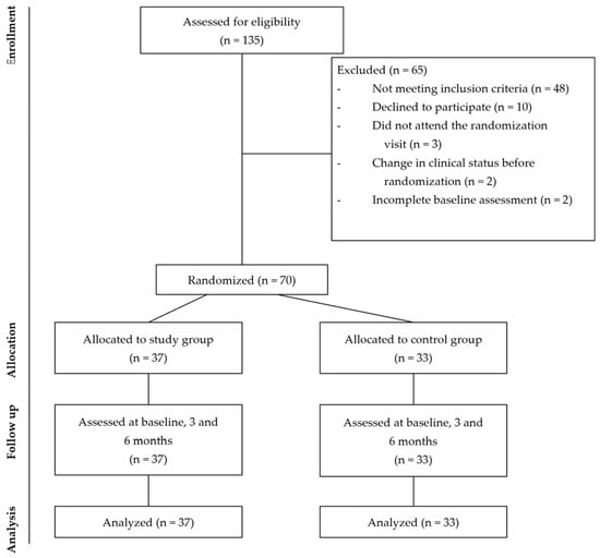
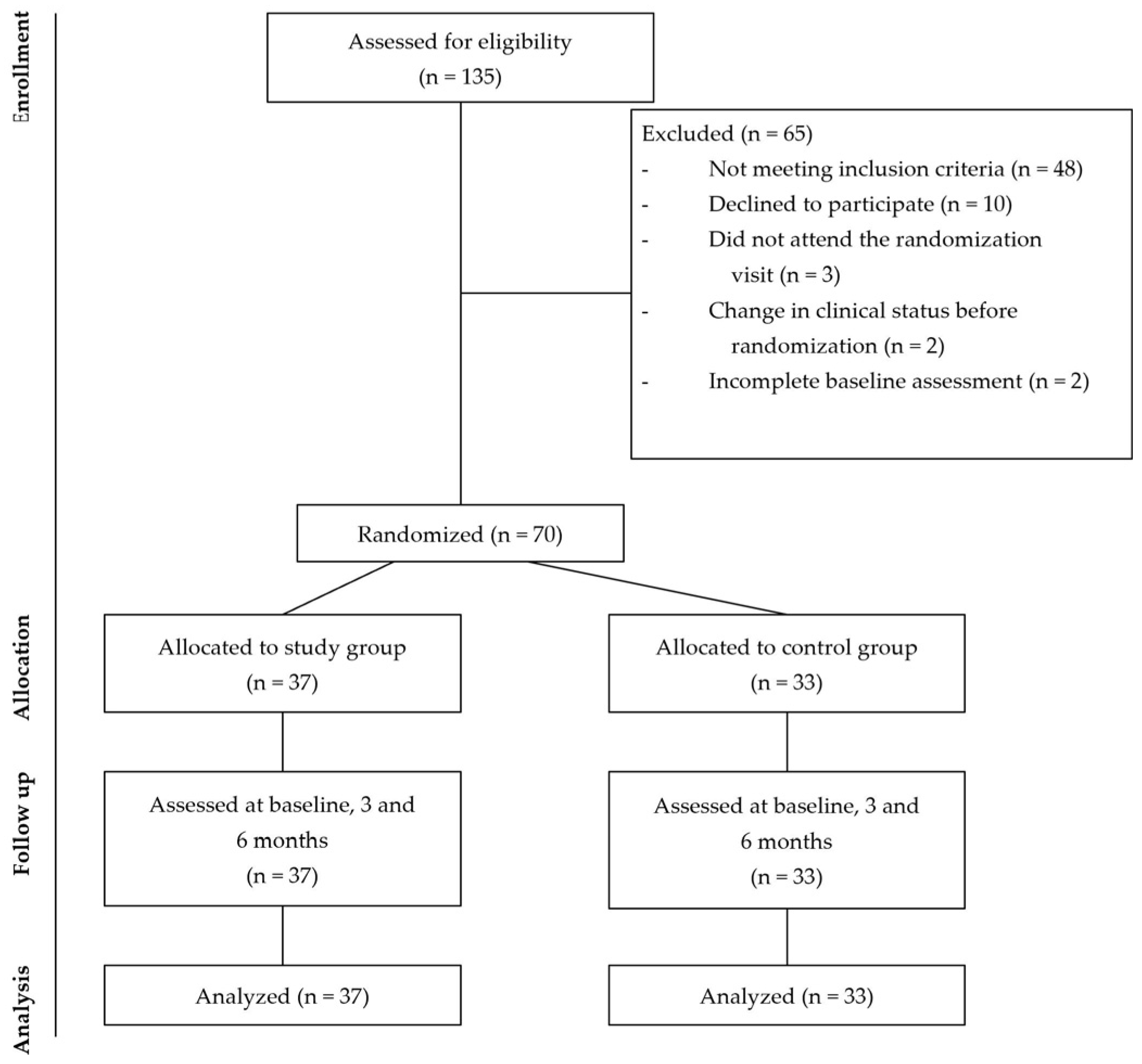
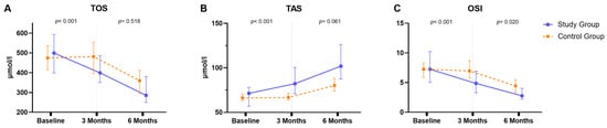
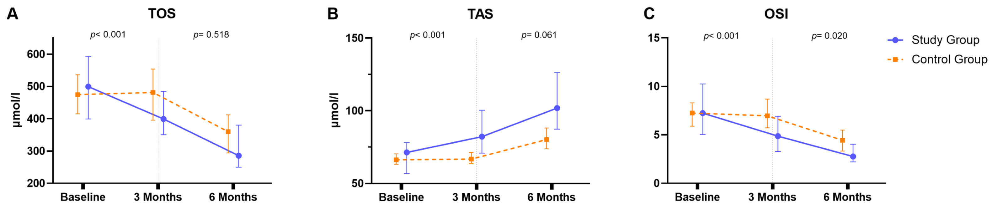
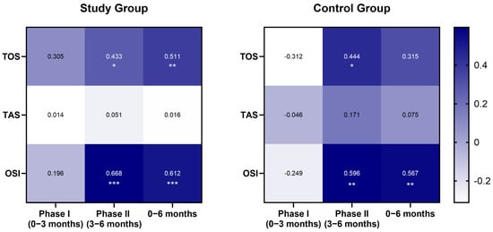
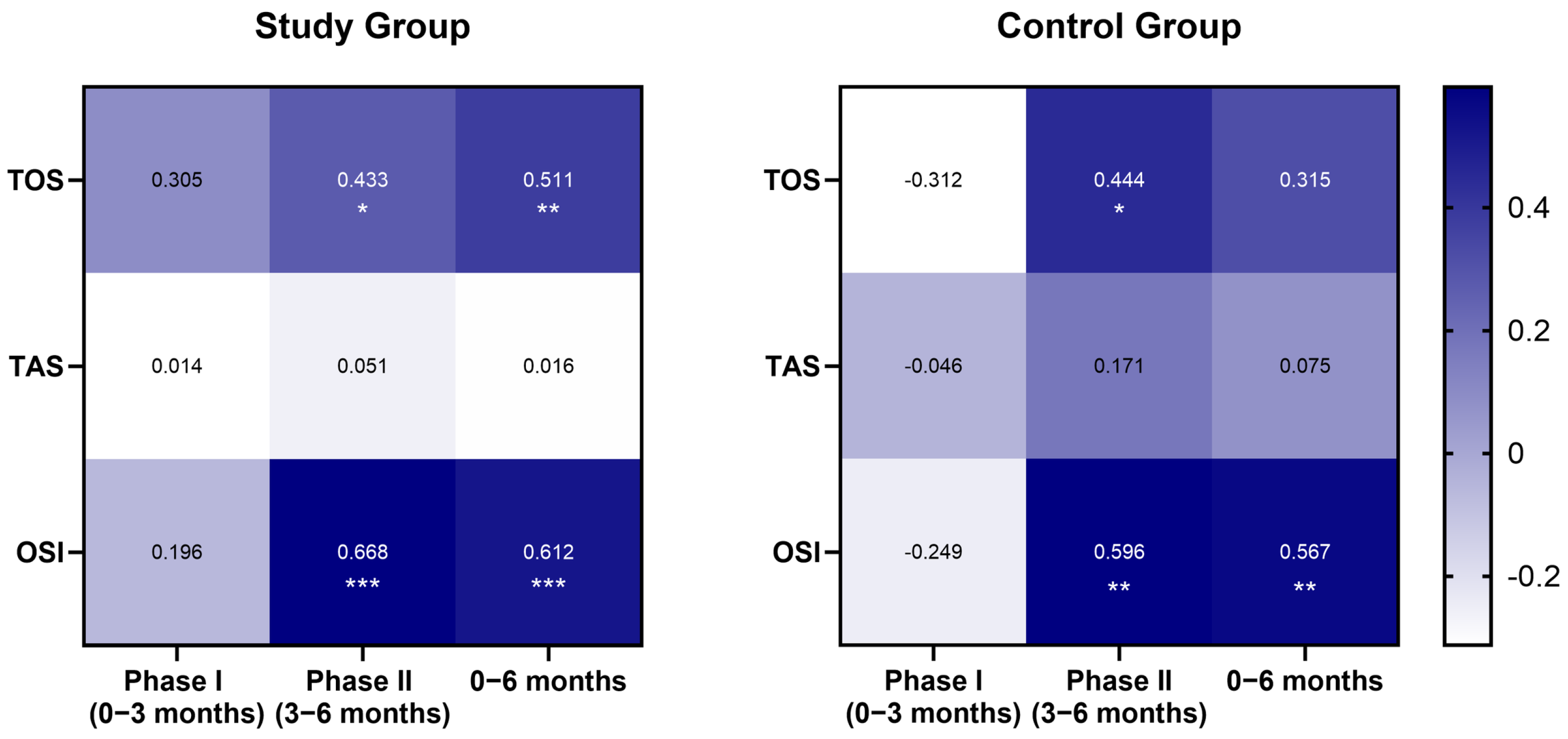
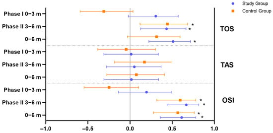
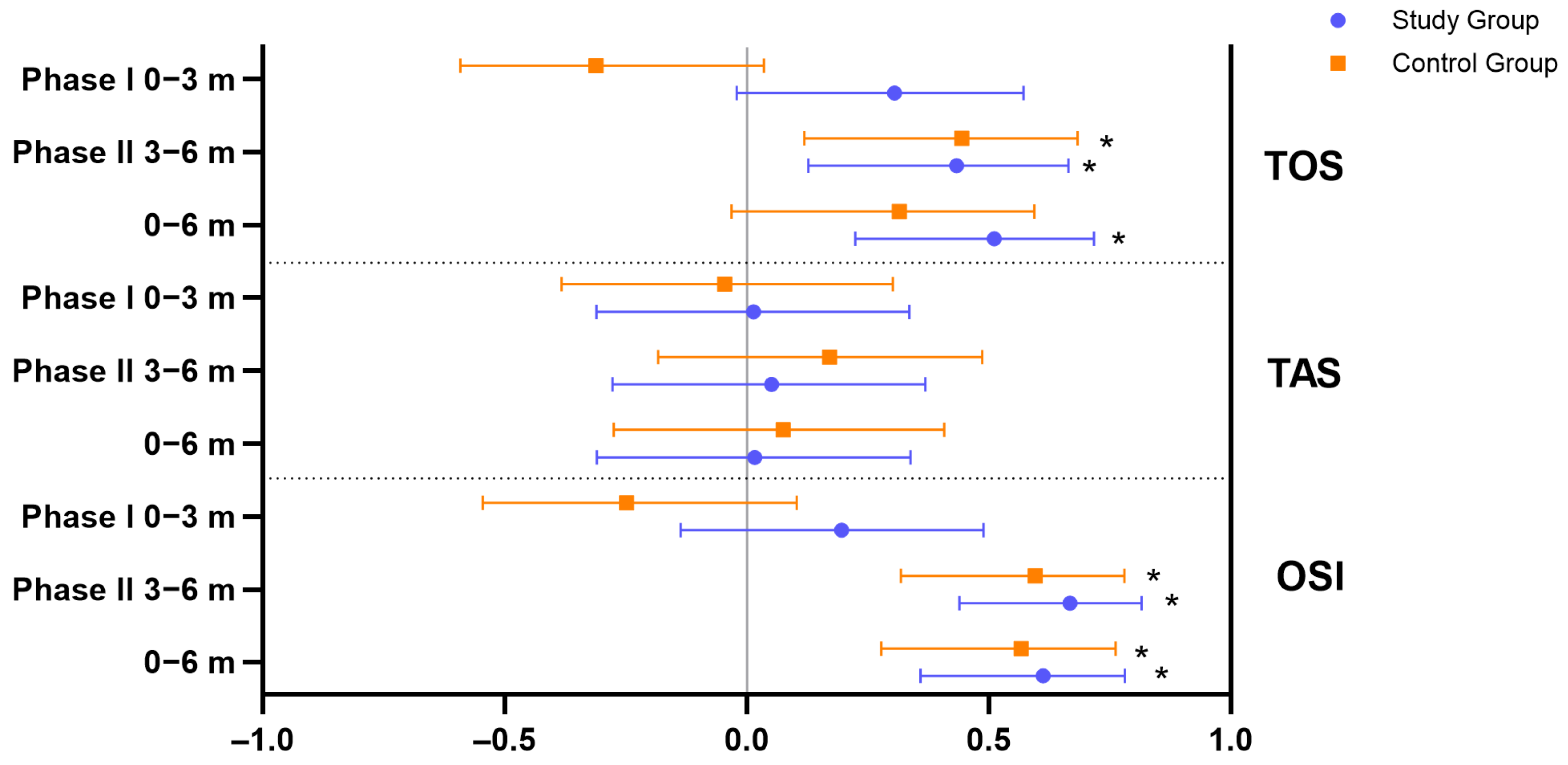
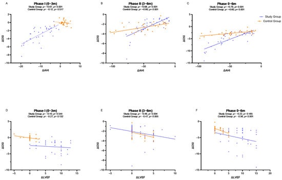
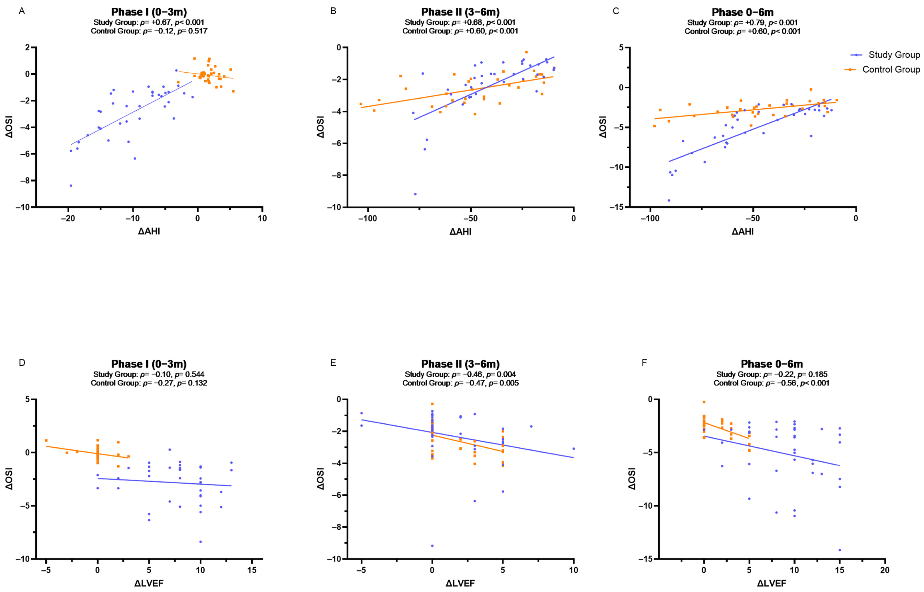
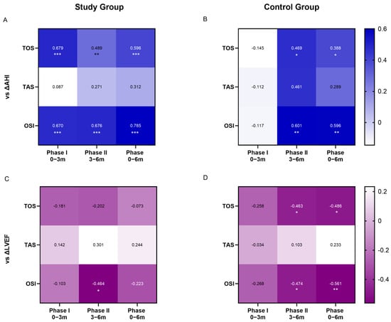
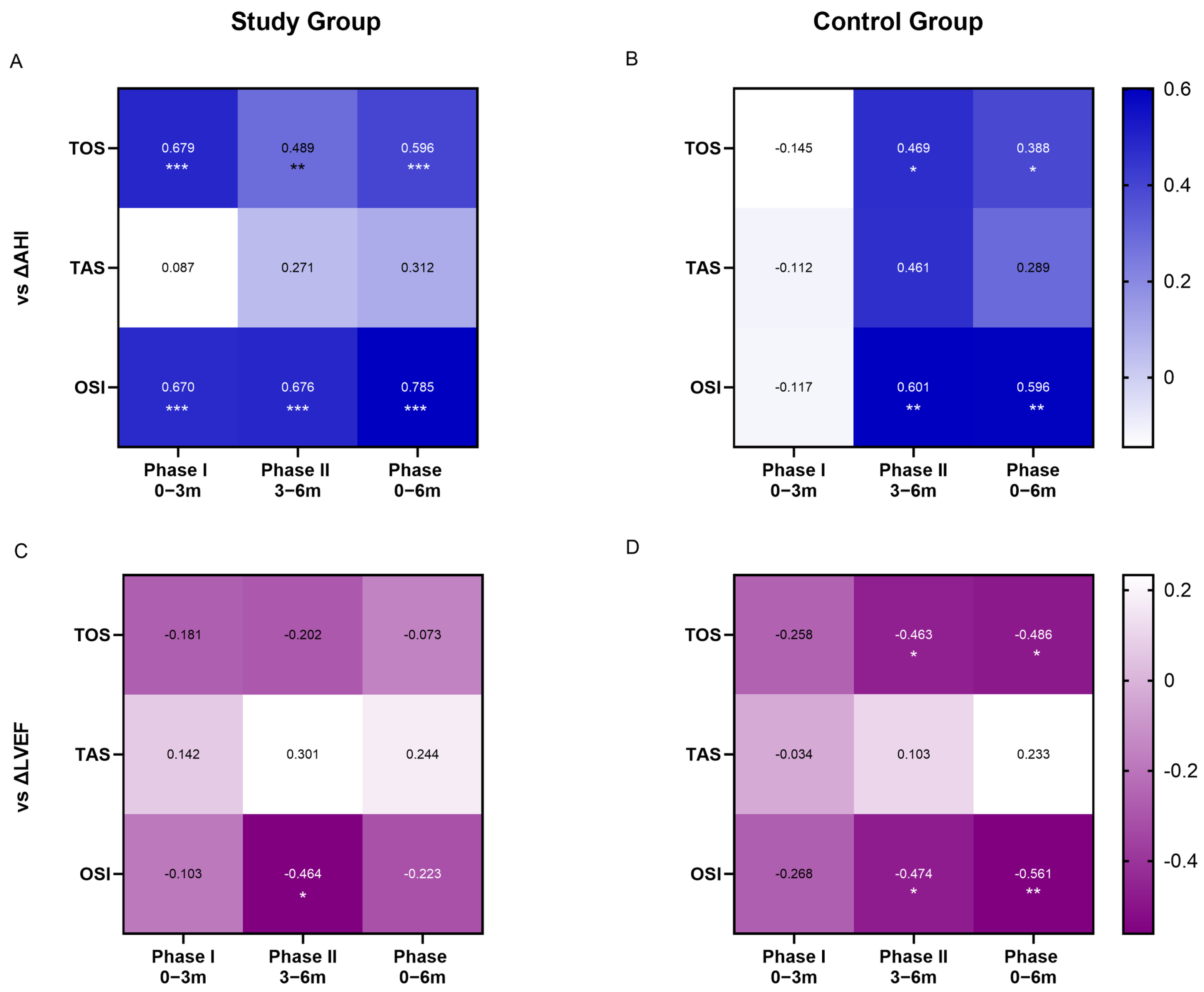
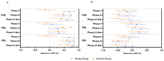
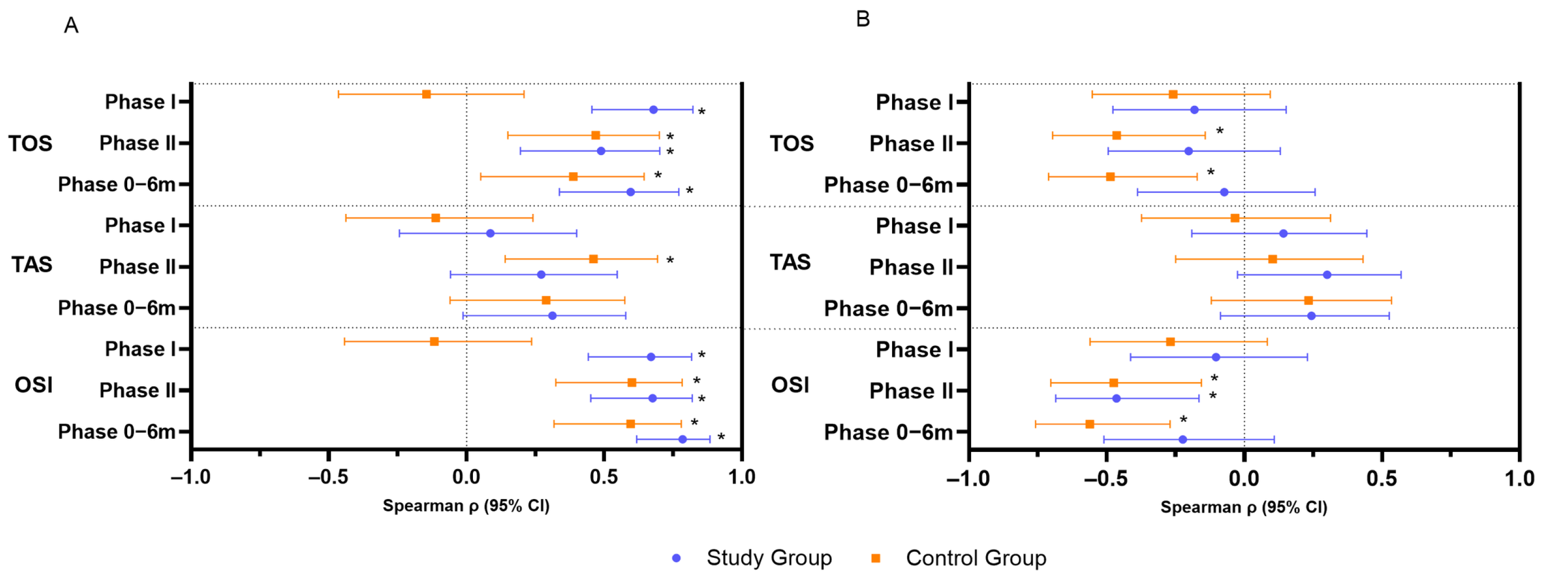

Disclaimer/Publisher’s Note: The statements, opinions and data contained in all publications are solely those of the individual author(s) and contributor(s) and not of MDPI and/or the editor(s). MDPI and/or the editor(s) disclaim responsibility for any injury to people or property resulting from any ideas, methods, instructions or products referred to in the content. |
© 2026 by the authors. Licensee MDPI, Basel, Switzerland. This article is an open access article distributed under the terms and conditions of the Creative Commons Attribution (CC BY) license.