


Abstract
In recent years, the growing research interests in the applications of plant and fruit extracts (synthetic/stabilization materials for the nanomaterials, medicinal applications, functional foods, and nutraceuticals) have led to the development of new analytical techniques to be utilized for identifying numerous properties of these extracts. One of the main properties essential for the applicability of these plant extracts is the antioxidant capacity (AOC) that is conventionally determined by spectrophotometric techniques. Nowadays, electrochemical methodologies are emerging as alternative tools for quantifying this particular property of the extract. These methodologies address numerous drawbacks of the conventional spectroscopic approach, such as the utilization of expensive and hazardous solvents, extensive sample pre-treatment requirements, long reaction times, low sensitivity, etc. The electrochemical methodologies discussed in this review include cyclic voltammetry (CV), square wave voltammetry (SWV), differential pulse voltammetry (DPV), and chronoamperometry (CAP). This review presents a critical comparison between both the conventional and electrochemical approaches for the quantification of the parameter of AOC and discusses the numerous applications of the obtained bioextracts based on the AOC parameter.
1. Introduction
During the metabolic conversions of food into the energy, excess amounts of the free radicals are constantly generated in the human body. These unstable and highly reactive free radical species are a constant threat to the constituents of the cells (particularly the genetic materials) and destroy the constituents via numerous destructive mechanisms [1]. Therefore, the modulation of the concentration of these free radical species is an essential task [2]. Though oxidative metabolism plays an essential role in the survival of our cells, the production of free radicals (along with some other reactive oxygen species) during this process still causes various oxidative changes [3]. However, we are not defenseless against these free radicals and their relentless attack. This is attributed to the presence of certain molecules in our bodies called antioxidants. Antioxidants operate in two ways in order to address the issue of the oxidative stress in the human body: (1) the antioxidants scavenge/quench these reactive oxygen species (ROS) or reactive nitrogen species (RNS) by chain-breaking mechanisms (these antioxidants are called primary antioxidants) [4]; and (2) the antioxidants that suppress the generation of the oxidation promoters (such as singlet oxygen, metal ions, and pro-oxidation enzymes, etc.) by chelation mechanisms (these antioxidants are called secondary or preventive antioxidants) [5]. The most familiar out of thousands of such antioxidant substances are Vitamin C, Vitamin E, beta carotenes, many related carotenoids, and minerals like selenium and manganese, etc. [6]. Antioxidants hold great interest for pharmacists and biochemists because of their capability to moderate the damages caused by RNS, ROS, or even chlorine-like reactive species [7]. The importance of antioxidants is highlighted in the food preservation applications as presented in Figure 1.
Figure 1.
Evolution of food antioxidants as a core topic of food science. Figure adapted with permission from Ref. [8]. Elsevier.
Growing interest has been recently observed in developing cost-effective and reliable techniques for screening and quantifying the antioxidants present in various biological/biogenic samples. The quantification of the antioxidant potential is done by using the parameter of the antioxidant capacity (AOC), which represents the concentration or number of moles of the specific free radical species scavenged by a particular antioxidant present in the sample [9]. The AOC parameter is a thermodynamic parameter and is found to be connected with the equilibrium constant of the process (scavenging reaction of antioxidant) [10]. Though a number of methodologies have been reported for estimating the AOC of the antioxidants, identifying the direct-action mechanism of antioxidants still remains a challenge in the field of free radical chemistry [10]. Antioxidant behavior is quantified using the two broad-term methodologies that reflect the focus on activity in foods (first category) and bioactivity in humans (second category). The category focusing on food systems involves the assessment of antioxidant efficacy, especially in fruits, vegetables and beverages so that a clear observation of their dietary burden and in vivo activity could be reported. As far as the antioxidant status in humans is concerned, there has been reported an obvious observation regarding an imbalance between the reactive oxygen species and defense/repair mechanisms in vivo [3,11]. Understanding both quantification methodologies (usually termed as antioxidant assays) is essential in developing a clear picture of the working of antioxidants.
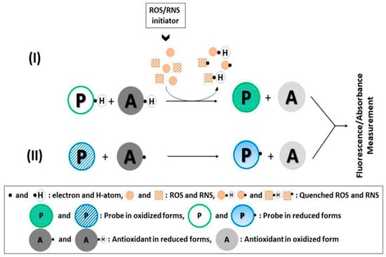
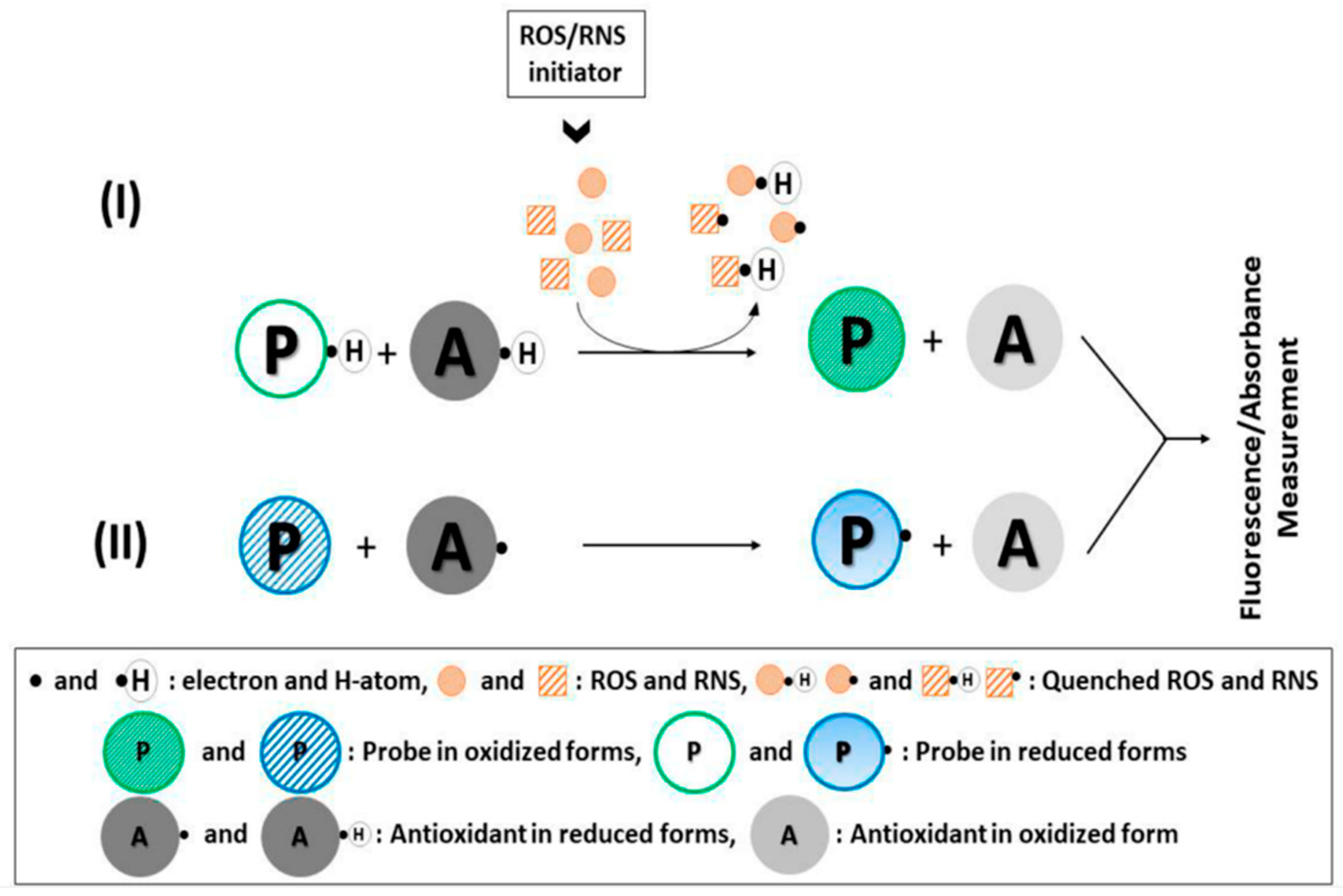
The antioxidant assays can be broadly categorized into two classes of direct antioxidant assays and indirect antioxidant assays [12]. The first category of direct assays is the competitive technique, and the added probe, as well as the antioxidants present in the sample both competes with each other to attack the free radical/free radical initiator. This makes these direct assays a little less accurate technique to detect the AOC [11]. The indirect antioxidant assays are non-competitive tests where an artificial probe (an oxidant) is added to the sample to observe the impact of the antioxidant action on this probe. The attack of antioxidant molecules generates structural changes in the added probe, which is measured by spectroscopic, fluorescence, electrochemical, or other methodologies. The indirect assays are more common and provide more accurate results in comparison to the direct assays owing to their non-competitive nature [12]. Figure 2 shows the schematics of the mechanism of direct or indirect assays used in this regard. Furthermore, it should also be mentioned here that the free radicals are scavenged by utilizing two mechanisms, including the electron transfer (ET) and hydrogen atom transfer (HAT) mechanism. In the ET mechanism, the compound of interest is reduced by the transference of one or more electrons from antioxidants to the unstable species, while in the HAT mechanism, the compound of interest is reduced by using the transference of the hydrogen atom to the probe [12].
Figure 2.
(I) Direct (competitive) antioxidant assay involving a fluorogenic or chromogenic 1263 probe and biologically relevant ROS/RNS; (II) indirect (noncompetitive) antioxidant assay in 1264 probe where the physiological redox reactions (i.e., oxidant–antioxidant interactions) are simulated on 1265, an artificial probe without biologically relevant ROS/RNS. Figure adapted with permission from Ref. [12]. Copyright 2019 American Chemical Society.
In this review article, critical comparisons of numerous antioxidant assays utilized for the estimation of AOC in the plant/fruit extracts are presented. The first section provides a brief description of the fundamentals of antioxidants. The second section represents a summary of the conventional methodologies, while the third section provides a detailed analysis of the electrochemical techniques utilized for the estimation of AOC in the biogenic extracts. Some peculiar case studies explaining the applicability of the electrochemical techniques for the estimation of AOC are summarized in the fourth section. The last section details the conclusion and future prospects associated with this particular research domain of electrochemical techniques used for the determination of AOC.
2. Conventional Assays/Methods for Determination of AOC
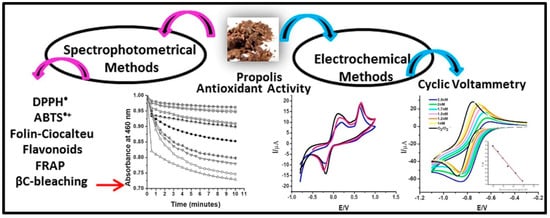
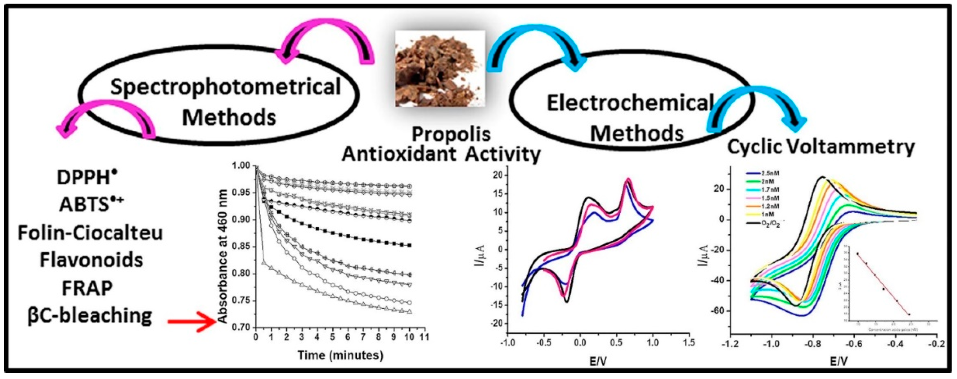
Many analytical techniques have been developed for the extraction, separation, identification and quantification of antioxidant compounds by using various conventional methodologies [13]. The classification of the numerous antioxidant assays utilized for the determination of AOC is presented in Table 1. AOC is either expressed in the form of equivalents of the selected reference antioxidants (particularly gallic acid and Trolox) in the case of radical/ROS-based scavenging assay or inhibition potential of the antioxidant against the oxidation of the added probe [14]. The oxidation of the added probe is determined by using analytical techniques such as fluorimetric, spectrophotometric, cyclic voltammetry, amperometric methods, and chemiluminescent, etc. [13]. The electrochemical methods fall under the category of non-radical/potential-based antioxidant assays and are not discussed in Table 1. The electrochemical methodologies are discussed in detail in the next section. A comparative study of different methodologies for the determination of the antioxidant activity of Venezuelan propolis, as an example, has been shown in Figure 3.
Table 1.
Conventional assays/methods utilized for the determination of AC.
Figure 3.
Comparative study of different methodologies for the determination of the antioxidant activity of Venezuelan propolis. Figure adapted with permission from Ref. [15]. Copyright 2020 Elsevier.
Advanced spectrometric methods have also been widely documented for analyzing the antioxidant properties of reported natural antioxidants found in the biogenic samples. However, these methodologies exhibit strong dependence on parameters such as optimization of temperature, time, concentration, and other physicochemical characterization of antioxidant substances, which is needed to be crucially considered before experimentation [27]. High-performance liquid chromatography has been primarily used for the separation, identification, and quantification of nutraceutical components for many years. However, mass spectrometry, when coupled with liquid chromatography, has recently been introduced as a detection method for many such investigations with even lower detection limits [28]. The summary of some case studies documenting the use of conventional and electrochemical detection methodologies used for the identification of AOC in natural and synthetic antioxidants, along with their applications, are provided in Table 2.
Table 2.
Some analytical methods applied to determine the antioxidant and total antioxidant capacities..
3. Electrochemical Methodologies
In recent years, electrochemical methodologies have emerged as the competing technique against the antioxidants for the determination of AOC in biogenic samples [41]. The increase in the applicability of these techniques for AOC determination is attributed to the fact that extremely low molecular weight anti-oxidants can be easily detected and screened by using these techniques. The electrochemical methods operate by calculating the total reducing power of the sample, which represents the ability of certain antioxidants to donate/accept electrons in a redox (oxidation–reduction) environment [42]. Electrochemical methods are considered very sensitive and rapid methodologies for the determination of antioxidant potential in both stationary as well as dynamic flow systems [43]. Moreover, it should be made clear that most electrochemical techniques provide the information regarding the total antioxidant capacity (TAC) values of the antioxidants, which represents the cumulative impact of all the antioxidants present in the sample in contrast to the determination of the individual antioxidant potentials measured via a parameter of AOC. Electrochemical techniques involved in evaluating the antioxidant potential of biogenic samples are discussed in detail in the subsection [44].
Electrochemical techniques address a few major drawbacks associated with chromatographic and spectroscopic techniques, such as long sample preparation times, use of hazardous solvents, long analysis time, undefined reaction time and expensive setups. Furthermore, the sensitivity and reproducibility of the electrochemical methods is also quite high in comparison to the above-mentioned conventional antioxidant assays. Stationary systems are known for their suitability in quantifying only a limited amount of analytes, but the study of simple as well as more complicated biomolecules in dynamic systems can also be easily conducted by using these methodologies [45]. A carbon working electrode can be used in the case of both fluid and solid systems. Carbon paste electrodes, printed carbon electrodes, paraffin, and silicon oils modified carbonaceous materials (carbon pastes) are also used for a similar purpose [42]. Sometimes, the addition of inorganic/organic nanomaterials is also done to increase the selectivity or specificity of these electrodes. These modified electrodes perform quite well when used in voltammetry, amperometry and cyclic voltammetry techniques [42,46].
3.1. Cyclic Voltammetry (CV)
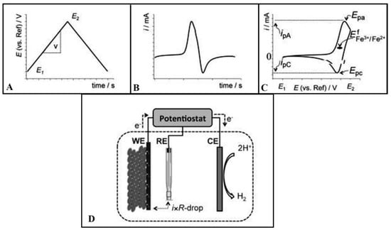
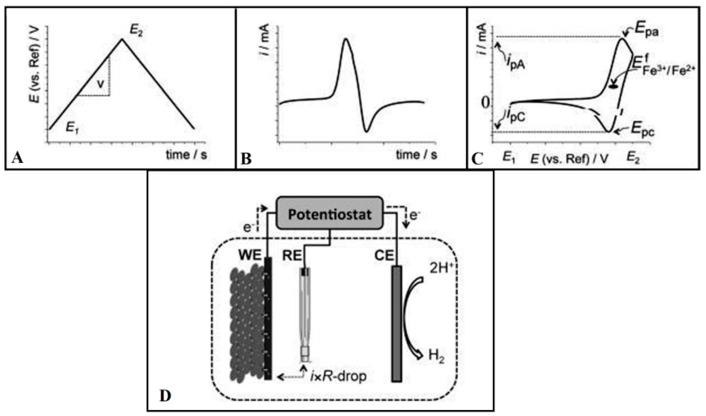
The CV is one of the most exploited electrochemical techniques for the determination of TAC in biogenic extracts. The CV comprises a stationary working electrode (WE) which is linearly ramped with respect to the time in a triangular waveform (i.e., the potential is increased from the lowest to the maximum value and after attaining the maximum potential, the potential is reversed to again attain the lowest value) [47]. Besides the WE, two other electrodes called reference electrodes (RE) and auxiliary/counter electrode (CE) is also part of the CV setup. The galvanostat/potentiostat is attached with the CV assembly that records the current values attained owing to the redox reactions occurring in the medium and the current versus potential graph is plotted for the understudy sample [48]. The typical setup utilized for CV, as well as the utilized waveform and typical voltammogram shape, is presented in Figure 4.
Figure 4.
In a CV, (A) the potential (with respect to RE) is applied to the WE. The potential start at the lowest potential value (E1), gains a maximum potential value (E2) and then is reduced back to the lowest potential value (E1); (B) The current generated owing to this potential change is plotted against time; and (C) The experimental CV setup indicating RE, CE, WE, and (D) A three-electrode potentiostatic experimental setup for anodic biofilm. Figure adapted with permission from Ref. [47]. Copyright 2012 John Wiley and Sons.
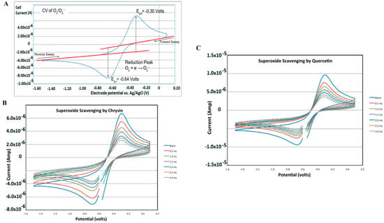
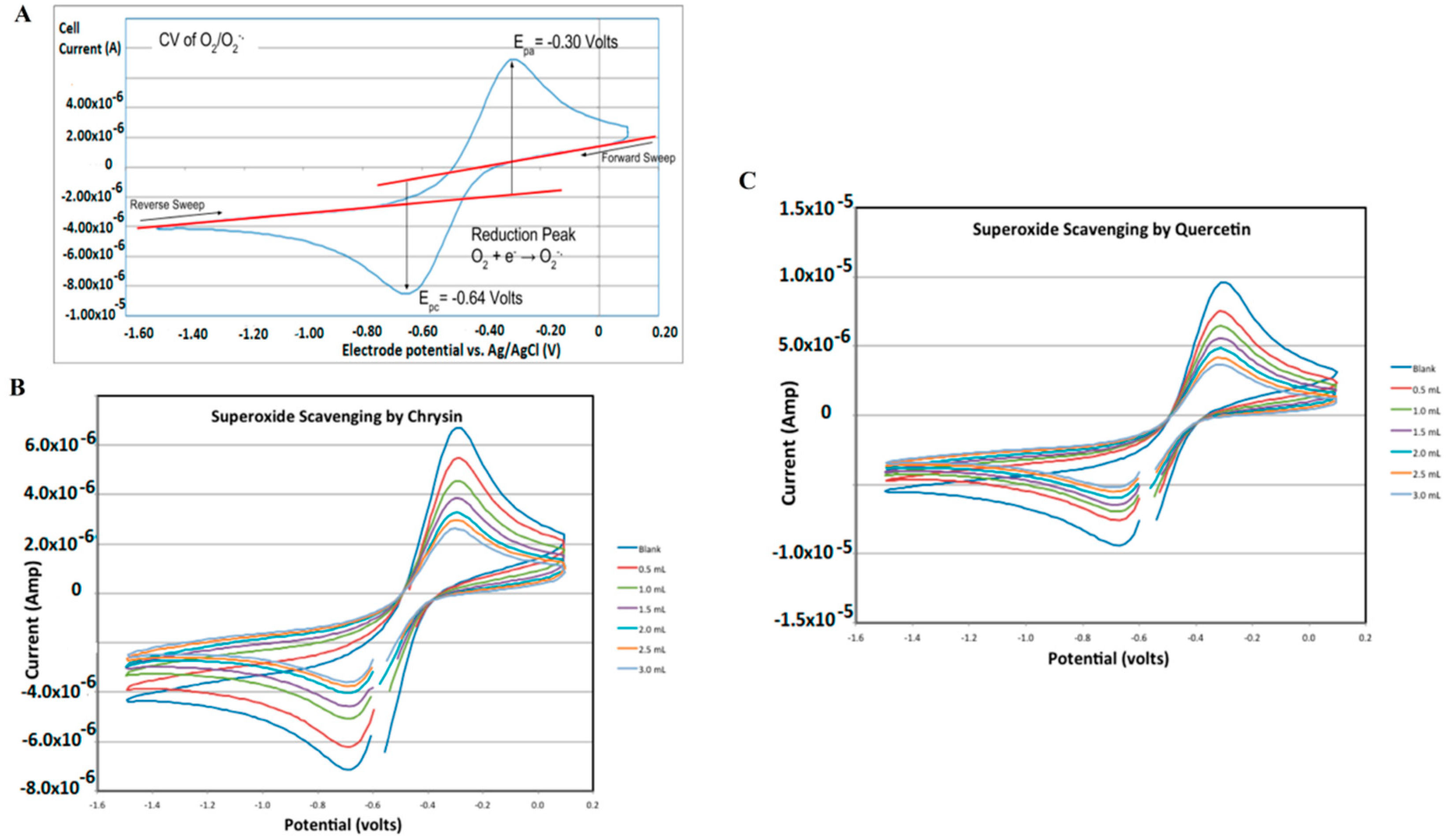
The reversible system is represented by the equation where O represents the oxidized form and R represents the reduced form. The typical CV voltammogram attained owing to the forward and reversed sweep in the case of superoxide radical is presented in Figure 5. As discussed previously, antioxidant scavenges the superoxide radicals, which results in a decrease in the current values observed in the presence of chrysin and quercetin (presented in Figure 5). This is a technique used to collect qualitative information while measuring the current response of any redox-active solution, such as the presence of intermediates in redox reactions to linearly cycled potential sweep between two or more set values [48]. Furthermore, this gives information about the characterization of redox systems, the number of redox states, stability of these states, and electron transfer kinetics of the redox reactions taking place in the medium [48,49]. The variation in the parameters of peak current (Ip) and peak potential (Ep) are the main tools utilized as a means to calculate the TAC value of antioxidants. It is used for determining the antioxidant activity of food, polyphenolic compounds [50], clinical samples [51], and pathological processes and infectious diseases [52,53].
Figure 5.
(A) CV voltammogram for superoxide radical ion; (B) Decrease in current values representing the superoxide radical ions in the presence of Chrysin; and (C) Decrease in current values representing the superoxide radical ions in the presence of Quercetin. Figure adapted with permission from Ref. [54]. Copy Right 2019 MDPI under Creative Commons Attribution Licence (CC BY 4.0).
3.2. Differential Pulse Voltammetry (DPV)
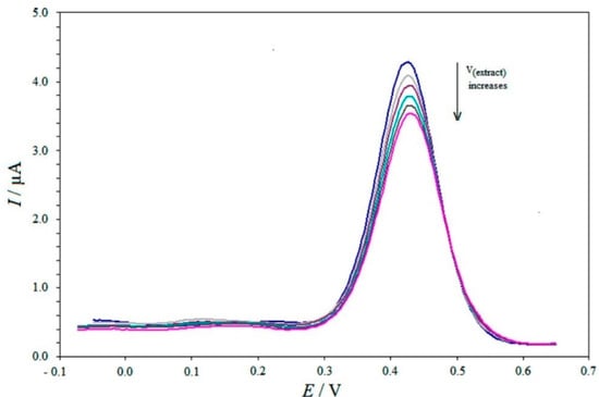
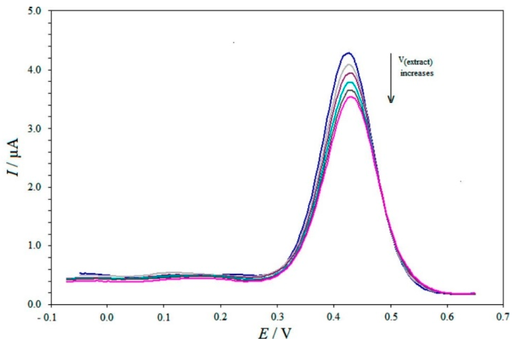
The quantitative chemical analysis, kinetics, thermodynamics and mechanisms of chemical reactions have also been studied by another electrochemical technique of DPV [55]. The DPV technique superimposes the fixed-magnitude pulses on a linear potential ramp. The potential-time curve acquired in the DPV technique is generated by recording the response current, which was sampled twice and the current difference is plotted against the potential to acquire the DPV voltammogram [56]. These measurements are advantageous because the effect of the charging current can be minimized, resulting in increased sensitivity in current measurements [57]. Figure 6 represents the DPV technique where the current peak corresponds to the concentration of cupric neocuproine complex (Cu(Nc)22+) and the addition of herbal tea extracts containing the antioxidants reduces this complex and consequently, peak current values are reduced. This reduction is utilized as a tool to measure the TAC of the antioxidants present in the tea extract [58]. Another important point is the extracted faradaic current which results in analyzing the electrode reactions more precisely [59]. It is also used effectively to measure the antioxidant capacity of food [60], plant extracts [61], and polyphenolic compounds such as procyanides and catechins from cone extracts which showed significant ability to reduce oxidative stress, scavenge free radicals, and transitions metal ions as well [62,63,64].
Figure 6.
DPV voltammogram representing the reduction in the peak current owing to the presence of the antioxidants in the herbal tea extract. Figure adapted with permission from Ref. [58]. Copyright 2014, American Chemical Society.
3.3. Square Wave Voltammetry (SWV)
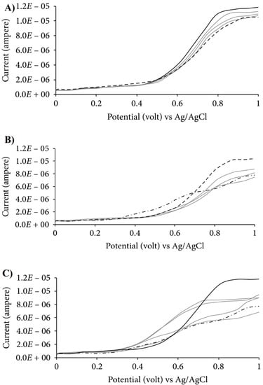
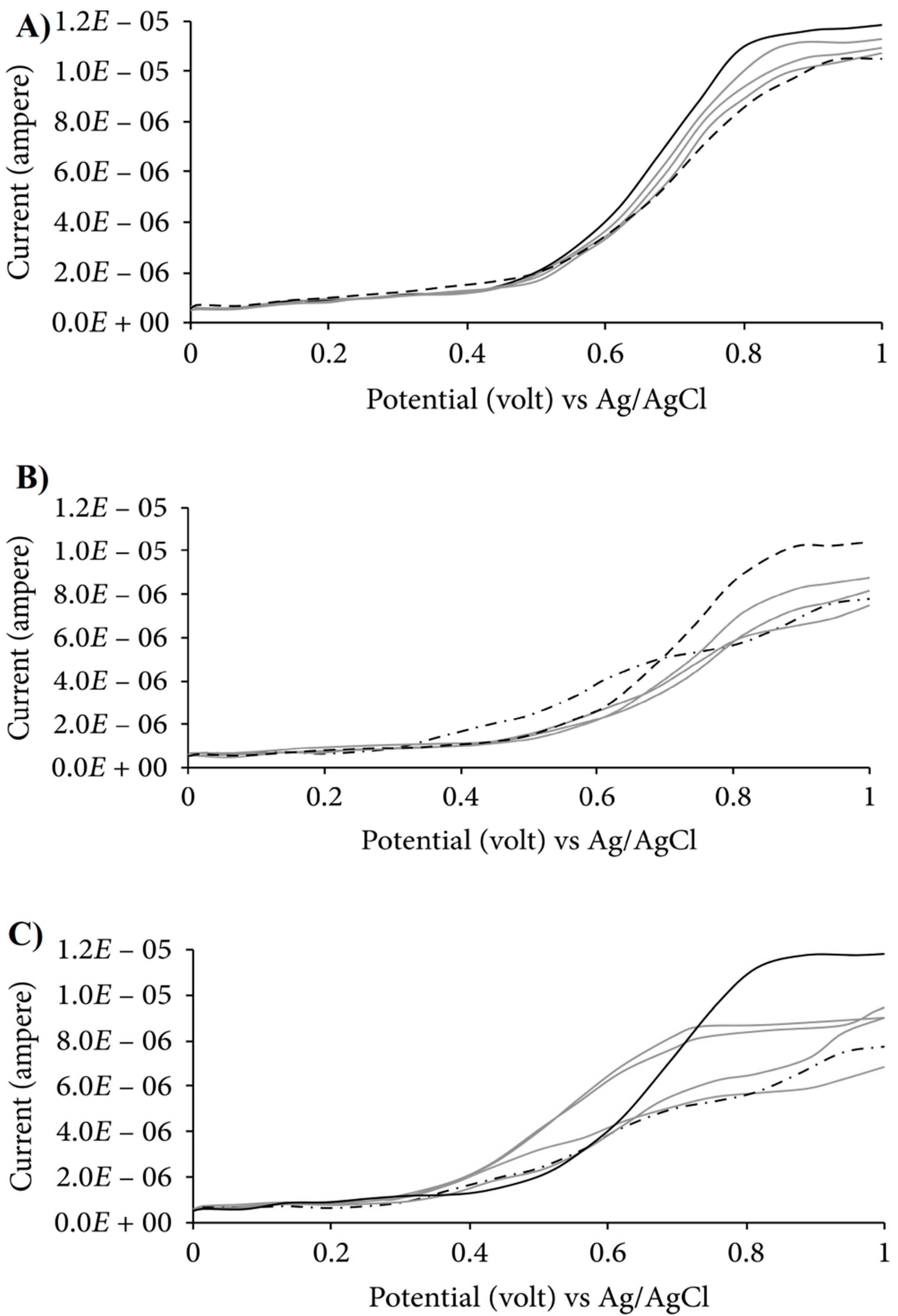
This pulsed voltammetry technique uses a potential waveform (where the entire symmetrical potential square wave is superimposed on the staircase potential waveform) that is implemented on the WE [65]. The typical voltammogram acquired in the SWV is presented in Figure 7. As observed in the case of an electrochemical technique, the voltammograms of the understudy compound exhibit peak/peaks that correspond to the numerous redox processes that analytes experiences in the medium. The peak potential/potentials (potential at the maxima of the peak) also provide essential information regarding the tendency of the antioxidants in order to perform the ET reactions. Moreover, the antioxidants display the peaks at the lower values of oxidation potential, which displays the tendency of the antioxidants to be oxidized [66]. In comparison to the linear sweep voltammetry, the SWV is more sensitive and has a more extended dynamic range with lower detection limits. This technique was used to identify and quantify the synthetic antioxidants tert-butylhydroquinone (TBHQ) and butylated hydroxy anisole (BHA) in the presence of the cationic surfactant CPB using a carbon black electrode and analysis of food samples and biodiesel, and also in tea samples [67].
Figure 7.
SWV voltammogram of binary mixture of extracts. (A) Blueberry and lemon; (B) blueberry and grape; (C) lemon and grape (where blueberry, lemon and grape extract are represented by a solid line, even dashed line and uneven dashed line, respectively, while gray lines represent the binary mixtures of the extracts in different ratios). Figure adapted with permission from Ref. [68]. Copyright 2019 Hindawi under Creative Commons Attribution Licence (CC BY 4.0).
3.4. Amperometric Measurements (Chronoamperometry)
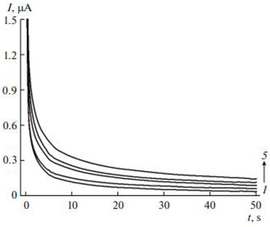
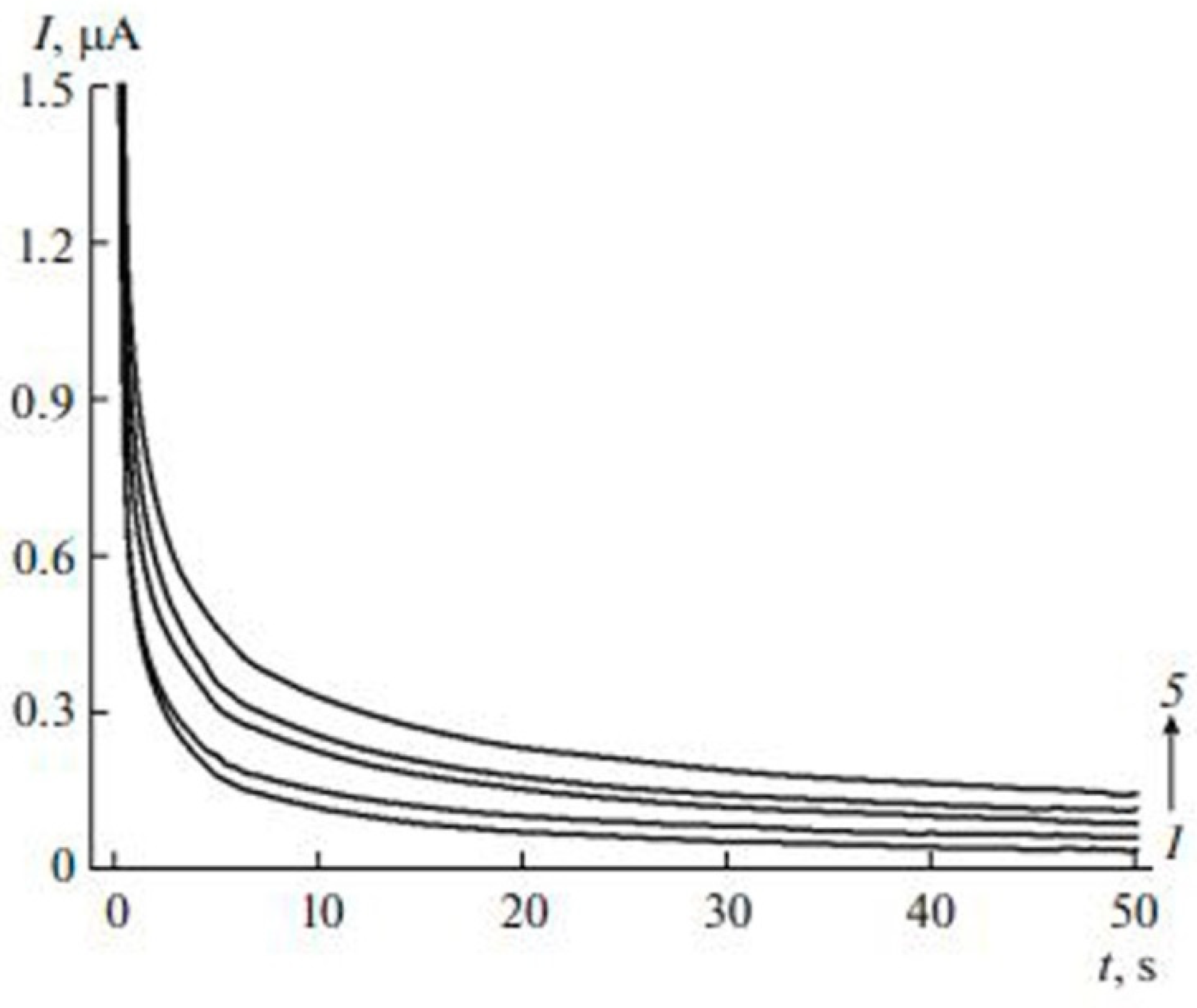
Chronoamperometry (CA) technique utilizes the stepping of the potential from the potential value at which there is no faradic reaction to the potential value and where the dose of electroactive species at the WE is essentially zero [69]. The variation in the response current is documented with respect to time to generate the CA potential-time graph. The typical chronoamperogram acquired in the case of the extracts of the tea leaves was performed by using the WE of glassy carbon electrode (GCE) that is modified with carbon nanotubes (MWCNTs) and electropolymerized quercetin (EPQ) is presented in Figure 8. Various antioxidant compounds have been analyzed for their antioxidant activity using amperometric techniques [70]. Disposable polyester screen-printed graphite macroelectrodes have been used coupled with a batch injection cell for measurement of consumption of DPPH. This technique measures electric current from oxidation of a substance such as food samples when studied on the surface of a working electrode at some fixed voltage potential using amperometric detectors [71].
Figure 8.
Chronoamperograms of (1) phosphate buffer, (2) blank, (3) semi-fermented tea leaves, (4) white tea leaves, and (5) green tea leaves. Figure adapted with permission from Ref. [72]. Copyright 2017 Springer Nature.
4. Applications of the Electrochemical Methods for the AOC
Different case studies utilizing the electrochemical methods as a means for the determination of AOC are discussed in the subsection below [69,70,71,72]. These particular case studies are classified on the basis of the origin and utility of the biomass extract for this review.
4.1. AOC Determination of the Fruit’s Extract
A literature survey indicated that among the utilized electrochemical techniques, CV is the most abundantly exploited electrochemical technique used for the determination of the AOC in the fruit’s extract [73]. Antioxidants having moderately low-molecular weight have been particularly documented to be determined with quite an ease via this technique [74]. Zeliǹska et al. [75] examined 11 randomly selected samples of apple cultivars and used the techniques of CV for documenting the AOC, reduction, and chelating potential of the apple’s (peel and flesh) extract. The parameter of first anodic peak potential (Epa) was utilized to investigate the reduction potential of the phytochemicals present in the extract. The Epa values of 0.815 V, 0.759 V, 0.618 V, 0.571 V, 0.515 V, 0.512 V, 0.511 V, 0.391 V, 0.390 V, 0.339 V and 0.334 V were recorded for the phytochemicals of phloretin, phloridzin, cyanidin 3-galactoside, catechin, quercetin 3-rhamnoside, quercetin 3-arabinoside, quercetin 3-glucoside, chlorogenic acid, rutin, epicatechin, and quercetin, respectively. The higher Epa values are attributed to the lower reducing activity of the phytochemicals and these results are translated into the high to moderate antioxidant power. Area under the current wave was also utilized to detect the order of the AOC potential. Comparison and correlation studies revealed that the results acquired from CV technique were found to be most compatible with Ferric reducing antioxidant power (FRAP) assay among all other spectroscopic and chromatographic techniques. The literature survey further indicated that the peak current, the area under the current curve, and peak potential are the most important parameters for studying the AOC of the extract [76,77]. The peak potential of the acquired phytochemicals correlates with the type of the reducing agent utilized [78]. The lower oxidation potential values were found to be associated with the enhanced strength of particular phytochemicals for the electron donation and, therefore, directly provide information regarding the AOC of the molecule [79]. The Randles–Sevcik equation (discussed in the above equation) also provides a means to access the antioxidant dose present in the extract via the value of the peak current ().
Jara-Palacios et al. [80] utilized the AOC values of the grape pomace (a solid residue composed of approximately 20% of total solid weight of the fruit of the grape, acquired as waste from the grape juice or wine industries) by using the CV technique and thiobarbituric acid reactive substances (TBARS) assay. The TBARS assay is a measure of inhibition of lipid peroxidation (the series of the particular chemical reactions responsible for the oxidative degradation of the lipids molecules). This inhibition is done owing to the presence of the antioxidants in the medium as peroxide molecules are scavenged by these molecules. Apart from identifying the AOC via CV, the electrochemical data was also used to study the by-products of these antioxidant reactions [80]. De Macêdo et al. [73] synthesized a laccase-based biosensor (LLB) and used it for the detection of antioxidants in the extracts of acerola, cranberry, camu-camu, and açai red fruits. The electroanalytical DPV, CV and SWV techniques were also used for this purpose and the parameter of the electrochemical index (EI, presented in Equation (1)) was evaluated for studying the antioxidants:
where the Ipa1 to Ipan and Epa1 to Epan represent the anodic current and anodic potential of each successive anodic peak from 1 to n. The LLB and EI both represented the same order for the AOC potential in different varieties of the red fruits, i.e., camu-camu cranberry açai = acerola. However, the order of AOC potential documented by the DPPH assay did not relate to the LLB and electrochemical techniques, indicating that further research is still needed in this domain to fully comprehend the usefulness of the electrochemical techniques for measuring the AOC values [73]. The electrochemical techniques were also utilized for investigating the AOC values in the extracts of the following edible nutraceuticals, including apricot, black currant, grape pomace (by CV) [64], carrots, persimmons, pumpkins (by DPV), [46], Vaccinium meridionale swart/Agraz (by CV) [81], hops (Barth-Haas, Hallertau, Columbus, and Cascade) and malted barley samples (Chateau Cristal, Pilsen, Chateau Munich, and Wheat), sour cherry, pomegranate, plum, prickly pear (cactus fruit), date, and nectarine (by CV) [82], etc.
4.2. AOC Determination of the Plant’s Extract
Similar to the trend observed in the case of the fruit extracts, the technique of CV was found to be most effectively utilized for investigating the antioxidants of the plant’s extract as well [83]. However, the utilization of other techniques of DPV and SWV was found to be more abundant in comparison to the fruit extracts [84]. Khiya et al. [83] determined the AOC, TPC, condensed tannin content (TCT), and TAC of the Salvia officinalis by using the following assays/techniques of FRAP, DPPH, phosphomolybdate reduction, and CV. The extract acquired from the Boulemane region exhibited the highest value of total phenolic content (176 mgGAE/g), while the extract acquired from Khenifra region exhibited the highest TCT values (162.53 mgEC/g). In the case of TAC (highest value: 108 mgGAE/g), the aqueous fraction of the S. officinalis leaves acquired from the Boulemane region exhibited the best results. However, the CV results exhibited different results in comparison to other assays, as the best AOC results (288.8 mgGAE/g) were found in the case of the hydro-methanolic extract of the particular plant. Interestingly, the authors also highlighted the fact that the CV can only be utilized to specify the generalized type of phytochemicals; the technique of HPLC is still required for the documentation/characterization of individual molecules present in the understudy extract [60]. The authors also performed the correlation studies and concluded that the phenolic components (rather than flavonoid compounds) were responsible for AOC. This study highlights that every assay/technique has its limitation and the selection of the type of extracting medium (such as aqueous, methanoic, ethanoic or hydro-methanoic, etc.) should be considered beforehand for selecting the technique used for measuring AOC.
Amamra et al. [85] investigated the AOC values for Thymus vulgaris by utilizing spectroscopic assays and electrochemical techniques. The authors investigated butanol, ethyl acetate, aqueous, chloroform, petroleum ether, and methanolic extracts and revealed that all the extracts contained phenolics, flavonoids and tannins components. In terms of CV, the methanolic extract exhibited the best results with a correlation coefficient of 0.964 and 0.989 for gallic acid equivalent (GAE) and ascorbic acid equivalent, respectively, with the other assays (DPHH, Iron chelation assay, and 2,2-azinobis-(3-ethylbenzothiazoline-6-sulfonic acid)-radical cation (ABTS+) assay) for phenolic contents. Chochevska et al. [86] presented a comparison of the AOC values for the homemade-fruit vinegar (HMV) and commercial vinegar by using the technique of CV and exhibited that all the samples of HMV (acquired from the fermentation of the fruits of blueberry (Vaccinium myrtillus); apple (Malus domestica), raspberry (Rubus idaeus), rose hip (Rosa canina), blackberry (Rubus fruticosus), and persimmon (Diospyros kaki)) exhibited at least ten times more AOC values in comparison to the commercial vinegar samples. Fuentes et al. [87] used the technique of the chronoamperometry and chronopotensiometry techniques as a means to detect the AOC in the extracts of Aliso (Alnusacuminate) plant. Table 3 summarizes the critical remarks observed to be associated with the electrochemical techniques utilized for the detection of AOC.
Table 3.
Summary of the case studies documenting the AC values and other parameters calculated by using the electrochemical techniques.
5. Conclusions
This recent literature review presents critical insights into the use of the electrochemical methodologies as a tool to detect the AOC values of the plant and fruit extracts. The techniques of CV, DPV, SWV, and AM were explored for achieving this purpose. It was found that the CV is the most extensively exploited technique among all other electrochemical techniques for the detection of the AOC in numerous extract-based samples. In most studies, the electrochemical methodologies were used as an additional tool alongside other conventional (spectroscopic and chromatographic) techniques for the AOC determination, which is a welcome approach as the literature survey indicates that a contradiction still exists regarding the documented values of correlation of these electrochemical techniques with other technique. Furthermore, the AMM technique is far less explored in comparison to all other techniques, indicating that further research is still needed to explore the full potential/advantage of the electrochemical techniques for the determination of AOC. The nature of phytochemicals was also found to be critical as the AOC values of the extract containing the high molecular weight antioxidants cannot be found by using the electrochemical techniques.
6. Future Perspectives
Innovation in every field needs a multiprobe approach. The use of simulations and data-driven techniques can speed up the development in the investigation of the antioxidant potential of plant/fruit extracts. The density functional theory can be used to study the antioxidant potential of molecules in plant/fruit extracts [92,93,94]. The information from the theoretical analysis can help in selecting methods for experimental work. Recently, the use of machine learning has been increasing in biological and material science [95,96]. The machine learning models also can be used to predict the antioxidant potential of plants/fruits. It will shorten the discovery of efficient antioxidants; it is easy and more cost-effective way.
Following future perspectives can be observed regarding the use of the electrochemical methodologies for AOC values.
- The electrochemical techniques should be applied in conjugation with the chromatographic technique as the electrochemical techniques were still not able to identify the phytochemicals present in the extract.
- During the study of the correlation of the electrochemical techniques with the conventional methodologies, the fact that the electrochemical techniques are used to measure the TAC rather than measuring the particular antioxidants for the particular reaction should not be ignored. The cumulative antioxidant potential should be considered to be a preferable approach in determining the selective antioxidants via particular reactions.
- The flow injection analysis alongside the stationary electrochemical techniques should also be performed for studying the AOC potential of the extract.
Funding
This work was supported by Al Bilad Bank Scholarly Chair for Food Security in Saudi Arabia, the Deanship of Scientific Research, Vice-Presidency for Graduate Studies and Scientific Research, King Faisal University, Saudi Arabia [Project No. CHAIR69].
Acknowledgments
This work was supported by Al Bilad Bank Scholarly Chair for Food Security in Saudi Arabia, the Deanship of Scientific Research, Vice-Presidency for Graduate Studies and Scientific Research, King Faisal University, Saudi Arabia [Project No. CHAIR69].
Conflicts of Interest
The authors declare no conflict of interest.
References
- Munteanu, I.G.; Apetrei, C. Analytical methods used in determining antioxidant activity: A review. Int. J. Mol. Sci. 2021, 22, 3380. [Google Scholar] [CrossRef] [Scilit] [PubMed]
- Masek, A.; Latos-Brozio, M.; Chrzescijanska, E.; Podsedek, A. Polyphenolic profile and antioxidant activity of Juglans regia L. leaves and husk extracts. Forests 2019, 10, 988. [Google Scholar] [CrossRef] [Scilit]
- Antolovich, M.; Prenzler, P.D.; Patsalides, E.; McDonald, S.; Robards, K. Methods for testing antioxidant activity. Analyst 2002, 127, 183–198. [Google Scholar] [CrossRef] [Scilit] [PubMed]
- Apak, R.; Özyürek, M.; Güçlü, K.; Çapanoğlu, E. Antioxidant activity/capacity measurement. 1. Classification, physicochemical principles, mechanisms, and electron transfer (ET)-based assays. J. Agric. Food Chem. 2016, 64, 997–1027. [Google Scholar] [CrossRef] [Scilit]
- Sadeer, B.N.; Montesano, D.; Albrizio, S.; Zengin, G.; Mahomoodally, M.F. The versatility of antioxidant assays in food science and safety—Chemistry, applications, strengths, and limitations. Antioxidants 2020, 9, 709. [Google Scholar] [CrossRef] [Scilit]
- Bunaciu, A.A.; Danet, A.F.; Fleschin, Ş.; Aboul-Enein, H.Y. Recent applications for in vitro antioxidant activity assay. Crit. Rev. Anal. Chem. 2016, 46, 389–399. [Google Scholar] [CrossRef] [Scilit]
- Shahidi, F.; Zhong, Y. Measurement of antioxidant activity. J. Funct. Foods 2015, 18, 757–781. [Google Scholar] [CrossRef] [Scilit]
- Cömert, E.D.; Gökmen, V. Evolution of food antioxidants as a core topic of food science for a century. Food Res. Int. 2018, 105, 76–93. [Google Scholar] [CrossRef] [Scilit]
- Ácsová, A.; Martiniaková, S.; Hojerová, J. Selected in vitro methods to determine antioxidant activity of hydrophilic/lipophilic substances. Acta Chim. Slov. 2019, 12, 200–211. [Google Scholar] [CrossRef] [Scilit]
- Dżugan, M.; Tomczyk, M.; Sowa, P.; Grabek-Lejko, D. Antioxidant activity as biomarker of honey variety. Molecules 2018, 23, 2069. [Google Scholar] [CrossRef] [Scilit]
- Siddeeg, A.; AlKehayez, N.M.; Abu-Hiamed, H.A.; Al-Sanea, E.A.; Al-Farga, A.M. Mode of action and determination of antioxidant activity in the dietary sources: An overview. Saudi J. Biol. Sci. 2021, 28, 1633–1644. [Google Scholar] [CrossRef] [Scilit] [PubMed]
- Apak, R. Current issues in antioxidant measurement. J. Agric. Food Chem. 2019, 67, 9187–9202. [Google Scholar] [CrossRef] [Scilit] [PubMed]
- Oroian, M.; Escriche, I. Antioxidants: Characterization, natural sources, extraction and analysis. Food Res. Int. 2015, 74, 10–36. [Google Scholar] [CrossRef] [Scilit] [PubMed]
- Brainina, K.Z.; Tarasov, A.V.; Kazakov, Y.E.; Vidrevich, M.B. Platinum electrode regeneration and quality control method for chronopotentiometric and chronoamperometric determination of antioxidant activity of biological fluids. J. Electroanal. Chem. 2018, 808, 14–20. [Google Scholar] [CrossRef] [Scilit]
- Mohtar, L.G.; Messina, G.A.; Bertolino, F.A.; Pereira, S.V.; Raba, J.; Nazareno, M.A. Comparative study of different methodologies for the determination the antioxidant activity of Venezuelan propolis. Microchem. J. 2020, 158, 105244. [Google Scholar] [CrossRef] [Scilit]
- Ilyasov, I.R.; Beloborodov, V.L.; Selivanova, I.A.; Terekhov, R.P. ABTS/PP decolorization assay of antioxidant capacity reaction pathways. Int. J. Mol. Sci. 2020, 21, 1131. [Google Scholar] [CrossRef] [Scilit]
- Alnemari, R.M.; Brüßler, J.; Keck, C.M. Assessing the Oxidative State of the Skin by Combining Classical Tape Stripping with ORAC Assay. Pharmaceuticals 2022, 15, 520. [Google Scholar] [CrossRef] [Scilit]
- Valgimigli, L.; Baschieri, A.; Amorati, R. Antioxidant activity of nanomaterials. J. Mater. Chem. B 2018, 6, 2036–2051. [Google Scholar] [CrossRef] [Scilit] [PubMed]
- Guerreiro, A.; Andrade, M.A.; Menezes, C.; Vilarinho, F.; Dias, E. Antioxidant and cytoprotective properties of cyanobacteria: Potential for biotechnological applications. Toxins 2020, 12, 548. [Google Scholar] [CrossRef] [Scilit]
- Shivakumar, A.; Kumar, Y.M.S. Critical review on the analytical mechanistic steps in the evaluation of antioxidant activity. Crit. Rev. Anal. Chem. 2018, 48, 214–236. [Google Scholar] [CrossRef] [Scilit] [PubMed]
- Sethi, S.; Joshi, A.; Arora, B.; Bhowmik, A.; Sharma, R.R.; Kumar, P. Significance of FRAP, DPPH, and CUPRAC assays for antioxidant activity determination in apple fruit extracts. Eur. Food Res. Technol. 2020, 246, 591–598. [Google Scholar] [CrossRef] [Scilit]
- Ravindranath, V.; Singh, J.; Jayaprakasha, G.K.; Patil, B.S. Optimization of Extraction Solvent and Fast Blue BB Assay for Comparative Analysis of Antioxidant Phenolics from Cucumis melo L. Plants 2021, 10, 1379. [Google Scholar] [CrossRef] [Scilit] [PubMed]
- Cai, R.; Wang, L.N.; Fan, J.J.; Geng, S.Q.; Liu, Y.M. New 4-N-phenylaminoquinoline derivatives as antioxidant, metal chelating and cholinesterase inhibitors for Alzheimer’s disease. Bioorg. Chem. 2019, 93, 103328. [Google Scholar] [CrossRef] [Scilit] [PubMed]
- Kiokias, S.; Proestos, C.; Oreopoulou, V. Effect of natural food antioxidants against LDL and DNA oxidative changes. Antioxidants 2018, 7, 133. [Google Scholar] [CrossRef] [Scilit] [PubMed]
- Coulombier, N.; Jauffrais, T.; Lebouvier, N. Antioxidant compounds from microalgae: A review. Mar. Drugs 2021, 19, 549. [Google Scholar] [CrossRef] [Scilit]
- Kellett, M.E.; Greenspan, P.; Pegg, R.B. Modification of the cellular antioxidant activity (CAA) assay to study phenolic antioxidants in a Caco-2 cell line. Food Chem. 2018, 244, 359–363. [Google Scholar] [CrossRef] [Scilit]
- Miller, N.; Rice-Evans, C. Spectrophotometric determination of antioxidant activity. Redox Rep. 1996, 2, 161–171. [Google Scholar] [CrossRef] [Scilit]
- Lu, X.; Rasco, B.A. Determination of antioxidant content and antioxidant activity in foods using infrared spectroscopy and chemometrics: A review. Crit. Rev. Food Sci. Nutr. 2012, 52, 853–875. [Google Scholar] [CrossRef] [Scilit]
- de Lima, A.B.S.; Batista, A.S.; Santos, M.R.C.; da Rocha, R.d.S.; da Silva, M.V.; Ferrão, S.P.B.; de Almeida, V.V.S.; Santos, L.S. Spectroscopy NIR and MIR toward predicting simultaneous phenolic contents and antioxidant in red propolis by multivariate analysis. Food Chem. 2022, 367, 130744. [Google Scholar] [CrossRef] [Scilit]
- Cui, Q.; Wang, Y.; Zhou, W.; He, S.; Yang, M.; Xue, Q.; Wang, Y.; Zhao, T.; Cao, J.; Khan, A. Phenolic composition, antioxidant and cytoprotective effects of aqueous-methanol extract from Anneslea fragrans leaves as affected by drying methods. Int. J. Food Sci. Technol. 2021, 56, 4807–4819. [Google Scholar] [CrossRef] [Scilit]
- Buldurun, K.; Turan, N.; Bursal, E.; Mantarcı, A.; Turkan, F.; Taslimi, P.; Gülçin, İ. Synthesis, spectroscopic properties, crystal structures, antioxidant activities and enzyme inhibition determination of Co (II) and Fe (II) complexes of Schiff base. Res. Chem. Intermed. 2020, 46, 283–297. [Google Scholar] [CrossRef] [Scilit]
- İlyasoğlu, H.; Nadzieja, M.; Guo, Z. Caffeic acid grafted chitosan as a novel dual-functional stabilizer for food-grade emulsions and additive antioxidant property. Food Hydrocoll. 2019, 95, 168–176. [Google Scholar] [CrossRef] [Scilit]
- Zamani, M.; Delfani, A.M.; Jabbari, M. Scavenging performance and antioxidant activity of γ-alumina nanoparticles towards DPPH free radical: Spectroscopic and DFT-D studies. Spectrochim. Acta A Mol. Biomol. Spectrosc. 2018, 201, 288–299. [Google Scholar] [CrossRef] [Scilit] [PubMed]
- Cirak, C.; Radusiene, J.; Raudone, L.; Vilkickyte, G.; Seyis, F.; Marksa, M.; Ivanauskas, L.; Yayla, F. Phenolic compounds and antioxidant activity of Achillea arabica populations. S. Afr. J. Bot. 2022, 147, 425–433. [Google Scholar] [CrossRef] [Scilit]
- Yalçın, S.; Uzun, M.; Karakaş, Ö.; Sözgen Başkan, K.; Okudan, E.Ş.; Apak, M.R. Determination of Total Antioxidant Capacities of Algal Pigments in Seaweed by the Combination of High-Performance Liquid Chromatography (HPLC) with A Cupric Reducing Antioxidant Capacity (CUPRAC) Assay. Anal. Lett. 2021, 54, 2239–2258. [Google Scholar] [CrossRef] [Scilit]
- Deseo, M.A.; Elkins, A.; Rochfort, S.; Kitchen, B. Antioxidant activity and polyphenol composition of sugarcane molasses extract. Food Chem. 2020, 314, 126180. [Google Scholar] [CrossRef] [Scilit] [PubMed]
- Tu, J.; Shi, D.; Wen, L.; Jiang, Y.; Zhao, Y.; Yang, J.; Liu, H.; Liu, G.; Yang, B. Identification of moracin N in mulberry leaf and evaluation of antioxidant activity. Food Chem. Toxicol. 2019, 132, 110730. [Google Scholar] [CrossRef] [Scilit]
- Moreno, M.T.; Rodríguez Mellado, J.M. Spectrophotometric and Electrochemical Assessment of the Antioxidant Capacity of Aqueous and Ethanolic Extracts of Citrus Flavedos. Oxygen 2022, 2, 9. [Google Scholar] [CrossRef] [Scilit]
- Arman, A.; Üzer, A.; Sağlam, Ş.; Erçağ, E.; Apak, R. Indirect electrochemical determination of antioxidant capacity with hexacyanoferrate (III) reduction using a gold nanoparticle-coated o-phenylenediamine-aniline copolymer electrode. Anal. Lett. 2019, 52, 1282–1297. [Google Scholar] [CrossRef] [Scilit]
- Deepa, R.R.; Arulraj, A.A.D.; Mideen, A.; Gandhidasan, R.R.; Vasantha, V. Evaluation of antioxidant property of quinones and calculation of their binding constant values with DNA by Electrochemical Technique. Der Pharma Chem. 2018, 10, 69–78. [Google Scholar]
- Sochor, J.; Dobes, J.; Krystofova, O.; Ruttkay-Nedecky, B.; Babula, P.; Pohanka, M.; Jurikova, T.; Zitka, O.; Adam, V.; Klejdus, B. Electrochemistry as a tool for studying antioxidant properties. Int. J. Electrochem. Sci. 2013, 8, 8464–8489. [Google Scholar]
- Ivanova, A.; Gerasimova, E.; Gazizullina, E. Study of antioxidant properties of agents from the perspective of their action mechanisms. Molecules 2020, 25, 4251. [Google Scholar] [CrossRef] [Scilit] [PubMed]
- David, I.G.; Popa, D.E.; Buleandră, M.; Cheregi, M.C. Electrochemical methods and (bio) sensors for rosmarinic acid investigation. Chemosensors 2020, 8, 74. [Google Scholar] [CrossRef] [Scilit]
- Adamcová, A.; Horna, A.; Šatínský, D. Determination of Phloridzin and Other Phenolic Compounds in Apple Tree Leaves, Bark, and Buds Using Liquid Chromatography with Multilayered Column Technology and Evaluation of the Total Antioxidant Activity. Pharmaceuticals 2022, 15, 244. [Google Scholar] [CrossRef] [Scilit]
- Bedlovičová, Z.; Strapáč, I.; Baláž, M.; Salayová, A. A brief overview on antioxidant activity determination of silver nanoparticles. Molecules 2020, 25, 3191. [Google Scholar] [CrossRef] [Scilit]
- Koc, T.B.; Kuyumcu Savan, E.; Karabulut, I. Determination of Antioxidant Properties and β-Carotene in Orange Fruits and Vegetables by an Oxidation Voltammetric Assay. Anal. Lett. 2022, 55, 891–903. [Google Scholar] [CrossRef] [Scilit]
- Harnisch, F.; Freguia, S. A basic tutorial on cyclic voltammetry for the investigation of electroactive microbial biofilms. Chem. Asian J. 2012, 7, 466–475. [Google Scholar] [CrossRef] [Scilit]
- Ricci, A.; Parpinello, G.P.; Teslić, N.; Kilmartin, P.A.; Versari, A. Suitability of the cyclic voltammetry measurements and DPPH• spectrophotometric assay to determine the antioxidant capacity of food-grade oenological tannins. Molecules 2019, 24, 2925. [Google Scholar] [CrossRef] [Scilit]
- Pastor, F.T.; Šegan, D.M.; Gorjanović, S.Ž.; Kalušević, A.M.; Sužnjević, D.Ž. Development of voltammetric methods for antioxidant activity determination based on Fe (III) reduction. Microchem. J. 2020, 155, 104721. [Google Scholar] [CrossRef] [Scilit]
- Jiao, Y.; Kilmartin, P.A.; Fan, M.; Quek, S.Y. Assessment of phenolic contributors to antioxidant activity of new kiwifruit cultivars using cyclic voltammetry combined with HPLC. Food Chem. 2018, 268, 77–85. [Google Scholar] [CrossRef] [Scilit]
- Wang, H.-W.; Bringans, C.; Hickey, A.J.; Windsor, J.A.; Kilmartin, P.A.; Phillips, A.R. Cyclic Voltammetry in Biological Samples: A Systematic Review of Methods and Techniques Applicable to Clinical Settings. Signals 2021, 2, 12. [Google Scholar] [CrossRef] [Scilit]
- Li, X.; Qin, Z.; Fu, H.; Li, T.; Peng, R.; Li, Z.; Rini, J.M.; Liu, X. Enhancing the performance of paper-based electrochemical impedance spectroscopy nanobiosensors: An experimental approach. Biosens. Bioelectron. 2021, 177, 112672. [Google Scholar] [CrossRef] [Scilit] [PubMed]
- Han, Q.; Pang, J.; Li, Y.; Sun, B.; Ibarlucea, B.; Liu, X.; Gemming, T.; Cheng, Q.; Zhang, S.; Liu, H. Graphene Biodevices for Early Disease Diagnosis Based on Biomarker Detection. ACS Sens. 2021, 6, 3841–3881. [Google Scholar] [CrossRef] [Scilit] [PubMed]
- Belli, S.; Rossi, M.; Molasky, N.; Middleton, L.; Caldwell, C.; McKenney, B.C.; Duong, M.; Chiu, J.; Gibbs, E.; Caldwell, A.; et al. Effective and novel application of hydrodynamic voltammetry to the study of superoxide radical scavenging by natural phenolic antioxidants. Antioxidants 2019, 8, 14. [Google Scholar] [CrossRef] [Scilit] [PubMed]
- Reddy, Y.M.; Kumar, S.P.; Saritha, K.V.; Gopal, P.; Reddy, T.M.; Gandara, S.J. Phytochemical profiling of methanolic fruit extract of Gardenia latifolia Ait. by LC-MS/MS analysis and evaluation of its antioxidant and antimicrobial activity. Plants 2021, 10, 545. [Google Scholar] [CrossRef] [Scilit] [PubMed]
- Percevault, L.; Limanton, E.; Nicolas, P.; Paquin, L.; Lagrost, C. Electrochemical Determination and Antioxidant Capacity Modulation of Polyphenols in Deep Eutectic Solvents. ACS Sustain. Chem. Eng. 2020, 9, 776–784. [Google Scholar] [CrossRef] [Scilit]
- David, M.; Serban, A.; Radulescu, C.; Danet, A.F.; Florescu, M. Bioelectrochemical evaluation of plant extracts and gold nanozyme-based sensors for total antioxidant capacity determination. Bioelectrochemistry 2019, 129, 124–134. [Google Scholar] [CrossRef] [Scilit]
- Tufan, A.N.; Baki, S.; Guclu, K.; Ozyurek, M.; Apak, R. A novel differential pulse voltammetric (DPV) method for measuring the antioxidant capacity of polyphenols-reducing cupric neocuproine complex. J. Agric. Food Chem. 2014, 62, 7111–7117. [Google Scholar] [CrossRef] [Scilit]
- Piszcz, P.; Głód, B.K. Antioxidative properties of selected polish honeys. J. Apic. Sci. 2019, 63, 81–91. [Google Scholar] [CrossRef] [Scilit]
- Della Pelle, F.; Compagnone, D. Nanomaterial-based sensing and biosensing of phenolic compounds and related antioxidant capacity in food. Sensors 2018, 18, 462. [Google Scholar] [CrossRef] [Scilit]
- Latos-Brozio, M.; Masek, A.; Chrzescijanska, E.; Podsędek, A.; Kajszczak, D. Characteristics of the Polyphenolic Profile and Antioxidant Activity of Cone Extracts from Conifers Determined Using Electrochemical and Spectrophotometric Methods. Antioxidants 2021, 10, 1723. [Google Scholar] [CrossRef] [Scilit] [PubMed]
- Belfar, A.; Bensaci, C.; Belguidoum, M. Evaluation of Antioxidant Capacity by Cyclic Voltammetry of Phoenix dactylifera L.(date palm). Asian J. Res. Chem. 2022, 15, 138–144. [Google Scholar] [CrossRef] [Scilit]
- Nikolic, M.; Pavlovic, A.; Mitic, S.; Tosic, S.; Mitic, M.; Kalicanin, B.; Manojlovic, D.; Stankovic, D. Use of cyclic voltammetry to determine the antioxidant capacity of berry fruits: Correlation with spectrophotometric assays. Eur. J. Hortic. Sci. 2019, 84, 152–160. [Google Scholar] [CrossRef] [Scilit]
- Vasyliev, G.S.; Vorobyova, V.I.; Linyucheva, O.V. Evaluation of reducing ability and antioxidant activity of fruit pomace extracts by spectrophotometric and electrochemical methods. J. Anal. Methods Chem. 2020, 2020, 8869436. [Google Scholar] [CrossRef] [Scilit]
- Aslışen, B.; Koçak, Ç.C.; Koçak, S. Electrochemical determination of sesamol in foods by square wave voltammetry at a boron-doped diamond electrode. Anal. Lett. 2020, 53, 343–354. [Google Scholar] [CrossRef] [Scilit]
- Karimi, F.; Bijad, M.; Farsi, M.; Vahid, A.; Bami, A.H.; Wen, Y.; Ganjali, M.R. A new nanostructure square wave voltammetric platform for determination of tert-butylhydroxyanisole in food samples. Curr. Anal. Chem. 2019, 15, 172–176. [Google Scholar] [CrossRef] [Scilit]
- Angelis, N.P.; Mendonça, C.J.; da Rocha, R.L.; Capelari, B.T.; Prete, C.M.; Segatelli, G.M.; Borsato, D.; Tarley, R.T.C. Feasibility of a Nano-Carbon Black Paste Electrode for Simultaneous Voltammetric Determination of Antioxidants in Food Samples and Biodiesel in the Presence of Surfactant. Electroanalysis 2020, 32, 1198–1207. [Google Scholar] [CrossRef] [Scilit]
- Vicuña, G.C.; Pizarro, S.; Labarca, B.V.; Delgadillo, A. A square wave voltammetry study on the antioxidant interaction and effect of extraction method for binary fruit mixture extracts. J. Chem. 2019, 2019, 8706061. [Google Scholar]
- Brainina, K.Z.; Varzakova, D.P.; Gerasimova, E.L. A chronoamperometric method for determining total antioxidant activity. J. Anal. Chem. 2012, 67, 364–369. [Google Scholar] [CrossRef] [Scilit]
- Porfírio, D.A.; Ferreira, Q.R.; Malagutti, A.R.; Valle, E.M. Electrochemical study of the increased antioxidant capacity of flavonoids through complexation with iron (II) ions. Electrochim. Acta 2014, 141, 33–38. [Google Scholar] [CrossRef] [Scilit]
- Tirawattanakoson, R.; Rattanarat, P.; Ngamrojanavanich, N.; Rodthongkum, N.; Chailapakul, O. Free radical scavenger screening of total antioxidant capacity in herb and beverage using graphene/PEDOT: PSS-modified electrochemical sensor. J. Electroanal. Chem. 2016, 767, 68–75. [Google Scholar] [CrossRef] [Scilit]
- Ziyatdinova, G.K.; Kozlova, E.V.; Budnikov, H.C. Chronoamperometric evaluation of the antioxidant capacity of tea on a polyquercetin-modified electrode. J. Anal. Chem. 2017, 72, 382–389. [Google Scholar] [CrossRef] [Scilit]
- de Macêdo, I.Y.L.; Garcia, L.F.; Neto, J.R.O.; de Siqueira Leite, K.C.; Ferreira, V.S.; Ghedini, P.C.; de Souza Gil, E. Electroanalytical tools for antioxidant evaluation of red fruits dry extracts. Food Chem. 2017, 217, 326–331. [Google Scholar] [CrossRef] [Scilit] [PubMed]
- Hoyos-Arbeláez, J.; Blandón-Naranjo, L.; Vázquez, M.; Contreras-Calderón, J. Antioxidant capacity of mango fruit (Mangifera indica). An electrochemical study as an approach to the spectrophotometric methods. Food Chem. 2018, 266, 435–440. [Google Scholar] [CrossRef] [Scilit]
- Zielińska, D.; Turemko, M. Electroactive phenolic contributors and antioxidant capacity of flesh and peel of 11 apple cultivars measured by cyclic voltammetry and HPLC–DAD–MS/MS. Antioxidants 2020, 9, 1054. [Google Scholar] [CrossRef] [Scilit]
- Amamra, S.; Charef, N.; Arrar, L.; Belhaddad, O.; Khennouf, S.; Zaim, K.; Baghiani, A. Phenolic content, antioxidant and antibacterial effects of fruit extracts of Algerian ash, Fraxinus excelsior. Indian J. Pharm. Sci. 2018, 80, 1021–1028. [Google Scholar] [CrossRef]
- Kostka, T.; Ostberg-Potthoff, J.J.; Stärke, J.; Guigas, C.; Matsugo, S.; Mirčeski, V.; Stojanov, L.; Veličkovska, S.K.; Winterhalter, P.; Esatbeyoglu, T. Bioactive phenolic compounds from Lingonberry (Vaccinium vitis-idaea L.): Extraction, chemical characterization, fractionation and cellular antioxidant activity. Antioxidants 2022, 11, 467. [Google Scholar] [CrossRef] [Scilit]
- Stevanović, M.; Stevanović, S.; Mihailović, M.; Kiprovski, B.; Bekavac, G.; Mikulič-Petkovšek, M.; Lović, J. Antioxidant capacity of dark red corn–Biochemical properties coupled with electrochemical evaluation. Rev. Chim. 2020, 71, 31–41. [Google Scholar] [CrossRef] [Scilit]
- Pilaquinga, F.; Morey, J.; Fernandez, L.; Espinoza-Montero, P.; Moncada-Basualto, M.; Pozo-Martinez, J.; Olea-Azar, C.; Bosch, R.; Meneses, L.; Debut, A. Determination of Antioxidant Activity by Oxygen Radical Absorbance Capacity (ORAC-FL), Cellular Antioxidant Activity (CAA), Electrochemical and Microbiological Analyses of Silver Nanoparticles Using the Aqueous Leaf Extract of Solanum mammosum L. Int. J. Nanomed. 2021, 16, 5879. [Google Scholar] [CrossRef] [Scilit]
- Jara-Palacios, M.J.; Escudero-Gilete, M.L.; Hernández-Hierro, J.M.; Heredia, F.J.; Hernanz, D. Cyclic voltammetry to evaluate the antioxidant potential in winemaking by-products. Talanta 2017, 165, 211–215. [Google Scholar] [CrossRef] [Scilit]
- García-Carvajal, E.; Blandón-Naranjo, L.; Villa-Montoya, V.; Vázquez, M.V.; Peláez-Jaramillo, C. Electrochemical Approach to the Study of the Antioxidant Capacity of Vaccinium Meridionale Swart at Different Ripening Stages. Port. Electrochim. Acta 2019, 37, 71–82. [Google Scholar] [CrossRef] [Scilit]
- Issaad, F.Z.; Fernandes, I.P.; Enache, T.A.; Mouats, C.; Rodrigues, I.A.; Oliveira-Brett, A.M. Flavonoids in selected Mediterranean fruits: Extraction, electrochemical detection and total antioxidant capacity evaluation. Electroanalysis 2017, 29, 358–366. [Google Scholar] [CrossRef] [Scilit]
- Khiya, Z.; Oualcadi, Y.; Gamar, A.; Berrekhis, F.; Zair, T.; Hilali, F.E. Correlation of total polyphenolic content with antioxidant activity of hydromethanolic extract and their fractions of the Salvia officinalis leaves from different regions of Morocco. J. Chem. 2021, 2021, 8585313. [Google Scholar] [CrossRef] [Scilit]
- Munteanu, I.G.; Apetrei, C. A Review on Electrochemical Sensors and Biosensors Used in Assessing Antioxidant Activity. Antioxidants 2022, 11, 584. [Google Scholar] [CrossRef] [Scilit] [PubMed]
- Amamra, S.; Cartea, M.E.; Belhaddad, O.E.; Soengas, P.; Baghiani, A.; Kaabi, I.; Arrar, L. Determination of total phenolics contents, antioxidant capacity of Thymus vulgaris extracts using electrochemical and spectrophotometric methods. Int. J. Electrochem. Sci 2018, 13, 7882–7893. [Google Scholar] [CrossRef] [Scilit]
- Chochevska, M.; Jančovska Seniceva, E.; Veličkovska, S.K.; Naumova-Leţia, G.; Mirčeski, V.; Rocha, J.M.F.; Esatbeyoglu, T. Electrochemical Determination of Antioxidant Capacity of Traditional Homemade Fruit Vinegars Produced with Double Spontaneous Fermentation. Microorganisms 2021, 9, 1946. [Google Scholar] [CrossRef] [Scilit]
- Fuentes, E.; Alcañiz, M.; Contat, L.; Baldeón, E.O.; Barat, J.M.; Grau, R. Influence of potential pulses amplitude sequence in a voltammetric electronic tongue (VET) applied to assess antioxidant capacity in aliso. Food Chem. 2017, 224, 233–241. [Google Scholar] [CrossRef] [Scilit]
- Guediri, I.; Boubekri, C.; Smara, O.; Lanez, T. Total phenolic contents and determination of Antioxidant activity by DPPH, FRAP, and cyclic voltammetry of the fruit of Solanum nigrum (black nightshade) growing in the south of Algeria. Asian J. Res. Chem. 2021, 14, 47–55. [Google Scholar] [CrossRef] [Scilit]
- Aravena-Sanhueza, F.; Pérez-Rivera, M.; Castillo-Felices, R.; Mundaca-Uribe, R.; Aranda Bustos, M.; Peña Farfal, C. Determination of antioxidant capacity (orac) of Greigia sphacelata and correlation with voltammetric methods. J. Chil. Chem. Soc. 2020, 65, 4925–4928. [Google Scholar] [CrossRef] [Scilit]
- de Menezes Peixoto, C.R.; Fraga, S.; da Rosa Justim, J.; Gomes, M.S.; Carvalho, D.G.; Jarenkow, J.A.; de Moura, N.F. Voltammetric determination of total antioxidant capacity of Bunchosia glandulifera tree extracts. J. Electroanal. Chem. 2017, 799, 519–524. [Google Scholar] [CrossRef] [Scilit]
- Oualcadi, Y.; Aityoub, A.; Berrekhis, F. Investigation of different antioxidant capacity measurements suitable for bioactive compounds applied to medicinal plants. J. Food Meas. Charact. 2021, 15, 71–83. [Google Scholar] [CrossRef] [Scilit]
- Barbosa, G.B.; Gomez, E.C.; Inutan, E.D.V. Cyclic voltammetry and spectrophotometric determination of antioxidant activities of selected Ginger Species. Asian J. Biol. Life Sci. 2018, 7, 98–104. [Google Scholar] [CrossRef] [Scilit]
- Mahmood, A.; Saqib, M.; Ali, M.; Abdullah, M.I.; Khalid, B. Theoretical investigation for the designing of novel antioxidants. Can. J. Chem. 2013, 91, 126–130. [Google Scholar] [CrossRef] [Scilit]
- Saqib, M.; Iqbal, S.; Mahmood, A.; Akram, R. Theoretical investigation for exploring the antioxidant potential of chlorogenic acid: A density functional theory study. Int. J. Food Prop. 2016, 19, 745–751. [Google Scholar] [CrossRef] [Scilit]
- Mahmood, A.; Wang, J.L. A time and resource efficient machine learning assisted design of non-fullerene small molecule acceptors for P3HT-based organic solar cells and green solvent selection. J. Mater. Chem. A 2021, 9, 15684–15695. [Google Scholar] [CrossRef] [Scilit]
- Mahmood, A.; Wang, J.L. Machine learning for high performance organic solar cells: Current scenario and future prospects. Energy Environ. Sci. 2021, 14, 90–105. [Google Scholar] [CrossRef] [Scilit]

Publisher’s Note: MDPI stays neutral with regard to jurisdictional claims in published maps and institutional affiliations. |
© 2022 by the authors. Licensee MDPI, Basel, Switzerland. This article is an open access article distributed under the terms and conditions of the Creative Commons Attribution (CC BY) license (https://creativecommons.org/licenses/by/4.0/).