


Highlights
What are the main findings?
- Visual field deficits were identified in 18 of 212 candidates assessed within a multidisciplinary return-to-driving pathway after severe acquired brain injury.
- Five of nine patients who completed Retimax rehabilitation met the visual field requirements and obtained license renewal.
What are the implications of the main findings?
- Visual field assessment is an important component of multidisciplinary return-to-driving evaluation after severe acquired brain injury.
- Controlled studies are needed to determine whether Retimax improves visual and functional outcomes.
Abstract
Background/Objectives: Visual field loss can hinder return to driving after severe acquired brain injury (sABI). We describe visual deficits and clinical outcomes in a multidisciplinary driving rehabilitation pathway. Methods: This retrospective case series included 18 adults with visual field loss among 212 candidates assessed between 2006 and 2018. Candidates were clinically stable and at least 12 months post-injury. Assessment included binocular Esterman 120-point perimetry. Results: Ten patients had hemianopic and eight had quadrantanopic defects. Eight patients assessed before Retimax became available received no specific visual field rehabilitation; none met the visual field requirements within the pathway. Ten patients started Retimax, one discontinued because of limited cooperation, and nine completed four months of treatment. Five of the nine completers met the visual field requirements and obtained license renewal: two with hemianopic and three with quadrantanopic defects. Four completers with hemianopic defects did not meet the requirements. Conclusions: This series documents visual field assessment and the use of Retimax within a return-to-driving pathway after sABI. Controlled studies with quantitative perimetry and functional outcomes are needed to evaluate its contribution to rehabilitation.
1. Introduction
Return to driving is an important rehabilitation goal after a severe acquired brain injury (sABI), with implications for personal independence, employment, and family life [1,2,3,4]. In a multicenter study of moderate-to-severe traumatic brain injury, driving status was associated with employment and community participation [5]. Assessment requires adequate recovery of cognitive, behavioral, motor, and sensory functions [1,2,3,4,6,7]. Visual field loss can prevent a patient from proceeding to practical driving assessment despite satisfactory recovery in other domains.
Brain injury can affect visual acuity, ocular alignment and motility, binocular vision, and visual fields, as well as spatial attention and higher-order visual processing [8,9,10]. These disorders require different approaches to assessment and treatment. Hemianopic and quadrantanopic defects are particularly relevant to driving because they can impair detection of hazards and exploration of the surrounding environment; the extent of functional adaptation varies between patients [11,12,13].
Patients referred for return-to-driving assessment represent a selected population. They have generally reached clinical stability and recovered sufficiently to consider resuming this activity. Describing their residual visual disorders can help clarify the contribution of neuro-ophthalmological assessment at this stage of rehabilitation.
Rehabilitation of visual field loss includes compensatory scanning training, prism-based approaches, and visual stimulation techniques. Evidence for restoration of the field remains limited [10,14]. Retimax Vision Trainer combines patterned visual stimulation with auditory feedback derived from visual evoked potentials. It has been investigated in age-related macular degeneration [15,16] and anisometropic amblyopia [17]. Evidence in patients with sABI includes a preliminary report of two cases [18], providing a basis for further clinical investigation.
The primary aim of this study was to describe patients with visual field deficits identified during assessment for return to driving after sABI, including their clinical characteristics, field-defect patterns, visual eligibility, and license-renewal outcomes. The clinical course of patients who received Retimax rehabilitation is reported as part of this descriptive account of the multidisciplinary pathway.
2. Materials and Methods
2.1. Study Design and Population
This retrospective descriptive case series examined neuro-ophthalmological assessments and Retimax rehabilitation within the multidisciplinary return-to-driving pathway at Centro Cardinal Ferrari KOS, a rehabilitation center in Fontanellato, Italy. We reviewed medical records of adults with sABI evaluated for return to driving between January 2006 and December 2018. Candidates had previously held a driving license and were assessed after the post-acute phase, once their clinical condition was stable. Eligibility required a Glasgow Coma Scale score of 8 or lower in the acute phase, a Level of Cognitive Functioning score of 8 at entry, and at least 12 months since the injury. Traumatic, vascular, and post-anoxic etiologies were included. The retrospective analysis received institutional scientific authorization from the Scientific Board of KOS (protocol 02/2026; 21 January 2026).
The patients belonged to the clinical cohort described by De Tanti et al. in their study of vehicle adaptations [2]. The present report addresses neuro-ophthalmological findings during the 2006–2018 observation period. The series comprises the 18 patients with visual field loss among 212 candidates assessed during that period. Four patients with isolated diplopia were outside the scope of this analysis.
Reporting was guided by the applicable items of the STROBE statement [19].
2.2. Multidisciplinary Return-to-Driving Pathway
The clinical assessment addressed seizure risk, cognitive and behavioral function, vision, motor performance, and the need for vehicle adaptations. Physicians, neuropsychologists, physiotherapists, ophthalmologists, orthoptists, and driving instructors contributed to the program. Patients who met the requirements of the initial assessments could proceed to driving simulation, protected-track testing, and on-road evaluation.
Neuro-ophthalmological assessment preceded practical driving evaluation. Visual eligibility was judged against the licensing requirements applicable at the time of assessment. The observation period spanned changes in the Italian licensing framework, including the 2010 decree and Legislative Decree No. 59/2011 [20,21].
2.3. Neuro-Ophthalmological Assessment and Case-Series Definition
The examination included uncorrected and corrected visual acuity, ocular motility, binocular function, stereopsis, color vision, and visual field assessment. Visual field eligibility for driving was assessed using binocular Esterman 120-point perimetry. During binocular Esterman testing, fixation was monitored visually by the operator using the instrument’s video eye monitor. At the time of clinical testing, test reliability was assessed using the false-positive and false-negative indices and the instrument-generated reliability flags; flagged examinations were regarded as unreliable.
The case series comprised patients with visual field defects that could prevent them from meeting the requirements for driving. Clinical characterization included age, sex, etiology, and type of visual field defect. Defects were classified as hemianopic or quadrantanopic, with side or quadrant specified. Patients with isolated diplopia were excluded; one patient with both a field defect and diplopia was included.
2.4. Retimax Vision Trainer Protocol
Retimax Vision Trainer (CSO, Scandicci, Florence, Italy) was introduced into the program in 2012. Selected patients with visual field loss were then offered treatment. Patients assessed before its introduction received no specific visual field rehabilitation.
The operating principle of Retimax has been described by Chiari et al. [18]. Patients view a black-and-white checkerboard with adjustable check size and contrast while cutaneous electrodes record visual evoked responses. Fast Fourier transform analysis separates stimulus-related components from background activity, and response amplitude is converted into auditory feedback. The operator helps the patient maintain fixation on the central target. Patients who completed the program received treatment twice weekly for four months, with each eye trained separately for 10 min per session.
2.5. Outcomes and Descriptive Analysis
Outcomes were fulfillment of the visual field requirements for progression through the driving assessment pathway and subsequent license renewal.
Age was summarized as the mean, sample standard deviation, and range, and categorical variables as counts and percentages. Clinical characteristics and outcomes were described for the 18-patient series and the nine Retimax completers. Treatment discontinuation was reported separately. Analyses were descriptive. AI assistance supported manuscript drafting and editing, reference checking, code-based verification of descriptive statistics, and preparation of tables and the participant flow diagram.
3. Results
3.1. Screening and Baseline Characteristics
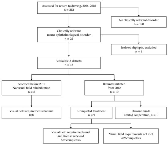
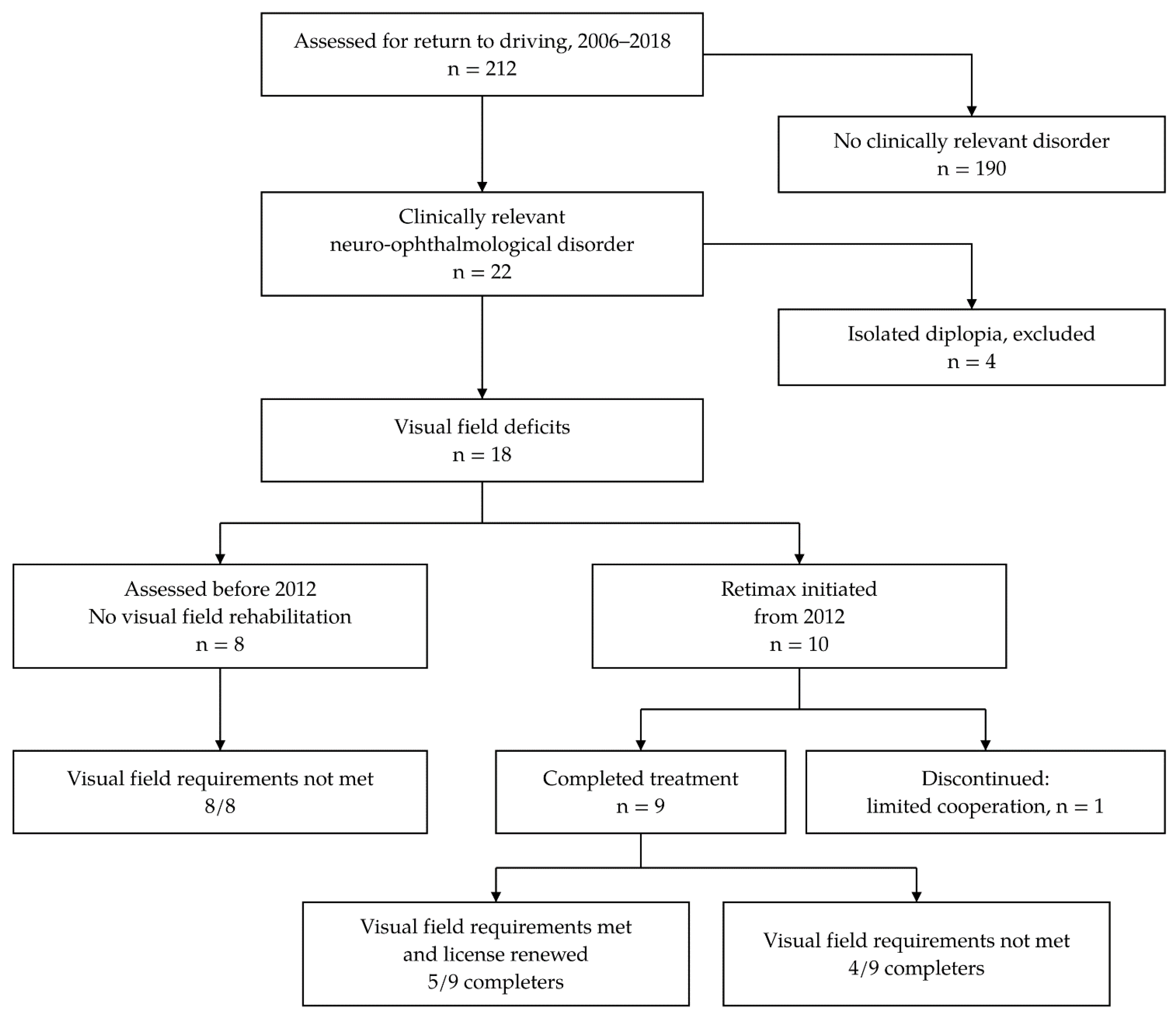
Of the 212 patients assessed for return to driving, 22 (10.4%) had a clinically relevant neuro-ophthalmological disorder. Four had isolated diplopia. The 18 patients with visual field involvement accounted for 8.5% of the assessed population (Figure 1).
Figure 1.
Selection and clinical course of patients with visual field loss in the multidisciplinary return-to-driving pathway.
Mean age was 40.2 years (SD 10.6; range 24–58), and 13 patients (72.2%) were men. Nine patients had a vascular etiology, eight had traumatic brain injury, and one had post-anoxic injury. Ten had hemianopic and eight quadrantanopic defects. The hemianopic defects were left-sided in six patients, right-sided in three, and bitemporal in one. Quadrantanopia involved the upper right field in five patients and the lower right, upper left, and lower left fields in one patient each. One patient with bitemporal hemianopia also had ocular misalignment and diplopia. No clinically relevant impairment of stereopsis or color vision was identified as a barrier to driving eligibility. Table 1 summarizes the baseline characteristics; Supplementary Table S1 presents the individual cases.
Table 1.
Baseline characteristics of the 18 patients with visual field involvement.
3.2. Clinical Pathway and Observed Visual Field Outcomes
Eight patients were evaluated before Retimax became available in 2012 and did not receive specific visual field rehabilitation. None met the visual field requirements for license renewal within the pathway.
Ten patients started Retimax rehabilitation from 2012 onward. One patient discontinued treatment because of limited cooperation during the rehabilitation sessions. The nine completers comprised six men and three women, with a mean age of 38.2 years (SD 8.9; range 24–51). Four had traumatic brain injury, four had a vascular etiology, and one had post-anoxic injury. Six had hemianopic defects, including the patient with concomitant ocular misalignment and diplopia, and three had quadrantanopic defects.
Five of the nine completers met the visual field requirements and obtained renewal of their driving licenses: two patients with hemianopic defects and all three with quadrantanopic defects. The other four completers, all with hemianopic defects, did not meet the visual field requirements. Of the two patients with hemianopic defects who obtained license renewal, one had a left-sided defect and the other had bitemporal hemianopia. By etiology, two of four completers with traumatic brain injury and two of four with vascular injury met the visual field requirements and obtained license renewal. The single completer with post-anoxic injury also met the requirements and obtained license renewal. Treatment course and outcomes are summarized in Table 2. Supplementary Table S2 presents the characteristics and outcomes of the nine individual completers.
Table 2.
Treatment course, visual field eligibility, and license renewal in the 18-patient series.
4. Discussion
This series describes the field-defect patterns and clinical course of patients undergoing visual assessment and rehabilitation within a multidisciplinary return-to-driving pathway after sABI. Visual field loss was identified in 18 of 212 candidates. Among the nine patients who completed Retimax rehabilitation, five met the visual field requirements and obtained license renewal.
The type of visual disorder matters for both rehabilitation and interpretation. Ocular misalignment and diplopia require assessment distinct from that of visual field loss. Likewise, a sensory field defect should not be equated with visuospatial neglect, which may independently affect exploration and driving performance [12]. These distinctions support joint neuro-ophthalmological and neuropsychological assessment within the multidisciplinary framework described by De Tanti et al. [2] and in our recent opinion article [1].
The study population comprised people with sABI who had survived beyond the acute phase, reached clinical stability, previously held a driving license, and been referred for return-to-driving assessment. All candidates were assessed at least 12 months after injury. This selection limits generalization to the wider population with sABI, including patients with more severe residual impairments or those not referred for driving assessment. The proportion with visual field loss describes candidates assessed in this pathway and should not be interpreted as a prevalence estimate for all patients with sABI. These candidates also differ from acute stroke populations in epidemiological studies [8].
Driving requires coordinated sensory, cognitive, and motor abilities [7,12]. Wood et al. found that some drivers with hemianopia or quadrantanopia were rated as safe during an on-road assessment, whereas others had difficulties with lane position, steering stability, and gap judgment [22]. Such variation supports individualized functional assessment within the applicable licensing framework. Perimetry assesses visual detection under controlled viewing conditions; practical driving assessment examines how a patient uses vision while performing the task. Both contribute to the assessment, with the applicable legal visual requirements determining eligibility to proceed. License renewal reflects the combined assessment of visual, cognitive, behavioral, and motor function within the applicable legal and administrative framework. It is therefore a multidimensional outcome rather than a surrogate measure of visual rehabilitation efficacy.
Visual field requirements form part of the legal criteria for driving eligibility [20,21]. Five completers in this series were recorded as meeting the applicable visual field requirements and subsequently obtained license renewal. This categorical outcome indicates that the clinical assessment supported progression through the licensing pathway; it does not specify the final field extent or the magnitude of change from baseline. The relevant criterion is the requirement applied at the time of assessment, given the changes in the licensing framework during the observation period.
Recent evidence also distinguishes perimetric eligibility from functional driving performance. In a matched retrospective study of 202 license applicants, Faraji et al. found substantial overlap in Esterman results between those who passed and failed an on-road driving test, with limited predictive value for individual decisions [23]. That population consisted mainly of older people with eye disease and differs from the present sABI series. Nevertheless, the findings support interpreting perimetry alongside functional assessment, rather than treating an Esterman result as a stand-alone measure of driving safety.
The five completers who obtained license renewal included three patients with quadrantanopia, one with left hemianopia, and one with bitemporal hemianopia accompanied by ocular misalignment and diplopia. Bitemporal hemianopia is clinically distinct from homonymous field loss. The retained nasal hemifields can provide complementary coverage during binocular testing, while ocular misalignment can alter that coverage and produce diplopia or a central gap [24]. Consequently, meeting binocular field requirements does not itself establish recovery of the monocular defects. When the bitemporal case is considered separately, one of the five completers with right- or left-sided hemianopia met the requirements and obtained license renewal (Supplementary Table S2). The small numbers and heterogeneous defects limit subgroup interpretation. Chiari et al. [18] previously described Retimax rehabilitation in two patients with sABI; the present series adds observations from its use in a driving rehabilitation pathway.
Compensatory scanning training seeks to improve exploration of the affected side, while Retimax combines patterned stimulation with electrophysiological feedback [18,25]. In a randomized trial, de Haan et al. reported improvements in peripheral detection and obstacle avoidance during walking after scanning training [25]. The more recent SEARCH trial found improvements in both visual scanning and sham-training groups after stroke, with no significant between-group difference in the primary vision-related quality-of-life outcome [26]. Neither trial evaluated Retimax or license renewal.
Recent trials of visual perceptual learning illustrate why treatment-specific evidence is needed. Cavanaugh et al. randomized 48 patients with stroke-related homonymous hemianopia to motion-discrimination training in the affected or the sighted field. Although the affected-field group improved from baseline, the between-group differences in perimetric mean deviation were not significant [27]. In contrast, Namgung et al. reported greater perimetric improvement after 12 weeks of personalized virtual-reality visual perceptual training than with no training in chronic poststroke field loss [28]. The latter trial used a passive comparator and lacked eye tracking, limiting separation of the specific training effect from nonspecific influences. Together with SEARCH [26], these findings show that the comparator, fixation assessment, and choice of outcome can materially affect interpretation. None of these interventions was Retimax, and their results cannot establish its efficacy or its effect on license renewal.
Measurement methods are central to evaluating visual field rehabilitation. Reinhard et al. studied a different visual restitution intervention and found no relevant change in the border of absolute homonymous defects in the central visual field using fundus-controlled perimetry [29]. Their findings emphasize the importance of fixation control and of distinguishing sensory change from compensatory behavior when assessing treatment outcomes.
Retimax may be considered a candidate adjunct for investigation within orthoptic rehabilitation because it combines visual stimulation with electrophysiological feedback. Studies in macular degeneration and anisometropic amblyopia concern different visual disorders and treatment goals [15,16,17], so their findings cannot be transferred directly to acquired cerebral field loss. For the population described here, a useful next study would test whether adding Retimax to a defined rehabilitation program improves prespecified visual or functional outcomes compared with the same program without Retimax. Recording monocular and binocular fields separately would help distinguish changes in the underlying defects from their combined functional expression, especially when ocular alignment is abnormal.
Future controlled studies should combine standardized perimetry and prespecified reliability criteria with functional driving assessment and follow-up of driving exposure and safety [1,7,12]. Patient-reported visual difficulties and activity-specific measures would also help evaluate Retimax within orthoptic rehabilitation for goals beyond driving.
Limitations
This small retrospective series concerns a selected population from one center. Treatment was not randomized, and the eight patients assessed before Retimax availability were evaluated in a different calendar period. Differences in baseline impairment, assessment timing, selection, clinical practice, and the licensing framework preclude a valid treatment-effect comparison. Assessment at least 12 months after injury places the series in the chronic phase, although later change and repeated-testing effects remain possible.
Treatment outcomes concern nine completers and may not represent all patients who start rehabilitation. Individual quantitative pre- and post-treatment perimetric measurements were not available for this analysis. The categorical eligibility outcomes therefore provide no estimate of baseline field-loss severity or of the magnitude and location of within-patient change. They cannot distinguish sensory improvement from compensatory visual behavior, test familiarity, or other influences on performance. Individual test-level reliability indices were unavailable for the present retrospective analysis, and operator-based fixation monitoring provides no objective record of eye position. The small etiological subgroups and heterogeneous visual field patterns preclude meaningful comparisons by etiology and limit generalization. Treatment exposure was described by session frequency and program duration, limiting assessment of dose–response relationships. Long-term driving exposure and safety were not evaluated. These limitations prevent conclusions about treatment efficacy.
5. Conclusions
This series describes visual field deficits and clinical outcomes within a multidisciplinary return-to-driving pathway after sABI, including the use of Retimax rehabilitation. Five of the nine patients who completed Retimax met the visual field requirements and obtained license renewal. Controlled studies with quantitative visual and functional outcomes are needed to evaluate Retimax efficacy.
Supplementary Materials
The following supporting information can be downloaded at: https://www.mdpi.com/article/10.3390/brainsci16111260/s1, Supplementary Table S1: Baseline characteristics of the 18 patients with visual field involvement, providing additional details on the study population. Supplementary Table S2: Characteristics and rehabilitation outcomes of the nine patients who completed Retimax rehabilitation, providing additional information on the treated subgroup.
Author Contributions
Conceptualization, M.C., D.S., and A.D.T.; methodology, M.C., S.B., A.D.T., and D.S.; formal analysis, S.B.; investigation, M.C., C.S., E.B., and A.S.; writing—original draft, S.B., D.S., and A.D.T.; writing—review and editing, M.C., S.B., A.D.T., and D.S.; supervision, D.S. and A.D.T. All authors have read and agreed to the published version of the manuscript.
Funding
This research received no funding.
Institutional Review Board Statement
The retrospective analysis received institutional scientific authorization from the Scientific Board of KOS (protocol 02/2026; 21 January 2026).
Informed Consent Statement
Under institutional procedures, patients or their legal representatives provide general consent at admission for the use of anonymized clinical data in retrospective research and scientific publications. The Scientific Board of KOS authorized this retrospective analysis on that basis. No additional assessments or interventions were performed for research purposes.
Data Availability Statement
The original contributions presented in this study are included in the article and Supplementary Tables S1 and S2. Further inquiries can be directed to the corresponding author.
Acknowledgments
Grammarly (online version, accessed on 27 October 2026; Grammarly Inc., San Francisco, CA, USA) was used for language editing. OpenAI Codex (GPT-6 Astra model; OpenAI, San Francisco, CA, USA) provided the additional assistance described in Section 2.5. Figure 1 and the graphical abstract were drawn as vector graphics from code prepared with the assistance of Claude (Sonnet 5.5 model; Anthropic, San Francisco, CA, USA); no generative image tools were used for these figures. The authors reviewed and verified all AI-assisted outputs and take full responsibility for the content, analyses, and references.
Conflicts of Interest
The authors declare no conflicts of interest, including financial or professional relationships with CSO. No equipment loans or free devices were provided by CSO.
References
- Saviola, D.; Bruni, S.; Cantoni, M.; De Tanti, A. Safe return to driving after severe acquired brain injury: Toward a tiered multidisciplinary rehabilitation pathway. Front. Neurol. 2026, 17, 1751388. [Google Scholar] [CrossRef] [Scilit] [PubMed]
- De Tanti, A.; Baldari, F.; Pedrazzi, G.; Inzaghi, M.G.; Benoldi, R.; Abbati, P.; Malangone, G.; Martini, C.; Saviola, D. Role of vehicle assistive devices for safe return to driving after severe acquired brain injury. Eur. J. Phys. Rehabil. Med. 2020, 56, 386–393. [Google Scholar] [CrossRef] [Scilit] [PubMed]
- Saviola, D.; De Tanti, A.; Conforti, J.; Posteraro, L.; Manfredini, A.; Bagattini, C.; Basagni, B. Safe return to driving following severe acquired brain injury: Role of a short neuropsychological assessment. Eur. J. Phys. Rehabil. Med. 2018, 54, 717–723. [Google Scholar] [CrossRef] [Scilit] [PubMed]
- Ross, P.E.; Di Stefano, M.; Charlton, J.; Spitz, G.; Ponsford, J.L. Interventions for resuming driving after traumatic brain injury. Disabil. Rehabil. 2018, 40, 757–764. [Google Scholar] [CrossRef] [Scilit] [PubMed]
- Novack, T.A.; Zhang, Y.; Kennedy, R.; Rapport, L.J.; Watanabe, T.K.; Monden, K.R.; Dreer, L.E.; Bergquist, T.; Bombardier, C.; Brunner, R.; et al. Return to driving after moderate-to-severe traumatic brain injury: A Traumatic Brain Injury Model System study. Arch. Phys. Med. Rehabil. 2021, 102, 1568–1575. [Google Scholar] [CrossRef] [Scilit] [PubMed]
- Egeto, P.; Badovinac, S.D.; Hutchison, M.G.; Ornstein, T.J.; Schweizer, T.A. A systematic review and meta-analysis on the association between driving ability and neuropsychological test performances after moderate to severe traumatic brain injury. J. Int. Neuropsychol. Soc. 2019, 25, 868–877. [Google Scholar] [CrossRef] [Scilit] [PubMed]
- Schultheis, M.T.; Whipple, E. Driving after traumatic brain injury: Evaluation and rehabilitation interventions. Curr. Phys. Med. Rehabil. Rep. 2014, 2, 176–183. [Google Scholar] [CrossRef] [Scilit] [PubMed]
- Rowe, F.J.; Hepworth, L.R.; Howard, C.; Hanna, K.L.; Cheyne, C.P.; Currie, J. High incidence and prevalence of visual problems after acute stroke: An epidemiology study with implications for service delivery. PLoS ONE 2019, 14, e0213035. [Google Scholar] [CrossRef] [Scilit] [PubMed]
- Hanna, K.L.; Hepworth, L.R.; Rowe, F.J. Screening methods for post-stroke visual impairment: A systematic review. Disabil. Rehabil. 2017, 39, 2531–2543. [Google Scholar] [CrossRef] [Scilit] [PubMed]
- Pollock, A.; Hazelton, C.; Rowe, F.J.; Jonuscheit, S.; Kernohan, A.; Angilley, J.; Henderson, C.A.; Langhorne, P.; Campbell, P. Interventions for visual field defects in people with stroke. Cochrane Database Syst. Rev. 2019, 5, CD008388. [Google Scholar] [CrossRef] [Scilit] [PubMed]
- Devos, H.; Akinwuntan, A.E.; Nieuwboer, A.; Truijen, S.; Tant, M.; De Weerdt, W. Screening for fitness to drive after stroke: A systematic review and meta-analysis. Neurology 2011, 76, 747–756. [Google Scholar] [CrossRef] [Scilit] [PubMed]
- Bowers, A.R. Driving with homonymous visual field loss: A review of the literature. Clin. Exp. Optom. 2016, 99, 402–418. [Google Scholar] [CrossRef] [Scilit] [PubMed]
- de Haan, G.A.; Heutink, J.; Melis-Dankers, B.J.M.; Brouwer, W.H.; Tucha, O. Difficulties in daily life reported by patients with homonymous visual field defects. J. Neuroophthalmol. 2015, 35, 259–264, Correction in J. Neuroophthalmol. 2016, 36, e15. [Google Scholar] [CrossRef] [Scilit] [PubMed]
- Bouwmeester, L.; Heutink, J.; Lucas, C. The effect of visual training for patients with visual field defects due to brain damage: A systematic review. J. Neurol. Neurosurg. Psychiatry 2007, 78, 555–564. [Google Scholar] [CrossRef] [Scilit] [PubMed]
- Verdina, T.; Piaggi, S.; Ferraro, V.; Russolillo, V.; Peschiera, R.; Chester, J.; Mastropasqua, R.; Cavallini, G.M. Efficacy of biofeedback rehabilitation based on visual evoked potentials analysis in patients with advanced age-related macular degeneration. Sci. Rep. 2020, 10, 20886. [Google Scholar] [CrossRef] [Scilit] [PubMed]
- Verdina, T.; Piaggi, S.; Peschiera, R.; Russolillo, V.; Ferraro, V.; Chester, J.; Mastropasqua, R.; Cavallini, G.M. Biofeedback low vision rehabilitation with Retimax Vision Trainer in patients with advanced age-related macular degeneration: A pilot study. Semin. Ophthalmol. 2020, 35, 164–169. [Google Scholar] [CrossRef] [Scilit] [PubMed]
- Nemeș-Drăgan, I.A.; Țîpcu, A.; Hapca, M.C.; Pașcalău, R.; Nicoară, S.D. Reconnecting anisometropic amblyopic eyes to the cortex: VEP-based auditory biofeedback. Diagnostics 2024, 14, 1861. [Google Scholar] [CrossRef] [Scilit] [PubMed]
- Chiari, M.; Savi, C.; Battagliola, E.; Saviola, D.; De Tanti, A. Visual rehabilitation with Retimax Vision Trainer in patients with severe acquired brain injury: Report of two cases. Neuropsychol. Trends 2014, 15, 35–40. [Google Scholar] [CrossRef] [Scilit]
- von Elm, E.; Altman, D.G.; Egger, M.; Pocock, S.J.; Gøtzsche, P.C.; Vandenbroucke, J.P. STROBE Initiative. The Strengthening the Reporting of Observational Studies in Epidemiology (STROBE) statement: Guidelines for reporting observational studies. PLoS Med. 2007, 4, e296. [Google Scholar] [CrossRef] [Scilit] [PubMed]
- Italian Ministry of Infrastructure and Transport. Decree of 30 November 2010. Transposition of Commission Directive 2009/112/EC of 25 August 2009 Amending Council Directive 91/439/EEC on Driving Licences. Gazzetta Ufficiale della Repubblica Italiana. 2010. Available online: https://www.gazzettaufficiale.it/eli/id/2010/12/27/10A15247/sg (accessed on 30 October 2026).
- Italian Republic. Legislative Decree 18 April 2011, No. 59. Implementation of Directives 2006/126/EC and 2009/113/EC on Driving Licences. In Official Gazette of the Italian Republic; 2011, 99, Annex III; Istituto Poligrafico e Zecca dello Stato: Rome, Italy, 2011. [Google Scholar]
- Wood, J.M.; McGwin, G.; Elgin, J.; Vaphiades, M.S.; Braswell, R.A.; DeCarlo, D.K.; Kline, L.B.; Meek, G.C.; Searcey, K.; Owsley, C. On-road driving performance by persons with hemianopia and quadrantanopia. Investig. Ophthalmol. Vis. Sci. 2009, 50, 577–585. [Google Scholar] [CrossRef] [Scilit] [PubMed]
- Faraji, Y.; Tan-Burghouwt, M.T.; Bredewoud, R.A.; van Nispen, R.M.A.; van Rijn, L.J. Predictive value of the Esterman visual field test on the outcome of the on-road driving test. Transl. Vis. Sci. Technol. 2022, 11, 20. [Google Scholar] [CrossRef] [Scilit] [PubMed]
- Peli, E.; Jung, J.H. Review: Binocular double vision in the presence of visual field loss. J. Vis. 2024, 24, 13. [Google Scholar] [CrossRef] [Scilit] [PubMed]
- de Haan, G.A.; Melis-Dankers, B.J.M.; Brouwer, W.H.; Tucha, O.; Heutink, J. The effects of compensatory scanning training on mobility in patients with homonymous visual field defects: A randomized controlled trial. PLoS ONE 2015, 10, e0134459, Correction in PLoS ONE 2016, 11, e0165863. [Google Scholar] [CrossRef] [Scilit] [PubMed]
- Rowe, F.J.; Brayshaw, E.; Brown, M.; Chatterjee, K.; Drummond, A.; Hazelton, C.; Helliwell, B.; Hepworth, L.R.; Howard, C.; Johnson, S.; et al. A randomized controlled trial of Scanning Eye trAining as a Rehabilitation Choice for Hemianopia after stroke (SEARCH). Int. J. Stroke 2025, 20, 968–976. [Google Scholar] [CrossRef] [Scilit] [PubMed]
- Cavanaugh, M.R.; Blanchard, L.M.; McDermott, M.; Lam, B.L.; Tamhankar, M.; Feldon, S.E. Efficacy of visual retraining in the hemianopic field after stroke: Results of a randomized clinical trial. Ophthalmology 2021, 128, 1091–1101. [Google Scholar] [CrossRef] [Scilit] [PubMed]
- Namgung, E.; Kim, B.J.; Kwon, J.H.; Han, M.-K.; Kim, H.Y.; Jung, J.-M.; Kim, J.G.; Park, K.-Y.; Koo, J.; Hong, K.-S.; et al. Personalized visual perceptual learning digital therapy for visual field defects following stroke: A randomized clinical trial. JAMA Netw. Open 2025, 8, e2511068. [Google Scholar] [CrossRef] [Scilit] [PubMed]
- Reinhard, J.; Schreiber, A.; Schiefer, U.; Kasten, E.; Sabel, B.A.; Kenkel, S.; Vonthein, R.; Trauzettel-Klosinski, S. Does visual restitution training change absolute homonymous visual field defects? A fundus controlled study. Br. J. Ophthalmol. 2005, 89, 30–35. [Google Scholar] [CrossRef] [Scilit] [PubMed]

Disclaimer/Publisher’s Note: The statements, opinions and data contained in all publications are solely those of the individual author(s) and contributor(s) and not of MDPI and/or the editor(s). MDPI and/or the editor(s) disclaim responsibility for any injury to people or property resulting from any ideas, methods, instructions or products referred to in the content. |
© 2026 by the authors. Licensee MDPI, Basel, Switzerland. This article is an open access article distributed under the terms and conditions of the Creative Commons Attribution (CC BY) license.