


Highlights
What are the main findings?
- Computed with the CIE S 026 melanopic action spectrum, the melanopic content per lux of clear-sky daylight at the eye (melanopic daylight efficacy ratio) falls by about a quarter between solar noon and a solar altitude of , and by about a third before sunset, while total illuminance changes much less.
- This spectral change is governed almost entirely by solar altitude: across three European latitudes and four dates it collapses onto a single curve, so its clock time is computable for any site and day, and moves with season, latitude and daylight-saving time.
What are the implications of the main findings?
- If afternoon light is a cue for sundowning, the relevant variable is spectral composition at a given solar altitude, not darkness and not a clock hour; lighting interventions should be dosed in melanopic units and timed to local solar conditions.
- The proposal can fail. Onset should track the Sun rather than the clock across a daylight-saving transition—a test needing no new instrumentation, and one whose negative outcome would count strongly against the hypothesis.
Abstract
Background/Objectives: Sundowning syndrome—recurrent late-afternoon agitation, disorientation and anxiety in people with dementia—is widely attributed to disturbed light exposure. Existing accounts remain qualitative, specifying neither which property of the ambient light field changes nor when, and so yield no prediction varying with latitude, season or sky conditions. This article is a computational hypothesis: it contains no patient data and is intended to make a light-based account specific enough to be tested. Methods: We combine standard solar geometry, an analytic clear-sky radiance model and the CIE photometric and CIE S 026 melanopic weighting functions to compute the spectral composition of the light reaching an observer as a continuous function of time, for single sky directions, for vertical (corneal) planes integrated over the whole sky, for turbidities 2–6, and for an observer indoors near a window, at three European latitudes over a full year. Results: The melanopic daylight efficacy ratio at the eye falls from about 1.33 at solar noon to about 1.02 at a solar altitude of and about 0.89 at (turbidity 2), while modelled total illuminance changes much less (by 3–10%). The decline is a function of solar altitude alone to within 0.005 across sites and dates, is flattened but not removed by aerosol loading and, in a modelled side-lit room with interreflections, by electric light. The Sun passes altitude 51–107 min before sunset, at clock times that vary by 3–6 h over the year. These are model estimates of spectral composition, least certain close to the horizon, and not measures of physiological melanopic stimulation. Conclusions: We propose that afternoon spectral reddening—rather than darkness itself—is a candidate physical cue for sundowning, possibly mediated by the diminished short-wavelength drive to melanopsin-expressing retinal ganglion cells. The proposal yields testable predictions, including an experimental design that separates spectral composition from light level, and implies that lighting interventions should be timed to local solar conditions rather than the clock.
1. Introduction
Life on Earth is organised around two astronomical periodicities. The rotation of the Earth imposes a 24 h alternation of light and dark to which almost all organisms have evolved endogenous circadian rhythms, and the tilt of its axis imposes an annual modulation of day length and solar elevation to which many respond with circannual rhythms in physiology and behaviour [1,2]. In humans, the dominant signal that entrains the circadian system to local time is light reaching the retina [1,3]. Both the daily and the annual cycle of that light depend on where and when it is received: at a given latitude and date, solar geometry fixes the times of sunrise and sunset, the maximum solar elevation and the rate at which the Sun descends in the afternoon.
Light also changes in spectral compositionthrough the day. As the Sun descends, the optical path through the atmosphere lengthens, short wavelengths are increasingly scattered out of the direct beam, and both direct and diffuse daylight become relatively richer in long wavelengths [4,5]. This matters physiologically because the non-image-forming (NIF) effects of light—circadian entrainment, melatonin suppression and alerting—are driven principally by intrinsically photosensitive retinal ganglion cells (ipRGCs) containing melanopsin, whose sensitivity peaks in the short-wavelength part of the visible spectrum [6,7,8,9]. Older adults, and particularly people with dementia, receive this signal through an ageing lens that transmits less short-wavelength light [10] and, in Alzheimer’s disease, through a retina in which ipRGCs are lost [11,12]; they also typically spend their days indoors under low light levels [13,14].
Dementia is among the leading causes of disability in older adults, and the number of affected individuals continues to rise as life expectancy increases [15,16]. Alzheimer’s disease accounts for the majority of cases; its aetiology and treatment remain subjects of active debate [17,18,19].
Among the neuropsychiatric symptoms that accompany dementia, one of the most disruptive is sundowning syndrome (SS): a recurrent worsening of agitation, disorientation, anxiety and aggression that emerges in the late afternoon or early evening and typically subsides overnight [20,21]. Reported prevalence varies enormously between studies, from about 2.5% to 66% depending on setting, operational definition and underlying disease [22,23], reflecting the absence of an agreed operational definition at least as much as real variation between populations [20,24]. At the low end, a study of 85 institutionalised patients using behavioural ratings collected every 15 min over three days classified only two individuals (2.4%) as sundowners, even though agitation showed a clear circadian component in most patients [25]. The low end of this range therefore reflects a strict definition of a discrete syndrome rather than the absence of late-day worsening, which clinicians have recognised for more than half a century [21]. The symptom imposes a substantial burden on caregivers and is a frequent precipitant of institutionalisation [24]. Whether sundowning constitutes a discrete, time-locked entity or a nonspecific exacerbation of pre-existing agitation remains contested [26].
Several mechanisms have been proposed, including circadian disruption, degeneration of the suprachiasmatic nucleus, sensory and perceptual impairment, accumulated fatigue, and features of the institutional environment such as evening staff changeover [20,22,25]. Of these, the light-dependent hypothesis has received the most attention, motivated by the coincidence of symptom onset with the daily transition from day to night. The same hypothesis has motivated therapeutic trials of bright-light exposure, whose results have been mixed: benefits for selected circadian, sleep, mood and agitation outcomes have been reported [27,28], whereas a systematic review found insufficient evidence of benefit for agitation specifically [29].
That the spectral composition of daylight shifts towards long wavelengths as the Sun descends, and that melanopsin sensitivity may be tuned to this natural progression, has already been noted in the circadian literature [30]. We do not claim the observation as new. What has not been done, to our knowledge, is to compute the resulting time course quantitatively in melanopic units for a specified place and date, so that the idea yields predictions rather than a narrative. A common feature of the existing light-dependent proposals is precisely that they remain qualitative. They do not specify which property of the ambient light field changes, by how much, or at what time. Consequently they yield no prediction that varies with the observer’s latitude, the season, or the state of the sky—even though all three are determined by well-understood physics.
The tools needed for such a calculation exist in solar engineering, atmospheric optics and daylighting practice. Solar geometry is standard [31]. Sky luminance distributions are described by the Perez all-weather model [32] and, in building design, by the CIE standard general sky [33]; analytic models that also give the chromaticity of each sky element are available from computer graphics [34,35], and the spectrum can be reconstructed from chromaticity with the CIE daylight basis [36]. Photometric and melanopic quantities are defined by CIE 018 and CIE S 026 [9,37]. Indoor daylight is commonly estimated with the split-flux method [38], and annual daylight performance with climate-based simulations, for which representative subsets of the year can be selected [39]. These tools have not, to our knowledge, been combined to ask how the melanopic composition of the light reaching a person changes through the afternoon.
In this paper we take that step. Combining standard solar geometry [31], an analytic clear-sky radiance model [32,34], the CIE daylight basis [36] and the CIE photometric and melanopic weighting functions [9,37], we compute the spectral composition of the light reaching an observer as a continuous function of time, for single sky directions, for the vertical plane of the eye integrated over the whole sky, for a range of atmospheric turbidities, and for an observer indoors in a side-lit room, including light reflected within the room. We present the work explicitly as a hypothesis: it contains no patient data, and its purpose is to replace a qualitative light-based account of sundowning with a quantitative one that makes predictions capable of failing.
The aim is therefore twofold: to quantify, for specified sites and dates, how the melanopic content per lux of the light at the eye changes from solar noon to sunset, outdoors and indoors; and to derive from it predictions that can confirm or refute a spectral account of sundowning. The work is restricted to mid-latitudes (about – N), where every day of the year contains a sunrise and a sunset; the polar day and polar night, in which these markers disappear, are outside its scope.
The paper is organised as follows. Section 2 describes the sites and dates, the solar geometry, the sky model, the photoreceptor weighting and the indoor scenario. Section 3 presents the computed spectral quantities and their timing. Section 4 discusses the visual and non-image-forming pathways, the relation to clinical observations, testable predictions, limitations and future research. Section 5 concludes.
2. Materials and Methods
2.1. Sites, Dates and Climatic Context
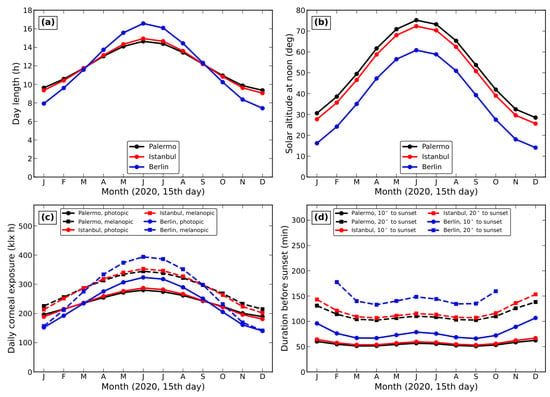
We use four sites. Ankara (39.93° N, 32.86° E; altitude 938 m) is used for the single-direction illustration (Section 3.1), on 29 October 2023. Palermo, Sicily (38.12° N, 13.36° E), İstanbul (41.01° N, 28.98° E) and Berlin (52.52° N, 13.40° E) span about 14° of latitude across Europe and were chosen because they bracket the latitudes of most of Europe’s population, include one site without daylight saving time (İstanbul, UTC+3 all year since 2016) and two with EU summer time (Palermo and Berlin, UTC+1/UTC+2), and have contrasting climates: Mediterranean (Palermo), transitional Mediterranean–humid subtropical (İstanbul) and temperate central European (Berlin), i.e., Köppen–Geiger classes Csa, Csa/Cfa and Cfb [40]. Table 1 summarises the sites, and Figure 1 shows, month by month, the astronomical daylight availability at each: day length, solar altitude at noon, the daily clear-sky photopic and melanopic exposure at the eye, and the duration of the late-afternoon interval from a solar altitude of 20° or 10° to sunset. Because we model clear skies only, climate enters the calculation solely through the atmospheric turbidity, which we vary between (very clear) and (hazy); cloud climatology is not modelled (Section 4.6).
Table 1.
Sites used in this work. Day length is geometric (sunrise to sunset of the solar centre, no refraction); civil times are those in force in 2020; climate class after Ref. [40].
Figure 1.
Astronomical daylight availability at Palermo (black), İstanbul (red) and Berlin (blue), computed for the 15th day of each month of 2020. (a) Day length. (b) Solar altitude at solar noon. (c) Daily exposure at the eye under a clear sky (, sky light only, orientation-averaged vertical plane), photopic (, solid) and melanopic (, dashed). (d) Duration of the interval from the Sun passing (solid) or (dashed) altitude to sunset; in Berlin the Sun does not reach from November to January. Cloud is not modelled, so (c) is an upper bound set by solar geometry, not a climatological mean.
Time series are computed for the March equinox (21 March 2020), the June solstice (21 June), the September equinox (22 September) and the December solstice (21 December), and the annual behaviour is computed every second day of 2020. All times are given as local civil clock time, including daylight saving time where it applies, because that is the time in which care routines and clinical observations are recorded.
Choice of dates and time series. Equinoxes and solstices bracket the annual range of solar geometry, but a few such milestone dates are not by themselves representative of the annual distribution of daylight, which also depends on the sky conditions of each day [39]. We have therefore used them only for illustration and drawn the general results from complete annual series: (i) we show that, in the model, the spectral index at the eye depends on solar altitude and turbidity but not on date or site (Section 3.3), so the result for any day follows from the solar altitudes reached on it; (ii) the clock times of fixed solar altitudes are computed for every second day of the year (Section 3.5); and (iii) the indoor quantities are computed for every fourth day (Section 3.4). Because cloud is not modelled, these are annual series of clear-sky days, not climate-based annual distributions.
2.2. Solar Geometry
We use the standard description of the apparent motion of the Sun following Ref. [31], and give only the relations needed to reproduce the calculation. The declination is
where n is the day of the year. Apparent solar time is obtained from the civil clock time as
where is the local longitude and the longitude of the meridian of the civil time zone in force on that day (both east-positive, in degrees; therefore changes by at a daylight-saving transition), and is the equation of time in minutes,
with . The hour angle is , negative before solar noon, and the solar zenith angle and altitude follow from
where is the latitude. The solar azimuth , measured from south and positive towards west, is computed with the two-argument form
which resolves the quadrant ambiguity of the single-argument expression. The hour angle at sunset follows from . Refraction and the finite solar disc are neglected, so the computed sunset is geometric and falls a few minutes before the tabulated civil sunset.
2.3. Sky Radiance Model
The spectral radiance of the clear sky is computed from the analytic model of Preetham et al. [34]. For a sky element at zenith angle that makes an angle with the Sun, each of the luminance Y and the chromaticity coordinates x and y is written as its zenith value multiplied by a ratio of Perez functions [32],
with . The coefficients and the zenith values , , are the linear and polynomial functions of the turbidity and the solar zenith angle given in Ref. [34]. The spectral radiance is then reconstructed from with the CIE daylight basis [36],
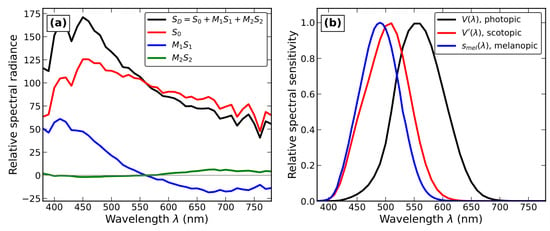
and scaled to absolute spectral radiance (W m−2sr−1nm−1) such that , with lm W−1. Figure 2a shows the decomposition of Equation (7) for one sky direction. In this model, therefore, the spectral shape of each sky element is fixed by its chromaticity; any spectral index we compute is a function of the chromaticities of the sky elements contributing to it, weighted by their luminances.
Figure 2.
(a) Reconstruction of a sky spectrum from the CIE daylight basis, Equation (7), for the sky element at zenith angle and azimuth west of south, Ankara, 29 October 2023, 12:00 local time (); , and are the three terms and their sum, in relative units normalised to ; the thin grey horizontal line marks zero, about which the basis terms and change sign. (b) Weighting functions used in this work: CIE photopic and scotopic luminous efficiency functions and the CIE S 026 melanopic action spectrum , each normalised to unit peak.
Three geometries are used. (i) Single sky direction. To retain continuity with the original submission, Figure 3 shows the radiance from one direction at zenith angle (i.e., above the horizon) and azimuth west of south. (ii) Vertical (corneal) plane. The irradiance on a vertical plane facing azimuth , a standard proxy for irradiance at the cornea of an observer with horizontal gaze [41], is obtained by integrating over the whole visible sky (angular grid ), where is the angle of incidence on the plane, and adding the light reflected by a Lambertian ground of spectrally flat albedo lit by the diffuse sky. We report results for = N, E, S, W and for their mean, which we call the orientation-averaged corneal irradiance. (iii) Indoor observer (Section 2.5). In all three geometries the direct solar beam is excluded: the observer is assumed not to look at, or be exposed to, the solar disc, and windows are assumed shaded against beam sunlight. The effect of this choice is discussed in Section 4.6.
Figure 3.
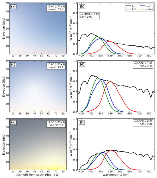
(a,c,e) Simulated appearance of the sky at Ankara on 29 October 2023 at 12:00, 15:00 and 17:30 local time (UTC+3), rendered from the analytic daylight model of Ref. [34] for turbidity with the same exposure in all three panels; the colour of each point is the computed colour of that sky element converted to sRGB, so that the yellowing near the horizon in (e) is the spectral reddening itself, and the darkening is the fall of sky luminance at fixed exposure. (b,d,f) Corresponding spectral radiance of the sky element at zenith angle and azimuth west of south (black), and its photopically (, red), scotopically (, blue) and melanopically (, green) weighted versions, all in absolute units on a common ordinate; where these curves cross or overlap, they do so because the weighted spectra have similar values at those wavelengths. Insets give mel-DER and .
The model is analytic and was developed for image synthesis rather than radiometry. Comparisons with measured clear skies show that it reproduces the angular structure of sky luminance but overestimates absolute sky radiance, by up to a factor of about two, and becomes unreliable near the horizon and at very low solar elevation [42,43]; more accurate but more complex models exist [35]. For this reason we (a) base our conclusions on spectral ratios, which are insensitive to the absolute scale, (b) restrict all time series to solar altitudes , and (c) treat absolute illuminances as indicative only. Ratios are not immune to the model’s errors, because the chromaticity of the sky elements, from which the spectral shape is reconstructed, is itself least reliable at low solar elevation. We therefore regard numerical values below a solar altitude of about as indicative, and we check (Section 3.3) that the reconstructed spectra stay within the colour-temperature range over which the CIE daylight basis is defined.
2.4. Spectral Weighting and Photometric Quantities
Three weighting functions are used (Figure 2b): the CIE photopic and scotopic luminous efficiency functions and [37], and the melanopic action spectrum of CIE S 026 [9,44], which describes the ipRGC-influenced responses of the standard observer at the cornea and peaks at 490 nm. From a spectral irradiance we compute the illuminance , the melanopic equivalent daylight (D65) illuminance
abbreviated mel-EDI (lx), and the melanopic daylight efficacy ratio
which equals 1 for CIE illuminant D65 [9]. mel-DER is a dimensionless measure of the melanopic content per lux of a spectrum: it is independent of the intensity of the light and of any normalisation convention, which is why we adopt it as our primary index of spectral composition. For comparison with lighting-engineering practice we also report the scotopic-to-photopic ratio , with lm W−1. We verified the implementation by reproducing the tabulated D65 spectrum from the daylight basis to within 0.5% and obtaining mel-DER and for D65.
In the original submission the sky spectrum was weighted with V and each normalised to unit peak, and the time at which the two weighted integrals were equal was taken as a “crossover”. That ratio is divided by , so the crossover was simply the moment at which passes , a value fixed by the photometric constants and carrying no physiological meaning. Moreover, describes rod-mediated vision at low light levels and is not a substitute for the melanopic action spectrum. The revised analysis therefore replaces the crossover with the continuous time course of mel-DER, which is normalisation-free and refers directly to the photoreceptor class relevant to the hypothesis.
2.5. Indoor Scenario
To approach the conditions of a care-home resident we consider a side-lit room 4.0 m wide (window wall), 7.0 m deep and 2.8 m high, with a single unobstructed window 1.5 m wide with sill and head at 0.9 m and 2.2 m, centred in the window wall. The glazed area is 1.95 m2 and the window-to-wall ratio 17%. The observer is seated on the centre line of the room with eyes 1.2 m above the floor, facing the window, at distances of 2, 4 and 6 m from it, i.e., from close to the glazed perimeter to near the back of the room. The parameters are collected in Table 2. The window faces north so that, in the northern hemisphere, it admits little or no direct sunlight in the afternoon; in addition we assume, as outdoors, that beam sunlight is excluded by shading. The glazing is clear double glazing with spectrally flat transmittance 0.78 above 420 nm falling linearly to 0.59 at 380 nm.
Table 2.
Parameters of the indoor scenario.
The corneal irradiance is the sum of two components. The direct component is the integral of the radiance of the sky (or, below the horizon, of the ground) seen through the aperture, weighted by the cosine of the angle of incidence on the vertical corneal plane and by the glazing transmittance . The internally reflected component is obtained with the split-flux method of daylighting practice [38], applied wavelength by wavelength,
where and are the spectral irradiances on the outside of the window from the sky and from the ground, W is the glazed area, m2 the total internal surface area, the area-weighted mean reflectance, and and the mean reflectances of the floor and walls below, and of the ceiling and walls above, the mid-height of the window (excluding the window wall). The surface reflectances (ceiling 0.80, walls 0.50, floor 0.20, glass 0.10) are typical of light-coloured interiors and are taken as spectrally neutral. The internally reflected light is treated as a uniform diffuse field and therefore adds equally to a vertical corneal plane at any position. External obstructions are not modelled.
Electric lighting is represented by a warm-white source contributing 200 lx at the eye, whose spectrum we approximate by a 3000 K Planckian radiator (mel-DER ); typical 3000 K light-emitting diodes have a somewhat lower mel-DER, so this proxy is conservative. The indoor quantities are computed through the afternoon of 21 March at İstanbul and, at solar noon and at a solar altitude of , for every fourth day of 2020.
2.6. Software and Reproducibility
All calculations were performed with a purpose-written package in Python (version 3.13; Python Software Foundation, Wilmington, DE, USA), using NumPy (version 2.5; https://numpy.org, accessed on 20 October 2026) and Matplotlib (version 3.11.2; https://matplotlib.org, accessed on 20 October 2026), implementing Section 2.2, Section 2.3, Section 2.4 and Section 2.5. The code, which regenerates every figure and number reported here, is provided as Supplementary Materials.
3. Results
3.1. Spectral Change at a Single Sky Direction
Figure 3 shows the sky at Ankara on 29 October 2023 (solar noon 12:32, sunset 17:42 local time) at three times of day. The left panels render the simulated appearance of the sky; the horizon reddens progressively as the Sun approaches it. The right panels show the spectral radiance of the sky element at , together with its photopically, scotopically and melanopically weighted versions, now all on one common ordinate.
Two features are visible. First, the absolute radiance does not decrease monotonically: at 15:00 the Sun (azimuth ) lies close to the viewing azimuth, and the circumsolar brightening raises the radiance above its noon value. Absolute radiance from a single direction therefore depends strongly on geometry and is not a useful index. Second, the shape of the spectrum changes steadily: the short-wavelength part is progressively depleted relative to the long-wavelength part, so that mel-DER falls from 1.23 at 12:00 (solar altitude ) to 1.02 at 15:00 () and 0.77 at 17:30 (), and falls from 2.92 to 2.50 and 2.02. It is this change of composition, rather than a change of level, that the remainder of the paper quantifies.
3.2. Time Course at the Eye at Three Latitudes
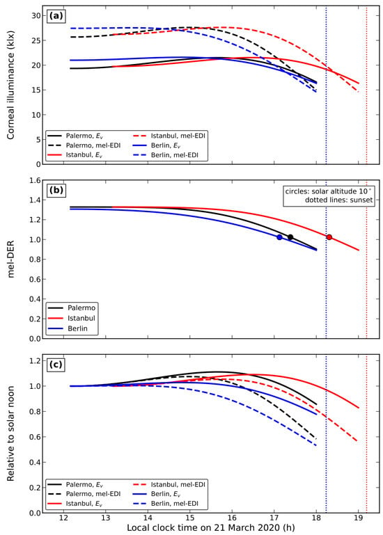
Figure 4 shows the orientation-averaged corneal illuminance , the mel-EDI and mel-DER from solar noon to a solar altitude of 2° on 21 March 2020 at Palermo, İstanbul and Berlin. At solar noon the corneal illuminance from the sky alone is 19–21 klx and mel-DER is 1.31–1.33 at all three sites. mel-DER then declines monotonically, slowly at first and more steeply as the Sun approaches the horizon, reaching 1.02 at a solar altitude of (17:23 in Palermo, 18:18 in İstanbul, 17:08 in Berlin; circles in Figure 4b) and 0.89–0.90 at .
Figure 4.
Orientation-averaged corneal quantities from solar noon to a solar altitude of on 21 March 2020 at Palermo (black), İstanbul (red) and Berlin (blue), , sky light only. (a) Illuminance (solid) and melanopic EDI (dashed). (b) Melanopic daylight efficacy ratio, Equation (9). (c) (solid) and mel-EDI (dashed) relative to their values at solar noon. Horizontal axis: local clock time (Palermo and Berlin UTC+1, İstanbul UTC+3). Vertical dotted lines mark sunset, in the colour of the site: red for İstanbul (19:12) and blue for Palermo and Berlin, whose sunsets coincide at 18:14 so that the black Palermo line lies under the blue one; circles in (b) mark the time at which the Sun passes altitude. Where curves of different sites cross, they do so because the sites reach the same values at the same clock time.
The melanopic and photopic quantities therefore do not decline together (Figure 4c). Between solar noon and a solar altitude of , falls to 0.97, 0.96 and 0.90 of its noon value at the three sites, whereas mel-EDI falls to 0.75, 0.73 and 0.70. In the model, then, the melanopic stimulus declines earlier and considerably more steeply than illuminance, as anticipated qualitatively in the original submission. The near-constancy of is partly an artefact of the sky model, which is known to overestimate the brightness of the low-sun sky [42,43]; with a more realistic decline of total sky radiance both curves in Figure 4a would fall faster, but mel-DER, a ratio, is unaffected by such a scale error. Even at a solar altitude of the modelled corneal illuminance is of order lx, so all of the change described here takes place under fully photopic conditions, well before the mesopic range is approached during twilight [45]. The corresponding mel-EDI, – lx, is two orders of magnitude above the daytime recommendation of 250 lx [41]. A fall of mel-DER at such levels describes a change in the spectral composition of the stimulus; it does not by itself imply a proportional change in melanopsin-driven signalling, which also depends on absolute retinal irradiance, adaptation and exposure history (Section 4.1).
3.3. Solar Altitude as the Governing Variable; Turbidity, Orientation and Colour Temperature
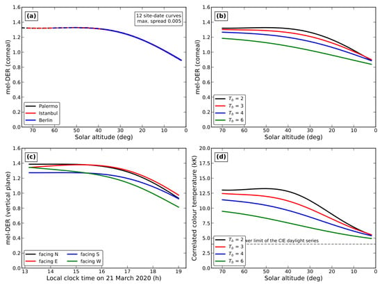
When mel-DER is plotted against solar altitude rather than clock time, the curves for the three sites and the four dates (equinoxes and solstices) collapse onto a single curve (Figure 5a). At solar altitudes of , , and the orientation-averaged corneal mel-DER is 1.26, 1.16, 1.02 and 0.94, and the spread across all twelve site–date combinations is at most 0.005. Within the clear-sky model, the afternoon spectral change at the eye is thus a function of solar altitude and turbidity only; latitude and date enter solely through the clock time at which a given altitude is reached. This removes the need for an arbitrary threshold: any hypothesis that ties symptom onset to spectral composition ties it to a solar altitude, whose clock time is computable. It also means that the size of the decline after noon depends on the season: where the Sun stays low all day, the noon value is itself low. In Berlin in December, for example, the Sun reaches only and the corneal mel-DER at noon is about 1.09, so the decline to is less than a tenth of a unit, against about 0.3 at the equinox.
Figure 5.
(a) Orientation-averaged corneal mel-DER against solar altitude for Palermo (black), İstanbul (red) and Berlin (blue) on 21 March, 21 June, 22 September and 21 December 2020 (solid, dashed, dash-dotted and dotted lines, respectively); the twelve curves are indistinguishable at this scale, and this overlap is the result shown: mel-DER depends on solar altitude, not on site or date. (b) Orientation-averaged corneal mel-DER against solar altitude for turbidities , 3, 4 and 6. (c) mel-DER on vertical planes facing north, east, south and west at İstanbul on 21 March 2020 against local clock time (UTC+3). (d) Correlated colour temperature of the orientation-averaged corneal irradiance against solar altitude for , 3, 4 and 6; the grey dashed line marks 4000 K, the lower limit of the CIE daylight series. All panels: sky light only, direct solar beam excluded; unless stated.
Aerosol loading lowers mel-DER at all altitudes and flattens its decline (Figure 5b). At a solar altitude of , mel-DER is 1.32, 1.26, 1.20 and 1.08 for , 3, 4 and 6; at it is 1.02, 1.01, 0.97 and 0.89. The afternoon decline between these altitudes is therefore 0.30 in very clear air and 0.19 in hazy air: turbidity reduces the amplitude of the change but does not remove it.
The orientation of the corneal plane modulates the levels and, to a lesser degree, the timing (Figure 5c, İstanbul, 21 March). At solar noon mel-DER ranges from 1.27 (facing south, towards the Sun) to 1.39 (facing north, towards the blue sky away from the Sun). In the afternoon a west-facing plane, which receives the reddened circumsolar and horizon sky, shows the earliest and deepest decline (0.94 at a solar altitude of ), and an east-facing plane the shallowest (1.12). The difference between orientations at a given time is comparable to the change from noon to a solar altitude of for any one orientation, so the direction a person faces is a material variable.
Figure 5d gives the correlated colour temperature (CCT) of the orientation-averaged corneal irradiance. It falls from 13,000 K at high solar altitude to 6800 K at and 5500 K at for , and stays above 4900 K for all turbidities considered, i.e., within the range 4000–25,000 K over which the CIE daylight series is defined [36]. The spectral reconstruction is therefore not extrapolated beyond its domain of definition at any point of our time series; because the direct solar beam is excluded, the reddening of the corneal light is moderate. This check concerns the internal consistency of the reconstruction, not the accuracy of the model’s chromaticities near the horizon, which remains the principal uncertainty of the low-altitude numbers. As a rough indication of their sensitivity, mel-DER at ranges from 0.84 to 0.90 over turbidities 2–6 (Figure 5b); the error of the clear-sky model itself at such elevations may be larger [42,43], so values below about should be read as indicative.
3.4. Indoor Exposure in a Side-Lit Room
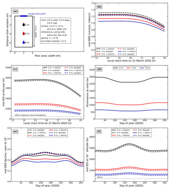
Figure 6 shows the indoor results for the room of Table 2. At solar noon on the equinox the daylight illuminance at the eye at 2, 4 and 6 m from the window is about 2800, 1090 and 620 lx (mel-EDI 3660, 1360 and 760 lx), of which about 150 lx is internally reflected light. The internally reflected component matters most at the back of the room, where it supplies a quarter of the daylight at the eye; because the interior surfaces are neutral and the light that reaches them comes largely from the blue sky, it slightly raises mel-DER there. Measured values in care facilities are substantially lower [13,14], owing to smaller or obstructed windows, deeper plans, darker finishes and occupants’ positions, so these figures should be read as an upper bound for a well-daylit room.
Figure 6.
Indoor exposure in the side-lit room of Table 2, İstanbul, , beam sunlight excluded; direct view of the window plus internally reflected light, Equation (10). (a) Plan of the room; the filled circles mark the seated observers at 2 m (black), 4 m (red) and 6 m (blue) from the window, and the small triangle on each circle shows the viewing direction (all facing the window, blue bar). (b) Corneal mel-DER and (c) melanopic EDI at the eye from solar noon to a solar altitude of on 21 March 2020, with daylight only (dashed) and with an additional 200 lx of 3000 K electric light (solid); the dotted line in (c) marks the daytime recommendation of 250 lx. (d) Daylight illuminance at the eye at solar noon, (e) decline of mel-DER from solar noon to a solar altitude of , and (f) mel-EDI at a solar altitude of , for every fourth day of 2020. In (b,e) the daylight-only curves for the three distances nearly coincide, because with daylight alone the spectral decline is the same at all distances (Section 3.4). Clear skies only: the seasonal variation shown is that of solar geometry, not of cloud.
With daylight alone, the spectral decline indoors follows that of the sky seen through the window: mel-DER falls by 0.38–0.40 from solar noon to a solar altitude of at all three distances (Figure 6b). Adding 200 lx of warm-white electric light dilutes the daylight signal, and the dilution grows with distance from the window: the decline is reduced to 0.37, 0.33 and 0.30 at 2, 4 and 6 m, i.e., to 94%, 87% and 78% of its daylight-only value. Far from windows, where electric light dominates, the afternoon spectral change reaching the eye is correspondingly small; close to windows it is preserved almost entirely. In absolute terms (Figure 6c) the melanopic EDI remains above the daytime recommendation of 250 lx [41] at all three positions until the Sun is above the horizon (440 lx at 6 m with daylight only), in this unobstructed room under a clear sky.
The equinox is one day of the year. Figure 6d–f gives the same quantities for every fourth day of 2020. Under clear skies, the noon illuminance near a north-facing window changes by only 10–20% over the year, because the luminance of the northern sky varies much less with season than the direct beam does; the large seasonal and day-to-day variation of indoor daylight in real rooms comes mainly from cloud, which is not modelled. The spectral quantities vary more: the decline of mel-DER from noon to a solar altitude of ranges from 0.17 to 0.27 over the year (daylight only) and is smallest in winter, when the Sun is low even at noon, and with electric light from 0.14 to 0.25. The equinox values used above therefore lie in the upper half of the annual range; they illustrate the effect but do not represent its annual distribution, which would require climate-based simulation with representative sky conditions [39].
3.5. Clock Time of the Spectral Transition over the Year
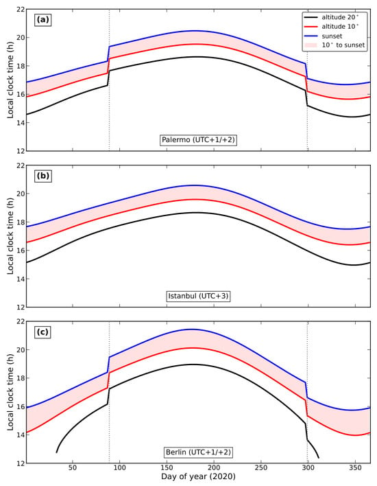
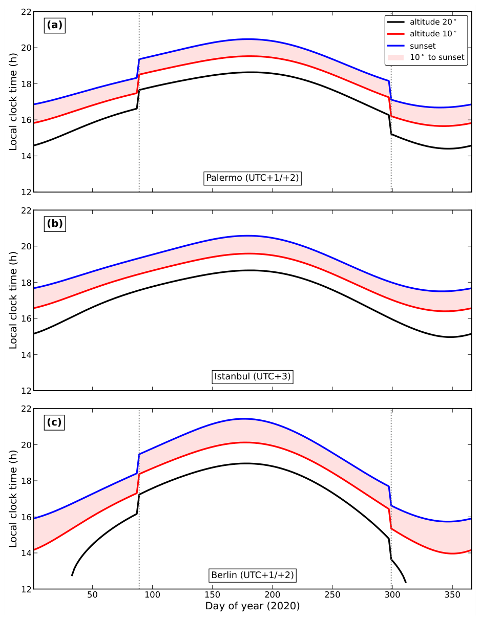
Because the spectral index is governed by solar altitude, its timing in clock time follows from solar geometry and civil time alone. Figure 7 shows, for every second day of 2020, the clock time at which the Sun descends through altitudes of and , and the time of sunset, at the three sites; Table 3 summarises the annual statistics. Three features are relevant to the hypothesis. First, the clock time of a given altitude, and hence of a given stage of the spectral decline, varies over the year by 3.9 h in Palermo, 3.2 h in İstanbul (where it is not shifted by summer time) and 6.2 h in Berlin. Second, at the two sites that observe summer time, it jumps by one hour at each transition, while the solar conditions on either side of the transition are essentially identical; at İstanbul there is no jump. Third, the interval between the Sun passing and sunset is nearly constant at low latitudes (51–62 min in Palermo, 53–67 min in İstanbul) but varies from 66 to 107 min in Berlin, where the Sun sets at a shallower angle.
Figure 7.
Local clock time at which the Sun descends through altitudes of (black) and (red), and of geometric sunset (blue), for every second day of 2020 at (a) Palermo, (b) İstanbul and (c) Berlin. The one-hour steps in (a,c) are the EU summer-time transitions (29 March and 25 October 2020); İstanbul keeps UTC+3 all year. In Berlin the Sun does not reach between early November and early February. The shaded band is the interval between the Sun passing and sunset; grey vertical dotted lines in (a,c) mark the summer-time transitions. The ordinate starts at 12:00 because no event of interest occurs in the morning.
Table 3.
Annual statistics (2020, every second day) of the local clock time of geometric sunset and of the Sun’s descent through altitude, and of the interval between the two; lead times are given as mean (range).
4. Discussion
4.1. Visual and Non-Image-Forming Pathways
Two retinal systems must be distinguished. The image-forming visual system is served by rods and cones. Rods mediate achromatic vision at low light levels, with the scotopic luminous efficiency peaking at 507 nm; cones mediate colour vision and acuity in daylight, with the photopic function peaking at 555 nm [37]. Which of the two operates is set by the absolute luminance: cone vision above roughly 5 cd m−2, rod vision below roughly cd m−2, with the mesopic range between [45]. The handover produces the Purkinje shift of spectral sensitivity towards shorter wavelengths during twilight [46,47]. Because afternoon illuminance remains of order – lx (Figure 4a), the eye is photopic throughout the interval considered here, and no account of sundowning resting on a change of visual regime in the late afternoon is compatible with the photometry. We do not advance one.
The non-image-forming system is served chiefly by melanopsin-expressing ipRGCs [6,7]. They project via the retinohypothalamic tract to the suprachiasmatic nucleus, the master circadian pacemaker, and to other brain regions regulating sleep, alertness and mood [2,3]. Their intrinsic sensitivity, referred to the cornea, is described by the melanopic action spectrum of CIE S 026 [9], which peaks at 490 nm—some 17 nm shorter than , from which it is distinct (Figure 2b). The two systems are not fully separate: ipRGCs also receive synaptic input from rods and cones, which contributes to NIF responses, particularly at low light levels and for short exposures [3,8]. For this reason CIE S 026 defines five -opic quantities, one for each photoreceptor class, of which the melanopic one is the most widely used predictor of circadian and alerting responses to light and is the basis of current recommendations: a melanopic EDI of at least 250 lx at the eye during the day, and at most 10 lx in the three hours before sleep [41]. We therefore express the spectral change in melanopic units (Section 2.4); the scotopic and photopic functions are retained only for comparison.
In these terms the results can be summarised simply. In the afternoon the eye remains in the photopic regime, total illuminance changes modestly, and the melanopic content per lux of daylight falls by about a quarter before the Sun reaches and by about a third before sunset. If the NIF system is sensitive to spectral composition as well as to melanopic dose—for instance through the colour-opponent (S-cone versus L/M-cone) input to ipRGCs that has been shown in mice to encode the colour changes of twilight and to contribute to circadian entrainment [48]—then the late afternoon offers such a signal. In people with dementia, whose lens transmits less short-wavelength light [10] and whose ipRGCs are depleted [11,12], the melanopic signal is already attenuated, and the afternoon decline would reduce it further.
Two quantities must, however, be kept apart: the spectral composition of the light, which mel-DER describes, and the physiologically effective melanopic stimulation, which it does not—mel-DER is a ratio. A fall of one quarter in mel-DER at constant illuminance is a fall of one quarter in mel-EDI, but what the retina does with that change depends on more than the ratio. It depends on the absolute retinal irradiance, which is set by mel-EDI, pupil size and the transmittance of the ageing lens [10]; on the state of adaptation and the preceding light history; on the timing and duration of exposure; and on the contributions of rods and cones to ipRGC output [8,41]. Models of melatonin suppression make the same point: melanopic EDI is the best single predictor, but timing and duration of exposure, and at some levels spectral composition, contribute independently [49]. Two consequences follow for the present results. Outdoors, where mel-EDI remains above lx throughout the interval we model, the melanopic dose is far above the levels at which non-image-forming responses are usually dose-limited, and a change in composition there need not translate into a proportional change in melanopsin-driven signalling. Indoors, where mel-EDI is of order – lx and measured exposures in care facilities are lower still [14], the same proportional change acts on a dose-sensitive part of the response. Our results quantify the first of the two quantities, the spectral composition at the eye, and should not be read as estimates of the second.
4.2. Interpretation and Relation to Clinical Observations
We advance this as a hypothesis, not as a demonstrated mechanism. The model shows that a well-defined property of natural light changes by a computable amount at a computable time within the broad window in which sundowning is clinically reported [20]. It does not establish that this change causes the behaviour.
Alternative readings should be kept in view. The coincidence between the computed spectral change and the reported onset window may be no more than that: both fall in the late afternoon, but so do a good many other things, including meals, staff changeover and the accumulated fatigue of the day. A spectral cue is one hypothesis among several, and the present calculation does not discriminate between them.
The clinical motivation is also weaker than a light-based account would wish. The empirical picture is mixed, and includes findings that count against the present proposal. Martin et al., using behavioural ratings collected every 15 min over three days in 85 institutionalised patients, reported a mean agitation acrophase of 14:38, classified only 2.4% of the sample as sundowners, and found no consistent seasonal pattern [25]. An ecological analysis of internet search activity found enquiries about sundowning to be roughly 11% higher on Sundays, when external caregiver support is least available, which is more readily explained by caregiver burden than by any optical effect; the same study, however, found search activity higher in winter, in less sunny states, and at more northerly latitudes, as a light-based account would predict [50]. Reviews agree that late-day worsening of behaviour is common in dementia even where a discrete syndrome is rare, and that light exposure, including the transition from daylight to artificial light in the late afternoon, is among its plausible modifiers [21,22]. Taken together, these results are consistent with sundowning having both an environmental-light component and a caregiver-related component, and they do not at present decide between them. The value of the present calculation is not that it establishes the first; it is that it makes the first testable, by stating which property of light changes, by how much, and at what clock time for a given place and day.
The agreement in timing is coarse. The spectral decline is gradual and is concentrated in the last one to two hours before sunset (Figure 4 and Figure 5a), which in clock time can fall anywhere between mid-afternoon in a northern winter and mid-evening in a southern summer (Figure 7); an agitation acrophase of 14:38 [25] lies earlier than this in most seasons. Clinical onset data are not precise or harmonised enough at present to constrain the model.
4.3. Separating Spectral Composition from Declining Light Level
In natural light the two are confounded. In the model the composition changes first, while illuminance at the eye is nearly constant down to a solar altitude of about (Figure 4c), but this separation is partly an artefact of a sky model that overestimates low-sun radiance, and below both decline together. Real afternoons therefore cannot by themselves distinguish a spectral cue from a cue of decreasing light. Three designs can.
(i) Controlled spectral manipulation at constant illuminance. With multi-channel tunable lighting it is possible to change melanopic content at fixed illuminance, and even at fixed colour temperature [51], or to modulate melanopsin while holding the cone signals constant by silent substitution [52]. A within-subject factorial design in the day rooms of a care facility, applied during the late afternoon, would compare: (a) constant illuminance with mel-DER lowered from about 1.3 to about 0.9, reproducing the natural spectral decline at constant level; (b) constant mel-DER with illuminance lowered by the same melanopic factor, reproducing the decline in level without the spectral change; (c) both together, as in nature; and (d) neither. A spectral account predicts that (a) and (c) increase late-day agitation relative to (d); an account based on light level predicts that (b) and (c) do. Daylight would have to be excluded or held constant, for example in interior rooms or with blinds.
(ii) Weather as a natural experiment. Overcast skies flatten the spectral change while lowering illuminance. Comparing behaviour on clear and overcast days matched for illuminance at the eye, measured with wearable spectral loggers, separates the two factors observationally (Prediction 3).
(iii) Position in the room. Residents near and far from windows receive similar clock-time routines but different spectral time courses (Figure 6b,c). This contrast, together with measured exposures, gives a further handle on the spectral factor (Prediction 4), although it is confounded by the reasons residents sit where they do.
4.4. Clinical Implications
If the proposal is correct, the practical consequence concerns timing and spectrum rather than intensity alone. Trials of bright-light therapy in dementia have generally administered light on fixed clock schedules, most often in the morning, and have reported inconsistent effects on agitation [27,29], although a tailored, all-day, high-melanopic intervention improved sleep, depression and agitation scores [28]. The present model suggests a specific target: supplementation of the short-wavelength component during the afternoon hours in which it is naturally depleted, timed to the local solar altitude rather than to the clock, and specified in melanopic rather than photopic units [9,41]. Figure 6 indicates a further, simpler lever: the position of a resident relative to a window determines how much of the natural afternoon signal reaches the eye at all.
4.5. Testable Predictions
The value of a quantitative proposal lies in its capacity to fail. The following predictions distinguish the present hypothesis from the alternatives above, and each is accessible with existing methods.
- Onset should track solar time, not clock time. The transitions to and from daylight saving time provide a natural experiment: the clock shifts by one hour overnight while solar conditions are essentially unchanged (Figure 7a,c). If sundowning is cued by the light field, symptom onset measured in clock time should shift by approximately one hour across the transition and remain fixed in solar time. If it is driven by institutional routine, which follows the clock, onset should remain fixed in clock time. Facilities in regions without daylight saving time, such as İstanbul, provide a control in which neither account predicts a shift. The test is useful but not definitive. A transition also shifts sleep timing, meals, medication and visits, and the circadian system itself adjusts to the new clock over days to weeks rather than overnight [53]; the two accounts therefore predict different patterns over the weeks following a transition rather than a clean step. The comparison requires continuous recording on both sides of the transition and an analysis that allows for gradual re-entrainment. It requires no new instrumentation.
- Onset should follow a fixed solar altitude through the year. Because the spectral index is a function of solar altitude (Figure 5a), the hypothesis predicts that onset, recorded in clock time, follows the seasonal curves of Figure 7, with an annual excursion of about 3–4 h at the latitude of Palermo or İstanbul and more than 6 h at that of Berlin. The altitude itself is a free parameter to be estimated from data, not a threshold chosen in advance. A null result—onset fixed in clock time through the year—would count against the hypothesis. The existing evidence is divided: one actigraphic study examined seasonality and found no consistent pattern [25], whereas geographic search-activity data show the latitude dependence a light-based account predicts [50].
- Overcast conditions should attenuate the effect. Cloud cover flattens the spectral evolution of daylight relative to the clear-sky case. If the spectral transition is the cue, its behavioural correlate should be weaker on overcast days, after controlling for total illuminance.
- Proximity to windows and room orientation should modulate the effect. Residents who spend the afternoon close to windows receive nearly the full natural spectral decline, those far from windows mainly electric light (Figure 6b); west-facing views show an earlier and deeper decline than east-facing ones (Figure 5c). Onset timing and severity should differ accordingly. This prediction is the one most readily confounded, but also the one most directly relevant to intervention. Because indoor daylight varies considerably through the year and from day to day, and our equinox examples and clear skies sample only part of that variation (Figure 6d–f; [39]), such comparisons should rest on exposures measured at the eye rather than on modelled values.
- Melanopic irradiance should decline earlier and more steeply than photopic illuminance through the afternoon. The present calculation confirms this within the clear-sky model (Figure 4c): between solar noon and a solar altitude of , mel-EDI falls by 25–30% while illuminance falls by 3–10%. The prediction can now be tested directly with calibrated spectroradiometry at the eye, outdoors and in care settings.
- Short-wavelength supplementation timed to the local solar altitude should outperform fixed-schedule bright light. The present model predicts that afternoon supplementation, timed to local solar conditions and dosed in melanopic equivalent daylight illuminance [41], should be more effective than the same melanopic dose delivered on a fixed clock schedule. A trial finding no difference between solar-timed and clock-timed protocols would substantially weaken the proposal.
Predictions 1 and 2 are the most direct tests of the timing, and the designs of Section 4.3 the most direct tests of the spectral factor itself. If symptom onset follows the clock rather than the Sun across daylight saving transitions and through the year, the hypothesis advanced here is very probably wrong, and the late-afternoon timing of sundowning is better explained by the structure of the institutional day than by the structure of the atmosphere.
4.6. Limitations
This is a computational hypothesis. It includes no patients, no intervention and no correlation between symptoms and measured light; its results describe the light field, not behaviour. The model is deliberately minimal, and its assumptions restrict what we can conclude from it.
Optical model. The sky radiance is computed from an analytic model developed for image synthesis rather than radiometry, which overestimates absolute sky radiance and is least reliable near the horizon and at low solar elevation [42,43]. Our conclusions rest on the spectral ratio mel-DER, which is insensitive to the absolute scale, and we stop at a solar altitude of ; the absolute illuminances in Figure 4a and Figure 6 are indicative only. The ratio is not free of model error, however, since the sky chromaticities from which it is computed are also least reliable at low solar elevation. The reconstructed spectra remain within the domain of the CIE daylight series down to (Figure 5d), but the numerical values of mel-DER below about , including the value of about 0.89 at , should be treated as indicative, with an uncertainty that we cannot quantify without measurements. We have not validated the computed spectra against measurements. The spectral shape is reconstructed from chromaticity through the three-component CIE daylight basis, which captures most of the variance of measured daylight with the Sun above the horizon but not the finer spectral structure of twilight [5].
Sky conditions and geometry. We have varied turbidity between 2 and 6 and computed vertical planes in four orientations integrated over the whole sky, but we assume a cloudless sky; cloud cover and its local climatology are not modelled, although they substantially modify the spectral composition of daylight. The direct solar beam is excluded; an observer facing the Sun, or a room lit by beam sunlight, would receive a larger share of the reddened direct component and hence a lower mel-DER in the late afternoon, so the exclusion makes our estimate of the decline conservative. The ground is modelled as spectrally flat, whereas vegetation and buildings have their own spectral reflectances.
From the sky to the retina. Patients are predominantly indoors. The indoor scenario of Section 2.5 is one room: a single unobstructed north-facing window with a window-to-wall ratio of 17%, clear glazing, neutral surfaces whose interreflection is treated by the split-flux approximation, beam sunlight excluded, and a single electric-light spectrum and level. Coated or tinted glazing attenuates short wavelengths preferentially; interreflections from coloured surfaces alter the spectrum; deeper plans, smaller or obstructed windows and darker finishes reduce the daylight reaching the eye; the switching schedule of electric lighting is itself a late-afternoon event; and actual exposure depends on posture, gaze and movement [54]. Measured exposures in dementia units are well below current recommendations for most of the day [13,14]. Our indoor results show the direction and approximate size of these effects, not the exposure of any particular resident.
Clinical confounds. Even granting the optics, the timing coincidence admits other explanations. The late afternoon in a residential care facility is also when staff shifts change, when the evening meal is prepared and served, when staffing levels typically fall, and when the day’s accumulated fatigue is greatest. Sundowning is largely identified from caregiver report, and caregiver observation is itself unevenly distributed across the day, which may bias the apparent timing of symptom onset. Whether sundowning is a discrete, time-locked phenomenon at all remains contested [26], and a model that predicts a specific time is of limited use if the phenomenon it predicts is not reliably time-locked. We have not attempted a quantitative comparison between computed timing and published onset distributions; such a comparison would require onset data recorded against local solar time with harmonised instrumentation across sites, and we are not aware of a dataset that presently permits it.
4.7. Future Research Directions
Three extensions follow directly. First, validation of the computed spectra and of the melanopic decline with calibrated spectroradiometry at eye level, outdoors under clear and overcast skies and inside care facilities of differing orientation and glazing, and replacement of the analytic sky model by a radiative-transfer calculation. Second, a full indoor coupling model including glazing spectra, obstruction, interreflection and the spectrum and schedule of electric lighting, combined with wearable light loggers on residents so that the exposure actually received can be related to behaviour. Third, a clinical test of Predictions 1 and 2 using continuous behavioural or actigraphic recording across daylight-saving transitions and through the year, ideally at facilities in regions with and without daylight saving time and at contrasting latitudes, and a controlled test of the spectral factor with tunable lighting (Section 4.3). For the indoor work, climate-based annual daylight simulation with representative sky conditions [39], extended to spectral quantities, would replace the clear-sky days used here.
5. Conclusions
We have presented a quantitative model linking solar geometry and atmospheric scattering to the spectral composition of the light available to an observer, and have used it to characterise how that composition evolves through the afternoon.
The principal result is that the melanopic and photopic content of daylight do not decline together. In clear-sky conditions the melanopic daylight efficacy ratio at the eye falls from about 1.33 at solar noon to about 1.02 at a solar altitude of and about 0.89 near sunset, while total illuminance changes much less and remains far above the mesopic range. This change is a function of solar altitude and turbidity alone; its clock time is therefore computable for any site and day, varies by 3–6 h over the year at European latitudes, and jumps by one hour at daylight-saving transitions. Aerosol loading and electric light indoors reduce its amplitude but do not remove it. Three qualifications apply. The result concerns spectral composition, not the physiologically effective melanopic stimulation, which also depends on absolute irradiance, adaptation, timing and duration. Its numerical values are least certain close to the horizon, where the analytic sky model is weakest. And its size depends on the season and on where a person sits: it is smaller in winter, when the Sun is low all day, and far from windows, where electric light dominates.
We advance the resulting proposal carefully. The model establishes that a well-defined property of natural light changes substantially, and at a computable time. It does not establish a causal link, it does not include patients, and it does not exclude the institutional and behavioural confounds that share the same time of day. Nor is the existing clinical evidence unanimous. These limitations and tensions are set out in Section 4.6.
What the present approach offers, and what qualitative light-based accounts of sundowning have lacked, is a specified quantity and a predicted time. That is enough to generate predictions capable of failing: that symptom onset should follow solar rather than clock time across daylight saving transitions and through the year, and that, under controlled lighting, a change of spectral composition at constant illuminance should matter. Should these tests come out the other way, the hypothesis advanced here would be very probably wrong; that it can be tested in this way is, we think, its principal merit.
Supplementary Materials
The following supporting information can be downloaded at: https://www.mdpi.com/article/10.3390/brainsci16111168/s1, Python code (package skymodel2 and script make_figures.py) regenerating all figures, Table 1 and Table 2 and the numbers quoted in the text.
Author Contributions
Conceptualisation, A.S.; methodology, E.C.A. and A.S.; software and formal analysis, E.C.A. and A.S.; visualisation, E.C.A. and A.S.; writing—original draft, E.C.A. and A.S.; writing—review and editing, E.C.A. and A.S.; supervision, A.S. All authors have read and agreed to the published version of the manuscript.
Funding
This research received no external funding.
Institutional Review Board Statement
Not applicable.
Informed Consent Statement
Not applicable.
Data Availability Statement
This study is entirely computational and generated no new experimental data. The CIE daylight basis functions, luminous efficiency functions and melanopic action spectrum were taken from Refs. [36,37,44]. The code that produces all results is provided as Supplementary Materials.
Acknowledgments
A.S. thanks the management of the Vocational School and also collaborators from the engineering and natural sciences faculty. During the preparation of this work the authors used Claude (Anthropic, San Francisco, CA, USA; https://claude.ai, accessed on 20 October 2026), ChatGPT (OpenAI, San Francisco, CA, USA; https://chatgpt.com, accessed on 20 October 2026), Jenni AI (https://jenni.ai, accessed on 20 October 2026) and Grammarly (Grammarly Inc., San Francisco, CA, USA; https://www.grammarly.com, accessed on 20 October 2026) for calculations, figure design and English-language improvement. The authors reviewed and edited all output and take full responsibility for the content of this publication.
Conflicts of Interest
The authors declare no conflicts of interest.
References
- Czeisler, C.A.; Duffy, J.F.; Shanahan, T.L.; Brown, E.N.; Mitchell, J.F.; Rimmer, D.W.; Ronda, J.M.; Silva, E.J.; Allan, J.S.; Emens, J.S.; et al. Stability, precision, and near-24-hour period of the human circadian pacemaker. Science 1999, 284, 2177–2181. [Google Scholar] [CrossRef] [Scilit]
- LeGates, T.A.; Fernandez, D.C.; Hattar, S. Light as a central modulator of circadian rhythms, sleep and affect. Nat. Rev. Neurosci. 2014, 15, 443–454. [Google Scholar] [CrossRef] [Scilit]
- Schmidt, T.M.; Chen, S.K.; Hattar, S. Intrinsically photosensitive retinal ganglion cells: Many subtypes, diverse functions. J. Physiol. 2011, 589, 2451–2458. [Google Scholar]
- Bohren, C.F.; Clothiaux, E.E. Fundamentals of Atmospheric Radiation: An Introduction with 400 Problems; Wiley-VCH: Weinheim, Germany, 2006. [Google Scholar]
- Spitschan, M.; Aguirre, G.K.; Brainard, D.H.; Sweeney, A.M. Variation of outdoor illumination as a function of solar elevation and light pollution. Sci. Rep. 2016, 6, 26756. [Google Scholar] [CrossRef] [Scilit]
- Berson, D.M.; Dunn, F.A.; Takao, M. Phototransduction by retinal ganglion cells that set the circadian clock. Science 2002, 295, 1070–1073. [Google Scholar] [CrossRef] [Scilit]
- Hattar, S.; Liao, H.W.; Takao, M.; Berson, D.M.; Yau, K.W. Melanopsin-containing retinal ganglion cells: Architecture, projections, and intrinsic photosensitivity. Science 2002, 295, 1065–1070. [Google Scholar] [CrossRef] [Scilit]
- Lucas, R.J.; Peirson, S.N.; Berson, D.M.; Brown, T.M.; Cooper, H.M.; Czeisler, C.A.; Figueiro, M.G.; Gamlin, P.D.; Lockley, S.W.; O’Hagan, J.B.; et al. Measuring and using light in the melanopsin age. Trends Neurosci. 2014, 37, 1–9. [Google Scholar] [CrossRef] [Scilit]
- International Commission on Illumination. CIE S 026/E:2018 System for Metrology of Optical Radiation for ipRGC-Influenced Responses to Light; CIE: Vienna, Austria, 2018. [Google Scholar] [CrossRef] [Scilit]
- Pokorny, J.; Smith, V.C.; Lutze, M. Aging of the human lens. Appl. Opt. 1987, 26, 1437–1440. [Google Scholar] [CrossRef] [Scilit]
- Lax, P.; Ortuño-Lizarán, I.; Gómez-Vicente, V.; Esquiva, G.; Cuenca, N. Photosensitive melanopsin-containing retinal ganglion cells in health and disease: Implications for circadian rhythms. Int. J. Mol. Sci. 2019, 20, 3164. [Google Scholar] [CrossRef] [Scilit]
- La Morgia, C.; Ross-Cisneros, F.N.; Sadun, A.A.; Carelli, V. Retinal ganglion cells and circadian rhythms in Alzheimer’s disease, Parkinson’s disease, and beyond. Front. Neurol. 2017, 8, 162. [Google Scholar] [CrossRef] [Scilit]
- Shochat, T.; Martin, J.; Marler, M.; Ancoli-Israel, S. Illumination levels in nursing home patients: Effects on sleep and activity rhythms. J. Sleep Res. 2000, 9, 373–379. [Google Scholar] [CrossRef] [Scilit]
- Kolberg, E.; Pallesen, S.; Hjetland, G.J.; Nordhus, I.H.; Thun, E.; Flo-Groeneboom, E. Insufficient melanopic equivalent daylight illuminance in nursing home dementia units across seasons and gaze directions. Light. Res. Technol. 2022, 54, 163–177. [Google Scholar] [CrossRef] [Scilit]
- OECD. Elderly Population (Indicator); OECD Publishing: Paris, France, 2020. [Google Scholar] [CrossRef] [Scilit]
- World Health Organization. Dementia: Fact Sheet; WHO: Geneva, Switzerland, 2017. [Google Scholar]
- National Institutes of Health. Alzheimer’s Disease Fact Sheet; National Institute on Aging: Bethesda, MD, USA, 2019.
- Burns, A.; Iliffe, S. Alzheimer’s disease. BMJ 2009, 338, b158. [Google Scholar] [CrossRef] [Scilit]
- Larson, E.B.; Yaffe, K.; Langa, K.M. New insights into the dementia epidemic. N. Engl. J. Med. 2013, 369, 2275–2277. [Google Scholar] [CrossRef] [Scilit]
- Khachiyants, N.; Trinkle, D.; Son, S.J.; Kim, K.Y. Sundown syndrome in persons with dementia: An update. Psychiatry Investig. 2011, 8, 275–287. [Google Scholar] [CrossRef] [Scilit]
- Bachman, D.; Rabins, P. “Sundowning” and other temporally associated agitation states in dementia patients. Annu. Rev. Med. 2006, 57, 499–511. [Google Scholar] [CrossRef] [Scilit]
- Canevelli, M.; Valletta, M.; Trebbastoni, A.; Sarli, G.; D’Antonio, F.; Tariciotti, L.; de Lena, C.; Bruno, G. Sundowning in dementia: Clinical relevance, pathophysiological determinants, and therapeutic approaches. Front. Med. 2016, 3, 73. [Google Scholar] [CrossRef] [Scilit]
- Boronat, A.C.; Ferreira-Maia, A.P.; Wang, Y.-P. Sundown syndrome in older persons: A scoping review. J. Am. Med. Dir. Assoc. 2019, 20, 664–671.e5. [Google Scholar] [CrossRef] [Scilit]
- Gallagher-Thompson, D.; Brooks, J.O.; Bliwise, D.; Leader, J.; Yesavage, J.A. The relations among caregiver stress, “sundowning” symptoms, and cognitive decline in Alzheimer’s disease. J. Am. Geriatr. Soc. 1992, 40, 807–810. [Google Scholar] [CrossRef] [Scilit]
- Martin, J.; Shochat, T.; Marler, M.; Ancoli-Israel, S. Circadian rhythms of agitation in institutionalized patients with Alzheimer’s disease. Chronobiol. Int. 2000, 17, 405–418. [Google Scholar] [CrossRef] [Scilit]
- Cohen-Mansfield, J.; Watson, V.; Meade, W.; Gordon, M.; Leeks, D.; Lawson, M.; Taylor, L. Does sundowning occur in residents of an Alzheimer’s unit? Int. J. Geriatr. Psychiatry 1989, 4, 293–298. [Google Scholar] [CrossRef] [Scilit]
- Riemersma-van der Lek, R.F.; Swaab, D.F.; Twisk, J.; Hol, E.M.; Hoogendijk, W.J.G.; Van Someren, E.J.W. Effect of bright light and melatonin on cognitive and noncognitive function in elderly residents of group care facilities: A randomized controlled trial. JAMA 2008, 299, 2642–2655. [Google Scholar] [CrossRef] [Scilit]
- Figueiro, M.G.; Plitnick, B.A.; Lok, A.; Jones, G.E.; Higgins, P.; Hornick, T.R.; Rea, M.S. Tailored lighting intervention improves measures of sleep, depression, and agitation in persons with Alzheimer’s disease and related dementia living in long-term care facilities. Clin. Interv. Aging 2014, 9, 1527–1537. [Google Scholar] [CrossRef] [Scilit]
- Forbes, D.; Blake, C.M.; Thiessen, E.J.; Peacock, S.; Hawranik, P. Light therapy for improving cognition, activities of daily living, sleep, challenging behaviour, and psychiatric disturbances in dementia. Cochrane Database Syst. Rev. 2014, 2, CD003946. [Google Scholar] [CrossRef] [Scilit]
- Bedrosian, T.A.; Nelson, R.J. Timing of light exposure affects mood and brain circuits. Transl. Psychiatry 2017, 7, e1017. [Google Scholar] [CrossRef] [Scilit]
- Duffie, J.A.; Beckman, W.A. Solar Engineering of Thermal Processes, 4th ed.; Wiley: Hoboken, NJ, USA, 2013. [Google Scholar]
- Perez, R.; Seals, R.; Michalsky, J. All-weather model for sky luminance distribution—Preliminary configuration and validation. Sol. Energy 1993, 50, 235–245. [Google Scholar] [CrossRef] [Scilit]
- International Commission on Illumination. CIE S 011/E:2003 Spatial Distribution of Daylight—CIE Standard General Sky; CIE: Vienna, Austria, 2003. [Google Scholar]
- Preetham, A.J.; Shirley, P.; Smits, B. A practical analytic model for daylight. In Proceedings of the 26th Annual Conference on Computer Graphics and Interactive Techniques (SIGGRAPH ’99), Los Angeles, CA, USA, 8–13 August 1999; pp. 91–100. [Google Scholar] [CrossRef] [Scilit]
- Hosek, L.; Wilkie, A. An analytic model for full spectral sky-dome radiance. ACM Trans. Graph. 2012, 31, 95. [Google Scholar] [CrossRef]
- International Commission on Illumination. CIE 015:2018 Colorimetry, 4th ed.; CIE: Vienna, Austria, 2018. [Google Scholar] [CrossRef] [Scilit]
- International Commission on Illumination. CIE 018:2019 The Basis of Physical Photometry, 3rd ed.; CIE: Vienna, Austria, 2019. [Google Scholar] [CrossRef] [Scilit]
- Hopkinson, R.G.; Longmore, J.; Petherbridge, P. An empirical formula for the computation of the indirect component of daylight factor. Trans. Illum. Eng. Soc. 1954, 19, 201–219. [Google Scholar] [CrossRef] [Scilit]
- Kent, M.G.; Schiavon, S.; Jakubiec, J.A. A dimensionality reduction method to select the most representative daylight illuminance distributions. J. Build. Perform. Simul. 2020, 13, 122–135. [Google Scholar] [CrossRef] [Scilit]
- Peel, M.C.; Finlayson, B.L.; McMahon, T.A. Updated world map of the Köppen–Geiger climate classification. Hydrol. Earth Syst. Sci. 2007, 11, 1633–1644. [Google Scholar] [CrossRef] [Scilit]
- Brown, T.M.; Brainard, G.C.; Cajochen, C.; Czeisler, C.A.; Hanifin, J.P.; Lockley, S.W.; Lucas, R.J.; Münch, M.; O’Hagan, J.B.; Peirson, S.N.; et al. Recommendations for daytime, evening, and nighttime indoor light exposure to best support physiology, sleep, and wakefulness in healthy adults. PLoS Biol. 2022, 20, e3001571. [Google Scholar] [CrossRef] [Scilit]
- Kider, J.T., Jr.; Knowlton, D.; Newlin, J.; Li, Y.K.; Greenberg, D.P. A framework for the experimental comparison of solar and skydome illumination. ACM Trans. Graph. 2014, 33, 180. [Google Scholar] [CrossRef] [Scilit]
- Bruneton, E. A qualitative and quantitative evaluation of 8 clear sky models. IEEE Trans. Vis. Comput. Graph. 2017, 23, 2641–2655. [Google Scholar] [CrossRef] [Scilit]
- International Commission on Illumination. CIE α-Opic Action Spectra (Dataset); CIE: Vienna, Austria, 2019. [Google Scholar] [CrossRef] [Scilit]
- International Commission on Illumination. CIE 191:2010 Recommended System for Mesopic Photometry Based on Visual Performance; CIE: Vienna, Austria, 2010. [Google Scholar]
- Purkinje, J.E. Beobachtungen und Versuche zur Physiologie der Sinne; Reimer: Berlin, Germany, 1825. [Google Scholar]
- Lamb, T.D. Evolution of phototransduction, vertebrate photoreceptors and retina. Prog. Retin. Eye Res. 2013, 36, 52–119. [Google Scholar] [CrossRef] [Scilit]
- Walmsley, L.; Hanna, L.; Mouland, J.; Martial, F.; West, A.; Smedley, A.R.; Bechtold, D.A.; Webb, A.R.; Lucas, R.J.; Brown, T.M. Colour as a signal for entraining the mammalian circadian clock. PLoS Biol. 2015, 13, e1002127. [Google Scholar] [CrossRef] [Scilit]
- Giménez, M.C.; Stefani, O.; Cajochen, C.; Lang, D.; Deuring, G.; Schlangen, L.J.M. Predicting melatonin suppression by light in humans: Unifying photoreceptor-based equivalent daylight illuminances, spectral composition, timing and duration of light exposure. J. Pineal Res. 2022, 72, e12786. [Google Scholar] [CrossRef] [Scilit]
- Madden, K.M.; Feldman, B. Weekly, seasonal, and geographic patterns in health contemplations about sundown syndrome: An ecological correlational study. JMIR Aging 2019, 2, e13302. [Google Scholar] [CrossRef] [Scilit]
- Souman, J.L.; Borra, T.; de Goijer, I.; Schlangen, L.J.M.; Vlaskamp, B.N.S.; Lucassen, M.P. Spectral tuning of white light allows for strong reduction in melatonin suppression without changing illumination level or color temperature. J. Biol. Rhythms 2018, 33, 420–431. [Google Scholar] [CrossRef] [Scilit]
- Spitschan, M.; Woelders, T. The method of silent substitution for examining melanopsin contributions to pupil control. Front. Neurol. 2018, 9, 941. [Google Scholar] [CrossRef] [Scilit]
- Kantermann, T.; Juda, M.; Merrow, M.; Roenneberg, T. The human circadian clock’s seasonal adjustment is disrupted by daylight saving time. Curr. Biol. 2007, 17, 1996–2000. [Google Scholar] [CrossRef] [Scilit]
- Webler, F.S.; Spitschan, M.; Foster, R.G.; Andersen, M.; Peirson, S.N. What is the ‘spectral diet’ of humans? Curr. Opin. Behav. Sci. 2019, 30, 80–86. [Google Scholar] [CrossRef] [Scilit]

Disclaimer/Publisher’s Note: The statements, opinions and data contained in all publications are solely those of the individual author(s) and contributor(s) and not of MDPI and/or the editor(s). MDPI and/or the editor(s) disclaim responsibility for any injury to people or property resulting from any ideas, methods, instructions or products referred to in the content. |
© 2026 by the authors. Licensee MDPI, Basel, Switzerland. This article is an open access article distributed under the terms and conditions of the Creative Commons Attribution (CC BY) license.