


Abstract
Flowable composites improve adaptability at the gingival margin but may affect mechanical properties. This study tested the surface hardness of proximal restorations as indicator of depth-of-cure and mechanical properties. Typodont teeth with 4 mm deep disto-occlusal preparations were (a) restored with three flowable composites in 4 mm bulk, and (b) restored with a thin layer of a flowable at the gingival margin under a universal composite. For the latter, two techniques were tested: ‘pre-cure’ and ‘snowplow’. In the pre-cure technique, the flowables were cured separately; in the snowplow technique, they were cured together with the universal composite. The universal composite placed in two 2 mm increments was the control. After 24 h, the Vickers hardness of the proximal restorations was measured every 0.5 mm from occlusal to gingival. The results were statistically analyzed using ANOVA and post hoc tests (α = 0.05). The sample size was 10/group. Bulk-filled restorations having undergone 40 s of light curing were significantly harder than those cured for 20 s. The pre-cure technique decreased hardness at depths of 3 mm and greater. The snowplow technique also led to significantly lower hardness at 4 mm, but less reductions at the 3.0 and 3.5 mm depths. The fiber-reinforced flowable composite increased the overall hardness of the restorations. In conclusion, using a flowable composite in a thin layer at the gingival margin to improve adaptation affected the hardness of the restoration. The snowplow technique reduced the softening effect associated with flowable composites compared to the pre-cure technique.
1. Introduction
Dental caries is the most common noncommunicable disease worldwide. Untreated dental caries in permanent teeth affects more than 2 billion people, and leads to repeated episodes of pain, difficulty chewing, and reduced quality of life, as well as productivity loss at work [1]. The high incidence of dental caries, together with the global effort to phase down dental amalgam following the Minamata Convention on Mercury, have increased demand for mercury-free restorative materials, particularly glass–ionomer cements and composite resins [2].
In recent years, resin-based composites have become the material of choice for tooth restorations, being preferred over mercury-containing amalgam [3,4]. In addition to their esthetic quality, composite restorations are able to bond to tooth structures [5,6]. Composite materials have the largest market share among restorative materials and have accounted for over 500 millions restorations placed internationally [7,8]. However, a systematic review and meta-analysis reported that composite restorations in posterior teeth showed less longevity than amalgam restorations, with a survival rate of 86% versus 93% at 55 months of follow-up [9].
The main cause of composite restoration failure was recurrent caries [9,10]. Among the clinically diagnosed secondary caries, more than 80% were located at gingival margins [11]. It has been postulated that recurrent caries at restoration margins originate from poor interface quality as a consequence of compromised adhesion, polymerization shrinkage, or restorative technique [12,13]. Achieving good adaptation and good curing are particularly challenging at the gingival margin of a proximal box [14].
Placing low-viscosity composite material at the gingival margin may promote the marginal seal by allowing improved adaptation and a reduction in stress from its low stiffness [15,16]. Flowable composites are a subclass of restorative composites designed to reduce viscosity via a lower filler content or adjusted formulation [17]. They can be used on their own or as a liner, i.e., a thin layer precedes the placement of a conventional composite [13]. A decade after the introduction of flowable composites, a new subclass, ‘bulk-fill’ composite, was introduced that allowed for placement and light curing in 4–5 mm thick layers instead of the traditional 2 mm increments [18]. Bulk-fill composites quickly gained popularity among clinicians because they simplify the restorative procedure and reduce chair time [19]. Because of their ease of use, more than 70% of dental practitioners in north America and Europe are using flowable composites; this pushed the estimated market size from USD 1.2 billion in 2024 to USD 1.8 billion by 2033 [20].
Advantages of flowable composites on marginal adaptation and achieving reductions in leakage are well studied [21,22], although positive clinical outcomes have yet to be demonstrated [23,24]. In addition to margin quality, the effect of flowable composites on the mechanical properties of the restoration should also be considered. Although the mechanical properties of the newer generation of flowable composites are still inferior to those of conventional composites, improved properties have been reported for a novel short fiber-reinforced flowable composite [25,26]. Restorative technique may also mitigate some of the mechanical property limitations of flowable materials. Flowable composites have been used in bulk as the sole restorative material [25,27] or as a thin layer under a universal composite in a ‘pre-cure’ or ‘snowplow’ technique [13]. In the pre-cure technique, a thin layer of flowable composite is light-cured prior to placing the first increment of the universal composite. In the snowplow technique, a thin layer of flowable is applied (but not yet cured), followed by the placement and condensation of the first increment of the universal composite on the uncured flowable to further disperse both materials along the cavity walls and margins before being light-cured simultaneously [13]. How well a fiber-reinforced flowable or regular bulk-fill flowable composite performs when used as proximal restoration or in the ‘pre-cure’ and ‘snowplow’ techniques still remains to be answered.
The objectives of this study were (a) to determine the depth-of-cure of three bulk-fill flowable composites in a simulated class II cavity with a 4 mm deep proximal box, and with a 20 or 40 s light curing time, and (b) to compare the hardness of the proximal surface when these cavities were restored with the pre-cure or snowplow technique. Restorations restored with a universal composite placed in standard 2 mm increments were used as a control.
2. Materials and Methods
The restorative composites used in this study were a light-cure universal composite and three light-cure bulk-fill flowable composites. The compositions of the tested composites are shown in Table 1. The universal composite is a nano-hybrid with pre-polymerized fillers, indicated for anterior and posterior direct restorations (TPH—TPH Spectra ST HV, Dentsply, Konstanz, Germany). Two of the three flowable composites are widely used particulate-filled composites (SDR—SDR Flow+ Bulk Fill Flowable, Dentsply Caulk, Milford, DE, USA; FTK—Filtek Bulk Fill Flowable Restorative, 3M Oral Care, St. Paul, MN, USA). The third flowable composite is fiber-reinforced (EXF—everX Flow Short-fiber Reinforced Flowable, GC Dental Products Corp., Aichi, Japan). All flowable composites used were easily adapted in deep posterior restorations.
Table 1.
Composition of the tested composites.
Restorations with Bulk-Fill Flowable Composites for Depth-of-Cure Evaluation
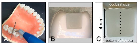
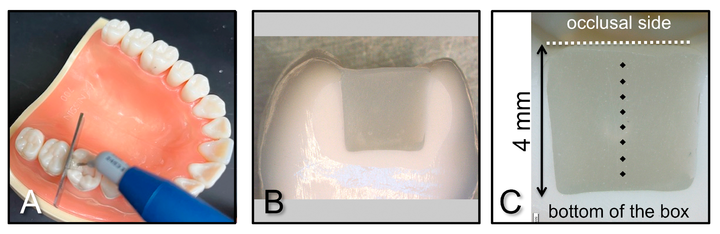
Sixty typodont maxillary first molars with a 4 mm proximal depth class II disto-occlusal (DO) preparation (A21AN-700-26-AJ-02, Kilgore International, Coldwater, MI, USA) were used to ensure standardized cavity size and shape. The distal surface was slightly flattened bucco-lingually by grinding with 600-grit SiC paper to create a flat surface for hardness measurements. The prepared tooth was secured in the typodont against a stainless steel matrix band (Tofflemire matrix band #1, Teledyne Densco, Denver, CO, USA) and restored with resin composites simulating clinical placement, as shown in Figure 1A. One of the three bulk-fill flowable composites was placed in the proximal box from the gingival margin to the height of the marginal ridge and light-cured from the occlusal direction for 20 or 40 s using the high-power mode of a Bluephase PowerCure curing light (Ivoclar Vivadent AG, Schaan, Liechtenstein) with an irradiance output of 1291 ± 13 mW/cm2. The irradiance was determined using the anterior sensor of a MARC Patient Simulator (BlueLight Analytics, Halifax, NS, Canada), and calculated as the mean of three measurements. Light curing was standardized by placing the curing tip over the occlusal plane, approximately 1 mm above the marginal ridge at the level of the matrix band. After the completion of light curing, the matrix band was removed and the restored teeth were kept in the dark for 24 h at room temperature. The sample size of the experimental groups was 10 per group for detecting a difference of 0.6 standard deviations between groups with 95 percent confidence [31].
Figure 1.
(A) Composite placement in a DO preparation against a stainless steel matrix band to simulate class II restoration; (B) proximal composite surface; (C) Diagram showing positions of Vickers hardness indentations from the occlusal to the gingival in 0.5 mm increments.
Restorations with Pre-cure or Snowplow Technique for Proximal Hardness Testing
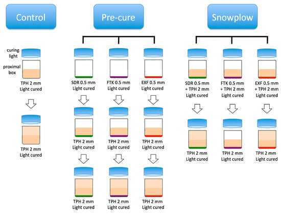
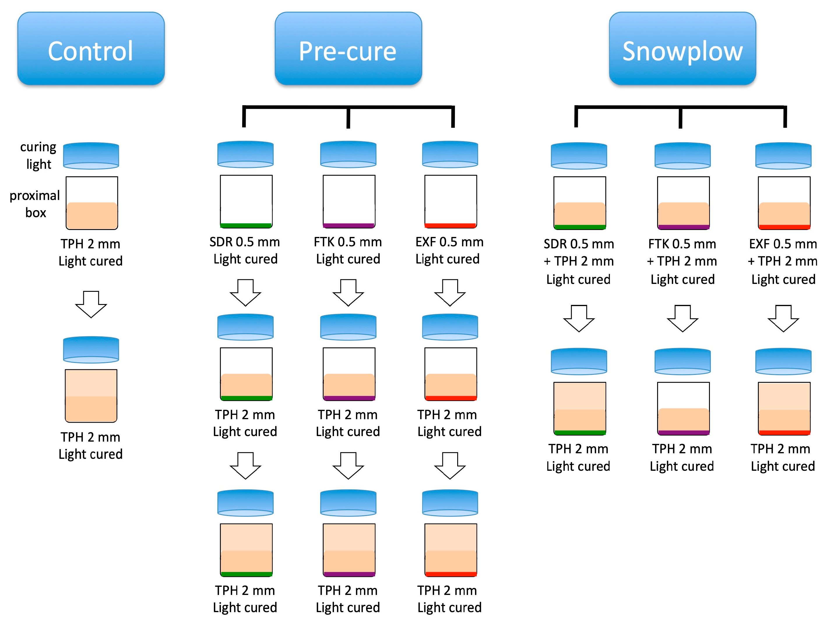
The experimental design consisted of 7 groups, as shown in the flow diagram (Figure 2). The universal composite, placed and light-cured in two 2 mm increments, was a control. Two restorative techniques were used, namely a pre-cure technique and a snowplow technique, as described later. Within each technique, one of the three flowable composites was used.
Figure 2.
Experimental design. TPH, SDR, FTK, and EXF are TPH Spectra ST HV universal composite, SDR Flow+ Bulk Fill Flowable, Filtek Bulk Fill Flowable Restorative, and everX Flow Short-Fiber Reinforced Flowable composites, respectively.
Seventy typodont maxillary first molars with a 4 mm proximal depth class II disto-occlusal (DO) preparation (A21AN-700-26-AJ-02, Kilgore International) were restored with resin composites to simulate clinical placement, as carried out previously, in the depth-of-cure test (Figure 1A). The two restorative techniques, pre-cure and snowplow, both consisted of a thin layer, measuring approximately 0.5 mm of one of the three bulk-fill flowables, placed on the gingival floor/margin under the universal composite. The flowable composites were injected directly into the proximal box from a compula tip (SDR) using a composite gun or from syringe tips (FTK and EXF). The operator used the tip to enhance leveling in addition to the self-leveling property of the flowables. In the pre-cure technique, the flowable composite was light-cured for 20 s before placing the first 2 mm increment of the universal composite by condensing it with a composite instrument PFI W3 (Hu-Friedy, Chicago, IL, USA). After the first increment of the universal composite was light-cured, the second 2 mm increment was placed and contoured to the level of the marginal ridge and light-cured. In the snowplow technique, the first 2 mm increment of the universal composite was condensed onto the uncured flowable, and both materials were simultaneously light-cured for 20 s. Then the second 2 mm increment was placed and light-cured. In the control group, two 2 mm increments of the universal composite were placed into the proximal box in horizontal layers and condensed with the composite instrument. The second increment was contoured to the level of the marginal ridge. Each increment was light-cured for 20 s from the occlusal direction. All light curing was performed using a high power mode of the Bluephase PowerCure curing light (Ivoclar Vivadent AG, Schaan, Liechtenstein). After the completion of light curing, the matrix band was removed and the restored teeth were kept in the dark for 24 h at room temperature. The sample size of the experimental groups was 10 per group, chosen to detect a difference of 0.6 standard deviations between groups with 95 percent confidence [31].
Hardness Measurements—The restored teeth were placed on a custom-made jig to adjust the proximal surface of the restoration in a horizontal plane for hardness measurement. The Vickers hardness of the proximal composite surface (Figure 1B) was measured (QV-1000 Micro Hardness Tester, Qualitest, Ft. Lauderdale, FL, USA) in 0.5 mm increments, starting from the top (marginal ridge) and moving down to a depth of 4 mm at the bottom (gingival) of the proximal box (Figure 1C). The indentation load was 50 g and the dwell time was 15 s. After indentation, the length of both indentation diagonals was measured using an attached optical microscope with 400 magnification, after which the Vickers hardness value (HV) could be calculated using the following:
where F is the 50 g indentation load and d is the mean of the two indentation diagonals in millimeters. The unit of the Vickers hardness number is kgf/mm2.
HV = 2 F sin(68°)/d2
Hardness values of cured resin-based materials have been shown to correlate well with the degree of conversion [32]. The ratio between the bottom hardness of each sample divided by the maximum hardness of each composite can therefore be used as an indicator of the depth-of-cure. Note, however, that hardness values of the proximal surface of restroations filled using a pre-cure or snowplow technique should not be interpreted as the depth-of-cure because the restorations contain a mix of different materials. In these cases, hardness should be considered just a mechanical property of the restoration [33].
After confirming normal distributions (Kolmogorov–Smirnov test of normality), the results were statistically analyzed using analysis of variance (ANOVA) followed by Student–Newman–Keuls post hoc tests (SuperANOVA, ABACUS Concepts, Inc, Berkeley, CA, USA). The significance level was 0.05.
3. Results
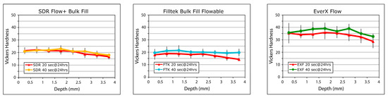
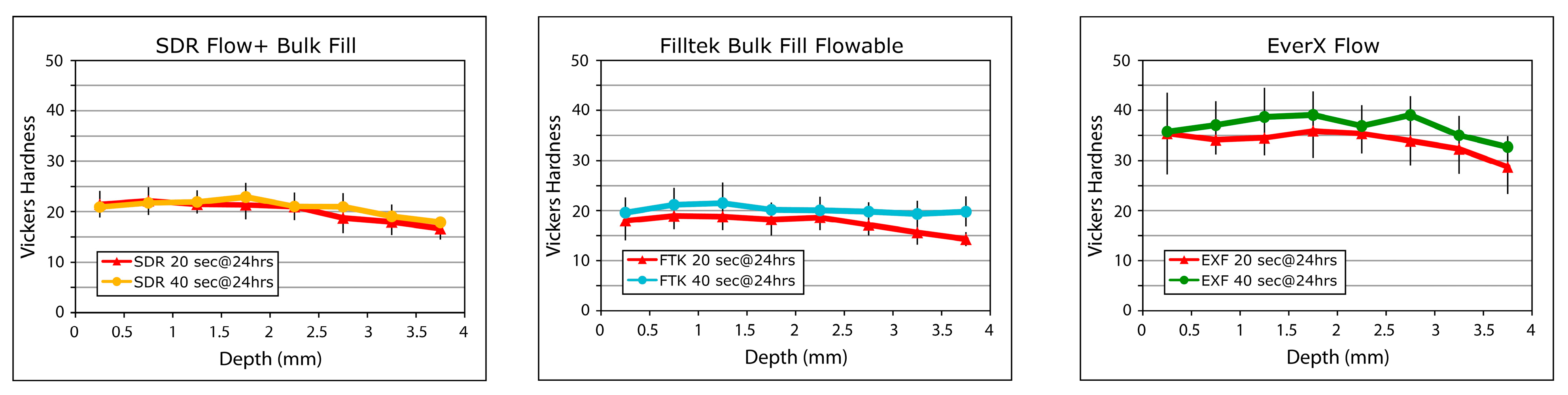
Depth-of-Cure of Bulk-Fill Flowable Composites—Figure 3 shows the Vickers hardness of the three bulk-filled flowable composites plotted through their depths. The fiber-reinforced composite (EXF) was harder than the other tested bulk-fill flowable composites. The ratios between the bottom hardness and the maximum hardness of each composite are listed in Table 2. Restorations with 40 s light curing time were significantly harder than those ligh-cured for 20 s, regardless of the composite used. There was a trend showing that the hardness values at a 4 mm depth were slightly lower than those at other depths for all tested bulk-fill flowable composites (Figure 3). With a 20 s light curing time, the bottom/max hardness ratios were 0.63–0.65. With a 40 s light curing time, the ratios increased to 0.73–0.85 at a 4 mm depth (Table 2).
Figure 3.
Hardness profiles of the bulk-filled flowable composites light-cured for 20 or 40 s.
Table 2.
Bottom/maximum hardness ratio (mean ± standard deviation) of the bulk-filled flowable composites light-cured for 20 or 40 s. The same letters denote values not significantly different (2-way ANOVA and pairwise comparisons; significance level, 0.05).
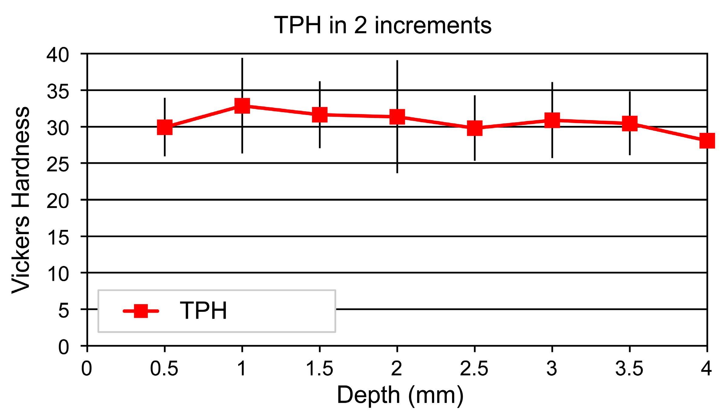
Hardness of Proximal Surfaces—The hardness values of the proximal surface of composite restorations in the control group, where the universal composite (TPH) was placed and light-cured in standard two 2 mm increments, were not significantly different among the depths (one-way ANOVA; p = 0.773). The hardness values through the depth are shown in Table 3 and Figure 4. The vertical bars depict the standard deviation values.
Table 3.
Hardness values (mean ± standard deviation) for the control group (TPH increments) and the pre-cure and snowplow techniques. Different letters indicate significant differences among depths within the same flowable composite (column). Note that there were no significant differences in hardness throughout the depth within the control group. (ANOVA and Student–Newman–Keuls post hoc test; significance level, 0.05).
Figure 4.
Hardness profile of the control group, a conventional composite TPH placed and light-cured in two 2 mm increments. There was no significant difference in hardness among the depths (ANOVA and Student–Newman–Keuls post hoc test; significance level 0.05).
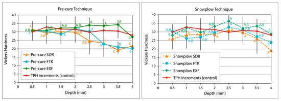
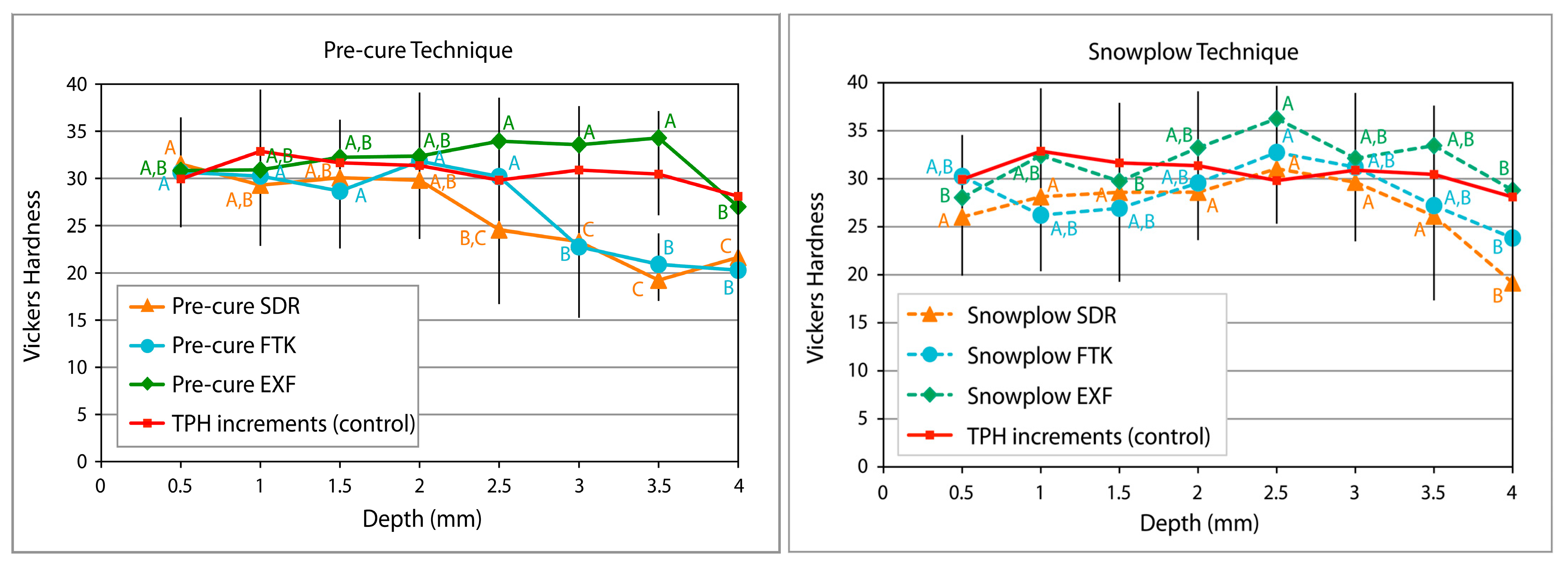
Hardness profiles of the proximal composites restored with a flowable composite at the gingival margin in the pre-cure and snowplow techniques are shown in Table 3 and Figure 5.
Figure 5.
Hardness profiles of the pre-cure and snowplow techniques. Different letters indicate significant difference among depths within the same flowable composite (ANOVA and Student–Newman–Keuls post hoc test; significance level, 0.05).
Significant differences in hardness values through the depth were detected for all composite materials and restorative technique combinations: p = 0.005 for the SDR snowplow technique, p = 0.026 for the FTK snowplow technique, p = 0.007 for the EXF snowplow technique, p < 0.001 for the SDR pre-cure technique, p < 0.001 for the FTK pre-cure technique, and p < 0.024 for the EXF pre-cure technique (one-way ANOVA). The pre-cure restorative technique had significantly lower hardness at a 4 mm depth for all flowables, and at 3.0 and 3.5 mm for SDR and FTK (Student–Newman–Keuls post hoc; 0.05 significance level). The snowplow restorative technique resulted in a significantly lower hardness of the proximal surface at a 4 mm depth for all flowable composites, similar to the pre-cure technique (Student–Newman–Keuls post hoc; 0.05 significance level). However, using the snowplow technique resulted in hardness values at 3.0 and 3.5 mm that were higher than those at 4 mm. Regardless of restorative technique, the fiber-reinforced flowable (EXF) had higher hardness values than the other two flowable composites (SDR and FTK).
4. Discussion
Hardness is a measure of a material’s resistance to localized surface deformation and could be considered an indicator of the mechanical properties that affect restoration longevity [33]. Hardness has also shown a strong correlation with degree of conversion within a composite material [32]. The hardness profile of the conventional composite filled in two 2 mm increments indicated good curing throughout the 4 mm depth of the deep proximal box (Figure 4). All three bulk-fill flowable composites tested in this study had better depth-of-cure with 40 s light curing time. Although these flowables allow placement and light curing in 4–5 mm layer thicknesses [34], they were placed and cured in a 0.5 mm thin layer for the pre-cure technique and in a 0.5 mm layer combined with the first 2 mm increment of universal composite in the snowplow technique. The hardness values at a 4 mm depth of both the pre-cure and snowplow restorations were approximately 20–25 for SDR and FTK (Figure 5), similar to the well-cured materials shown in Figure 3. The hardness values at a 4 mm depth of EXF in both the pre-cure and snowplow restorations were approximately 30 (Figure 5), slightly lower than the well-cured values of 35 (Figure 3). This observation suggested that an EXF composite might need to be light-cured for 40 s regardless of the restorative technique used. It should be noted that the composites in the snowplow restorative technique were subjected to less total light curing time than those in the pre-cure technique.
As previously discussed, the correlation of hardness with the degree of conversion only applies within a composite material because different composites have inherently different hardness when fully cured. Therefore, hardness values of the proximal surfaces when using flowable composites at the gingival margin should be viewed solely as an indicator of mechanical properties, not depth-of-cure. Any variations in measured hardness reflect the blending of the conventional composite and flowable composite layers. Hence, the statistical analysis of our results focused only on hardness differences among depths within the same restoration technique and the same flowable composite (Table 3).
The present study found that placing flowable composites as a liner at the gingival margin under the universal composite could affect the hardness of the proximal surface up to 1.5 mm from the gingival margin, depending on the restorative technique and type of flowable composite (Figure 5). Such changes in hardness can be explained by the presence of flowable composites on the proximal surface of the restoration. While a decrease in hardness may have been expected with the SDR and FTK flowable composites due to their softer nature and low filler content [17,23], the softening effect was less when the snowplow technique was used (Figure 5). In the snowplow technique, the softer flowable material was mixed with the harder universal composite when the operator condensed the two materials together before light curing. This created a blended resin with lower hardness than the universal composite alone. On the other hand, the cured flowable composite in the pre-cure technique did not allow mixing with the uncured conventional composite during placement. Hence, the exposed flowable composite was not reinforced through mixing with the conventional composite. The fiber-reinforced flowable (EXF) had a higher hardness than the other flowable composites tested, and similar or higher hardness than the conventional composite (Figure 3). Consequently, the fiber-reinforced flowable composite layer increased the hardness at the bottom half of the proximal surface of the restoration. It should be noted that although the operator only placed a thin layer measuring approximately 0.5 mm of the flowable composites, the hardness results indicate a presence of flowable composite up to 1.5 mm above the gingival margin in the pre-cure SDR group, and up to 1 mm above the gingival margin in the pre-cure FTK group (Figure 5, left graph). We speculated that a thin layer of flowable composites was pulled upward by the metal matrix band due to the surface tension effect or that the operator applied the composite at the cavosurface margin along the matrix band before light curing, hence making the flowable material on the band slightly higher than the material on the gingival floor. The snowplow technique showed lower hardness at 3.5 mm for the SDR and FTK groups because any flowable resin drawn up into the matrix band before curing was incorporated into the universal composite, effectively reducing its hardness. This increased variations in the hardness values, but reflected the reality of clinical situations.
Flowable composites with lower viscosity were developed to provide better adaptability at the cavity margins than universal composites [15,16]. About a decade after flowable composites were introduced, bulk-fill composites became available and quickly gained popularity due to the simplified restorative placement procedures they allowed for [18,19]. Bulk-fill composites could be light-cured in 4–5 mm layers to reduce chair time, contrasting to the 2 mm incremental technique generally used for universal composites. The composites used in the current study were bulk-fill flowables. SDR Flow+ is a successor of the first bulk-fill composite, which used a specially developed resin named ‘Stress Decreasing Resin’ (SDR) [18,28]. SDR Flow+ has 70.5 wt% fillers consisting of silanated barium–alumino-fluoro-borosilicate glass silanated strontium alumino-fluoro-silicate glass, surface-treated fume silicas, ytterbium fluoride, and pigments [28]. FiltekTM Bulk Fill Flowable Restorative has 64.5% wt% fillers consisting of zirconia/silica and ytterbium trifluoride, with an average particle size of 0.6 μm [29]. Although the exact filler composition and percentage are slightly different, Figure 5 shows that both particulate-filled composites had similar hardness. EverX Flow, a fiber-reinforced composite, has randomly orientated E-glass (electrical glass) fibers in addition to particulate inorganic fillers to resist crack propagation, the second most frequent cause of failure of composite restorations [10,26]. It was introduced as a bulk-fill material that would mimic the ‘stress-absorbing’ properties of dentin [26]. The higher hardness of the fiber-reinforced composite could be expected due to the presence of 25 wt% short-fiber glasses in addition to that of 42–52 wt% barium glass fillers [30].
The durability of composite restorations depends not only on the restorative technique and material choice for their respective properties, but also on how clinicians handle such materials. Flowable composites enhance adaptation, while the bulk-fill characteristics enable deep curing. Well-cured composites attain their optimal mechanical properties. Although deep curing is easier to achieve in bulk-fill composites compared to conventional light-cured restorative composites, they still need sufficient light energy from the curing light to reach optimal curing. The light intensity (irradiance), duration of light curing (exposure time), and distance/angle of the curing light tip should all be taken into consideration. As shown in Figure 3 and Table 2, 40 s light curing provided higher hardness than 20 s light curing when the composites were filled in 4 mm bulk. When used as a liner under a universal composite, the bulk-fill flowable composites were light-cured directly for 20 s in a 0.5 mm thin layer (pre-cure technique) or combined with 2 mm of universal composite (snowplow technique). Then, the second increment of the universal composite received another 20 s of light curing. Therefore, the total light curing times of the proximal box for the pre-cure and snowplow techniques were 60 and 40 s, respectively. This may explain why the hardness values at a 4 mm depth (bottom layer) of the pre-cure and snowplow techniques were similar to the hardness values of the well-cured flowable composites in Figure 3.
Another effect of a restorative technique on the durability of a restored tooth is residual shrinkage stresses that may accompany polymerization. Polymerization stress from restorative resins can negatively affect the integrity at an interface. Although assessment of shrinkage stress caused by the two investigated restorative techniques was not a topic in the current study, this subject should be mentioned since shrinkage stress can be affected by the restoration technique. The actual level of shrinkage stress is determined by many factors, including the mechanical and physical properties of both the restorative material and tooth structure, the shape and size of both the restoration and tooth, marginal adaptation, composite bonding, curing parameters, etc. Regarding the material properties, manufacturers attempt to reduce the potential for significant polymerization shrinkage stress development in their products. For example, the previously mentioned Stress Decreasing Resin in SDR Flow+ was designed to reduce polymerization shrinkage stress [28]. The eventual shrinkage stress, however, is not a property of any material but the result of the many interactions among noted factors, which include geometry and the structure to which the composite material is attached. Fiber-reinforced composites have been reported to decrease shrinkage strains. This has been attributed to the short fiber fillers and plasticization of the polymer matrix [26]. Strain, however, is not stress. In a stiff material, a small strain causes high stress. A recent study showed that among the three composites tested in the current study, the fiber-reinforced composite caused significantly higher residual stress in restored teeth compared to the other two flowable composites [35]. This finding was attributed to the high elastic modulus that developed rapidly during the initial stage of polymerization due to the fiber fillers. A rapid rise in elastic modulus prevents stress relief early on in the polymerization process and can consequently lead to high residual polymerization shrinkage stresses when the bonding to the cavity walls restricts the polymerization shrinkage strains. One mechanism that can reduce the restriction of inevitable polymerization shrinkage is accommodating the shrinkage strain with a layer of low-elastic-modulus material between the composite and tooth structure. Such a ‘soft’ layer is likely to reduce the shrinkage stress that is transferred into the tooth structure as well as the stress in the composite material because the higher deformation of the soft layer poses less rigid restrictions on the shrinking restoration. However, the soft layer that experiences a relatively high deformation will therefore be relatively higher stressed. The significance of such elevated stresses in a soft interfacial layer is determined by the strength properties of that layer material because the relevance of any stress for any particular material depends on its strength. Although evaluation of polymerization shrinkage stresses was not the objective of the present study, it is conceivable that the demonstrated changes in hardness at the gingival margins for the pre-cure and snowplow techniques may also have consequences for the shrinkage stresses in a restored tooth, and this could be subject of future studies.
This study compared the hardness of proximal restoration surfaces when a pre-cure or snowplow technique was used. The results showed that the gingival portion of restorations was softer with SDR or FTK flowable composites. Such softening was found to occur less with the snowplow technique. The presence of a softer material at the gingival margin may have clinical consequences. Lower hardness has been associated with higher wear rates [36]. Posterior composite restorations reportedly showed 200-micron proximal wear after 2 years of clinical service compared to the 20-micron wear on an enamel surface [37]. Hardness is only one property among many other mechanical properties that may affect clinical performance. However, hardness is often used as an indication of various important characteristics, such as softening, cure, modulus, wear, etc. Future studies, for example the testing of toothbrush abrasion using the experimental configuration used in this study, could shed further light regarding the observed softening effect on material wear rate. In addition, how other mechanical bulk properties, such as elastic modulus, creep, and fracture strength of the composite restorations, are affected could also be assessed in future studies.
Our experimental model simulated a clinical procedure to restore a class II cavity with a deep proximal box. To our knowledge, such a configuration has not been used before in any in vitro study. The curing light was placed above the occlusal surface, at approximately 1 mm above the marginal ridge at the level of the matrix band, similar to the procedures used in clinical practice. Pre-prepped typodont teeth were chosen in this study instead of extracted teeth in order to standardize cavity size and shape. Typodont teeth may have slightly different opacity/translucency than enamel and dentin, but they should have had a minimal effect on composite curing in this setting because they were not light-cured through the tooth. In all groups, the universal composite was placed in standard 2 mm increments and the flowable composites were placed in 0.5 mm increments by an experienced operator. As in clinical practice, the thickness of each layer of composite was not exact, which may have contributed to some variability in the results. Another source of variability was the presence of fibers in the fiber-reinforced flowable (EXF). EXF contains randomly orientated electrical glass (E-glass) fibers to resist crack propagation and shrinkage of the matrix [26]. The presence of the fibers increased the inhomogeneity of the material, likely explaining the higher standard deviation in the hardness measurements. Only three flowable composites were tested in this study. Future studies could apply the experimental model to test other flowables, micro/nanohybrid composites, or other restorative techniques, such as preheated universal composites before placement.
This study found that the classic incremental technique (control) had better mechanical properties than techniques that use a flowable composite. However, the rationale for applying a flowable composite is to improve adaptation. Clinicians often like to use flowable composites as a liner at the gingival margin under a conventional composite to improve adaptation and marginal seal [15,21,22]. This study demonstrated that a layer of flowable composite might affect the hardness of the gingival portion of the proximal restoration surface. When the universal composites were condensed onto the flowable and simultaneously light-cured in the snowplow technique, the hardness at the gingival portion improved compared to that under the pre-cure technique when the flowable was light-cured before the placement of the universal composite. The highest hardness of the surface at the gingival portion of the proximal restoration among the three flowable composites was achieved by the fiber-reinforced flowable, which potentially surpassed the hardness of the universal composite used in this study.
In summary, when bulk-fill flowable composites were placed in 4 mm bulk, light curing for 40 s yielded better depth-of-cure than 20 s. When bulk-fill flowable composites were placed as a thin layer at the gingival floor under a universal composite, the snowplow technique provided better outcomes in terms of mechanical properties of the proximal restoration than the pre-cure technique.
5. Conclusions
A light curing time of 40 s provided better depth-of-cure in proximal restorations than a 20 s light curing time when bulk-fill flowable composites were placed in 4 mm bulk. When simulating the clinical placement of proximal composite restorations with a thin layer of bulk-fill flowable composite placed under a universal composite, the hardness of the proximal restorations was lower at depths of 3 mm and greater for the pre-cure technique. The snowplow technique minimized reductions in hardness, in contrast to the pre-cure technique. The inherently harder fiber-reinforced flowable composite tended to increase overall hardness of the restorations’ proximal surface.
Author Contributions
Conceptualization, D.T. and A.V.; methodology, K.M.F., R.E.M. and D.T.; formal analysis, A.V.; data curation, K.M.F., R.E.M. and D.T.; writing—original draft preparation, D.T. and A.V.; writing—review and editing, D.T., K.M.F., R.E.M. and A.V. All authors have read and agreed to the published version of the manuscript.
Funding
This research was funded by the UTHSC College of Dentistry Alumni Endowment Fund and the Tennessee Dental Association Foundation.
Institutional Review Board Statement
Not applicable.
Informed Consent Statement
Not applicable.
Data Availability Statement
The raw data supporting the conclusions of this article will be made available by the authors on request.
Acknowledgments
Supported by the UTHSC College of Dentistry Alumni Endowment Fund and the Tennessee Dental Association Foundation.
Conflicts of Interest
The authors declare no conflicts of interest.
References
- World Health Organization. Global Oral Health Status Report: Towards Universal Health Coverage for Oral Health by 2030; World Health Organization: Geneva, Switzerland, 2022; Available online: https://www.who.int/team/noncommunicable-diseases/global-status-report-on-oral-health-2022/ (accessed on 24 October 2025).
- World Health Organization. Report of the Informal Global WHO Consultation with Policymakers in Dental Public Health: Monitoring Country Progress in Phasing Down the Use of Dental Amalgam; World Health Organization: Geneva, Switzerland, 2021; Available online: https://www.who.int/publications/i/item/9789240038424 (accessed on 24 October 2025).
- Beltrán-Aguilar, E.D.; Thornton-Evans, G.; Wei, L.; Bernal, J. Prevalence and mean number of teeth with amalgam and nonamalgam restorations, United States, 2015 through 2018. J. Am. Dent. Assoc. 2023, 154, 417–426. [Google Scholar] [CrossRef] [Scilit]
- Estrich, C.G.; Eldridge, L.A.; Lipman, R.D.; Araujo, M.W.B. Posterior dental restoration material choices in privately insured people in the United States, 2017 through 2019. J. Am. Dent. Assoc. 2023, 154, 393–402. [Google Scholar] [CrossRef] [Scilit]
- Blatz, M.B.; Chiche, G.; Bahat, O.; Roblee, R.; Coachman, C.; Heymann, H.O. Evolution of aesthetic dentistry. J. Dent. Res. 2019, 98, 1294–1304. [Google Scholar] [CrossRef] [Scilit]
- Perdigão, J.; Araujo, E.; Ramos, R.Q.; Gomes, G.; Pizzolotto, L. Adhesive dentistry: Current concepts and clinical considerations. J. Esthet. Restor. Dent. 2021, 33, 51–68. [Google Scholar] [CrossRef] [Scilit]
- Global Market Insight. Tooth Filling Materials Market—By Product, By End Use—Global Forecast, 2025 to 2034; Report ID: GMI816, January 2025; Global Market Insight: Selbyville, DE, USA, 2025; Available online: https://www.gminsights.com/industry-analysis/tooth-filling-materials-market (accessed on 24 October 2025).
- Market Reports World. Dental Composite Resins Market Size, Share, Growth, and Industry Analysis, By Type (Macrofilled Filler, Microfilled Filler, Hybrid Filler, Nanofilled Filler, Bulk Filler), By Application (Dental Clinic, Hospital, Others), Regional Insights and Forecast to 2033; SKU ID: 14716067, Last Updated Date: 06 October 2025; Global Market Insight: Selbyville, DE, USA, 2025; Available online: https://www.marketreportsworld.com/market-reports/dental-composite-resins-market-14716067 (accessed on 24 October 2025).
- Moraschini, V.; Fai, C.K.; Alto, R.M.; Dos Santos, G.O. Amalgam and resin composite longevity of posterior restorations: A systematic review and meta-analysis. J. Dent. 2015, 43, 1043–1050. [Google Scholar] [CrossRef] [Scilit]
- Opdam, N.J.M.; van de Sande, F.H.; Bronkhorst, E.; Cenci, M.S.; Pottenberg, P.; Pallesen, U.; Gaengler, P.; Lindberg, A.; Huysmans, M.C.D.N.J.M.; van Dijken, J.W. Longevity of posterior composite restorations: A systematic review and meta-analysis. J. Dent. Res. 2014, 93, 943–949. [Google Scholar] [CrossRef] [Scilit] [PubMed]
- Mjör, I.A. The location of clinically diagnosed secondary caries. Quintessence Int. 1998, 29, 313–317. [Google Scholar]
- Demarco, F.F.; Cenci, M.S.; Montagner, A.F.; de Lima, V.P.; Correa, M.B.; Moraes, R.R.; Opdam, N.J.M. Longevity of composite restorations is definitely not only about materials. Dent. Mater. 2023, 39, 1–12. [Google Scholar] [CrossRef] [Scilit] [PubMed]
- Ferracane, J.L.; Lawson, N.C. Probing the hierarchy of evidence to identify the best strategy for placing class II dental composite restorations using current materials. J. Esthet. Restor. Dent. 2021, 33, 39–50. [Google Scholar] [CrossRef] [Scilit] [PubMed]
- Nedeljkovic, I.; De Munck, J.; Vanloy, A.; Declerck, D.; Lambrechts, P.; Peumans, M.; Teughels, W.; Van Meerbeek, B.; Van Landuyt, K.L. Secondary caries: Prevalence, characteristics, and approach. Clin. Oral Investig. 2020, 24, 683–691. [Google Scholar] [CrossRef] [Scilit] [PubMed]
- Baltacioğlu, İ.H.; Demirel, G.; Öztürk, B.; Aydin, F.; Orhan, K. Marginal adaptation of bulk-fill resin composites with different viscosities in class II restorations: A micro-CT evaluation. BMC Oral Health 2024, 24, 228. [Google Scholar] [CrossRef] [Scilit] [PubMed]
- Kemp-Scholte, C.M.; Davidson, C.L. Complete marginal seal of Class V resin composite restorations effected by increased flexibility. J. Dent. Res. 1990, 69, 1240–1243. [Google Scholar] [CrossRef] [Scilit]
- Bayne, S.C.; Thompson, J.Y.; Swift, E.J., Jr.; Stamatiades, P.; Wilkerson, M. A characterization of first-generation flowable composites. J. Am. Dent. Assoc. 1998, 129, 567–577. [Google Scholar] [CrossRef] [Scilit]
- Ilie, N.; Hickel, R. Investigations on a methacrylate-based flowable composite based on the SDRTM technology. Dent. Mater. 2011, 27, 348–355. [Google Scholar] [CrossRef] [Scilit]
- de Menezes, A.J.O.; do Nascimento Barbosa, L.; Leite, J.V.C.; Barbosa, L.M.M.; Montenegro, R.V.; Dantas, R.V.F.; de Souza, G.M.; de Andrade, A.K.M.; Lima, R.B.W. Clinical outcomes of bulk-fill resin composite restorations: A 10-year mapping Review and evidence gap map. J. Esthet. Restor. Dent. 2025, 37, 920–933. [Google Scholar] [CrossRef] [Scilit]
- Market Growth Reports. Dental Flowable Composite Materials Market Size, Share, Growth, and Industry Analysis, By Type (Nano-hybrid, Micro-hybrid, Microfill), By Application (Cavity Filling, Base Liner, Restorative Dentistry), Regional Insights and Forecast to 2033; Report ID: 114319, Last Updated Date: 06 October 2025; Global Market Insight: Selbyville, DE, USA, 2025; Available online: https://www.marketgrowthreports.com/market-reports/dental-flowable-composite-materials-market-114319 (accessed on 24 October 2025).
- Sadeghi, M.; Lynch, C.D. The effect of flowable materials on the microleakage of Class II composite restorations that extend apical to the cemento-enamel junction. Oper. Dent. 2009, 34, 306–311. [Google Scholar] [CrossRef] [Scilit]
- Aggarwal, V.; Singla, M.; Yadav, S.; Yadav, H. Effect of flowable composite liner and glass ionomer liner on class II gingival marginal adaptation of direct composite restorations with different bonding strategies. J. Dent. 2014, 42, 619–625. [Google Scholar] [CrossRef] [Scilit] [PubMed]
- Borouziniat, A.; Khaki, H.; Majidinia, S. Retrospective evaluation of the clinical performance of direct composite restorations using the snow-plow technique: Up to 4 years follow-up. J. Clin. Exp. Dent. 2019, 11, e964–e968. [Google Scholar] [CrossRef] [Scilit]
- Nguyen, A.D.; Bitter, K.; Gernhardt, C.R. Class-I and Class-II restorations with the application of a flowable composite as an intermediate layer-A narrative review of clinical trials. J. Funct. Biomater. 2025, 16, 111. [Google Scholar] [CrossRef] [Scilit] [PubMed]
- Tzimas, K.; Pappa, E.; Fostiropoulou, M.; Papazoglou, E.; Rahiotis, C. Highly filled flowable composite resins as sole restorative materials: A systematic review. Materials 2025, 18, 3370. [Google Scholar] [CrossRef] [Scilit]
- Garoushi, S.; Gargoum, A.; Vallittu, P.K.; Lassila, L. Short fiber-reinforced composite restorations: A review of the current literature. J. Investig. Clin. Dent. 2018, 9, e12330. [Google Scholar] [CrossRef] [Scilit] [PubMed]
- Us, Y.Ö.; Aydınoğlu, A.; Erşahan, Ş.; Hepşenoğlu, Y.E.; Sağır, K.; Üşümez, A. A comparison of the effects of incremental and snowplow techniques on the mechanical properties of composite restorations. Aust. Dent. J. 2024, 69, 40–48. [Google Scholar]
- Dentsply Sirona. Scientific Manual SDR® Flow+ Bulk Fill Flowable. 2017. Available online: https://assets.dentsplysirona.com/master/regions-countries/north-america/product-procedure-brand/restorative/class-2-restorations/documents/RES-EN-Document-SDR-flow-Scientific-Manual.pdf (accessed on 15 January 2026).
- 3M Oral Care. 3M Filtek Bulk Fill Flowable Restorative Technical Data Sheet. 2021. Available online: https://multimedia.3m.com/mws/media/2084830O/3m-filtek-bulk-fill-flowable-restorative-technical-data-sheet.pdf (accessed on 11 December 2025).
- GC. everX Flow Comprehensive Guide Version 1.0. February 2020. Available online: https://www.gc.dental/europe/sites/europe.gc.dental/files/products/downloads/everxflow/manual/MAN_Comprehensive_Guide.pdf (accessed on 15 January 2026).
- Devore, J.; Peck, R. Statistics: The Exploration and Analysis of Data, 2nd ed.; Duxbury Press: Belmont, CA, USA, 1993. [Google Scholar]
- Santos, G.B.; Medeiros, I.S.; Fellows, C.E.; Muench, A.; Braga, R.R. Composite depth of cure obtained with QTH and LED units assessed by microhardness and micro-Raman spectroscopy. Oper. Dent. 2007, 32, 79–83. [Google Scholar] [CrossRef] [Scilit]
- Nair, K.C.; Dathan, P.C.; Sreeba, S.B.; Soman, A.K. Hardness of dental materials is an essential property that determines the life of restorations—An overview. Acta Sci. Dent. Sci. 2022, 6, 129–134. [Google Scholar] [CrossRef] [Scilit]
- Hirokane, E.; Takamizawa, T.; Tamura, T.; Shibasaki, S.; Tsujimoto, A.; Barkmeier, W.W.; Latta, M.A.; Miyazaki, M. Handling and mechanical properties of low-viscosity bulk-fill resin composites. Oper. Dent. 2021, 46, E185–E198. [Google Scholar] [CrossRef] [Scilit]
- Tantbirojn, D.; Quave, J.; Hill, A.E.; Conner, C.S.; Versluis, A. Shrinkage stress is determined by the elastic modulus during shrinkage, not the final elastic modulus. Dent. Mater. 2025, 41, 1388–1394. [Google Scholar] [CrossRef] [Scilit] [PubMed]
- Osiewicz, M.A.; Werner, A.; Roeters, F.J.M.; Kleverlaan, C.J. Wear of bulk-fill resin composites. Dent. Mater. 2022, 38, 549–553. [Google Scholar] [CrossRef] [Scilit]
- Wendt, S.L., Jr.; Ziemiecki, T.L.; Leinfelder, K.F. Proximal wear rates by tooth position of resin composite restorations. J. Dent. 1996, 24, 33–39. [Google Scholar] [CrossRef] [Scilit]

Disclaimer/Publisher’s Note: The statements, opinions and data contained in all publications are solely those of the individual author(s) and contributor(s) and not of MDPI and/or the editor(s). MDPI and/or the editor(s) disclaim responsibility for any injury to people or property resulting from any ideas, methods, instructions or products referred to in the content. |
© 2026 by the authors. Licensee MDPI, Basel, Switzerland. This article is an open access article distributed under the terms and conditions of the Creative Commons Attribution (CC BY) license.