


Abstract
Calcium silicate-based sealers are widely used in contemporary endodontics, but their strong interaction with dentinal substrates may complicate their removal during nonsurgical retreatment and potentially hinder canal disinfection. This ex vivo study evaluated the effectiveness of different irrigation activation techniques in removing a calcium silicate-based sealer from oval-shaped root canals. Sixty extracted single-rooted teeth were instrumented and obturated using the single-cone technique with NeoSealer Flo, followed by retreatment using a reciprocating system. Specimens were randomly assigned to four final irrigation protocols: conventional needle irrigation (CNI) with NaOCl/EDTA, ultrasonic activation (US), diode laser activation (LI), and Er:YAG laser activation using the SWEEPS mode (SW) (n = 15). Residual filling material was quantified before and after final irrigation using stereomicroscopic imaging and ImageJ (version 1.54) analysis. Dentinal surface morphology and residual sealer were further evaluated using SEM–EDS. Statistical analysis included one-way ANOVA and chi-square tests (p < 0.05). All protocols significantly reduced residual filling material compared with mechanical retreatment alone (US 15.08%, CNI 7.89%, LI 8.01%, SW 7.20%) (p < 0.01). US resulted in significantly greater sealer removal compared with CNI, LI, and SW, with mean differences ranging from 7.08% to 7.88% (p < 0.05). These findings indicate that irrigation activation enhances the removal of NeoSealer Flo calcium silicate-based sealer, with ultrasonic activation demonstrating greater effectiveness among the evaluated techniques, under the conditions of this experimental setup.
1. Introduction
Nonsurgical endodontic retreatment is the primary option when initial root canal therapy fails, offering a minimally invasive approach to eliminate persistent or secondary intraradicular infections [1,2]. Although nonsurgical retreatment can achieve favorable outcomes [3,4], the effectiveness of the procedure largely depends on the ability to remove existing filling materials and improve canal cleanliness, particularly by eliminating the sealer, which may harbor residual microorganisms and impede disinfection of the root canal system [5,6]. Complete removal of obturation materials allows for a better penetration of irrigants, as well as improved adaptation of the subsequent root filling [7,8].
Calcium silicate-based sealers, such as NeoSealer Flo (Avalon Biomed, Houston, TX, USA), have gained popularity in contemporary endodontics due to their bioactivity, good healing results, dimensional stability, and favorable sealing ability [9,10,11,12]. However, NeoSealer Flo’s physical and chemical properties, including strong adhesion to dentin using an ultra-thin layer of sealer, low solubility, and mechanical interlocking with the dentinal tubules, present significant challenges for removal during retreatment [13,14,15,16,17]. The complete removal of this material, used in conjunction with the single cone technique, was shown to be difficult [13]. Oval-shaped canals further complicate this process due to their irregular geometry and difficulty in instrumenting the entire canal circumference [18,19].
Different shaping instruments were used in the removal of root filling materials, such as manual, continuous rotation, or reciprocating movement files, with none of them showing a superior efficacy [20,21]. Consequently, various techniques have been proposed to enhance the removal of root-filling materials, including the use of different irrigants and activation methods.
The conventional combination of sodium hypochlorite (NaOCl) and ethylenediaminetetraacetic acid (EDTA) remains the standard for root canal irrigation, facilitating tissue dissolution and smear layer removal [22]. Ultrasonic activation of irrigants demonstrated improved removal of residual filling by generating acoustic streaming and cavitation effects [23,24]. More recently, laser-based techniques that operate through photoacoustic mechanisms, like diode lasers and erbium-doped yttrium aluminum garnet (Er:YAG) lasers, and the advanced SWEEPS (Shock Wave Enhanced Emission Photoacoustic Streaming) technique, have been introduced as promising methods to improve debridement and sealer removal [25,26,27,28].
However, in the context of endodontic retreatment, conventional needle irrigation alone has a limited ability to detach and remove residual filling materials adhered to dentinal walls [18,29]. Ultrasonic activation effectiveness remains limited, particularly in anatomically complex or oval-shaped canals [30], and its performance may vary depending on the sealer composition [23], while regarding laser-based activation, the literature reports inconsistent findings, with no clear evidence of a universally superior method, depending on canal morphology and experimental conditions [15].
Despite the growing interest in irrigation activation techniques, comparative evidence regarding their effectiveness in removing calcium silicate-based sealers remains limited, with most available data focusing either on single activation techniques, instrument efficacy, or different sealers. In particular, there is a lack of studies directly comparing different activation methods for the removal of NeoSealer Flo in anatomically challenging oval-shaped root canals [28,31].
In this context, the present study was designed to evaluate and compare the effectiveness of four final irrigation protocols—conventional needle irrigation with NaOCl and EDTA, ultrasonic-activated irrigation, diode laser activation, and Er:YAG laser activation using the SWEEPS mode—in removing NeoSealer Flo from oval-shaped single-rooted canals.
This ex vivo study aims to use stereomicroscope photography for evaluating and comparing the effectiveness of the four final irrigation techniques, and SEM-EDS complementary analysis for characterizing residual material and dentinal surface morphology. The null hypothesis will assume no significant difference among the irrigation protocols regarding the bioceramic sealer removal.
2. Materials and Methods
2.1. Specimen Preparation
Sixty single-rooted extracted teeth for periodontal reasons, having one straight, oval-shaped root canal, were selected for this study. The research protocol was approved by the Ethics Committee of the University of Medicine and Pharmacy “Iuliu Hatieganu” Cluj-Napoca (AVZ267/07.03.2025). Sample size calculation (G*Power 3.1.9.7 for Windows) was performed for a one-way ANOVA design comparing four independent groups. An effect size of 0.567, derived from a previous study with a similar design [13], was applied with an alpha error of 0.05 and a power of 0.95, resulting in a minimum of 15 specimens per group (n = 15), which was adopted in the present study.
Each tooth was radiographed (Planmeca, Helsinki, Finland) in buccal-oral and mesial-distal incidences to determine the ratio between the long and short diameters to exclude round root canals. Root canals were considered oval when the ratio measured in the two coronal thirds was greater than two [30]. Teeth with more than one apical foramen, or having defects like root resorptions, radicular caries, or previous endodontic treatments, were excluded from this study, along with roots presenting a curvature greater than 5°, calculated using the Schneider Criteria [32].
2.2. Initial Treatment
All teeth were cut at the enamel-cemental junction with a diamond disk (Drendel + Zweiling Diamant GmbH, Kalletal, Germany), obtaining roots with lengths between 14 and 19 mm [33]. A reservoir for the irrigation solution was created using a round 2 mm diamond bur (Acurata, Thurmansbang, Germany) at the entry of the root canal [34]. The working length was determined by subtracting 1 mm from the length between the coronal reference point of the root and the apical foramen, when the first 10 K-File used (VDW, Munich, Germany) was visualized exiting, using the stereomicroscope at 4× magnification (Leica, Wetzlar, Germany) [35]. Teeth with an initial apical diameter greater than 15 were excluded from this study, along with root canals in which patency could not be obtained [18]. All specimens were embedded in silicone to mimic periodontal tissues [36]. This mold was used throughout the whole protocol. The canals were instrumented using an engine-driven (X-Smart, Dentsply, Konstanz, Germany) system, ProTaper Ultimate (Dentsply, Konstanz, Germany) up to F3 (size 30), following the manufacturer’s recommended protocol. Between files, the root canals were irrigated with 2.5% NaOCl (Cerkamed, Stalowa Wola, Poland) with a side-vented irrigation needle (Cerkamed, Stalowa Wola, Poland). Final irrigation was performed using a flushing speed of 1 mL/min, using 3 mL EDTA 17% (Meta-Biomed, Cheongju-si, Republic of Korea), 1 mL saline solution, and 3 mL NaOCl 2.5%. Next, NeoSealer Flo (NF) (Avalon Biomed, Houston, TX, USA), a premixed calcium silicate-based sealer, which contains tricalcium silicate (<25%), dicalcium silicate (<10%), calcium aluminate (<25%), calcium aluminum oxide (<6%), tricalcium aluminate (<5%), and radiopacifying agents such as tantalite (50%) [37], was used to fill the root canals using the single-cone technique. The master guttapercha cone (Dentsply, Konstanz, Germany) corresponding to the last-used F3 ProTaper Ultimate instrument was checked in the root canal for tug-back at the working length. Root canals were dried using two sterile F3 (size 30) ProTaper Ultimate paper points (Dentsply, Konstanz, Germany). Care was taken to avoid overdrying, in order to preserve the residual dentinal moisture required for the setting of calcium silicate-based sealers [38]. The canals were considered adequately dried when no visible moisture was present on the paper points, while the dentinal walls were not excessively desiccated. The same drying protocol was applied to all specimens. The sealer was applied using the 25.5-gauge flexible Flex Flo Tip (Avalon Biomed, Houston, TX, USA), as recommended by the manufacturer. The tip was introduced in the middle third, and the sealer was dispensed up to the coronal orifice. The master cone was introduced with a slight pumping motion to the working length, and the coronal excess was cut with a heat plugger. The coronal orifice was sealed with flowable composite (Ivoclar, Schaan, Liechtenstein), and buccal and proximal radiographs were obtained to assess the homogeneity of the fillings. The specimens were stored at 100% humidity for 3 weeks to ensure complete setting of the sealer. All specimen preparation steps were performed by the same operator.
2.3. Root Canal Retreatment
The root canal was re-accessed for retreatment using the same round diamond bur as in root canal preparation. A Gates-Glidden number 3 (Dentsply Maillefer, Ballaigues, Switzerland) was used for gutta-percha removal in the coronal third. Retreatment was performed with a Reciproc R40 file (VDW, Munich, Germany) in a reciprocating motion driven by an electric motor (X-Smart, Dentsply, Konstanz, Germany). The instruments were used in a pecking motion with an amplitude of up to 3 mm. After three entries, the flutes of the instrument were cleaned using a cotton compress, and the root canal was irrigated with 1 mL 2.5% NaOCl. The retreatment was considered finished when the working length was reached, and no evident remnants of gutta-percha were visible on the R40 instrument [18]. The root canal was irrigated again with 1 mL 2.5% NaOCl. The patency of the root canal was checked with a 10 K-File and recorded as either achieved or unachieved. When the retreatment was considered finished, a groove was created using a diamond disk on the circumference of the root, taking care not to enter the root canal. A chisel was used to split the root longitudinally. Each half was photographed at a stereomicroscope (Stemi 2000-C, Carl Zeiss, Oberkochen, Germany) at 6.5× magnification and a digital camera (Canon EOS 1300D, Canon Inc., Tokyo, Japan), to evaluate root canal filling after instrumentation [18]. The roots were carefully repositioned and reassembled using flowable composite (Ivoclar, Schaan, Liechtenstein) in the grooves created by the cutting disc, and their alignment was verified under the stereomicroscope and then inserted into the original silicone mold [39].
After completion of mechanical retreatment, all specimens were numbered sequentially. The specimens were then randomly assigned to four experimental groups (n = 15) using a computer-generated randomization sequence (www.random.org; accessed on 5 June 2025). A simple randomization method was applied, and specimens were allocated according to the generated sequence into groups:
- Conventional needle irrigation (CNI)
- Ultrasonic activated irrigation (US)
- Diode laser-activated irrigation (LI)
- SWEEPS laser-activated irrigation (SW).
A chi-square test was used to confirm the proper distribution of roots regarding root lengths between groups.
CNI consisted of 3 mL 2.5% NaOCl for 1 min, followed by 3 mL 17% EDTA for 1 min, and 3 mL 2.5% NaOCl for 1 min. Irrigation was performed by the same operator using a side-vented irrigation needle (Cerkamed, Stalowa Wola, Poland), inserted as deep as possible, without binding. The same technique was used for all the other three groups with the following modifications: in the US group, using the Endo Ultra device (Vista Apex, Chicago, IL, USA), a titanium 20/02 tip was placed 1–2 mm from the working length, and every ml of NaOCl and EDTA was activated for 20 s (180 s in total) [40]; for the LI group, the Germ Reduction setting (wavelength 970 nm, maximum power 14 W) on SiroLaser Blue (Dentsply Sirona, York, PA, USA), with an EasyTip Endo 200 μm tip, was used for the same amount of time, which was divided between the apical, medial, and coronal thirds of the root canal [40]; for SW group, an Er:YAG laser (Fotona, Ljubljana, Slovenia), with a 2940 nm wavelength and a radial tip 600/9 that was placed 2–3 mm inside the root canal, was used in Auto-SWEEPS mode using the following parameters: pulse energy, 20 mJ; pulse repetition rate, 15 Hz; power, 0.60 W; and peak power, 800 W for activation [28]. The irrigant was activated for the same amount of time as in the other groups. Finally, all roots were flushed with 1 mL of distilled water, and then split again into two using a chisel and photographed.
2.4. Sealer Removal Evaluation
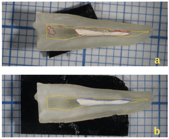
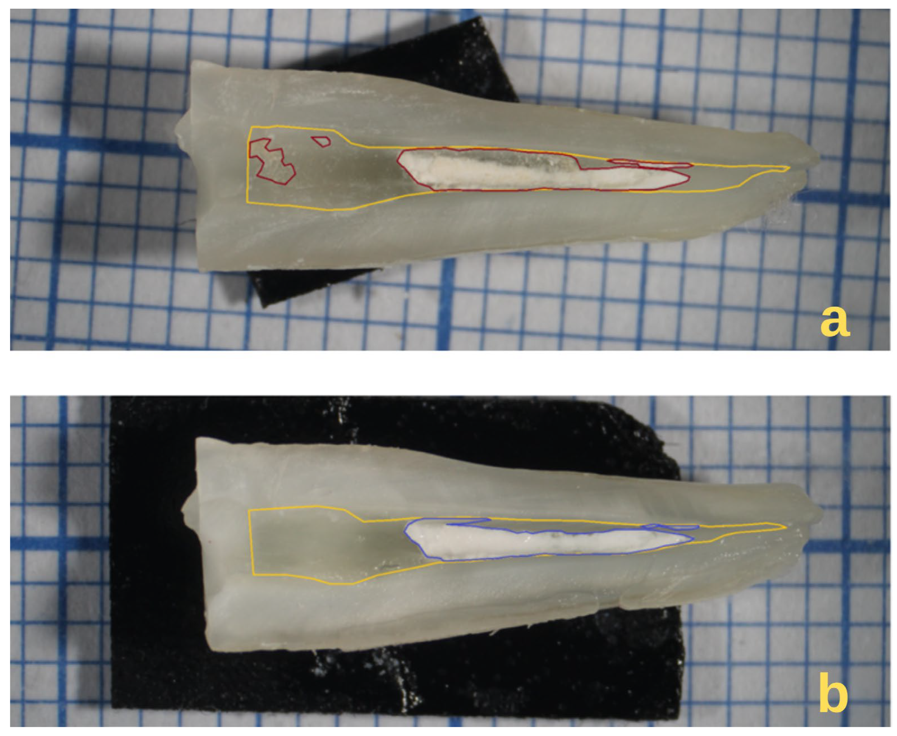
Every half of each root (n = 30) was photographed using a stereomicroscope (Stemi 2000-C, Carl Zeiss, Oberkochen, Germany) at 6.5× magnification and a Canon EOS 1300D (Canon Inc., Tokyo, Japan) digital camera before and after the final irrigation protocol. The images were analyzed using ImageJ software (version 1.54, National Institutes of Health, Bethesda, MD, USA). The polygon tool was used to measure the area of the root canal and the area covered by sealer and gutta-percha (Figure 1) [18]. The measurements were performed in pixels, and the amount of residual filling was expressed as a percentage. An average percentage between the two halves was calculated for each specimen. Sealer removed by final irrigation protocols (FI) was calculated as the percentage of sealer remnant before FI minus that after FI. For the root canal thirds analysis, every root canal’s length was measured in pixels and divided equally into three parts (apical, middle, and coronal).
Figure 1.
Example illustrating root canal sealer remnants evaluation ((a) before final irrigation; (b) after final irrigation). The yellow contour represents the root canal area, red represents the sealer remnant before final irrigation, and blue represents the sealer remnant after final irrigation.
2.5. SEM-EDS Evaluation
For scanning electron microscopy (SEM) analysis, following the application of the final irrigation protocol specific to each group, five specimens were randomly selected from each group using the website www.random.org (accessed on 10 September 2025). From each root, one half obtained after longitudinal splitting was randomly chosen for analysis. SEM observations were performed at magnifications of 200× and 3000× to evaluate dentin morphology and surface characteristics, as well as the presence or absence of residual dentinal debris and obturation material [41]. For each specimen, nine images were acquired at 3000× magnification, with three images obtained from each root canal third (apical, middle, and coronal), focusing on the central canal area as the region of interest. SEM analysis was used as a complementary approach to support and interpret the quantitative stereomicroscopic findings.
The morphology and local elemental composition of the samples were analyzed using a Scanning Electron Microscope (Tescan Vega G4, Brno, Czech Republic) with a resolution of 3 nm, equipped with a QUANTAX Energy Dispersive X-ray Spectrometer (Bruker, Billerica, MA, USA) (EDS). Samples were mounted on stubs using double-sided adhesive carbon tape. Imaging was conducted under high vacuum conditions, with an accelerating voltage of 25 kV and a beam current of 1 nA. A 4Q BSE COMPO backscattered electron detector (Bruker, Billerica, MA, USA) was used to capture the images.
The analysis of dentinal surfaces was performed using a categorical scoring system (A1–A4), representing a semi-quantitative evaluation of debris and residual filling material distribution, as follows:
- A1—open dentinal tubules, with no residual dentinal debris and no or minimal remaining obturation material;
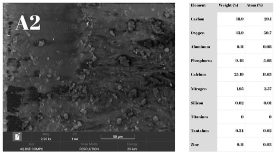
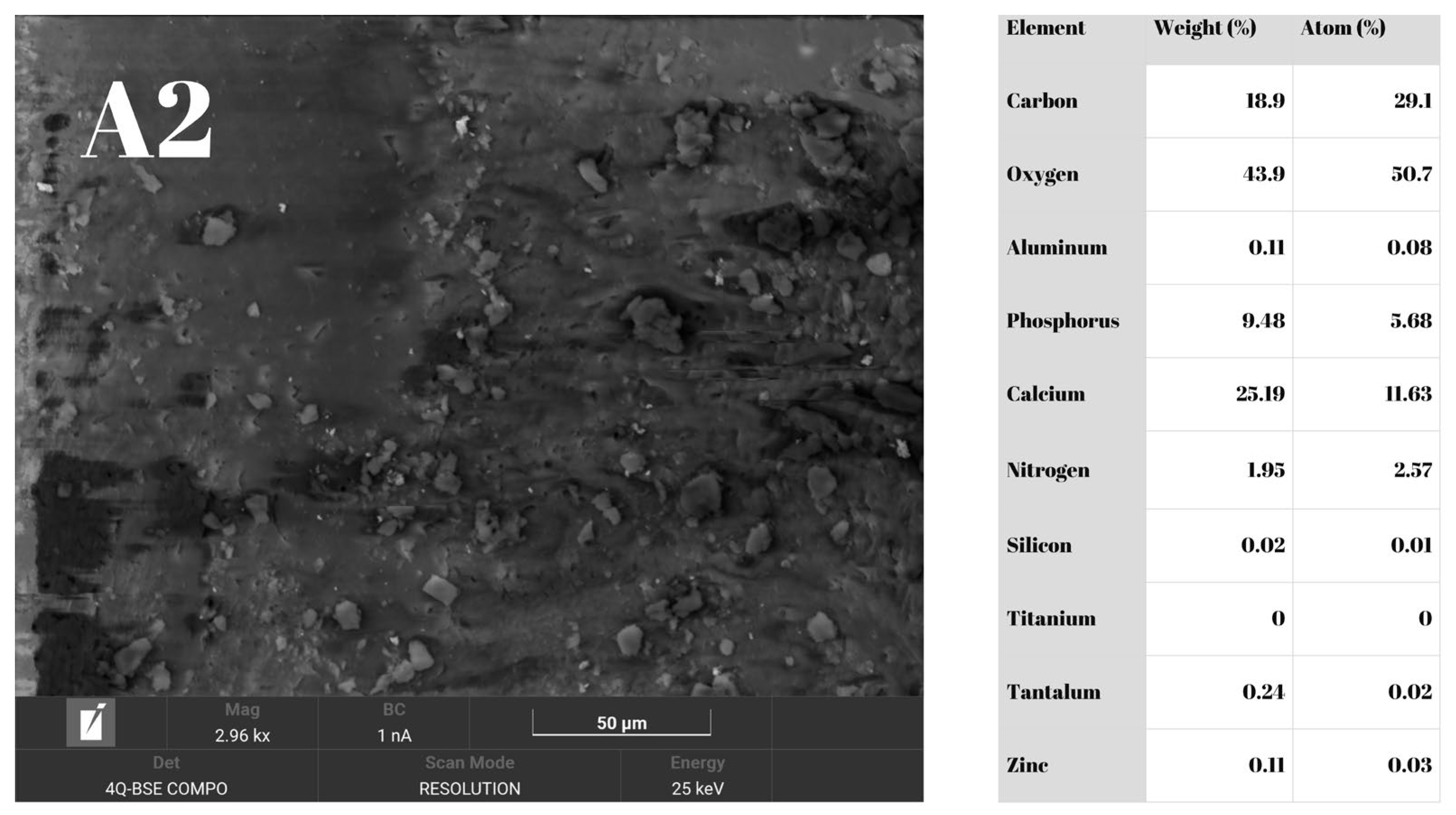
- A2—dentinal tubules covered by residual dentinal debris, with no or minimal remaining obturation material;
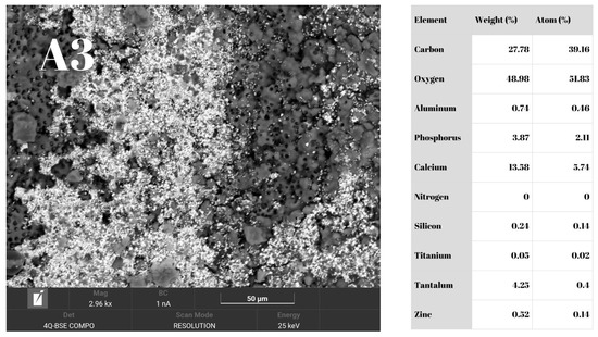
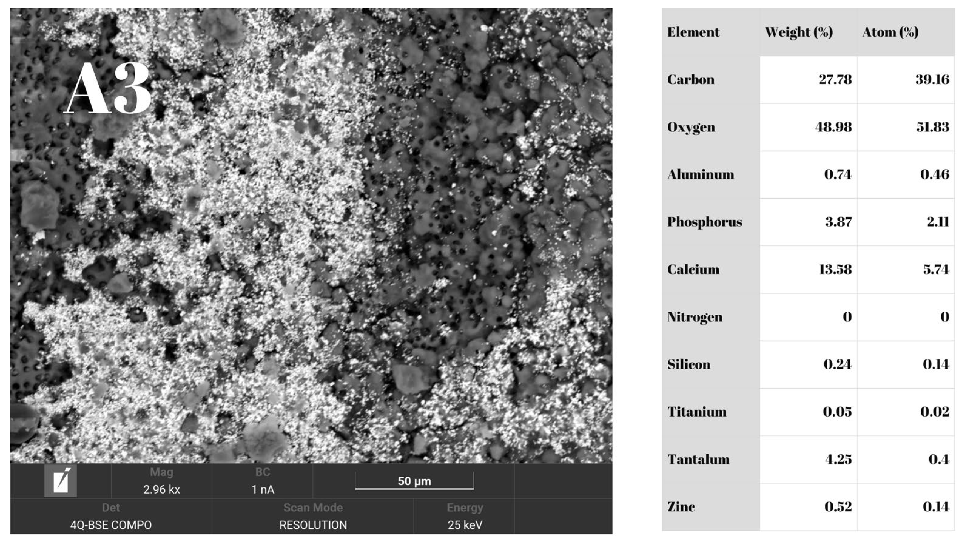
- A3—dentinal tubules partially covered by remaining obturation material, with or without residual dentinal debris;
- A4—dentin surface completely covered by residual obturation material [21].
Energy-dispersive X-ray spectroscopy (EDS) analysis was conducted qualitatively on each of the nine images obtained per specimen. The analysis aimed to identify elements characteristic of dentin (calcium, phosphorus, nitrogen) and those specific to the sealer (aluminum, silicon, tantalum) or gutta-percha (zinc, titanium), in order to confirm the presence or absence of residual obturation material. For calibration purposes, EDS analysis was also performed on the NF sealer and on a gutta-percha cone [21].
2.6. Statistical Analysis
Data distribution was assessed for normality using the Shapiro–Wilk test prior to parametric analysis. To evaluate differences between groups for continuous variables, one-way analysis of variance (ANOVA) for independent samples was applied, followed by Tukey and Bonferroni post hoc tests, as appropriate. For within-specimen comparisons before and after final irrigation, repeated-measures ANOVA was used. For categorical variables, the chi-square test was used to assess statistical significance, and post hoc analysis based on standardized residuals was performed for comparisons between groups and regions. The level of statistical significance was set at p < 0.05 for all statistical tests, while a threshold of z ≥ |2| was considered statistically significant for standardized residuals. All graphical representations and statistical analyses were performed using JASP software (version 0.19.0).
3. Results
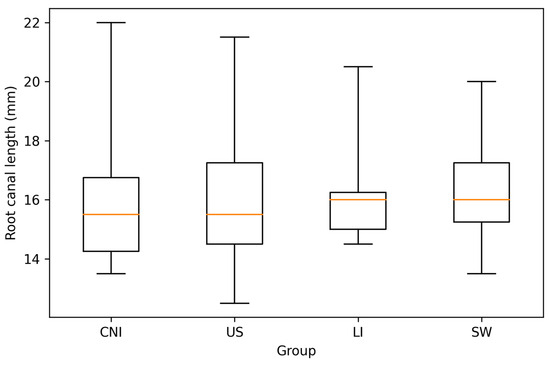
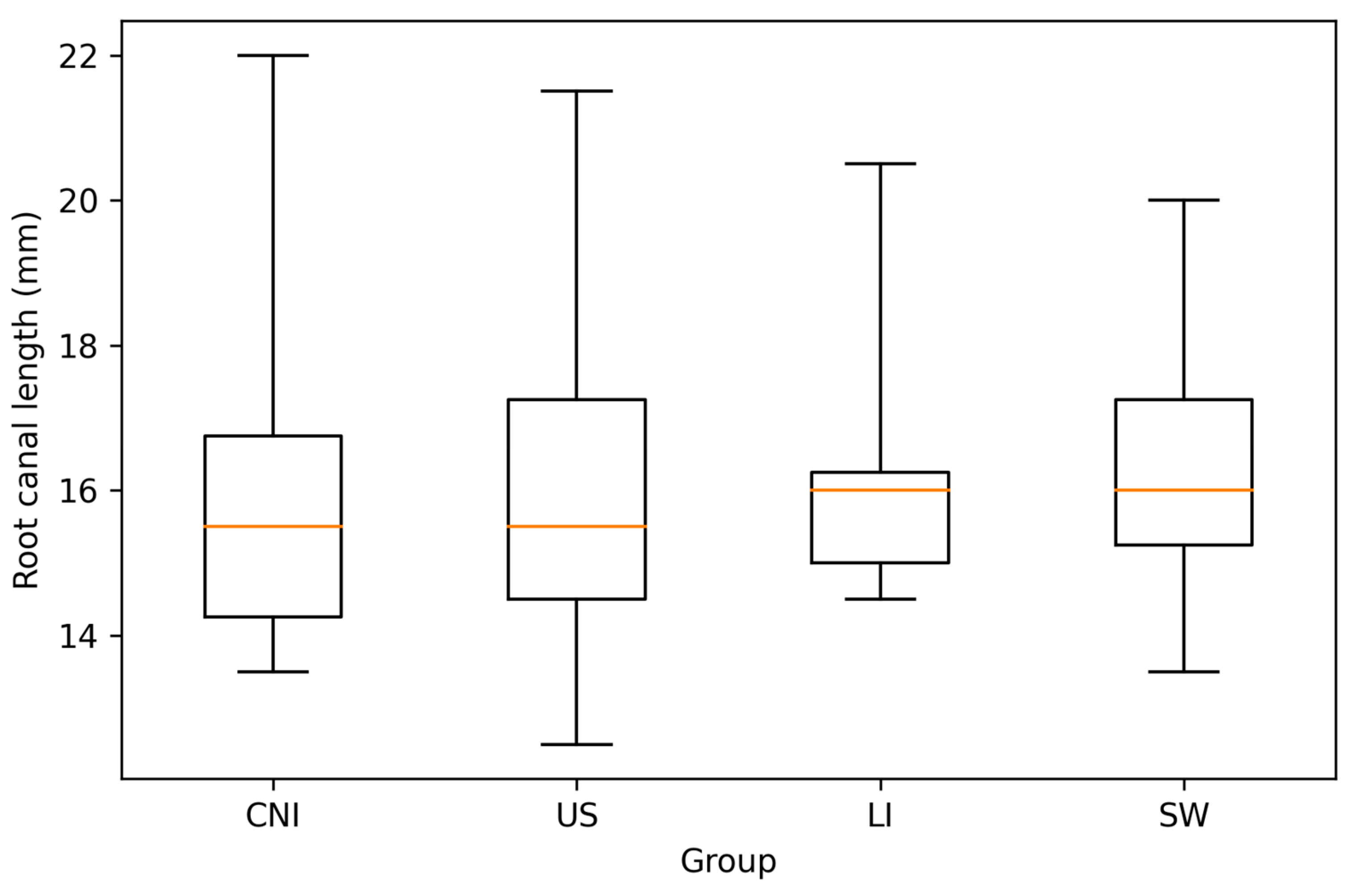
In all root canals, the initial working length was achieved during retreatment. Among these, apical patency was not achieved in 11 specimens, including 3 in the CNI group, 2 in the US group, 4 in the LI group, and 2 in the SW group. The distribution of root canal length was comparable among the four experimental groups (CNI, US, LI, and SW), with similar medians observed across all groups (Figure 2). No significant differences regarding working lengths were observed between groups (p > 0.05).
Figure 2.
Box-and-whisker plots illustrating the distribution of root canal length in the four experimental groups (CNI, conventional needle irrigation; US, ultrasonic-activated irrigation; LI, diode-laser activated irrigation; SW, SWEEPS-laser activated irrigation). The boxes represent the interquartile range (Q1–Q3), the horizontal line indicates the median, and whiskers denote the minimum and maximum values.
3.1. Stereomicroscope Images Evaluation
Statistical analysis revealed that the percentage of root canal covered by sealer was significantly reduced (p < 0.01) by any type of irrigation, compared with the pre-final irrigation rate. Mean percentages of sealer remnants before and after final irrigation are shown in Table 1.
Table 1.
Mean percentages and standard deviation of sealer remnants before and after final irrigation, and of sealer removed by each irrigation protocol (repeated ANOVA, and Bonferroni post hoc tests).
When the percentage of sealer removed before and after final irrigation was compared across groups, which reflects the specific contribution of the irrigation protocol used, the US group achieved significantly more filling removal than the other three groups (p < 0.05). Mean values for the removed sealer by each irrigation protocol across all groups are presented in Table 1, and mean differences between groups are presented in Table 2. The variability of the data, expressed as standard deviation (Table 1), indicates a heterogeneous distribution of sealer remnants within groups. Therefore, differences between groups should be interpreted considering the observed within-group variability.
Table 2.
Mean differences between groups (n = 15) for sealer removal percentage after final irrigation using one-way ANOVA and Tukey post hoc comparison tests.
The difference in percentages between the sealer remnants before and after final irrigations, by root canal region, can be seen in Table 3. One-way ANOVA revealed a statistically significant difference among irrigation techniques in the apical region (p = 0.011). Tukey post hoc analysis showed significantly better sealer removal in this region for US compared to SW (p = 0.009). No statistically significant differences were observed among groups in the middle (p = 0.081) or coronal regions (p = 0.136).
Table 3.
Mean differences by root canal region for sealer removal percentage after final irrigation.
3.2. SEM-EDS Results
The SEM–EDS analysis provided qualitative and semi-quantitative support for the stereomicroscopic results, allowing detailed assessment of dentinal surface morphology and confirmation of residual material composition.
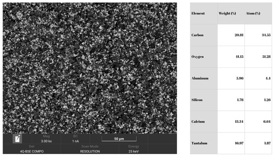
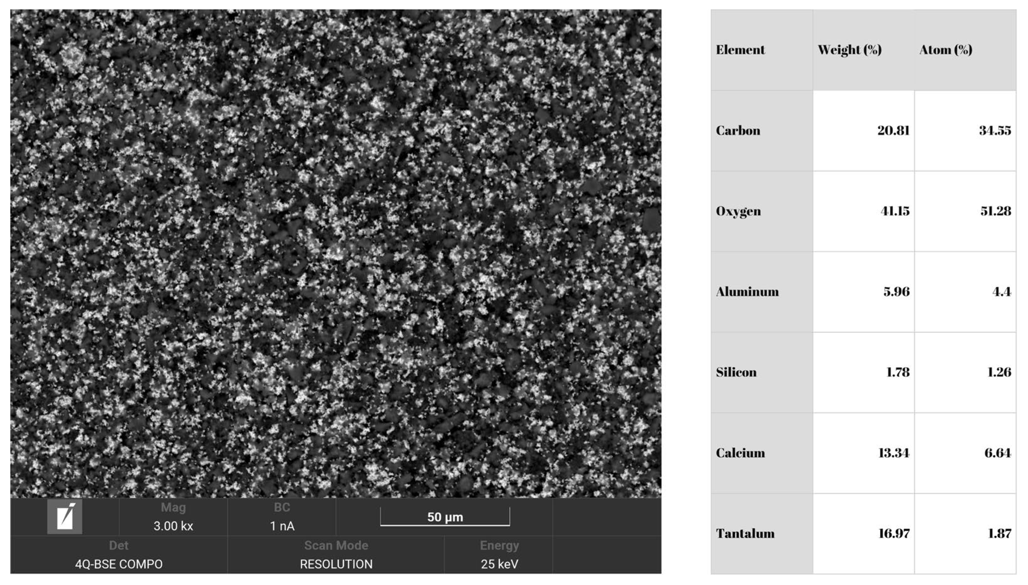
SEM–EDS analysis of the gutta-percha cone confirmed the presence of titanium and zinc in its composition. The EDS analysis of the NF sealer is shown in Figure 3, confirming the presence of aluminum, tantalum, and silicon.
Figure 3.
SEM-EDS analysis of NF Sealer.
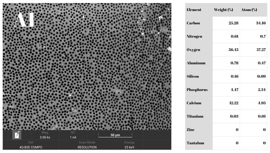
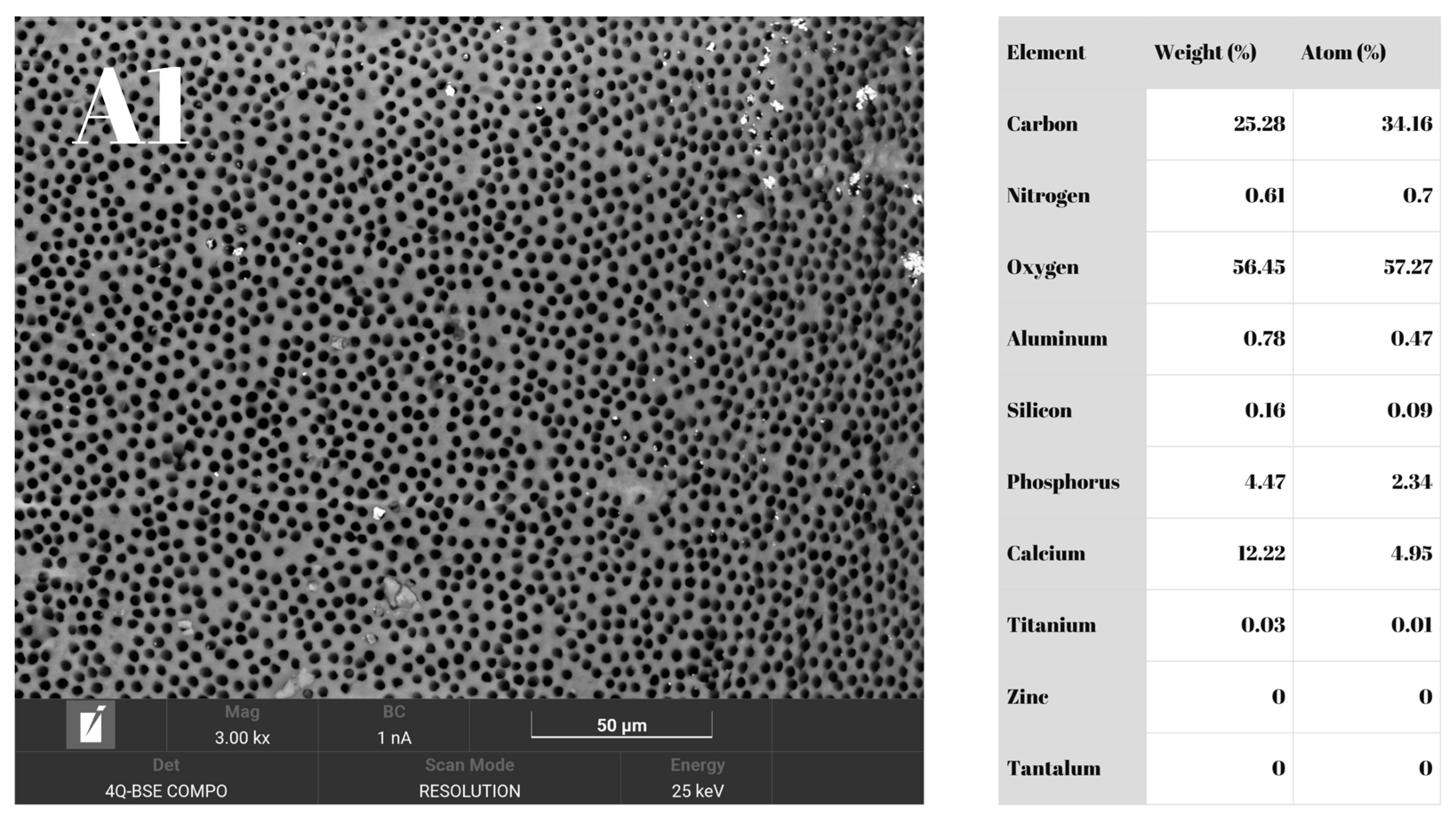
The identified dentin surface areas are illustrated in Figure 4, Figure 5, Figure 6 and Figure 7. Within areas A1 and A3, EDS analyses revealed spectra with and without nitrogen in the composition.
Figure 4.
SEM-EDS analysis showing A1 dentinal surface (open dentinal tubules) with minimal quantities of NF components (Aluminum, Silicon, Tantalum).
Figure 5.
SEM-EDS analysis showing A2 dentinal surface (tubules covered by residual debris) with minimal quantities of NF components (Aluminum, Silicon, Tantalum).
Figure 6.
SEM-EDS analysis showing A3 dentinal surface (partially covered by sealer) showing higher quantities of NF components (Aluminum, Tantalum) and Zinc (guttapercha component).
Figure 7.
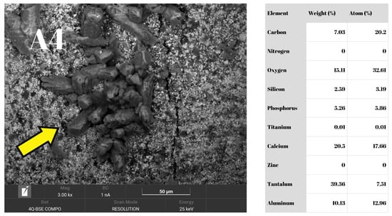
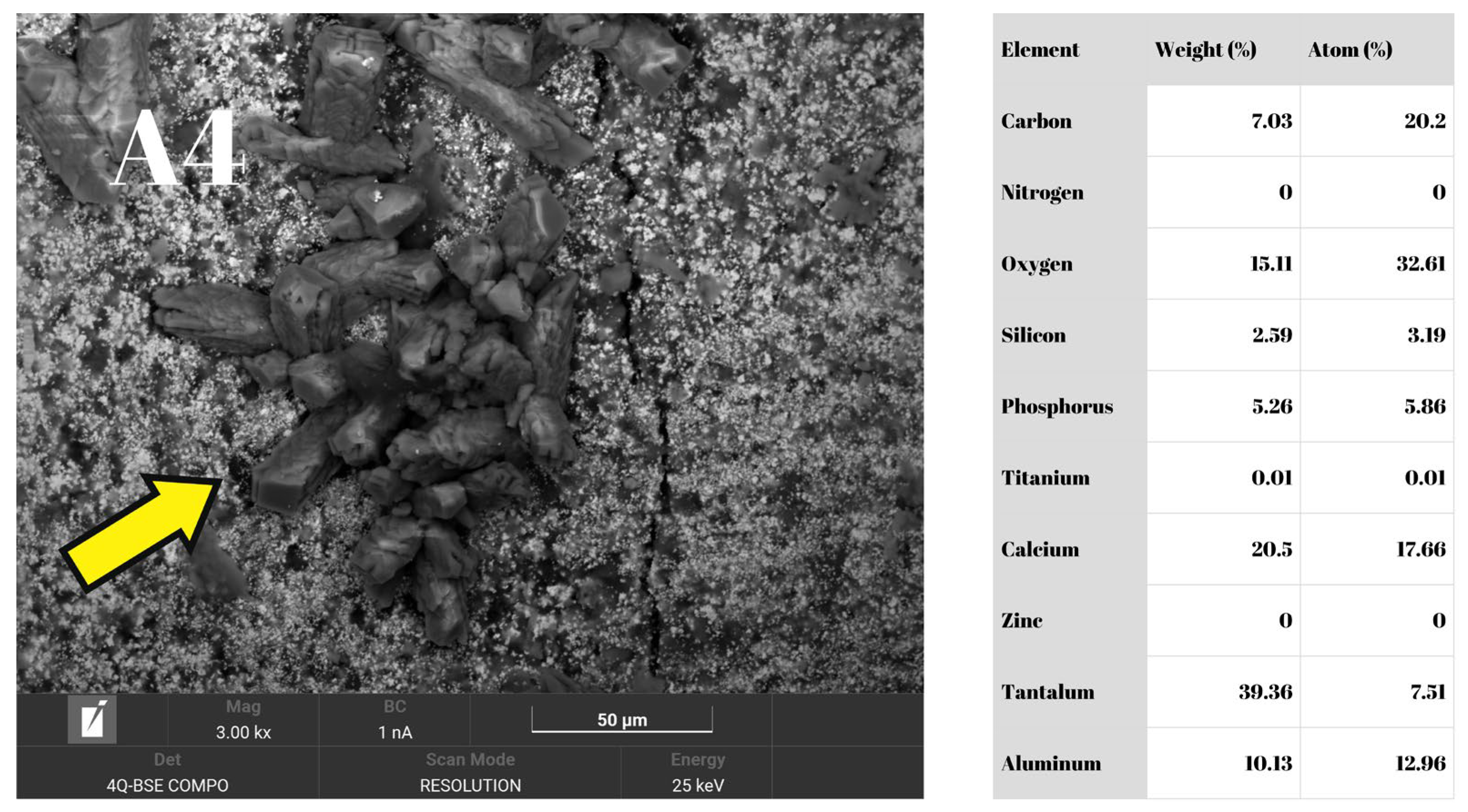
SEM-EDS analysis showing A4 dentinal surface completely covered by NF sealer, confirmed by large quantities of Tantalum, Aluminum, and Silicon. The yellow arrow indicates a prismatic crystal representing ettringite formed during NF setting, which resulted from the contact between tricalcium aluminate and calcium sulfate.
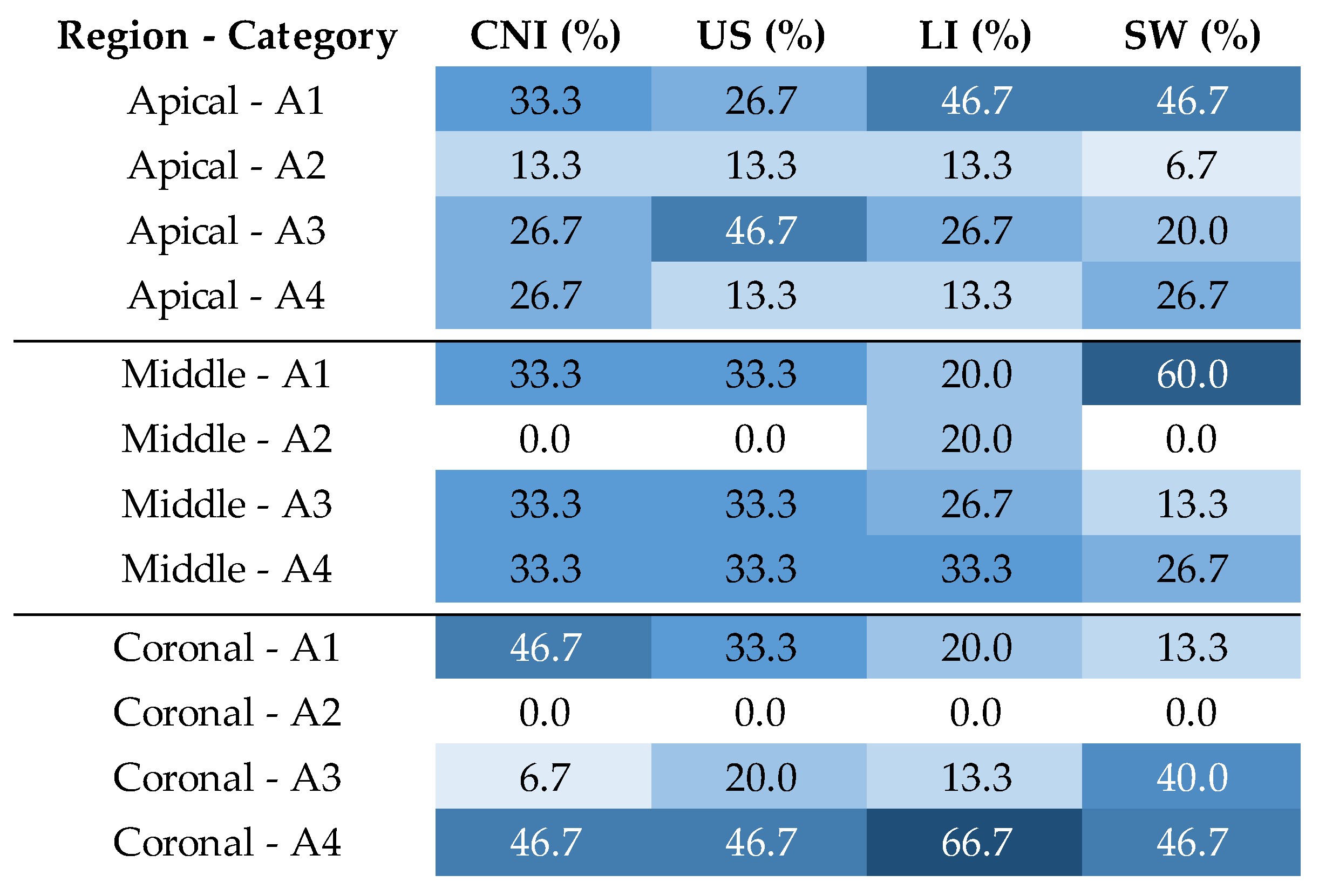
Across the four groups, 34.4% of A1 areas, 5.5% of A2 areas, 25.5% of A3 areas, and 34.4% of A4 areas were identified. Chi-square tests revealed no statistically significant differences (p > 0.05) regarding the distribution of dentin surface areas within individual groups or overall, both across the entire root length or along root canal thirds.
Post hoc analysis within groups showed that, in the LI group, significantly more A4 areas were present in the coronal third (z = 2.83) and significantly fewer in the apical third (z = −2.39). In the CNI group, a significantly higher number of A2 areas was observed in the apical third (z = 2.05), while in the SW group, significantly fewer A1 areas were identified in the coronal third (z = −2.58).
Statistical analysis across root canal thirds (apical, middle, coronal) between groups revealed no statistically significant differences (p > 0.05). However, post hoc analysis by thirds between groups showed that, in the middle third, significantly more A1 areas were present in the SW group than in the other groups (z = 2.17), whereas significantly more A2 areas were observed in the LI group (z = 3.08).
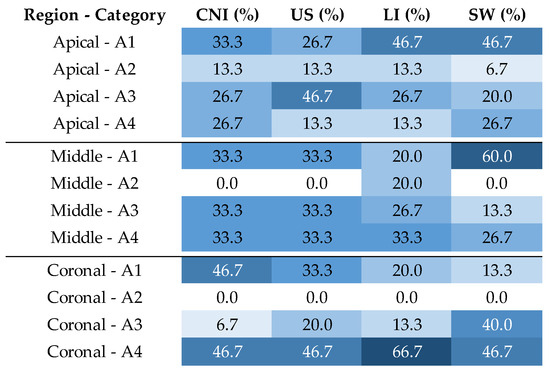
The distribution of dentin surface areas in percentages by region and group is illustrated in Figure 8.
Figure 8.
Heatmap showing the distribution (percentages) of dentin surface categories (A1–A4) across irrigation activation protocols (CNI, US, LI, SW) and root canal thirds (apical, middle, coronal). Percentages are displayed within cells. Color intensity represents the magnitude of the percentages, with lighter colors indicating lower values and darker colors indicating higher values. CNI, conventional needle irrigation; LI, diode laser-activated irrigation; SW, SWEEPS laser-activated irrigation; US, ultrasonic-activated irrigation.
4. Discussion
The removal of root canal filling materials during retreatment is influenced not only by instrumentation techniques but also by the physicochemical interaction between the filling material and dentinal substrates. Calcium silicate-based sealers can form bioactive interfacial layers and penetrate dentinal tubules, which increases their adhesion to canal walls and may complicate their removal during retreatment procedures [13,14,15,16]. Residual bacterial biofilms are a major cause of endodontic treatment failure, making effective removal of root canal filling materials essential during nonsurgical retreatment [5,6].
The main contribution of the present study is the controlled comparison of contemporary irrigation activation techniques (conventional NaOCl/EDTA, ultrasonic activation, diode laser activation, and Er:YAG laser using SWEEPS mode), and their interaction with the same calcium silicate-based sealer under standardized retreatment conditions, without demonstrating the complete bioceramic removal. By focusing on relative performance trends rather than absolute cleanliness, the present findings provide comparative data regarding the effectiveness of different irrigation activation technologies in removing calcium silicate-based sealer residues.
In the present study, all four irrigation protocols enhanced the root canal filling removal after the instrumentation alone, underscoring the importance of final irrigation. However, all protocols left some material residue, supporting the consensus that complete sealer removal is difficult to achieve [18,26,42]. Volponi et al. reported a significant reduction in filling material following supplementary irrigation, rather than instrumentation alone, confirming this research’s findings [29]. The US demonstrated higher effectiveness in the present experimental setting among the four protocols; therefore, the null hypothesis is rejected. No differences were found between CNI, LI, and SW groups, suggesting that under the conditions of the present study, the tested laser-based protocols did not demonstrate superior effectiveness compared with conventional irrigation. This finding contrasts with some previous studies reporting enhanced performance of laser-activated irrigation techniques, which may be related to differences in tooth type, canal anatomy, laser parameters, and irrigation protocols (volume, time, and concentration) [26,28]. The higher effectiveness of US suggests that the acoustic streaming and cavitation associated with passive ultrasonics throughout the root canal length, phenomena that generate localized hydrodynamic shear forces and microstreaming within the irrigant, may contribute to a more effective disruption and removal of sealer remnants compared to the tested laser-based techniques under the present experimental conditions. The findings of the present research concur with previous studies showing enhanced sealer/gutta-percha removal when ultrasonic activation is used as the final step [25,38]. However, other studies showed different results, with some authors finding no significant differences between ultrasonic and laser-based techniques (SWEEPS or PIPS), or even superior performance of laser activation methods [26,27,28]. These discrepancies may be attributed to differences in root canal anatomy, experimental design (concentration, volume, activation, and irrigation time, and type of irrigation solution), type of sealer used, and evaluation methods. The observed differences should be interpreted cautiously, as experimental ex vivo conditions cannot fully reproduce the complexity of clinical retreatment scenarios. In addition, variability in methodological approaches across studies further complicates direct comparisons between different irrigation techniques. Nevertheless, the higher effectiveness of ultrasonic-activated irrigation suggests that its routine use as an adjunctive step may increase the likelihood of improved canal debridement, particularly in oval-shaped root canals where mechanical instrumentation alone is insufficient.
Although none of the tested protocols achieved complete removal of the filling material, the presence of residual sealer may have potential clinical implications, as it could interfere with effective disinfection and may harbor microorganisms. However, the exact clinical relevance of these residual amounts remains unclear. The combination of different irrigation techniques, such as ultrasonic and laser activation, may represent a promising strategy to enhance retreatment efficacy, given their different mechanisms of action.
Our findings indicate that significant remnants can persist after standard instrumentation, necessitating supplementary irrigation techniques to improve cleanliness. The same was reported by Donnermeyer et al., who concluded that none of the four different instruments used was able to remove the material fully [18]. However, the percentage of sealer remnants after mechanical retreatment was lower in the aforementioned study than in our findings. This can be explained by the use of round root canals in the experimental setup, whereas oval root canals were used in the present study. Oval root canals need more sealer to exhibit a three-dimensional filling, and a round retreatment instrument might touch fewer parts of the irregular root canal and remove less sealer, especially in the coronal part [13,19].
The type of bioceramic sealers and their composition could also influence the retreatment efficacy. This was confirmed by Cikrik et al., who found that two different bioceramic sealers could not be entirely removed by any method, but ultrasonics (UltraX) was more effective on a calcium-silicate-based sealer, while sonic (EDDY) activation significantly decreased the residual sealer for the MTA-based material [23]. In the present study, despite NF’s good adhesion to the dentin, its relative softness could allow ultrasonics to micro-fracture it and lift it from the dentinal walls. Furthermore, patency was regained in the majority of the samples, the same being reported by Spinelli et al. [31]. When patency was not achieved, the working length was reached in every case; therefore, its potential influence on irrigant penetration and overall results is likely limited. In addition, the moisture condition of the dentinal walls, influenced by the drying protocol, may affect the setting and adhesion of calcium silicate-based sealers, potentially influencing their retreatability [43]. In the present study, two paper points were used in order to avoid overdrying.
The present findings may be cautiously extrapolated to other calcium silicate-based sealers with similar properties, particularly those labeled as soft-setting sealers [15,44] characterized by strong adhesion to dentin and the ability to penetrate dentinal tubules and form a thin interfacial layer. However, variations in composition, radiopacifiers, and obturation techniques may influence retreatability and should be considered when interpreting the results.
A 2.5% NaOCl solution was selected to preserve dentinal structure and ensure standardized conditions. Although higher concentrations may improve organic tissue dissolution [45], the aim of the present study was to compare irrigation activation techniques rather than irrigant concentration. Similarly, both the total irrigation time and the volume of irrigant were standardized across all groups to minimize potential confounding factors. It should be noted that variations in concentration, activation time, and irrigant volume may influence the effectiveness of irrigation protocols, particularly in techniques relying on hydrodynamic or photoacoustic effects [26,46]. Therefore, the results should be interpreted within the context of these controlled parameters.
The SEM–EDS findings should be interpreted as complementary to the quantitative stereomicroscopic analysis, providing qualitative and semi-quantitative insights into dentinal surface morphology and residual material distribution.
The SEM images were classified into categories A1–A4 as a semi-quantitative assessment of surface cleanliness to enable evaluation of the removal capacity of both residual root canal filling material and dentinal debris [21]. Statistical analysis did not reveal significant differences among the four groups with respect to the canal thirds (p < 0.05). However, in the middle third, the SW group exhibited a significantly higher number of images classified as A1 (z = 2.17), indicating a greater effectiveness of the Er:YAG laser operating in SWEEPS mode to remove dentinal debris and residual filling material in this region, under the conditions of the present study. In contrast, the diode laser group showed the highest number of A2-classified images among the four irrigation techniques (z = 3.08). Still, only 20% of the areas were classified as A2, suggesting that the diode laser continues to produce an acceptable effect on residual dentinal debris in this region. Yang et al. similarly reported that the Er:YAG laser used with the PIPS program resulted in small amounts of residual dentinal debris and bioceramic sealer in the middle third of the canal, with a high number of open dentinal tubules, compared with ultrasonic activation and conventional irrigation [26]. The CNI group demonstrated lower capacity for dentinal debris removal in the apical third compared with the other regions, suggesting more limited irrigant penetration in the deeper portions of the canal, which confirms the findings of Yang et al. [26]. A study evaluating two types of instruments combined with different final irrigation protocols, without activation methods, for the removal of root canal fillings using the resin-based sealer AH Plus reported predominantly open dentinal tubules (A1) or dentinal tubules covered by smear layer (A2). In contrast, in the present study, the distribution of surface areas (A1, A3, A4) was more balanced, with A2 observed in only 5.5% of cases [21]. This finding suggests that the bioceramic sealer NF is more difficult to remove from dentinal walls and that the irrigation protocols evaluated in the present study are more effective in removing dentinal debris. These observations support the stereomicroscopic results, without representing a primary quantitative outcome measure.
Complementary EDS analysis confirmed the identity of the residual material on the dentinal walls. Areas of dentin covered by NF residues exhibited peaks of aluminum, tantalum, and silicon, while regions with small amounts of remaining gutta-percha showed the presence of titanium and zinc. These elements are consistent with the known compositions of the NF sealer [37] and gutta-percha [47]. This analysis allowed clear identification of residual filling materials, thereby confirming the area classification based on SEM images. The presence of nitrogen within A1 and A3 areas suggests the persistence of undissolved organic matter (collagen fibers), which may be attributed to the use of a 2.5% NaOCl concentration [48].
In this research, the stereomicroscope-based imaging approach coupled with ImageJ analysis provided an accessible and reproducible means of quantifying residual NF. Several studies used this method in evaluating sealer remnants after the endodontic retreatment [18,49,50]. Alternative methods, such as micro-computed tomography, allow three-dimensional assessment of residual filling material and may provide a more comprehensive evaluation [29]. Radiographic and CBCT-based approaches have also been used in combination with microscopic techniques for remnant detection [29,51]; however, these imaging techniques may result in artifacts and distortions [52,53]. Nevertheless, special care was required to prevent the diamond disk from entering the root canal during longitudinal splitting of the specimens. This study was designed to measure sealer remnants before and after the final irrigation protocol. A possible limitation of splitting the roots is the impossibility of observing them at two different time points [49]. To address this issue, the roots were glued back together before the final irrigation following a protocol described by Sharki et al. [39]. Consequently, the findings should be interpreted with appropriate caution. In addition to the analysis based on photographic images, SEM evaluation allowed visualization of dentinal microstructure and assessment of the ability of the irrigation protocols to open dentinal tubules [26,30]. A limitation of this analytical method is the inability to evaluate the entire canal surface [54]. In addition, two-dimensional measurements on split roots provide only partial information regarding the spatial distribution of residual material, and SEM–EDS analysis was performed on a limited number of specimens per group (n = 5), because of its high complexity [16,55]. Given that SEM–EDS provides localized qualitative and semi-quantitative information, this number was considered sufficient to identify general patterns, although it may not fully represent the variability of the entire sample. These aspects should be considered when interpreting the findings.
This study has several limitations that should be considered when interpreting the results. First, the ex vivo design and the use of standardized single-rooted teeth with oval canals with removed crowns, although beneficial for experimental control, may limit the generalizability of the findings to more complex clinical situations, as crown morphology and access cavity design can influence instrumentation and irrigation dynamics. Sealer setting behavior may differ in vivo, and the anatomy of the endodontic system can be more complex in multirooted teeth with curved canals, particularly in the presence of isthmuses or multiple canals within the same root. Furthermore, the stereomicroscopic evaluation provides two-dimensional measurements of residual material, which may not fully reflect its three-dimensional distribution within the root canal system. As a result, thin remnants and irregularities may be underestimated or missed using the split-root and reassembly approach, which represents a limitation of the present study. Although this method allows direct visualization of residual material, it may influence the results by potentially dislodging filling remnants, inducing microcracks, or altering canal geometry and irrigant dynamics during subsequent procedures. Despite the fact that the alignment of the reassembled specimens was verified under an optical microscope, no standardized method was used to quantitatively assess the integrity of the reassembly.
Future research should investigate the influence of sealer composition, material hardness, and interfacial bonding mechanisms on retreatability, as well as the hydrodynamic behavior of activated irrigants within complex root canal geometries. In addition, future studies should include three-dimensional evaluation methods, such as micro-computed tomography, to better characterize the spatial distribution of residual materials. Future research should focus on roots with more diverse and clinically relevant anatomical configurations, on evaluating different solvents that may assist in removing residual filling materials, increasing the irrigant volume, NaOCl concentration, and irrigation time, on combining different activation techniques, and on clinical studies assessing the long-term impact of these irrigation techniques on the success rates of nonsurgical endodontic retreatment of teeth obturated with bioceramic sealer.
5. Conclusions
Within the limitations of this ex vivo study, ultrasonic-activated irrigation demonstrated greater effectiveness in reducing residual bioceramic sealer compared with the other evaluated protocols (conventional needle irrigation, diode laser-activated irrigation, and SWEEPS mode-activated irrigation). However, none of the tested techniques achieved complete sealer removal. These findings suggest that ultrasonic activation enhances the disruption and removal of NeoSealer Flo calcium silicate-based sealer residues during the retreatment of oval-shaped root canals under controlled experimental conditions, while the clinical relevance of residual sealer and the potential benefits of combined irrigation strategies require further investigation.
Author Contributions
Conceptualization, A.G.D. and S.I.C.; methodology, M.M. and S.I.C.; validation, I.C.B. and S.C.; formal analysis, S.I.C. and I.S.P.-C.; investigation, E.A., I.C.B., V.-A.S. and S.C.; resources, A.G.D. and S.C.; data curation, V.-A.S. and E.A.; writing—original draft preparation, M.M.; writing—review and editing, M.M. and I.S.P.-C.; visualization, V.-A.S. and I.C.B.; supervision, A.G.D. All authors have read and agreed to the published version of the manuscript.
Funding
This research received no external funding.
Institutional Review Board Statement
The study was conducted in accordance with the Declaration of Helsinki and approved by the Ethics Committee of the University of Medicine and Pharmacy “Iuliu Hatieganu” Cluj-Napoca, Romania (number AVZ267/07.03.2025).
Informed Consent Statement
Written informed consent was obtained from all subjects involved in the study.
Data Availability Statement
The data presented in this study are available on request from the corresponding author.
Conflicts of Interest
The authors declare no conflicts of interest.
Abbreviations
The following abbreviations are used in this manuscript:
| ANOVA | Analysis of variance |
| CNI | Conventional needle irrigation |
| EDS | Energy-dispersive X-ray spectroscopy |
| EDTA | Ethylenediaminetetraacetic acid |
| Er:YAG | Erbium-doped yttrium aluminum garnet |
| FI | Final irrigation |
| LI | Diode laser-activated irrigation |
| NaOCl | Sodium hypochlorite |
| NF | NeoSealer Flo |
| SEM | Scanning electron microscopy |
| SW | SWEEPS laser-activated irrigation |
| SWEEPS | Shock Wave Enhanced Emission Photoacoustic Streaming |
| US | Ultrasonic-activated irrigation |
References
- Siqueira, J.F.; Rôças, I.N. Clinical Implications and Microbiology of Bacterial Persistence after Treatment Procedures. J. Endod. 2008, 34, 1291–1301.e3. [Google Scholar] [CrossRef] [Scilit]
- Sabeti, M.A.; Karimpourtalebi, N.; Shahravan, A.; Dianat, O. Clinical and Radiographic Failure of Nonsurgical Endodontic Treatment and Retreatment Using Single-Cone Technique with Calcium Silicate-Based Sealers: A Systematic Review and Meta-Analysis. J. Endod. 2024, 50, 735–746.e1. [Google Scholar] [CrossRef] [Scilit]
- Olivieri, J.G.; Encinas, M.; Nathani, T.; Miró, Q.; Duran-Sindreu, F. Outcome of Root Canal Retreatment Filled with Gutta-Percha Techniques: A Systematic Review and Meta-Analysis. J. Dent. 2024, 142, 104809. [Google Scholar] [CrossRef] [Scilit]
- Stueland, H.; Ørstavik, D.; Handal, T. Treatment Outcome of Surgical and Non-Surgical Endodontic Retreatment of Teeth with Apical Periodontitis. Int. Endod. J. 2023, 56, 686–696. [Google Scholar] [CrossRef] [Scilit]
- Vieira, A.R.; Pacheco-Yanes, J.; Gazzaneo, I.D.; Neves, M.A.S.; Siqueira, J.F., Jr.; Gonçalves, L.S. Factors Influencing the Outcome of Nonsurgical Root Canal Treatment and Retreatment: A Retrospective Study. Aust. Endod. J. 2024, 50, 245–259. [Google Scholar] [CrossRef] [Scilit]
- Sabeti, M.; Chung, Y.J.; Aghamohammadi, N.; Khansari, A.; Pakzad, R.; Azarpazhooh, A. Outcome of Contemporary Nonsurgical Endodontic Retreatment: A Systematic Review of Randomized Controlled Trials and Cohort Studies. J. Endod. 2024, 50, 414–433. [Google Scholar] [CrossRef] [Scilit] [PubMed]
- Martins, M.P.; Duarte, M.A.H.; Cavenago, B.C.; Kato, A.S.; da Silveira Bueno, C.E. Effectiveness of the ProTaper Next and Reciproc Systems in Removing Root Canal Filling Material with Sonic or Ultrasonic Irrigation: A Micro–Computed Tomographic Study. J. Endod. 2017, 43, 467–471. [Google Scholar] [CrossRef] [Scilit]
- Camilleri, J. Sealers and Warm Gutta-Percha Obturation Techniques. J. Endod. 2015, 41, 72–78. [Google Scholar] [CrossRef] [Scilit] [PubMed]
- Basta, D.G.; Reslan, M.R.; Rayyan, M.; Sayed, M. Evaluation of Antibacterial Effect of New Sealer “Neoseal” and Two Commercially Used Endodontic Sealers against Enterococcus Faecalis: An In Vitro Study. J. Contemp. Dent. Pract. 2023, 24, 871–876. [Google Scholar] [CrossRef] [Scilit] [PubMed]
- Sebastian, S.; El-Sayed, W.; Adtani, P.; Zaarour, R.F.; Nandakumar, A.; Elemam, R.F.; Eid, B.; Elsewify, T. Evaluation of the Antibacterial and Cytotoxic Properties of TotalFill and NeoSEALER Flo Bioceramic Sealers. J. Conserv. Dent. Endod. 2024, 27, 491–497. [Google Scholar] [CrossRef] [Scilit]
- Drukteinis, S.; Camilleri, J. (Eds.) Bioceramic Materials in Clinical Endodontics; Springer: Berlin/Heidelberg, Germany, 2025; Available online: https://link.springer.com/book/10.1007/978-3-030-58170-1 (accessed on 24 April 2025).
- Algar, J.; Docampo-Vázquez, C.; Rico-Romano, C.; Boquete-Castro, A.; Obispo-Díaz, C.; Aragoneses, J.M. Randomised Clinical Trial: Effect of AH Plus and Neosealer Flo on Postoperative Pain and Healing of Periapical Lesions. Bioengineering 2025, 12, 376. [Google Scholar] [CrossRef] [Scilit]
- Elzanaty, T.K.; Elashiry, M.M.; Mahran, A.H. Retreatability of NeoSEALER Flo Obturated with Warm Vertical Compaction versus Single-Cone Technique Using Two Different Retreatment Systems. J. Conserv. Dent. Endod. 2024, 27, 286–292. [Google Scholar] [CrossRef] [Scilit] [PubMed]
- Creazzo, G.; de Barros Ciribelli Alves, B.M.; de Assis, H.C.; Villamayor, K.G.G.; de Sousa-Neto, M.D.; Mazzi-Chaves, J.F.; Lopes-Olhê, F.C. Bond Strength and Adhesive Interface Quality of New Pre-Mixed Bioceramic Root Canal Sealer. Microsc. Res. Tech. 2025, 88, 1989–2000. [Google Scholar] [CrossRef] [Scilit] [PubMed]
- Al akam, H.; Kim, H.-C.; Jeong, J.W. Retreatment Strategies for Cases Containing Calcium Silicate-Based Root Canal Sealers: A Comprehensive Review. Dent. J. 2024, 12, 41. [Google Scholar] [CrossRef] [Scilit]
- Shim, E.; Son, J.W.; Kwon, J.; Kim, H.-J.; Jang, J.-H.; Chang, S.W.; Oh, S. Retrieval of AH Plus Bioceramic and Ceraseal Versus AH Plus in Endodontic Retreatment. J. Clin. Med. 2025, 14, 1826. [Google Scholar] [CrossRef] [Scilit]
- Donnermeyer, D.; Bürklein, S.; Dammaschke, T.; Schäfer, E. Endodontic Sealers Based on Calcium Silicates: A Systematic Review. Odontology 2019, 107, 421–436. [Google Scholar] [CrossRef] [Scilit]
- Donnermeyer, D.; Bunne, C.; Schäfer, E.; Dammaschke, T. Retreatability of Three Calcium Silicate-Containing Sealers and One Epoxy Resin-Based Root Canal Sealer with Four Different Root Canal Instruments. Clin. Oral Investig. 2018, 22, 811–817. [Google Scholar] [CrossRef] [Scilit]
- Crozeta, B.M.; Lopes, F.C.; Menezes Silva, R.; Silva-Sousa, Y.T.C.; Moretti, L.F.; Sousa-Neto, M.D. Retreatability of BC Sealer and AH Plus Root Canal Sealers Using New Supplementary Instrumentation Protocol during Non-Surgical Endodontic Retreatment. Clin. Oral Investig. 2021, 25, 891–899. [Google Scholar] [CrossRef] [Scilit]
- Helvacioglu-Yigit, D.; Yilmaz, A.; Kiziltas-Sendur, G.; Aslan, O.S.; Abbott, P.V. Efficacy of Reciprocating and Rotary Systems for Removing Root Filling Material: A Micro-Computed Tomography Study. Scanning 2014, 36, 576–581. [Google Scholar] [CrossRef] [Scilit]
- Prati, C.; Zamparini, F.; Spinelli, A.; Pelliccioni, G.A.; Pirani, C.; Gandolfi, M.G. Secondary Root Canal Treatment with Reciproc Blue and K-File: Radiographic and ESEM-EDX Analysis of Dentin and Root Canal Filling Remnants. J. Clin. Med. 2020, 9, 1902. [Google Scholar] [CrossRef] [Scilit] [PubMed]
- Siqueira, J.F., Jr.; Rôças, I.d.N.; Marceliano-Alves, M.F.; Pérez, A.R.; Ricucci, D. Unprepared Root Canal Surface Areas: Causes, Clinical Implications, and Therapeutic Strategies. Braz. Oral Res. 2018, 32, e65. [Google Scholar] [CrossRef] [Scilit] [PubMed]
- Cıkrık, B.N.; İnce Yusufoğlu, S. Efficacy of Irrigation Activation Methods in Removal of Bioceramic-Based Sealer in Retreatment. Odontology 2025, 113, 1119–1127. [Google Scholar] [CrossRef] [Scilit]
- Tandon, J.; Yadav, R.K.; Tikku, A.P.; Shakya, V.K.; Singh, S.K. Comparative Evaluation of Different Irrigating and Irrigant Activation System in Removal of Gutta-Percha/Sealer during Retreatment: An in Vitro Micro-CT Study. J. Oral Biol. Craniofac. Res. 2022, 12, 444–448. [Google Scholar] [CrossRef] [Scilit]
- Almohareb, R.A.; Barakat, R.M.; Aljarallah, N.; Mudhish, H.; Almutairi, A.; Algahtani, F.N. Efficiency of Diode Laser and Ultrasonic-Activated Irrigation in Retreatment of Gutta Percha and Bioceramic Sealer: An in Vitro Study. Aust. Endod. J. 2023, 49, 318–323. [Google Scholar] [CrossRef] [Scilit] [PubMed]
- Yang, R.; Han, Y.; Liu, Z.; Xu, Z.; Liu, H.; Wei, X. Comparison of the Efficacy of Laser-Activated and Ultrasonic-Activated Techniques for the Removal of Tricalcium Silicate-Based Sealers and Gutta-Percha in Root Canal Retreatment: A Microtomography and Scanning Electron Microscopy Study. BMC Oral Health 2021, 21, 275. [Google Scholar] [CrossRef] [Scilit] [PubMed]
- Bago, I.; Plotino, G.; Katić, M.; Ferenac, A.; Petričević, G.K.; Gabrić, D.; Anić, I. Effect of a Novel Laser-Initiated Photoacoustic Activation of a Solvent or Sodium Hypochlorite in the Removal of Filling Remnants after Retreatment of Curved Root Canals. Photodiagnosis Photodyn. Ther. 2021, 36, 102535. [Google Scholar] [CrossRef] [Scilit]
- Petričević, G.K.; Katić, M.; Anić, I.; Salarić, I.; Vražić, D.; Bago, I. Efficacy of Different Er:YAG Laser-Activated Photoacoustic Streaming Modes Compared to Passive Ultrasonic Irrigation in the Retreatment of Curved Root Canals. Clin. Oral Investig. 2022, 26, 6773–6781. [Google Scholar] [CrossRef] [Scilit]
- Volponi, A.; Pelegrine, R.A.; Kato, A.S.; Stringheta, C.P.; Lopes, R.T.; Silva, A.S.d.S.; Bueno, C.E.d.S. Micro–Computed Tomographic Assessment of Supplementary Cleaning Techniques for Removing Bioceramic Sealer and Gutta-Percha in Oval Canals. J. Endod. 2020, 46, 1901–1906. [Google Scholar] [CrossRef] [Scilit]
- Bernardes, R.A.; Duarte, M.a.H.; Vivan, R.R.; Alcalde, M.P.; Vasconcelos, B.C.; Bramante, C.M. Comparison of Three Retreatment Techniques with Ultrasonic Activation in Flattened Canals Using Micro-Computed Tomography and Scanning Electron Microscopy. Int. Endod. J. 2016, 49, 890–897. [Google Scholar] [CrossRef] [Scilit]
- Spinelli, A.; Zamparini, F.; Lenzi, J.; Carboni, D.; Gandolfi, M.G.; Prati, C. Retreatability of Bioceramic-Filled Teeth: Comparative Analysis of Single-Cone and Carrier-Based Obturation Using a Reciprocating Technique. Appl. Sci. 2024, 14, 6444. [Google Scholar] [CrossRef] [Scilit]
- Merfea, M.; Cimpean, S.I.; Chiorean, R.S.; Antoniac, A.; Delean, A.G.; Badea, I.C.; Badea, M.E. Comparative Assessment of Push-Out Bond Strength and Dentinal Tubule Penetration of Different Calcium-Silicate-Based Endodontic Sealers. Dent. J. 2024, 12, 397. [Google Scholar] [CrossRef] [Scilit]
- Akçay, A.; Gorduysus, M.; Aydin, B.; Gorduysus, M.O. Evaluation of Different Irrigation Techniques on Dentin Erosion and Smear Layer Removal: A Scanning Electron Microscopy Study. J. Conserv. Dent. Endod. 2022, 25, 311. [Google Scholar] [CrossRef]
- Suman, S.; Verma, P.; Prakash-Tikku, A.; Bains, R.; Kumar-Shakya, V. A Comparative Evaluation of Smear Layer Removal Using Apical Negative Pressure (EndoVac), Sonic Irrigation (EndoActivator) and Er:YAG Laser -An In Vitro SEM Study. J. Clin. Exp. Dent. 2017, 9, e981–e987. [Google Scholar] [CrossRef] [Scilit]
- Cimpean, S.I.; Burtea, A.L.C.; Chiorean, R.S.; Dudescu, M.C.; Antoniac, A.; Robu, A.; Campian, R.S.; Timis, L.I. Evaluation of Bond Strength of Four Different Root Canal Sealers. Materials 2022, 15, 4966. [Google Scholar] [CrossRef] [Scilit]
- Virdee, S.S.; Farnell, D.J.J.; Silva, M.A.; Camilleri, J.; Cooper, P.R.; Tomson, P.L. The Influence of Irrigant Activation, Concentration and Contact Time on Sodium Hypochlorite Penetration into Root Dentine: An Ex Vivo Experiment. Int. Endod. J. 2020, 53, 986–997. [Google Scholar] [CrossRef] [Scilit]
- Zamparini, F.; Prati, C.; Taddei, P.; Spinelli, A.; Di Foggia, M.; Gandolfi, M.G. Chemical-Physical Properties and Bioactivity of New Premixed Calcium Silicate-Bioceramic Root Canal Sealers. Int. J. Mol. Sci. 2022, 23, 13914. [Google Scholar] [CrossRef] [Scilit] [PubMed]
- Gouveia, P.; Limoeiro, A.G.; Silva, C.B.F.; Marceliano-Alves, M.F.V.; Fontana, C.E.; Rocha, D.G.P.; Bueno, C.E.d.S.; De Martin, A.S. Evaluation of the Retreatability of BC Sealer, BC Sealer Hiflow, and AH Plus: A Micro-Computed Tomography Study. J. Endod. 2025, 51, 748–754. [Google Scholar] [CrossRef] [Scilit]
- Sharki, A.M.; Ali, A.H. Three-Dimensional Measurement of Obturation Quality of Bioceramic Materials in Filling Artificial Internal Root Resorption Cavities Using Different Obturation Techniques: An In Vitro Comparative Study. J. Endod. 2024, 50, 997–1003. [Google Scholar] [CrossRef] [Scilit] [PubMed]
- Cîmpean, S.I.; Pop-Ciutrila, I.-S.; Matei, S.-R.; Colosi, I.A.; Costache, C.; Nicula, G.Z.; Badea, I.C.; Colceriu Burtea, L. Effectiveness of Different Final Irrigation Procedures on Enterococcus Faecalis Infected Root Canals: An In Vitro Evaluation. Materials 2022, 15, 6688. [Google Scholar] [CrossRef] [Scilit] [PubMed]
- Park, S.Y.; Kang, M.K.; Choi, H.W.; Shon, W.-J. Comparative Analysis of Root Canal Filling Debris and Smear Layer Removal Efficacy Using Various Root Canal Activation Systems during Endodontic Retreatment. Medicina 2020, 56, 615. [Google Scholar] [CrossRef] [Scilit]
- Sherif, D.A.; Farag, A.M.; Darrag, A.M.; Shaheen, N.A. Evaluation of Root Canals Filled with Bioceramic Sealer after Retreatment with Two Different Techniques. Tanta Dent. J. 2017, 14, 76. [Google Scholar] [CrossRef] [Scilit]
- Wang, J.-S.; Bai, W.; Wang, Y.; Liang, Y.-H. Effect of Different Dentin Moisture on the Push-out Strength of Bioceramic Root Canal Sealer. J. Dent. Sci. 2023, 18, 129–134. [Google Scholar] [CrossRef] [Scilit]
- Carrillo, C.A.; Kirkpatrick, T.; Freeman, K.; Makins, S.R.; Aldabbagh, M.; Jeong, J.W. Retrievability of Calcium Silicate-Based Root Canal Sealers During Retreatment: An Ex Vivo Study. J. Endod. 2022, 48, 781–786. [Google Scholar] [CrossRef] [Scilit] [PubMed]
- Stojicic, S.; Zivkovic, S.; Qian, W.; Zhang, H.; Haapasalo, M. Tissue Dissolution by Sodium Hypochlorite: Effect of Concentration, Temperature, Agitation, and Surfactant. J. Endod. 2010, 36, 1558–1562. [Google Scholar] [CrossRef] [Scilit]
- van der Sluis, L.W.M.; Versluis, M.; Wu, M.K.; Wesselink, P.R. Passive Ultrasonic Irrigation of the Root Canal: A Review of the Literature. Int. Endod. J. 2007, 40, 415–426. [Google Scholar] [CrossRef] [Scilit] [PubMed]
- Carvalho, C.N.; Wang, Z.; Shen, Y.; Gavini, G.; Martinelli, J.R.; Manso, A.; Haapasalo, M. Comparative Analyses of Ion Release, pH and Multispecies Biofilm Formation between Conventional and Bioactive Gutta-percha. Int. Endod. J. 2016, 49, 1048–1056. [Google Scholar] [CrossRef] [Scilit]
- Krah-Sinan, A.A.; Adou-Assoumou, M.; Xavier Djolé, S.; Diemer, F.; Gurgel, M. The Effects of Sodium Hypochlorite on Organic Matters: Influences of Concentration, Renewal Frequency and Contact Area. Iran. Endod. J. 2020, 15, 18–22. [Google Scholar] [CrossRef] [Scilit]
- Da Silva, V.; Loroño, G.; Valencia de Pablo, O.; Estevez, R.; Conde, A.J.; Rossi-Fedele, G.; Cisneros, R. The Supplementary Use of XP-Endo Finisher R after Reciproc Enhances the Removal of a Bioceramic Sealer in Oval Root Canals. Aust. Endod. J. 2023, 49, 156–161. [Google Scholar] [CrossRef] [Scilit]
- Koçak, M.M.; Koçak, S.; Türker, S.A.; Sağlam, B.C. Cleaning Efficacy of Reciprocal and Rotary Systems in the Removal of Root Canal Filling Material. J. Conserv. Dent. 2016, 19, 184–188. [Google Scholar] [CrossRef] [Scilit]
- Spinelli, A.; Zamparini, F.; Buonavoglia, A.; Pisi, P.; Gandolfi, M.G.; Prati, C. Reciprocating System for Secondary Root Canal Treatment of Oval Canals: CBCT, X-Rays for Remnant Detection and Their Identification with ESEM and EDX. Appl. Sci. 2022, 12, 11671. [Google Scholar] [CrossRef] [Scilit]
- Özyürek, T.; Demiryürek, E.Ö. Efficacy of Different Nickel-Titanium Instruments in Removing Gutta-Percha during Root Canal Retreatment. J. Endod. 2016, 42, 646–649. [Google Scholar] [CrossRef] [Scilit]
- Siqueira Zuolo, A.; Zuolo, M.L.; Silveira Bueno, C.E.; Chu, R.; Cunha, R.S. Evaluation of the Efficacy of TRUShape and Reciproc File Systems in the Removal of Root Filling Material: An Ex Vivo Micro-Computed Tomographic Study. J. Endod. 2016, 42, 315–319. [Google Scholar] [CrossRef] [Scilit] [PubMed]
- Hassan, R.; Elzahar, S. Cleaning Efficiency of XP Finisher, XP Finisher R and Passive Ultrasonic Irrigation Following Retreatment of Teeth Obturated with TotalFill HiFlow Bioceramic Sealer. Eur. Endod. J. 2022, 7, 143–149. [Google Scholar] [CrossRef] [Scilit] [PubMed]
- Bardini, G.; Cotti, E.; Congiu, T.; Caria, C.; Aru, D.; Mercadè, M. Medium- and Long-Term Re-Treatment of Root Canals Filled with a Calcium Silicate-Based Sealer: An Experimental Ex Vivo Study. Materials 2022, 15, 3501. [Google Scholar] [CrossRef] [Scilit] [PubMed]

Disclaimer/Publisher’s Note: The statements, opinions and data contained in all publications are solely those of the individual author(s) and contributor(s) and not of MDPI and/or the editor(s). MDPI and/or the editor(s) disclaim responsibility for any injury to people or property resulting from any ideas, methods, instructions or products referred to in the content. |
© 2026 by the authors. Licensee MDPI, Basel, Switzerland. This article is an open access article distributed under the terms and conditions of the Creative Commons Attribution (CC BY) license.