


Featured Application
This study establishes submerged culture—particularly the culture liquid—as the promising source of basidiomycete antimicrobial metabolites, with active synthesis occurring at the vegetative stage. These findings suggest that submerged cultivation technologies may offer a viable alternative to fruiting body-based approaches, providing a rapid, scalable, and reproducible production platform.
Abstract
Antimicrobial metabolite production in basidiomycetes varies by strain and growing conditions. This study compared the antimicrobial activity of extracts from nine fungal strains at both their vegetative and reproductive stages. Wild-growing fungal fruiting bodies were collected and identified through both morphological characterization and molecular sequencing. Extracts from fruiting bodies, mycelia, and culture liquids were tested using the agar well diffusion method and by determining the minimum inhibitory concentration (MIC). Analysis revealed that the highest antimicrobial activity was associated with culture liquid extracts. Antimicrobial properties were detected in the submerged mycelium extracts of only two strains: Stereum hirsutum 1 and Flammulina rossica 16. For fruiting bodies, activity was restricted to extracts of strains from the genus Fomitopsis. The strain S. hirsutum 1 was determined to be the most effective producer of antibacterial compounds. The highest activity was exhibited by the S. hirsutum 1 culture liquid extract, with an MIC of 320 µg/mL against clinical Gram-positive strains (Staphylococcus aureus, S. epidermidis, S. haemolyticus, vancomycin-resistant Enterococcus faecium) and Gram-negative Proteus vulgaris. The studied strains demonstrated higher production of antimicrobial metabolites during vegetative growth, with the active compounds being primarily extracellular. Submerged cultivation of basidiomycetes offers an efficient method for obtaining antimicrobial metabolites, permitting their subsequent isolation, physicochemical characterization, and biomedical evaluation.
1. Introduction
In recent decades, antimicrobial resistance has become a global health threat, making the search for novel antimicrobial compounds a critical priority [1]. Natural compounds are particularly valuable, as their structural complexity and biological activity provide a foundation for the development of new pharmaceuticals. These metabolites possess considerable potential for chemical modification, enabling the production of semi-synthetic analogs with enhanced pharmacological properties.
Basidiomycetes represent a promising source of biologically active compounds. Their fruiting bodies have been extensively investigated for antimicrobial activity [2], with numerous studies reporting antibacterial and antifungal properties in wild fungal specimens from diverse regions, including France [3], Egypt [4], India [5], and Kenya [6], among others [7,8]. Most studies follow a standard approach, involving the collection of fruiting bodies, taxonomic identification (either by morphological features or sequencing) and extract preparation using solvents of varying polarity. Commonly used solvents include methanol, ethanol, ethyl acetate, and water [2]. The antimicrobial activity of the obtained extracts is evaluated using in vitro methods, such as the agar well diffusion method and minimum inhibitory concentration (MIC) determination being that are widely employed. These assays evaluate activity against Gram-positive and Gram-negative bacteria, as well as against yeast-like and filamentous fungal pathogens [2], including clinical and drug-resistant strains [3,9] and phytopathogenic isolates [10]. However, they are mainly descriptive, lacking subsequent investigations to isolate the active metabolites and determine their structures and activity spectra.
The main limitation of these studies is their poor reproducibility due to difficulties in obtaining the required quantities of basidiomes and the impossibility of re-collecting fruiting bodies of identical biochemical composition. It is preferable to analyze extracts from laboratory-cultivated fruiting bodies grown under controlled conditions and submerged cultures established from wild strains.
Few studies have examined the antimicrobial properties of metabolites produced at different ontogenetic stages of the same basidiomycete strain. Most of the studies have compared the antimicrobial activity of fruiting body and submerged mycelium extracts, while, to our knowledge, no studies have included culture liquid extracts in such comparisons. Antimicrobial activity has been detected in both fruiting body and submerged mycelium extracts. However, the ratio of activities between such extracts varies. For example, Ganoderma lucidum and Inonotus hispidus fruiting body extracts demonstrated higher antimicrobial activity than submerged mycelium extracts [11,12]. Similarly, the antibacterial and antifungal activity of Pleurotus ostreatoroseus fruiting body extract was higher than that from the mycelium extract, except against Micrococcus flavus, Escherichia coli, and Penicillium ochrochloron, where mycelium extracts demonstrated higher activity [13]. In Phellinus robiniae, both fruiting body and mycelium extracts were active against Gram-positive and Gram-negative bacteria, with the fruiting body extract demonstrating higher activity [14]. On the other hand, a study on Tricholosporum goniospermum [15] revealed that mycelium extracts are generally more active than those from fruiting bodies. Sequential extraction with three solvents of differing polarity revealed that, in most cases, mycelium extracts demonstrated higher activity regardless of the solvent used, except for hexane extracts tested against E. coli and methanol extracts tested against Bacillus cereus [16].
Studies of basidiomycete metabolites at different stages of development are not limited to antimicrobial compounds. Differences in the content of proteins, carbohydrates (including polysaccharides), and low-molecular-weight compounds (phenolic compounds, triterpenes, ergosterol, lovastatin, serotonin, and others) have been reported in fruiting bodies and mycelium of basidiomycetes [17,18,19,20,21,22,23,24,25,26].
The observed differences in primary and secondary metabolite production between mycelium and fruiting bodies correlate with stage-specific gene expression patterns during basidiomycete ontogeny [16]. In Lentinula edodes, 9092 unique genes were identified, of which 2080 genes were differentially expressed. Among these, 1503 and 577 genes were upregulated in the mycelium and fruiting body, respectively [27]. Fruiting body-specific transcripts were associated with replication, karyogamy, and meiosis occurring during fruiting body maturation [27]. A transcriptomic study on Agaricus blazei revealed 3721 differentially expressed genes: 2164 were upregulated in mycelia and 1557 in primordia. Functional analysis revealed that genes associated with stress response, ribosome biogenesis, and the biosynthesis of steroids and arginine were more active during fruiting body formation [28]. In Stropharia rugosoannulata, 9690 genes were differentially expressed across developmental stages. In particular, during the primordium and fruiting body stages, expression of genes related to carbon dioxide sensitivity and glycosyltransferase activity increased, which could be associated with cell wall formation [29].
The study of antimicrobial metabolites of basidiomycetes at different ontogenetic stages is of particular scientific and practical importance. However, a comparative analysis limited to antimicrobial metabolites from fruiting bodies and mycelium alone does not provide a complete assessment of differences in biosynthetic activity across developmental stages. Antimicrobial metabolites primarily serve protective and signaling functions, implying their release into the extracellular environment, where they accumulate during synthesis. During artificial cultivation of mycelium in liquid media, such compounds accumulate in the culture liquid. Therefore, an objective evaluation of the basidiomycetes’ biosynthetic potential at different developmental stages should include both intracellular and extracellular metabolites. The present study reports the comparison of antimicrobial and antifungal properties of extracts from fruiting bodies, submerged mycelium, and culture liquids of nine basidiomycete strains.
2. Materials and Methods
2.1. Geographic Origin and Isolation of Basidiomycete Strains
Fruiting bodies were collected during systematic field surveys conducted in Moscow and the Moscow Region, Russia, in the autumn of 2024, following a standardized sampling protocol [30]. For axenic culture isolation, trama fragments were aseptically transferred onto modified Wort-agar medium (supplemented with 5 g/L yeast extract), containing ceftriaxone (0.75 mg/mL; RUE Belmedpreparaty, Minsk, Belarus) to prevent bacterial contamination.
Species identification was based on macromorphological and micromorphological characteristics of both fruiting bodies and mycelia, following standard taxonomic keys [31,32,33]. Species identification was verified using the Index Fungorum database (CABI Bioscience, Wallingford, UK).
To characterize colony morphology, strains were cultured on modified PDA medium supplemented with 5 g/L yeast extract and incubated at 25 °C for 7 days. Colony texture and color were described according to Stalpers [34], and radial growth was measured.
2.2. Molecular Identification
Genomic DNA was extracted from mycelial cultures using the CTAB buffer method. For taxonomic confirmation, the ITS1-5.8S-ITS2 ribosomal DNA region was amplified using the fungal primers ITS1F and ITS4. PCR amplification was performed on a Bio-Rad T100 amplifier (Bio-Rad, Hercules, CA, USA) with Phire Plant Direct Master Mix (Thermo Fisher Scientific, Waltham, MA, USA). PCR products were purified using the CleanMag DNA magnetic particle-based purification kit (Evrogen, Moscow, Russia). Sanger dideoxy sequencing was performed on an Applied Biosystems 3730xl DNA Analyzer (Applied Biosystems, Waltham, MA, USA). Sequence analysis was conducted using the BLAST version 2.1.2 algorithm against the NCBI GenBank database, with species-level identification defined as ≥99.5% sequence similarity to validated reference sequences. The sequences were deposited in GenBank.
2.3. Submerged Cultivation
Fungal strains were initially cultivated on PDA solid medium at 26 °C until full surface colonization. Stock cultures were maintained at 4 °C. Submerged cultivation was conducted in two stages: first, the preparation of liquid inoculum; second, the production of submerged culture for further analysis. The inoculum medium [35] contained (g/L): glucose—20, peptone—10, soy flour—10, KH2PO4—2.5, MgSO4—0.25. The fermentation medium [36] consisted of (g/L): glucose—20, peptone—10, yeast extract—2, KH2PO4—2.5, MgSO4—0.25. Media were adjusted to pH 6.1 ± 0.1 prior to sterilization at 0.25 atm for 20 min.
Submerged cultivation was carried out in Erlenmeyer flasks on an Infors RC-406 orbital shaker (Infors AG, Bottmingen, Switzerland) (25 °C, 200 rpm). The liquid inoculum was grown for 6 days; fermentation duration ranged from 5 to 7 days depending on the strain’s growth rate. The inoculum volume was maintained at 10% of the total medium volume. Mycelium was separated from the culture liquid by filtration and freeze-dried (LS-500 freeze dryer, Prointech LLC, Moscow, Russia). Dry mass yield was determined gravimetrically.
2.4. Extraction of Bioactive Compounds
Fresh fruiting bodies were sliced using a scalpel. Both fruiting body slices and submerged mycelial biomass were frozen at −49 °C and freeze-dried. Freeze-dried samples were then homogenized using a mortar and pestle. Fruiting body samples were extracted with 96% ethanol (1:15 w/v) for 24 h. Submerged mycelial biomass was extracted with 96% ethanol (1:10 w/v) for 15 min under constant agitation. Culture liquids of all strains were extracted with ethyl acetate (EtOAc) (Vecton, Saint Petersburg, Russia) at a 1:1 ratio (v/v). The S. hirsutum 1 culture liquid was also extracted with chloroform (CHCl3) (Vecton, Saint Petersburg, Russia) at a 1:1 ratio (v/v) during further studies. All extracts were concentrated using a KNF RC600 rotary evaporator (KNF, Freiburg im Breisgau, Germany) at 40 °C. The dry mass yield of extracts was determined gravimetrically.
2.5. Agar Well Diffusion Assay
The following microbial strains were used in this assay: Gram-positive bacteria Bacillus subtilis ATCC 6633, Micrococcus luteus ATCC 9341, and Staphylococcus aureus ATCC 43300; Gram-negative bacteria Pseudomonas aeruginosa ATCC 27853; yeast-like fungus Candida albicans ATCC 14053; and filamentous fungi Aspergillus niger ATCC 16404 and Fusarium oxysporum VKPM F-890.
Bacterial strains were cultured on Mueller-Hinton Agar (AcuMedia, San Bernardino, CA, USA), while fungal strains were grown on the same medium supplemented with 2% glucose, following the protocol described in [37]. Prior to inoculation, microbial suspensions were adjusted to 0.5 McFarland standard using a DEN-1 densitometer (BioSan, Riga, Latvia) and were evenly spread on the surface of the agar medium in Petri dishes.
Dry extracts from fruiting bodies, submerged mycelial biomass, and culture liquids were dissolved in 25% ethanol to a final concentration of 10 mg/mL. Wells of 9 mm diameter were aseptically cut into inoculated agar, into which 100 µL of each extract solution was added. Agar plates were incubated for 20–24 h at 28 °C for F. oxysporum and at 35 °C for the other test organisms. Antimicrobial activity was evaluated by measuring the diameter (mm) of the growth inhibition zones surrounding each well.
2.6. Minimum Inhibitory Concentration (MIC)
The following microbial strains were used in this assay: Gram-positive bacteria Staphylococcus aureus 10, S. epidermidis 533, S. haemolyticus 585, and Enterococcus faecium 569; and Gram-negative bacteria Acinetobacter baumannii ATCC 5696, Escherichia coli ATCC 25922, Klebsiella pneumoniae ATCC 13883, Salmonella choleraesuis ATCC 14028, and Proteus vulgaris ATCC 13315.
Antibacterial activity was evaluated by determining the MIC using a serial microdilution method in Mueller-Hinton broth, following CLSI guidelines [38]. Levofloxacin (RUE Belmedpreparaty, Minsk, Belarus) was used as a positive control. Fungal extracts were dissolved in 5% DMSO to a stock concentration of 10 mg/mL.
For bacterial cultivation, tryptic soy agar (BBL, Singapore) was used for A. baumanii, E. coli, K. pneumoniae, P. vulgaris, S. choleraesuis, Staphylococcus spp., while Columbia Agar Base (BBL, Singapore) was used for E. faecium.
Twenty-four-hour bacterial cultures were adjusted to 0.5 McFarland standard in isotonic saline and diluted in Mueller-Hinton broth to 5 × 10⁵ CFU/mL. In sterile 96-well plates, 100 µL of broth was added into each well. Serial dilutions were performed as follows: the initial extract was added to the first well at a concentration of 2560 µg/mL (or levofloxacin 32 µg/mL), then 100 µL was sequentially transferred to the next wells, receiving double dilutions up to 10 µg/mL for extracts and 0.25 µg/mL for levofloxacin. Subsequently, 100 µL of bacterial suspension was added to each well, resulting in a final 1:2 dilution. Plates were incubated at 36 °C for 24 h.
MIC was defined as the minimum concentration that completely inhibited visible bacterial growth. Negative (media sterility, DMSO in final used concentrations) and positive (bacterial growth without treatment) controls were included. All tests were performed in triplicate, and reproducibility was confirmed within a tolerance of one dilution step. To interpret the results, we used a system originally developed for ranking the antimicrobial activity of plant extracts [39] and adjusted the MIC thresholds to reflect the concentrations of extracts tested. This approach has also been used in studies on fungal extracts [40,41]. Based on this system, MIC values ≤ 100 µg/mL were considered highly active, values between 100 and 640 µg/mL were seen as moderately active, and values ≥ 640 µg/mL indicated low activity.
2.7. Evaluation of Total Phenolic Content
The culture liquid extracts were dissolved in 95% ethanol to a concentration of 10 mg/mL and centrifuged. To 100 µL of the sample, 200 µL of 10% Folin–Ciocalteu reagent (PanReac AppliChem ITW Reagents, Monza, Italy) was added, and the mixture was vortexed (Multi Speed Vortex MSV-3500, BioSan, Riga, Latvia). Then, 800 µL of a 700 mM Na2CO3 solution was added, and the samples were incubated in the dark for 2 h. A calibration curve was constructed using gallic acid (Dia-m, Moscow, Russia) solutions prepared by the same method. Absorbance was measured at 765 nm using a spectrophotometer (PD-303S, Apel, Kawaguchi, Japan) [42].
2.8. Qualitative and Quantitative Analysis of Total Terpenoid Content
The culture liquid extracts were dissolved in 95% ethanol to a concentration of 10 mg/mL and centrifuged. To 200 µL of the extract, 1.5 mL of chloroform was added, and the mixture was vortexed and left to stand for 3 min. Then, 100 µL of concentrated H2SO4 was added to each sample, cooling the tube on ice if necessary. The formation of a red-brown precipitate was considered a qualitative indicator of terpenoids. The samples were then kept in the dark for 2 h. A calibration curve was constructed using linalool (Acros Organics, Geel, Belgium) solutions prepared identically but without the dark incubation step. Absorbance was measured at 538 nm using a spectrophotometer [43].
2.9. Statistical Analysis
All experiments on the strains’ submerged cultivation and determination of the extracts’ antimicrobial activity were carried out in three repetitions. The results are presented as the mean and standard deviation. The results of the study were processed using the Microsoft Excel 2024 software package.
3. Results
3.1. Morphological Characteristics of Basidiomycetes
Pure cultures of basidiomycetes were isolated from the collected fruiting bodies. A total of nine, one per species, were selected for this study. Taxonomic identification was performed based on the macromorphological and micromorphological characteristics of both the fruiting bodies and the mycelia (Table 1).
Table 1.
Fungal strains used in the study.
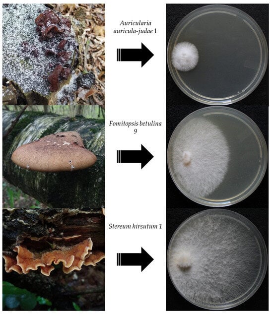
All strains formed light-colored colonies with smooth margins on a modified PDA medium. Four distinct colony types (cottony, felty, pellicular, and wooly) were observed, following the classification by Stalpers [34]. Stereum hirsutum strain 1 demonstrated the most rapid mycelial growth, while Auricularia auricula-judae 1 and Ganoderma applanatum 3 strains had the slowest growth rates (Table 2, Figure 1).
Table 2.
Morphological characteristics of fungal strains.
Figure 1.
Fruit bodies and seven-day mycelial colonies of Auricularia auricula-judae 1, Fomitopsis betulina 9, and Stereum hirsutum 1 strains cultured on a modified PDA medium.
3.2. Submerged Cultivation of Fungal Strains
Active biosynthesis of secondary metabolites in mycelial fungi during their submerged cultivation primarily takes place in idiophase. To identify the onset of the transition from the trophophase to the idiophase, the time point of maximum biomass accumulation was determined. The cultivation period required to achieve peak biomass yield ranged from 5 to 7 days (Table 3). Complete data for 5–7 cultivation days on submerged biomass yields and culture fluid pH values are provided in Supplementary Materials, Table S1. The highest biomass yields were observed in strains Flammulina rossica 16 and S. hirsutum 1. Fomitopsis pinicola strain 6 demonstrated the fastest growth, achieving maximum biomass on day 5. In contrast, A. auricula-judae strain 1 and Fomitopsis betulina strain 9 showed the slowest biomass accumulation, reaching a maximum only on day 7. Most of the studied strains acidified the culture medium, with the exception of Pleurotus ostreatus strain 38, which caused alkalization of the medium. The lowest pH values were observed in the culture media of F. pinicola 6 and F. betulina 9 strains.
Table 3.
Characteristics of submerged strain cultivation.
3.3. Extraction
Lyophilized fruiting bodies and submerged mycelial biomass of the studied strains were finely powdered and extracted with 96% ethanol, while culture liquids were extracted with ethyl acetate. The dry mass yields of the obtained extracts are presented in Table 4. The highest yield of culture liquid extract (>500 mg/L) was recorded for the F. pinicola strain 6 (644.20 mg/L), followed by the F. betulina strain 9 (591.97 mg/L). The F. pinicola strain 6 yielded the highest mycelial extract yield (164.04 mg/g), followed by F. betulina strain 9 (78.02 mg/g). In contrast, the remaining strains demonstrated lower mycelial extract yields.
Table 4.
Yields of fruiting bodies, submerged mycelium, and culture liquid extracts.
Among the fruiting body extracts, the highest yield values were recorded for F. rossica 16 (91.23 mg/g), F. betulina 9 (57.10 mg/g) and P. ostreatus 38 (54.06 mg/g) strains. F. betulina 9 and F. pinicola 6 strains yielded substantial amounts of extract from all three sample types (fruiting bodies, submerged mycelial biomass, and culture liquids), suggesting high overall biosynthetic activity.
The largest difference in extract yields was observed in the F. pinicola strain 6, where the mycelial extract yield was almost four times higher than that from the fruiting bodies. In contrast, in the F. rossica strain 16, the fruiting body extract yield was 3.6 times higher than that from the mycelium.
3.4. Agar Well Diffusion Assay
Preliminary assays showed that 25% ethanol did not inhibit growth of the test organisms. Results of the antimicrobial activity evaluation are presented in Table 5.
Table 5.
Antimicrobial activity of fruiting bodies, submerged mycelium, and culture liquid extracts.
Culture liquid extracts demonstrated higher antimicrobial activity compared to extracts from submerged mycelium and fruiting bodies. All culture liquid extracts showed an inhibitory effect against Gram-positive bacteria. The culture liquid extract of S. hirsutum strain 1 showed the highest activity against S. aureus, with an inhibition zone diameter of 50 mm. The next most active were, in descending order, extracts from culture liquids of A. auricula-judae 1 (35 mm), F. betulina 9 (30 mm), F. pinicola 6 (26 mm) and F. rossica 16 (25 mm) strains. Culture liquid extracts of Bjerkandera adusta 1, G. applanatum 3, and P. ostreatus 38 strains demonstrated low activity against S. aureus, with inhibition zones reaching 12, 15, and 14 mm, respectively. The culture liquid extract of Trametes hirsuta strain 12 was inactive against S. aureus but showed low activity against other Gram-positive bacteria, including B. subtilis and M. luteus. In general, culture liquid extracts of S. hirsutum 1, A. auricula-judae 1, F. pinicola 6, F. betulina 9, and F. rossica 16 strains demonstrated the highest activity against Gram-positive bacteria. Moderate activity against the Gram-negative bacterium Pseudomonas aeruginosa was observed only in the culture liquid extracts of F. betulina 9, F. pinicola 6, and S. hirsutum 1 strains, with inhibition zones measuring 17, 17, and 14 mm, respectively. No activity against the tested microorganisms is marked as ‘n/a’.
The highest antifungal activity was observed in culture liquid extracts of A. auricula-judae 1 and S. hirsutum 1 strains, which showed an inhibitory effect against both yeast-like and filamentous fungi. The culture liquid extract of the A. auricula-judae strain 1 was more active in inhibiting C. albicans than the growth of F. oxysporum. In contrast, the culture liquid extract of S. hirsutum strain 1 was more effective against F. oxysporum than C. albicans. Extracts from the culture liquids of F. rossica 16, F. betulina 9, and F. pinicola 6 strains showed low antifungal activity. No antifungal activity was detected in the culture liquid extracts of strains B. adusta 1, G. applanatum 3, P. ostreatus 38, and T. hirsuta 12.
Among the submerged mycelium extracts, antimicrobial activity was detected only in the S. hirsutum 1 and F. rossica 16 strains. Mycelium extract of S. hirsutum strain 1 showed weak to moderate activity against Gram-positive bacteria, with inhibition zones ranging from 12 to 18 mm, and weak activity against filamentous fungi with an inhibition zone of 12 mm. The submerged mycelium extract of the F. rossica strain 16 demonstrated antibacterial activity only against Gram-positive bacteria, showing high activity against S. aureus and moderate activity against B. subtilis and M. luteus.
Most fruiting body extracts showed no antimicrobial activity, with the exception of those from F. betulina 9 and F. pinicola 6 strains. The fruiting body extract of F. betulina strain 9 was active against Gram-positive bacteria, showing the highest activity against B. subtilis with an inhibition zone of 20 mm, and lower activity against M. luteus and S. aureus, with inhibition zones of 14 and 12 mm, respectively. The fruiting body extract of the F. pinicola strain 6 demonstrated only low activity against B. subtilis with an inhibition zone of 10 mm.
3.5. Evaluation of the Minimum Inhibitory Concentration (MIC)
Culture liquid extracts from strains that demonstrated the highest antibacterial activity in the previous assay (inhibition zones > 25 mm) were selected for MIC evaluation against an expanded panel of bacterial test cultures. A. auricula-judae 1, F. rossica 16, F. betulina 9, F. pinicola 6, and S. hirsutum 1 strains were the most active producers of antimicrobial metabolites. In addition, a culture liquid chloroform extract was prepared from a strain of S. hirsutum 1. Based on the preliminary screening results, which revealed the largest growth inhibition zones for the fungal extract, the MIC was further investigated for two selected extracts.
The results of evaluating the MIC of culture liquid extracts are presented in Table 6. The antibiotic levofloxacin, used as a positive control, demonstrated MIC values ranging from 0.25 to 2 µg/mL. The most active was a culture liquid extract of the S. hirsutum strain 1, with an MIC value of 320 µg/mL against clinical Gram-positive bacterial strains, including S. aureus, S. epidermidis, S. haemolyticus, and a vancomycin-resistant strain of E. faecium, as well as the Gram-negative bacterium P. vulgaris.
Table 6.
MIC values of culture liquid extracts against Gram-positive and Gram-negative bacteria.
The chloroform extract of S. hirsutum strain 1 was less active than the ethyl acetate extract. Weak activity was observed only against the Gram-positive bacteria S. epidermidis and E. faecium, with an MIC value of 640 µg/mL.
The culture liquid extract of the A. auricula-judae strain 1 showed comparable activity against all tested Gram-positive strains, as well as two Gram-negative bacteria, E. coli and P. vulgaris. MIC values against E. coli and P. vulgaris strains were 640 µg/mL, at the upper limit of moderate activity. The extract showed low activity against A. baumannii, K. pneumoniae, and P. vulgaris.
The culture liquid extract of the F. rossica 16 demonstrated moderate activity, with an MIC value of 640 µg/mL, against the Gram-positive bacteria S. epidermidis and S. haemolyticus and the Gram-negative bacterium P. vulgaris. Activity against other test organisms was low. The culture liquid extract of the F. betulina 9 strain showed moderate activity (MIC value of 640 µg/mL) against Staphylococcus strains. In contrast, the extract of the F. pinicola strain 6 demonstrated low activity against all test organisms (MIC value ≥ 1280 µg/mL).
3.6. Total Phenol and Terpenoid Content of Strain S. hirsutum 1 Extracts
The ethyl acetate extract from the culture liquid of the S. hirsutum strain 1 showed the highest antimicrobial activity among all extracts tested. For comparison, the total content of phenolic compounds and terpenoids was quantified spectrophotometrically in this extract and in the chloroform extract from the same culture (Table 7).
Table 7.
Total phenol and terpenoid content of S. hirsutum 1 extracts.
The content of phenolic compounds in the ethyl acetate extract was 20 times higher than in the chloroform extract. Terpenoids were detected only in the ethyl acetate extract, with a quantified content below 2 mg linalool equivalents per gram. They were absent in the chloroform extract.
3.7. Molecular Identification
The taxonomic identity of the A. auricula-judae 1 and S. hirsutum 1 strains, considered the most promising producers of antimicrobial metabolites in this study, was confirmed through Sanger sequencing of the ITS1-5.8S-ITS2 ribosomal DNA region. Resulting sequences were compared against the NCBI GenBank database using the BLAST algorithm, with species-level identification requiring ≥99.5% sequence similarity to validated reference sequences. The A. auricula-judae strain 1 exhibited 99.61% similarity to the voucher specimen LE 254030 (Komarov Botanical Institute of the Russian Academy of Sciences; GenBank accession KJ698425.1), while the S. hirsutum strain 1 showed a 99.84% similarity to the voucher specimen SFC20170809_10 (School of Biological Sciences, Seoul National University; GenBank accession MT044393.1). Sequences were deposited in GenBank under accession number PV918714 for A. auricula-judae strain 1 and PV918712 for S. hirsutum strain 1.
4. Discussion
4.1. Results Discussion
The life cycle of basidiomycete macromycetes (division Basidiomycota) consists of two primary stages: a vegetative phase, represented by the mycelium, and a reproductive phase, characterized by fruiting body formation and basidiospore maturation. The transition between these stages, as well as fruiting body development, is regulated by transcriptional reprogramming and changes in gene expression [44], including genes involved in the biosynthesis of primary and secondary metabolites [17]. The fungal secondary metabolome is characterized by high chemical and functional diversity, making it a promising source of biologically active compounds, particularly those with antimicrobial properties. Its composition depends not only on the developmental stage but also on fungal taxonomy, environmental factors, and cultivation parameters.
For a comparative evaluation of the antibacterial and antifungal activity of basidiomycete extracts obtained at vegetative and reproductive stages, extracts from fruiting bodies, culture liquids and submerged mycelium were analyzed. Fruiting bodies used for both pure culture isolation and extraction were collected from a single region during the autumn of 2024 in order to minimize variability associated with natural, climatic, and seasonal factors. Vegetative mycelium was obtained by submerged cultivation, allowing for the separation of endometabolites accumulated in the mycelium from exometabolites secreted into the culture liquid. Thus, metabolites associated with the vegetative stage were represented by submerged mycelium and culture liquid extracts, while metabolites of the reproductive stage were represented by fruiting body extracts.
The choice of solvents for extracting metabolites from basidiomycete biomass and culture liquids was based on a literature review. This analysis revealed that ethanol is the most commonly used solvent for biomass extraction (from both fruit bodies and mycelium). In most cases, the use of ethanol was associated with antimicrobial activity. For example, ethanol extracts of A. bisporus fruit bodies exhibited high antimicrobial activity, whereas ethyl acetate extracts were inactive. Ethyl acetate, on the other hand, is predominantly used for extracting culture liquids when screening for producers of antimicrobial metabolites [2].
Extracts of culture liquids from all nine strains exhibited antimicrobial activity. In contrast, such activity was detected in extracts from fruit bodies of only two strains and from mycelium of two strains. Thus, antimicrobial activity was considerably less common in submerged mycelium and fruit bodies among the strains studied.
The study showed that culture liquid extracts demonstrated the highest antibacterial and antifungal activity. Culture liquid extracts from all nine strains studied were active against Gram-positive bacteria, while activity against Gram-negative bacteria was observed in the culture liquid extracts from only three strains. The highest antibacterial activity was detected in culture liquid extracts of strains S. hirsutum 1, A. auricula-judae 1, F. betulina 9, F. pinicola 6 and F. rossica 16. These five strains also demonstrated antifungal activity. For the chloroform and ethyl acetate extracts of S. hirsutum 1, the total content of phenolic and terpenoid compounds was quantified. The ethyl acetate extract contained higher levels of both compound classes. Together with the MIC data, these results indicate that the antimicrobial activity of strain S. hirsutum 1 extract may be influenced by the combined presence of phenolics and terpenoids. This is supported by the lower activity of the chloroform extract compared to the ethyl acetate extract. The difference in activity was most pronounced against the Gram-positive bacteria S. aureus and S. haemolyticus, as well as against the Gram-negative bacterium P. vulgaris.
Only two submerged mycelium extracts showed antimicrobial activity: the S. hirsutum strain 1 extract was active against both Gram-positive bacteria and fungi, while the F. rossica strain 16 mycelium extract inhibited the growth of Gram-positive bacteria only. The activity of submerged mycelium extracts was lower than that of culture liquid extracts. Antimicrobial activity of fruiting body extracts was observed exclusively in strains of the genus Fomitopsis, limited to Gram-positive bacteria, being most pronounced in the extract of F. betulina 9.
Thus, under the experimental conditions used, the strains studied produced antimicrobial metabolites more actively during the vegetative stage, with the target metabolites accumulating in the culture liquid in most of the strains tested. The results are consistent with the role of antimicrobial metabolites in allowing vegetative mycelium to compete effectively for substrate during growth.
The study identified A. auricula-judae 1 and S. hirsutum 1 as the most promising strains for further investigation. The culture liquid extract of A. auricula-judae strain 1 demonstrated high activity against both Gram-positive bacteria and fungi. While there are few data on the antimicrobial properties of this species, existing studies report bacteriostatic effects against S. aureus and B. subtilis, as well as moderate antifungal activity [45,46,47]. Most of the studies on A. auricula-judae have focused on the study of total extracts and fractions activity: polysaccharide extracts showed activity against S. aureus and E. coli with MICs of 6.25 and 12.5 µg/mL, respectively [46]; melanin isolated from fruiting bodies demonstrated significant antibacterial activity against Gram-negative bacteria with an MIC of 160–640 µg/mL and suppressed biofilm formation by 60–70% at a concentration of 80 µg/mL [47].
Stereum species are known for producing diverse bioactive compounds, including those with antitumor, hepatoprotective, cytotoxic, antiparasitic, and antimicrobial properties [48,49,50,51,52,53]. Stereumamides A and D showed MICs of 12.5 µg/mL against E. coli and S. aureus, while sterostrein Q was characterized by a higher MIC; these compounds were also isolated from the culture liquid [50]. Sterenin D had fungistatic activity against Botrytis cinerea at a concentration of 20 µg/mL and fungicidal activity at 50 µg/mL [52]. Epidoxysterols 1 and 2, sterol compounds isolated from fruit bodies, showed activity against Mycobacterium tuberculosis with MIC 16 µg/mL [53].
The preferential accumulation of antimicrobial metabolites of basidiomycetes in the culture liquid allows the advantages of submerged cultivation to be exploited in the development of biotechnologies for obtaining these compounds. The processes for obtaining submerged cultures of basidiomycetes are significantly shorter than the processes for obtaining their fruiting bodies. The duration of submerged cultivation can be 3–5 days, while obtaining fruiting bodies in culture requires from one to several months [54,55,56,57,58]. To increase the efficiency of submerged cultivation biotechnologies for basidiomycetes, experimental design methods are successfully used.
4.2. Limitations of the Study
This study had several limitations. First, different solvents were used for the extraction of biomass and culture fluids, although their selection was guided by a literature review. Second, the study was limited to nine strains, which may not fully represent the diversity of basidiomycetes.
5. Conclusions
The secondary metabolome of basidiomycetes is a rich but underexplored source of bioactive compounds, including antimicrobial agents. Successful screening for such metabolites requires careful material selection based on consistent compound content and reproducible sample acquisition. Potential sources include fruiting bodies, spores, mycelia, and culture liquids. Comparative analysis of nine basidiomycete strains showed that the vegetative stage, particularly via secreted exometabolites, yields the highest antimicrobial activity. This supports the ecological role of these compounds in defending the substrate-colonizing mycelium. These findings guide future efforts in identifying novel antimicrobials from basidiomycetes.
Supplementary Materials
The following supporting information can be downloaded at: https://www.mdpi.com/article/10.3390/app16063031/s1, Figure S1: Fruit bodies and seven-day mycelial colonies of Bjerkandera adusta 1, Flammulina rossica 16, Fomitopsis pinicola 6; Figure S2: Fruit bodies and seven-day mycelial colonies of Ganoderma applanatum 3, Pleurotus ostreatus 38, Trametes hirsuta 12; Table S1: Complete data for 5–7 cultivation days on submerged biomass yields and culture fluid pH values; Table S2: ITS1-5.8S-ITS2 region sequences of studied strains.
Author Contributions
Conceptualization, L.K.; methodology, L.K., V.L., O.S., E.I. and N.K.; software, N.K.; validation, L.K., M.Z. and V.L.; investigation, M.Z., M.L., N.K., M.D., O.S., E.I. and V.L.; resources, L.K. and M.D.; data curation, L.K., M.Z., M.L., V.L., O.S., E.I. and N.K.; writing—original draft preparation, M.Z., V.L. and N.K.; writing—review and editing, L.K., M.Z., N.K. and V.L.; visualization, M.Z., N.K. and V.L.; supervision, L.K. and M.Z.; administration, M.D. All authors have read and agreed to the published version of the manuscript.
Funding
This research received no external funding.
Institutional Review Board Statement
Not applicable.
Informed Consent Statement
Not applicable.
Data Availability Statement
The original data are available from the corresponding author on request.
Conflicts of Interest
The authors declare no conflicts of interest.
Abbreviations
The following abbreviations are used in this manuscript:
| MIC | Minimum inhibitory concentration |
| DNA | Deoxyribonucleic acid |
| PDA | Potato Dextrose Agar |
| DMSO | Dimethyl sulfoxide |
| ITS | Internal Transcribed Spacer |
| EtOAc | Ethyl acetate |
| CHCl3 | Chloroform |
| ATCC | American Type Culture Collection |
| VKPM | Russian Collection of Pathogenic Microorganisms |
| CFU | Colony Forming Unit |
| GAE | Gallic Acid Equivalents |
References
- World Health Organization. Antimicrobial Resistance Fact Sheet. Available online: https://www.who.int/news-room/fact-sheets/detail/antimicrobial-resistance (accessed on 21 November 2023).
- Lysakova, V.; Krasnopolskaya, L.; Yarina, M.; Ziangirova, M. Antibacterial and Antifungal Activity of Metabolites from Basidiomycetes: A Review. Antibiotics 2024, 13, 1026. [Google Scholar] [CrossRef] [Scilit] [PubMed]
- Morel, S.; Vitou, M.; Masnou, A.; Jumas-Bilak, E.; Rapior, S.; Licznar-Fajardo, P. Antibacterial activity of wild mushrooms from France. Int. J. Med. Mushrooms 2021, 23, 79–89. [Google Scholar] [CrossRef] [Scilit]
- Soliman, E.R.; El-Sayed, H. Molecular identification and antimicrobial activities of some wild Egyptian mushrooms: Bjerkandera adusta as a promising source of bioactive antimicrobial phenolic compounds. J. Genet. Eng. Biotechnol. 2021, 19, 106–111. [Google Scholar] [CrossRef] [Scilit] [PubMed]
- Ragupathi, V.; Stephen, A.; Arivoli, D.; Kumaresan, S. In vitro antibacterial activity of methanolic extract of wild mushrooms from southern Western Ghats, India. Int. J. Phytopharm. Res. 2018, 9, 32–39. [Google Scholar]
- Sum, W.C.; Indieka, S.A.; Matasyoh, J.C. Antimicrobial activity of Basidiomycetes fungi isolated from a Kenyan tropical forest. Afr. J. Biotechnol. 2019, 18, 112–123. [Google Scholar] [CrossRef] [Scilit]
- Clericuzio, M.; Bivona, M.; Gamalero, E.; Bona, E.; Novello, G.; Massa, N.; Robotti, E. A systematic study of the antibacterial activity of basidiomycota crude extracts. Antibiotics 2021, 10, 1424. [Google Scholar] [CrossRef] [Scilit]
- Ait Hamadouche, Y.; Dib, S.; Fortas, Z. Antifungal Basidiomycete Extracts with Phytosanitary Potential. South Asian J. Exp. Biol. 2021, 11, 86–95. [Google Scholar] [CrossRef] [Scilit]
- Garcia, J.; Rodrigues, F.; Castro, F.; Aires, A.; Marques, G.; Saavedra, M.J. Antimicrobial, antibiofilm, and antioxidant properties of Boletus edulis and Neoboletus luridiformis against multidrug-resistant ESKAPE pathogens. Front. Nutr. 2022, 8, 773346. [Google Scholar] [CrossRef] [Scilit]
- Covino, S.; D’Ellena, E.; Tirillini, B.; Angeles, G.; Arcangeli, A.; Bistocchi, G.; Venanzon, R.; Angelini, P. Characterization of biological activities of methanol extract of Fuscoporia torulosa (Basidiomycetes) from Italy. Int. J. Med. Mushrooms 2019, 21, 1051–1063. [Google Scholar] [CrossRef] [Scilit]
- Mehta, S.; Jandaik, S. In vitro comparative evaluation of antibacterial activity of fruiting body and mycelial extracts of Ganoderma lucidum against pathogenic bacteria. J. Pure Appl. Microbiol. 2012, 6, 1997–2001. [Google Scholar]
- Angelini, P.; Girometta, C.; Tirillini, B.; Moretti, S.; Covino, S.; Cipriani, M.; D’Ellena, E.; Angeles, G.; Federici, E.; Savino, E.; et al. A comparative study of the antimicrobial and antioxidant activities of Inonotus hispidus fruit and their mycelia extracts. Int. J. Food Prop. 2019, 22, 768–783. [Google Scholar] [CrossRef] [Scilit]
- Corrêa, R.C.G.; de Souza, A.H.P.; Calhelha, R.C.; Barros, L.; Glamoclija, J.; Sokovic, M.; Peralta, R.M.; Bracht, A.; Ferreira, I.C. Bioactive formulations prepared from fruiting bodies and submerged culture mycelia of the Brazilian edible mushroom Pleurotus ostreatoroseus Singer. Food Funct. 2015, 6, 2155–2164. [Google Scholar] [CrossRef] [Scilit] [PubMed]
- Nguyen, T.M.; Kim, J.; Hung, N.T.; Tho, N.H.; Due, D.B.; Hien, D.T. Antioxidant and Antimicrobial Properties of Fruiting Body and Submerged Mycelium of Medicinal Mushroom Phellinus robiniae (Agaricomycetes). Int. J. Med. Mushrooms 2023, 25, 37–46. [Google Scholar] [CrossRef] [Scilit] [PubMed]
- Vaquiro, H.A.; Suárez, H.; Murillo, W. Probiotic growth-stimulating capacity and antimicrobial activities of aqueous extracts of Lentinus crinitus (L.) Fr (polyporales, basidiomycota). Heliyon 2023, 9, e18738. [Google Scholar] [CrossRef] [Scilit] [PubMed]
- Angelini, P.; Venanzoni, R.; Flores, G.A.; Tirillini, B.; Orlando, G.; Recinella, L.; Chiavaroli, A.; Brunetti, L.; Leone, S.; Di Simone, S.C.; et al. Evaluation of antioxidant, antimicrobial and tyrosinase inhibitory activities of extracts from Tricholosporum goniospermum, an edible wild mushroom. Antibiotics 2020, 9, 513. [Google Scholar] [CrossRef] [Scilit]
- Berger, R.G.; Bordewick, S.; Krahe, N.-K.; Ersoy, F. Mycelium vs. Fruiting Bodies of Edible Fungi—A Comparison of Metabolites. Microorganisms 2022, 10, 1379. [Google Scholar] [CrossRef] [Scilit]
- Chen, J.; Yu, M.; Yang, C.; Huang, Z.; He, L.; Bian, J.; Sun, S.; Li, J. Study on the nutritional relationships in mycelia and fruiting bodies of Hypsizygus marmoreus under defined nutrient conditions. Food Chem. 2025, 467, 142323. [Google Scholar] [CrossRef] [Scilit]
- Brazkova, M.; Angelova, G.; Mihaylova, D.; Stefanova, P.; Pencheva, M.; Gledacheva, V.; Stefanova, I.; Krastanov, A. Bioactive Metabolites from the Fruiting Body and Mycelia of Newly-Isolated Oyster Mushroom and Their Effect on Smooth Muscle Contractile Activity. Foods 2022, 11, 3983. [Google Scholar] [CrossRef] [Scilit]
- Onuoha, S.C.; Okoroh, P.N.; Tom-Quinn, R.A. Proximate Composition, Essential Heavy Metal Concentrations and Nutrient Density of the Mycelium and Fruiting Bodies of Organically Cultivated Pleurotus ostreatus. J. Appl. Life Sci. Int. 2021, 24, 44–52. [Google Scholar] [CrossRef] [Scilit]
- Yang, Z.; Zeng, Y.; Hu, Y.; Zhou, T.; Li, J.; He, L.; Fan, J. Comparison of chemical property and in vitro digestion behavior of polysaccharides from Auricularia polytricha mycelium and fruit body. Food Chem. X 2023, 17, 100570. [Google Scholar] [CrossRef] [Scilit]
- Campi, M.; Mancuello, C.; Ferreira, F.; Maubet, Y.; Centurión, E.L.C.; Gayozo, E.; Robledo, G.L. Does the source matter? Phenolic compounds and antioxidant activity from mycelium in liquid medium, wild and cultivated fruiting bodies of the neotropical species Ganoderma tuberculosum. J. Microbiol. Biotechnol. Food Sci. 2023, 127, 1034–1044. [Google Scholar] [CrossRef] [Scilit]
- Xie, C.-L.; Yan, S.-W.; Zhang, Z.-M.; Gong, W.-B.; Zhu, Z.-H.; Zhou, Y.-J.; Yan, L.; Hu, Z.-X.; Ai, L.-Z.; Peng, Y.-D. Mapping the metabolic signatures of fermentation broth, mycelium, fruiting body and spores powder from Ganoderma lucidum by untargeted metabolomics. LWT-Food Sci. Technol. 2020, 129, 109494. [Google Scholar] [CrossRef] [Scilit]
- Cardoso, R.V.C.; Fernandes, Â.; Oliveira, M.; Calhelha, R.C.; Barros, L.; Martins, A.; Ferreira, I. Development of nutraceutical formulations based on the mycelium of Pleurotus ostreatus and Agaricus bisporus. Food Funct. 2017, 8, 2155–2164. [Google Scholar] [CrossRef] [Scilit] [PubMed]
- Kała, K.; Kryczyk-Poprawa, A.; Rzewińska, A.; Muszyńska, B. Fruiting bodies of selected edible mushrooms as a potential source of lovastatin. Eur. Food Res. Technol. 2020, 246, 713–722. [Google Scholar] [CrossRef] [Scilit]
- Sułkowska-Ziaja, K.; Korczyński, M.; Trepa, M.; Galanty, A.; Lazur, J.; Kubica, P.; Kała, K.; Paśko, P.; Muszyńska, B. A Comparative Study on the Mycelium and Fruiting Body of Meripilus giganteus: Chemical Composition and Biological Activity. Curr. Issues Mol. Biol. 2025, 47, 302. [Google Scholar] [CrossRef] [Scilit]
- Song, H.-Y.; Kim, D.-H.; Kim, J.-M. Comparative transcriptome analysis of dikaryotic mycelia and mature fruiting bodies in the edible mushroom Lentinula edodes. Sci. Rep. 2018, 8, 8983. [Google Scholar] [CrossRef] [Scilit]
- Lu, Y.P.; Liao, J.H.; Guo, Z.J.; Cai, Z.X.; Chen, M.Y. Genome Survey and Transcriptome Analysis on Mycelia and Primordia of Agaricus blazei. Biomed. Res. Int. 2020, 2020, 1824183. [Google Scholar] [CrossRef] [Scilit]
- Hao, H.; Zhang, J.; Wang, Q.; Huang, J.; Juan, J.; Kuai, B.; Chen, H. Transcriptome and Differentially Expressed Gene Profiles in Mycelium, Primordium and Fruiting Body Development in Stropharia rugosoannulata. Genes 2022, 13, 1080. [Google Scholar] [CrossRef] [Scilit]
- Bondartsev, A.S.; Zinger, R.A. Guide to Collecting Higher Basidiomycetes for Scientific Study; Izdatel’stvo Akademii nauk SSSR: Moscow, Russia; Saint Petersburg, Russia, 1950; p. 45. (In Russian) [Google Scholar]
- Hansen, L.; Knudsen, H. (Eds.) Nordic Macromycetes. Vol. 2: Polyporales, Boletales, Agaricales, Russulales; Nordsvamp: Copenhagen, Denmark, 1992; p. 474. [Google Scholar]
- Hansen, L.; Knudsen, H. (Eds.) Nordic Macromycetes. Vol. 3: Heterobasidioid, Aphyllophoroid and Gastromycetoid Basidiomycetes; Nordsvamp: Copenhagen, Denmark, 1997; p. 444. [Google Scholar]
- Ryvarden, L.; Melo, I. Poroid Fungi of Europe; Synopsis Fungorum 31; Fungiflora: Oslo, Norway, 2014; p. 455. [Google Scholar]
- Stalpers, J.A. Identification of Wood-Inhabiting Aphyllophorales in Pure Culture; Centraalbureau voor Schimmelcultures: Baarn, The Netherlands, 1978; Volume 16, p. 248. [Google Scholar]
- Krasnopolskaya, L.M.; Belitsky, I.V.; Fedorova, G.B.; Katrukha, G.S. Pleurotus djamor: Cultivation Methods and Antimicrobial Properties. Mikol. Fitopatol. 2001, 35, 62–67. (In Russian) [Google Scholar]
- Lysakova, V.S.; Sineva, O.N.; Bychkova, O.P.; Krasnopolskaya, L.M. Screening of Antibacterial and Antifungal Activities of Basidiomycetes Extracts. Antibiot. Khimioter. 2024, 69, 11–18. (In Russian) [Google Scholar] [CrossRef] [Scilit]
- Russian Guidelines. Determination of Microorganism Susceptibility to Antimicrobial Agents, Versiya 2025-01; MAKMAKH, SSU: Smolensk, Russia, 2025; p. 208. (In Russian) [Google Scholar]
- CLSI. Performance Standards for Antimicrobial Susceptibility Testing, 30th ed.; CLS Supplement M100; Clinical and Laboratory Standarts Institute: Wayne, PA, USA, 2020. [Google Scholar]
- Kuete, V. Potential of Cameroonian plants and derived products against microbial infections: A review. Planta Med. 2010, 76, 1479–1491. [Google Scholar] [CrossRef] [Scilit] [PubMed]
- Fadeyi, O.G.; Olou, B.A.; Tchuenguem, R.T.; Tsakem, B.; Sinda, P.V.K.; Dzoyem, J.P.; Yorou, N.S. Antimicrobial and Antioxidant Activities of a Brown Wood Rotting Mushroom Piptoporellus baudonii from Benin (Tropical Africa). Adv. Biol. Chem. 2024, 14, 145–170. [Google Scholar] [CrossRef]
- Anwar, J.; Iqbal, Z. Effect of growth conditions on antibacterial activity of Trichoderma harzianum against selected pathogenic bacteria. Sarhad J. Agric. 2017, 33, 501–510. [Google Scholar] [CrossRef] [Scilit]
- Ainsworth, E.A.; Gillespie, K.M. Estimation of total phenolic content and other oxidation substrates in plant tissues using Folin–Ciocalteu reagent. Nat. Protoc. 2007, 2, 875–877. [Google Scholar] [CrossRef] [Scilit]
- Ghorai, N.; Chakraborty, S.; Gucchait, S.; Saha, S.K.; Biswas, S. Estimation of total terpenoids concentration in plant tissues using a monoterpene, linalool as standard reagent. Protoc. Exch. 2012. [Google Scholar] [CrossRef] [Scilit]
- Nagy, L.G.; Vonk, P.J.; Künzler, M.; Földi, C.; Virágh, M.; Ohm, R.A.; Hennicke, F.; Bálint, B.; Csernetics, Á.; Hegedüs, B.; et al. Lessons on fruiting body morphogenesis from genomes and transcriptomes of Agaricomycetes. Stud. Mycol. 2023, 104, 1–85. [Google Scholar] [CrossRef] [Scilit]
- Oli, A.N.; Edeh, P.A.; Al-Mosawi, R.M.; Mbachu, N.A.; Al-Dahmoshi, H.O.M.; Al-Khafaji, N.S.K.; Ekuma, U.O.; Okezie, U.M.; Saki, M. Evaluation of the phytoconstituents of Auricularia auricula-judae mushroom and antimicrobial activity of its protein extract. Eur. J. Integr. Med. 2020, 38, 101176. [Google Scholar] [CrossRef] [Scilit]
- Cai, M.; Lin, Y.; Luo, Y.; Liang, H.; Sun, P. Extraction, antimicrobial, and antioxidant activities of crude polysaccharides from the wood ear medicinal mushroom Auricularia auricula-judae (Higher Basidiomycetes). Int. J. Med. Mushrooms 2015, 17, 591–600. [Google Scholar] [CrossRef] [Scilit]
- Li, B.; Li, W.; Chen, X.; Jiang, M.; Dong, M. In vitro antibiofilm activity of the melanin from Auricularia auricula, an edible jelly mushroom. Ann. Microbiol. 2012, 62, 1523–1530. [Google Scholar] [CrossRef] [Scilit]
- Tian, M.; Zhao, P.; Li, G.; Zhang, K. In depth natural product discovery from the basidiomycetes Stereum species. Microorganisms 2020, 8, 1049. [Google Scholar] [CrossRef] [Scilit]
- Yun, B.S.; Lee, I.K.; Cho, Y.; Cho, S.M.; Yoo, I.D. New Tricyclic Sesquiterpenes from the Fermentation Broth of Stereum hirsutum. J. Nat. Prod. 2002, 65, 786–788. [Google Scholar] [CrossRef] [Scilit] [PubMed]
- Duan, Y.C.; Feng, J.; Bai, N.; Li, G.H.; Zhang, K.Q.; Zhao, P.J. Four novel antibacterial sesquiterpene-alpha-amino acid quaternary ammonium hybrids from the mycelium of mushroom Stereum hirsutum. Fitoterapia 2018, 128, 213–217. [Google Scholar] [CrossRef] [Scilit] [PubMed]
- Ma, K.; Bao, L.; Han, J.; Jin, T.; Yang, X.; Zhao, F.; Li, S.; Song, F.; Liu, M.; Liu, H. New benzoate derivatives and hirsutane type sesquiterpenoids with antimicrobial activity and cytotoxicity from the solid-state fermented rice by the medicinal mushroom Stereum hirsutum. Food Chem. 2014, 143, 239–245. [Google Scholar] [CrossRef] [Scilit] [PubMed]
- Aqueveque, P.; Cespedes, C.L.; Becerra, J.; Aranda, M.; Sterner, O. Antifungal activities of secondary metabolites isolated from liquid fermentations of Stereum hirsutum (Sh134-11) against Botrytis cinerea (grey mould agent). Food Chem. Toxicol. 2017, 109, 1048–1054. [Google Scholar] [CrossRef] [Scilit]
- Cateni, F.; Doljak, B.; Zacchigna, M.; Anderluh, M.; Piltaver, A.; Scialino, G.; Banfi, E. New biologically active epidioxysterols from Stereum hirsutum. Bioorg. Med. Chem. Lett. 2007, 17, 6330–6334. [Google Scholar] [CrossRef] [Scilit]
- Krasnopolskaya, L.; Leontieva, M.; Avtonomova, A.; Isakova, E.; Belitskii, I.V.; Usov, A.I.; Buchman, V.M. Antitumor properties of submerged cultivated biomass and extracts of medicinal mushrooms of genus Hypsizygus Singer. Int. J. Med. Mushrooms 2008, 10, 25–35. [Google Scholar] [CrossRef] [Scilit]
- Krasnopolskaya, L.; Shuktueva, M.; Golyshkin, A.; Almyasheva, N.; Yarina, M. Optimization of the nutrient medium for Flammulina velutipes submerged biomass production and micromorphology of its mycelium. Fermentation 2021, 7, 180. [Google Scholar] [CrossRef] [Scilit]
- Stamets, P. Growing Gourmet and Medicinal Mushrooms, 3rd ed.; Ten Speed Press: Berkeley, CA, USA, 2000; p. 592. [Google Scholar]
- Desisa, B.; Muleta, D.; Dejene, T.; Jida, M.; Goshu, A.; Martin-Pinto, P. Substrate optimization for shiitake (Lentinula edodes (Berk.) Pegler) mushroom production in Ethiopia. J. Fungi 2023, 9, 811. [Google Scholar] [CrossRef] [Scilit]
- Nghien, N.X.; Thuy, N.T.B.; Luyen, N.T.; Thu, N.T.; Quan, N.D. Morphological characteristics, yield performance, and medicinal value of some lingzhi mushroom (Ganoderma lucidum) strains cultivated in Tam Dao, Vietnam. Vietnam J. Agric. Sci. 2019, 2, 321–331. [Google Scholar]

Disclaimer/Publisher’s Note: The statements, opinions and data contained in all publications are solely those of the individual author(s) and contributor(s) and not of MDPI and/or the editor(s). MDPI and/or the editor(s) disclaim responsibility for any injury to people or property resulting from any ideas, methods, instructions or products referred to in the content. |
© 2026 by the authors. Licensee MDPI, Basel, Switzerland. This article is an open access article distributed under the terms and conditions of the Creative Commons Attribution (CC BY) license.