


Abstract
Background: Auricular defects resulting from congenital anomalies, trauma, or oncologic resection pose significant functional and psychosocial challenges. When autologous reconstruction is not feasible or not desired, implant-retained auricular prostheses represent a reliable alternative with high patient satisfaction. This study aimed to systematically evaluate the clinical performance of craniofacial implants used for auricular prosthetic rehabilitation, focusing on implant survival, prosthetic outcomes, workflow typologies, and complications. A secondary objective was to illustrate the long-term validity of a minimally invasive navigation technique through a clinical case with 6-year follow-up. Methods: A systematic review was conducted according to PRISMA guidelines. Clinical studies published between 2005 and 2025 reporting outcomes of implant-retained auricular prostheses were searched in PubMed and Scopus databases. Data were extracted on implant type, survival rates, prosthetic performance, workflow, and complications. Risk of bias was assessed using appropriate tools based on each study design. Results: A total of thirty-two studies were included, comprising fifteen case reports, fifteen case series, one cohort study, and one prospective observational study. Implant survival was consistently high across all workflow categories, with failures predominantly associated with irradiated or anatomically compromised bone. Prosthetic outcomes were favorable, showing excellent esthetics, stable retention, and high patient satisfaction irrespective of manufacturing method, although digital and navigation-assisted workflows improved reproducibility, symmetry, and planning precision. Complication rates were low and generally limited to mild peri-abutment inflammation manageable with conservative care. The clinical case confirmed these findings, showing stable osseointegration, healthy soft tissues, and uncompromised prosthetic function at 6-year follow-up. Conclusions: Implant-retained auricular prostheses show predictable long-term success, independent of whether traditional, hybrid, or fully digital workflows are employed. Digital technologies enhance surgical accuracy, minimize morbidity, and streamline prosthetic fabrication, although high-quality comparative studies remain limited.
1. Introduction
Auricular loss (whether congenital, traumatic, or oncologic) represents a significant reconstructive and psychosocial challenge. Congenital anomalies such as microtia and anotia occur in approximately one per five to seven thousand live births, whereas acquired auricular defects often result from ablative oncologic surgery or severe trauma [1,2,3,4]. Traditional autologous reconstruction using costal cartilage or porous polyethylene frameworks can provide excellent esthetic results in experienced hands [5,6,7,8]. However, these techniques remain technically demanding, require multi-stage surgery, and are contraindicated in patients with scarring, previous irradiation, or inadequate soft-tissue quality [9]. When autologous reconstruction is not feasible or not desired, implant-retained auricular prostheses offer a predictable and minimally invasive alternative to restore facial symmetry and social well-being [10,11,12]. Since their introduction in the late 1970s, osseointegrated craniofacial implants have become the gold standard for anchoring auricular epitheses [13,14,15]. Contemporary clinical series consistently report implant survival rates exceeding 95%, favorable soft-tissue behavior, and high patient satisfaction, with complications typically limited to manageable peri-abutment inflammation or skin overgrowth. Compared with adhesive-retained prostheses, implant anchorage provides superior retention, hygiene, durability, and day-to-day comfort [16]. Over the past decade, the surgical and prosthetic workflow for auricular rehabilitation has undergone a profound transformation with the advent of digital technologies. Computer-aided design/manufacturing (CAD-CAM), three-dimensional (3D) imaging, and virtual surgical planning (VSP) enable accurate replication of the contralateral anatomy, prosthetically driven implant positioning, and reduced dependence on manual sculpting [17,18,19,20,21,22]. Stereolithographic guides have improved the precision of extraoral implant placement, while significantly reducing laboratory time and increasing reproducibility [23,24,25,26]. More recently, dynamic navigation systems (originally developed for neurosurgery and oral implantology) have been introduced into craniofacial implant surgery. These systems provide real-time visual feedback on drill trajectory relative to the patient’s anatomy, enabling high-precision flapless implant placement with minimal morbidity [27,28,29,30,31,32,33]. Navigation-assisted workflows improve intraoperative visibility, reduce surgical duration, and lower the risk of injury to delicate anatomic structures, including the facial nerve and auricular vasculature. Early experimental and clinical studies have shown sub-millimetric accuracy comparable to that achieved in guided intraoral implantology [34]. Despite these promising developments, the application of dynamic navigation to auricular implant placement remains scarcely documented and long-term clinical outcomes are still largely missing. In parallel, the scientific literature on implant-retained auricular prostheses remains heterogeneous with respect to defect etiology, implant systems, retention mechanisms, and reported outcome measures. While several systematic reviews on implant-retained auricular prosthesis have already been published, only a limited number has specifically addressed comparison between different surgical and prosthetic workflows. Moreover, available data on digital and navigation-assisted approaches remain heterogeneous and are largely derived from small observational studies with short-term follow-up. In this context, the present study was designed with a dual purpose: (i) to provide an updated overview of the available clinical evidence by comparatively analyzing traditional, hybrid, and digital workflows for implant-retained auricular prosthetic rehabilitation, focusing on implant survival, complications, and prosthetic outcomes; and (ii) to complement this evidence with the presentation of an innovative, navigation-guided, flapless clinical case with six-year clinical and radiographic follow-up, serving as an illustrative example of the long-term feasibility and biological stability of a fully digital approach.
2. Materials and Methods
This systematic review aimed to comprehensively evaluate the clinical outcomes of osseointegrated craniofacial implants used for auricular prosthetic rehabilitation. The included studies investigated surgical protocols, implant survival, peri-implant soft-tissue behavior, prosthesis-related outcomes, and patient-reported satisfaction across different clinical scenarios. Only clinical studies involving human participants were included to ensure real-world clinical relevance. Eligible designs comprised prospective and retrospective cohorts, as well as case series and case reports, that reported outcomes related to both the implant and the retained auricular prosthesis. Experimental, animal, in vitro, technical notes without clinical data, and studies focusing exclusively on bone-anchored hearing aids without auricular prosthetic rehabilitation were excluded. The review protocol was prospectively registered in the PROSPERO database (CRD420251181874). The review was conducted in accordance with the PRISMA 2020 (Preferred Reporting Items for Systematic Reviews and Meta-Analyses) guidelines to ensure methodological transparency, reproducibility, and adherence to high-quality standards [35].
2.1. Search Stretegy
A comprehensive literature search was conducted across PubMed and Scopus databases between 29 October and 2 November 2025. Search strings were adapted to each database while maintaining semantic consistency. A last comprehensive search was conducted on 5 November 2025 to perform a final screening of eligible articles across all databases.
The following search strategy was employed to systematically retrieve relevant studies:
| (auricular[Title/Abstract] OR microtia[Title/Abstract] OR “ear prosthesis”[Title/Abstract] OR “auricular prosthesis”[Title/Abstract]) AND (“osseointegrated implant”[Title/Abstract] OR “craniofacial implant”[Title/Abstract] OR “extraoral implant”[Title/Abstract] OR “bone anchored”[Title/Abstract]) AND (prosthesis[Title/Abstract] OR rehabilitation[Title/Abstract] OR digital[Title/Abstract] OR “CAD CAM”[Title/Abstract] OR navigation[Title/Abstract]) |
Reference lists of selected articles were manually screened to identify additional relevant studies.
2.2. Inclusion and Exclusion Criteria
Studies were considered eligible for inclusion if they were published between 2005 and 2025 and reported clinical applications of osseointegrated craniofacial implants used for auricular prosthetic rehabilitation in human patients. Only clinical studies were included to ensure real-world applicability and to capture outcomes related to both the implant and the retained auricular prosthesis. Eligible studies needed to involve implant placement in the auricular region and include postoperative evaluation of implant survival, soft-tissue response, prosthetic retention, patient satisfaction, and functional/esthetic outcomes.
Both traditional and digitally assisted workflows (virtual surgical planning, CAD/CAM, navigation-guided or guided-surgery protocols) were included, provided that they involved craniofacial implants specifically used to retain an auricular prosthesis. Only articles published in English and available in full text were included to ensure methodological transparency and complete data extraction.
Exclusion criteria comprised in vitro, animal, and cadaveric studies, as well as reports describing implants not placed in the auricular region or not used to retain a prosthesis (e.g., bone-anchored hearing devices without prosthetic rehabilitation). Studies were excluded if they did not involve the use of craniofacial osseointegrated implants, if they focused solely on autologous reconstruction without a prosthetic component, or if they addressed hearing rehabilitation only. Reviews, systematic reviews, editorials, conference abstracts without full text and technical notes were excluded. Finally, studies published in languages other than English or without accessible full text were omitted to ensure consistent methodological quality and reliable data synthesis.
2.3. Article Selection Process
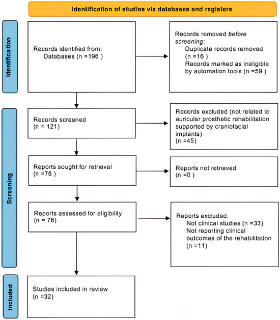
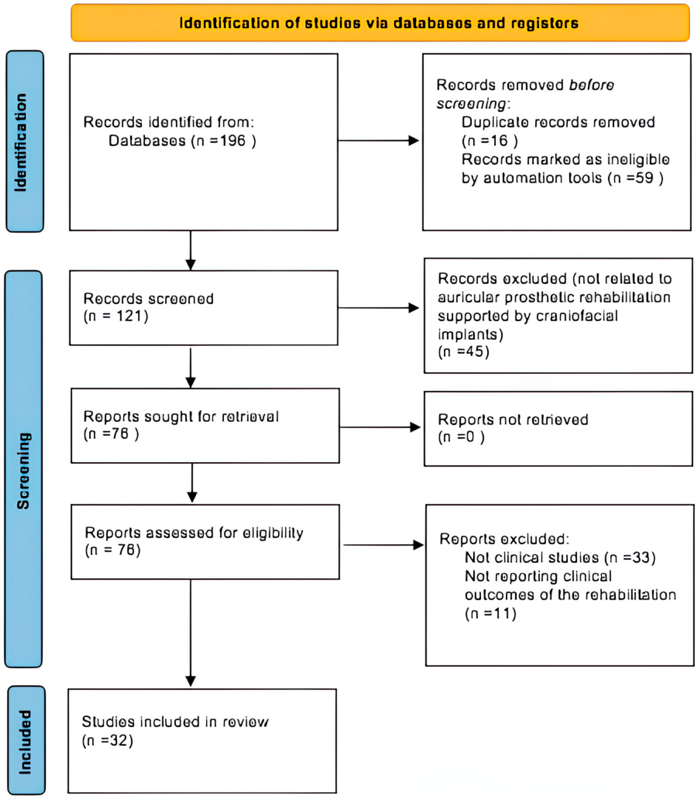
The initial database search identified 196 records. After applying filters for publication language (English) and publication date (2005–2025), 59 records were excluded, resulting in 137 records: 58 from PubMed and 79 from Scopus. Following the removal of 16 duplicates, 121 studies were screened based on titles and abstracts. Of these, 45 records were excluded for being unrelated to auricular prosthetic rehabilitation supported by craniofacial implants. A total of 76 full-text articles were sought for retrieval. Of these, 43 were excluded for the following reasons: 11 did not report clinical outcomes (neither implant-related nor prosthesis-related) and 33 were non-clinical studies. A total of 32 studies met all inclusion criteria and were included in this systematic review. The selection process was conducted independently by two expert reviewers, with disagreements resolved through discussion with a third reviewer. The full selection process is summarized in the PRISMA flow diagram (Figure 1).
Figure 1.
This diagram illustrates the systematic process of identifying, screening, and selecting studies for inclusion in the systematic review.
2.4. Data Extraction and Quality Assessment
Two independent reviewers performed data extraction using a standardized Excel spreadsheet.
The following information was collected from each included study: first author, year of publication, study design, number of patients, type of implant, prosthesis material, type of workflow, implant outcomes, prosthesis outcomes, comparison with other rehabilitation strategies, and follow up.
Any discrepancies between the two reviewers regarding study selection or risk of bias assessment were resolved through discussion with a third reviewer.
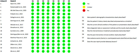
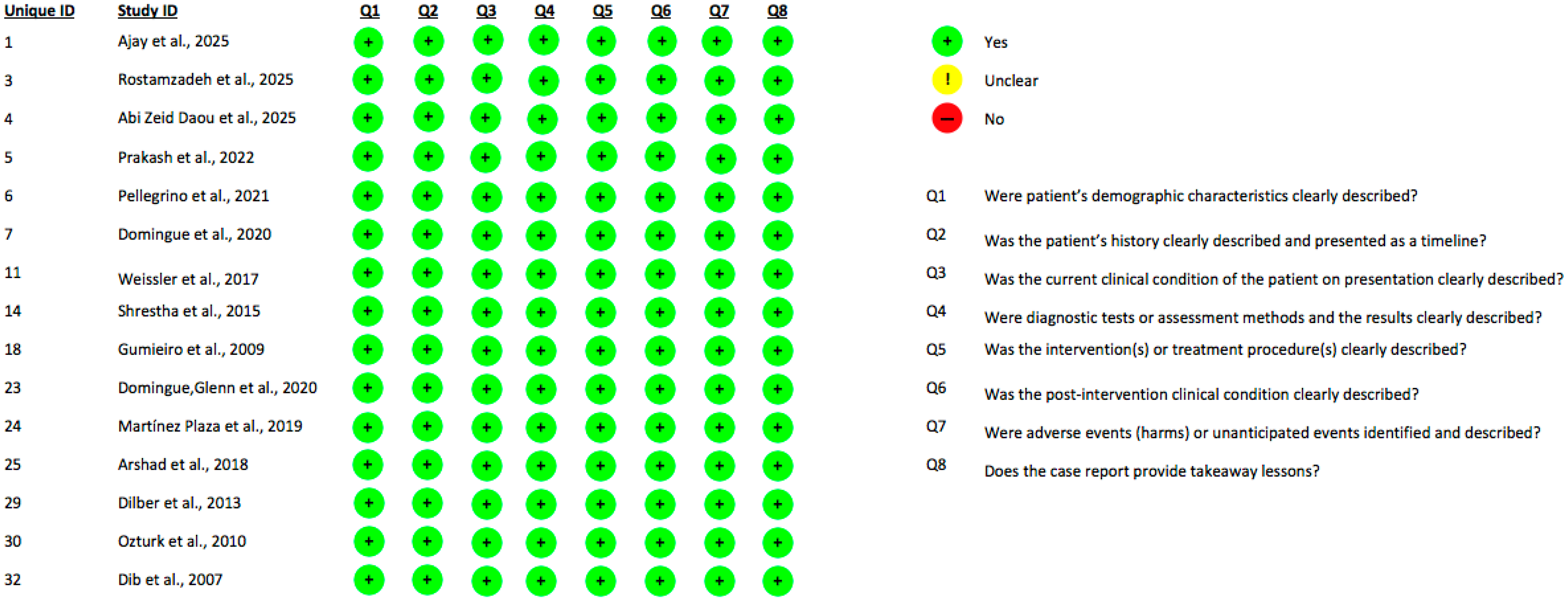
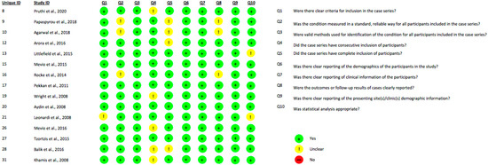
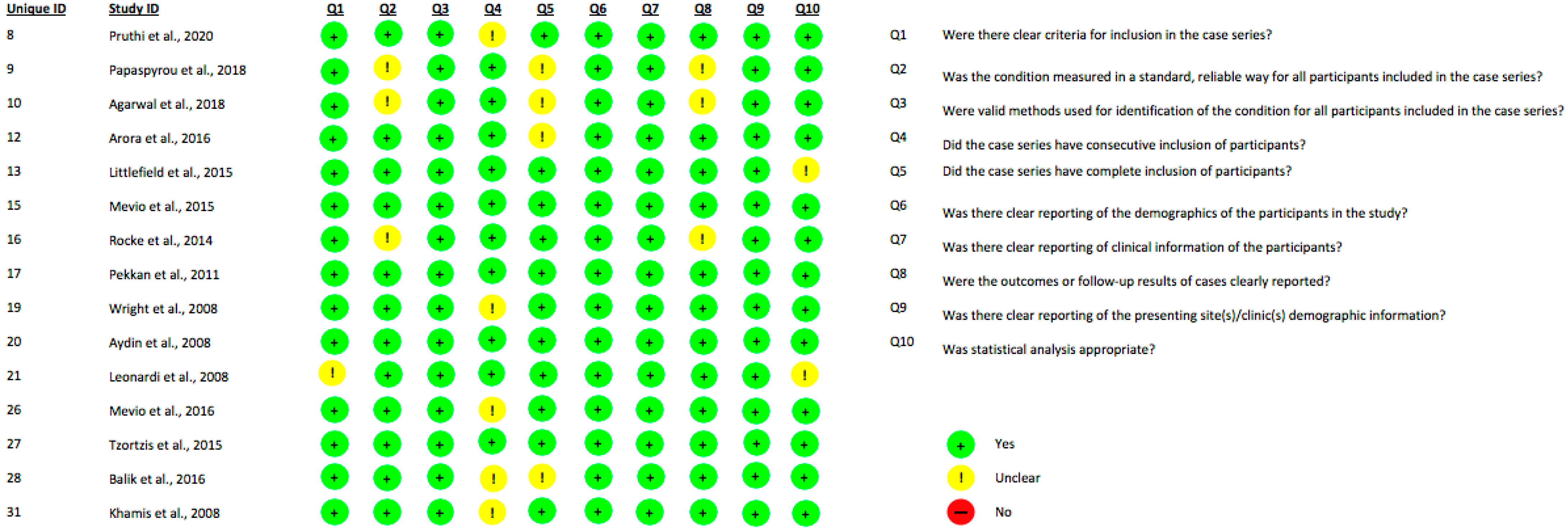
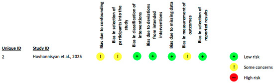
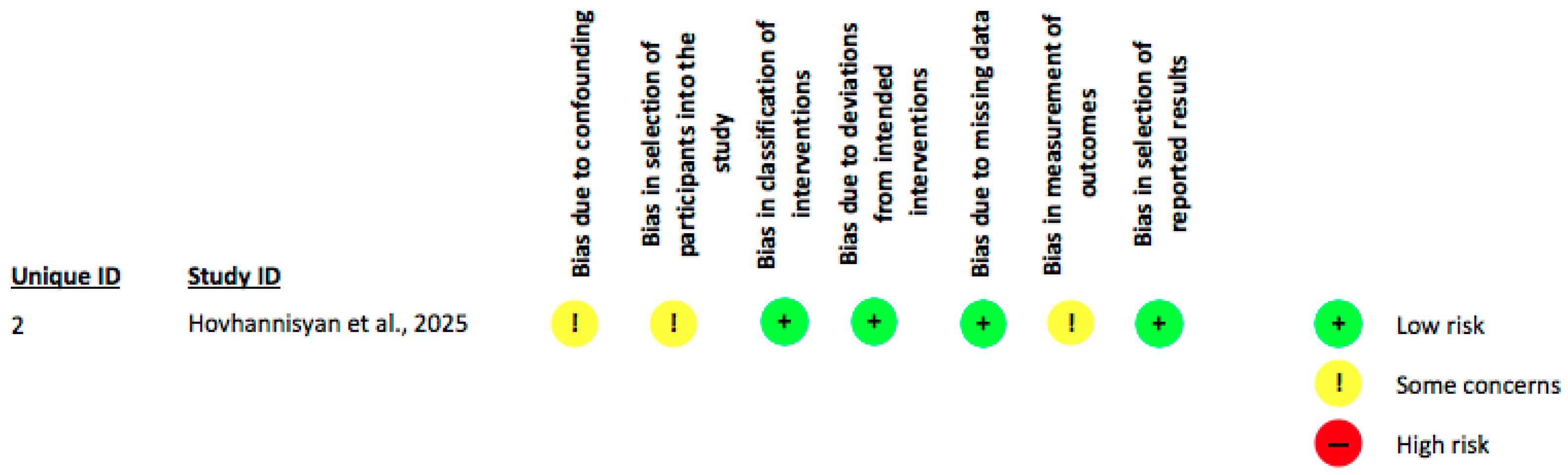
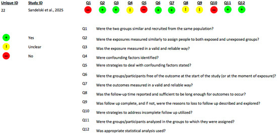
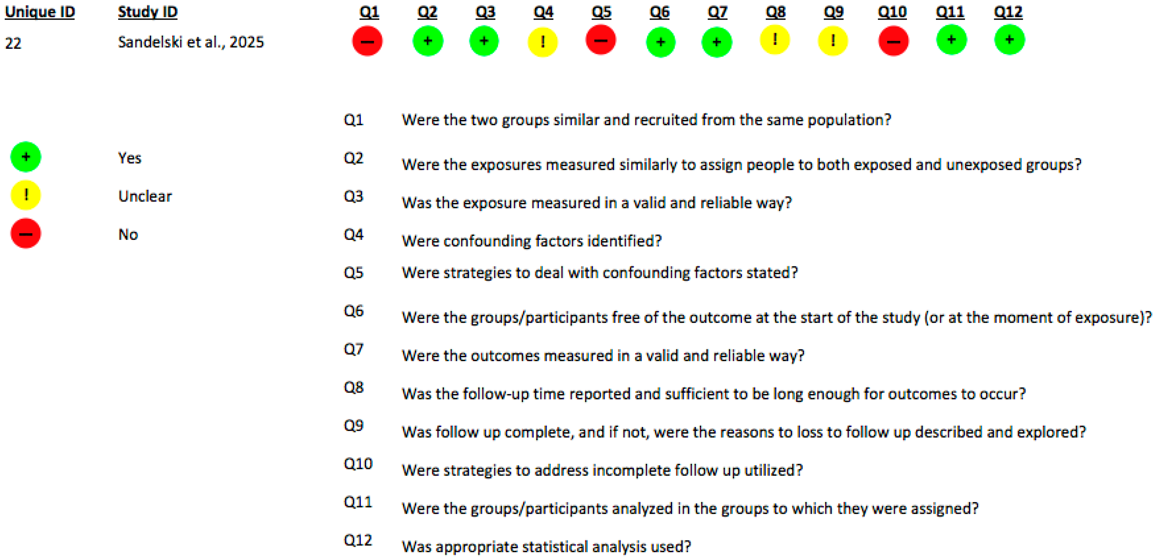
The present systematic review incorporated studies involving diverse methodological designs, including the following: fifteen case series, one prospective observational study, one retrospective cohort study, and fifteen case reports. Accordingly, risk of bias assessments were conducted employing tools specifically validated for each study design. For case reports and case series, the Joanna Briggs Institute (JBI) critical appraisal checklist was applied to evaluate study rigor and relevance [36] (Figure 2 and Figure 3). The ROBINS-I tool was utilized to appraise the risk of bias of the prospective observational study [37] (Figure 4). The included cohort study was evaluated using the JBI checklist for cohort studies [38] (Figure 5). This stratified approach ensured an appropriate and robust critical appraisal tailored to the heterogeneity of included evidence.
Figure 2.
This table presents the assessment of the risk of bias for each included case report, evaluated across the eight domains of the JBI Critical Appraisal Checklist for case reports [39,40,41,42,43,44,45,46,47,48,49,50,51,52,53].
Figure 3.
This table presents the assessment of the risk of bias for each included case series, evaluated across the ten domains of the JBI Critical Appraisal Checklist for case series [54,55,56,57,58,59,60,61,62,63,64,65,66,67,68].
Figure 4.
This table presents the assessment of the risk of bias for the only prospective observational study included, evaluated across the seven domains of the ROBINS-I tool [69].
Figure 5.
This table presents the assessment of the risk of bias for the only retrospective cohort study included, evaluated across the twelve domains of the JBI Critical Appraisal Checklist for cohort studies [70].
2.5. Surgical Workflow of the Case
An illustrative clinical case was incorporated into the present review to exemplify the practical application of the digital and navigated surgical workflows identified in the literature. The case was selected because it met predefined methodological criteria: (1) complete preoperative imaging and digital planning data, (2) use of a fully guided or navigation-assisted implant placement workflow, (3) documentation of prosthetic design and fabrication, and (4) availability of long-term follow-up. The case report presented here was originally included among the clinical studies identified in the systematic review; however, we provide an updated 7-year follow-up to further support the evidence on long-term implant survival, peri-implant stability, and high patient satisfaction associated with dynamically guided craniofacial implant placement for auricular rehabilitation. All diagnostic and operative steps followed standard institutional protocols, including Cone-Beam Computed Tomography (CBCT) imaging, 3D facial scanning, digital mirroring of the contralateral auricle, virtual planning of implant positioning, and execution of a minimally invasive flapless procedure using a dynamic navigation system. Prosthetic rehabilitation was carried out through an entirely digital workflow based on optical scanning and CAD/CAM design. Ethical approval and patient consent were obtained prior to treatment. A detailed clinical description is provided in Section 3.
3. Results
3.1. Literature Synthesis
A total of 32 clinical studies met the inclusion criteria and were analyzed. The studies were published between 2005 and 2025 and collectively present a broad spectrum of clinical scenarios involving osseointegrated craniofacial implants for auricular prosthetic rehabilitation. Most reports were retrospective case series, prospective observational studies, or detailed clinical case reports, reflecting the relative scarcity of large-scale prospective research in this field. A summary table of the various outcomes reported in the included studies has been provided as Table 1.
Table 1.
Synthesis of the 32 articles included in the review.
3.1.1. Implant Outcomes
Across the entire body of evidence, implants exhibited consistently high stability and durable long-term performance, independent of surgical technique, retention design or the complexity of the reconstructive scenario. The reliability of the mastoid as an anchorage site was particularly evident in auricular-only cohorts such as those described by Wright et al. (2008) [62] and Aydın et al. (2008) [63], both of which reported complete or near-complete osseointegration without implant failures. Similar outcomes were reflected in various case reports (Gumieiro et al. 2009 [47]; Shrestha et al. 2015 [46]; Arshad et al. 2018 [50]; Ozturk et al. 2010 [52] and Dilber et al. 2013 [51]), with each documenting stable unilateral or bilateral fixtures with no loss of integration. Larger multisite series reinforced these findings: Leonardi et al. (2008) [64] observed successful integration in all auricular cases examined, while Pekkan et al. (2011) [61] emphasized the consistently favorable bone architecture of the mastoid region. Long-term observational data from Tzortzis et al. (2015) [66] and Balik et al. (2016) [67] further confirmed the robustness of implant survival over extended follow-up periods. The more recent literature from 2020 to 2025 aligns seamlessly with these earlier trends. Ajay et al. (2025) [39] reported complete osseointegration of two digitally guided implants within three months and no biological complications, while Hovhannisyan et al. (2025) [69] documented 100% survival in staged placements, noting only a transient Holgers 2 reaction. Additional contemporary evidence further reinforces this consistency. Notably, stable bilateral osseointegration in severe burn trauma was reported by Rostamzadeh et al. (2025) [40], while Abi Zeid Daou et al. (2025) [41] documented complication-free immediate loading. Prakash et al. (2022) [42] likewise achieved full osseointegration under local anesthesia and Pellegrino et al. (2021) [43] reported excellent outcomes using virtual planning combined with navigated flapless surgery. Likewise, Pruthi et al. (2020) [54] confirmed complete implant survival in their retrospective series and Domingue et al. (2020) [44] described uneventful integration within four months when using a digital workflow. Overall, the body of evidence indicates that high implant stability is achievable across digital, hybrid, analog, and even immediate-loading approaches. Failures remain uncommon and tend to occur primarily in cases involving previously irradiated or structurally compromised bone.
3.1.2. Prosthesis Outcomes
Esthetic and functional outcomes were consistently excellent across all studies, independent of whether prostheses were produced through traditional sculpting or fully digital workflows. Patients in the series by Agarwal et al. (2018) [56], Wright et al. (2008) [62], Aydın et al. (2008) [63], and Mevio et al. (2015) [59] reported high satisfaction with comfort, natural appearance, and secure retention, with Mevio et al. (2016) [65] further documenting measurable improvements in quality-of-life scores. Similar impressions emerged from case reports by Gumieiro et al. (2009) [47], Shrestha et al. (2015) [46], Arshad et al. (2018) [50], Ozturk et al. (2010) [52], and Dilber et al. (2013) [51], each describing prostheses that blended naturally with surrounding tissues and maintained stable function even in complex traumatic or congenital defects. Larger multi-site studies echoed these findings, with Leonardi et al. (2008) [64] highlighting excellent long-term adherence in auricular cases. Digital approaches added an additional layer of esthetic precision. Weissler et al. (2017) [45] achieved remarkably natural contours using mirrored 3D anatomy, while Domingue et al. (2020) [44] produced highly accurate silicone prostheses through a combined CBCT, optical scanning, and 3D-printing workflow. Martínez Plaza et al. (2019) [49] likewise proved excellent anthropometric harmony in bilateral microtia by optimizing virtual ear positioning. More recent studies further confirmed these favorable outcomes: Ajay et al. (2025) [39] reported outstanding symmetry and retention using CAD/CAM-generated molds and Hovhannisyan et al. (2025) [69] reported better performance of implant retention over adhesives. Rostamzadeh et al. (2025) [40] produced highly natural bilateral results using conventional fabrication, while Abi Zeid Daou et al. (2025) [41] successfully delivered a final prosthesis intraoperatively within an “ear-in-a-day” workflow. Prakash et al. (2022) [42] achieved excellent hygiene and esthetics through a custom bar-and-clip system and Pellegrino et al. (2021) [43] obtained a precisely adapted prosthesis by leveraging mirrored digital anatomy. In addition, Pruthi et al. (2020) [54] reported satisfaction rates exceeding 85% and predictable prosthesis longevity, while Domingue, Glenn et al. (2020) [48] confirmed excellent fit and comfort using integrated CBCT and digital scans. Taken together, these findings show that implant-retained auricular prostheses reliably provide high-quality esthetics, durable retention, and strong patient satisfaction across both analog and advanced digital workflows.
3.1.3. Long-Term Performance and Maintenance
Long-term follow-up data consistently indicate that, while osseointegrated craniofacial implants show high survival and biological stability over extended periods, the silicone auricular prosthesis itself requires periodic maintenance and replacement. Several authors have explicitly reported that material-related degradation, including discoloration, marginal tearing, and loss of adaptation, represents the most frequent long-term prosthetic complication. In a large retrospective series, Pruthi et al. [54] reported that refabrication of the silicone prosthesis was generally required after approximately 1.5 to 2 years due to progressive material deterioration, despite stable implant integration and satisfactory peri-implant soft tissue conditions. Similar observations emerge from long-term cohort studies. Papaspyrou et al. [55], in their retrospective analysis of 99 patients rehabilitated with facial prostheses, described regular outpatient follow-up visits (two to four per year) and reported that prosthetic maintenance and replacement constituted a routine component of long-term care, even in the absence of implant failure. Extended follow-up data further confirm that biological complications tend to remain limited and manageable over time, whereas prosthetic-related issues are more frequent. In a ten-year review of 131 auricular prostheses, Tzortzis et al. [66] reported postoperative follow-up periods ranging from 2 to 14 years, with skin reactions generally mild and reversible, while prosthetic use and maintenance remained ongoing requirements throughout the observation period. Complex reconstructions involving extensive soft tissue and bony defects show comparable trends. In cases of total auricular rehabilitation with combined reconstructive approaches, Littlefield et al., 2015 [58] reported stable long-term implant support with continued prosthesis use over follow-up periods of up to 54 months, emphasizing the need for sustained prosthetic care rather than implant-related revisions. Collectively, these findings highlight that long-term success of implant-retained auricular rehabilitation depends not only on implant survival but also on structured follow-up protocols addressing routine prosthesis maintenance and periodic refabrication.
3.1.4. Patient-Reported Outcomes
Patient satisfaction and quality-of-life outcomes were reported across all included studies, although assessed using different approaches. Overall, auricular implant rehabilitation was consistently associated with high levels of patient satisfaction, particularly with regard to prosthesis stability, ease of daily management and esthetic outcome. Most studies described patient satisfaction qualitatively, reporting good acceptance of the prosthesis, improved comfort compared with adhesive-retained solutions, and positive psychosocial impact. A subset of studies employed structured or quantitative patient-reported outcome measures. Mevio et al. in both the 2015 and 2016 study [59,65] assessed quality of life using the validated SF-12 questionnaire and showed a statistically significant improvement in quality-of-life scores following auricular rehabilitation. Agarwal et al. (2018) [56] evaluated patient satisfaction using a structured Likert-scale questionnaire covering multiple domains, including comfort, stability, esthetics, and self-consciousness, and reported consistently high satisfaction scores. Papaspyrou et al. (2018) [55] retrospectively classified patient satisfaction using predefined categories, while Pruthi et al. (2020) [54] reported a long-term patient acceptance rate of 87.5% based on continued prosthesis use and compliance. Despite the heterogeneity of assessment methods, all studies reported favorable patient perception of implant-retained auricular prostheses, indicating high overall satisfaction with both functional handling and esthetic integration.
3.1.5. Different Workflows: Where Are the Differences?
The combined evidence reveals three complementary workflow philosophies: traditional, digital, and hybrid, whereby all are capable of producing reliable auricular rehabilitation.
The comparison of different workflows emerging from the studies included should be interpreted descriptively, as the available evidence does not allow direct or quantitative comparisons between traditional, hybrid, and digital approaches. Traditional analog workflows (Rostamzadeh et al., 2025 [40]; Gumieiro et al., 2009 [47]; Arshad et al., 2018 [50] and Ozturk et al., 2010 [52]) used manual impressions, hand-sculpted wax patterns, and conventional prosthetic fabrication, providing dependable esthetic outcomes when executed meticulously. Hybrid workflows, such as those described by Hovhannisyan et al. (2025) [69], Prakash et al. (2022) [42] and Pruthi et al. (2020) [54], combined digital elements (CT planning or CAD-designed bars) with traditional prosthetic artistry, maintaining a balance between digital accuracy and manual refinement. Fully digital workflows (Ajay et al., 2025 [39]; Pellegrino et al., 2021 [43]; Domingue et al., 2020 [44]; Weissler et al., 2017 [45]; Martínez Plaza et al., 2019 [49]) represented the most technologically advanced approach. These protocols integrated CBCT-based virtual planning, mirrored anatomical modeling, surgical navigation, flapless placement, and 3D printing of molds, surgical guides or prosthetic elements. Digital workflows consistently achieved the highest level of precision, reproducibility, and prosthetic adaptation, particularly in anatomically complex or bilateral cases. Taken together, the literature shows that all three workflows (traditional, hybrid, and digital) can achieve stable and successful auricular rehabilitation. Digital pathways generally offer superior planning accuracy and workflow efficiency; however, traditional and hybrid approaches remain equally dependable when performed by experienced teams, underscoring the robustness of auricular implant protocols across diverse technological settings.
3.1.6. Complications
Complications across all included studies were consistently low in frequency, mild in severity, and highly manageable, confirming the overall safety of implant-retained auricular rehabilitation. Auricular-specific cohorts, including Wright et al., 2008 [62] and both Mevio’s studies (2015 and 2016) [59,65], reported no implant failures and only rare soft-tissue issues, with peri-implant tissues remaining stable throughout follow-up. Aydın et al. (2008) [63] and Pruthi et al. (2020) [54] similarly observed mostly minor Holgers 1–2 reactions, typically presenting as transient redness or slight granulation and none of these episodes progressed to deeper infection or threatened fixture stability. In mixed craniofacial cohorts, more pronounced complications (such as soft-tissue overgrowth, peri-abutment inflammation, or isolated infection) were generally confined to patients with irradiated or surgically compromised bone, a finding particularly emphasized by Rocke et al. (2014) [60]. Even in these cases, conservative measures or minor revision were typically sufficient and prosthesis use was rarely affected thanks to redundancy of fixtures and stable surrounding implants. Digital case series echoed this pattern: Martínez Plaza et al. (2019) [49] reported a single episode of peri-implant hyperplasia resolved through limited soft-tissue correction, while Domingue et al. (2020) [44] documented early soft-tissue swelling that subsided after replacing the healing abutment with a taller component. The newly added literature from 2020 to 2025 showed similarly reassuring profiles. Ajay et al. (2025) [39], Rostamzadeh et al. (2025) [40], Pellegrino et al. (2021) [43], and Prakash et al. (2022) [42] reported no biological complications, even in complex anatomical or traumatic contexts. Hovhannisyan et al. (2025) [69] noted a single Holgers 2 reaction that resolved with routine care, while Abi Zeid Daou et al. (2025) [41] encountered only a minor magnet misalignment in their accelerated “ear-in-a-day” workflow. Pruthi et al. (2020) [54] described occasional mild inflammation and one drilling deviation, neither of which impacted long-term osseointegration. Overall, the cumulative evidence clearly indicates that implant-retained auricular rehabilitation has an excellent safety profile, with complications that are predominantly minor, reversible, and rarely disruptive to either implant stability or prosthetic function. This consistency across analog, digital, hybrid, and immediate-loading workflows underscores the robustness and clinical reliability of the technique.
3.1.7. Limitations
The interpretation of the present findings must consider several limitations inherent to the available literature on implant-retained auricular prosthetic rehabilitation. Most included studies were retrospective case series or case reports, lacking control groups, standardized methodologies, and consistent long-term follow-up. This predominance of low-level evidence introduces risks of selection and reporting bias and restricts the generalizability of outcomes beyond highly specialized centers. As a consequence, the findings of this review should be interpreted as descriptive and hypothesis-generating rather than confirmatory.
A further challenge arises from the marked heterogeneity among studies. Patient populations varied widely, from congenital microtia to oncologic or traumatic defects, each presenting different anatomical and biological conditions that influence implant survival and prosthetic success. Surgical protocols also differed substantially, encompassing one-stage, two-stage, freehand, flapless, and digitally guided techniques, often without precise stratification. Similar variability was observed in prosthetic workflows, ranging from fully analog approaches to advanced digital or hybrid systems, making direct comparisons difficult and precluding meta-analytic synthesis. As a result, no definitive statements regarding the comparative effectiveness of different workflows can be supported. Outcome definitions were not uniform across studies and objective quantitative metrics were often absent. Follow-up durations ranged from a few months to more than a decade, further complicating the interpretation of long-term performance and maintenance requirements. Technological evolution over the study period also limits comparability: earlier works relied solely on analog methods and older implant designs, while more recent studies integrate CBCT-based planning, CAD/CAM frameworks, surgical navigation, and improved silicone materials. This temporal variability acts as a confounder, making it difficult to isolate the effect of technique from broader technological progress. Finally, evidence for specific subgroups (such as pediatric patients, irradiated fields or bilateral reconstructions) remains sparse, preventing firm conclusions for these more complex clinical scenarios. Overall, these limitations underscore the need for prospective, multicenter studies with standardized protocols, validated outcome measures, and longer follow-up to strengthen the evidence base and refine clinical recommendations.
3.2. Case Report
A 26-year-old male patient affected by right-sided hemifacial microsomia (Pruzansky type II) with complete agenesis of the right auricle was treated in 2019 at the Maxillofacial Surgery Unit of S. Orsola Hospital, Bologna. Following multidisciplinary evaluation, implant-retained prosthetic rehabilitation was selected as the definitive treatment option [43].
3.2.1. Digital Planning and Workflow
A cone-beam CT scan (VGi, NewTom, Verona, Italy) was obtained while the patient wore a customized intraoral marker plate containing fiducial markers for navigation calibration. Simultaneously, a 3D surface scan of the face (SLS3, David Vision System, Amsterdam, The Netherlands) was acquired to record soft-tissue morphology. The healthy contralateral ear was digitally mirrored and superimposed onto the defect side to simulate the missing auricle. DICOM (CBCT) and STL (surface scan) datasets were integrated using anatomical landmarks (glabella, nasal apex, alar base) and refined with an iterative closest-point (ICP) algorithm to achieve precise alignment between skeletal and soft-tissue models. The resulting composite dataset allowed accurate identification of optimal implant sites based on bone availability and the projected prosthetic volume.
3.2.2. Surgical Procedure
The procedure was performed under local anesthesia without intravenous or oral sedation. After calibration of the dynamic navigation system (ImplaNav, BresMedical, Sydney, Australia), the marker plate was repositioned on the maxillary teeth and connected to the patient reference tool. The handpiece reference was calibrated in front of the navigation camera, enabling real-time, dynamically guided osteotomy preparation. No soft-tissue flap was raised. Osteotomies were prepared transcutaneously using a 2 mm lance drill followed by conical drills according to the manufacturer’s protocol. Two hybrid craniofacial implants (4 × 8.5 mm; MSc-IBT, Southern Implants, Irene, South Africa) were placed in the mastoid bone following the prosthetically driven plan, leaving the coronal machined portion extraosseous to allow transcutaneous bar connection. Hemostasis was obtained without the need for sutures. Postoperative healing was uneventful, with minimal discomfort and swelling reported by the patient. After a 3-month healing period, the implants were exposed. During uncovering, digital impressions were taken using an intraoral scanner (CEREC Omnicam, Dentsply Sirona, York, PA, USA) combined with a laser facial scan, enabling precise superimposition of implant positions with the patient’s facial morphology.
3.2.3. Prosthetic Rehabilitation
A custom titanium retention bar was digitally designed to accommodate three clips for optimal stress distribution and hygiene access, maintaining at least 2 mm of clearance from the skin. The bar and substructure were fabricated via selective laser melting (Ti6Al4V). The transcutaneous cylinders were coated with dental ceramic to reduce bacterial colonization. A medical-grade silicone auricular prosthesis (ETR 4750, Biesterfield, UK) was fabricated based on the printed auricular model. Pigmented silicone layers were applied to reproduce the patient’s skin color and texture. The prosthesis was retained on the titanium bar using mechanical clips (SO-line, New Ancorvis, Italy).
3.2.4. Six-Year Follow-Up
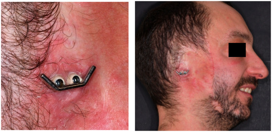
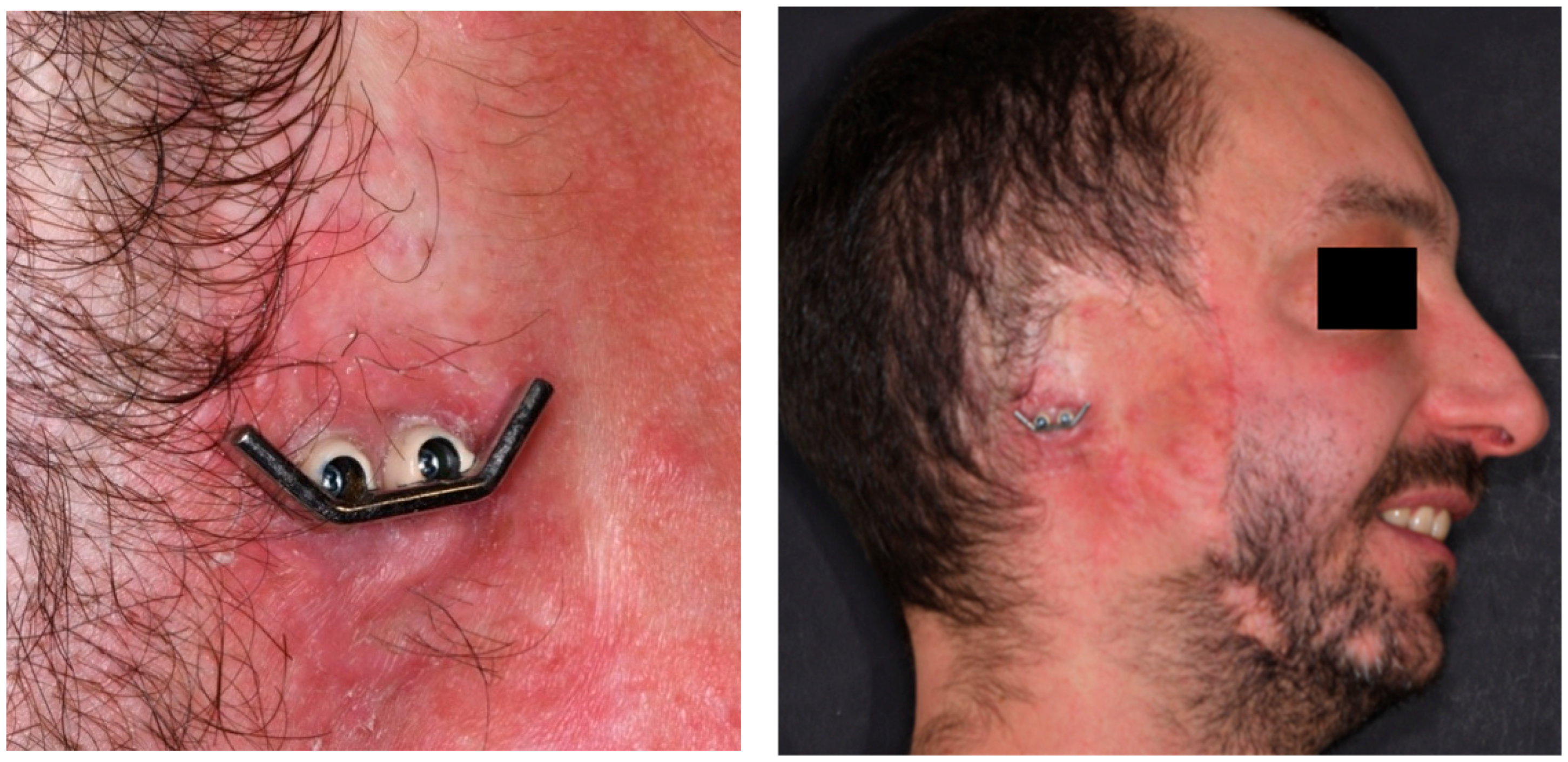
At the six-year follow-up, the patient reported continuous daily prosthesis use and full satisfaction with comfort and retention. Stability remained consistent during mastication, speech, and facial movements, with no episodes of loosening or detachment. Clinical examination revealed healthy peri-implant skin, well-adapted margins, and good hygiene, with no erythema, hypertrophy, ulceration or discharge. Palpation and visual inspection confirmed the absence of tenderness or signs of inflammation, indicating long-term soft-tissue stability. The titanium bar and clip-retention system were mechanically intact, showing no screw loosening, corrosion or component fatigue. Overall, the findings of this clinical case showed excellent long-term integration of the implants and supported the biological reliability and durability of the navigated flapless approach (Figure 6).
Figure 6.
Clinical view of the implant framework and surrounding soft tissues at the six-year follow-up.
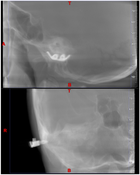
A new cone-beam CT scan confirmed complete osseointegration of both implants, with no radiolucency or peri-implant bone loss. The implant heads remained flush with the skin surface, maintaining stable transcutaneous emergence (Figure 7).
Figure 7.
CBCT view of the implants at the six-year follow-up.
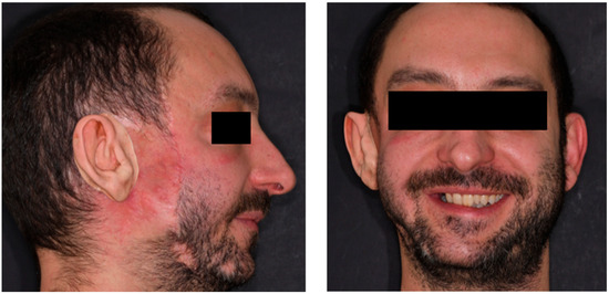
The silicone auricular prosthesis maintained accurate external contours and stable retention (Figure 8). However, progressive color fading, surface wear, and decreased elasticity were noted, particularly along anterior and superior margins exposed to sunlight. Mild surface roughness was also evident, consistent with long-term silicone aging. These changes were attributed to ultraviolet exposure, temperature fluctuations, and repetitive mechanical cleaning. Despite the esthetic deterioration, functional retention remained satisfactory. Nevertheless, the degree of material fatigue and surface degradation justified replacement of the silicone prosthesis to restore optimal esthetics, hygiene, and mechanical performance. The replacement process will reuse the existing digital design and implant framework, minimizing patient morbidity and laboratory time while reestablishing the original esthetic outcome.
Figure 8.
Prosthesis at the six-year follow-up.
4. Discussion
4.1. Principal Findings
This systematic review supports the view that implant-supported auricular prosthetic rehabilitation remains a highly reliable and predictable treatment modality, characterized by excellent implant survival, favorable soft-tissue tolerance, and consistently strong esthetic and functional outcomes. Across a twenty-year span and a diverse range of clinical scenarios (including congenital, traumatic, and oncologic defects), osseointegration in the mastoid region proved uniformly stable, independent of surgical protocol, prosthetic workflow, or retention system. The collective findings of this review confirm that the mastoid continues to represent an ideal anchorage site and that implant-supported prostheses provide superior retention and esthetic integration compared with adhesive alternatives.
4.2. Comparison with Existing Literature
The results of the present review are broadly consistent with the established evidence base for auricular prosthetic rehabilitation. Early foundational literature, such as the overview by Sharma et al. (2012) [71], already emphasized the high predictability of mastoid osseointegration and the clear advantages of implant retention in terms of comfort, hygiene, and prosthetic stability. Their review also underscored the pivotal role of soft-tissue management in long-term success, an aspect that remains central in the current synthesis where most reported complications were minor peri-abutment reactions easily resolved with conservative measures. Subsequent work through the 2010s further reinforced these themes. Datarkar et al. (2017) [72] highlighted the equivalence of various retention mechanisms (bar–clip, magnetic, ball-and-socket) when paired with meticulous hygiene and tissue maintenance, concluding that biological stability is determined more by peri-implant care than by the choice of attachment system. More recent studies have provided larger and more methodologically structured clinical cohorts. Notably, Vijverberg et al. (2019) [73] reported a 100% implant survival rate over an average 2.7-year follow-up, accompanied by positive patient-reported outcomes and a manageable incidence of soft-tissue reactions. Their findings closely mirror those of our included studies and further substantiate the durability and safety of contemporary craniofacial implant systems. Together, these comparisons prove strong convergence between the present review and the broader literature: implant survival remains exceptionally high across decades, soft-tissue complications are generally mild and reversible, and implant-retained prostheses consistently outperform adhesive alternatives in stability, comfort, and esthetics.
4.3. Evolution and Clinical Implications of Workflow Selection
Over the past decade, auricular implant rehabilitation has been characterized by a progressive diversification of surgical and prosthetic workflows, ranging from traditional analog approaches to hybrid and fully digital, navigation-assisted protocols.
Earlier literature referenced digital tools only in exploratory terms, but contemporary evidence, including the technologically focused systematic review by Tanveer et al. (2023) [20], documents the widespread adoption of CBCT-based planning, mirrored anatomy, CAD/CAM frameworks, and 3D-printed surgical guides, which have been associated with improved planning accuracy, reproducibility, and prosthetic symmetry. The present review confirms these benefits, particularly in digitally guided implant placement and prosthesis design. However, the data also show that analog and hybrid workflows can achieve comparably strong clinical outcomes in experienced hands. Importantly, the heterogeneity of study designs, outcome measures, and follow-up durations prevents robust comparative conclusions regarding the relative effectiveness of different workflows. Within this context, workflow selection in clinical practice should be guided not by technological sophistication alone, but by a combination of anatomical complexity, defect characteristics, patient-related factors, resource availability, and organizational considerations. Digital and navigation-assisted workflows may be particularly advantageous in complex or bilateral defects, in cases requiring high symmetry or minimally invasive approaches, whereas traditional and hybrid protocols remain reliable and widely applicable options in settings with limited access to advanced technologies or straightforward anatomical conditions. In this framework, the presented long-term navigation-guided clinical case should be interpreted as an illustrative application of a fully digital workflow rather than as evidence of superiority. Its six-year follow-up complements the review findings by extending the temporal observation of digital protocols and by showing long-term implant stability and expected prosthetic maintenance, thereby reinforcing a central conclusion of the literature: successful auricular implant rehabilitation depends primarily on sound surgical principles, careful soft-tissue management, and structured long-term follow-up, rather than on the degree of digital sophistication.
4.4. Connection Between the Systematic Review and the Clinical Case
The systematic review highlights that digital and navigation-assisted workflows for auricular implant rehabilitation are predominantly documented through case reports and small case series, with follow-up periods generally limited to the short or medium term. Across the included studies, digitally assisted protocols were mainly evaluated in terms of planning accuracy, feasibility, and early clinical outcomes, while data on long-term biological and prosthetic stability remain limited. In this context, the presented navigation-guided clinical case should be interpreted as a complementary illustration rather than confirmatory evidence. With a six-year follow-up, the case extends the temporal observation window reported in most digital workflow studies included in the review and provides insight into long-term peri-implant tissue behavior, implant stability, and prosthetic maintenance within a fully digital approach. Notably, the need for prosthesis replacement due to material-related degradation observed at long-term follow-up mirrors findings reported in long-term observational studies included in the review, reinforcing that prosthetic refabrication represents an expected aspect of long-term management rather than a complication.
4.5. General Complications
Complication rates across the included studies were low and predominantly limited to mild soft-tissue reactions, reflecting the biological robustness of craniofacial implants. This pattern mirrors earlier findings from Sharma et al. (2012) [71], Datarkar et al. (2017) [72], and Vijverberg et al. (2019) [73], all of whom reported rare implant losses and soft-tissue complications that were generally reversible and non-threatening to long-term osseointegration. The convergence of evidence supports the view that biological stability is reliably maintained when patients adhere to hygiene protocols and when peri-implant tissues are handled carefully during surgery. However, a different risk profile has been reported in patients with a history of radiotherapy, in whom compromised vascularity and reduced healing capacity may increase the incidence of implant failure and soft-tissue complications. In these cases, careful patient selection, adapted surgical protocols, and closer long-term follow-up are recommended to mitigate biological risks.
4.6. Economic and Organizational Considerations
Economic and organizational aspects represent important determinants in the selection of surgical and prosthetic workflows. Fully digital and navigation-assisted approaches are generally associated with higher initial investment costs related to hardware acquisition, software licensing, and staff training. As reported by several authors, CAD/CAM-based workflows require dedicated software, advanced imaging, and additive manufacturing resources, which contribute to higher initial procedural and infrastructural costs compared with conventional analog techniques [39]. Similarly, navigation-assisted implant placement has been described as a technically demanding approach requiring additional equipment, preoperative imaging, and operator training, potentially increasing initial intervention-related costs [43]. Conversely, long-term observational studies based on analog workflows have highlighted the cumulative organizational and economic burden associated with repeated follow-up visits, prosthetic adjustments, and periodic replacement of silicone prostheses due to material aging [42]. Although these approaches require lower initial investment, authors have emphasized that maintenance-related aspects may significantly influence long-term costs and clinical workload. At the same time, digital and navigation-guided approaches may reduce intraoperative uncertainty, remakes, and adjustments over time, potentially offsetting the higher initial investment in selected clinical scenarios. Despite these considerations, robust comparative cost-effectiveness analyses are currently lacking, and the available literature does not allow definitive conclusions regarding the overall economic balance of different workflows. Future studies specifically designed to assess costs, accessibility, and organizational burden are therefore warranted.
4.7. Evolution of Craniofacial Implant Design and Surfaces
Given the 20-year time span covered by the present review, changes in craniofacial implant design and surface characteristics should be considered when interpreting the reported clinical outcomes. Early clinical studies predominantly employed machined-surface implants with limited surface roughness, while more recent investigations increasingly report the use of moderately roughened or surface-treated implants designed to enhance primary stability and osseointegration. Over time, refinements in implant macro-design, thread geometry, and surface characteristics have been introduced, with the aim of improving bone-to-implant contact, particularly in anatomically challenging or compromised craniofacial sites such as the mastoid region. These technological advancements may have contributed to the consistently high implant survival rates reported in more recent cohorts, independently of the surgical workflow adopted. As a consequence, comparisons across studies conducted in different time periods should be interpreted with caution, as improvements in implant technology may act as a confounding factor when evaluating clinical outcomes attributed to surgical technique or prosthetic workflow. Although the present review did not aim to compare specific implant systems or surface treatments, acknowledging this evolution is essential to provide a balanced interpretation of the available evidence.
4.8. Limitations of the Evidence Base
Despite the strength of clinical outcomes, the quality of available evidence remains limited. Consistent with observations by Sharma et al. (2012) [71] and more recently by Tanveer et al. (2023) [20], the literature is dominated by retrospective case series, small cohorts, and heterogeneous methodologies, which represent low levels of clinical evidence and inherently limit the possibility of drawing strong or comparative conclusions. Additionally, differences in implant systems, surface characteristics, retention designs, and prosthetic materials across decades introduce substantial variability. The rapid technological development from purely analog workflows to advanced digital platforms further complicates cross-study comparison.
4.9. Clinical Implications and Future Directions
Overall, the findings of this systematic review reaffirm that implant-supported auricular prostheses represent a highly effective, esthetically acceptable, and durable rehabilitative option for patients with external ear defects. The increasing adoption of digital workflows has contributed to improved accuracy in planning and execution, as well as more streamlined clinical and laboratory procedures. Nevertheless, conventional and hybrid approaches continue to provide predictable outcomes when performed by experienced and well-coordinated clinical teams. To further strengthen the available evidence, future research should focus on prospective multicenter studies, the use of standardized definitions for implant and prosthetic outcomes, uniform reporting of complications, and extended follow-up periods. In this context, comparative investigations assessing digital versus analog workflows, particularly in terms of clinical efficiency, costs, patient-reported outcomes, and peri-implant tissue health, appear warranted. As digital technologies continue to evolve, advanced solutions such as robotic-assisted surgery may represent a logical extension of precision-guided approaches, potentially offering enhanced control during implant placement in anatomically complex or prosthetically demanding cases.
5. Conclusions
Implant-supported auricular prostheses show consistently high implant survival, excellent esthetic outcomes, and low complication rates across traditional, hybrid, and digital workflows. Digital technologies enhance precision and workflow efficiency, but experienced teams continue to achieve reliable results with analog techniques. Future prospective, standardized, and multicenter studies are needed to clarify the relative advantages of emerging digital systems and improve long-term evidence quality.
Author Contributions
Conceptualization, G.P. and L.C.; methodology, C.B.; software, S.T.; validation, G.P., P.F. and L.C.; formal analysis, M.S.; investigation, G.P. and L.C.; resources, M.S.; data curation, C.B. and S.T.; writing—original draft preparation, S.T. and M.S.; writing—review and editing, C.B. and S.T.; visualization, C.A.; supervision, G.P. and L.C.; project administration, P.F. All authors have read and agreed to the published version of the manuscript.
Funding
This research received no external funding.
Institutional Review Board Statement
Approved by the Ethics Committee of S. Orsola Hospital, Bologna (protocol no. 217/2017/O/Sper).
Informed Consent Statement
Written informed consent was obtained from the patient for treatment, follow-up, and publication of anonymized data and images.
Data Availability Statement
All relevant data are available from the corresponding author upon reasonable request.
Conflicts of Interest
The authors declare no conflicts of interest.
Abbreviations
The following abbreviations are used in this manuscript:
| CAD/CAM | Computer-aided design/manufacturing |
| 3D | Three-dimensional |
| VSP | Virtual Surgical Planning |
| PRISMA | Preferred Reporting Items for Systematic Reviews and Meta-Analyses |
| JBI | Joanna Briggs Institute |
| CBCT | Cone-Beam Computed Tomography |
| ICP | Iterative Closest Point |
References
- Harris, J.; Källén, B.; Robert, E. The epidemiology of anotia and microtia. J. Med. Genet. 1996, 33, 809–813. [Google Scholar] [CrossRef] [Scilit] [PubMed] [PubMed Central]
- Bartel-Friedrich, S. Congenital Auricular Malformations: Description of Anomalies and Syndromes. Facial Plast. Surg. 2015, 31, 567–580. [Google Scholar] [CrossRef] [Scilit] [PubMed]
- Andrews, J.; Kopacz, A.A.; Hohman, M.H. Ear Microtia. In StatPearls [Internet]; StatPearls Publishing: Treasure Island, FL, USA, 2025. [Google Scholar] [PubMed]
- Park, C.; Yoo, Y.S.; Hong, S.T. An update on auricular reconstruction: Three major auricular malformations of microtia, prominent ear and cryptotia. Curr. Opin. Otolaryngol. Head Neck Surg. 2010, 18, 544–549. [Google Scholar] [CrossRef] [Scilit] [PubMed]
- Bly, R.A.; Bhrany, A.D.; Murakami, C.S.; Sie, K.C. Microtia Reconstruction. Facial Plast. Surg. Clin. N. Am. 2016, 24, 577–591. [Google Scholar] [CrossRef] [Scilit] [PubMed] [PubMed Central]
- Cabin, J.A.; Bassiri-Tehrani, M.; Sclafani, A.P.; Romo, T., 3rd. Microtia reconstruction: Autologous rib and alloplast techniques. Facial Plast. Surg. Clin. N. Am. 2014, 22, 623–638. [Google Scholar] [CrossRef] [Scilit] [PubMed]
- Kim, Y.J.; Min, K.; Kim, Y.S.; Roh, T.S.; Zhang, H.S.; Yun, I.S. Auricle reconstruction with autologous costal cartilage versus polyethylene implants in microtia patients: A meta-analysis. Arch. Craniofac. Surg. 2024, 25, 179–186. [Google Scholar] [CrossRef] [Scilit] [PubMed] [PubMed Central]
- Hussein, S.M.; Sharaf, B.A.; Mardini, S.; Gibreel, W. Advancing Auricular Reconstruction: The Evolution and Outcomes of Auricular Reconstruction Using a Porous Polyethylene (PPE) Framework. J. Clin. Med. 2025, 14, 4116. [Google Scholar] [CrossRef] [Scilit]
- Brent, B. Technical advances in ear reconstruction with autogenous rib cartilage grafts: Personal experience with 1200 cases. Plast Reconstr. Surg. 1999, 104, 319–334, discussion 335–338. [Google Scholar] [CrossRef] [Scilit] [PubMed]
- Davies, I.; Jenkinson, A.; Key, S.; Evans, P.L.; Shah, K.; Kittur, M.A. Craniofacial defect rehabilitation: Long-term outcomes of implant-retained ear, nasal, and orbital prostheses—The Morriston experience. Br. J. Oral Maxillofac. Surg. 2025, 63, 589–595. [Google Scholar] [CrossRef] [Scilit] [PubMed]
- Alberga, J.; Eggels, I.; Visser, A.; van Minnen, B.; Korfage, A.; Vissink, A.; Raghoebar, G. Outcome of implants placed to retain craniofacial prostheses—A retrospective cohort study with a follow-up of up to 30 years. Clin. Implant Dent. Relat. Res. 2022, 24, 643–654. [Google Scholar] [CrossRef] [Scilit] [PubMed] [PubMed Central]
- Miles, B.A.; Sinn, D.P.; Gion, G.G. Experience with Cranial Implant-Based Prosthetic Reconstruction. J. Craniofac. Surg. 2006, 17, 889–897. [Google Scholar] [CrossRef] [Scilit]
- Benscoter, B.J.; Jaber, J.J.; Kircher, M.L.; Marzo, S.J.; Leonetti, J.P. Osseointegrated implant applications in cosmetic and functional skull base rehabilitation. Skull Base 2011, 21, 303–308. [Google Scholar] [CrossRef] [Scilit] [PubMed] [PubMed Central]
- Roumanas, E.D.; Freymiller, E.G.; Chang, T.L.; Aghaloo, T.; Beumer, J., 3rd. Implant-retained prostheses for facial defects: An up to 14-year follow-up report on the survival rates of implants at UCLA. Int. J. Prosthodont. 2002, 15, 325–332. [Google Scholar] [PubMed]
- Subramaniam, S.S.; Breik, O.; Cadd, B.; Peart, G.; Wiesenfeld, D.; Heggie, A.; Gibbons, S.D.; Nastri, A. Long-term outcomes of craniofacial implants for the restoration of facial defects. Int. J. Oral Maxillofac. Surg. 2018, 47, 773–782. [Google Scholar] [CrossRef] [Scilit] [PubMed]
- Gupta, D.; Thakkur, R.; Shah, B. A case report on implant retained auricular prosthesis: Bringing back hope and smile. Natl. J. Maxillofac. Surg. 2023, 14, 152–156. [Google Scholar] [CrossRef] [Scilit] [PubMed] [PubMed Central]
- Ciocca, L.; Bacci, G.; Mingucci, R.; Scotti, R. CAD-CAM construction of a provisional nasal prosthesis after ablative tumour surgery of the nose: A pilot case report. Eur. J. Cancer Care 2009, 18, 97–101. [Google Scholar] [CrossRef] [Scilit] [PubMed]
- Ciocca, L.; De Crescenzio, F.; Fantini, M.; Scotti, R. Rehabilitation of the nose using CAD/CAM and rapid prototyping technology after ablative surgery of squamous cell carcinoma: A pilot clinical report. Int. J. Oral Maxillofac. Implants 2010, 25, 808–812. [Google Scholar] [PubMed]
- Qiu, J.; Gu, X.Y.; Xiong, Y.Y.; Zhang, F.Q. Nasal prosthesis rehabilitation using CAD-CAM technology after total rhinectomy: A pilot study. Support. Care Cancer 2011, 19, 1055–1059. [Google Scholar] [CrossRef] [Scilit] [PubMed]
- Tanveer, W.; Ridwan-Pramana, A.; Molinero-Mourelle, P.; Forouzanfar, T. Applications of CAD/CAM Technology for Craniofacial Implants Placement and Manufacturing of Auricular Prostheses—Systematic Review. J. Clin. Med. 2023, 12, 5950. [Google Scholar] [CrossRef] [Scilit] [PubMed]
- Esposito, M.; Barausse, C.; Balercia, A.; Pistilli, R.; Ippolito, D.R.; Felice, P. Conventional drills vs piezoelectric surgery preparation for placement of four immediately loaded zygomatic oncology implants in edentulous maxillae: Results from 1-year split-mouth randomised controlled trial. Eur. J. Oral Implantol. 2017, 10, 147–158. [Google Scholar] [PubMed]
- Unkovskiy, A.; Roehler, A.; Huettig, F.; Geis-Gerstorfer, J.; Brom, J.; Keutel, C.; Spintzyk, S. Simplifying the digital workflow of facial prostheses manufacturing using a three-dimensional (3D) database: Setup, development, and aspects of virtual data validation for reproduction. J. Prosthodont. Res. 2019, 63, 313–320. [Google Scholar] [CrossRef] [Scilit] [PubMed]
- Salem, D.; Reher, P.; Evans, J.L.; Mansour, M.H. Exploring digital technologies used in the design and manufacture of craniofacial implant surgical guides: A scoping review. J. Prosthet. Dent. 2024, 131, 1264–1270. [Google Scholar] [CrossRef] [Scilit] [PubMed]
- Kernen, F.; Kramer, J.; Wanner, L.; Wismeijer, D.; Nelson, K.; Flügge, T. A review of virtual planning software for guided implant surgery—Data import and visualization, drill guide design and manufacturing. BMC Oral Health 2020, 20, 251. [Google Scholar] [CrossRef] [Scilit]
- Abad-Coronel, C.; Vandeweghe, S.; Vela Cervantes, M.D.; Tobar Lara, M.J.; Mena Córdova, N.; Aliaga, P. Accuracy of Implant Placement Using Digital Prosthetically-Derived Surgical Guides: A Systematic Review. Appl. Sci. 2024, 14, 7422. [Google Scholar] [CrossRef] [Scilit]
- Flügge, T.; Kramer, J.; Nelson, K.; Nahles, S.; Kernen, F. Digital implantology-a review of virtual planning software for guided implant surgery. Part II: Prosthetic set-up and virtual implant planning. BMC Oral Health 2022, 22, 23. [Google Scholar] [CrossRef] [Scilit] [PubMed] [PubMed Central]
- Pellegrino, G.; Taraschi, V.; Andrea, Z.; Ferri, A.; Marchetti, C. Dynamic navigation: A prospective clinical trial to evaluate the accuracy of implant placement. Int. J. Comput. Dent. 2019, 22, 139–147. [Google Scholar] [PubMed]
- Younis, H.; Lv, C.; Xu, B.; Zhou, H.; Du, L.; Liao, L.; Zhao, N.; Long, W.; Elayah, S.A.; Chang, X.; et al. Accuracy of dynamic navigation compared to static surgical guides and the freehand approach in implant placement: A prospective clinical study. Head Face Med. 2024, 20, 30. [Google Scholar] [CrossRef] [Scilit] [PubMed] [PubMed Central]
- Aydemir, C.A.; Arısan, V. Accuracy of dental implant placement via dynamic navigation or the freehand method: A split-mouth randomized controlled clinical trial. Clin. Oral Implant. Res. 2020, 31, 255–263. [Google Scholar] [CrossRef] [Scilit] [PubMed]
- Marques-Guasch, J.; Bofarull-Ballús, A.; Giralt-Hernando, M.; Hernández-Alfaro, F.; Gargallo-Albiol, J. Dynamic Implant Surgery-An Accurate Alternative to Stereolithographic Guides-Systematic Review and Meta-Analysis. Dent. J. 2023, 11, 150. [Google Scholar] [CrossRef] [Scilit] [PubMed] [PubMed Central]
- Block, M.S.; Emery, R.W.; Cullum, D.R.; Sheikh, A. Implant Placement Is More Accurate Using Dynamic Navigation. J. Oral Maxillofac. Surg. 2017, 75, 1377–1386. [Google Scholar] [CrossRef] [Scilit] [PubMed]
- Zhang, S.; Cai, Q.; Chen, W.; Lin, Y.; Gao, Y.; Wu, D.; Chen, J. Accuracy of implant placement via dynamic navigation and autonomous robotic computer-assisted implant surgery methods: A retrospective study. Clin. Oral Implant. Res. 2024, 35, 220–229. [Google Scholar] [CrossRef] [Scilit] [PubMed]
- Gargallo-Albiol, J.; Barootchi, S.; Salomó-Coll, O.; Wang, H.L. Advantages and disadvantages of implant navigation surgery. A systematic review. Ann. Anat. 2019, 225, 1–10. [Google Scholar] [CrossRef] [Scilit] [PubMed]
- Wu, D.; Zhou, L.; Yang, J.; Zhang, B.; Lin, Y.; Chen, J.; Huang, W.; Chen, Y. Accuracy of dynamic navigation compared to static surgical guide for dental implant placement. Int. J. Implant Dent. 2020, 6, 78. [Google Scholar] [CrossRef] [Scilit] [PubMed]
- Page, M.J.; McKenzie, J.E.; Bossuyt, P.M.; Boutron, I.; Hoffmann, T.C.; Mulrow, C.D.; Shamseer, L.; Tetzlaff, J.M.; Akl, E.A.; Brennan, S.E.; et al. The PRISMA 2020 statement: An updated guideline for reporting systematic reviews. BMJ 2021, 372, n71. [Google Scholar] [CrossRef] [Scilit] [PubMed] [PubMed Central]
- Munn, Z.; Barker, T.H.; Moola, S.; Tufanaru, C.; Stern, C.; McArthur, A.; Stephenson, M.; Aromataris, E. Methodological quality of case series studies: An introduction to the JBI critical appraisal tool. JBI Evid. Synth. 2020, 18, 2127–2133. [Google Scholar] [CrossRef] [Scilit] [PubMed]
- Sterne, J.A.; Hernán, M.A.; Reeves, B.C.; Savović, J.; Berkman, N.D.; Viswanathan, M.; Henry, D.; Altman, D.G.; Ansari, M.T.; Boutron, I.; et al. ROBINS-I: A tool for assessing risk of bias in non-randomised studies of interventions. BMJ 2016, 355, i4919. [Google Scholar] [CrossRef] [Scilit] [PubMed] [PubMed Central]
- Barker, T.H.; Hasanoff, S.; Aromataris, E.; Stone, J.C.; Leonardi-Bee, J.; Sears, K.; Habibi, N.; Klugar, M.; Tufanaru, C.; Moola, S.; et al. The revised JBI critical appraisal tool for the assessment of risk of bias for cohort studies. JBI Evid. Synth. 2025, 23, 441–453. [Google Scholar] [CrossRef] [Scilit] [PubMed]
- Ajay, Y.; Vijayamohan, M.; Krishnadas, A.; Babu, A.S.; Iyer, S.; Xavier, S. Digital Workflow for Implant-retained Auricular Prosthesis Using Modified 3D Surgical Guide and Negative Mold: A Case Report. J. Contemp. Dent. Pract. 2025, 26, 710–715. [Google Scholar] [CrossRef] [Scilit] [PubMed]
- Rostamzadeh, M.; Rahimi, F. Bilateral implant-retained auricular prosthesis for a patient with fire-related missing ears: A case report. Natl. J. Maxillofac. Surg. 2025, 16, 362–366. [Google Scholar] [CrossRef] [Scilit] [PubMed] [PubMed Central]
- Abi Zeid Daou, C.; Landeen, K.C.; Lee, B.J.; Patel, K.G. One-stage Osseointegrated Implant, Abutment, and Loading for an Auricular Prosthesis. Plast. Reconstr. Surg. Glob. Open 2025, 13, e6638. [Google Scholar] [CrossRef] [Scilit] [PubMed] [PubMed Central]
- Bhandari, S.; Prakash, P.; Singh, K. Rehabilitation of a Patient with Anotia with Implant-retained Silicone Auricular Prosthesis Using Custom Fabricated Abutments and Bar: A Case Report. Int. J. Prosthodont. Restor. Dent. 2022, 12, 42–45. [Google Scholar] [CrossRef] [Scilit]
- Pellegrino, G.; Ferri, A.; Cercenelli, L.; Marcelli, E.; Marchetti, C.; Tarsitano, A.; Ciocca, L. 3D planning of ear prosthesis and navigated flapless surgery for craniofacial implants: A pilot study. J. Stomatol. Oral Maxillofac. Surg. 2021, 122, 391–396. [Google Scholar] [CrossRef] [Scilit] [PubMed]
- Domingue, D.; Sinada, N.; White, J.R. Digital surgical planning and placement of osseointegrated implants to retain an auricular prosthesis using implant software with cone-beam computed tomography and 3D-printed surgical guides: A case report. Clin. Case Rep. 2020, 9, 203–209. [Google Scholar] [CrossRef] [Scilit] [PubMed] [PubMed Central]
- Weissler, J.M.; Sosin, M.; Dorafshar, A.H.; Garcia, J.R. Combining Virtual Surgical Planning, Intraoperative Navigation, and 3-Dimensional Printing in Prosthetic-Based Bilateral Microtia Reconstruction. J. Oral Maxillofac. Surg. 2017, 75, 1491–1497. [Google Scholar] [CrossRef] [Scilit] [PubMed]
- Shrestha, B.; Fatmasari, F.; Thaworanunta, S.; Srithavaj, T. Prosthodontic rehabilitation of congenital auricular defect: A clinical report. Cleft Palate Craniofac. J. 2015, 52, 229–233. [Google Scholar] [CrossRef] [Scilit] [PubMed]
- Gumieiro, E.H.; Dib, L.L.; Jahn, R.S.; Santos Junior, J.F.; Nannmark, U.; Granström, G.; Abrahão, M. Bone-anchored titanium implants for auricular rehabilitation: Case report and review of literature. Sao Paulo Med. J. 2009, 127, 160–165. [Google Scholar] [CrossRef] [Scilit] [PubMed] [PubMed Central]
- Domingue, D.; Glenn, N.C.; Vest, A.; White, J.R. Osseointegrated implant-retained auricular prosthesis constructed using cone-beam computed tomography and a prosthetically driven digital workflow: A case report. Clin. Case Rep. 2020, 9, 37–45. [Google Scholar] [CrossRef] [Scilit] [PubMed] [PubMed Central]
- Martínez Plaza, A.; Peréz de Perceval Tara, M.; Marín Fernández, A.B.; Bullejos Martínez, E.; Román Ramos, M.; Fernández Valadés, R.; España López, A. Bilateral auricular reconstruction with osseointegrated implant-retained prostheses. Optimization of aesthetic outcomes using virtual planning. J. Stomatol. Oral Maxillofac. Surg. 2019, 120, 579–583. [Google Scholar] [CrossRef] [Scilit] [PubMed]
- Arshad, M.; Shirani, G.; Refoua, S. Rehabilitation of an Auricular Defect Using Surgical Stent. World J. Plast. Surg. 2018, 7, 113–117. [Google Scholar] [PubMed] [PubMed Central]
- Dilber, E.; Koc, O.; Ozturk, A.N.; Karamese, M. Craniofacial implant-retained auricular prosthesis: A case report. J. Oral Implantol. 2013, 39, 479–482. [Google Scholar] [CrossRef] [Scilit] [PubMed]
- Ozturk, A.N.; Usumez, A.; Tosun, Z. Implant-retained auricular prosthesis: A case report. Eur. J. Dent. 2010, 4, 71–74. [Google Scholar] [CrossRef] [Scilit] [PubMed] [PubMed Central]
- Dib, L.L.; de Oliveira, J.A.; Neves, R.I.; Sandoval, R.L.; Nannmark, U. Auricular rehabilitation by means of bone grafting from the iliac crest in combination with porous extraoral implants: A case report. Clin. Implant. Dent. Relat. Res. 2007, 9, 228–232. [Google Scholar] [CrossRef] [Scilit] [PubMed]
- Pruthi, G.; Bansal, K.; Jain, V.; Kumar Koli, D. Retrospective study of treatment outcomes with implant retained auricular prostheses at a tertiary referral care centre. J. Oral Biol. Craniofac Res. 2020, 10, 266–275. [Google Scholar] [CrossRef] [Scilit] [PubMed] [PubMed Central]
- Papaspyrou, G.; Yildiz, C.; Bozzato, V.; Bohr, C.; Schneider, M.; Hecker, D.; Schick, B.; Al Kadah, B. Prosthetic supply of facial defects: Long-term experience and retrospective analysis on 99 patients. Eur. Arch. Otorhinolaryngol. 2018, 275, 607–613. [Google Scholar] [CrossRef] [Scilit] [PubMed]
- Agarwal, C.A.; Johns, D.; Tanner, P.B.; Andtbacka, R.H.I. Osseointegrated Prosthetic Ear Reconstruction in Cases of Skin Malignancy: Technique, Outcomes, and Patient Satisfaction. Ann. Plast. Surg. 2018, 80, 32–39. [Google Scholar] [CrossRef] [Scilit] [PubMed]
- Arora, V.; Sahoo, N.K.; Gopi, A.; Saini, D.K. Implant-retained auricular prostheses: A clinical challenge. Int. J. Oral Maxillofac. Surg. 2016, 45, 631–635. [Google Scholar] [CrossRef] [Scilit] [PubMed]
- Littlefield, P.D.; Tolisano, A.M.; Sabol, J.V.; Herberg, M.E.; Coppit, G.L., 3rd. Total Auricular Rehabilitation: Combined Cosmetic Functional Lateral Temporal Bone Reconstruction. J. Craniofac. Surg. 2015, 26, 1467–1470. [Google Scholar] [CrossRef] [Scilit] [PubMed]
- Mevio, E.; Facca, L.; Mullace, M.; Sbrocca, M.; Gorini, E.; Artesi, L.; Mevio, N. Osseointegrated implants in patients with auricular defects: A case series study. Acta Otorhinolaryngol. Ital. 2015, 35, 186–190. [Google Scholar] [PubMed] [PubMed Central]
- Rocke, D.J.; Tucci, D.L.; Marcus, J.; McClennen, J.; Kaylie, D. Osseointegrated implants for auricular defects: Operative techniques and complication management. Otol. Neurotol. 2014, 35, 1609–1614. [Google Scholar] [CrossRef] [Scilit] [PubMed]
- Pekkan, G.; Tuna, S.H.; Oghan, F. Extraoral prostheses using extraoral implants. Int. J. Oral Maxillofac. Surg. 2011, 40, 378–383. [Google Scholar] [CrossRef] [Scilit] [PubMed]
- Wright, R.F.; Zemnick, C.; Wazen, J.J.; Asher, E. Osseointegrated implants and auricular defects: A case series study. J. Prosthodont. 2008, 17, 468–475. [Google Scholar] [CrossRef] [Scilit] [PubMed]
- Aydin, C.; Karakoca, S.; Yilmaz, H.; Yilmaz, C. Implant-retained auricular prostheses: An assessment of implant success and prosthetic complications. Int. J. Prosthodont. 2008, 21, 241–244. [Google Scholar] [PubMed]
- Leonardi, A.; Buonaccorsi, S.; Pellacchia, V.; Moricca, L.M.; Indrizzi, E.; Fini, G. Maxillofacial prosthetic rehabilitation using extraoral implants. J. Craniofac. Surg. 2008, 19, 398–405. [Google Scholar] [CrossRef] [Scilit] [PubMed]
- Mevio, E.; Facca, L.; Schettini, S.; Mullace, M. Bone-Anchored Titanium Implants in Patients with Auricular Defects: Three Years and 27 Patients’ Experience. Int. J. Otolaryngol. 2016, 2016, 9872048. [Google Scholar] [CrossRef] [Scilit] [PubMed] [PubMed Central]
- Tzortzis, S.; Tzifa, K.; Tikka, T.; Worrollo, S.; Williams, J.; Reid, A.P.; Proops, D. A ten-year review of soft tissue reactions around percutaneous titanium implants for auricular prosthesis. Laryngoscope 2015, 125, 1934–1939. [Google Scholar] [CrossRef] [Scilit] [PubMed]
- Balik, A.; Ozdemir-Karatas, M.; Peker, K.; Cifter, E.D.; Sancakli, E.; Gökcen-Röhlig, B. Soft Tissue Response and Survival of Extraoral Implants: A Long-Term Follow-up. J. Oral Implantol. 2016, 42, 41–45. [Google Scholar] [CrossRef] [Scilit] [PubMed]
- Khamis, M.M.; Medra, A.; Gauld, J. Clinical evaluation of a newly designed single-stage craniofacial implant: A pilot study. J. Prosthet. Dent. 2008, 100, 375–383. [Google Scholar] [CrossRef] [Scilit] [PubMed]
- Hovhannisyan, S.; Mashinyan, K.; Ordoyan, M.; Harutyunyan, A.; Poghosyan, A.; Hakobyan, G. Comparative Assessment of Effectiveness of Various Fixation Methods for Auricular Prostheses. Aesthetic Plast. Surg. 2025, 49, 2925–2931. [Google Scholar] [CrossRef] [Scilit] [PubMed]
- Sandelski, M.M.; Martini, D.; Kubon, T.M.; Gion, G.G.; Pittman, A.L. General and Treatment-Specific Outcomes with Osseointegrated Implants in Auricular, Nasal, and Orbital Prosthetic Reconstruction. Craniomaxillofac. Trauma Reconstr. 2025, 18, 16. [Google Scholar] [CrossRef] [Scilit] [PubMed] [PubMed Central]
- Sharma, A.; Rahul, G.R.; TPoduval, S.; Shetty, K. Implant-Supported Auricular Prosthesis—An Overview. J. Oral Implantol. 2012, 1. [Google Scholar] [CrossRef] [Scilit] [PubMed]
- Datarkar, A.; Daware, S.; Dande, R.; Datarkar, U. Rehabilitation of Unilateral Congenital Microtia by Implant-retained Prosthesis. Ann. Maxillofac. Surg. 2017, 7, 291–295. [Google Scholar] [CrossRef] [Scilit] [PubMed] [PubMed Central]
- Vijverberg, M.A.; Verhamme, L.; van de Pol, P.; Kunst, H.P.M.; Mylanus, E.A.M.; Hol, M.K.S. Auricular prostheses attached to osseointegrated implants: Multidisciplinary work-up and clinical evaluation. Eur. Arch. Otorhinolaryngol. 2019, 276, 1017–1027. [Google Scholar] [CrossRef] [Scilit] [PubMed] [PubMed Central]

Disclaimer/Publisher’s Note: The statements, opinions and data contained in all publications are solely those of the individual author(s) and contributor(s) and not of MDPI and/or the editor(s). MDPI and/or the editor(s) disclaim responsibility for any injury to people or property resulting from any ideas, methods, instructions or products referred to in the content. |
© 2026 by the authors. Licensee MDPI, Basel, Switzerland. This article is an open access article distributed under the terms and conditions of the Creative Commons Attribution (CC BY) license.