


Abstract
This study evaluated the influence of different sealer placement techniques on void formation in flattened root canals obturated with BC Universal Sealer using the single-cone technique. Thirty-six extracted human mandibular premolars with flattened canals were randomly assigned to four groups (n = 9): syringe delivery alone, syringe delivery with indirect ultrasonic activation (IUA), direct sonic agitation (DSA), or Lentulo spiral placement. All canals were prepared to size 35/0.06 and obturated with BC Universal Sealer and a matched gutta-percha cone. Voids were assessed by two blinded examiners using radiographs and cross-sections at 3, 6, and 9 mm from the apex. Lower scores indicated fewer voids. Data were analyzed using the Kruskal–Wallis and Dunn’s post hoc tests (α = 0.05). Radiographic analysis showed significant differences among groups (p < 0.001), with IUA producing higher void scores than DSA and Lentulo placement (p = 0.001). Cross-sectional analysis revealed significant differences in the coronal (p = 0.006) and middle thirds (p = 0.009), but not in the apical third (p = 0.083), with IUA again showing more voids than DSA and Lentulo. No technique differed significantly from syringe delivery alone. Within the study limitations, DSA and Lentulo placement yielded fewer voids than IUA.
1. Introduction
Successful endodontic treatment depends on several factors, including the quality of a three-dimensional and fluid-tight root canal filling [1]. An effective obturation should provide a fluid-tight seal that prevents microbial leakage and reinfection while supporting periapical healing and long-term treatment success [2]. Such a requirement is supported by the microbiological basis of apical periodontitis, since residual bacteria and their byproducts, either remaining within the root canal system or entering through coronal or apical leakage, may maintain periradicular inflammation and adversely affect treatment outcomes [1,3].
Calcium silicate-based sealers (CSBSs) have emerged as an important development in endodontics because of their favorable physicochemical and biological properties, including biocompatibility, bioactivity, alkaline pH, dimensional stability, and the ability to promote mineralized tissue formation [4]. In addition, their hydraulic setting reaction allows them to perform reliably in the moist environment of the root canal system, making them particularly attractive for clinical use [4]. The introduction of CSBSs has also contributed to the growing popularity of the single-cone obturation technique, in which a matched gutta-percha cone is used in conjunction with a sealer and a larger proportion of the filling mass consists of sealer when compared with conventional obturation techniques [2,5]. Consequently, the quality of the root canal filling becomes highly dependent on the distribution and adaptation of the sealer within the canal [6]. An ideal sealer should adequately fill anatomical irregularities and adapt closely to the dentinal walls [2]; voids within the filling material or at the sealer–dentin interface are undesirable because they may create pathways for fluid penetration and microbial leakage, potentially affecting the long-term outcome of treatment [6].
Several techniques have been proposed to improve the distribution of calcium silicate-based sealers within the root canal system, including placement with a Lentulo spiral and the use of sonic or ultrasonic activation devices [6,7,8]. The rationale behind these approaches is to promote a more homogeneous distribution of the sealer, enhance its adaptation to canal walls and anatomical irregularities, and reduce the occurrence of voids within the filling mass. However, the available evidence remains inconsistent. While some previous studies have reported reduced void formation with the use of sonic or ultrasonic devices compared with conventional syringe delivery [9,10], others have failed to demonstrate significant advantages [7,8], and excessive agitation has been suggested to promote air entrapment [9].
The influence of sealer placement techniques may be particularly relevant when the single-cone technique is used in anatomically challenging canals, such as flattened root canals. These canals frequently contain buccolingual extensions, recesses and irregularities, increasing the difficulty of achieving a homogeneous filling [11,12]. Successful obturation relies heavily on the ability of the sealer to flow into and adapt to these critical regions [2]. Previous studies have suggested that void formation may be influenced not only by the obturation material and technique but also by canal morphology itself, with root canal anatomy playing a major role in the distribution of filling defects [6,13]. Flattened root canals thus represent a clinically relevant model for evaluating the influence of sealer placement techniques on void formation.
BC Universal Sealer (FKG Dentaire, Ballaigues, Switzerland) is a recently introduced single-syringe calcium silicate-based sealer, and the information available on its properties remains limited [14]. The evidence on placement techniques derives mainly from other premixed materials, such as TotalFill BC Sealer, Endoseal MTA and Endocem MTA [7,8,10]. Since calcium silicate-based sealers differ considerably in particle size, viscosity, flow and setting behavior, the effect of an agitation method observed with one material cannot be assumed for another [4,14]. To the best of our knowledge, the influence of sealer placement techniques on the filling quality of BC Universal Sealer has not been investigated, either in flattened root canals or in other canal configurations.
Accordingly, the aim of this laboratory study was to evaluate the influence of four sealer placement techniques (syringe delivery alone, or combined with indirect ultrasonic activation, direct sonic agitation or a Lentulo spiral) on void formation within the filling material and at the sealer–dentin interface in flattened root canals obturated with BC Universal Sealer using the single-cone technique. The null hypothesis was that no significant differences would exist among the techniques in the radiographic evaluation or cross-sectional assessment.
2. Materials and Methods
This study was approved by the local Ethics Committee (Approval No. 11/2025), and was conducted in accordance with the Preferred Reporting Items for Laboratory Studies in Endodontology (PRILE) 2021 guidelines [15]. A flowchart summarizing the methodological design and main findings is provided in the Supplementary Material (Supplementary Figure S1).
2.1. Sample Size Calculation
The sample size was calculated a priori using G*Power software (version 3.1.9.7; Heinrich Heine University, Düsseldorf, Germany), following the approach described by Sfeir et al. [7]. A power analysis for a one-way ANOVA with four groups, a power of 0.80, an α level of 0.05 and an effect size (Cohen’s f) of 0.6, indicated that a total of 36 specimens (nine per group) would be required.
2.2. Sample Selection and Standardization
From an initial pool of 100 human mandibular premolars extracted for orthodontic or periodontal reasons, 36 teeth were selected. Mesiodistal and buccolingual radiographs were obtained to select teeth with a single flattened root canal and without caries, extensive restorations, resorptive defects, calcifications, or pronounced canal curvatures. A flattened canal configuration was established when the buccolingual diameter measured at least four times the mesiodistal diameter at a level 9 mm from the radiographic apex, as previously described [16]. Teeth were stored in distilled water until use and subsequently disinfected in 1% sodium hypochlorite (NaOCl) solution. The crown of each tooth was reduced, without involving the cementoenamel junction, to create a stable coronal reference point and a standardized tooth length of approximately 19 mm, measured from this reference point to the apex.
2.3. Chemomechanical Preparation
After access cavity preparation, the working length was established 1 mm short of the apical foramen. Apical patency was verified using a size 10 K-file (Dentsply Sirona, Ballaigues, Switzerland). All root canals were prepared using the WaveOne Gold system (Dentsply Sirona, Ballaigues, Switzerland) up to the Medium instrument (35/0.06), according to the manufacturer’s recommendations. During canal preparation, irrigation was performed with 1% sodium hypochlorite delivered through a 30-gauge needle. Final irrigation was performed with 2 mL of 17% ethylenediaminetetraacetic acid (EDTA) for 1 min, followed by 3 mL of 1% NaOCl for 1 min and a final rinse with 3 mL of distilled water. The canals were subsequently dried with paper points, taking care to avoid excessive dentin dehydration. A minimum of three paper points (35/0.06) were used in each canal.
2.4. Root Canal Obturation
Root canals were obturated using the single-cone technique with a size 35/0.06 gutta-percha cone (Dentsply Sirona, Ballaigues, Switzerland) and a calcium silicate-based sealer (BC Universal Sealer; FKG Dentaire, Ballaigues, Switzerland). Proper adaptation and positioning of the master cone were verified by tactile tug-back at the working length and radiographically. To simulate a closed-end canal system, all roots were embedded in putty silicone (VOCO, Cuxhaven, Germany), covering the external root surface up to the cementoenamel junction. Specimens were randomly allocated to the four experimental groups (n = 9 per group) according to the sealer placement technique. The allocation was performed by an investigator not involved in the obturation procedures, using a computer-generated random sequence (random.org). For all groups, BC Universal Sealer was initially delivered into the root canal using the manufacturer-provided syringe and intracanal tip. The tip was positioned 2 mm short of the working length and the sealer was dispensed while gradually withdrawing the tip coronally until sealer backflow was observed at the canal entrance.
2.4.1. Group 1: Syringe
Following sealer placement, the master gutta-percha cone (35/0.06) was slowly inserted to the full working length.
2.4.2. Group 2: Syringe + Indirect Ultrasonic Activation (IUA)
Following sealer placement, a 35/0.06 master gutta-percha cone was seated to full working length. The cone was held in place with metallic tweezers, and indirect ultrasonic activation was performed for 3 s using a Start-X #3 ultrasonic tip (Dentsply Sirona, Bensheim, Germany) attached to a P5 XS Newtron ultrasonic unit (Acteon Satelec, Mérignac, France) set at power level 8. The ultrasonic tip was applied to the tweezers, allowing vibration to be transmitted through the gutta-percha cone and indirectly activate the sealer.
2.4.3. Group 3: Syringe + Direct Sonic Agitation (DSA)
Following sealer placement, the sealer was agitated using the EndoActivator system (Dentsply Sirona, Ballaigues, Switzerland) with a size 25, 0.04 taper tip, operated at 10,000 cycles/min and positioned 1 mm short of the working length. Agitation was performed for 3 s before insertion of the master gutta-percha cone, which was subsequently seated to the working length.
2.4.4. Group 4: Syringe + Lentulo Spiral
After sealer placement, a size 30 Lentulo spiral (Sensipast; FKG Dentaire, Switzerland) mounted on a low-speed contra-angle handpiece was inserted 2 mm short of the working length and operated clockwise at 300 rpm for 3 s. The master gutta-percha cone was subsequently inserted to working length. In Groups 2, 3, and 4, the 3-s activation period was timed from the moment the device reached the indicated position (the ultrasonic tip in contact with the tweezers holding the seated master cone in Group 2; the sonic tip 1 mm short of the working length in Group 3; and the Lentulo spiral 2 mm short of the working length in Group 4).
In all groups, the master gutta-percha cone was cut off at the level of the canal orifice and lightly condensed vertically with a plugger. All chemomechanical preparation and obturation procedures were performed by a single operator.
2.5. Obturation Quality Assessment
2.5.1. Radiographic Evaluation
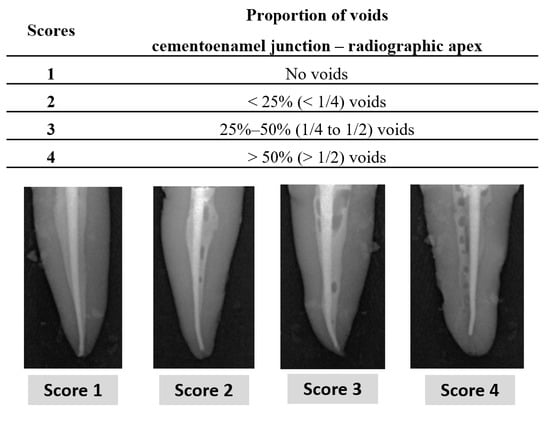
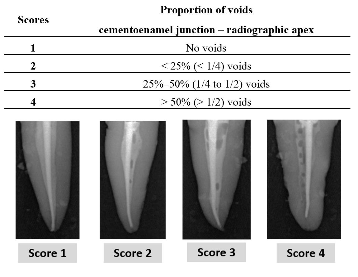
Immediately after obturation, all specimens were radiographed in both buccolingual and mesiodistal projections. The radiographs were independently evaluated by two experienced examiners who were blinded to group allocation. Obturation quality was assessed by analyzing the proportion of voids extending from the cementoenamel junction to the radiographic apex. The radiographic scoring system (Figure 1), based on the proportion of voids along the filling, was adopted from Galvão et al. [17].
Figure 1.
Radiographic scoring system and representative examples used for the evaluation of voids within root canal fillings. Scores were assigned according to the proportion of voids extending from the cementoenamel junction to the radiographic apex.
Radiographs were acquired using a Trophy IRIX 70 X-ray unit (70 kV, 8 mA, 0.2 s). All specimens were positioned on the same support at a fixed 10-cm source-to-sensor distance to standardize image acquisition. Images were subsequently analyzed using VistaSoft software (version 2.4.1; Dürr Dental, Bietigheim-Bissingen, Germany). Each specimen received a single radiographic score based on the evaluation of both projections; when the two projections showed different amounts of voids, the final score was assigned by consensus between the two examiners.
2.5.2. Cross-Sectional Evaluation
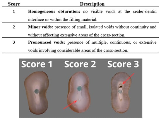
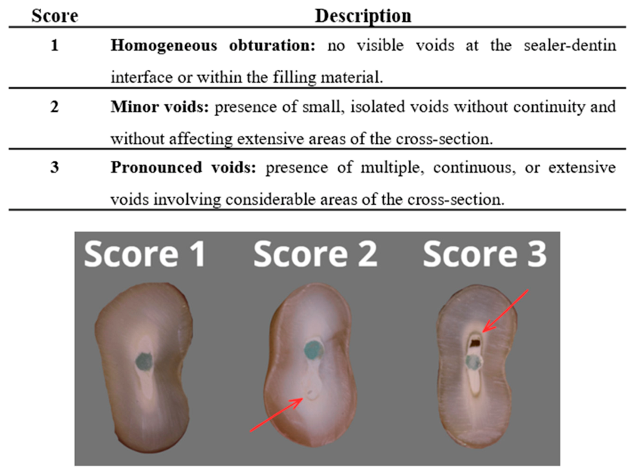
After the radiographic evaluation, the access cavities were sealed with a temporary restorative material (IRM; Dentsply Sirona), and the specimens were stored at 37 °C and 100% relative humidity for 5 weeks. This storage period exceeded the 28-day interval previously used to characterize the hydration and setting behavior of BC Universal Sealer under humid conditions [14]. The roots were then sectioned perpendicular to their long axis at 3, 6 and 9 mm from the apex using a low-speed diamond disc under water cooling (section thickness: 2.0 ± 0.1 mm), in order to obtain sections representing the apical, middle and coronal levels of the apical 9 mm of the root canal [18]. The obtained sections were photographed with a digital camera (Canon EOS 1000D, Canon, Tokyo, Japan) equipped with a 100-mm macro lens (Canon EF 100 mm f/2.8L) mounted on a tripod. Each section was individually assessed for the presence of voids either within the obturation material or at the interface between the filling material and the root canal walls. Voids formation was evaluated using a scoring system (Figure 2). A semiquantitative ordinal scale was chosen to allow standardized comparison of void formation between groups rather than precise quantification of void dimensions. All images were independently assessed by two experienced examiners who were blinded to the experimental groups.
Figure 2.
Representative cross-sectional images illustrating the scoring system used to evaluate obturation quality. Score 1: homogeneous filling with no visible voids. Score 2: presence of small isolated voids (arrow). Score 3: presence of extensive voids involving a larger area of the cross-section (arrow).
2.6. Statistical Analysis
Statistical analysis was performed using IBM SPSS Statistics software (version 30.0; IBM Corp., Armonk, NY, USA). Inter-examiner agreement was assessed using linear-weighted Cohen’s kappa on the independent scores of the two examiners. Disagreements were resolved by discussion between the two examiners until consensus was reached. Descriptive statistics were performed for each group to characterize the distribution of the assigned scores. Lower scores indicated better obturation quality, reflecting a lower prevalence of voids. For the cross-sectional evaluation, the scores of each root third (coronal, middle, and apical) were analyzed separately, and the mean score of the three thirds was calculated for each specimen. Differences among the four experimental groups were assessed with the Kruskal–Wallis test. When statistically significant differences were detected, pairwise comparisons were performed using Dunn’s post hoc test with Bonferroni correction, and Bonferroni-adjusted p-values are reported. The level of statistical significance was set at α = 0.05.
3. Results
3.1. Radiographic Evaluation
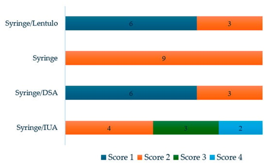
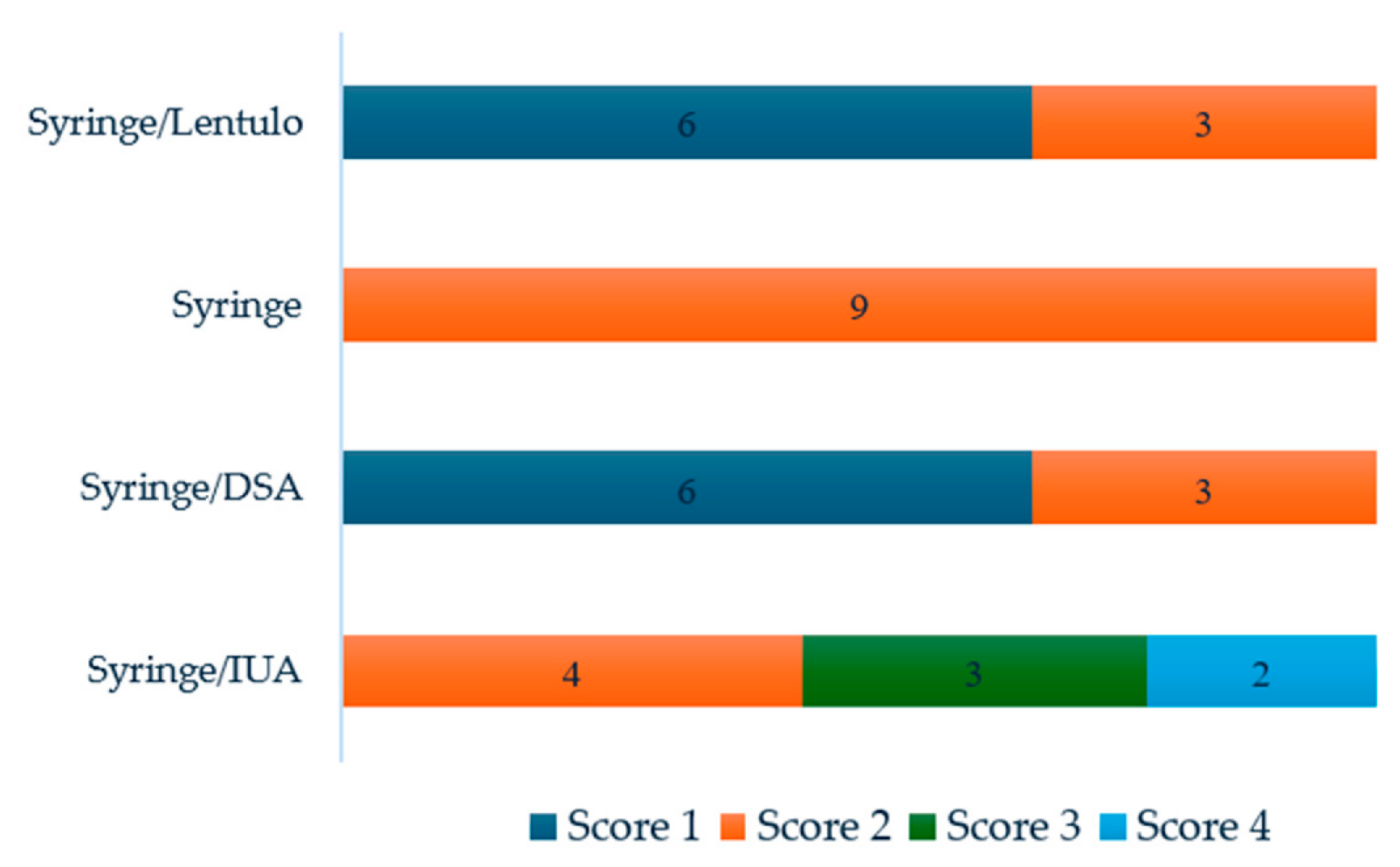
Inter-examiner agreement was almost perfect (weighted κ = 0.813; 95% CI: 0.648–0.978). Score distribution differed across the four experimental groups (Figure 3). Lower scores (Scores 1 and 2) indicate a lower proportion of voids.
Figure 3.
Distribution of obturation quality scores across the four experimental groups.
The Kruskal–Wallis test revealed significant differences among groups (H(3) = 20.771, p < 0.001). In pairwise comparisons, the Syringe + IUA group had significantly higher scores than both the Syringe + DSA group (p = 0.001) and the Syringe + Lentulo group (p = 0.001). The syringe delivery alone group did not differ significantly from the Syringe + DSA group, the Syringe + Lentulo group (p = 0.128 for both), or the Syringe + IUA group (p = 0.823). No difference was observed between Syringe + DSA and Syringe + Lentulo (p = 1.000).
3.2. Cross-Sectional Evaluation
Inter-examiner agreement was almost perfect in all three root thirds (weighted κ = 0.867, 95% CI: 0.741–0.994 for the coronal third; 0.881, 95% CI: 0.747–1.000 for the middle third; and 0.898, 95% CI: 0.695–1.000 for the apical third).
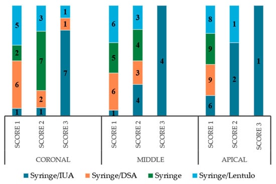
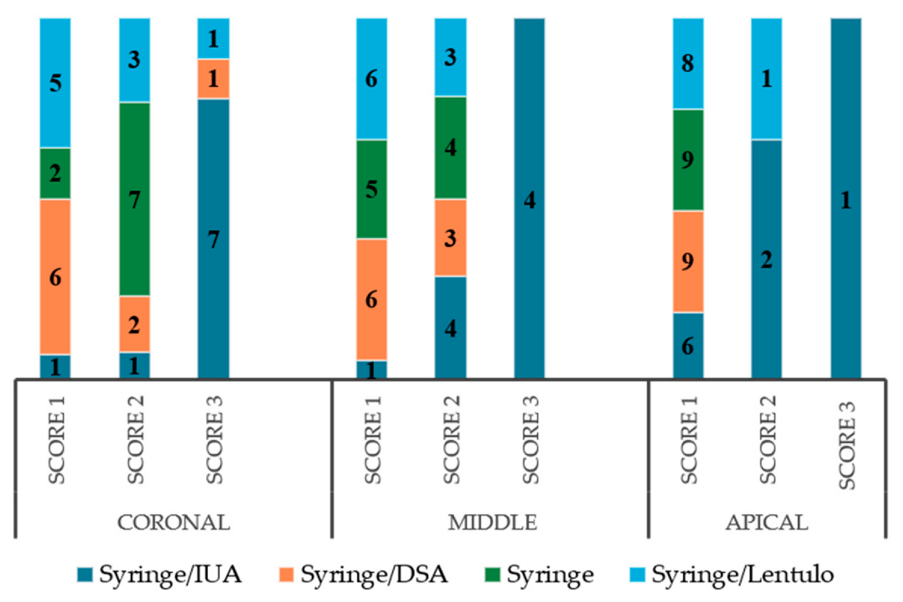
The distribution of obturation quality scores according to group and root third is presented in Figure 4.
Figure 4.
Distribution of filling-quality scores by experimental group and root third (coronal, middle, apical).
Significant differences among groups were found in the coronal (H(3) = 12.329, p = 0.006) and middle thirds (H(3) = 11.485, p = 0.009), but not in the apical third (H(3) = 6.668, p = 0.083). In both the coronal and middle thirds, the Syringe + IUA group had significantly higher scores than the Syringe + DSA group (p = 0.009 and 0.024, respectively) and the Syringe + Lentulo group (p = 0.025 and 0.024, respectively), whereas the syringe delivery alone group did not differ significantly from any other group (p ≥ 0.079). When the mean score of the three thirds was considered, significant differences were also found (H(3) = 15.081, p = 0.002): Syringe + IUA scored significantly higher than Syringe + DSA (p = 0.004) and Syringe + Lentulo (p = 0.006), with no significant differences among the remaining pairs (p ≥ 0.085).
4. Discussion
The present ex vivo study evaluated the influence of different sealer placement techniques on void formation in flattened root canals obturated with BC Universal Sealer. The results showed that the sealer placement technique significantly affected void formation, leading to rejection of the null hypothesis. Indirect ultrasonic activation (IUA) was consistently associated with a higher prevalence of voids than direct sonic agitation (DSA) and Lentulo-assisted placement, in both radiographic and cross-sectional assessments. In contrast, DSA using the EndoActivator and Lentulo-assisted placement produced the most favorable results. However, neither technique differed significantly from the syringe-only group.
The unfavorable performance of IUA contrasts with the findings of Kim et al. [10], who evaluated the premixed calcium silicate sealer Endoseal MTA in ribbon-shaped canals of maxillary premolars. In that study, micro-computed tomography showed no significant differences among groups, whereas cross-sectional evaluation revealed more voids in the single-cone group without ultrasonic activation than in the group in which ultrasonic energy was transmitted through the gutta-percha cone. The authors attributed this benefit to the vibration reducing friction between sealer particles and facilitating the escape of entrapped air bubbles. Differences in the assessment methods and sealer composition may account for the discrepant findings. In contrast, Shin et al. [8], using a nanoscale fluid filtration device to evaluate IUA with the premixed calcium silicate cement Endocem MTA, reported no significant reduction in void formation compared with the non-activated technique. Moreover, the ultrasonically activated specimens exhibited significantly greater leakage, indicating that improved energy transmission does not necessarily translate into enhanced sealing ability. Sfeir et al. [7] evaluated different placement techniques for TotalFill BC Sealer using micro-computed tomography and observed that both DSA and IUA tended to reduce void occurrence compared with conventional sealer placement, although these differences did not reach statistical significance.
The protocol of Kim et al. [10] was similar to the present one in the way ultrasonic energy was transmitted. The same ultrasonic unit and tip were used, and ultrasonic activation was applied at the same power setting to an instrument holding the master cone, namely a cotton plier in their study and metal tweezers in the present investigation. Consequently, vibration was transmitted indirectly to the sealer through the gutta-percha cone, and the activation time was also comparable. However, some relevant differences should be considered. In the study by Kim et al. [10], vibration was applied while the cone was gradually advanced to the working length, and the ribbon-shaped canals were prepared as two separate canals, each obturated with an individual cone. In contrast, in the present study, the cone was first seated to the full working length and only then activated, within a single flattened canal. These differences may help explain the divergent findings, although the underlying mechanisms were not assessed directly. First, because the master cone had already reached the working length when activation was performed, most sealer displacement had likely occurred before ultrasonic energy was applied. In a flattened canal, where a round gutta-percha cone contacts only a limited portion of the canal walls, vibration transmitted through a fully seated cone may be insufficient to redistribute the sealer into the buccolingual extensions. This differs from DSA, in which the sealer was agitated before insertion of the master cone and could therefore flow more freely throughout the canal space. Second, vibration of the cone against the canal walls may have displaced sealer from anatomical recesses or promoted the incorporation and coalescence of air bubbles which, in the closed-end model used in the present study, had no apical pathway for escape. Third, ultrasonic energy and the associated increase in temperature may alter the rheological behavior of the sealer, potentially increasing its viscosity or accelerating the initial stages of setting. Finally, Kim et al. [10], suggested that gentle ultrasonic activation may be necessary, since excessive ultrasonication may adversely affect void formation. The authors also suggested that vibration was attenuated as it passed through the ultrasonic tip, the plier, and the gutta-percha cone, thereby reducing the amplitude ultimately reaching the sealer. A similar attenuation likely occurred in the present study, where vibration was transmitted through the ultrasonic tip, the tweezers, and the cone. However, because the energy effectively delivered to the sealer was not measured in either investigation, it is not possible to determine whether comparable levels of ultrasonic energy reached the filling material.
Direct sonic agitation and Lentulo-assisted placement produced the lowest void scores in the present study, although neither differed significantly from the syringe-only group. Sonic agitation has been proposed to enhance irrigant distribution by generating hydrodynamic movement that may promote sealer flow along canal walls and into anatomical irregularities [19,20]. In the present study, agitation was performed directly on the sealer, before insertion of the master cone, which may have facilitated a more homogeneous sealer distribution and reduced air entrapment during obturation. This is consistent with Sfeir et al. [7], in whose study DSA with the EndoActivator yielded the lowest percentage of voids, although without statistically significant differences among groups. The Lentulo spiral may act through a different mechanism, as its rotational movement mechanically carries the sealer along the canal walls and into canal extensions that are difficult to reach with syringe delivery alone. Evidence on the use of Lentulo spirals with modern calcium silicate-based sealers remains limited, and the present findings do not allow conclusions regarding their superiority over syringe delivery.
The absence of significant differences between DSA, Lentulo-assisted placement, and the syringe-delivery group should be interpreted with caution. DSA and Lentulo-assisted placement consistently showed more favorable score distributions across the radiographic and cross-sectional evaluations. Although these differences did not reach statistical significance, they suggest a potential improvement in sealer distribution that warrants further investigation in future studies.
The discrepancies among studies may also be attributed to material-related and anatomical factors. Calcium silicate-based sealers differ considerably in particle size, viscosity, flow, setting behavior, and rheological response to mechanical agitation [4]. Therefore, the beneficial effect reported for one material cannot be assumed for another. This is particularly relevant because BC Universal Sealer is a recently introduced material for which limited scientific evidence is available [14]. In addition, root canal anatomy may have played a critical role. The present study was conducted in flattened canals, a configuration in which conventional gutta-percha cones contact only a limited portion of the canal walls, leaving buccolingual extensions highly dependent on sealer distribution. Previous investigations have evaluated different anatomical models [7,8,10], and the performance of a placement technique in one configuration may not be replicated in another. Another factor that may influence the behavior of calcium silicate-based sealers is the moisture condition within the root canal. Because these materials undergo a hydraulic setting reaction, the availability of residual moisture may affect their physicochemical behavior and interaction with the surrounding environment. Recently, Spinelli et al. [21] reported that moisture conditions influenced the short-term radiographic behavior of extruded NeoSealer Flo, highlighting the importance of moisture management when using premixed calcium silicate-based sealers. In the present study, canals were dried with paper points while avoiding excessive dentin dehydration. Nevertheless, the influence of different moisture levels on the distribution and adaptation of BC Universal Sealer was not investigated and warrants further research.
The differences observed between the techniques in the cross-sectional assessment may also be related to the anatomical characteristics of flattened canals. The coronal and middle thirds frequently contain buccolingual extensions that are not occupied by a round gutta-percha cone. Similar findings have been reported in previous studies. Celikten et al. [13] suggested that void formation in the apical region is influenced more strongly by canal anatomy than by the obturation technique itself, whereas Santos-Junior et al. [22] found significant differences among obturation techniques in the cervical and middle thirds of flattened canals but not in the apical third. Likewise, Moazami et al. [23] observed no significant differences among obturation techniques in the apical third when evaluating Endoseal MTA using cross-sectional analysis. The middle third of oval canals has also been identified as a particularly challenging region in studies evaluating other endodontic outcomes. In non-surgical retreatment of oval canals, greater amounts of residual filling material were observed in the middle third, where remnants tended to accumulate and extend into the buccolingual recesses [24]. Although a different outcome was investigated, these findings further highlight the influence of complex canal anatomy on endodontic procedures.
Although void formation is generally considered undesirable because it may compromise the seal of the root canal filling, the clinical significance of small differences in void prevalence remains unclear. In a recent 5-year randomized clinical trial comparing sealer-based obturation with a calcium silicate-based sealer and continuous wave condensation, the presence of voids was not significantly associated with treatment success. Therefore, while techniques that reduce void formation may improve the technical quality of the obturation, the extent to which these differences translate into improved long-term clinical outcomes remains to be established. Further long-term clinical studies are needed to clarify the relationship between obturation voids and treatment outcome [25].
Micro-computed tomography is widely regarded as one of the most comprehensive methods for evaluating root canal filling quality because it provides a non-destructive, three-dimensional assessment of void distribution [7,11]. However, the radiopacity of filling materials may interfere with the identification of small defects, and discrepancies between micro-computed tomography and direct microscopic examination of sectioned specimens have been reported. Notably, in the study by Kim et al. [10], micro-computed tomography detected no significant differences among techniques, whereas stereomicroscopic evaluation of the sections revealed significant differences in void occurrence. The authors attributed this to the limited resolution of micro-computed tomography for small voids and, alternatively, to material loss during sectioning, and recommended sectioning as a complementary approach. Methodological differences in void quantification may also influence the interpretation of results. Sfeir et al. [7] evaluated only open porosity, defined as voids at the interface between the filling material and the canal wall, excluding internal voids completely surrounded by filling material. In contrast, the radiographic and cross-sectional evaluations used in the present study did not distinguish between open and closed voids, so all detectable defects contributed to the assessment.
The strengths of this study include the selection of teeth according to previously described criteria for flattened canals, adherence to the PRILE guidelines, the use of two complementary evaluation methods, blinded and independent assessment by two examiners, and the comparison of four clinically relevant techniques under identical experimental conditions. Some limitations should nevertheless be acknowledged. As an ex vivo investigation, the study cannot fully reproduce clinical conditions, and the closed-end silicone model does not reproduce periapical tissues, which may influence sealer backflow and air entrapment. The ordinal scoring system introduces a degree of subjectivity, although inter-examiner agreement was high. In particular, the cross-sectional categories were defined qualitatively, without numerical thresholds. Internal voids and voids at the sealer–dentin interface were scored together, although they may have different implications for sealing ability, and some defects may have been produced during sectioning.
Future studies should investigate sealer placement techniques in other anatomical configurations, including curved canals and canals with isthmuses, and should determine the optimal parameters for ultrasonic and sonic activation with different calcium silicate-based sealers.
5. Conclusions
Within the limitations of this study, IUA of BC Universal Sealer was associated with more voids than DSA and Lentulo spiral placement in flattened root canals, whereas none of the techniques differed significantly from syringe delivery alone.
Supplementary Materials
The following supporting information can be downloaded at: https://www.mdpi.com/article/10.3390/app162111043/s1, Figure S1: PRILE flowchart illustrating the study design, experimental groups, outcome assessment methods, key findings, and conclusions.
Author Contributions
Conceptualization, I.P.-V. and I.F.; methodology, B.T.; formal analysis, A.C.B.; investigation, B.T. and I.F.; writing—original draft preparation, B.T. and I.F.; writing—review and editing, I.F., A.C.B. and I.P.-V.; supervision, I.F.; All authors have read and agreed to the published version of the manuscript.
Funding
This research received no external funding.
Institutional Review Board Statement
The study was conducted in accordance with the Declaration of Helsinki and approved by the Ethics Committee of the Faculty of Dental Medicine, University of Porto (approval no. 11/2025; approval date: 18 June 2025).
Informed Consent Statement
Informed consent was obtained from all subjects who donated the extracted teeth used in this study.
Data Availability Statement
The data presented in this study are available from the corresponding author upon reasonable request.
Conflicts of Interest
The authors declare no conflicts of interest.
References
- Ng, Y.L.; Mann, V.; Rahbaran, S.; Lewsey, J.; Gulabivala, K. Outcome of primary root canal treatment: Systematic review of the literature—Part 2. Influence of clinical factors. Int. Endod. J. 2008, 41, 6–31. [Google Scholar] [CrossRef] [Scilit] [PubMed]
- Tait, C.; Camilleri, J.; Blundell, K. Non-surgical endodontics—Obturation. Br. Dent. J. 2025, 238, 487–496. [Google Scholar] [CrossRef] [Scilit] [PubMed]
- Sundqvist, G.; Figdor, D.; Persson, S.; Sjögren, U. Microbiologic analysis of teeth with failed endodontic treatment and the outcome of conservative re-treatment. Oral Surg. Oral Med. Oral Pathol. Oral Radiol. Endodontology 1998, 85, 86–93. [Google Scholar] [CrossRef] [Scilit] [PubMed]
- Donnermeyer, D.; Bürklein, S.; Dammaschke, T.; Schäfer, E. Endodontic sealers based on calcium silicates: A systematic review. Odontology 2019, 107, 421–436. [Google Scholar] [CrossRef] [Scilit] [PubMed]
- Pirani, C.; Camilleri, J. Effectiveness of root canal filling materials and techniques for treatment of apical periodontitis: A systematic review. Int. Endod. J. 2023, 56, 436–4454. [Google Scholar] [CrossRef] [Scilit] [PubMed]
- Komora, P.; Vámos, O.; Agócs, G.; Szentes, B.L.; Hegyi, P.; Szalai, E.; Wenning, A.S.; Varga, G.; Vág, J.; Kerémi, B. In Vitro Comparison of Void Formation Between Calcium Silicate-Based Sealers and AH Plus: A Systematic Review and Meta-Analysis. Dent. J. 2026, 14, 496. [Google Scholar] [CrossRef] [Scilit] [PubMed]
- Sfeir, G.; Bukiet, F.; Kaloustian, M.K.; Kharouf, N.; Slimani, L.; Casel, B.; Zogheib, C. Evaluation of the Impact of Calcium Silicate-Based Sealer Insertion Technique on Root Canal Obturation Quality: A Micro-Computed Tomography Study. Bioengineering 2023, 10, 1331. [Google Scholar] [CrossRef] [Scilit] [PubMed]
- Shin, B.; Shon, W.-J.; Yoo, Y.-J. Effect of indirect ultrasonic activation on the root canal obturation with premixed calcium silicate cement: An in vitro study. BMC Oral Health 2025, 25, 570. [Google Scholar] [CrossRef] [Scilit] [PubMed]
- Pauletto, G.; Padoin, Y.; de Pellegrin, S.F.; Bier, C.A.S. The use of sonic and ultrasonic activation of endodontic sealer to improve sealer filling quality in the root canal: A scoping review. Odontology 2024, 112, 657–671. [Google Scholar] [CrossRef] [Scilit] [PubMed]
- Kim, J.A.; Hwang, Y.C.; Rosa, V.; Yu, M.K.; Lee, K.W.; Min, K.S. Root Canal Filling Quality of a Premixed Calcium Silicate Endodontic Sealer Applied Using Gutta-percha Cone-mediated Ultrasonic Activation. J. Endod. 2018, 44, 133–138. [Google Scholar] [CrossRef] [Scilit] [PubMed]
- Tavares, K.; Pinto, J.C.; Santos-Junior, A.O.; Torres, F.F.E.; Guerreiro-Tanomaru, J.M.; Tanomaru-Filho, M. Micro-CT evaluation of filling of flattened root canals using a new premixed ready-to-use calcium silicate sealer by single-cone technique. Microsc. Res. Tech. 2021, 84, 976–981. [Google Scholar] [CrossRef] [Scilit] [PubMed]
- Versiani, M.A.; Carvalho, K.K.T.; Mazzi-Chaves, J.F.; Sousa-Neto, M.D. Micro-computed Tomographic Evaluation of the Shaping Ability of XP-endo Shaper, iRaCe, and EdgeFile Systems in Long Oval-shaped Canals. J. Endod. 2018, 44, 489–495. [Google Scholar] [CrossRef] [Scilit] [PubMed]
- Celikten, B.; Uzuntas, C.F.; Orhan, A.I.; Orhan, K.; Tufenkci, P.; Kursun, S.; Demiralp, K. Evaluation of root canal sealer filling quality using a single-cone technique in oval shaped canals: An In vitro Micro-CT study. Scanning 2016, 38, 133–140. [Google Scholar] [CrossRef] [Scilit] [PubMed]
- Raman, V.; Camilleri, J. Characterization and Assessment of Physical Properties of 3 Single Syringe Hydraulic Cement-based Sealers. J. Endod. 2024, 50, 381–388. [Google Scholar] [CrossRef] [Scilit] [PubMed]
- Nagendrababu, V.; Murray, P.E.; Ordinola-Zapata, R.; Peters, O.A.; Rôças, I.N.; Siqueira, J.F., Jr.; Priya, E.; Jayaraman, J.; Pulikkotil, S.J.; Camilleri, J.; et al. PRILE 2021 guidelines for reporting laboratory studies in Endodontology: A consensus-based development. Int. Endod. J. 2021, 54, 1482–1490. [Google Scholar] [CrossRef] [Scilit] [PubMed]
- Tavares, K.; Pinto, J.C.; Santos-Junior, A.O.; Esteves Torres, F.F.; Guerreiro-Tanomaru, J.M.; Tanomaru-Filho, M. Combination of a new ultrasonic tip with rotary systems for the preparation of flattened root canals. Restor. Dent. Endod. 2021, 46, e56. [Google Scholar] [CrossRef] [Scilit] [PubMed]
- Galvão, T.; Camargo, B.; Armada, L.; Alves, F. Efficacy of three methods for inserting calcium hydroxide-based paste in root canals. J. Clin. Exp. Dent. 2017, 9, e762–e766. [Google Scholar] [CrossRef] [Scilit] [PubMed][Green Version]
- Pérez-Alfayate, R.; Mercade, M.; Algar-Pinilla, J.; Cisneros-Cabello, R.; Foschi, F.; Cohen, S. Root canal filling quality comparison of a premixed calcium silicate endodontic sealer and different carrier-based obturation systems. J. Clin. Med. 2021, 10, 1271. [Google Scholar] [CrossRef] [Scilit] [PubMed]
- Aliberti, A.; Piscopo, M.; Gasparro, R.; Sammartino, G.; Trosino, O.; Riccitiello, F.; Ausiello, P. Modern Endodontic Irrigation and Activation: From Disinfection to Dentin Substrate Conditioning—A Narrative Review. Appl. Sci. 2026, 16, 4593. [Google Scholar] [CrossRef] [Scilit]
- Ferreira, I.; Babo, P.S.; Braga, A.C.; Gomes, M.E.; Pina-Vaz, I. Effect of Sonic Agitation of a Binary Mixture of Solvents on Filling Remnants Removal as an Alternative to Apical Enlargement-A Micro-CT Study. J. Clin. Med. 2020, 9, 2465. [Google Scholar] [CrossRef] [Scilit] [PubMed]
- Spinelli, A.; Pergola, V.; Prati, C.; Gandolfi, M.G.; Zamparini, F. Short-term radiographic changes of extruded NeoSealer Flo in a simulated enlarged-apex model: Influence of moisture and periapical support. Clin. Oral Investig. 2026, 30, 499. [Google Scholar] [CrossRef] [Scilit] [PubMed]
- Santos-Junior, A.O.; Tanomaru-Filho, M.; Pinto, J.C.; Tavares, K.I.M.C.; Torres, F.F.E.; Guerreiro-Tanomaru, J.M. Effect of obturation technique using a new bioceramic sealer on the presence of voids in flattened root canals. Braz. Oral Res. 2021, 35, e028. [Google Scholar] [CrossRef] [Scilit] [PubMed]
- Moazami, F.; Naseri, M.; Malekzadeh, P. Different application methods for endoseal MTA sealer: A comparative study. Iran. Endod. J. 2020, 15, 44. [Google Scholar] [CrossRef] [Scilit] [PubMed]
- Spinelli, A.; Zamparini, F.; Buonavoglia, A.; Pisi, P.; Gandolfi, M.G.; Prati, C. Reciprocating System for Secondary Root Canal Treatment of Oval Canals: CBCT, X-rays for Remnant Detection and Their Identification with ESEM and EDX. Appl. Sci. 2022, 12, 11671. [Google Scholar] [CrossRef] [Scilit]
- Seog, M.; Kwon, S.K.; Kim, D.; Shin, S.J.; Jung, I.Y. Five-Year Outcomes of Root Canal Treatment Comparing Sealer-Based Obturation with Calcium Silicate-Based Sealer to Continuous Wave of Condensation: A Randomized Controlled Trial. J. Endod. 2025, 51, 538–546. [Google Scholar] [CrossRef] [Scilit] [PubMed]
Disclaimer/Publisher’s Note: The statements, opinions and data contained in all publications are solely those of the individual author(s) and contributor(s) and not of MDPI and/or the editor(s). MDPI and/or the editor(s) disclaim responsibility for any injury to people or property resulting from any ideas, methods, instructions or products referred to in the content. |
© 2026 by the authors. Licensee MDPI, Basel, Switzerland. This article is an open access article distributed under the terms and conditions of the Creative Commons Attribution (CC BY) license.