


Abstract
This in vitro study evaluated the effects of remineralizing products on enamel microhardness and the shear bond strength (SBS) of composite resin to enamel. A total of 110 human premolars were allocated to five groups, with 100 teeth allocated for microhardness and SBS testing (n = 20 per group) and 10 for scanning electron microscopy (SEM; n = 2 per group). The groups were sound enamel, demineralized enamel, casein phosphopeptide–amorphous calcium phosphate with fluoride (CPP-ACPF), 5% sodium fluoride (NaF), and NovaMin. Enamel demineralization was induced using 0.5% citric acid twice daily for three days. NaF varnish was applied once for 5 min, whereas CPP-ACPF- and NovaMin-containing products were applied twice daily for seven days. Composite bonding was performed using Clearfil S3 Bond Universal. Enamel microhardness, SBS, and failure modes were assessed using Vickers hardness testing, a universal testing machine, and stereomicroscopy, respectively. Demineralized enamel exhibited the lowest microhardness (202.61 ± 15.77 VHN) and SBS (11.81 ± 3.69 MPa). All remineralizing products significantly increased both parameters compared with demineralized enamel (p < 0.05). However, microhardness remained significantly lower than that of sound enamel (p < 0.001). SBS in the NaF group did not differ significantly from that of sound enamel (p = 0.175), whereas the CPP-ACPF and NovaMin groups exhibited significantly lower values (p < 0.05). No significant SBS differences were observed among the remineralizing product groups (p > 0.05). Adhesive failure was most frequent in the demineralized enamel group (50%), although the overall distribution of failure modes did not differ significantly among the groups (p > 0.05). The tested remineralizing products improved enamel microhardness and immediate composite bond strength compared with demineralized enamel, without significant differences in SBS among the products.
1. Introduction
Demineralization of dental hard tissues occurs when acid exposure disrupts the balance between mineral dissolution and deposition, resulting in progressive mineral loss. Acidic challenges may arise from bacterial metabolism or dietary sources, potentially compromising enamel integrity and mechanical properties. Therefore, preventing enamel demineralization and promoting remineralization are important objectives of contemporary minimally invasive dentistry [1]. Under normal oral conditions, demineralization and remineralization exist in a dynamic balance. However, when the local pH decreases below the critical threshold, mineral dissolution may exceed mineral deposition. Remineralization involves the deposition of calcium and phosphate ions within demineralized enamel, contributing to mineral recovery and improved mechanical properties [2]. Beyond conventional remineralization strategies, biomimetic approaches have demonstrated that calcium phosphate-based mineral deposition can contribute to the reconstruction of enamel-like structures [3].
Various agents have been developed to enhance enamel remineralization. Among these, fluoride remains the most widely used and extensively investigated remineralization agent [4]. Topical fluoride products, including toothpastes, mouthwashes, and varnishes, are commonly recommended for the prevention and management of early enamel lesions [5,6]. Fluoride promotes remineralization and reduces enamel solubility by facilitating the formation of fluoridated apatite. Compared with hydroxyapatite, fluoridated apatite exhibits greater resistance to acidic conditions [6]. Although fluoride is effective and safe when appropriately administered, excessive exposure may increase the risk of dental fluorosis, particularly during tooth development [7]. Consequently, alternative and complementary remineralization agents have received increasing attention.
Casein phosphopeptide–amorphous calcium phosphate (CPP-ACP) is a remineralization system that stabilizes calcium and phosphate ions, maintaining their availability for mineral deposition on enamel surfaces [8]. The incorporation of fluoride into this system results in casein phosphopeptide–amorphous calcium phosphate with fluoride (CPP-ACPF), which provides calcium, phosphate, and fluoride ions simultaneously [9,10,11]. The availability of these ions may promote mineral deposition and the formation of more acid-resistant fluoridated apatite structures, potentially contributing to enamel remineralization [12].
Calcium sodium phosphosilicate (NovaMin) is another bioactive material investigated for enamel remineralization. Upon contact with an aqueous environment, NovaMin undergoes ion exchange, releasing calcium, phosphate, and sodium ions while increasing the surrounding pH [13]. These reactions may promote the precipitation of a hydroxycarbonate apatite layer on the tooth surface. Because this mineral phase resembles the apatite found in natural dental tissues, NovaMin has been proposed as a potential agent for restoring mineral content in demineralized enamel [2,4,13].
The remineralization potential of these agents has been investigated using several analytical methods, including quantitative light-induced fluorescence [14,15], laser fluorescence [11], micro-computed tomography [16,17], and surface microhardness measurements [4,17]. Among these methods, the Vickers microhardness test is frequently used to assess changes in the mechanical properties of enamel following demineralization and remineralization procedures. Since enamel microhardness is closely associated with its mineral content, changes in hardness may provide an indirect indication of mineral loss or recovery [18]. Previous investigations of biomimetic remineralization strategies have also highlighted the importance of material composition and enamel surface conditions in determining mineralization outcomes [19,20]. However, previous studies have reported inconsistent findings concerning the relative effectiveness of different remineralization agents, potentially due to variations in enamel substrates, demineralization models, application protocols, and adhesive strategies [21].
In addition to their effects on enamel mechanical properties, the influence of remineralization treatments on subsequent adhesive procedures is clinically relevant, as differences in adhesive strategies may contribute to variations in bond strength outcomes reported across studies. Changes in enamel surface characteristics following remineralization may affect acid-etching patterns, resin infiltration, and the resulting bond strength. This consideration is particularly important when restorative or orthodontic bonding procedures are required after remineralization treatment [22,23,24,25]. Although the potential benefits of fluoride varnish, CPP-ACPF, and NovaMin on enamel microhardness have been widely investigated, it remains unclear whether these benefits are accompanied by corresponding improvements in composite resin bond strength. Understanding this relationship is particularly relevant when adhesive restorations are planned following remineralization treatment.
Therefore, the aim of this in vitro study was to evaluate the effects of three remineralizing products (5% NaF varnish, CPP-ACPF, and NovaMin) on enamel surface microhardness and the shear bond strength of composite resin to enamel subjected to an erosive demineralization challenge.
The null hypotheses were that (1) the different remineralizing products would not significantly affect enamel surface microhardness and (2) the different remineralizing products would not significantly affect the shear bond strength of composite resin to enamel.
2. Materials and Methods
2.1. Study Design and Sample Size Calculation
This in vitro study was approved by the Ordu University Non-Interventional Clinical Research Ethics Committee (Approval No. 220, 20 December 2024) and conducted in accordance with the principles of the Declaration of Helsinki. Written informed consent was obtained from all participants before tooth extraction, permitting the use of their extracted teeth for research purposes.
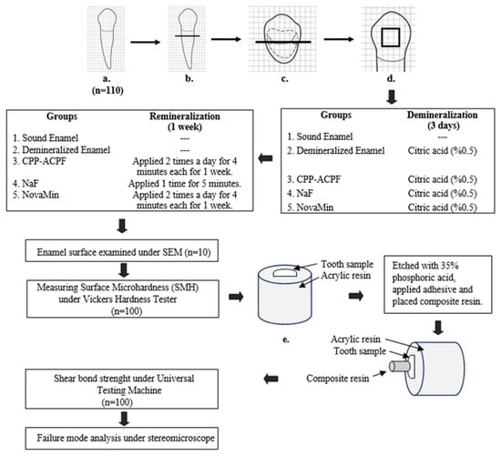
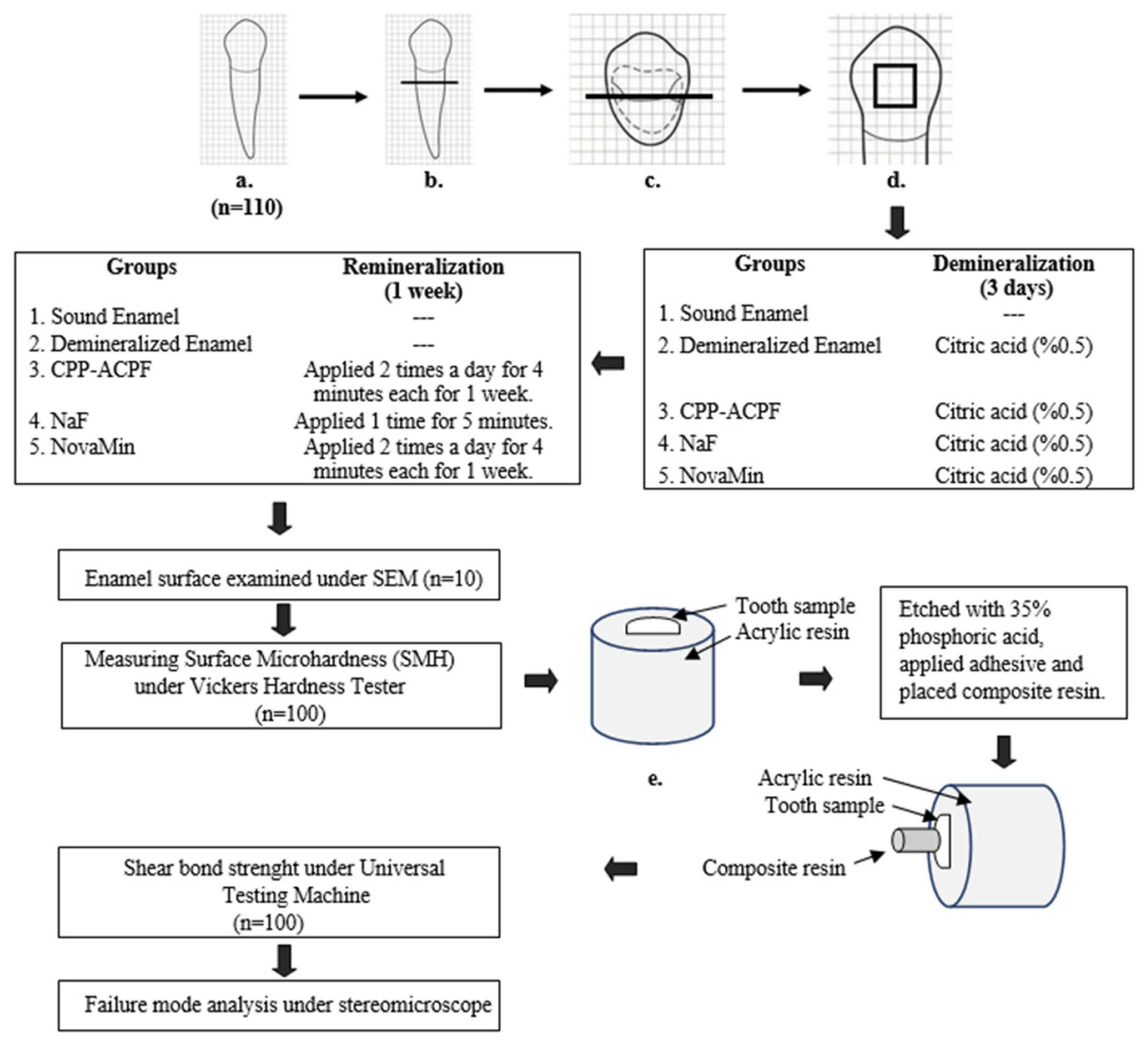
The sample size was calculated using G*Power software (version 3.1.9.7; Heinrich Heine University, Düsseldorf, Germany). Based on a one-way analysis of variance (ANOVA), an effect size of 0.40, a significance level of 0.05, and a statistical power of 85%, the minimum required sample size was determined to be 90 specimens (n = 18 per group). To account for potential specimen loss during preparation and testing, the sample size was increased to 100 specimens (n = 20 per group), with an additional 10 specimens (n = 2 per group) being allocated to qualitative SEM evaluation, resulting in a total of 110 teeth. A schematic overview of the experimental design is presented in Figure 1.
Figure 1.
Summary of the study design.(a) Extracted premolars; (b) sectioning 3 mm below the cementoenamel junction; (c) buccolingual sectioning of crowns; (d) standardization of the enamel test surface; (e) specimens embedded in acrylic resin.
2.2. Specimen Preparation
A total of 110 sound human premolars extracted exclusively for orthodontic purposes were obtained from 110 donors aged 13–18 years, with only one tooth collected from each donor. Ten teeth were reserved for qualitative surface evaluation using scanning electron microscopy (SEM; n = 2 per group), while the remaining 100 teeth were used for quantitative microhardness and shear bond strength (SBS) measurements (n = 20 per group).
All extracted teeth were examined under magnification. Teeth exhibiting caries, restorations, developmental enamel defects, hypoplasia, extraction-related damage, or signs of abrasion, erosion, or attrition were excluded. Teeth extracted for periodontal reasons were also excluded. Residual periodontal tissues and calculus were removed using an ultrasonic scaler. The teeth were subsequently polished with fluoride-free prophylaxis paste and disinfected in 0.5% chloramine-T solution for seven days [4]. The teeth were collected over approximately three months. Following disinfection, the specimens were stored in distilled water at 4 °C for a maximum of four months before preparation, with the storage medium replaced weekly, in accordance with ISO/TS 11405:2015 recommendations [26].
The teeth were sectioned approximately 3 mm below the cementoenamel junction using a diamond separating disc under continuous water cooling. Each tooth was then sectioned mesiodistally into buccal and lingual halves. Buccal specimens were allocated to SBS testing, whereas lingual specimens were used for microhardness measurements. This approach allowed microhardness and SBS measurements to be performed on separate enamel surfaces obtained from the same tooth.
Both buccal and lingual enamel surfaces were sequentially polished using silicon carbide abrasive papers (600–3000 grit), followed by water-based diamond polishing pastes (1–0.25 µm), to obtain standardized surfaces for shear bond strength and microhardness measurements, respectively [27]. Based on the average mesiodistal and cervico-occlusal dimensions of the collected premolars, a 3 × 3 mm area was identified as the maximum flat enamel surface that could be standardized across specimens. Two layers of acid-resistant nail varnish were applied to the surrounding enamel, leaving the designated experimental window exposed and preventing exposure of adjacent surfaces to the experimental treatments. For shear bond strength testing, the 3-mm-diameter composite cylinders were positioned centrally within the exposed window.
The specimens were randomly assigned to five experimental groups (n = 20 per group) using a computer-generated random number sequence without stratification:
Group 1 (Sound enamel): Positive control without demineralization.
Group 2 (Demineralized enamel): Negative control subjected to demineralization without subsequent remineralization treatment.
Group 3 (CPP-ACPF): Demineralized enamel treated with MI Paste Plus (GC, Leuven, Belgium).
Group 4 (NaF): Demineralized enamel treated with 5% sodium fluoride varnish (Profluorid Varnish, VOCO, Cuxhaven, Germany).
Group 5 (NovaMin): Demineralized enamel treated with Sensodyne Repair & Protect (GlaxoSmithKline, Brentford, UK).
2.3. Demineralization Protocol
Specimens in Group 1 were maintained as sound enamel controls and were not subjected to demineralization. Specimens in Groups 2–5 were exposed to 0.5% citric acid (pH 2.68) for 30 min twice daily over three consecutive days to induce erosive enamel demineralization, as described in previous enamel research [22]. Following each acid exposure, the specimens were rinsed with distilled water and stored in artificial saliva between demineralization cycles. After completion of the demineralization protocol, the specimens were evaluated using DIAGNOdent (KaVo, Biberach, Germany), with readings between 10 and 25 used as an additional assessment criterion, based on the approach described by Shirani et al. [28].
Artificial saliva was prepared by dissolving sodium chloride (NaCl, 1.280 g/L), magnesium chloride hexahydrate (MgCl2·6H2O, 0.125 g/L), potassium chloride (KCl, 0.095 g/L), potassium acetate (CH3COOK, 1.508 g/L), calcium chloride (CaCl2, 0.167 g/L), potassium phosphate tribasic trihydrate (K3PO4·3H2O, 0.386 g/L), and phosphoric acid (H3PO4, 0.05 mL/L) in distilled water to obtain a final volume of 1000 mL. The final pH was adjusted to 6.97 ± 0.2 [29].
The pH of the solutions was monitored every 24 h using a digital pH meter (Mettler-Toledo GmbH, Schwerzenbach, Switzerland). All solutions were renewed daily throughout the experimental period.
2.4. Application of Remineralization Agents
The remineralizing products and their compositions are presented in Table 1.
Table 1.
Remineralizing products used in the study.
Following the demineralization procedure, specimens in Group 2 received no remineralization treatment and served as the negative control. Throughout the seven-day experimental period, specimens in Groups 1 and 2 were maintained in artificial saliva at 37 °C without the application of any remineralizing products.
In Group 3 (CPP-ACPF), MI Paste Plus (GC, Leuven, Belgium) was applied to the exposed enamel surfaces using a microbrush to form a thin layer covering the entire exposed area. The product was applied for 4 min twice daily for seven consecutive days, according to the manufacturer’s instructions. Following each application, the paste was rinsed off with distilled water.
In Group 4 (NaF), 5% sodium fluoride varnish (Profluorid Varnish, VOCO, Cuxhaven, Germany) was applied once to the demineralized enamel surfaces using a microbrush in a thin layer sufficient to cover the entire exposed enamel area. The varnish was left undisturbed for 5 min, after which its surface was moistened with distilled water to facilitate setting, without removing the varnish [30]. The specimens were then stored in artificial saliva at 37 °C until completion of the seven-day experimental period.
In Group 5 (NovaMin), Sensodyne Repair & Protect toothpaste (GlaxoSmithKline, Brentford, UK) was mixed with deionized water at a ratio of 1:3 using a laboratory blender (FTDS11, SCI Finetech, Seoul, Republic of Korea) [31]. Each specimen was immersed in 2 mL of the prepared suspension for 4 min twice daily for seven consecutive days.
Following each CPP-ACPF and NovaMin treatment, the specimens were rinsed with distilled water and stored in artificial saliva at 37 °C. CPP-ACPF and NovaMin treatments were performed twice daily for seven consecutive days, whereas NaF varnish was applied only once. At the end of the seven-day experimental period, the specimens were cleaned using an electric toothbrush (Oral-B Precision Clean, Procter & Gamble, Cincinnati, OH, USA) and distilled water for 10 s per specimen, with an estimated brushing force of approximately 2 N, to remove residual deposits. Following toothbrushing, the specimen surfaces were covered with moist cotton to prevent dehydration until the adhesive procedure. Adhesive procedures were performed 24 h after completion of the seven-day treatment period.
2.5. Scanning Electron Microscopy
Following completion of the experimental protocols, enamel surface morphology was qualitatively assessed using SEM. Two specimens from each experimental group (n = 10 in total) were coated with gold–palladium (Au–Pd) to improve electrical conductivity and minimize charging artifacts.
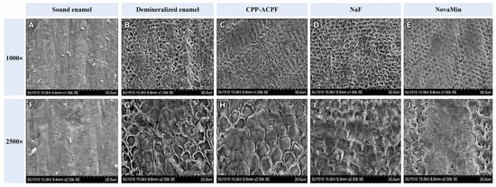
The specimens were examined with a scanning electron microscope (SU-1510, Hitachi, Tokyo, Japan) at an accelerating voltage of 15 kV, with secondary electron (SE) imaging under high-vacuum conditions. The working distance ranged from 8.0 to 9.6 mm, and images were obtained at magnifications of 1000× and 2500×. Representative SEM images are presented in Figure 2.
Figure 2.
Representative scanning electron microscopy (SEM) images of enamel surfaces in the experimental groups at 1000× (A–E) and 2500× (F–J) magnifications: sound enamel (A,F), demineralized enamel (B,G), CPP-ACPF (C,H), NaF varnish (D,I), and NovaMin (E,J). Scale bars: 50 µm (A–E) and 20 µm (F–J).
2.6. Surface Microhardness Measurement
Lingual enamel specimens were fixed to wax blocks with their polished surfaces positioned parallel to the indenter. Surface microhardness was measured using a Vickers hardness tester (Duramin, Struers, Germany), whose calibration was verified with a reference hardness block before testing.
A load of 300 g (2.94 N) was applied for 15 s at 40× magnification. Three indentations were made at standardized locations on each specimen, including the right, central, and left regions of the exposed enamel surface, with a minimum center-to-center distance of 200 µm. Irregular or defective indentations were excluded and repeated. The mean of the three valid measurements was calculated and expressed as the Vickers hardness number (VHN). All measurements were performed by an investigator blinded to the experimental group assignments.
2.7. Shear Bond Strength Test
Buccal enamel specimens were embedded in acrylic resin, ensuring that the exposed enamel surfaces were positioned parallel to the loading axis. All specimen preparation and adhesive procedures were performed by a single operator to ensure procedural consistency.
The enamel surfaces were etched with 35% phosphoric acid (K-Etchant Syringe, Kuraray Noritake, Tokyo, Japan), rinsed, and air-dried according to the manufacturer’s instructions. Subsequently, a universal adhesive system (Clearfil S3 Bond Universal, Kuraray Noritake, Tokyo, Japan) was applied according to the manufacturer’s instructions and light-cured for 10 s using an LED curing unit (Elipar S10, 3M ESPE, St. Paul, MN, USA, 1200 mW/cm2) after verifying the light irradiance with a radiometer.
A transparent polyethylene tube with an internal diameter of 3 mm and a height of 2 mm was positioned perpendicular to each enamel surface, providing a bonding area of 7.07 mm2. Composite resin (Clearfil Majesty Esthetic, Kuraray Noritake, Tokyo, Japan) was inserted into the mold and light-cured for 20 s.
The compositions and application procedures of the phosphoric acid, adhesive system, and composite resin are provided in Table 2.
Table 2.
Information on materials used for shear bond strength testing.
Following polymerization, the specimens were stored in distilled water at 37 °C for 24 h. The polyethylene tubes were then carefully removed using a scalpel.
SBS was measured using a universal testing machine (Autograph AGS-X, Shimadzu Corporation, Kyoto, Japan). A 0.5-mm-thick loading blade was positioned as close as possible to the composite–enamel interface, and shear force was applied at a crosshead speed of 0.5 mm/min until failure occurred.
The SBS values were calculated by dividing the maximum failure load (N) by the bonded surface area (mm2) and were expressed in megapascals (MPa).
2.8. Failure Mode Analysis
Following SBS testing, the fractured surfaces were examined using a stereomicroscope (SMZ25, Nikon, Tokyo, Japan) at 30× magnification. Failure modes were classified as adhesive (<20%), cohesive (>80%), or mixed (20–80%) based on the amount of composite resin remaining on the enamel surface and the location of fracture. All cohesive failures occurred within the composite resin, and no cohesive fractures within the enamel were observed. Before failure mode assessment, the examiner was calibrated through repeated evaluation of selected specimens to ensure consistency in classification. The examiner was blinded to group allocation throughout the evaluation.
2.9. Statistical Analysis
Statistical analyses were performed using SPSS software (version 22.0; IBM Corp., Armonk, NY, USA). The normality of the data was assessed using the Shapiro–Wilk test, and homogeneity of variance was evaluated using Levene’s test. Microhardness and SBS data satisfied the assumptions of normality and homogeneity of variance (p > 0.05). Therefore, differences among experimental groups were analyzed using one-way ANOVA, followed by Tukey’s post hoc test for pairwise comparisons. Additionally, mean differences with Tukey-adjusted 95% confidence intervals and Hedges’ g effect sizes were reported. Failure mode frequencies were analyzed using two-sided Fisher’s exact tests, comparing each group with the remaining groups combined for each failure category. Bonferroni correction was applied for 15 comparisons, with the adjusted significance threshold set at p < 0.0033. For all other statistical analyses, the significance level was set at α = 0.05.
3. Results
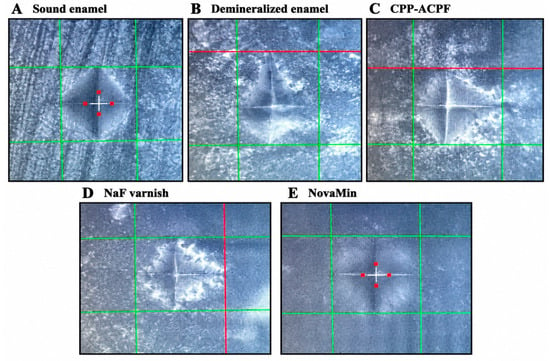
Table 3 presents the enamel surface microhardness values of all groups. A statistically significant difference in microhardness was observed among the groups (p < 0.001). In Group 2 (demineralized enamel; 202.61 ± 15.77), microhardness values were significantly lower than those of the sound enamel group and all remineralizing product groups (p < 0.001). Although microhardness values in the remineralizing product groups were significantly higher than those in demineralized enamel, they remained significantly lower than those in the sound enamel group (p < 0.001). Among the remineralizing product groups, the highest microhardness value was observed in Group 4 (NaF; 292.78 ± 15.12). No statistically significant difference was found between Group 4 (NaF) and Group 3 (CPP-ACPF; 280.04 ± 18.25) (p = 0.081). However, the microhardness value of Group 4 was significantly higher than that of Group 5 (NovaMin; 260.88 ± 16.55) (p < 0.001). Overall, the microhardness values followed the descending order: sound enamel > NaF > CPP-ACPF > NovaMin > demineralized enamel. Representative Vickers indentation images from each experimental group are presented in Figure 3.
Table 3.
Surface microhardness results and pairwise comparisons.
Figure 3.
Representative Vickers indentation images of the experimental groups: (A) sound enamel, (B) demineralized enamel, (C) CPP-ACPF, (D) NaF varnish, and (E) NovaMin. Images were selected from specimens exhibiting microhardness values close to the mean values of their respective groups.
Table 4 presents the shear bond strength (SBS) values for each group. Statistically significant differences in SBS were detected among the groups (p < 0.001). The lowest bond strength was observed in Group 2 (demineralized enamel; 11.81 ± 3.70), which was significantly lower than the values observed in the sound enamel and remineralizing product groups (p < 0.05). The highest bond strength was observed in Group 1 (sound enamel; 21.24 ± 3.84). In all remineralizing product groups, the SBS values were significantly higher than those of the negative control group (demineralized enamel) (p = 0.001 for CPP-ACPF; p < 0.001 for NaF and NovaMin). When compared with the positive control group, no statistically significant difference was observed between Group 4 (NaF) and sound enamel (p = 0.175). In contrast, Group 3 (CPP-ACPF) (p = 0.004) and Group 5 (NovaMin) (p = 0.024) exhibited significantly lower SBS values than sound enamel. No statistically significant differences in SBS were detected among the remineralizing product groups (p > 0.05).
Table 4.
Shear bond strength results and pairwise comparisons.
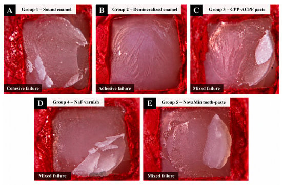
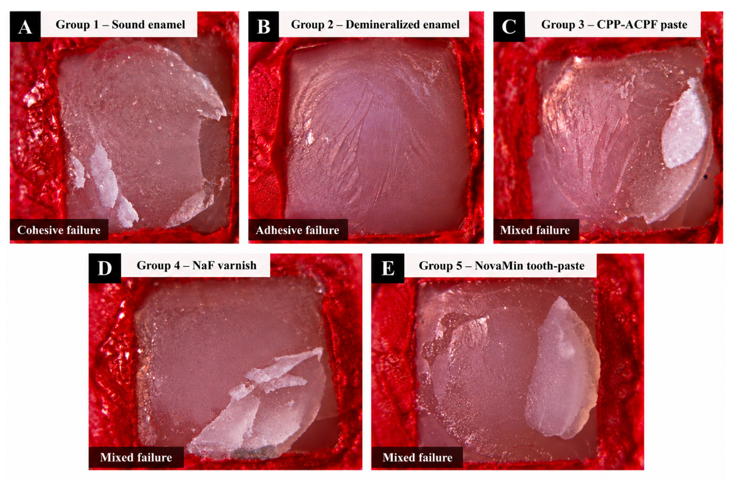
Table 5 summarizes the distribution of failure modes among the experimental groups. Adhesive failure was most frequent in Group 2 (demineralized enamel; 50%), whereas mixed failure predominated in the other groups. Stereomicroscopic examination revealed that all cohesive failures occurred within the composite resin, with no cohesive enamel fractures observed. Following reanalysis using two-sided Fisher’s exact tests, no statistically significant differences in failure mode frequencies were detected between individual groups and the remaining groups combined after Bonferroni correction (adjusted significance threshold: p < 0.0033). Selected stereomicroscopic images illustrating different failure modes in the experimental groups are presented in Figure 4.
Table 5.
Distribution of failure modes among experimental groups.
Figure 4.
Representative stereomicroscopic images illustrating different failure modes following shear bond strength testing: (A) sound enamel (cohesive failure), (B) demineralized enamel (adhesive failure), (C) CPP-ACPF (mixed failure), (D) NaF varnish (mixed failure), and (E) NovaMin (mixed failure).
SEM examination revealed morphological differences among the experimental groups (Figure 2). Sound enamel exhibited a relatively smooth and compact surface, whereas demineralized enamel showed an irregular, porous surface with prominent prism-like structures. Following treatment, enamel surfaces treated with CPP-ACPF exhibited granular surface features, NaF-treated surfaces exhibited irregular grooves and relatively compact areas, and NovaMin-treated surfaces showed a heterogeneous surface with residual porosities and smoother regions. These observations were qualitative and descriptive and did not provide direct evidence of mineral deposition or changes in enamel mineral composition.
4. Discussion
Various remineralizing approaches, including fluoride-based products, bioactive glasses, nanohydroxyapatite, and calcium phosphate systems, have been investigated to improve the properties of demineralized enamel [15,22]. These approaches may enhance enamel surface microhardness and resistance to acidic challenges by promoting conditions favorable for remineralization. Beyond their potential effects on enamel surface properties, their interaction with adhesive procedures is clinically relevant, particularly when composite restorations are planned following remineralizing treatments. Therefore, the bonding performance of treated enamel and the compatibility of remineralizing products with adhesive systems remain important considerations [22,31,32].
This study was conducted in vitro to ensure controlled and standardized experimental conditions. In vitro models are widely used to evaluate the efficacy of remineralizing products under reproducible conditions, typically using either bovine or human enamel. However, bovine enamel differs from human enamel in several aspects, including greater surface roughness, lower hardness, higher carbonate content, lower fluoride concentration, and smaller, irregular enamel prisms, all of which may influence experimental outcomes [33]. To avoid these potential confounding factors, human enamel specimens were selected in the present study.
Premolar teeth were selected because they are commonly extracted for orthodontic purposes and are generally free of caries and restorations, allowing the use of relatively standardized human enamel specimens. Furthermore, their availability facilitated specimen collection while minimizing prolonged storage before preparation. In the present study, erosive enamel demineralization was induced using repeated citric acid exposures with artificial saliva storage between cycles, based on a previously reported experimental approach [22].
In the first step of this study, the effects of remineralizing products on enamel surface microhardness were evaluated with a Vickers microhardness test, one of the most widely used and reliable methods for this purpose [18]. The findings of the study revealed that demineralization and remineralizing treatments significantly affected enamel microhardness; therefore, the first null hypothesis was rejected. Microhardness in demineralized enamel was significantly lower than that in sound enamel (p < 0.001). This outcome was expected due to the surface alterations associated with erosive demineralization, consistent with findings from previous studies [27,34]. SEM images (Figure 2B,G) showed an irregular, porous surface morphology with altered enamel prism structures following demineralization. However, these qualitative observations do not provide a direct quantitative assessment of mineral loss.
All three remineralizing products significantly increased enamel microhardness compared with demineralized enamel; however, values closest to those of sound enamel were observed in the NaF varnish group (Group 4). Similarly, Imren and Güven reported significantly greater surface microhardness recovery with conventional 5% NaF varnish than with the untreated control following citric acid-induced enamel erosion [30]. This outcome may be explained by the high fluoride concentration of NaF varnish (22,600 ppm), which may favor the formation of fluorapatite through the substitution of hydroxyl ions within enamel crystals. In addition, the deposition of calcium fluoride (CaF2) on the enamel surface at high fluoride concentrations (≥300 ppm) may have contributed to the observed increase in microhardness [35]. However, the formation of specific mineral phases was not directly investigated in the present study. Furthermore, the observed differences among the products may also be related to variations in their application protocols.
In the present study, microhardness in the CPP-ACPF group (Group 3) was significantly higher than that in demineralized enamel (p < 0.001), consistent with the findings of Shetty et al. [34], and no significant difference was found between the CPP-ACPF and NaF varnish groups (p = 0.081). This may be associated with the ability of GC MI Paste Plus®, a product containing CPP-ACPF, to release calcium and phosphate ions at the enamel surface, potentially creating conditions favorable for remineralization. In addition, its fluoride content (900 ppm) may contribute to fluoride-mediated mineral interactions [11,16].
NovaMin, one of the tested remineralizing components, crystallizes in an aqueous medium to form hydroxycarbonate apatite, which is chemically and structurally equivalent to hydroxyapatite [13]. Bologa et al. reported that a single application of NovaMin at concentrations higher than 3% can block at least 75% of the opened pores and resist acidic environmental factors [36]. In our study, the significantly higher microhardness in the NovaMin group (Group 5) compared with demineralized enamel supports the effectiveness of NovaMin in blocking the porous structure on the enamel surface. SEM observations of the NovaMin-treated enamel (Figure 2E,J) revealed a heterogeneous surface morphology with residual porosity, indicating that the surface alterations were not uniform.
Despite the significant improvements observed with all remineralizing products, microhardness values remained lower than those of sound enamel (p < 0.05). This finding may be related to the severity of the experimentally induced erosive challenge and the relatively short treatment period, as both the characteristics of the demineralization model and the duration of remineralizing treatments may influence enamel microhardness recovery [37]. Previous studies have also suggested that longer application periods may enhance remineralization outcomes [2]. Although the NovaMin group exhibited significantly lower surface microhardness values than the NaF and CPP-ACPF groups, this finding should not be interpreted as evidence of lower overall remineralization efficacy. Since subsurface mineral recovery and penetration depth were not evaluated, the comparison of remineralizing products was limited to changes in surface microhardness. Although DIAGNOdent measurements were used as an auxiliary criterion during specimen selection, they did not provide a quantitative assessment of enamel mineral loss or lesion depth.
Remineralizing products can alter enamel surface characteristics such as microstructure, mineral composition, and surface energy, all of which play critical roles in adhesive bonding [24]. However, studies on the bonding of adhesive resin to remineralized enamel remain limited [24,35,38,39]. In the second step of this study, the effect of remineralizing products on the bond strength of composite resin to enamel was investigated. The findings revealed significant differences in shear bond strength among the experimental groups, leading to the rejection of the second null hypothesis. The demineralized enamel group exhibited significantly lower bond strength than sound enamel (p < 0.001). This reduction in bond strength may be attributed to the loss of interprismatic minerals and alterations in enamel surface morphology, which may compromise the adhesion of adhesive monomers to demineralized enamel and reduce the bonding ability of adhesive resins [40]. SEM images revealed disruption of the enamel prism structure and morphological alterations in demineralized enamel (Figure 2B,G). In contrast, intact enamel displayed a homogeneous and smooth appearance (Figure 2A,F). Additionally, adhesive failure was numerically more frequent in demineralized enamel (50%), consistent with the lower bond strength values observed in this group.
All remineralizing products significantly increased bond strength compared with demineralized enamel, consistent with previous reports [22,24,41]. This improvement may be related to partial restoration of mineral content and surface integrity, which are essential for effective adhesive bonding [38].
In the present study, no significant difference was found among the remineralizing products in terms of bond strength (p > 0.05). Although the CPP-ACPF and NovaMin groups exhibited significantly lower SBS values compared with sound enamel, no statistically significant difference was observed between the NaF varnish and sound enamel groups (p = 0.175). Fluorapatite and calcium fluoride (CaF2) crystals have been reported to partially or completely fill interprismatic spaces, altering the enamel surface’s wettability. Such changes may reduce bond strength and increase susceptibility to water-related degradation [35,42]. Since all three remineralizing products tested in this study contain fluoride at varying concentrations (Table 1), fluoride-related mineral deposition may have influenced the SBS values observed in the treated groups. However, the formation of these mineral phases was not directly investigated in the present study.
A possible explanation for the bond strength values observed in the remineralizing product groups may be related to the etch-and-rinse adhesive protocol used in this study. Following remineralization, mineral deposition within enamel prisms has been reported to occur heterogeneously, with greater accumulation in the peripheral prism regions [43]. This differential mineral distribution may increase the contrast between prism core and periphery during phosphoric acid application, potentially resulting in a more pronounced Type I etching pattern and enhanced micromechanical retention [28]. The application of 35% phosphoric acid for 30 s may therefore have standardized the heterogeneous enamel surface created by remineralization and facilitated consistent resin infiltration across groups.
In addition, the etch-and-rinse system may effectively remove superficial calcium fluoride deposits, promote micro-retentive patterns, and improve adhesive wettability by reducing the contact angle between the adhesive and enamel surface [23,44]. Although the NaF varnish group exhibited SBS values comparable to those of sound enamel, the decrease in bond strength compared with sound enamel remained significant for the CPP-ACPF and NovaMin groups. This difference may be related to potential variations in the depth of mineral deposition associated with CPP-ACPF, NovaMin, and NaF treatments [45]. However, the penetration depth of these products was not directly evaluated in the present study. Since high fluoride concentrations have been reported to cause rapid mineral precipitation, leading primarily to superficial remineralization [46], the removal of superficial deposits during phosphoric acid etching may have contributed to the SBS values observed in the NaF varnish group.
Shalaby et al. reported that increasing nano-chitosan concentrations in glass-ionomer cement did not consistently improve bond strength, with higher concentrations resulting in reduced values [47]. Similarly, in the present study, NaF varnish produced the highest enamel microhardness among the remineralizing products but did not demonstrate significantly greater bond strength than the other remineralizing products. These findings suggest that improvements in material-related or enamel surface properties do not necessarily translate into proportional improvements in adhesive performance.
Arslandaş Dinçtürk et al., based on FE-SEM analysis, reported that self-etch application produced fewer surface alterations and shallower etching patterns compared with etch-and-rinse groups [48], and that limited resin tag formation and reduced micromechanical interlocking could potentially lead to lower SBS values [49]. Conversely, although the etch-and-rinse strategy may enhance micromechanical retention, it has also been reported to induce demineralization of the superficial enamel layer and increase long-term susceptibility to acid challenge and caries risk [50]. Therefore, some studies have proposed self-etch systems as a more conservative alternative capable of providing adequate adhesive bonding performance [50,51]. The absence of a direct comparison between adhesive strategies represents a limitation of this study.
The limitations of this study include the absence of thermal cycling, the lack of quantitative assessment of enamel mineral loss and lesion depth, and the absence of repeated toothbrushing simulation. Furthermore, SEM observations were qualitative, and no elemental or crystallographic analyses were performed to confirm mineral deposition or changes in enamel composition. Future studies incorporating SEM-EDX analysis may provide further insight into elemental changes associated with remineralizing treatments. Additionally, variations in demineralization protocols, remineralization durations, and adhesive strategies across studies complicate direct comparisons. Since SBS was evaluated after only 24 h of water storage, the findings reflect immediate bond strength and cannot establish long-term adhesive durability. Future studies incorporating artificial aging procedures are therefore necessary. Moreover, the remineralizing products were applied according to their respective recommended protocols. While NaF varnish was applied once, CPP-ACPF and NovaMin were applied twice daily for seven days. Therefore, differences in application frequency and fluoride concentration should be considered when interpreting the comparative effects of these products.
As all tested products contained fluoride and fluoride-free controls were not included, the individual contributions of CPP-ACP and NovaMin could not be isolated. Enamel polishing may have altered natural surface characteristics, and neither the thickness of the removed enamel layer nor surface roughness (Ra) was measured. The 1:3 dilution of the NovaMin-containing toothpaste may have influenced fluoride availability, and the immersion protocol did not fully reproduce clinical application conditions. The absence of baseline microhardness measurements, the use of a single adhesive system, the limited number of specimens examined by SEM, and the limited statistical power for failure mode comparisons should also be considered when interpreting the findings.
Another important limitation affecting the clinical applicability of the findings is the short treatment period. In the present study, the effects of short-term remineralizing treatments on erosively demineralized enamel were evaluated, which may be relevant when restorative treatment is planned following remineralization. However, in clinical practice, patients generally use remineralizing agents for longer periods, which may alter the effects of these pretreatments on bond strength.
Nevertheless, Oda and Güler reported that the application of remineralizing products at different time intervals (1, 15, and 30 days) did not produce a significant difference in SBS and that bonding performance remained stable over time [52]. Although longer clinical application periods may influence remineralization outcomes, further studies evaluating the effects of extended application durations on bond strength are required.
5. Conclusions
Within the limitations of this in vitro study, the following conclusions can be drawn:
- The tested remineralizing products significantly increased the surface microhardness of artificially demineralized enamel. However, microhardness values remained significantly lower than those of sound enamel.
- The tested remineralizing products significantly increased the shear bond strength of composite resin to demineralized enamel. However, no statistically significant differences in bond strength were observed among the remineralizing products.
- The observed changes in enamel surface microhardness and bond strength suggest that remineralizing treatments may influence the mechanical properties and adhesive performance of erosively demineralized enamel. However, these findings reflect short-term outcomes under the specific experimental conditions and application protocols evaluated.
Author Contributions
Conceptualization: A.K.B. and S.A.K.; Methodology: A.K.B. and S.A.K.; Investigation: A.K.B. and S.A.K.; Formal analysis: S.A.K.; Resources: A.K.B. and S.A.K.; Supervision: A.K.B. and S.A.K.; Writing—original draft preparation: A.K.B. and S.A.K.; Writing—review and editing: S.A.K. All authors have read and agreed to the published version of the manuscript.
Funding
This research received no external funding.
Institutional Review Board Statement
This study was approved by the Ordu University Non-Interventional Clinical Research Ethics Committee (Approval No. 220, 20 December 2024). Written informed consent was obtained from all participants prior to tooth extraction for the use of their extracted teeth in research.
Informed Consent Statement
Informed consent was obtained from all participants involved in the study.
Data Availability Statement
The data supporting the findings of this study are available from the corresponding author upon reasonable request.
Acknowledgments
The authors would like to thank the Department of Mechanical Engineering at Karadeniz Technical University for their technical support with the microhardness measurements.
Conflicts of Interest
The authors declare no conflicts of interest.
References
- Neel, E.A.A.; Aljabo, A.; Strange, A.; Ibrahim, S.; Coathup, M.; Young, A.M.; Bozec, L.; Mudera, V. Demineralization-remineralization dynamics in teeth and bone. Int. J. Nanomed. 2016, 11, 4743–4763. [Google Scholar] [CrossRef] [Scilit]
- Tulumbacı, F.; Oba, A.A. Efficacy of different remineralization agents on treating incipient enamel lesions of primary and permanent teeth. J. Conserv. Dent. 2019, 22, 281–286. [Google Scholar] [CrossRef] [Scilit]
- Shao, C.; Jin, B.; Mu, Z.; Lu, H.; Zhao, Y.; Wu, Z.; Yan, L.; Zhang, Z.; Zhou, Y.; Pan, H.; et al. Repair of Tooth Enamel by a Biomimetic Mineralization Frontier Ensuring Epitaxial Growth. Sci. Adv. 2019, 5, eaaw9569. [Google Scholar] [CrossRef] [Scilit]
- Shihabi, S.; AlNesser, S.; Comisi, J.C. Comparative Remineralization Efficacy of Topical NovaMin and Fluoride on Incipient Enamel Lesions in Primary Teeth: Scanning Electron Microscope and Vickers Microhardness Evaluation. Eur. J. Dent. 2021, 15, 420–424. [Google Scholar] [CrossRef] [Scilit]
- Sonesson, M.; Twetman, S.; Bondemark, L. Effectiveness of high-fluoride toothpaste on enamel demineralization during orthodontic treatment-a multicenter randomized controlled trial. Eur. J. Orthod. 2014, 36, 678–682. [Google Scholar] [CrossRef] [Scilit]
- Arifa, M.K.; Ephraim, R.; Rajamani, T. Recent Advances in Dental Hard Tissue Remineralization: A Review of Literature. Int. J. Clin. Pediatr. Dent. 2019, 12, 139–144. [Google Scholar] [CrossRef] [Scilit]
- Ullah, R.; Zafar, M.S.; Shahani, N. Potential fluoride toxicity from oral medicaments: A review. Iran. J. Basic Med. Sci. 2017, 20, 841–848. [Google Scholar] [CrossRef] [Scilit]
- Agob, J.N.; Aref, N.S.; Al-Wakeel, E.E.S. Effect of casein phosphopeptide-amorphous calcium phosphate on fluoride release and micro-shear bond strength of resin-modified glass ionomer cement in caries-affected dentin. Restor. Dent. Endod. 2018, 43, e45. [Google Scholar] [CrossRef] [Scilit]
- Tuloglu, N.; Bayrak, S.; Tunc, E.S.; Ozer, F. Effect of fluoride varnish with added casein phosphopeptide-amorphous calcium phosphate on the acid resistance of the primary enamel. BMC Oral Health 2016, 16, 103. [Google Scholar] [CrossRef] [Scilit]
- Tao, S.; Zhu, Y.; Yuan, H.; Tao, S.; Cheng, Y.; Li, J.; He, L. Efficacy of fluorides and CPP-ACP vs fluorides monotherapy on early caries lesions: A systematic review and meta-analysis. PLoS ONE 2018, 13, e0196660. [Google Scholar] [CrossRef] [Scilit]
- Dhanya, K.; Chandra, P.; Anandakrishna, L.; Karuveettil, V. A Comparison of NovaMin™ and Casein Phosphopeptide-Amorphous Calcium Phosphate Fluoride on Enamel Remineralization—An In vitro Study Using Scanning Electron Microscope and DIAGNOdent®. Contemp. Clin. Dent. 2021, 12, 301–307. [Google Scholar] [CrossRef] [Scilit]
- Fan, Y.; Sun, Z.; Moradian-Oldak, J. Effect of fluoride on the morphology of calcium phosphate crystals grown on acid-etched human enamel. Caries Res. 2009, 43, 132–136. [Google Scholar] [CrossRef] [Scilit]
- Khijmatgar, S.; Reddy, U.; John, S.; Badavannavar, A.N.; Souza, T.D. Is there evidence for Novamin application in remineralization?: A Systematic review. J. Oral Biol. Craniofac. Res. 2020, 10, 87–92. [Google Scholar] [CrossRef] [Scilit]
- Veeramani, R.; Shanbhog, R.; Bhojraj, N.; Kaul, S.; Anoop, N.K. Evaluation of mineral loss in primary and permanent human enamel samples subjected to chemical demineralization by international caries detection and assessment system II and quantitative light-induced fluorescence™: An in vitro study. J. Indian Soc. Pedod. Prev. Dent. 2020, 38, 355–360. [Google Scholar] [CrossRef] [Scilit]
- Krishnan, G.; George, S.; Anandaraj, S.; John, S.A.; Mathew, V.; Shanavas, N.M. Efficacy of Four Remineralizing Agents on Primary Teeth: In Vitro Evaluation Using Microhardness Testing and Quantitative Light-induced Fluorescence. Pediatr. Dent. 2017, 39, 233–237. [Google Scholar]
- İlisulu, S.C.; Gürcan, A.T.; Şişmanoğlu, S. Remineralization efficiency of three different agents on artificially produced enamel lesions: A micro-CT study. J. Esthet. Restor. Dent. 2024, 36, 1536–1546. [Google Scholar] [CrossRef] [Scilit]
- François, P.; Benoit, A.; Slimani, L.; Dufresne, A.; Gouze, H.; Attal, J.-P.; Mangione, F.; Dursun, E. In vitro remineralization by various ion-releasing materials of artificially demineralized dentin: A micro-CT study. Dent. Mater. 2024, 40, 520–526. [Google Scholar] [CrossRef] [Scilit]
- Sakar-Deliormanli, A.; Güden, M. Microhardnes’s and fracture toughness of dental materials by indentation method. J. Biomed. Mater. Res. B Appl. Biomater. 2006, 76, 257–264. [Google Scholar] [CrossRef] [Scilit]
- Guanipa Ortiz, M.I.; Gomes de Oliveira, S.; de Melo Alencar, C.; Baggio Aguiar, F.H.; Alves Nunes Leite Lima, D. Remineralizing effect of the association of nano-hydroxyapatite and fluoride in the treatment of initial lesions of the enamel: A systematic review. J. Dent. 2024, 145, 104973. [Google Scholar] [CrossRef] [Scilit]
- Seredin, P.; Goloshchapov, D.; Kashkarov, V.; Emelyanova, A.; Buylov, N.; Barkov, K.; Ippolitov, Y.; Khmelevskaia, T.; Mahdy, I.A.; Mahdy, M.A.; et al. Biomimetic Mineralization of Tooth Enamel Using Nanocrystalline Hydroxyapatite under Various Dental Surface Pretreatment Conditions. Biomimetics 2022, 7, 111. [Google Scholar] [CrossRef] [Scilit]
- Ahmed, T.; Fareen, N.; Alam, M.K. Effect of re-mineralizing surface treatment on the shear bond strength of orthodontic bracket: A systematic review and meta-analysis of in-vitro studies. Int. Orthod. 2023, 21, 100807. [Google Scholar] [CrossRef] [Scilit]
- Muntean, A.; Dârgău, C.M.; Pacurar, M.; Neagoe, S.; Delean, A.G. Effect of Remineralizing Agents on Shear Bond Strength of Orthodontic Brackets-In Vitro Study. Children 2023, 10, 268. [Google Scholar] [CrossRef] [Scilit]
- Alahdal, K.Y.; Almohareb, T.; Aldeeb, L.; Maawadh, A.; Alshamrani, A.S.; Alrahla, A. Use of Reparative Agents Topical Fluoride Activated by CO2 Laser and Curodont™ Repair and NR-5™ on Vickers Hardness and Micro-Shear Bond Strength of Eroded Enamel to Composite Restoration. Appl. Sci. 2023, 13, 12717. [Google Scholar] [CrossRef] [Scilit]
- Mobarak, E.H.; Ali, N.; Daifalla, L.E. Microshear Bond Strength of Adhesives to Enamel Remineralized Using Casein Phosphopeptide Agents. Oper. Dent. 2015, 40, E180–E188. [Google Scholar] [CrossRef] [Scilit]
- Daneshkazemi, P.; Sadeghian, S.; Khodaei, M. Shear bond strength of orthodontic brackets on intact and demineralized enamel after application of resin infiltrant, fluoride varnish and casein phosphopeptide-amorphous calcium phosphate remineralizing agents: In-vitro study. Int. Orthod. 2021, 19, 259–268. [Google Scholar] [CrossRef] [Scilit]
- ISO/TS 11405:2015; Dentistry—Testing of Adhesion to Tooth Structure. International Organization for Standardization (ISO): Geneva, Switzerland, 2015.
- Deswal, R.; Kukreja, N.; Chhabra, S.; Trivedi, S.; Sharma, A.; Thakur, A. Comparative Assessment of Enamel Microhardness Using Various Remineralising Agents on Artificially Demineralized Human Enamel. Cureus 2022, 14, e30281. [Google Scholar] [CrossRef] [Scilit]
- Shirani, F.; Malekipour, M.R.; Azary, M.; Manesh, V.S.; Gilani, A.; Mazdak, A. The effect of remineralization pretreatments on the enamel bond strength of demineralized and sound enamel: An in vitro study. Dent. Res. J. 2025, 22, 1. [Google Scholar] [CrossRef] [Scilit]
- Shannon, I.L. Fluoride treatment programs for high-caries-risk patients. Clin. Prev. Dent. 1982, 4, 11–20. [Google Scholar]
- Imren, E.; Güven, Y. Therapeutic and protective effects of light-cured varnishes on erosive lesions: An in vitro study. BMC Oral Health 2025, 25, 555. [Google Scholar] [CrossRef] [Scilit]
- Fernandes, N.L.S.; Juliellen, L.D.C.; Andressa, F.B.O.; D’Alpino, H.P.P.; Sampaio, C.F. Resistance against Erosive Challenge of Dental Enamel Treated with 1,450-PPM Fluoride Toothpastes Containing Different Biomimetic Compounds. Eur. J. Dent. 2021, 15, 433–439. [Google Scholar] [CrossRef] [Scilit]
- Skucha-Nowak, M.; Gibas, M.; Tanasiewicz, M.; Twardawa, H.; Szklarski, T. Natural and Controlled Demineralization for Study Purposes in Minimally Invasive Dentistry. Adv. Clin. Exp. Med. 2015, 24, 891–898. [Google Scholar] [CrossRef] [Scilit]
- Lippert, F.; Juthani, K. Fluoride dose-response of human and bovine enamel artificial caries lesions under pH-cycling conditions. Clin. Oral Investig. 2015, 19, 1947–1954. [Google Scholar] [CrossRef] [Scilit]
- Shetty, S.; Hegde, M.N.; Bopanna, T.P. Enamel remineralization assessment after treatment with three different remineralizing agents using surface microhardness: An in vitro study. J. Conserv. Dent. 2014, 17, 49–52. [Google Scholar] [CrossRef] [Scilit]
- Al-Qahtani, A.; Inoue, G.; Abdou, A.; Nikaido, T.; Tagami, J. Effects of potassium and sodium fluoride in different concentrations on micro-shear bond strength and inhibition of demineralization. Dent. Mater. J. 2021, 40, 356–363. [Google Scholar] [CrossRef] [Scilit]
- Bologa, E.; Stoleriu, S.; Nica, I.; Tărăboanță, I.; Georgescu, A.; Matei, R.I.; Andrian, S. The Effect of Three Desensitizing Toothpastes on Dentinal Tubules Occlusion and on Dentin Hardness. Biomedicines 2023, 11, 2464. [Google Scholar] [CrossRef] [Scilit]
- Eisenburger, M.; Addy, M.; Hughes, J.A.; Shellis, R.P. Effect of time on the remineralisation of enamel by synthetic saliva after citric acid erosion. Caries Res. 2001, 35, 211–215. [Google Scholar] [CrossRef] [Scilit]
- Abdelmegid, F.Y.; Salama, F.S.; Abouobaid, E.I.; Halawany, H.S.; Alhadlaq, M.K. Effect of Remineralizing Agents on Bond Strength of Resin-Composites to Primary Enamel. J. Clin. Pediatr. Dent. 2019, 43, 331–336. [Google Scholar] [CrossRef] [Scilit]
- Patel, D.; Sambrook, R.; Eder, A. The Effect of Demineralization and Remineralization Cycles on the Bond Strength of Resin Composite to Enamel. Eur. J. Prosthodont. Restor. Dent. 2021, 29, 93–101. [Google Scholar] [CrossRef] [Scilit]
- Ghadirian, H.; Geramy, A.; Shallal, W.; Heidari, S.; Noshiri, N.; Keshvad, M.A. The Effect of Remineralizing Agents With/Without CO2 Laser Irradiation on Structural and Mechanical Properties of Enamel and its Shear Bond Strength to Orthodontic Brackets. J. Lasers Med. Sci. 2020, 11, 144–152. [Google Scholar] [CrossRef] [Scilit]
- Velİ, I.; Akin, M.; Baka, Z.M.; Uysal, T. Effects of different pre-treatment methods on the shear bond strength of orthodontic brackets to demineralized enamel. Acta Odontol. Scand. 2016, 74, 7–13. [Google Scholar] [CrossRef] [Scilit]
- Wang, H.; Shimada, Y.; Tagami, J. Effect of fluoride in phosphate buffer solution on bonding to artificially carious enamel. Dent. Mater. J. 2007, 26, 722–727. [Google Scholar] [CrossRef] [Scilit][Green Version]
- Soares, R.; De Ataide, I.N.; Fernandes, M.; Lambor, R. Assessment of Enamel Remineralisation After Treatment with Four Different Remineralising Agents: A Scanning Electron Microscopy (SEM) Study. J. Clin. Diagn. Res. 2017, 11, ZC136–ZC141. [Google Scholar] [CrossRef] [Scilit]
- Pashley, D.H.; Tay, F.R. Aggressiveness of contemporary self-etching adhesives. Part II: Etching effects on unground enamel. Dent. Mater. 2001, 17, 430–444. [Google Scholar] [CrossRef] [Scilit]
- Bakry, A.S.; Marghalani, H.Y.; Amin, O.A.; Tagami, J. The effect of a bioglass paste on enamel exposed to erosive challenge. J. Dent. 2014, 42, 1458–1463. [Google Scholar] [CrossRef] [Scilit]
- Cochrane, N.J.; Cai, F.; Huq, N.L.; Burrow, M.F.; Reynolds, E.C. New approaches to enhanced remineralization of tooth enamel. J. Dent. Res. 2010, 89, 1187–1197. [Google Scholar] [CrossRef] [Scilit]
- Shalaby, H.A.; Soliman, N.K.; Al-Saudi, K.W. Antibacterial and preventive effects of newly developed modified nano-chitosan/glass-ionomer restoration on simulated initial enamel caries lesions: An in vitro study. Dent. Med. Probl. 2024, 61, 353–362. [Google Scholar] [CrossRef] [Scilit]
- Arslandaş Dinçtürk, B.; Kedici Alp, C. Evaluation of the Effect of Different Universal Adhesives on Remineralized Enamel by Shear Bond Strength and Fe-SEM/EDX Analysis. J. Funct. Biomater. 2025, 16, 23. [Google Scholar] [CrossRef] [Scilit]
- Arslandaş Dinçtürk, B.; Kedici Alp, C.; Altinişik, H.; Sari, C. Do universal adhesives applied with different modes affect remineralized enamel surfaces? A SEM study. Odontology 2026, 114, 2244–2255. [Google Scholar] [CrossRef] [Scilit]
- Baka, Z.M.; Akin, M.; Ileri, Z.; Basciftci, F.A. Effects of remineralization procedures on shear bond strengths of brackets bonded to demineralized enamel surfaces with self-etch systems. Angle Orthod. 2016, 86, 661–667. [Google Scholar] [CrossRef] [Scilit]
- Bishara, S.E.; VonWald, L.; Laffoon, J.F.; Warren, J.J. Effect of a self-etch primer/adhesive on the shear bond strength of orthodontic brackets. Am. J. Orthod. Dentofac. Orthop. 2001, 119, 621–624. [Google Scholar] [CrossRef] [Scilit]
- Oda, G.; Guler, O.M. Effects of different remineralization agents on shear bond strength of orthodontic brackets: In vitro study. Clin. Oral Investig. 2025, 29, 224. [Google Scholar] [CrossRef] [Scilit]

Disclaimer/Publisher’s Note: The statements, opinions and data contained in all publications are solely those of the individual author(s) and contributor(s) and not of MDPI and/or the editor(s). MDPI and/or the editor(s) disclaim responsibility for any injury to people or property resulting from any ideas, methods, instructions or products referred to in the content. |
© 2026 by the authors. Licensee MDPI, Basel, Switzerland. This article is an open access article distributed under the terms and conditions of the Creative Commons Attribution (CC BY) license.