


Abstract
Prostate cancer remains the most frequently diagnosed malignancy in men and a leading cause of cancer-related mortality. Multiparametric MRI (mpMRI) has become the gold standard for non-invasive diagnosis, staging, and follow-up. Yet, its widespread adoption is hampered by long acquisition times, inter-reader variability, and interpretative complexity. Though most papers focus on specific applications without offering a cohesive therapeutic perspective, artificial intelligence (AI) has recently attracted attention as a potential solution to these shortcomings. For instance, deep learning models can help optimize imaging protocols for biparametric and multiparametric MRI, and AI-based reconstruction techniques have shown promise for reducing acquisition times without sacrificing diagnostic performance. Several systems have produced outcomes in the diagnostic phase that are comparable to those of skilled radiologists, as demonstrated in multicenter settings such as PI-CAI. Radiomics and radiogenomics provide more detailed insights into the biology of the disease by extracting quantitative features associated with tumor aggressiveness, extracapsular expansion, and treatment response, in addition to detection. Despite these developments, methodological variability, a lack of multicenter validation, proprietary algorithms, and unresolved standardization and governance difficulties continue to restrict clinical translation. Our work emphasizes the maturity of existing technologies, ongoing gaps, and the progressive integration necessary for successful clinical adoption by presenting AI applications aligned with the patient pathway. In this context, this review aims to outline how AI can support the entire patient journey—from acquisition and protocol selection to detection, quantitative analysis, treatment assessment, and follow-up—while maintaining a clinically centered perspective that emphasizes practical relevance over theoretical discussion, potentially enabling more reliable, effective, and customized patient care in the field of prostate cancer.
1. Introduction
Prostate cancer is the second most common cancer in men worldwide, with an estimated 1.4 million diagnoses in 2020, 19.8% of all cancers in men [1,2]. This increase has been partly driven by preventive campaigns and the widespread use of prostate-specific antigen (PSA) testing, which has also led to the diagnosis of a substantial number of clinically insignificant tumors [3]. Despite this, prostate cancer remains a significant health burden, with about 7200 deaths estimated in Italy in 2021. Survival rates, however, are among the highest in oncology, with a 5-year survival of 91%, primarily due to earlier diagnosis and therapeutic improvements [2]. Despite the growing number of publications on AI in prostate MRI, existing reviews typically focus on specific components—such as acquisition acceleration, computer-aided detection, or radiomics—without providing an integrated, patient-journey perspective. Moreover, most reviews do not critically evaluate limitations related to reproducibility, dataset heterogeneity, regulatory issues, and clinical scalability. This review addresses these gaps by offering a unified, cross-sectional synthesis and outlining what is required for real-world clinical translation. Multiparametric magnetic resonance imaging (mpMRI) is now established as the gold standard for the non-invasive evaluation of prostate cancer. The Prostate Imaging Reporting and Data System (PI-RADS v2.1) guidelines [4] provide a structured framework for acquisition and interpretation, integrating anatomical (T2-weighted) with functional techniques (diffusion-weighted imaging, dynamic contrast-enhanced imaging). Moreover, several randomized multicenter trials—including PRECISION [5], 4M [6], and MRI-FIRST [7]—have demonstrated the superiority of the “MRI pathway” (targeted biopsy after positive MRI) over the traditional approach of systematic biopsy, confirming the pivotal role of MRI in diagnosis, staging, and post-treatment surveillance. Table 1 summarizes the key differences between the traditional biopsy-based diagnostic pathway and the MRI-guided diagnostic pathway for prostate cancer.
Table 1.
Comparison between the traditional diagnostic pathway and the MRI-guided pathway in prostate cancer diagnosis.
However, multiparametric MRI (mpMRI) has significant limitations: examinations are relatively long and technically demanding, interpretation requires expertise, and inter-reader variability remains high. These factors hinder its widespread adoption and standardization. Artificial intelligence (AI), defined as the ability of computer systems to simulate human-like reasoning and decision-making [8], offers a potential solution. In radiology, AI methods can automatically identify complex imaging patterns, provide quantitative assessments, and enhance workflow efficiency [9]. In prostate MRI, applications range from acquisition acceleration and image quality improvement to automated lesion detection, radiomics, and decision-support systems. Several recent reviews have addressed the role of artificial intelligence across the prostate cancer care continuum, including high-level overviews of diagnostics, genomics, and treatment decision support. In contrast, the present review provides a focused, MRI-centered perspective that follows the imaging-driven patient journey from acquisition and reconstruction to cancer detection, characterization, and post-treatment assessment. Emphasizing methodological standardization, publicly available datasets, and translational readiness, this work complements existing reviews by offering a detailed analysis of AI applications specific to prostate MRI workflows. With this perspective, we ultimately aim to highlight not only the technical innovations but also the clinical impact, methodological limitations, and future directions needed for the translation of AI into daily practice. Specifically, this review aims to (i) synthesize current AI applications across the prostate MRI-driven patient journey, (ii) critically assess their methodological maturity and clinical readiness, and (iii) identify remaining gaps and priorities for real-world translation (Figure 1).
Figure 1.
Overview of AI applications across the prostate MRI patient journey, from image acquisition and workflow optimization to cancer detection, risk stratification, and longitudinal follow-up.
In parallel with methodological advances, the availability of publicly accessible prostate MRI datasets has been crucial for the development, benchmarking, and reproducibility of AI models. A concise overview of the main open-access prostate MRI datasets and community challenges frequently used in the literature is provided in Table 2.
Table 2.
Publicly available prostate MRI datasets and challenges used for AI research.
2. AI in Image Acquisition and Workflow Optimization
MpMRI, as recommended by PI-RADS v2.1, remains the cornerstone of prostate imaging, typically requiring 30–45 min for acquisition [15,16,17]. This relatively long examination time, driven mainly by T2-weighted imaging (T2WI) that accounts for approximately 60% of the total scan time, poses a barrier to widespread adoption and patient compliance, motivating the growing interest in AI-driven strategies for acquisition acceleration, reconstruction, and workflow optimization. Of note, deep learning (DL)-based reconstruction algorithms can reduce scan time by 30–50% in T2WI and diffusion-weighted imaging (DWI), while simultaneously mitigating noise and artifacts [15,16,17]. Several reports describe improvements in quantitative metrics such as signal-to-noise (SNR) and contrast-to-noise ratios (CNR), especially for T2WI. However, findings for DWI remain inconsistent: some groups observed degraded SNR/CNR that was only partially restored with denoising, while others reported neutral or marginal improvements [18,19]. Despite these technical variations, diagnostic performance has generally been preserved, with no significant differences in lesion detection or local staging between AI-enhanced and conventional mpMRI [18,20]. Super-resolution (SR) techniques have recently emerged as a potential strategy to enhance image quality in prostate MRI, although their application remains less explored compared with other anatomical districts. DL–based SR approaches, including convolutional neural networks and generative adversarial networks, have been investigated to improve spatial resolution and edge sharpness in T2WI, with the potential to reduce acquisition time or compensate for lower-resolution protocols [21]. While reported benefits include improved lesion conspicuity and signal-to-noise characteristics, SR methods also carry inherent risks, particularly the introduction of hallucinated features that may alter subtle pathological findings [22].
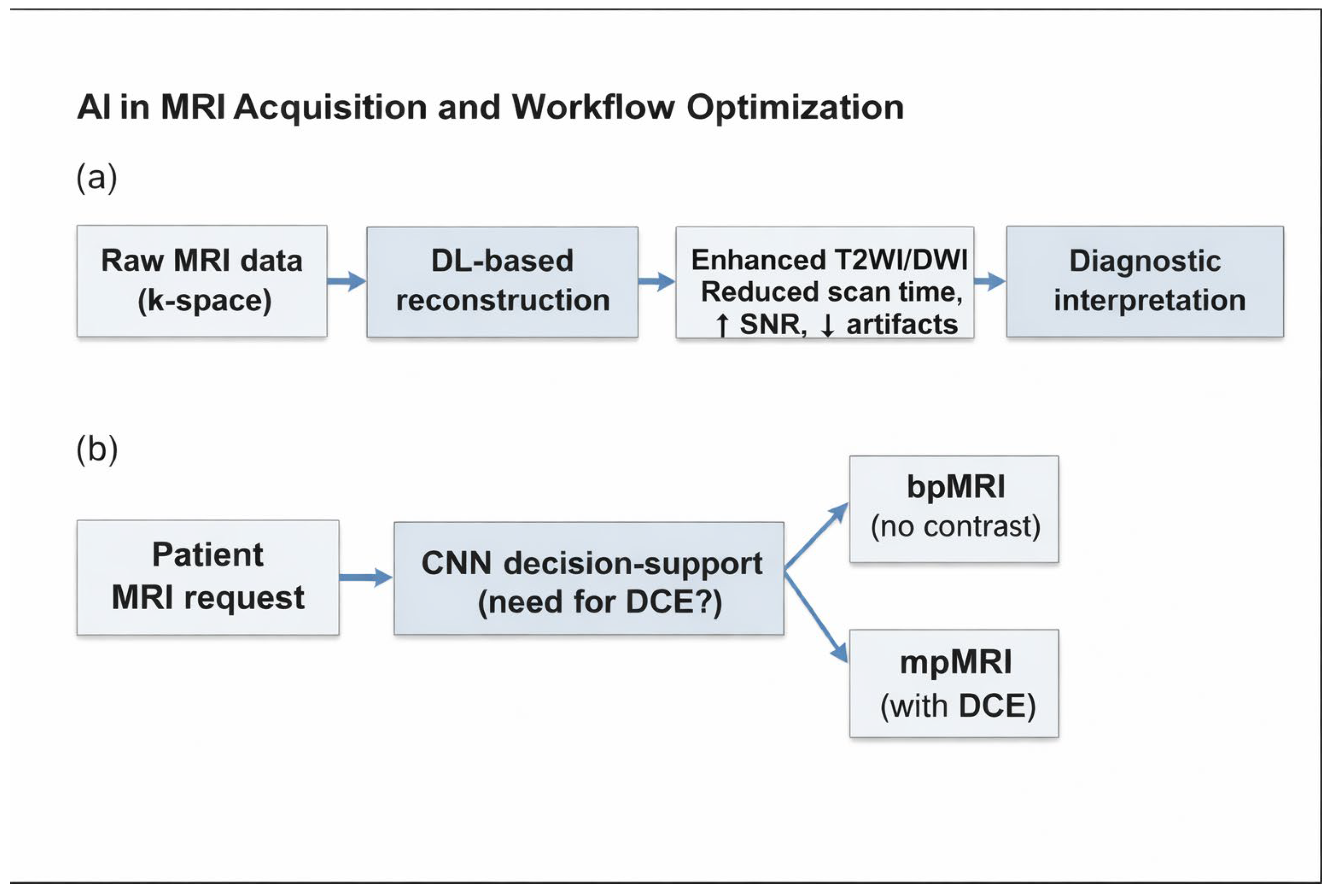
Beyond acceleration, AI has been explored for protocol simplification. Biparametric MRI (bpMRI), which omits dynamic contrast-enhanced (DCE) imaging, has been proposed as a shorter, safer alternative [23,24,25]. While increasingly adopted, bpMRI may not be universally reliable: DCE remains useful in challenging cases, such as for readers with limited experience or patients with degraded image quality from metallic implants or artifacts [23,24,25]. In this context, Hötker et al. showed that convolutional neural networks (CNNs) could autonomously decide whether contrast is required, achieving >94% sensitivity [26]. Such models represent a step towards adaptive, patient-specific workflows (Table 3).
Table 3.
Summary of artificial intelligence (AI) applications for prostate MRI acquisition and workflow optimization. ↑—increase; ↓—decrease.
Despite these encouraging results, several limitations temper the current enthusiasm for AI-enhanced acquisition. Most studies have been conducted in relatively small, monocentric cohorts, often under optimized technical conditions, which restricts their generalizability. Methodological heterogeneity—ranging from scanner field strength and coil configurations to vendor-specific algorithms and training datasets—further complicates comparisons across publications [27,28]. External validation remains scarce, with very few models tested outside the institutions where they were developed and no large-scale prospective trials available to date. Moreover, while improvements in scan duration and image quality are frequently reported, the clinical benefit is less clear [29], as evidence that these advances translate into reduced costs, improved outcomes, or meaningful changes in clinical decision-making remains lacking. Regulatory and integration challenges persist, since most deep learning–based reconstruction techniques are highly vendor-dependent, raising questions about interoperability and standardization across platforms [30].
In parallel, recent methodological advances in deep learning have increasingly focused on robustness to noisy or weak labels, a common limitation in large prostate MRI datasets where annotations may be heterogeneous or partially unreliable. Progressive Sample Selection with Contrastive Loss (PSSCL) has been proposed as a framework that iteratively identifies reliable samples while learning discriminative feature representations through contrastive objectives. Such approaches have demonstrated improved robustness and generalization compared with conventional supervised training and are particularly relevant for prostate MRI applications, where lesion-level ground truth is often uncertain. These developments reflect a broader shift toward noise-aware and data-efficient learning strategies [31].
Recent systematic evidence further supports the growing adoption of deep learning–based reconstruction techniques in prostate MRI. A 2025 systematic review and meta-analysis by Ursprung et al. reported consistent improvements in image quality alongside the potential for accelerated acquisition protocols across diverse cohorts. Together, these findings underscore a broader transition toward DL-enabled workflows that aim to balance image fidelity with clinical throughput, while highlighting the need for rigorous validation and standardized evaluation before widespread clinical deployment [32]. These AI-driven strategies for acquisition acceleration and protocol optimization are schematically summarized in Figure 2, which illustrates how deep learning–based reconstruction and decision-support models integrate into the prostate MRI acquisition workflow.
Figure 2.
Schematic overview of AI applications in prostate MRI acquisition. (a) Deep learning–based reconstruction for acquisition acceleration and image quality improvement in T2WI and DWI. (b) CNN–based decision support for protocol tailoring, enabling individualized selection between bp- and mp-MRI. ↑—increase; ↓—decrease.
3. AI in Cancer Detection and Characterization
AI has shown significant promise in characterizing and detecting prostate cancer using MRI, with DL–based systems supporting multiple stages of the diagnostic process, including lesion detection, segmentation, and grading. These approaches span a range of model architectures and levels of automation, reflecting different degrees of clinical integration and interpretability. Recent years have seen a rapid expansion of deep-learning–based approaches for prostate cancer detection and characterization on MRI. CNNs, including 2D and 3D U-Net–derived architectures, have been widely adopted for lesion detection, segmentation, and clinically significant prostate cancer (csPCa) classification on bpMRI and mpMRI. Several studies have reported performance comparable to, or exceeding, that of expert radiologists, particularly for csPCa detection at the patient or lesion level. More recent works have explored weakly supervised and multi-task learning strategies to reduce annotation burden, as well as multimodal models integrating imaging features with clinical variables to improve risk stratification. Transformer-based and hybrid CNN–transformer architectures have also been introduced (e.g., UNETR, SwinUNETR and variants), aiming to better capture long-range contextual information and inter-sequence relationships [33,34].
One of the most thorough is the DL model known as AutoProstate, which includes modules for segmenting prostatic zones, detecting and segmenting csPCa lesions, and generating automated reports. It showed significant improvements in prostate volume and PSA density estimation compared with radiologists and contributed to streamlining both the diagnostic and reporting sections. The performance was compared to that of an experienced radiologist with 10 years’ experience in reading prostate mpMRI, who scored lesions using a five-point Likert scale [35]. Different technologies have been applied to the field with excellent results; for example, algorithms based on CNNs, such as U-Net architectures. Yu et al. [36] introduced a human-in-the-loop AI model that combines UNet-Seg for lesion detection and segmentation with a 3D-Resnet for diagnosis, achieving high agreement with subspecialists in scoring target lesions. This model demonstrated the potential to match or exceed the performance of general radiologists in diagnosing clinically significant prostate cancer. In this context, semi-automated systems typically require partial human input for task initialization, whereas human-in-the-loop approaches involve continuous clinician interaction for validation and refinement of AI outputs. Other models, such as the one described by Pellicer-Valero et al. [37], added Gleason grade group estimation to segmentation and detection, validated across two datasets (ProstateX and Valencian Oncology Institute). Their model achieved lesion-level AUC = 0.96, sensitivity = 1.00, and specificity ≈ 0.79 for International Society of Urological Pathology (ISUP) ≥ 2 disease in the ProstateX test set. Similarly, strong performance in the IVO dataset demonstrates good performance at both lesion and patient levels.
Among the various topics in the field, the dispute over the grade of segmentation automation is relatively central: Sushentsev et al. conducted a systematic review of 27 studies that applied AI segmentation techniques to both fully automated and semi-automated systems [38]. They discovered that both AI models performed well overall and showed encouraging diagnostic accuracy. Because semi-automated models benefit from expert advice, they tend to perform slightly better in terms of sensitivity. However, because fully automated systems do not rely on expert input and scale more readily within clinical workflows, they have the potential to be widely adopted and have demonstrated satisfying specificity. While technically integrable, practical workflow adoption remains constrained by usability and organizational factors (Table 4).
Table 4.
AI approaches for prostate cancer detection and characterization in MRI, including semi-automated, fully automated, and human-in-the-loop systems. ✓: pros; ✗: cons.
The importance of these application in prostatic imaging is highlighted by the PI-CAI trial [11], the most extensive study on the topic to date, which demonstrated that an AI system could detect clinically significant prostate cancer (Gleason grade group 2 or greater) on MRI with a sensitivity and specificity comparable to, and in some cases superior to, radiologists using the PI-RADS 2.1. The AI system achieved an area under the receiver operating characteristic curve (AUROC) of 0.91, outperforming the average AUROC of 0.86 achieved by a pool of 62 radiologists. In this study, AI developers worldwide could opt in, download an annotated public dataset of 1500 MRI cases, and train AI models for clinically significant prostate cancer detection at bpMRI. For each examination, AI models were required to complete two tasks: localize and classify each lesion with clinically significant cancer (if any), using a 0–100 likelihood score, and classify the overall case using a 0–100 likelihood score for a clinically substantial cancer diagnosis. To this end, AI models could use imaging data and several metadata associated with the examination to inform their predictions.
Overall, the clinical studies reviewed demonstrate that AI systems for prostate cancer detection on MRI, whether based on CNNs, human-in-the-loop frameworks, or fully automated pipelines, achieve high diagnostic accuracy, often comparable to or exceeding radiologists. However, there are notable differences in their applicability and complexity. Fully automated systems like AutoProstate and cascaded DL models tend to offer greater scalability and easier integration into clinical workflows, especially when trained on large, annotated datasets. These models often use mpMRI, which provides richer input but requires more computational resources and greater data quality control. In contrast, bpMRI-based systems are more streamlined and faster, though potentially less sensitive in specific contexts. Human-in-the-loop models, while achieving excellent alignment with expert interpretations, require radiologist input and are therefore less scalable, but may be better suited for settings that require high interpretability and oversight. In terms of training, CNN-based models generally require substantial data and careful tuning, while hybrid approaches balance performance with expert guidance. While no single approach consistently outperforms across all metrics, fully automated mpMRI-based systems appear most promising for widespread adoption, provided that external validation and interpretability continue to improve [39,40,41].
4. Artificial Intelligence in Prostate MRI Workflows for Active Surveillance
Active surveillance (AS) is increasingly adopted as a management strategy for low-risk prostate cancer, yet monitoring progression remains challenging due to subtle MRI changes and inter-reader variability, creating a potential role for AI-based tools to support longitudinal assessment and decision-making. AI has been investigated as a tool to enhance MRI’s diagnostic role in this setting. Oerther et al. demonstrated that a fully automated DL algorithm achieved a sensitivity of 96.4% for detecting ISUP grade ≥ 2 lesions in men under AS, indicating that AI may reliably identify clinically significant disease requiring intervention [42]. Similarly, Arif et al. developed a CNN for the detection and segmentation of clinically significant prostatic cancer in low-risk patients, reporting sensitivities of 82–92% and specificities of 43–76%. These findings suggest that AI could complement radiologists by confirming low-risk status while flagging lesions suspicious for progression [43]. Of note, Bozgo et al. reviewed the current state of AI in AS and highlighted the potential benefits and challenges. They noted that while AI shows promise in various diagnostic tasks, including localization and prognostication, the evidence is currently insufficient to support a shift to AI-based AS due to limitations such as small dataset sizes and heterogeneous outcome definitions [44]. While AI-based models show promise for lesion localization, risk stratification, and prognostication, current evidence remains insufficient for clinical adoption. The main barriers include the use of relatively small, homogeneous datasets, heterogeneous outcome definitions across studies, and limited validation in external cohorts. From a broader perspective, these limitations reflect recurrent challenges in prostate MRI AI research. Models often demonstrate high sensitivity in controlled settings but reduced generalizability across scanners, institutions, and patient populations. Small lesion detection remains problematic, potentially undermining the detection of clinically relevant disease. Ethical and governance issues, including data privacy, algorithm transparency, and accountability, and further complicate clinical translation. Moreover, developing robust AI for AS will require access to large, diverse, and longitudinally annotated datasets, which remain scarce. In summary, AI-enhanced MRI has the potential to refine active surveillance by improving reproducibility and early detection of progression, particularly in subtle or equivocal cases.
5. Radiomics and Prognostication
Radiomics is an emerging discipline that extracts large numbers of quantitative features from medical images. These extracted features, which may include length, shape, and intensity metrics, can support diagnosis, prognosis, and therapy planning [45,46]. In prostate cancer, radiomics applied mpMRI has been investigated as a tool to complement visual interpretation and provide imaging biomarkers of tumor biology. Traditional radiomics relies on predefined handcrafted features, while deep learning–based radiomics automatically learns high-dimensional representations from imaging data; however, these two paradigms differ substantially in interpretability, reproducibility, and need for large, annotated datasets. Several studies have shown that MRI-derived radiomic signatures can predict Gleason score with moderate to high accuracy, with reported AUCs ranging from 0.50 to 0.92 in systematic reviews [47,48,49]. Beyond Gleason grading, radiomic models have been applied to predict extracapsular extension (EPE) and pelvic lymph node involvement. A recent systematic review by Wen et al. reported pooled sensitivities and specificities of 0.77 and 0.75, respectively, for EPE prediction. At the same time, integrative models combining radiomic and clinical features achieved AUCs above 0.90 for lymph node invasion [50]. Notably, radiomic assessment of lymph node status has shown promise in identifying occult metastases even in morphologically normal nodes, suggesting a potential role in refining nodal staging and treatment planning. Radiomics has also been used to assess treatment efficacy, particularly after radiotherapy, by tracking longitudinal changes in tumor heterogeneity and morphology. Quantitative MRI-based radiomic features can capture post-treatment tissue changes and distinguish between residual tumor, radiation-induced fibrosis, and benign post-therapy alterations, supporting more accurate response assessment and early relapse detection [51]. Recent evidence further suggests that radiomic profiling may contribute to personalized radiotherapy planning and adaptive strategies by predicting treatment response and toxicity patterns, extending its role beyond diagnosis toward comprehensive disease management [52]. Integration with AI techniques enhances the predictive power of radiomics, while radiogenomic approaches that link imaging features with molecular profiles offer additional insights into tumor behavior. Early studies have demonstrated associations between radiomic features and genomic alterations, such as PTEN loss, ERG fusions, and DNA repair gene mutations, suggesting that radiogenomics could serve as a bridge between imaging phenotypes and tumor molecular biology [53]. Applications in other organs, such as lung cancer, have already demonstrated the feasibility of DL–based radiomics for survival prediction and virtual biopsy [47], underscoring the translational potential of these methods across oncology. Despite encouraging results, radiomics in prostate MRI is still far from clinical translation. In fact, no radiomic model has been prospectively tested or integrated into clinical decision-making for prostate cancer, underscoring the gap between research and real-world implementation. Many studies are retrospective, use heterogeneous acquisition protocols, and lack external validation, resulting in poor reproducibility across centers. Harmonization methods such as ComBat have been proposed to mitigate scanner- and vendor-related variability, but robust cross-site feature stability remains challenging to achieve. Feature extraction pipelines remain unstandardized, and reporting quality is variable, as highlighted by the Radiomics Quality Score (RQS) framework [54]. Moreover, while predictive models often show high AUCs, their added value over existing clinical and pathological tools is rarely demonstrated. Future research should prioritize prospective, multicenter validation, harmonization of feature extraction, and integration of radiomics into multiparametric and multimodal models that combine clinical, imaging, and molecular data (Table 5).
Table 5.
Possible applications of radiomics in prostate MRI for risk stratification and treatment assessment.
6. Future Challenges and Opportunities
Numerous AI-based tools for prostate cancer imaging have recently obtained regulatory approval and are increasingly entering clinical workflows (Table 6). Nevertheless, the widespread clinical implementation of AI and radiomics in prostate MRI remains constrained by multiple technical, methodological, and regulatory barriers. Current models often rely on heterogeneous datasets acquired under variable imaging protocols, lacking standardized preprocessing or harmonization. As emphasized by studies such as that of Cuocolo et al. [55], reproducibility and external validation remain critical weaknesses in prostate MRI radiomics studies, limiting generalizability across institutions and vendors. The persistent scarcity of large, balanced, and well-annotated datasets further hinders model robustness, while biases in data acquisition and labeling may propagate into diagnostic or prognostic algorithms. In this context, methodological rigor and transparency are essential to advance from proof-of-concept studies to reliable clinical translation. The RQS proposed by Lambin et al. [56] continues to serve as a benchmark for assessing study quality, while initiatives such as the Image Biomarker Standardization Initiative (IBSI) [57] aim to improve feature reproducibility. Despite these efforts, scanner- and protocol-related variability still compromises feature stability. Harmonization strategies, including ComBat and domain adaptation techniques, have shown promise in mitigating inter-scanner effects [58], although robust cross-site reproducibility is not yet routinely achieved in clinical practice. Data governance and patient privacy represent additional obstacles to large-scale AI development. Strict confidentiality regulations limit the creation of centralized multicenter imaging repositories. Federated learning (FL) has emerged as a promising alternative, enabling collaborative model training without direct data sharing. In addition to foundational work by Sheller et al. [59], recent surveys have systematically reviewed methodological advances and open challenges of FL in medical image analysis [60,61]. Importantly, prostate MRI–specific FL studies have begun to demonstrate feasibility across multiple sites for tasks such as prostate segmentation and clinically significant cancer detection, highlighting the potential of FL to improve robustness under scanner and domain heterogeneity [62]. Nevertheless, implementations in prostate MRI remain limited and require further validation. Additional barriers include model explainability, algorithm auditability, and medicolegal accountability. Additionally, future progress will depend on the ability to integrate AI and radiomics with multimodal imaging. PSMA PET/MRI has already demonstrated superior diagnostic performance compared to MRI alone for staging and restaging [63]. AI-driven fusion of mpMRI, PSMA-PET, and potentially MR spectroscopy imaging (MRSI) could enhance lesion characterization and biological risk profiling. Radiogenomic studies, such as those by Stoyanova et al. [64], have shown that imaging-derived phenotypes can correlate with molecular alterations, such as PTEN loss or ERG rearrangements, providing a non-invasive window into tumor biology. Explainable AI models capable of generating interpretable outputs are essential for building clinician trust and meeting regulatory standards. AI-assisted structured reporting—including automated PI-RADS scoring and lesion quantification—could streamline workflow integration and reduce inter-reader variability [65]. Finally, the emergence of foundation and multimodal models, trained on extensive imaging and non-imaging datasets, promises a paradigm shift from narrow, task-specific algorithms to comprehensive, generalizable decision-support systems [66].
Table 6.
Commercially available AI tools for prostate MRI analysis. Information compiled from publicly available vendor documentation and regulatory databases. Abbreviations—MDR: Medical Device Regulation; MDD: Medical Device Directive.
7. Conclusions
Artificial intelligence is reshaping the role of multiparametric MRI along the entire prostate cancer care pathway—from image acquisition to post-treatment follow-up. AI-driven acceleration and quality enhancement are addressing long-standing technical limitations, while automated detection and characterization tools promise greater diagnostic consistency and efficiency. Radiomics and radiogenomics further extend the value of imaging, transforming it from a purely morphological technique into a source of quantitative biomarkers that can inform prognosis, treatment selection, and monitoring. Yet, despite rapid progress, proper clinical integration remains limited by methodological heterogeneity, lack of standardization, and insufficient prospective validation. The future of prostate MRI will depend on collaborative efforts among radiologists, data scientists, and clinicians to develop transparent, interoperable, and clinically validated AI systems. In this vision, imaging becomes not an isolated diagnostic test but a dynamic component of precision oncology—guiding decisions across the patient journey and bringing us closer to genuinely personalized prostate cancer care. Ultimately, the successful clinical translation of AI in prostate MRI will depend not only on technical performance, but also on rigorous validation, transparent design, and close collaboration between developers and clinicians, positioning AI as a supportive and trustworthy component of the diagnostic workflow.
Author Contributions
Conceptualization, G.P.; methodology, C.P. and D.B.; software, F.A. and M.F.G.; validation, G.C. and S.T.; formal analysis, M.F.G.; investigation, C.P.; resources, G.P.; data curation, C.P.; writing—original draft preparation, G.P.; writing—review and editing, C.P.; visualization, D.B.; supervision, G.C.; project administration, S.T. All authors have read and agreed to the published version of the manuscript.
Funding
This research received no external funding.
Data Availability Statement
The original contributions presented in this study are included in the article. Further inquiries can be directed to the corresponding author.
Conflicts of Interest
The author declares no conflicts of interest.
References
- Sung, H.; Ferlay, J.; Siegel, R.L.; Laversanne, M.; Soerjomataram, I.; Jemal, A.; Bray, F. Global Cancer Statistics 2020: GLOBOCAN Estimates of Incidence and Mortality Worldwide for 36 Cancers in 185 Countries. CA Cancer J. Clin. 2021, 71, 209–249. [Google Scholar] [CrossRef] [Scilit]
- Associazione Italiana di Oncologia Medica (AIOM). Linee guida AIOM 2024: Neoplasie della prostata. Available online: https://www.aiom.it/wp-content/uploads/2025/11/2024_NDC-web-21.11.25.pdf (accessed on 1 January 2026).
- Auvinen, A.; Moss, S.M.; Tammela, T.L.J.; Taari, K.; Roobol, M.J.; Schröder, F.H.; Bangma, C.H.; Carlsson, S.; Aus, G.; Zappa, M.; et al. Absolute Effect of Prostate Cancer Screening: Balance of Benefits and Harms by Center within the European Randomized Study of Prostate Cancer Screening. Clin. Cancer Res. 2016, 22, 243–249. [Google Scholar] [CrossRef] [Scilit]
- Turkbey, B.; Rosenkrantz, A.B.; Haider, M.A.; Padhani, A.R.; Villeirs, G.; Macura, K.J.; Tempany, C.M.; Choyke, P.L.; Cornud, F.; Margolis, D.J.; et al. Prostate Imaging Reporting and Data System Version 2.1: 2019 Update of Prostate Imaging Reporting and Data System Version 2. Eur. Urol. 2019, 76, 340–351. [Google Scholar] [CrossRef] [Scilit]
- Kasivisvanathan, V.; Rannikko, A.S.; Borghi, M.; Panebianco, V.; Mynderse, L.A.; Vaarala, M.H.; Briganti, A.; Budäus, L.; Hellawell, G.; Hindley, R.G.; et al. MRI-Targeted or Standard Biopsy for Prostate-Cancer Diagnosis. N. Engl. J. Med. 2018, 378, 1767–1777. [Google Scholar] [CrossRef] [Scilit]
- Alkema, N.G.; Hoogeveen, S.F.J.S.; Cauberg, E.C.C.; Witte, L.P.W.; van ’t Veer-Ten Kate, M.; de Boer, E.; Hoogland, M.A.M.; Blanker, M.H.; Boomsma, M.F.; Steffens, M.G. Magnetic Resonance Imaging–targeted Prostate Biopsy Compared with Systematic Prostate Biopsy in Biopsy-naïve Patients with Suspected Prostate Cancer. Eur. Urol. Open Sci. 2022, 44, 125–130. [Google Scholar] [CrossRef] [Scilit]
- Rouvière, O.; Puech, P.; Renard-Penna, R.; Claudon, M.; Roy, C.; Mège-Lechevallier, F.; Decaussin-Petrucci, M.; Dubreuil-Chambardel, M.; Magaud, L.; Remontet, L.; et al. Use of prostate systematic and targeted biopsy on the basis of multiparametric MRI in biopsy-naive patients (MRI-FIRST): A prospective, multicentre, paired diagnostic study. Lancet Oncol. 2019, 20, 100–109. [Google Scholar] [CrossRef] [Scilit] [PubMed]
- Ramesh, A.; Kambhampati, C.; Monson, J.; Drew, P. Artificial intelligence in medicine. Ann. R. Coll. Surg. Engl. 2004, 86, 334–338. [Google Scholar] [CrossRef] [Scilit]
- Hosny, A.; Parmar, C.; Quackenbush, J.; Schwartz, L.H.; Aerts, H.J.W.L. Artificial intelligence in radiology. Nat. Rev. Cancer 2018, 18, 500–510. [Google Scholar] [CrossRef] [Scilit] [PubMed]
- PROSTATEX—The Cancer Imaging Archive (TCIA). Available online: https://www.cancerimagingarchive.net/collection/prostatex/ (accessed on 13 December 2025).
- Saha, A.; Bosma, J.S.; Twilt, J.J.; van Ginneken, B.; Noordman, C.R.; Slootweg, I.; Roest, C.; Fransen, S.J.; Sunoqrot, M.R.S.; Bathen, T.F.; et al. Artificial intelligence and radiologists in prostate cancer detection on MRI (PI-CAI): An international, paired, non-inferiority, confirmatory study. Lancet Oncol. 2024, 25, 879–887. [Google Scholar] [CrossRef] [Scilit] [PubMed]
- Litjens, G.; Toth, R.; van de Ven, W.; Hoeks, C.; Kerkstra, S.; van Ginneken, B.; Vincent, G.; Guillard, G.; Birbeck, N.; Zhang, J.; et al. Evaluation of prostate segmentation algorithms for MRI: The PROMISE12 challenge. Med. Image Anal. 2014, 18, 359–373. [Google Scholar] [CrossRef] [Scilit]
- ISBI-MR-PROSTATE-2013—The Cancer Imaging Archive (TCIA). Available online: https://www.cancerimagingarchive.net/analysis-result/isbi-mr-prostate-2013/ (accessed on 13 December 2025).
- MICCAI Registered Challenges. Available online: https://miccai.org/index.php/special-interest-groups/challenges/miccai-registered-challenges/ (accessed on 13 December 2025).
- Lee, K.-L.; Kessler, D.A.; Dezonie, S.; Chishaya, W.; Shepherd, C.; Carmo, B.; Graves, M.J.; Barrett, T. Assessment of deep learning-based reconstruction on T2-weighted and diffusion-weighted prostate MRI image quality. Eur. J. Radiol. 2023, 166, 111017. [Google Scholar] [CrossRef] [Scilit]
- Park, J.C.; Park, K.J.; Park, M.Y.; Choi, S.H.; Lee, H.-Y.; Kim, J.K.; Kim, M.H.; Jeong, I.G.; Cho, J.Y.; Kim, S.K.; et al. Fast T2-Weighted Imaging With Deep Learning-Based Reconstruction: Evaluation of Image Quality and Diagnostic Performance in Patients Undergoing Radical Prostatectomy. J. Magn. Reson. Imaging 2022, 55, 1735–1744. [Google Scholar] [CrossRef] [Scilit]
- Gassenmaier, S.; Afat, S.; Nickel, D.; Mostapha, M.; Herrman, J.; Othman, A.E. Deep learning–accelerated T2-weighted imaging of the prostate: Reduction of acquisition time and improvement of image quality. Eur. J. Radiol. 2021, 137, 109600. [Google Scholar] [CrossRef] [Scilit]
- Ueda, T.; Ohno, Y.; Yamamoto, K.; Sato, H.; Tanaka, M.; Takahashi, M.; Hori, M.; Nakayama, T.; Suzuki, T.; Watanabe, Y.; et al. Deep Learning Reconstruction of Diffusion-weighted MRI Improves Image Quality for Prostatic Imaging. Radiology 2022, 303, 373–381. [Google Scholar] [CrossRef] [Scilit]
- Ursprung, S.; Herrmann, J.; Joos, N.; Agrotis, G.; van Houdt, P.J.; Bickelhaupt, S.; Davis, B.A.; Hahn, D.; Schoenberg, S.O.; Behrendt, F.; et al. Accelerated diffusion-weighted imaging of the prostate using deep learning image reconstruction: A retrospective comparison with standard diffusion-weighted imaging. Eur. J. Radiol. 2023, 165, 110953. [Google Scholar] [CrossRef] [Scilit]
- Johnson, P.M.; Tong, A.; Donthireddy, A.; Melamud, K.; Petrocelli, R.; Smereka, P.; Qian, K.; Keerthivasan, M.B.; Chandarana, H.; Knoll, F. Deep Learning Reconstruction Enables Highly Accelerated Biparametric MR Imaging of the Prostate. J. Magn. Reson. Imaging 2022, 56, 184–195. [Google Scholar] [CrossRef] [Scilit] [PubMed]
- Shiraishi, K.; Nakaura, T.; Kobayashi, N.; Yokoyama, K.; Ito, K.; Tanaka, M.; Yamamoto, K.; Watanabe, Y.; Sasaki, Y.; Otake, Y.; et al. Enhancing thin slice 3D T2-weighted prostate MRI with super-resolution deep learning reconstruction: Impact on image quality and PI-RADS assessment. Magn. Reson. Imaging 2025, 117, 110308. [Google Scholar] [CrossRef] [Scilit] [PubMed]
- Bischoff, L.M.; Peeters, J.M.; Weinhold, L.; Krausewitz, P.; Ellinger, J.; Katemann, C.; Isaak, A.; Weber, O.M.; Kuetting, D.; Attenberger, U.; et al. Deep Learning Super-Resolution Reconstruction for Fast and Motion-Robust T2-weighted Prostate MRI. Radiology 2023, 308, e230427. [Google Scholar] [CrossRef] [Scilit]
- Weiss, J.; Martirosian, P.; Notohamiprodjo, M.; Kaufmann, S.; Othman, A.E.; Grosse, U.; Nikolaou, K.; Gatidis, S. Implementation of a 5-Minute Magnetic Resonance Imaging Screening Protocol for Prostate Cancer in Men with Elevated Prostate-Specific Antigen Before Biopsy. Investig. Radiol. 2018, 53, 186–190. [Google Scholar] [CrossRef] [Scilit] [PubMed]
- Kuhl, C.K.; Bruhn, R.; Krämer, N.; Nebelung, S.; Heidenreich, A.; Schrading, S. Abbreviated Biparametric Prostate MR Imaging in Men with Elevated Prostate-specific Antigen. Radiology 2017, 285, 493–505. [Google Scholar] [CrossRef] [Scilit]
- Padhani, A.R.; Schoots, I.; Villeirs, G. Contrast Medium or No Contrast Medium for Prostate Cancer Diagnosis. That Is the Question. J. Magn. Reson. Imaging 2021, 53, 13–22. [Google Scholar] [CrossRef] [Scilit] [PubMed]
- Hötker, A.M.; Da Mutten, R.; Tiessen, A.; Konukoglu, E.; Donati, O.F. Improving workflow in prostate MRI: AI-based decision-making on biparametric or multiparametric MRI. Insights Imaging 2021, 12, 112. [Google Scholar] [CrossRef] [Scilit]
- Turkbey, B.; Choyke, P.L. Future Perspectives and Challenges of Prostate MR Imaging. Radiol. Clin. N. Am. 2018, 56, 327–337. [Google Scholar] [CrossRef] [Scilit]
- Jeong, J.; Yeom, S.K.; Choi, I.Y.; Cha, S.H.; Han, J.S.; Lee, C.H.; Sung, D.J.; Choi, J.-W.; Bae, J.H.; Choi, H.S.; et al. Deep learning image reconstruction of diffusion-weighted imaging in evaluation of prostate cancer focusing on its clinical implications. Quant. Imaging Med. Surg. 2024, 14, 3432–3446. [Google Scholar] [CrossRef] [Scilit]
- McBee, M.P.; Awan, O.A.; Colucci, A.T.; Ghobadi, C.W.; Kadom, N.; Kansagra, A.P.; Tridandapani, S.; Auffermann, W.F. Deep Learning in Radiology. Acad. Radiol. 2018, 25, 1472–1480. [Google Scholar] [CrossRef] [Scilit]
- Reinhardt, C.; Briody, H.; MacMahon, P.J. AI-accelerated prostate MRI: A systematic review. Br. J. Radiol. 2024, 97, 1234–1242. [Google Scholar] [CrossRef] [Scilit] [PubMed]
- Zhang, Q.; Zhu, Y.; Cordeiro, F.R.; Chen, Q. PSSCL: A progressive sample selection framework with contrastive loss designed for noisy labels. Pattern Recognit. 2025, 161, 111284. [Google Scholar] [CrossRef] [Scilit]
- Ursprung, S.; Agrotis, G.; van Houdt, P.J.; ter Beek, L.C.; Boellaard, T.N.; Beets-Tan, R.G.H.; Yakar, D.; Padhani, A.R.; Schoots, I.G. Prostate MRI Using Deep Learning Reconstruction in Response to Cancer Screening Demands—A Systematic Review and Meta-Analysis. J. Pers. Med. 2025, 15, 284. [Google Scholar] [CrossRef] [Scilit]
- Alzate-Grisales, J.A.; Mora-Rubio, A.; García-García, F.; Tabares-Soto, R.; de la Iglesia-Vayá, M. SAM-UNETR: Clinically Significant Prostate Cancer Segmentation Using Transfer Learning from Large Model. IEEE Access 2023, 11, 118217–118228. [Google Scholar] [CrossRef] [Scilit]
- Abudalou, S.; Qi, J.; Choi, J.; Gage, K.; Pow-Sang, J.; Yilmaz, Y.; Balagurunathan, Y. Improving Prostate Gland Segmenting Using Transformer based Architectures. arXiv 2025, arXiv:2506.14844. [Google Scholar] [CrossRef] [Scilit]
- Mehta, P.; Antonelli, M.; Singh, S.; Grondecka, N.; Johnston, E.W.; Ahmed, H.U.; Emberton, M.; Punwani, S.; Ourselin, S. AutoProstate: Towards Automated Reporting of Prostate MRI for Prostate Cancer Assessment Using Deep Learning. Cancers 2021, 13, 6138. [Google Scholar] [CrossRef] [Scilit] [PubMed]
- Yu, R.; Jiang, K.-W.; Bao, J.; Hou, Y.; Yi, Y.; Wu, D.; Song, Y.; Hu, C.-H.; Yang, G.; Zhang, Y.-D. PI-RADSAI: Introducing a new human-in-the-loop AI model for prostate cancer diagnosis based on MRI. Br. J. Cancer 2023, 128, 1019–1029. [Google Scholar] [CrossRef] [Scilit]
- Pellicer-Valero, O.J.; Marenco Jiménez, J.L.; Gonzalez-Perez, V.; Casanova Ramón-Borja, J.L.; Martín García, I.; Barrios Benito, M.; Pelechano Gómez, P.; Rubio-Briones, J.; Rupérez, M.J.; Martín-Guerrero, J.D. Deep learning for fully automatic detection, segmentation, and Gleason grade estimation of prostate cancer in multiparametric magnetic resonance images. Sci. Rep. 2022, 12, 2975. [Google Scholar] [CrossRef] [Scilit]
- Sushentsev, N.; Moreira Da Silva, N.; Yeung, M.; Barrett, T.; Sala, E.; Roberts, M.; Rundo, L. Comparative performance of fully-automated and semi-automated artificial intelligence methods for the detection of clinically significant prostate cancer on MRI: A systematic review. Insights Imaging 2022, 13, 59. [Google Scholar] [CrossRef] [Scilit]
- Roest, C.; Fransen, S.J.; Kwee, T.C.; Yakar, D. Comparative Performance of Deep Learning and Radiologists for the Diagnosis and Localization of Clinically Significant Prostate Cancer at MRI: A Systematic Review. Life 2022, 12, 1490. [Google Scholar] [CrossRef] [Scilit]
- He, M.; Cao, Y.; Chi, C.; Yang, X.; Ramin, R.; Wang, S.; Yang, G.; Mukhtorov, O.; Zhang, L.; Kazantsev, A.; et al. Research progress on deep learning in magnetic resonance imaging–based diagnosis and treatment of prostate cancer: A review on the current status and perspectives. Front. Oncol. 2023, 13, 1189370. [Google Scholar] [CrossRef] [Scilit]
- Turkbey, B.; Haider, M.A. Artificial Intelligence for Automated Cancer Detection on Prostate MRI: Opportunities and Ongoing Challenges, From the AJR Special Series on AI Applications. Am. J. Roentgenol. 2022, 219, 188–194. [Google Scholar] [CrossRef] [Scilit] [PubMed]
- Oerther, B.; Engel, H.; Nedelcu, A.; Schlett, C.L.; Grimm, R.; von Busch, H.; Sigle, A.; Gratzke, C.; Bamberg, F.; Benndorf, M. Prediction of upgrade to clinically significant prostate cancer in patients under active surveillance: Performance of a fully automated AI-algorithm for lesion detection and classification. Prostate 2023, 83, 871–878. [Google Scholar] [CrossRef] [Scilit]
- Arif, M.; Schoots, I.G.; Castillo Tovar, J.; Bangma, C.H.; Krestin, G.; Bouts, M.; Niessen, W.; Veenland, J.F. Clinically significant prostate cancer detection and segmentation in low-risk patients using a convolutional neural network on multi-parametric MRI. Eur. Radiol. 2020, 30, 6582–6592. [Google Scholar] [CrossRef] [Scilit]
- Bozgo, V.; Roest, C.; van Oort, I.; Yakar, D.; Huisman, H.; de Rooij, M. Prostate MRI and artificial intelligence during active surveillance: Should we jump on the bandwagon? Eur. Radiol. 2024, 34, 7698–7704. [Google Scholar] [CrossRef] [Scilit] [PubMed]
- Gillies, R.J.; Kinahan, P.E.; Hricak, H. Radiomics: Images Are More than Pictures, They Are Data. Radiology 2016, 278, 563–577. [Google Scholar] [CrossRef] [Scilit]
- Aerts, H.J.W.L.; Rios Velazquez, E.; Leijenaar, R.T.H.; Parmar, C.; Grossmann, P.; Carvalho, S.; Bussink, J.; Monshouwer, R.; Haibe-Kains, B.; Rietveld, D.; et al. Decoding tumour phenotype by noninvasive imaging using a quantitative radiomics approach. Nat. Commun. 2014, 5, 4006. [Google Scholar] [CrossRef] [Scilit]
- Calimano-Ramirez, L.F.; Virarkar, M.K.; Hernandez, M.; Ozdemir, S.; Kumar, S.; Gopireddy, D.R.; Lall, C.; Balaji, K.C.; Mete, M.; Gumus, K.Z. MRI-based nomograms and radiomics in presurgical prediction of extraprostatic extension in prostate cancer: A systematic review. Abdom. Radiol. 2023, 48, 2379–2400. [Google Scholar] [CrossRef] [Scilit]
- Zheng, H.; Miao, Q.; Liu, Y.; Afshari Mirak, S.; Hosseiny, M.; Scalzo, F.; Raman, S.S.; Sung, K. Multiparametric MRI-based radiomics model to predict pelvic lymph node invasion for patients with prostate cancer. Eur. Radiol. 2022, 32, 5688–5699. [Google Scholar] [CrossRef] [Scilit]
- Spohn, S.K.B.; Bettermann, A.S.; Bamberg, F.; Benndorf, M.; Mix, M.; Nicolay, N.H.; Fechter, T.; Hölscher, T.; Grosu, R.; Chiti, A.; et al. Radiomics in prostate cancer imaging for a personalized treatment approach—Current aspects of methodology and a systematic review on validated studies. Theranostics 2021, 11, 8027–8042. [Google Scholar] [CrossRef] [Scilit] [PubMed]
- Wen, J.; Liu, W.; Zhang, Y.; Shen, X. MRI-based radiomics for prediction of extraprostatic extension of prostate cancer: A systematic review and meta-analysis. Radiol. Med. 2024, 129, 702–711. [Google Scholar] [CrossRef] [Scilit] [PubMed]
- Delgadillo, R.; Ford, J.C.; Abramowitz, M.C.; Machtay, M.; Hahn, S.M.; Metz, J.M.; Deville, C.; Plastaras, J.P.; Bekelman, J.E.; Swisher-McClure, S.; et al. The role of radiomics in prostate cancer radiotherapy. Strahlenther. Onkol. 2020, 196, 900–912. [Google Scholar] [CrossRef] [Scilit]
- Stanzione, A.; Ponsiglione, A.; Alessandrino, F.; Cuocolo, R.; Castaldo, A.; Baggiano, A.; Del Prete, A.; Salvatore, M.; Imbriaco, M. Beyond diagnosis: Is there a role for radiomics in prostate cancer management? Eur. Radiol. Exp. 2023, 7, 13. [Google Scholar] [CrossRef] [Scilit] [PubMed]
- Ferro, M.; de Cobelli, O.; Vartolomei, M.D.; Lucarelli, G.; Crocetto, F.; Barone, B.; Sciarra, A.; Busetto, G.M.; Maggi, M.; Del Giudice, F.; et al. Prostate Cancer Radiogenomics—From Imaging to Molecular Characterization. Int. J. Mol. Sci. 2021, 22, 9971. [Google Scholar] [CrossRef] [Scilit]
- Stanzione, A.; Gambardella, M.; Cuocolo, R.; Castaldo, A.; Del Prete, A.; Ponsiglione, A.; Romeo, V.; Salvatore, M.; Imbriaco, M. Prostate MRI radiomics: A systematic review and radiomic quality score assessment. Eur. J. Radiol. 2020, 129, 109095. [Google Scholar] [CrossRef] [Scilit]
- Cuocolo, R.; Imbriaco, M. Machine learning solutions in radiology: Does the emperor have no clothes? Eur. Radiol. 2021, 31, 3783–3785. [Google Scholar] [CrossRef] [Scilit]
- Lambin, P.; Rios-Velazquez, E.; Leijenaar, R.; Carvalho, S.; van Stiphout, R.G.; Granton, P.; Zegers, C.M.; Gillies, R.J.; Boellard, R.; Dekker, A.; et al. Radiomics: Extracting more information from medical images using advanced feature analysis. Eur. J. Cancer 2012, 48, 441–446. [Google Scholar] [CrossRef] [Scilit]
- Zwanenburg, A.; Vallières, M.; Abdalah, M.A.; Aerts, H.J.W.L.; Andrearczyk, V.; Apte, A.; Ashrafinia, S.; Bakas, S.; Beukinga, R.J.; Boellaard, R.; et al. The Image Biomarker Standardization Initiative: Standardized Quantitative Radiomics for High-Throughput Image-based Phenotyping. Radiology 2020, 295, 328–338. [Google Scholar] [CrossRef] [Scilit]
- Orlhac, F.; Eertink, J.J.; Cottereau, A.-S.; Zijlstra, J.M.; Thieblemont, C.; Meignan, M.; Boellaard, R.; Buvat, I. A Guide to ComBat Harmonization of Imaging Biomarkers in Multicenter Studies. J. Nucl. Med. 2022, 63, 172–179. [Google Scholar] [CrossRef] [Scilit] [PubMed]
- Sheller, M.J.; Edwards, B.; Reina, G.A.; Martin, J.; Pati, S.; Kotrotsou, A.; Milchenko, M.; Xu, W.; Marcus, D.; Colen, R.R.; et al. Federated learning in medicine: Facilitating multi-institutional collaborations without sharing patient data. Sci. Rep. 2020, 10, 12598. [Google Scholar] [CrossRef] [Scilit] [PubMed]
- Guan, H.; Yap, P.T.; Bozoki, A.; Liu, M. Federated learning for medical image analysis: A survey. Pattern Recognit. 2024, 151, 110424. [Google Scholar] [CrossRef] [Scilit] [PubMed]
- da Silva, F.R.; Camacho, R.; Tavares, J.M.R.S.; Campilho, A.; Viana, M.; Morais, P.; Cunha, A.; Pereira, S.; Silva, C.; Oliveira, H.P.; et al. Federated Learning in Medical Image Analysis: A Systematic Survey. Electronics 2023, 13, 47. [Google Scholar] [CrossRef] [Scilit]
- Moradi, A.; Zerka, F.; Bosma, J.S.; Litjens, G.; Huisman, H.; van Ginneken, B.; Padhani, A.R.; Schoots, I.G.; Yakar, D.; Dekker, A. Optimizing Federated Learning Configurations for MRI Prostate Segmentation and Cancer Detection: A Simulation Study. Radiol. Artif. Intell. 2025, 7, e240485. [Google Scholar] [CrossRef] [Scilit]
- Ferdinandus, J.; Fendler, W.P.; Farolfi, A.; Giesel, F.L.; Ceci, F.; Uprimny, C.; Kroiss, A.; Lawhn-Heath, C.; Grubmüller, B.; Lopci, E.; et al. PSMA PET Validates Higher Rates of Metastatic Disease for European Association of Urology Biochemical Recurrence Risk Groups: An International Multicenter Study. J. Nucl. Med. 2022, 63, 76–80. [Google Scholar] [CrossRef] [Scilit]
- Stoyanova, R.; Zavala-Romero, O.; Kwon, D.; Tschudi, Y.; Marquardt, M.D.; Salama, J.K.; Parra, N.A.; Gillies, R.J.; Pollack, A.; Aerts, H.J.W.L.; et al. Clinical-Genomic Risk Group Classification of Suspicious Lesions on Prostate Multiparametric-MRI. Cancers 2023, 15, 5240. [Google Scholar] [CrossRef] [Scilit]
- Bleker, J.; Yakar, D.; van Noort, B.; Roest, C.; van der Leest, M.; Schoots, I.G.; de Rooij, M.; Huisman, H. Single-center versus multi-center biparametric MRI radiomics approach for clinically significant peripheral zone prostate cancer. Insights Imaging 2021, 12, 150. [Google Scholar] [CrossRef] [Scilit] [PubMed]
- Lundervold, A.S.; Lundervold, A. An overview of deep learning in medical imaging focusing on MRI. Z. Med. Phys. 2019, 29, 102–127. [Google Scholar] [CrossRef] [Scilit] [PubMed]



Disclaimer/Publisher’s Note: The statements, opinions and data contained in all publications are solely those of the individual author(s) and contributor(s) and not of MDPI and/or the editor(s). MDPI and/or the editor(s) disclaim responsibility for any injury to people or property resulting from any ideas, methods, instructions or products referred to in the content. |
© 2026 by the authors. Licensee MDPI, Basel, Switzerland. This article is an open access article distributed under the terms and conditions of the Creative Commons Attribution (CC BY) license.