


Abstract
Recent progress in machine learning (ML) has fueled the emergence of intelligent clinical decision support systems (CDSSs) designed to optimize diagnostic and prognostic accuracy through the analysis of complex and heterogeneous medical data. The analysis provides a comprehensive perspective on the use of machine learning in the medical field by integrating a bibliometric assessment of the recent literature and a detailed examination of the algorithms used in current studies. The bibliometric component highlights the evolution of publications, the thematic distribution of research and emerging directions within various medical specialties. In addition, the evaluation of selected articles sheds light on the concrete ways of applying ML algorithms, as well as the methodological limitations encountered in clinical practice. Random forest and gradient boosting are commonly used in internal medicine and cardiology, while convolutional neural networks (CNNs) dominate neuroimaging in neurology and image-based analyses in oncology and radiology.
1. Introduction
Artificial intelligence (AI) has become a technology capable of processing and generating natural language, analyzing complex data and transforming many fields, from education and research to healthcare and industry [1,2]. Artificial intelligence encompasses machine learning (ML), focused on developing algorithms that learn from data, and deep learning (DL), which uses neural networks to analyze complex data and provide fast and objective assessments.
Recent advances in artificial intelligence, and in particular machine learning, have opened up new opportunities for the development of clinical decision support systems (CDSSs) [3,4]. These tools aim to support physicians in diagnostic and prognostic activities by analyzing complex, large and often heterogeneous data sets. In a context where the volume of medical information is growing exponentially—from radiological images and structured clinical data to multi-omic data and electronic patient records—it becomes imperative to identify algorithmic solutions capable of integrating and interpreting these resources.
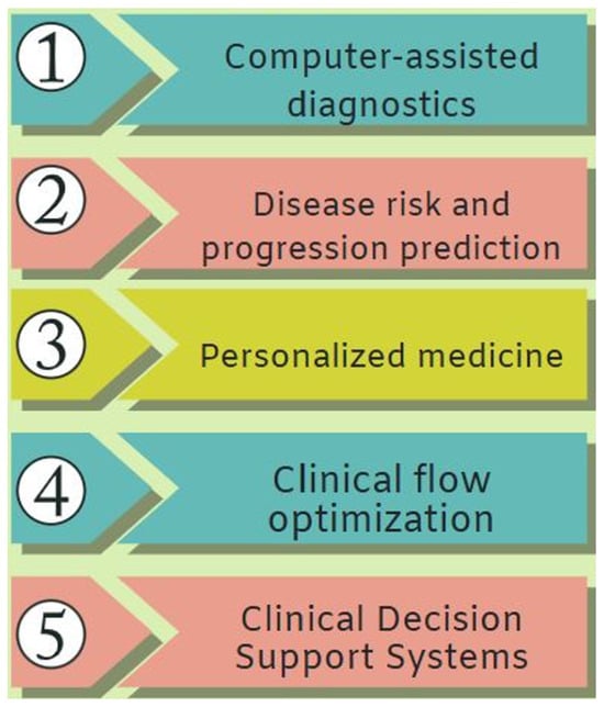
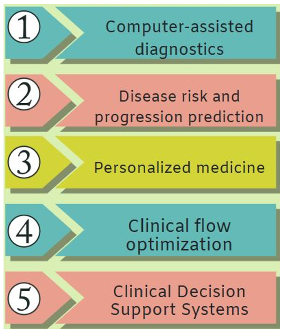
Applications of machine learning in medicine include the following (Figure 1):
Figure 1.
Applications of machine learning in medicine.
- Computer assisted diagnostics: machine learning (ML) algorithms can analyze medical images such as X-rays, CT scans or magnetic resonance imaging, helping to detect tumors, lesions or other abnormalities early;
- Disease risk and progression prediction: predictive models developed based on clinical, genetic and demographic data make it possible to estimate the risk of developing chronic diseases and anticipate the progression of patients over time;
- Personalized medicine: machine learning facilitates the selection of treatments tailored to each patient, with particular applicability in oncology;
- Clinical flow optimization: allows for prioritization of critical cases and a reduction in administrative errors;
- Clinical decision support systems: machine-learning-based platforms can provide recommendations based on practice guidelines, clinical evidence and automatically generated predictions.
In recent years, machine learning techniques have been increasingly used due to their ability to identify complex patterns in medical data and support disease prediction. For classifying risk factors and estimating the probability of a stroke event, algorithms such as extreme gradient boosting (XGBoost), random forest and logistic regression [5] were used and compared in terms of efficiency in the study [6]. XGBoost handles unbalanced datasets and builds accurate models by gradient boosting. Random forest increases classification accuracy by combining the results of multiple decision trees, and logistic regression is effective in binary classification problems, making the results easier to interpret. In the study [7], a comparison was made between XGBoost, LightGBM and CatBoost, with the results highlighting that CatBoost offers the best generalization accuracy, LightGBM stands out for its training speed and XGBoost is second in terms of both accuracy and training speed.
Machine learning relies on a variety of algorithms that enable computers to learn from data, and selecting the right one requires expertise with modern techniques [8,9]. However, the adoption of ML in medicine is not without challenges. Algorithmic models must cope with issues related to data variability and quality in real practice, class imbalances in clinical distributions or difficulties generalizing under external conditions. At the same time, to be truly useful and clinically acceptable, these systems must offer not only predictive performance but also transparency, explainability and ethical compliance.
The literature on the application of ML in medicine has expanded significantly in the past decade, but the directions of development are fragmented between specialties and data types. Thus, an overview of publication trends and the medical subfields in which ML is most present is necessary, as well as a critical analysis of the methodological and conceptual challenges that accompany this integration process.
2. Bibliometric Analysis
The development of machine learning applications in medicine has been accompanied by a steady increase in the volume of scientific literature published in recent years. Existing bibliometric analyses highlight not only a quantitative increase in the number of articles but also a progressive diversification of the clinical fields involved. There is an expansion in oncology, general internal medicine, radiology and neurology.
2.1. Temporal Dynamics of Publications
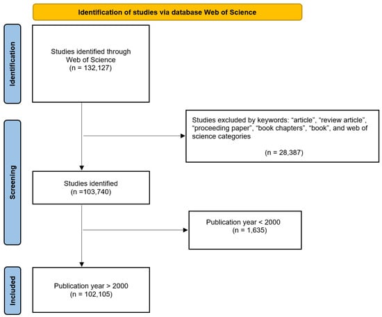
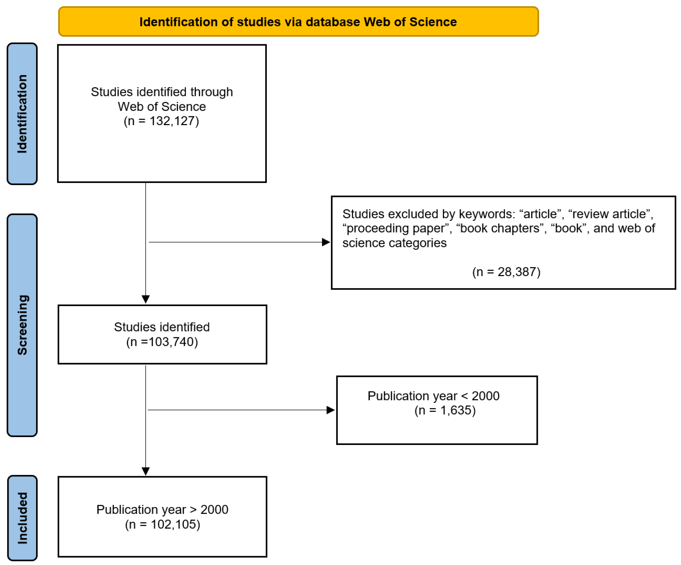
For this study, a systematic search was conducted in the Web of Science Core Collection database (Clarivate Analytics). The query was performed in mid-October 2025 and included the following keywords: “machine learning in medicine”, “AI in medicine”, “clinical decision”, “personalized medicine”, “precision medicine”, “predictive preventive personalized medicine”, and “clinical decision support systems”, yielding 132,127 results. These were then filtered by year, publication type—article, review article, proceeding paper, book chapters, book—and web of science categories, such as branches of medicine and interdisciplinary informatics, yielding 103,740 results, of which 1635 articles were published up to the year 2000, and 102,105 thereafter. The study selection process is summarized in Figure 2 through a PRISMA flow diagram, detailing the identification, screening, eligibility and inclusion stages.
Figure 2.
Study selection.
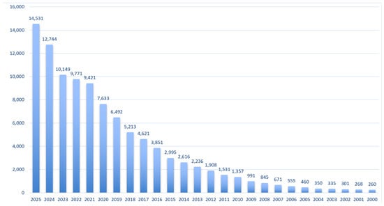
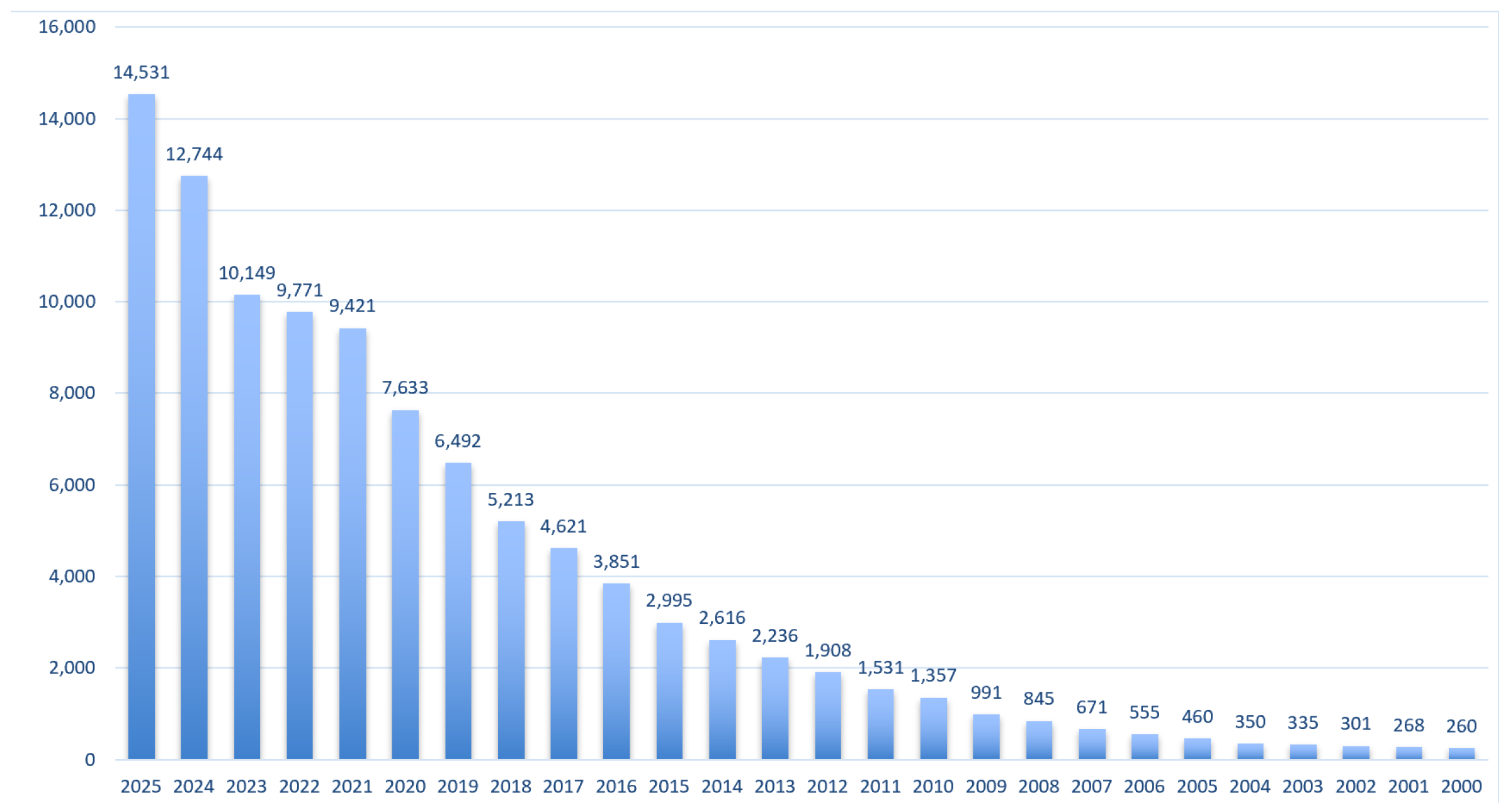
In 1975, only three publications were registered, and after 2000, their number reached several hundred. In recent years, the specialized literature has expanded to several thousand articles (2024: 12,744 publications; mid-October 2025: 14,531 publications), reflecting considerable scientific interest and rapid development of the field (Figure 3).
Figure 3.
Evolution of publications between 2000 and mid-October 2025.
Their distribution over time reveals a clear upward trajectory, particularly after 2020, when publication volumes increased sharply. This phenomenon can be correlated with the progress in the field of deep learning and the emergence of large medical datasets, which have facilitated the application and validation of algorithms. In addition, the COVID-19 pandemic has led to increased interest in automated clinical data analysis solutions.
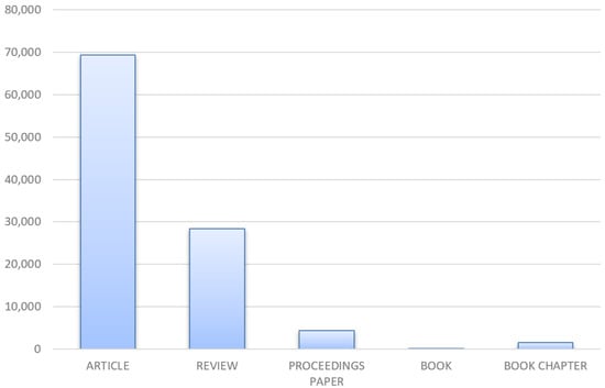
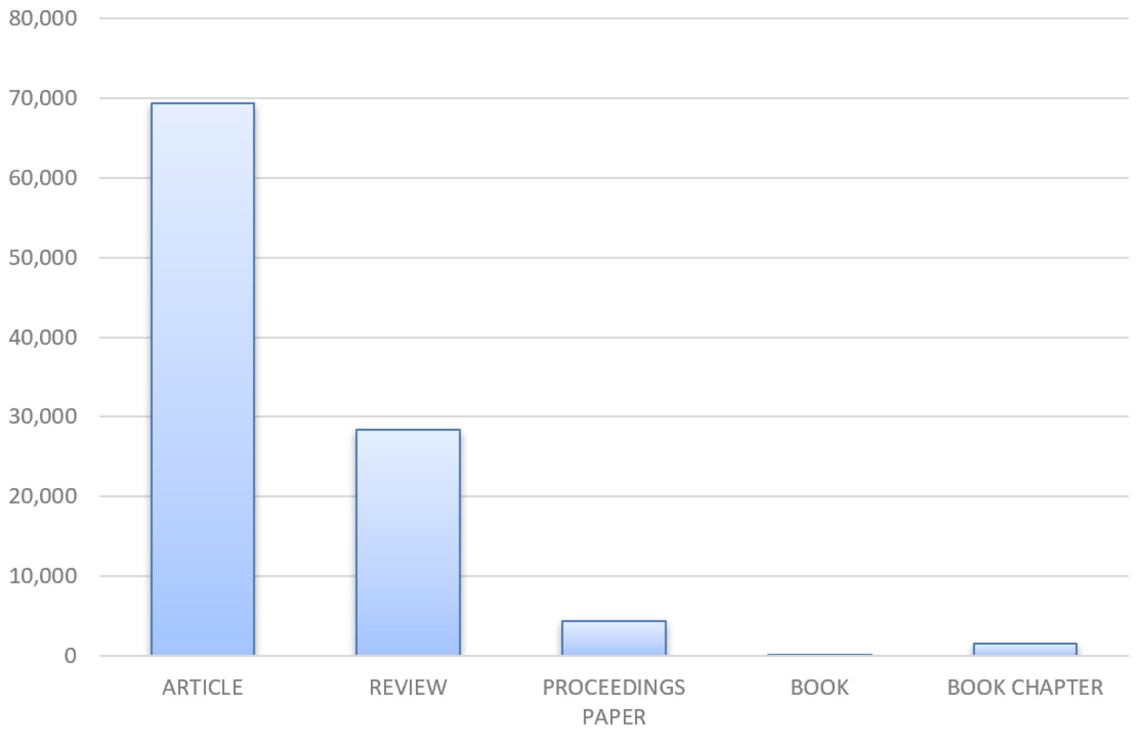
The dataset consists predominantly of research articles, followed by review articles, proceedings papers and book chapters, reflecting the methodological diversity of the analyzed literature (Figure 4).
Figure 4.
Distribution of publications by document type.
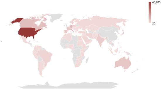
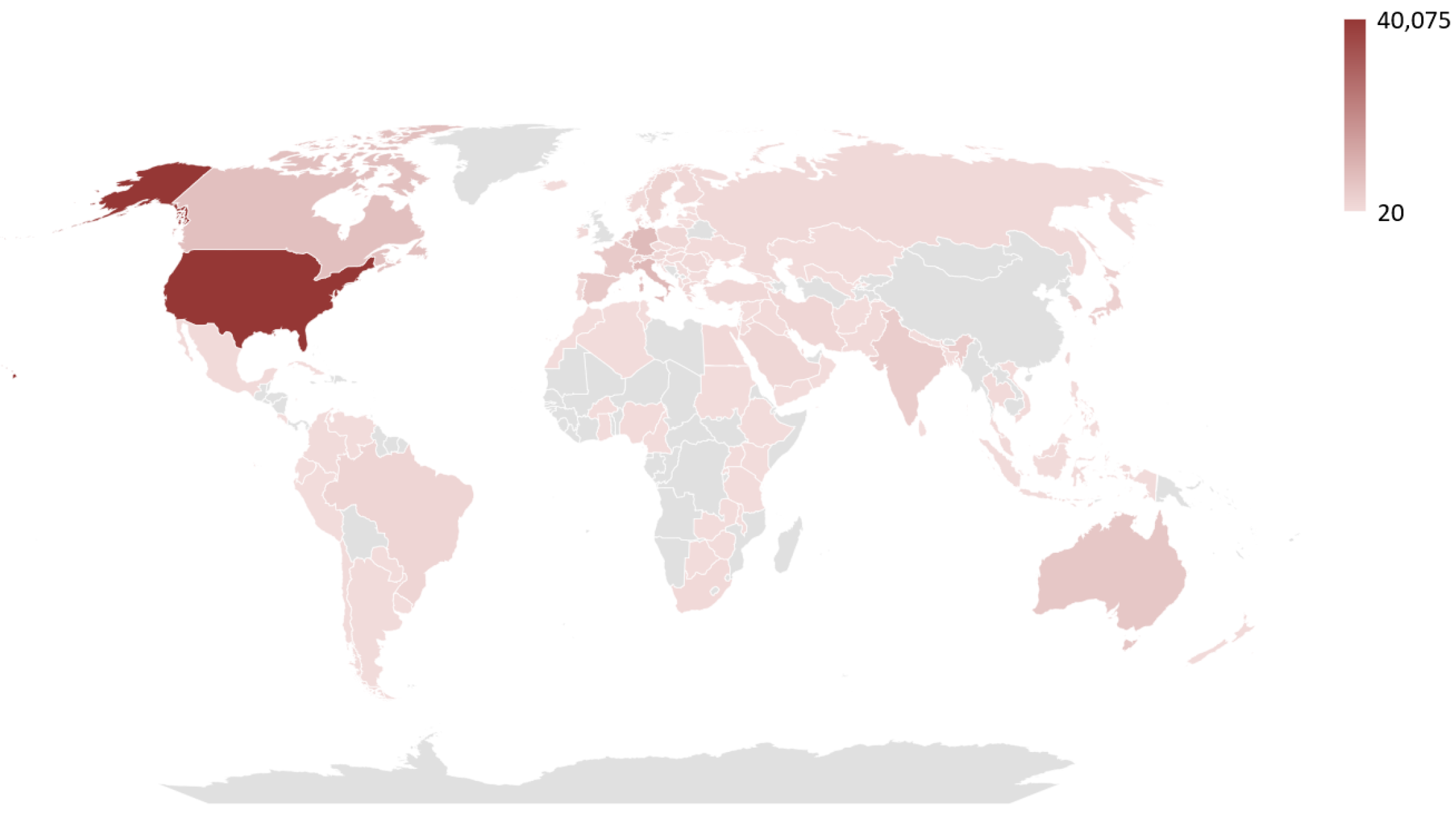
The majority of publications originated from USA (40,075), followed by China (16,329), England (9442), Italy (8413) and Germany (8092), reflecting regional research activity and publication trends in the field (Figure 5).
Figure 5.
Distribution of articles by country of origin.
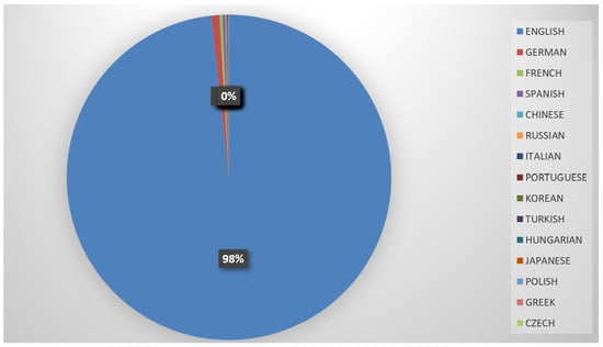
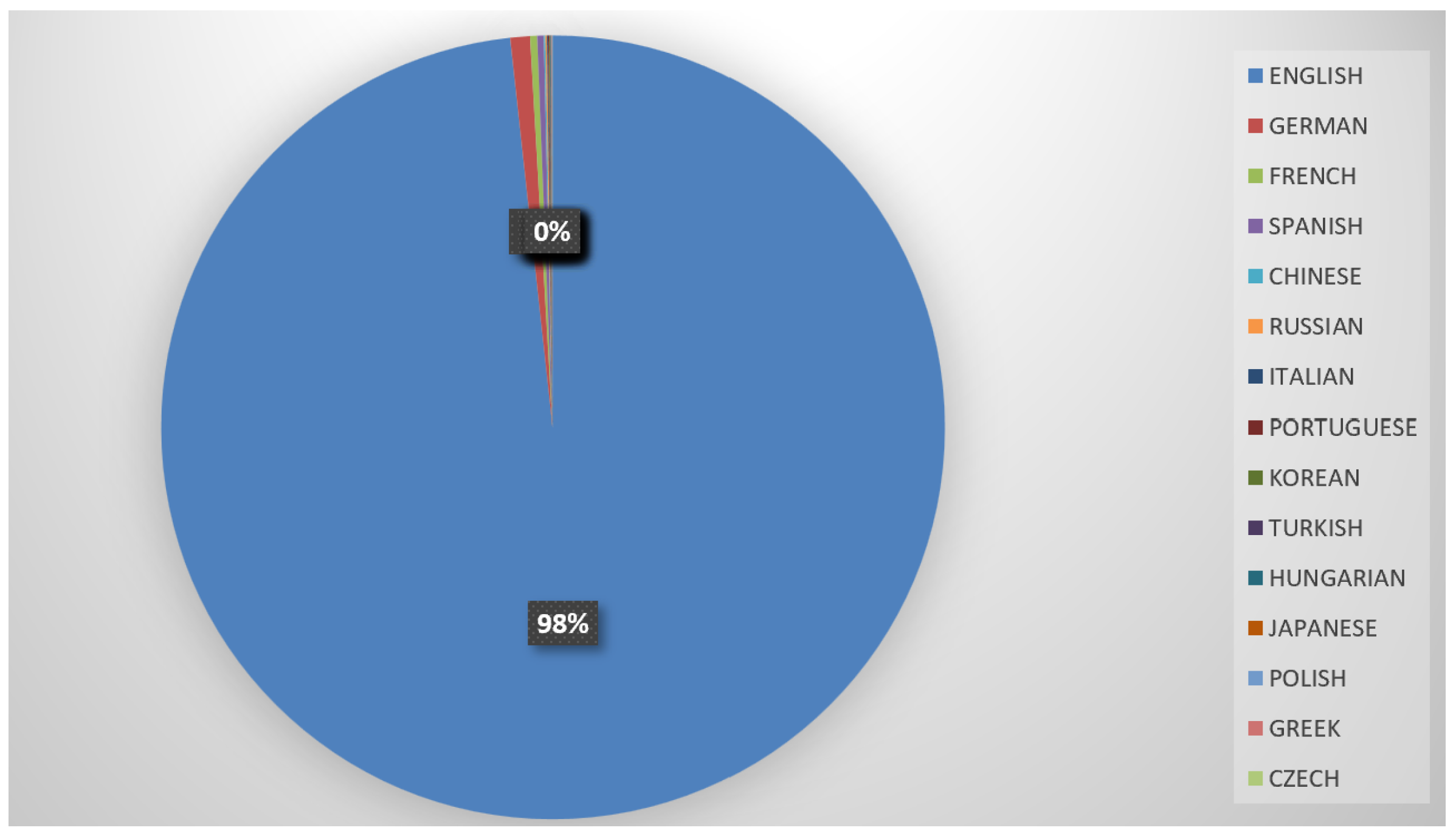
Most studies were published in English, with a smaller proportion appearing in German, French and Spanish, indicating the predominance of English-language scholarship in this research area (Figure 6).
Figure 6.
Distribution of articles by publication language.
2.2. Distribution Across Medical Subfields
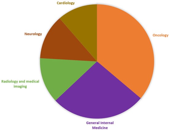
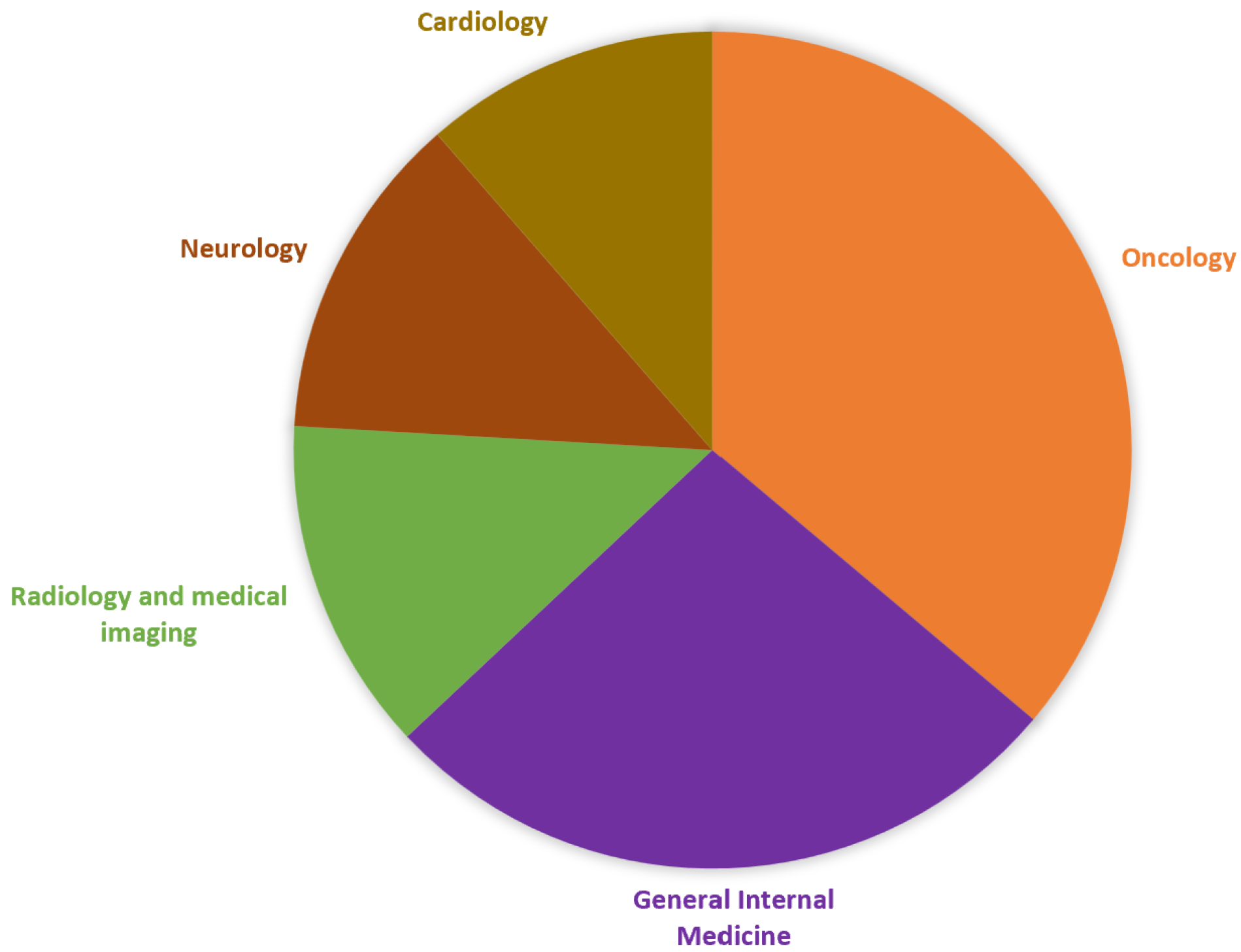
The interest in machine learning applications in medicine is also reflected in the uneven distribution of articles across different specializations, highlighting the areas where research has been most intensively developed. Oncology dominates, followed by general internal medicine, radiology and medical imaging, neurology and cardiology, which present a comparable number of articles (Figure 7). In contrast, areas such as dermatology or rheumatology are less addressed in the specialized literature.
Figure 7.
Distribution of articles across medical subfields.
Machine learning algorithms are used in the following:
- Oncology: to analyze genomic data and predict response to personalized therapies;
- General internal medicine: to predict patient outcomes, detect early signs of chronic diseases and personalize therapeutic strategies;
- Radiology and medical imaging: automated lesion detection, tumor classification and optimization of image interpretation workflows;
- Neurology: recognition of neurodegenerative diseases using imaging data and clinical biomarkers;
- Cardiology: assessment of cardiovascular risk, ECG interpretation and patient monitoring using portable devices.
3. Distribution of Machine Learning Methods Across Medical Domains
Machine learning algorithms have been applied in various medical specialties for diagnosis, prognosis and treatment optimization. This analysis included the latest published studies in oncology, radiology, neurology, internal medicine and cardiology, with the aim of identifying the most commonly used algorithms in each field.
3.1. Oncology
Advances in computational methods, algorithms and the accessibility of datasets are increasingly strengthening the role of machine learning in precision oncology.
The study [10] used RNA-Seq gene expression data and advanced machine learning techniques to identify distinct profiles between male and female patients with pancreatic ductal adenocarcinoma (PDAC) and developed sex-specific predictive models for 3-year survival. The sex-specific models outperformed the general model, with random forest as the most effective predictor. The models were integrated into Gap-App, a web application that provides free services to patients and clinicians, using the patient’s gene profile and sex for personalized predictions, highlighting the importance of sex-differentiated analysis and the advancement of personalized medicine.
In the article [11], the use of machine learning is highlighted in three key areas of thyroid cancer care: detection of malignancies, prediction of metastases and estimation of survival, using algorithms such as support vector machines, which are most effective for lateral lymph node metastasis; logistic regression; random forest, which showed the best results for malignancy detection and lung metastasis prediction; multi-layer perceptron; decision tree; naive Bayes. Additional ML models were used in specific contexts:
- Malignancy prediction: Ridge regression; FP-growth; XGBoost (for short-term survival); gradient boosting.
- Metastasis prediction: Gradient boosting decision tree (for central nodes); K-nearest neighbors; AdaBoost; convolutional neural network (most effective for lateral lymph node metastasis and achieved the best results in patients diagnosed via biopsy); multivariate adaptive regression splines (for central nodes).
- Survival models: Inductive logic programming; LightGBM; Partitioning Around Medoids (for short-term survival).
Gynecological cancers, including breast, cervical and ovarian cancers, are often detected late due to non-specific symptoms and limited screening methods. Early prediction is crucial to improve survival and guide personalized treatment. Standard algorithms like support vector machines, random forests and deep learning models, including convolutional neural networks, show high potential in cancer detection, monitoring progression and survival modeling [12].
Table 1 summarizes several studies in oncology, highlighting the machine learning algorithms employed and the most frequently addressed cancer types.
Table 1.
AI in oncology.
Risks and challenges: Technical challenges such as time-consuming manual annotations, inter-observer variability, a lack of automated integration of radiomic and genomic data, and computational constraints, as well as clinical limitations such as small sample sizes, a lack of external validation and exclusion of imaging data, which hinder clinical adoption. These issues can be addressed through standardized protocols, noise-robust algorithms, integrated multi-omics platforms and improved computational infrastructure.
3.2. General Internal Medicine
In internal medicine, artificial intelligence and machine learning enable rapid and integrated analysis of complex clinical data—including vital signs, laboratory results, imaging and patient history—to improve early diagnosis, risk stratification and treatment personalization, thus providing predictive tools that can support clinical decisions and optimize chronic disease management.
Table 2 summarizes several studies in general internal medicine, highlighting the machine learning algorithms used.
Table 2.
AI in general internal medicine.
Risks and challenges: Modern models for predicting the evolution and treatment of chronic wounds are mainly based on clinical characteristics of the patient (wound size and depth, drug treatment, presence of infection), but for the time being they do not have applicability for particular, highly specialized situations.
3.3. Radiology and Medical Imaging
Medical imaging is an essential component of modern medicine, allowing for visualization of the internal structures of the body through techniques such as radiography, computed tomography (CT), magnetic resonance imaging (MRI) and ultrasound, and complemented by techniques that capture and quantify data without generating images such as electroencephalography (EEG), magnetoencephalography (MEG) and electrocardiography. These technologies facilitate accurate diagnosis, monitoring of disease progression and treatment planning, providing important information. AI, through machine learning algorithms, computer vision and generative models, automates workflows and optimizes image acquisition and interpretation. A historical perspective on radiology, as well as new imaging techniques and trends in the field, is detailed in recent articles [24,25].
Table 3 summarizes several studies in radiology and medical imaging, highlighting the machine learning algorithms used.
Table 3.
AI in radiology and medical imaging.
Risks and challenges: AI adoption remains limited, often applied only in specific areas such as thoracic imaging, neuroimaging and musculoskeletal imaging. Issues of transparency, explainability, ethics, professional accountability and cost continue to pose challenges. However, AI does not replace radiologists but rather augments their capabilities, providing them with tools for interdisciplinary collaboration and more accurate decisions. Current challenges in medical imaging include the development of portable devices for use at the patient’s bedside in CT procedures and MRI, improving image quality and expanding the applications of elastography in sonography.
3.4. Neurology
In neurology, artificial intelligence offers faster and more refined interpretation of complex data. Its increasingly accurate diagnostic capabilities support earlier identification of neurological conditions while its personalized treatment suggestions help to develop care plans tailored to each patient’s profile. Artificial intelligence systems continue to advance at a rapid pace, thereby contributing to improved clinical decision making, more efficient workflows and ultimately better patient outcomes. In the article [33] various ML and DL algorithms were presented, and from the analyzed studies it emerged that the most frequently used were logistic regression, support vector machine, and random forest, which are models that improve clinical decision making and patient outcomes.
The introduction and continued refinement of CT and MRI modalities have had a major impact on medical imaging. Medical specialties that use imaging were early adopters of artificial intelligence methods, which led to vascular imaging becoming one of the first AI applications in stroke diagnosis and management [34,35,36]. A few examples of AI applications in neurology are presented in Table 4. There are also ample data in the field of stroke rehabilitation.
Table 4.
AI in neurology.
Risks and challenges: Although AI holds great potential in neurology, particularly for analyzing complex data and predicting clinical events, its safe and effective use relies heavily on human clinical expertise and rigorous model validation; otherwise, it may produce erroneous results due to mislabeled data.
3.5. Cardiology
Cardiovascular diseases continue to represent the main threat to population health globally. The use of machine learning techniques allows for accurate cardiovascular predictions, which can contribute to early diagnosis and reduce mortality. In the paper [45] various problems and factors affecting the heart were analyzed, using machine-learning-based classification algorithms—such as decision tree, support vector machines, random forest, naive Bayes, artificial neural network, K-nearest neighbors and linear regression—to identify heart conditions. The study [46] presents current applications of AI in interventional cardiology, highlighting their main functions, limitations and advantages, such as AI-QCT (artificial intelligence quantitative computed tomography) [47,48]; FFR-CT (fractional flow reserve computed tomography) [49,50]; AI-QCA (artificial intelligence quantitative coronary angiography) [51,52,53]; AI-FFR (artificial intelligence fractional flow reserve) [54,55,56]; AI-OCT (artificial intelligence optical coherence tomography) [57,58]; R-PCI (robotic-assisted percutaneous coronary intervention) [59,60]; MACEs (major adverse cardiovascular events) [61,62].
Table 5 summarizes several studies in cardiology, highlighting the machine learning algorithms used.
Table 5.
AI in Cardiology.
In the recent literature we also find constant approaches to topics such as the use of artificial intelligence in aortic valve stenosis [67], the application of AI in cardiac magnetic resonance imaging for precision medicine in coronary artery disease [68] and the role of ML in neurocardiology [69], highlighting the growing interest in integrating these technologies into cardiovascular practice.
Risks and challenges: Cardiovascular disease remains the leading cause of morbidity and mortality, and the limitations of traditional risk scores highlight the need for more refined approaches, which artificial intelligence and machine learning can provide by integrating complex and varied data, leading to more accurate assessment and personalized care. The paper [70] analyzes how machine learning can transform cardiovascular risk assessment and prevention by identifying new therapeutic targets, despite challenges related to data quality, transparency and model validation, highlighting the need for personalized strategies in precision cardiology.
In paper [65] the SHAP approach identified left ventricular ejection fraction (LVEF), left ventricular end-systolic diameter (LVD) and lactate dehydrogenase (LDH) as the three most influential features for predicting heart failure risk in patients with acute myocardial infarction. Individualized risk assessment was enabled through SHAP visualizations, including summary and waterfall plots. Furthermore, SHAP analysis improves the interpretability of the XGBoost model, thereby supporting clinical decision making.
4. Discussion
The specialized literature reflects a marked increase in interest in the integration of artificial intelligence into the medical field. These contributions highlight the evident trend in contemporary research toward developing digital tools that enhance diagnosis, prognosis and the personalization of therapeutic interventions.
Despite this diversification, the bibliometric analysis reveals several important gaps. Fields such as preventive medicine and public health remain relatively underrepresented in the current literature on automated medicine.
Machine learning algorithms bring significant benefits by increasing diagnostic accuracy and reducing human error, supporting complex decision making by analyzing large volumes of data in a short time, reducing costs and preventing complications by predicting risks.
Convolutional neural networks (CNNs) are the most frequently applied algorithms in oncology, particularly for medical imaging tasks such as tumor detection and characterization, due to their ability to learn complex spatial features directly from images.
In radiology, CNNs remain dominant because of their superior performance in analyzing diverse imaging modalities, enabling accurate segmentation and classification of anatomical structures and pathological findings.
In neurology, recurrent neural networks are often employed for sequential and time-series data such as EEG or longitudinal patient records, allowing for effective modeling of temporal patterns in neurological assessments.
In internal medicine, gradient boosting algorithms are predominantly used for tabular clinical data, providing high predictive accuracy for risk stratification and outcome prediction while handling heterogeneous and partially missing data efficiently.
For cardiology applications, random forest algorithms are commonly applied to structured patient data, supporting cardiovascular risk prediction and diagnostic decision making through robust handling of feature interactions and noise in clinical datasets.
However, AI does not replace healthcare professionals but instead enhances their skills, providing them with the necessary tools to support the decision-making process in medical practice.
The effective and safe use of algorithms crucially depends on human clinical expertise and rigorous validation of models; otherwise, there is a risk of producing erroneous results due to mislabeled data.
Sometimes, small sample sizes, lack of external validation and exclusion of imaging data hinder clinical adoption in many areas of medicine.
In addition to methodological and clinical aspects, ethical implications must be considered when deploying AI-based systems in healthcare. Key ethical considerations include patient data confidentiality and security, as well as the risk of inaccuracies in algorithmic predictions. In addition, clinicians’ responsibility for overseeing AI-assisted decisions, informed patient consent and equitable access to AI-supported healthcare are essential to ensuring alignment with established ethical principles in clinical practice.
5. Conclusions
The bibliometric analysis of the existing literature highlights both the evolution and distribution of machine learning (ML) applications in the medical field, as well as the main algorithmic challenges and data limitations that influence model design and validation. The number of publications followed an exponential growth trend, especially after 2020, when publication volumes increased sharply, with the annual average reaching approximately 10.708 in 2020–2025, compared to 4.298 publications in 2014–2019.
Machine learning algorithms bring significant benefits by increasing diagnostic accuracy and reducing human errors, supporting complex decision making by analyzing large volumes of data in a short time, reducing costs and preventing complications by predicting risks. Certain machine learning algorithms appear recurrently in various medical applications, such as random forest (RF), extreme gradient boost (XGBoost), support vector machine (SVM) and logistic regression (LR), reflecting their adaptability to varied clinical datasets and predictive tasks.
Integration of machine learning algorithms requires rigorous clinical validation, ethical regulations and collaboration between IT specialists and healthcare professionals.
6. Future Directions
In future work, we plan to expand the literature search to include additional scientific databases beyond Web of Science in order to obtain a more comprehensive and representative overview of machine learning applications in clinical decision support systems. This will allow us to reduce potential selection bias and strengthen the robustness of the bibliographic analysis.
We aim to extend the analysis to additional medical specialties beyond those addressed in the present study in order to examine the applicability of machine learning approaches across diverse healthcare domains.
Finally, we plan to explore real-world implementation aspects, including external validation, ethical considerations and integration into clinical workflows, to better understand the practical feasibility and impact of machine-learning-based decision support systems in routine medical practice.
Author Contributions
The work presented here was carried out as a collaboration between the two authors A.R. and E.-L.P. The authors contributed equally and significantly to the writing of this article. The authors contributed to the manuscript. All authors have read and agreed to the published version of the manuscript.
Funding
Project financed by Lucian Blaga University of Sibiu through the research grant LBUS-IRG-2023.
Institutional Review Board Statement
Not applicable.
Informed Consent Statement
Not applicable.
Data Availability Statement
No new data were created or analyzed in this study. Data sharing is not applicable to this article.
Conflicts of Interest
The authors declare no conflicts of interest. The funders had no role in the design of the study; in the collection, analyses or interpretation of data; in the writing of the manuscript, or in the decision to publish the results.
References
- Sakurada, K.; Ishikawa, T.; Oba, J.; Kuno, M.; Okano, Y.; Sakamaki, T.; Tamura, T. Medical AI and AI for Medical Sciences. Jpn. Med. Assoc. 2025, 8, 26–37. [Google Scholar]
- Adege, A.B. Advancing Biomedical Engineering With Artificial Intelligence and Machine Learning: A Systematic Review. Int. J. Clin. Pract. 2025, 2025, 9888902. [Google Scholar] [CrossRef] [Scilit]
- Derksen, C.; Walter, F.M.; Akbar, A.B.; Parmar, A.V.; Saunders, T.S.; Round, T.; Rubin, G.; Scott, S.E. The implementation challenge of computerised clinical decision support systems for the detection of disease in primary care: Systematic review and recommendations. Implement. Sci. 2025, 20, 33. [Google Scholar] [CrossRef] [Scilit]
- Sutton, R.T.; Pincock, D.; Baumgart, D.C.; Sadowski, D.C.; Fedorak, R.N.; Kroeker, K.I. An overview of clinical decision support systems: Benefits, risks, and strategies for success. Digit. Med. 2020, 3, 17. [Google Scholar] [CrossRef] [Scilit] [PubMed]
- Mridha, K.; Ghimire, S.; Shin, J.; Aran, A.; Uddin, M.M.; Mridha, M.F. Automated stroke predictionusing machine learning: An explainable and exploratory study with a web application for early intervention. IEEE Access 2023, 11, 52288–52308. [Google Scholar] [CrossRef] [Scilit]
- Sitompul, L.; Nababan, A.; Manihuruk, M.; Ponsen, W.; Supriyandi, S. Comparison of Xgboost, Random Forest and Logistic Regression Algorithms in Stroke Disease Classification. Sinkron 2025, 9, 957–968. [Google Scholar] [CrossRef] [Scilit]
- Bentéjac, C.; Csörgő, A.; Martínez-Muñoz, G. A comparative analysis of gradient boosting algorithms. Artif. Intell. Rev. 2021, 54, 1937–1967. [Google Scholar] [CrossRef] [Scilit]
- Yeo, C.J.J.; Ramasamy, S.; Leong, F.J.; Nag, S.; Simmons, Z. A neuromuscular clinician’s primer on machine learning. J. Neuromuscul. Dis. 2025, 13, 20–42. [Google Scholar] [CrossRef] [Scilit]
- Toh, Z.; Thambawita, V.; Grotle, M.; He, H.G.; Pikkarainen, M.; Hey, H.W.D.; Solberg, T.; Ingebrigtsen, T.; Berg, B. Development of machine learning models for predicting patient-perceived benefit following lumbar disc herniation or spinal stenosis surgery. Eur. Spine J. 2025. Online ahead of print. [Google Scholar] [CrossRef] [Scilit]
- Ojha, A.; Zhao, S.J.; Akpunonu, B.; Zhang, J.T.; Simo, K.A.; Liu, J.Y. Gap-App: A sex-distinct AI-based predictor for pancreatic ductal adenocarcinoma survival as a web application open to patients and physicians. Cancer Lett. 2025, 622, 217689. [Google Scholar] [CrossRef] [Scilit]
- Lixandru-Petre, I.-O.; Dima, A.; Musat, M.; Dascalu, M.; Gradisteanu Pircalabioru, G.; Iliescu, F.S.; Iliescu, C. Machine Learning for Thyroid Cancer Detection, Presence of Metastasis, and Recurrence Predictions—A Scoping Review. Cancers 2025, 17, 1308. [Google Scholar] [CrossRef] [Scilit]
- Garg, P.; Krishna, M.; Kulkarni, P.; Horne, D.; Salgia, R.; Singhal, S.S. Machine Learning Models for Predicting Gynecological Cancers: Advances, Challenges, and Future Directions. Cancers 2025, 17, 2799. [Google Scholar] [CrossRef] [Scilit]
- Hormaty, S.; Seiwan, A.N.; Rasheed, B.H.; Parvaz, H.; Gharahzadeh, A.; Ghaznavi, H. A Review on Biomarker-Enhanced Machine Learning for Early Diagnosis and Outcome Prediction in Ovarian Cancer Management. Cancer Med. 2025, 14, e71224. [Google Scholar] [CrossRef] [Scilit]
- Tao, H.; Wang, L.; Ding, Y.; Yi, L.; Han, M.; Gu, M.; Wu, J. Microbial and Metabolic Disorders in Cervical Cancer: Structural Insights, Biomarkers, Mechanisms, and Therapeutic Strategies. Cancer Sci. 2025, 116, 3228–3238. [Google Scholar] [CrossRef] [Scilit]
- Ramouz, A.; Adeliansedehi, A.; Khajeh, E.; März, K.; Michael, D.; Wagner, M.; Müller-Stich, B.P.; Mehrabi, A.; Majlesara, A. Introducing and Validating theMultiphasic Evidential Decision-MakingMatrix (MedMax) for ClinicalManagement in Patients with Intrahepatic Cholangiocarcinoma. Cancers 2025, 17, 52. [Google Scholar]
- Hagen, J.; Hornung, L.; Barham, W.; Mukhopadhyay, S.; Bess, A.; Contrera, K.; Basu, D.; Sandulache, V.; Spielmann, G.; Kansara, S. Artificial Intelligence in Head and Neck Cancer: Towards Precision Medicine. Cancers 2025, 17, 3023. [Google Scholar] [CrossRef] [Scilit] [PubMed]
- Kim, J.W.; Lee, J.; Lee, S.H.; Ahn, S.; Park, K.H. Machine Learning–Based Prognostic Gene Signature for Early Triple-Negative Breast Cancer. Cancer Res. Treat. 2025, 57, 731–740. [Google Scholar] [CrossRef] [Scilit]
- Rafiepoor, H.; Banoei, M.M.; Ghorbankhanloo, A.; Muhammadnejad, A.; Razavirad, A.; Soleymanjahi, S.; Amanpour, S. Exploring the potential of machine learning in gastric cancer: Prognostic biomarkers, subtyping, and stratification. BMC Cancer 2025, 25, 809. [Google Scholar] [CrossRef] [Scilit] [PubMed]
- Wang, Z.; Xiao, Z.; Zhang, T.; Lu, M.; Li, H.; Cao, J.; Zheng, J.; Zhou, Y.; Dai, J.; Wang, C.; et al. Development and validation of a novel artificial intelligence algorithm for precise prediction the postoperative prognosis of esophageal squamous cell carcinoma. BMC Cancer 2025, 25, 134. [Google Scholar] [CrossRef] [Scilit]
- Spinazzola, E.; Picaud, G.; Becchi, S.; Pittarello, M.; Ricci, E.; Chaumont, M.; Subsol, G.; Pareschi, F.; Teot, L.; Secco, J. Chronic Ulcers Segmentation and Healing Prediction Through Machine Learning Approaches: Preliminary Results on Diabetic Foot Ulcers Case Study. J. Clin. Med. 2025, 14, 2943. [Google Scholar] [CrossRef] [Scilit]
- Chakraborty, A.; Pant, M.D. Machine Learning Models for Pancreatic Cancer Survival Prediction: A Multi-Model Analysis Across Stages and Treatments Using the Surveillance, Epidemiology, and End Results (SEER) Database. J. Clin. Med. 2025, 14, 4686. [Google Scholar] [CrossRef] [Scilit]
- Obeagu, E.I.; Ezeanya, C.U.; Ogenyi, F.C.; Ifu, D.D. Big data analytics and machine learning in hematology. Transformative insights, applications and challenges. Medicine 2025, 104, 10. [Google Scholar] [CrossRef] [Scilit]
- Li, S.; Yue, R.; Lu, S.; Luo, J.; Wu, X.; Zhang, Z.; Liu, M.; Fan, Y.; Zhang, Y.; Pan, C.; et al. Artificial intelligence and machine learning in acute respiratory distress syndrome management: Recent advances. Front. Med. 2025, 12, 1597556. [Google Scholar] [CrossRef] [Scilit]
- Kantarcı, M.; Aydın, S.; Oğul, H.; Kızılgöz, V. New imaging techniques and trends in radiology. Diagn. Interv. Radiol. 2025, 31, 505–517. [Google Scholar] [CrossRef] [Scilit]
- Schlemmer, H.P. Navigating the AI revolution: Will radiology sink or soar? Jpn. J. Radiol. 2025, 43, 1628–1633. [Google Scholar] [CrossRef] [Scilit] [PubMed]
- Cao, M.; Xu, R.; You, Y.; Huang, C.; Tong, Y.; Zhang, R.; Zhang, Y.; Yu, P.; Wang, Y.; Chen, W.; et al. Development and validation of CT-based fusion model for preoperative prediction of invasion and lymph node metastasis in adenocarcinoma of esophagogastric junction. BMC Med. Imaging 2025, 25, 242. [Google Scholar] [CrossRef] [Scilit] [PubMed]
- Cozzi, J.L.; Li, H.; Fuhrman, J.D.; Lan, L.; Williams, J.; Finnerty, B.; Fahey, T.J., III; Tumati, A.; Genender, J.; Keutgen, X.M.; et al. Multi-institutional development and testing of attention-enhanced deep learning segmentation of thyroid nodules on ultrasound. Int. J. Comput. Assist. Radiol. Surg. 2025, 20, 259–267. [Google Scholar] [CrossRef] [Scilit]
- Zou, D.; Lyu, F.; Pan, Y.; Fan, X.; Du, J.; Mai, X. Enhancing diagnostic precision for thyroid C-TIRADS category 4 nodules: A hybrid deep learning and machine learning model integrating grayscale and elastographic ultrasound features. Quant. Imaging Med. Surg. 2025, 19, 7951–7963. [Google Scholar] [CrossRef] [Scilit]
- Dong, S.; Fu, A.; Liu, J. Prediction of metastases in confusing mediastinal lymph nodes based on flourine-18 fluorodeoxyglucose (18F-FDG) positron emission tomography/computed tomography (PET/CT) imaging using machine learning. Quant. Imaging Med. Surg. 2024, 14, 4723–4734. [Google Scholar] [CrossRef] [Scilit] [PubMed]
- Ichikawa, H.; Ichikawa, S.; Sawane, Y. Machine learning-based estimation of patient body weight from radiation dose metrics in computed tomography. J. Appl. Clin. Med. Phys. 2024, 25, 9. [Google Scholar] [CrossRef] [Scilit]
- Kendall, J.; Gaspar, G.; Berger, D.; Levman, L. Machine Learning and Feature Selection in Pediatric Appendicitis. Tomography 2025, 11, 90. [Google Scholar] [CrossRef] [Scilit]
- Khateri, M.; Mofrad, F.B.; Geramifar, P.; Jenabi, E. Machine learning-based analysis of 68Ga-PSMA-11 PET/CT images for estimation of prostate tumor grade. Phys. Eng. Sci. Med. 2024, 47, 741–753. [Google Scholar] [CrossRef] [Scilit]
- Issaiy, M.; Zarei, D.; Kolahi, S.; Liebeskind, D.S. Machine learning and deep learning algorithms in stroke medicine: A systematic review of hemorrhagic transformation prediction models. J. Neurol. 2025, 271, 37. [Google Scholar] [CrossRef] [Scilit]
- Gupta, S.; Sharma, D.K.; Gupta, M.K. Artificial Intelligence in Diagnosis and Management of Ischemic Stroke. Biomed. J. Sci. Tech. Res. 2019, 13, 3. [Google Scholar] [CrossRef] [Scilit]
- Soun, J.E.; Chow, D.S.; Nagamine, M.; Takhtawala, R.S.; Filippi, C.G.; Yu, W.; Chang, P.D. Artificial Intelligence and Acute Stroke Imaging. AJNR Am. J. Neuroradiol. 2021, 42, 2–11. [Google Scholar] [CrossRef] [Scilit]
- Lee, E.J.; Kim, Y.H.; Kim, N.; Kang, D.W. Deep into the Brain: Artificial Intelligence in Stroke Imaging. J. Stroke 2017, 19, 277–285. [Google Scholar] [CrossRef] [Scilit]
- Albers, G.; Wald, M.; Mlynash, M.; Endres, J.; Bammer, R.; Straka, M.; Maier, A.; Hinson, H.; Sheth, K.; Kimberly, W.; et al. Automated Calculation of Alberta Stroke Program Early CT Score: Validation in Patients With Large Hemispheric Infarct. Stroke 2019, 50, 11. [Google Scholar] [CrossRef] [Scilit] [PubMed]
- Attia, Z.I.; Noseworthy, P.A.; Jimenez, F.L.; Asirvatham, S.J.; Deshmukh, A.J.; Gersh, B.J.; Carter, R.E.; Yao, X.; Rabinstein, A.A.; Erickson, B.J.; et al. An artificial intelligence-enabled ECG algorithm for the identification of patients with atrial fibrillation during sinus rhythm: A retrospective analysis of outcome prediction. Lancet 2019, 394, 861–867. [Google Scholar] [CrossRef] [Scilit] [PubMed]
- Haberfehlner, H.; Ven, S.; Burg, S.; Huber, F.; Georgievska, S.; Aleo, I.; Harlaar, J.; Bonouvrié, L.; van der Krogt, M.; Buizer, A. Towards automated video-based assessment of dystonia in dyskinetic cerebral palsy: A novel approach using markerless motion tracking and machine learning. Front. Robot. AI 2023, 10, 1108114. [Google Scholar] [CrossRef] [Scilit]
- Jing, J.; Ge, W.; Hong, S.; Fernandes, M.B.; Lin, Z.; Yang, C.; An, S.; Struck, A.F.; Herlopian, A.; Karakis, I.; et al. Development of Expert-Level Classification of Seizures and Rhythmic and Periodic Patterns During EEG Interpretation. Neurology 2023, 100, 1750–1762. [Google Scholar] [CrossRef] [Scilit]
- Beniczky, S.; Aurlien, H.; Brøgger, J.C.; Hirsch, L.J.; Schomer, D.L.; Trinka, E.; Pressler, R.M.; Wennberg, R.; Visser, G.H.; Eisermann, M.; et al. Standardized computer-based organized reporting of EEG: SCORE—Second version. Clin. Neurophysiol. 2017, 128, 2334–2346. [Google Scholar] [CrossRef] [Scilit] [PubMed]
- AbuAlrob, M.A.; Itbaisha, A.; Mesraoua, B. Unlocking new frontiers in epilepsy through AI: From seizure prediction to personalized medicine. Epilepsy Behav. 2025, 166, 110327. [Google Scholar] [CrossRef] [Scilit]
- Ervin, B.; Arya, R. Artificial Intelligence and Machine Learning in Neuromodulation for Epilepsy. J. Clin. Neurophysiol. 2025, 42, 493–504. [Google Scholar] [CrossRef] [Scilit]
- Hossain, K.; Ashraf, A.; Islam, M.; Sourav, H.S.; Shimul, M.H. Optimizing Alzheimer’s disease prediction through ensemble learning and feature interpretabilitywith SHAP-based feature analysis. Alzheimer’s Dement. Diagn. Assess. Dis. Monit. 2025, 17, e70162. [Google Scholar]
- Azmi, J.; Arif, M.; Nafis, T.; Alam, M.A.; Tanweer, S.; Wang, G. A systematic review on machine learning approaches for cardiovascular disease prediction using medical big data. Med. Eng. Phys. 2022, 105, 103825. [Google Scholar] [CrossRef] [Scilit] [PubMed]
- Veneziano, F.A.; Cocco, N.; Veneziano, R. Artificial intelligence in interventional cardiology: Current applications and future clinical integration. Vessel Plus 2025, 9, 3. [Google Scholar] [CrossRef] [Scilit]
- Omori, H.; Matsuo, H.; Fujimoto, S.; Sobue, Y.; Nozaki, Y.; Nakazawa, G.; Takahashi, K.; Osawa, K.; Okubo, R.; Kaneko, U.; et al. Determination of lipid-rich plaques by artificial intelligence-enabled quantitative computed tomography using near-infrared spectroscopy as reference. Atherosclerosis 2023, 386, 117363. [Google Scholar] [CrossRef] [Scilit]
- Khan, H.; Bansal, K.; Griffin, W.F.; Cantlay, C.; Sidahmed, A.; Nurmohamed, N.S.; Zeman, R.K.; Katz, R.J.; Blankstein, R.; Earls, J.P.; et al. Assessment of atherosclerotic plaque burden: Comparison of AI-QCT versus SIS, CAC, visual and CAD-RADS stenosis categories. Int. J. Cardiovasc. Imaging 2024, 40, 1201–1209. [Google Scholar] [CrossRef] [Scilit]
- Chiou, A.; Hermel, M.; Sidhu, R.; Hu, E.; van Rosendael, A.; Bagsic, S.; Udoh, E.; Kosturakis, R.; Aziz, M.; Ruiz, C.R.; et al. Artificial intelligence coronary computed tomography, coronary computed tomography angiography using fractional flow reserve, and physician visual interpretation in the per-vessel prediction of abnormal invasive adenosine fractional flow reserve. Eur. Heart J.-Imaging Methods Pract. 2024, 2, qyae035. [Google Scholar] [CrossRef] [Scilit]
- Chen, M.; Almeida, S.O.; Sayre, J.W.; Karlsberg, R.P.; Packard, R.R.S. Distal-vessel fractional flow reserve by computed tomography to monitor epicardial coronary artery disease. Eur. Heart J. Cardiovasc. Imaging 2024, 25, 163–172. [Google Scholar] [CrossRef] [Scilit]
- Kim, Y.I.; Roh, J.-H.; Kweon, J.; Kwon, H.; Chae, J.; Park, K.; Lee, J.-H.; Jeong, J.-O.; Kang, D.-Y.; Lee, P.H.; et al. Artificial intelligence-based quantitative coronary angiography of major vessels using deep-learning. Int. J. Cardiol. 2024, 405, 131945. [Google Scholar] [CrossRef] [Scilit]
- Sen, T.; Kilit, C.; Astarcioglu, M.A.; Asarcikli, L.D.; Aksu, T.; Kafes, H.; Parspur, A.; Gozubuyuk, G.; Amasyali, B. Comparison of quantitative and qualitative coronary angiography: Computer versus the eye. Cardiovasc. J. Afr. 2018, 29, 278–282. [Google Scholar] [CrossRef] [Scilit]
- Jo, J.I.; Koo, H.J.; Kang, J.W.; Kim, Y.H.; Yang, D.H. Artificial Intelligence-Driven Assessment of Coronary Computed Tomography Angiography for Intermediate Stenosis: Comparison with Quantitative Coronary Angiography and Fractional Flow Reserve. Am. J. Cardiol. 2025, 239, 82–89. [Google Scholar] [CrossRef] [Scilit] [PubMed]
- Parikh, R.V.; Liu, G.; Plomondon, M.E.; Sehested, T.S.; Hlatky, M.A.; Waldo, S.W.; Fearon, W.F. Utilization and outcomes of measuring fractional flow reserve in patients with stable ischemic heart disease. J. Am. Coll. Cardiol. 2020, 75, 409–419. [Google Scholar] [CrossRef] [Scilit]
- Choi, K.H.; Lee, S.H.; Lee, J.M.; Hwang, D.; Zhang, J.; Kim, J.; Im, S.Y.; Kim, H.K.; Nam, C.-W.; Doh, J.-H.; et al. Clinical relevance and prognostic implications of contrast quantitative flow ratio in patients with coronary artery disease. Int. J. Cardiol. 2021, 325, 23–29. [Google Scholar] [CrossRef] [Scilit] [PubMed]
- Asano, T.; Tanigaki, T.; Hoshino, M.; Yasunaga, M.; Nonaka, H.; Emori, H.; Katagiri, Y.; Miyazaki, Y.; Sotomi, Y.; Kogame, N.; et al. Quantitative flow ratio versus fractional flow reserve for Heart Team decision-making in multivessel disease: The randomised, multicentre DECISION QFR trial. EuroIntervention 2024, 20, 561–570. [Google Scholar] [CrossRef] [Scilit]
- Alikarami, M.; Faraj, T.A.; Hama, N.H.; Hosseini, A.S.; Alavi, M.; Kashani, M.; Aminnezhad, S. Artificial intelligence in advancing optical coherence tomography for disease detection and cancer diagnosis: A scoping review. Eur. J. Surg. Oncol. 2025, 51, 9. [Google Scholar] [CrossRef] [Scilit] [PubMed]
- Tsai, T.Y.; Kageyama, S.; He, X.; Pompilio, G.; Andreini, D.; Pontone, G.; La Meir, M.; De Mey, J.; Tanaka, K.; Doenst, T.; et al. Feasibility and accuracy of real-time 3D-holographic graft length measurements. Eur. Heart J.-Digit. Health 2024, 5, 101–104. [Google Scholar] [CrossRef] [Scilit]
- Subhan, S.; Malik, J.; Haq, A.U.; Qadeer, M.S.; Zaidi, S.M.J.; Orooj, F.; Zaman, H.; Mehmoodi, A.; Majeedi, U. Role of Artificial Intelligence and Machine Learning in Interventional Cardiology. Curr. Probl. Cardiol. 2023, 48, 101698. [Google Scholar] [CrossRef] [Scilit]
- Dash, D.; Dash, U.; Abu-Dakka, B. Artificial intelligence-driven techniques for interventional cardiology: A current appraisal. Cardiovasc. Revasc. Med. 2025, 81, 154–163. [Google Scholar] [CrossRef] [Scilit] [PubMed]
- Bosco, E.; Hsueh, L.; McConeghy, K.W.; Gravenstein, S.; Saade, E. Major adverse cardiovascular event definitions used in observational analysis of administrative databases: A systematic review. BMC Med. Res. Methodol. 2021, 21, 241. [Google Scholar] [CrossRef] [Scilit]
- Savic, L.; Simic, D.; Lasica, R.; Krljanac, G.; Stankovic, S.; Mrdovic, I.; Asanin, M. Predictors of Major Adverse Cardiovascular Events in Stable Patients After ST Elevation Myocardial Infarction. Clin. Pract. 2025, 15, 106. [Google Scholar] [CrossRef] [Scilit]
- Islam, A.; Varadarajan, J.; Sufian, A.; Mishra, B.K.; Rasel, R.A. Integrated Deep Learning Framework for Cardiac Risk Stratification and Complication Analysis in Leigh’s Disease. Cardiogenetics 2025, 15, 19. [Google Scholar] [CrossRef] [Scilit]
- Gonzalez-Lopez, E.; Maurer, M.S.; Garcia-Pavia, P. Transthyretin amyloid cardiomyopathy: A paradigm for advancing precision medicine. Eur. Heart J. 2025, 46, 999–1013. [Google Scholar] [CrossRef] [Scilit] [PubMed]
- Lin, Q.; Zhao, W.; Zhang, H.; Chen, W.; Lian, S.; Ruan, Q.; Qu, Z.; Lin, Y.; Chai, D.; Lin, X. Predicting the risk of heart failure after acute myocardial infarction using an interpretable machine learning model. Front. Cardiovasc. Med. 2025, 12, 1444323. [Google Scholar] [CrossRef] [Scilit]
- Zhang, Z.; Ye, J. Predicting mortality in critically ill patients with hypertension using machine learning and deep learning models. Front. Cardiovasc. Med. 2025, 12, 1568907. [Google Scholar] [CrossRef] [Scilit]
- Chen, S.; Wu, C.; Zhang, Z.; Liu, L.; Zhu, Y.; Hu, D.; Jin, C.; Fu, H.; Wu, J.; Liu, S. The role of artificial intelligence in aortic valve stenosis: A bibliometric analysis. Front. Cardiovasc. Med. 2025, 12, 1568907. [Google Scholar] [CrossRef] [Scilit] [PubMed]
- Jiménez-Jara, C.; Salas, R.; Díaz-Navarro, R.; Chabert, S.; Andia, M.E.; Vega, J.; Urbina, J.; Uribe, S.; Sekine, T.; Raimondi, F.; et al. AI Applied to Cardiac Magnetic Resonance for Precision Medicine in Coronary Artery Disease: A Systematic Review. J. Cardiovasc. Dev. Dis. 2025, 12, 345. [Google Scholar] [CrossRef] [Scilit] [PubMed]
- Basem, J.; Mani, R.; Sun, S.; Gilotra, K.; Dianati-Maleki, N.; Dashti, R. Clinical applications of artificial intelligence and machine learning in neurocardiology: A comprehensive review. Front. Cardiovasc. Med. 2025, 12, 1525966. [Google Scholar] [CrossRef] [Scilit]
- Wang, Y.; Aivalioti, E.; Stamatelopoulos, K.; Zervas, G.; Mortensen, M.B.; Zeller, M.; Liberale, L.; Di Vece, D.; Schweiger, V.; Camici, G.G.; et al. Machine learning in cardiovascular risk assessment: Towards a precision medicine approach. Eur. J. Clin. Investig. 2025, 55, e70017. [Google Scholar] [CrossRef] [Scilit]

Disclaimer/Publisher’s Note: The statements, opinions and data contained in all publications are solely those of the individual author(s) and contributor(s) and not of MDPI and/or the editor(s). MDPI and/or the editor(s) disclaim responsibility for any injury to people or property resulting from any ideas, methods, instructions or products referred to in the content. |
© 2026 by the authors. Licensee MDPI, Basel, Switzerland. This article is an open access article distributed under the terms and conditions of the Creative Commons Attribution (CC BY) license.