


Abstract
The long-term success of orthopedic implants depends not only on their mechanical performance but also on their ability to establish a stable and biologically active interface with surrounding tissues. Despite the widespread clinical use of metallic, ceramic, and polymeric biomaterials, implant failure remains associated with insufficient osseointegration, bacterial infection, wear, corrosion, and adverse immune responses. This review provides a comprehensive overview of conventional biomaterials used in orthopedic implants and critically examines current surface engineering strategies developed to improve implant performance and longevity. Particular emphasis is placed on coating technologies, including sol–gel processing, electrochemical deposition, plasma spraying, physical and chemical vapor deposition, and CoBlast™, highlighting their influence on coating adhesion, bioactivity, and clinical performance. Recent developments in bioactive, antibacterial, immunomodulatory, and stimuli-responsive coatings are discussed, together with advances in therapeutic ion incorporation, extracellular matrix-inspired functionalization, and smart drug-delivery systems. Furthermore, the emerging role of osteoimmunomodulation, additive manufacturing, and patient-specific implant design is examined as a key driver for the next generation of orthopedic devices. By integrating materials science, surface engineering, and biological mechanisms, this work highlights current challenges and future opportunities in orthopedic implant technology, offering valuable insights for the development of safer, longer-lasting, and more biologically responsive implant systems.
1. Introduction
The use of biomedical devices to restore musculoskeletal function has become a key part of modern surgical practice, especially in response to trauma, degenerative pathologies, and congenital skeletal abnormalities. Over recent decades, the clinical demand for such technologies has expanded significantly, driven by an aging global population, the rise in chronic bone-related diseases, and the need to improve the quality of life in affected individuals. These interventions are designed not only to recover anatomical structure and mobility but also to perform reliably in environments subject to continuous mechanical loading, biological variability, and microbial exposure [1,2].
Historically, the first generations of structural implants were developed using metals with proven mechanical robustness and resistance to body fluids. Alloys containing titanium, iron-chromium-nickel combinations, and cobalt have been widely used due to their high load-bearing capacity, resistance to fatigue, and manufacturing versatility [3]. However, their long-term interaction with surrounding tissues has exposed certain biological limitations. These materials typically do not establish direct molecular communication with living cells, which can compromise their stability in vivo and increase the risk of complications such as chronic inflammation, fibrous encapsulation, or device migration over time [4].
To address these drawbacks, significant attention has been directed toward modifying the interaction between implantable materials and biological systems. A key focus has been on optimizing their capacity to influence cellular responses such as adhesion, proliferation, and differentiation, particularly of bone-forming cells [5]. This resulted in the development of external layers and surface designs that are intended to create a more favorable biological environment, either by mimicking the natural extracellular matrix or by releasing chemical signals that stimulate tissue regeneration [6]. However, balancing biological integration with long-term mechanical stability remains a significant challenge, particularly when these components are subjected to repetitive loading, micromovements, or chemical degradation in situ [3].
In parallel, other types of implant materials have gained recognition for their adaptability, especially in contexts where structural agreement or time-dependent degradation is desired. These systems can be engineered to accommodate dynamic physiological environments, offering solutions for temporary support structures or slow degradation that correspond with healing phases. In particular, blends or composites that combine rigid and flexible components have shown promise in supporting more natural mechanical behavior and improved biocompatibility [7].
The interaction between structural and functional performance has also influenced how these technologies are manufactured. Over the past decade, innovative fabrication platforms have emerged, allowing control over architecture at micro- and nanoscales. Techniques capable of adjusting geometric complexity, spatial resolution, and physicochemical features at the surface have opened new avenues for personalizing design to individual patient needs. In many cases, this has enabled the inclusion of substances with therapeutic action such as ions with antibacterial or osteoinductive potential, or bioactive molecules that modulate immune response [8].
A growing area of research focuses on coatings that react dynamically to their surrounding environment. These platforms aim to adapt their behavior in response to local chemical or mechanical signals, such as the presence of inflammatory mediators, variations in fluid composition, or external forces [9]. One notable example is the use of polymeric matrices capable of releasing pharmaceutical agents in response to specific biological stimuli, such as cytokines or pH alterations. These approaches have great potential to reduce early complications and improve implant osseointegration, defined as the direct structural and functional connection between living bone and the surface of a load-bearing implant without the interposition of fibrous tissue [10].
Therefore, the integration of engineering principles, biological insight, and material science has become central to advancing the next generation of bone-interfacing devices. This review explores the state-of-the-art in such technologies, examining the structural bases, interface mechanisms, fabrication routes, and current limitations that shape their development.
This review aims to provide a comprehensive and integrative overview of conventional orthopedic implant materials and recent advances in surface modification strategies, with particular emphasis on the interplay between physicochemical properties, biological responses, and clinical performance. Unlike previous reviews that often focus on individual material classes or isolated coating technologies, the present work adopts a multidisciplinary perspective, linking material selection, surface engineering approaches, and emerging biological mechanisms involved in osseointegration and implant longevity. The manuscript is intended for a broad readership including biomaterials scientists, biomedical engineers, clinicians, and researchers entering the orthopedic field, offering both foundational knowledge and critical insights into translational challenges. By highlighting current limitations, comparative advantages of different technologies, and future research directions, this review seeks to contribute to the development of next-generation orthopedic implants with improved functionality, durability, and patient-specific adaptability.
Literature Search Strategy
This review was conducted to provide a comprehensive overview of conventional biomaterials and surface engineering strategies for next-generation orthopedic implants. A structured literature search was initiated in 2024 and continuously updated throughout the preparation of the manuscript to incorporate recent advances in the field.
Electronic databases, including Web of Science, Scopus, PubMed, and ScienceDirect, were searched to identify relevant publications. The search included combinations of keywords such as orthopedic implants, biomaterials, surface engineering, surface modification, coating technologies, osseointegration, bioactive coatings, antibacterial coatings, smart coatings, drug delivery systems, and implant surface functionalization. The reference lists of selected articles and recent review papers were also manually screened to identify additional relevant studies.
Priority was given to peer-reviewed publications published primarily during the last decade that addressed conventional implant materials, coating deposition techniques, biological responses at the implant–tissue interface, and emerging strategies for improving implant performance and longevity. Classical studies and international standards were also included whenever necessary to provide historical context or describe well-established concepts that remain fundamental to the field.
The selected literature was critically analyzed according to its scientific relevance, methodological quality, and contribution to understanding current advances, existing challenges, and future perspectives in orthopedic biomaterials and implant surface engineering. This approach enabled a comprehensive synthesis of established concepts together with emerging technologies aimed at improving the biological and clinical performance of orthopedic implants.
2. Orthopedic Implants: History and Market
Orthopedic implants are devices surgically inserted into the body, developed to restore function by replacing or reinforcing a structure (joint, bone, or cartilage) due to damage or deformity. One of the earliest records mentioning orthopedic surgery is the Edwin Smith Papyrus from ancient Egypt, which described bone fractures and surgical treatments. Between 430 BC and 330 BC, the Greek physician Hippocrates was recognized as the father of medicine due to his contributions to the understanding of the musculoskeletal system. His anatomical observations and surgical practices laid the foundation for orthopedic progress. This evolution continued through the Roman era, where amputations and primitive prostheses began to be implemented. However, during the Middle Ages, scientific advancement slowed significantly due to religious influence that inhibited free thought and forbade human dissection, thereby limiting the study of human anatomy and delaying progress in orthopedic surgery [11,12].
The recovery of medical knowledge occurred in the 11th century with the establishment of academic centers such as the University of Bologna, which enabled a gradual separation from ecclesiastical control. The expansion of universities encouraged the dissemination of surgical techniques across Europe. In the 20th century, particularly during and after the World Wars, orthopedic surgery experienced its greatest transformation. The demand for effective trauma care catalyzed the development of surgical techniques and the introduction of metallic implants for bone fixation and structural replacement [11,13,14]. These historical milestones resulted from the contributions of pioneering surgeons and researchers across Europe, fostering the evolution of implant materials and surgical procedures (Figure 1) [15,16,17].
Figure 1.
Historical milestones and important events related to orthopedic surgery. This timeline emphasizes the progressive shift from empirical fracture management to technologically advanced implant designs, highlighting how historical milestones laid the groundwork for contemporary orthopedic practice.
Currently, the growing demand for orthopedic implants is primarily attributed to the aging population and increased participation in sports activities. Geriatric injuries are often linked to bone loss (osteoporosis), cartilage damage (osteoarthritis), and falls. Additionally, post-pandemic work patterns have contributed to sedentary lifestyles, which are associated with spinal disorders and bone diseases related to obesity [18,19].
To address implant failures and improve integration, the orthopedic industry has increasingly focused on patient-specific designs. Nevertheless, the sector still faces limitations, including high production costs and limited access for certain populations. As a result, some patients seek less invasive alternatives such as physiotherapy, rehabilitative, or pharmacological treatments [18].
According to Grand View Research, the global orthopedic implants market was valued at USD 27 billion in 2025 and is expected to reach USD 36.6 billion by 2033, growing at a compound annual rate of 3.9%. This growth was temporarily slowed by the COVID-19 pandemic, which led to a decline in elective surgeries, including non-urgent orthopedic procedures, and disrupted global supply chains [18].
Regarding regional distribution in 2025, North America held the largest share of the orthopedic implant market at 45.1%. Europe ranked second, led by Germany, the United Kingdom, France, and Belgium. Asia-Pacific is the third-largest region, driven by increased healthcare infrastructure, the growing geriatric population, and the rise in medical tourism in countries such as India, China, and Japan [18,20].
The orthopedic implant market can be categorized based on application as follows:
- Joint implants (hip, knee, upper/lower extremities);
- Spinal implants;
- Trauma and craniomaxillofacial implants;
- Dental implants;
- Orthobiologic implants (demineralized bone matrix, allografts, bone morphogenetic proteins, synthetic bone substitutes, among others).
Lower extremity implants represent the main segments of the orthopedic implant market in 2025, as shown in Figure 2. This dominance is largely attributed to the growing prevalence of obesity, high-impact sports injuries, and traffic accidents, which place significant strain on the knee joint, a major load-bearing structure particularly vulnerable to degeneration [18].
Figure 2.
Global orthopedic implant market by product for the period 2026–2033 [18,19].
In addition to these essential needs of the population, the market is further shaped by the strong presence of global manufacturers and distributors. Companies such as Zimmer Biomet and Stryker offer comprehensive ranges covering nearly all types of prostheses and fixation devices, while Johnson & Johnson MedTech focuses on fixation devices and spinal implants. Figure 3 highlights the main players responsible for supplying the various categories of orthopedic implants [21].
Figure 3.
Main manufacturers and distributors in the orthopedic implant market. Ranking based on estimated segment-specific sales leadership, global commercial presence, and breadth of the segment-specific portfolio. Data sources used for the market estimates: Zimmer Biomet 2025 Annual Report; Smith & Nephew 2025 Annual Report; Stryker FY2025 Results and 2024 Comprehensive Report; Johnson & Johnson 2025 Form 10-K and DePuy Synthes investor information; Medtronic FY2025 Annual Report and investor disclosures.
The historical evolution of orthopedic implantology and the continued growth of the global market highlight the strategic relevance of the materials used in these devices. Clinical performance, long-term durability, and biological interaction are closely tied to the nature and physicochemical properties of the biomaterials employed. In this context, it is essential to analyze the main classes of materials used in orthopedic implants, as well as their limitations and current challenges, opening the way for innovative solutions that enhance biocompatibility, improve bone integration, and extend the longevity of implanted devices.
3. Materials for Orthopedic Implants
The increasing demand for orthopedic implants has driven research toward the development of materials capable of maintaining structural integrity and proper function over extended periods, minimizing the risk of mechanical failure and the need for revision surgeries. To achieve these objectives, numerous materials have been investigated, individually or in combination, to meet the complex mechanical, biological, and chemical requirements associated with the specific anatomical site where the implant will be placed.
In the early years of the industry, especially between the 1930s and 1950s, metals were the most commonly used materials due to their strength and availability. From the 1950s onwards, with advances in chemistry, polymers began to emerge and become popular, bringing greater lightness and versatility to products. In the following decades, especially from the 1970s and 1980s onwards, there was a growing diversification of materials, including composites, advanced ceramics, and special metal alloys, allowing for improvements in performance and efficiency [22,23,24,25]. Nowadays, materials are chosen according to function, durability, and sustainability, combining different types as needed. The historical progression of implant materials over the past century is summarized in Figure 4. This historical progression highlights the evolution from predominantly metallic devices toward more diverse material platforms, including ceramics, polymers, and composites, enabling better tuning of mechanical performance and biological response.
Figure 4.
History of the implant materials in the last century [23,25].
For optimal performance and long-term success of an implant, the selection of materials must consider a combination of mechanical, chemical, and biological properties (Figure 5). Mechanically, implants require high fatigue and wear resistance, adequate ductility, and an elastic modulus compatible with bone. Chemically, high corrosion resistance and favorable wettability are essential to ensure stability in the physiological environment. From a biological perspective, materials must be biocompatible, non-toxic, non-allergenic, and non-carcinogenic to minimize adverse host responses. In addition to these fundamental requirements, recent advances have emphasized the importance of functional characteristics such as bioactivity, osteoinductivity, osteoconductivity, and antimicrobial effects, including antifungal activity, which play a critical role in implant integration and longevity [24,25,26].
Figure 5.
Main material properties required for successful implant integration. Taken together, these properties demonstrate that successful implant performance arises from a balance of mechanical reliability, chemical stability, and biological functionality rather than from any single material parameter.
Building on these fundamental requirements, increasing attention has been directed toward the bioactivity of biomaterials, defined as the capacity of a material to induce a specific biological response at the interface with living tissue, leading to the formation of a stable and functional bond. This concept has gained particular relevance as an alternative to earlier bioinert materials, which are designed to remain chemically stable and biologically passive in the physiological environment. Although bioinert materials minimize immediate adverse reactions, their limited interaction with surrounding tissues may contribute to long-term complications such as fibrous encapsulation, mechanical loosening, or infection, occasionally necessitating revision surgery [3,4,27].
From a biological interaction perspective, materials used in orthopedic applications can be broadly classified according to their degradability and in vivo biological behavior. Bioinert materials aim to avoid significant biological engagement and maintain long-term structural stability. In contrast, bioactive materials are characterized by their ability to trigger a controlled biological response at the material–tissue interface, frequently promoting osseointegration, defined as the direct structural and functional connection between living bone and an implant surface. Calcium phosphates and bioactive glasses are canonical examples, forming a bone-like apatite layer in contact with body fluids and correlating with in vivo bone bonding capacity [23,24,28].
Conventionally, orthopedic biomaterials are grouped into three principal classes: metals, ceramics, and polymers. Each class presents distinct combinations of mechanical performance, chemical stability, and biological behavior. These material groups are discussed in detail in the following subsections, with emphasis on their intrinsic properties, clinical applications, limitations, and their ability to address specific functional requirements in orthopedic implantology. The main methods used to assess these functional properties, their advantages, and limitations are discussed in Section 4.4.
3.1. Conventional Metals
Metallic materials remain the most commonly used class in orthopedic surgery, particularly for permanent implants and temporary fixation devices. Their popularity stems from their excellent mechanical performance, including high strength, hardness, fatigue resistance, and ductility, as well as corrosion resistance and biocompatibility. These attributes are particularly important in load-bearing implants, such as total joint replacements and fracture fixation systems, which must withstand repeated mechanical loads without deformation or failure. Stainless steel, cobalt-based alloys, magnesium alloys, and titanium alloys represent the main metallic biomaterials currently in clinical use and have been approved by regulatory agencies such as the United States Food and Drug Administration (FDA) [24]. Figure 6 shows the variety of clinical applications of metal-based implants in orthopedics [26].
Figure 6.
The clinical application of metal implants (adapted from [26]), licensed under CC BY 4.0.
3.1.1. Stainless Steel
Stainless steel is an iron-based alloy that typically contains 16–18% chromium and 10–14% nickel, along with smaller amounts of molybdenum (2–3%), manganese (2%), and carbon (0.03%). Trace elements such as sulphur, silicon, phosphorus, and nitrogen may also be present in reduced concentrations [29]. This alloy was one of the first metals applied in orthopedic implants, dating back to the 1920s, primarily due to its corrosion resistance and mechanical hardness. These properties can be adjusted by modifying the alloy’s microstructure and elemental composition. Chromium and nickel are particularly relevant for corrosion resistance, as they promote the formation of a stable and passive oxide layer that minimizes interactions with physiological fluids. However, excessive release of these elements may provoke tissue irritation, immune responses, and systemic toxicity. Nickel, in particular, has been associated with increased susceptibility to infections caused by nickel-dependent bacteria, as it supports enzymatic activity in such organisms. Conversely, molybdenum and nitrogen play key roles in improving mechanical strength and corrosion resistance, respectively [29,30]. Among stainless steels, AISI (American Iron and Steel Institute) 316L is the most commonly used in biomedical applications. It is present in various surgical instruments (scalpels, tweezers, scissors) and remains widely employed in fixation devices such as bone plates and screws. Nevertheless, its application in long-term or load-bearing orthopedic implants is limited due to its relatively low fatigue and wear resistance [31,32].
3.1.2. Cobalt Alloys
Cobalt-based alloys were first used in the biomedical field during the early 20th century, initially in orthodontics and later in orthopedic implants. Typical compositions include 62–68% cobalt, 27–30% chromium, 5–7% molybdenum, and less than 2.5% nickel, in addition to traces of tungsten and titanium [29]. By adjusting these elemental ratios, different cobalt alloys can be tailored for both short- and long-term medical applications. Similar to stainless steel, the presence of chromium, molybdenum, and nickel enhances corrosion resistance by forming protective oxide layers. However, the corrosion resistance of cobalt alloys is generally one order of magnitude greater than that of stainless steel, making them particularly advantageous in harsh physiological environments. These alloys also exhibit excellent wear and fatigue resistance, making them suitable for load-bearing prostheses such as total joint replacements. Nonetheless, their high Young’s modulus can lead to the stress shielding effect (as discussed in Section 4.1), which may compromise long-term implant success. Under optimal conditions, cobalt-based implants may remain functional for up to 70 years [29]. One of the most widely used biomedical cobalt alloys is ASTM (American Society for Testing and Materials) F75 CoCrMo, which is applied in cemented total hip and knee arthroplasty, as well as in metal-on-metal bearings [33]. Despite their mechanical advantages, concerns remain regarding the potential release of toxic ions (Co, Cr, Ni) and the associated immunological responses [25], driving research into alternative materials such as titanium alloys.
3.1.3. Titanium Alloys
Titanium and its alloys exhibit distinct advantages over stainless steel and cobalt-based materials, particularly in terms of biocompatibility, corrosion resistance, and fatigue strength. While stainless steel has higher hardness and ductility and a greater modulus of elasticity, titanium alloys demonstrate superior fatigue performance, especially under cyclic loading conditions. The naturally formed titanium oxide layer (TiO2), which is highly adherent and regenerates rapidly in physiological environments, provides excellent protection against corrosion. This passive layer forms instantly upon exposure to oxygen and contributes significantly to the alloy’s long-term stability in vivo. Titanium’s non-ferromagnetic nature and low magnetic susceptibility also make it ideal for postoperative imaging via magnetic resonance [29]. The most commonly used titanium-based biomaterials include grade 4 titanium (ASTM F67), Ti6Al4V (ASTM F136), and commercially pure titanium (CP-Ti) [29,31]. CP-Ti was first applied in dental implants in the 1940s and was later introduced in orthopedics due to its mechanical compatibility with bone and its osseointegration capability [29]. Currently, Ti6Al4V is the most used alloy in orthopedic and trauma implants, including complete prosthetic replacements such as total hip and knee prostheses (Figure 7). This titanium alloy, strengthened with aluminum and vanadium, delivers excellent fatigue resistance, exceeding that of 316L stainless steel and comparable to cobalt-based alloys’ performance. Its modulus of elasticity is closer to that of cortical bone, which minimizes stress concentration and the risk of bone resorption or implant failure [31].
Figure 7.
Ti6Al4V femoral stem (a,b) parts of knee replacement (femoral component and tibial baseplate). These examples highlight the versatility of Ti6Al4V for both hip and knee prostheses, demonstrating how a single alloy can be adapted to distinct joint geometries while maintaining favorable mechanical and biological performance.
Despite these advantages, titanium alloys are not free from limitations. Their wear and fatigue resistance may be insufficient in certain demanding applications, although surface treatments have been proposed to enhance these properties. Additionally, there are reports of allergic reactions associated with aluminum and vanadium content. To address these concerns, newer β-type and near-β titanium alloys incorporating β-stabilizing elements such as niobium, molybdenum, tantalum, iron, and, in some compositions, zirconium have been developed. These alloys eliminate or reduce potentially problematic aluminum and vanadium additions and may exhibit lower Young’s modulus values, typically in the range of 50–65 GPa, thereby improving biomechanical compatibility with bone. However, further investigation is still required to fully validate their long-term biocompatibility and clinical performance [29,30].
3.1.4. Magnesium Alloys
Magnesium alloys have attracted significant attention as the new generation of biodegradable metals in orthopedic implantology. Often referred to as the most promising metallic biomaterials of the 21st century, magnesium alloys have seen a rapid rise in research interest since the early 2000s. Their mechanical properties, particularly elastic modulus and density, closely match those of natural bone, reducing stress shielding. Moreover, magnesium is biocompatible and bioresorbable, which enables its gradual degradation in the body while simultaneously supporting bone regeneration, thus eliminating the need for surgical removal. Magnesium alloys can be employed in orthopedic implants, such as temporary plates, screws, or pins for the fixation of fractures in small to medium-sized bones, particularly in pediatric patients where implant removal would be difficult, and for osteosynthesis of peripheral bones, including the hand, wrist, ankle, or face, where mechanical loading is low [33]. However, challenges remain regarding their degradation rate. Magnesium alloys tend to degrade rapidly in physiological environments, which may compromise their mechanical integrity before the healing process is complete. The degradation also leads to hydrogen gas release, potentially causing tissue toxicity or gas pocket formation [24,32]. To overcome these limitations, alloying elements such as aluminum, manganese, calcium, zinc, and rare earths have been added to control the degradation rate and improve mechanical performance [31]. Table 1 summarizes the key advantages and limitations of conventional metallic biomaterials for orthopedic implants. Stainless steel and cobalt alloys provide robust mechanical performance at relatively low cost, whereas titanium and magnesium alloys offer superior elastic modulus matching to bone but present ongoing challenges in wear resistance, corrosion control, and processing complexity.
Table 1.
Critical comparison of metallic materials used in orthopedic implants.
3.2. Conventional Ceramics
Complications associated with metal-on-metal (MoM) orthopedic implants, such as the release of metal ions and wear particles that can trigger hypersensitivity reactions, have limited their clinical use and stimulated the search for alternative materials. In this context, ceramic materials have gained attention, particularly due to their superior chemical stability, wear resistance, and biological performance. Similar to metallic systems, ceramics can be classified as bioinert or bioactive, depending on their interaction with host tissues. Bioinert ceramics are used to replace or repair skeletal structures and are often employed as structural components in joint replacements. In contrast, bioactive ceramics are primarily used as coatings or scaffolds to support bone regeneration and integration [34].
Bioinert ceramics do not elicit significant reactions when implanted in living tissues and remain physically and chemically stable for long periods, typically exhibiting excellent compressive strength, high hardness, good biocompatibility, and exceptional wear resistance. However, their low ductility and brittleness limit their use in dynamic fixation devices. Among the most studied bioinert ceramics are alumina (Al2O3) and zirconia (ZrO2), both of which have been widely investigated for orthopedic and dental applications. Conversely, bioactive ceramics interact directly with bone tissue, forming a chemical bond at the interface and supporting osseointegration. These materials are commonly used in small bone defect fillings or as bioactive coatings, as their intrinsic brittleness precludes application in load-bearing devices. One of the most representative bioactive ceramics is hydroxyapatite (HA), which will be discussed in detail below [24,25].
3.2.1. Alumina and Zirconia
Alumina was first introduced in the 1970s for the fabrication of femoral heads in hip prostheses, primarily to enhance wear resistance and biocompatibility. The material is composed of strong ionic and covalent bonds between Al3+ and O2− ions, which provide high chemical stability and excellent biocompatibility. Alumina exhibits high hardness, compressive strength, and resistance to wear and acidic attack. Its surface shows high wettability due to the chemisorption of hydroxyl groups, which facilitates the adsorption of water molecules and proteins. These properties have contributed to its widespread use in dental and orthopedic implants. However, its inertness may also result in fibrous tissue development at the bone–implant interface, potentially compromising implant stability. Furthermore, the low tensile and flexural strength of alumina limits its application in load-bearing or fracture fixation devices [32].
Zirconia, particularly in the form of yttria-stabilized tetragonal zirconia polycrystals (Y-TZP), emerged in the 1990s as an alternative to alumina in hip prostheses due to its higher fracture toughness, twice that of alumina, and greater flexural strength. Y-TZP is biocompatible, presents superior tribological properties, and has a lower elastic modulus compared to alumina, closer to that of titanium-based alloys, allowing for more flexible prosthetic designs. Due to its mechanical limitations in orthopedics, zirconia has found broader use in dental applications. A notable feature of zirconia is its light transmission, which enhances the esthetics of dental prostheses, especially in cases involving visible implant components or thin soft tissues. Additionally, it shows lower bacterial adhesion and reduced plaque accumulation. Nevertheless, zirconia-based materials may undergo structural degradation in the presence of water over time, a phenomenon known as low-temperature degradation or aging. This process leads to the formation of surface microcracks and roughness, resulting in decreased mechanical stability and wear performance over the long term [31,34,35].
To overcome these limitations, composite ceramics combining the hardness of alumina with the toughness of zirconia have been developed. One example is alumina-toughened zirconia (ATZ), which exhibits superior crack resistance compared to pure Y-TZP. These composites also maintain low wear rates and excellent long-term biocompatibility, making them suitable for applications in arthroplasty [29,34].
In 2003, the company CeramTec GmbH (Plochingen, Germany) introduced Biolox® Delta, a high-performance ceramic consisting of 82% Al2O3 and 17% ZrO2 with trace elements. This material has become a clinical standard for total hip replacements due to its fine and homogeneously dispersed zirconia grains, which improve mechanical strength and fracture resistance [31,34].
3.2.2. Hydroxyapatite
HA is a calcium phosphate ceramic that closely resembles the mineral component of natural bone. The typical Ca/P molar ratio of stoichiometric HA is 1.67, although biological apatites are generally calcium-deficient and carbonated [36]. The chemical similarity enhances its biocompatibility and osteoconductive properties, making it one of the most widely used bioactive ceramics in orthopedics. Osteoconduction refers to the ability of a material to serve as a three-dimensional scaffold that supports the adhesion, migration, proliferation, and differentiation of osteogenic cells, thereby promoting new bone formation along its surface and within its porous structure. This process is primarily determined by physicochemical surface properties, porosity, and ionic exchange at the implant–tissue interface, which collectively regulate cellular responses and bone remodeling dynamics [37]. Table 2 summarizes the Ca/P ratio of various biologically relevant calcium phosphates. The comparison shows that variations in Ca/P ratio span from calcium-deficient to calcium-rich phases, which directly affect solubility and bioactivity and therefore their suitability for coatings, cements, or resorbable scaffolds.
Table 2.
Materials, formulas, and Ca/P ratio of the biologically significant calcium phosphates [38,39,40].
HA facilitates direct bonding to host bone by creating a biological interface that supports new bone growth without producing inflammatory or cytotoxic responses. Despite being bioactive, biodegradable, and exhibiting excellent integration with bone tissue, HA presents insufficient mechanical strength and toughness for load-bearing implant applications. Consequently, its primary applications are in the filling of small bone defects as well as in spinal and short bone surgeries [31,32,36].
Synthetic HA is widely utilized as a coating for metallic implants, particularly titanium-based alloys, to enhance their biological performance. This strategy promotes early stabilization and improves fixation by forming a direct bond between the implant and surrounding tissues. Although HA-coated implants have achieved extensive clinical success, certain challenges remain. These include poor coating adhesion to the metallic substrate, which is strongly influenced by the deposition technique used [25,33].
Given the diverse properties and clinical implications of ceramic materials such as alumina, zirconia, and HA, their main advantages and limitations are summarized in Table 3. Overall, alumina and zirconia are best suited for wear-resistant bearing components, while HA is primarily employed as a bioactive phase to improve bone bonding despite its limited structural strength.
Table 3.
Critical comparison of ceramic materials used in orthopedic implants.
3.3. Conventional Polymers
Polymers have been used as biomaterials in orthopedic surgery for several decades. Their initial application focused on minimizing friction between articulating metal components and reducing wear, thereby preventing the generation and accumulation of toxic particles in surrounding tissues. Over time, the versatility of polymers has expanded their use into a broad range of orthopedic applications, including total joint replacements, soft tissue reconstruction, joint fusion procedures, and fracture fixation devices [42]. One of the major advantages of polymers lies in their inherent biocompatibility, which reduces the likelihood of adverse immune responses. Moreover, polymers offer considerable flexibility in terms of chemical composition, mechanical properties, and processing techniques, allowing them to be tailored to specific clinical needs. Their excellent machinability also enables fabrication into complex, patient-specific geometries, making them suitable for a variety of implant configurations [43].
Among the wide range of polymers investigated for orthopedic use, polyaryletheretherketone (PEEK), polymethylmethacrylate (PMMA) and poly (caprolactone) (PCL) are recognized for their established clinical performance and distinct functional roles.
3.3.1. Polyaryletheretherketone (PEEK)
Initially investigated in the 1980s as a candidate for orthopedic implants, PEEK soon became recognized as a high-performance thermoplastic suitable for replacing metallic components in load-bearing applications. Its combination of high strength, chemical and wear resistance, corrosion stability, and excellent processability has made it increasingly attractive for diverse clinical uses. Today, PEEK is employed in intervertebral fusion devices, cranial and maxillofacial reconstructions, dental implants, joint replacements (e.g., hip and knee) and fixation systems. Clinical translation of PEEK into large joint arthroplasty has also been demonstrated, including femoral and tibial components for total knee replacement. In the orthodontic field, the addition of carbon fibers to form CFR-PEEK (carbon fiber-reinforced polyetheretherketone) has enabled the material to reach elastic modulus values close to those of cortical bone and dentin, with tensile properties resembling those of bone, enamel, and dentin. However, a limitation of this composite is its slight cytotoxicity, which may restrict its use in some clinical scenarios [44].
One of the main challenges associated with PEEK is its biological inertness, which results in limited osseointegration. This property can hinder direct bonding with surrounding bone tissue. To address this, several strategies have been developed, including surface modifications and the incorporation of bioactive materials, such as HA or bioactive glass. These modifications have significantly improved its biological response, making it a viable and adaptable option in modern orthopedic implantology [31,45,46].
3.3.2. Poly(methyl methacrylate) (PMMA)
Widely recognized for its role in bone cement formulations, PMMA is frequently used as a primary component of bone cement for anchoring cemented orthopedic prostheses, particularly in hip and knee arthroplasty procedures, as well as in spinal interventions such as vertebroplasty and kyphoplasty. PMMA provides immediate fixation of the prosthesis and acts as an elastic shock absorber due to its relatively low Young’s modulus, which helps in distributing loads and reducing interfacial stress concentrations. Despite its widespread clinical use, PMMA presents several limitations, particularly related to its polymerization process. The curing of PMMA involves an exothermic reaction that can lead to local thermal necrosis of the adjacent bone tissue. In addition, the presence of residual monomers may lead to systemic complications, such as embolism, and local adverse effects, including inflammatory responses. The mechanical performance of PMMA-based cement is also compromised by volumetric shrinkage during polymerization, which may result in gaps at both the cement–bone and cement–prosthesis interfaces, reducing overall stability. The curing process further influences the porosity of the cement matrix; larger pores, in particular, are detrimental to mechanical strength and may serve as initiation sites for crack propagation. Over time, wear debris and degradation products from the cement can interact with the surrounding tissue, eliciting chronic inflammatory responses and promoting bone resorption at the interface, which may compromise long-term implant fixation [29,30,31].
3.3.3. Poly(caprolactone) (PCL)
PCL, a synthetic and biodegradable polymer broadly used in tissue engineering, combines flexibility with ease of fabrication into complex structures. Its elastic behavior allows it to return to its original shape after deformation, making it well-suited for applications that require resistance to repeated mechanical stress or strain cycles. While PCL does not offer the same load-bearing capacity as metallic materials, its strength is sufficient for use as a temporary support structure or scaffold, particularly in non-load-bearing environments. One of its most important features is its controlled biodegradability, with degradation occurring gradually over a period of approximately 3 to 4 years. This prolonged degradation profile is especially advantageous in applications such as nerve regeneration or long-term tissue repair, where extended structural support is required during the healing process. Moreover, this extended timeline enhances its utility in sustained drug delivery systems, allowing for controlled release over prolonged periods [32,47]. Furthermore, its processability via advanced manufacturing techniques, such as 3D printing, enables the creation of patient-specific implants and porous structures that mimic the native extracellular matrix, thereby enhancing cell adhesion, proliferation, and differentiation [48]. These features highlight its potential as a versatile and valuable material for orthopedic applications, particularly in the repair of critical-size bone defects and the development of bioresorbable implants.
The advantages and disadvantages associated with these polymeric materials are summarized in Table 4. Collectively, these data indicate that PMMA and PEEK are favored for their mechanical robustness and handling, whereas PCL is mainly exploited for its degradability and drug delivery potential in lower load-bearing contexts.
Table 4.
Critical comparison of polymeric materials used in orthopedic implants.
When compared with ceramics and polymeric biomaterials, metallic implants generally exhibit superior mechanical properties, including higher tensile and compressive strength, fracture toughness, and fatigue resistance, which are essential for applications involving significant load-bearing and cyclic mechanical stresses, such as hip and knee arthroplasty or fracture fixation devices [29,42]. These characteristics, combined with their ductility and reliability under long-term mechanical loading, have established metals, particularly titanium alloys, stainless steels, and cobalt–chromium alloys, as the clinical gold standard for structural orthopedic applications. Nevertheless, most metallic materials are intrinsically bioinert and do not actively promote biological bonding with surrounding bone tissue, which may result in fibrous tissue formation at the implant–bone interface and increase the risk of long-term loosening. Consequently, additional surface modification strategies are frequently required to enhance osseointegration and biological fixation [3,25,33].
In contrast, ceramic biomaterials, especially calcium phosphate–based compounds such as HA and tricalcium phosphate, exhibit excellent bioactivity and osteoconductivity due to their chemical similarity to the mineral phase of bone, enabling direct bonding with host tissue and promoting new bone formation. However, their inherent brittleness, low fracture toughness, and limited resistance to tensile and shear stresses significantly restrict their use as bulk load-bearing components, confining their clinical application mainly to coatings, fillers, or low-load environments [37,40]. Polymeric biomaterials, on the other hand, offer advantages related to lower elastic modulus, closer to that of natural bone, reduced stress shielding effects, ease of processing, and the potential for biodegradability or radiolucency, which can be advantageous for imaging compatibility and temporary implants. Despite these benefits, polymers generally present inferior mechanical strength, creep behavior under sustained loading, and potential degradation-related limitations that may compromise long-term stability in high-load orthopedic applications [42,46].
Therefore, the selection of implant materials typically involves balancing mechanical reliability with biological performance, as no single material class simultaneously satisfies all structural, biological, and clinical requirements. This trade-off has driven the development of hybrid systems and multifunctional surface engineering strategies that combine the mechanical robustness of metals with the bioactivity of ceramics or the functional versatility of polymers, aiming to optimize implant integration, longevity, and patient-specific therapeutic outcomes [25,33].
4. Mechanical, Chemical, and Biological Properties of Conventional Implant Materials
The functional performance of orthopedic implant materials is determined by a combination of mechanical, chemical, surface, and biological properties, which collectively determine structural reliability, degradation behavior, tissue integration, and resistance to infection. These properties determine the implant’s response to mechanical loading, its chemical interaction with the physiological environment and the biological reaction of adjacent tissues. An ideal biomaterial must not only support structural loads but also integrate effectively with bone, resist degradation over time, and avoid triggering adverse immune or inflammatory reactions. Balancing mechanical strength, corrosion resistance, and biocompatibility remains a central challenge in the development of advanced implant materials, particularly as the clinical demand drives the search for more durable, patient-specific, and minimally invasive orthopedic solutions [42].
4.1. Stress and Strain
The mechanical behavior of implant materials under physiological loads is a critical determinant of their clinical performance. Understanding how materials respond to stress and strain is essential to ensure structural integrity, promote osseointegration, and avoid long-term complications such as implant loosening or failure.
The stress–strain curve (Figure 8) illustrates the behavior of the material when subjected to an external load. This curve can be divided into two distinct regions corresponding to the elastic and plastic behavior of the material. From this relationship, several mechanical properties can be derived, including Young’s modulus, yield strength, and ultimate tensile strength. The elastic modulus, or Young’s modulus, quantifies the material’s resistance to elastic (non-permanent) deformation. Within the elastic region, the material deforms under applied stress but fully recovers its original dimensions once the load is removed. When the strain exceeds the elastic limit and reaches the yield point, the material transitions into the plastic region, where deformation becomes permanent even after the applied stress is released [50].
Figure 8.
Typical stress–strain curve showing key transition points.
Ideally, the Young’s modulus of the implant should approximate that of cortical bone (~18 GPa) to minimize mechanical mismatch and ensure effective load sharing between the implant and surrounding bone tissue. Insufficient compliance with this condition may result in a phenomenon known as stress shielding, in which the implant bears most of the mechanical load, reducing the stimulus necessary for bone remodeling. According to Wolff’s law, bone adapts to mechanical stresses, and in their absence, bone mass can decrease due to reduced osteoblastic activity and increased resorption. This schematic highlights how an elastic mismatch between implant and bone can redistribute loads away from the surrounding tissue, ultimately promoting bone resorption and aseptic loosening (Figure 9) [51,52].
Figure 9.
Mechanical load behavior before and after implant insertion (created by S.R. Gavinho in Biorender). The red arrows indicate the direction of the applied load.
The mechanical performance of the most commonly used orthopedic materials is summarized in Table 5. The data confirm that metals far exceed bone and polymers in strength and stiffness, whereas polymers and some ceramics provide elastic moduli closer to bone but at the expense of load-bearing capacity.
Table 5.
Mechanical properties of the bone and the implant materials [53,54].
Beyond elastic behavior, ductility is another vital property, especially for metallic implants. It refers to the ability of a material to undergo significant plastic deformation before rupture. Ductile materials exhibit a characteristic necking region during tensile testing and are preferred for implants due to their superior machinability and resistance to brittle fracture during service. In addition to ductility and stiffness, high tensile and compressive strength, elevated yield strength, and excellent fatigue resistance are essential for withstanding repeated loading cycles over time. These properties ensure the mechanical durability and functional reliability of the implant, particularly in load-bearing applications such as hip, knee, or spinal prostheses [55].
4.2. Wear and Corrosion Resistance
In the physiological environment, orthopedic implants are exposed to complex mechanical and chemical conditions that can compromise their long-term integrity. Among the primary degradation mechanisms, wear and corrosion are critical factors that directly affect implant performance, biocompatibility, and longevity. Corrosion is a major contributor to the failure of metallic implants, occurring through various mechanisms such as fretting corrosion, electrochemical corrosion, and galvanic corrosion.
Fretting corrosion arises when two surfaces in contact undergo repeated relative motion, as commonly observed in joint replacements. This mechanical interaction generates microscopic wear particles, especially at the modular connections of prostheses. In metals, this process disrupts the protective native oxide layer, leaving the underlying surface vulnerable to further corrosion. In ceramics, although less prone to electrochemical attack, repetitive micromovements can cause surface damage and particle release. These particles tend to accumulate in surrounding tissues and elicit an immune response, triggering macrophage activation and subsequent recruitment of osteoclasts, which promote localized bone resorption and may lead to implant loosening and failure [56]. In addition to mechanical degradation, these processes also affect the structural and mechanical integrity of the implant. Loss of material mass reduces hardness and fatigue resistance, increasing the likelihood of mechanical fracture under cyclic loading [57].
Electrochemical corrosion is a general phenomenon that occurs when a single metal undergoes redox reactions in the presence of an electrolyte, such as physiological fluids. In this process, anodic and cathodic sites form on the metal surface, where oxidation and reduction reactions take place simultaneously. The electrolyte facilitates ionic transport between these regions, sustaining the electrochemical circuit and progressively dissolving the metal, thereby degrading the implant surface [57,58]. Galvanic corrosion, in contrast, is a specific type of electrochemical corrosion that arises when two dissimilar metals are in direct contact within an electrolyte. In this scenario, the less noble metal functions as the anode and corrodes at an accelerated rate, while the more noble metal acts as the cathode and is relatively protected. This phenomenon is particularly relevant in modular implants that combine different metallic components [58,59].
Although many metallic biomaterials form a passive oxide layer that temporarily inhibits corrosion, this protective film can be compromised by wear. In physiological environments, the rate of re-passivation is often insufficient to prevent progressive degradation, especially under dynamic mechanical loads. Thus, corrosion resistance becomes a decisive factor in material selection. To address this challenge, passivated layers can be designed through surface oxidation or the application of biocompatible and non-toxic coatings, with the aim of preventing direct interaction between the implant and body fluids [58,59].
Surface modification has emerged as a widely adopted strategy to improve both corrosion resistance and biological performance. By altering surface properties, such as surface charge, roughness, and wettability, implants can be tailored not only to resist degradation but also to promote osseointegration and reduce the cytotoxic effects of corrosion products. These modifications aim both to prolong the implant’s functional lifespan and to enhance its integration with surrounding biological tissues [60].
4.3. Surface Modifications
Early implant designs prioritized improving overall mechanical properties, whereas contemporary approaches have increasingly focused on surface modifications to influence biological interactions. Surface modification has emerged as a powerful approach to improve the performance of orthopedic implants by enhancing corrosion and wear resistance, promoting biocompatibility, and fostering favorable biological responses at the implant-tissue interface. Modifications may involve changes in surface topography, chemical composition, or the application of bioactive coatings to mitigate adverse effects such as inflammation, infection, and fibrous encapsulation, ultimately contributing to the long-term survival of implants [61].
Beyond improving biocompatibility, surface treatments aim to influence interactions with host cells, extracellular matrix proteins, and the vascular system, facilitating osseointegration and functional integration. Additionally, strategies that endow implant surfaces with antimicrobial and anti-inflammatory properties are increasingly important in minimizing post-surgical complications and improving patient outcomes. These surface modifications have proven particularly beneficial for load-bearing orthopedic and dental implants, where mechanical demands and biological challenges are both pronounced [62].
4.3.1. Protective Coatings
Among the most effective strategies for enhancing implant durability, the application of protective coatings stands out for its ability to shield metallic surfaces from corrosion and wear in the aggressive physiological environment. In addition to acting as physical barriers, these surface modifications can enhance the mechanical properties of implants by increasing their hardness, strength, and surface finish. Various bioinert materials, including titanium nitride (TiN), thermoplastic polymers, and carbon-based compounds, have been widely applied as coatings, particularly for metallic orthopedic devices [57].
TiN coatings, originally developed for decorative and industrial purposes, have gained increasing attention in the biomedical field, particularly in the field of dental implants (Figure 10). Their application in orthopedic implants has demonstrated significant improvements in corrosion and wear resistance, reduction in friction coefficients, and effective suppression of metal ion release due to their function as a diffusion barrier. Studies have explored different titanium-based coatings on commercially pure titanium substrates, including titanium nitride, titanium oxynitride (TiOxNy), and titanium aluminum nitride (TiAlN). Among these, TiAlN has exhibited superior corrosion resistance, positioning it as a promising candidate for biomedical applications, although the potential risks associated with aluminum content must be carefully considered [60].
Figure 10.
Dental implant fixation screw and abutment with TiN coating applied by physical vapor deposition (PVD).
Another material of interest is PEEK, a thermoplastic polymer with excellent mechanical and thermal properties. When used as a surface coating, PEEK improves the tribological performance of implants by enhancing wear resistance and thermal stability, making it particularly suitable for sliding or load-bearing components. Similarly, carbon-based coatings, such as nanocrystalline diamond films, offer comparable benefits. These coatings provide high corrosion and wear resistance, reduce friction, and exhibit good biocompatibility, making them effective alternatives or complements to polymeric protective layers [61,63].
4.3.2. Bioactive Coatings
In addition to reducing the release of toxic ions, bioactive coating enhances osseointegration by promoting chemical and physical bonding between the implant and host bone. Their performance depends on the combined effects of chemical composition, surface topography, wettability, dissolution behavior, coating adhesion, and biological stability. These properties regulate protein adsorption, immune-cell recruitment, osteogenic cell adhesion, bacterial colonization, and ultimately the quality of the bone–implant interface [62].
Coatings with osteoconductive and osteoinductive properties, such as calcium phosphate-based materials, are particularly effective. Among them, HA coatings (Figure 11), due to their chemical similarity to bone mineral, act as a source of calcium phosphate (CaP) at the bone-implant interface, stimulating new bone formation and providing a favorable environment for the adhesion of non-collagenous proteins and osteogenic cells [36,38,40]. The combination of HA with amorphous phases, such as tricalcium phosphate, has shown to accelerate bone growth through rapid dissolution and osteoblast recruitment. The effectiveness of these coatings depends on the balance between crystallinity and stability of HA with the solubility of amorphous components (e.g., amorphous calcium phosphate (ACP)) [64]. Excessive dissolution may compromise coating integrity, whereas very high crystallinity may reduce biological reactivity. Ion substitution is increasingly used to tailor the biological and physicochemical properties of calcium phosphate coatings. Therapeutic ions such as strontium, zinc, magnesium, copper, and silver can provide osteogenic, angiogenic, immunomodulatory, or antibacterial functionality. For example, strontium-functionalized titanium surfaces have been associated with enhanced peri-implant bone formation and reduced osteoclast activity, whereas zinc-containing surfaces may simultaneously support osteogenic differentiation and antibacterial activity. Nevertheless, ion concentration and release kinetics must be carefully controlled, since insufficient release may limit biological efficacy, while excessive concentrations may induce cytotoxicity or impair tissue regeneration [65].
Figure 11.
(a) Metal acetabular prosthesis and (b) metal tibial component coated with pure HA by plasma spray.
Among ceramic-based coatings, bioactive glass has been extensively investigated for metallic implants because of its inherent bioactivity and its capacity to support osteoconduction and bone bonding. Its controlled dissolution and subsequent formation of a hydroxyapatite-like interfacial layer can promote strong bonding with surrounding bone tissue and improve implant stability [28,66,67]. The clinical performance of these coatings nevertheless depends on adequate adhesion to the metallic substrate, controlled dissolution, compatibility between the thermal expansion coefficients of the coating and substrate, and resistance to cracking or delamination during processing and clinical use [67].
The biological response to coated implants is not determined exclusively by osteoblast activity. Early immune events, particularly macrophage behavior at the implant–tissue interface, strongly influence subsequent bone regeneration. Surface nanotopography, wettability, chemical composition, and ion release can modulate macrophage phenotype and the balance between pro-inflammatory and reparative signaling. Nanostructured titanium surfaces have been reported to promote a shift toward M2-like macrophage phenotypes while simultaneously enhancing mesenchymal stem-cell osteogenic differentiation and improving bone–implant contact in vivo [68]. Nanoscale surface features may also regulate integrin-mediated signaling and cytoskeletal organization, thereby influencing cell adhesion, differentiation, and osseointegration [69,70]. These findings support the concept of osteoimmunomodulation, in which implant surfaces are designed to actively regulate the early immune response and create a microenvironment that favors tissue regeneration rather than chronic inflammation.
The host response to biomedical implants is also strongly influenced by interactions between bone cells and the extracellular matrix (ECM). Accordingly, coatings containing ECM-derived proteins, such as collagen, gelatin, fibronectin, vitronectin, and fibrinogen, have been investigated as biomimetic strategies for promoting bone healing [71]. These coatings can influence protein adsorption, ligand availability, and integrin-mediated cell adhesion, thereby regulating osteogenic cell attachment, spreading, proliferation, and differentiation. A particularly promising approach combines inorganic calcium phosphate coatings with bioactive ECM proteins. For example, collagen-infiltrated HA coatings can form fibrous organic–inorganic networks that more closely reproduce the composition of bone and enhance the adhesion, proliferation, and differentiation of mesenchymal stem cells [71,72]. However, the stability of these biological components during processing, sterilization, storage, and implantation remains an important translational challenge.
Biological molecules can also be incorporated into implant coatings to provide more specific control over cellular behavior. Growth factors such as bone morphogenetic proteins, insulin-like growth factor, transforming growth factor-β, and fibroblast growth factor regulate osteogenic differentiation, matrix production, angiogenesis, and tissue remodeling. Their controlled release from coatings may enhance local bone regeneration while reducing the systemic exposure associated with conventional administration [38]. Nevertheless, clinical translation is limited by dose-dependent adverse effects, short biological half-life, burst release, loss of activity during sterilization, and difficulties in maintaining long-term coating stability. These concerns have encouraged the investigation of shorter bioactive peptides and extracellular vesicles, which may provide more controlled and physiologically relevant signaling. Osteogenic peptides can reproduce selected functional domains of larger proteins while offering greater chemical stability, whereas extracellular vesicles may deliver proteins, lipids, and nucleic acids involved in intercellular communication [73]. However, reproducible production, loading efficiency, storage stability, and long-term in vivo safety still require further investigation.
Beyond coatings that release therapeutic agents continuously, stimuli-responsive systems are being developed to provide localized treatment only when specific pathological conditions arise. These coatings may respond to changes in pH, enzymatic activity, inflammatory mediators, temperature, or external physical stimuli. For example, pH-responsive polymer coatings can be designed to release antimicrobial agents preferentially under the acidic conditions commonly associated with infection and inflammation, thereby reducing bacterial colonization while limiting unnecessary exposure of host cells [74]. Enzyme-responsive systems may similarly exploit bacterial or inflammatory enzymes to trigger drug release at the implant interface. Although these approaches offer improved temporal and spatial control, their increased structural complexity creates additional challenges related to reproducibility, sterilization, storage stability, manufacturing scale-up, and regulatory validation.
Infection remains one of the most important causes of orthopedic implant failure. Although systemic antibiotic prophylaxis is widely used, it may provide insufficient drug concentrations at the implant surface and contribute to antimicrobial resistance. Consequently, both passive and active antibacterial surface strategies have been developed. Passive approaches modify surface roughness, charge, wettability, or nanoscale architecture to reduce bacterial adhesion or induce contact-mediated membrane damage (Figure 12) [57,71,75]. Active approaches rely on the local release of antibiotics, metallic ions, nanoparticles, or other antimicrobial agents. Silver, copper, and zinc-containing coatings have received particular attention because they can disrupt bacterial membranes, interfere with metabolic processes, and promote reactive oxygen species formation [71].
Figure 12.
Interactions of bacteria with different surface charges, different degrees of wettability, and levels of roughness (created by S.R. Gavinho in Biorender). The illustration reinforces that fine control of surface charge, wettability, and roughness is crucial to simultaneously discourage bacterial colonization and support favorable cell–material interactions.
Recent strategies increasingly combine antibacterial activity with osteogenic and immunomodulatory functions. Hybrid coatings containing silver nanoparticles together with osteogenic ions have shown the ability to reduce bacterial adhesion while maintaining or enhancing osteoblast activity [76]. Dual-functional surfaces have also demonstrated reduced biofilm formation together with improved peri-implant bone formation and bone–implant contact in vivo [77]. However, antibacterial efficacy must be carefully balanced against cytocompatibility, since the same mechanisms responsible for bacterial killing may damage osteogenic or immune cells at excessive concentrations. The clinical translation of multifunctional coatings therefore depends on controlled release kinetics, coating adhesion, long-term mechanical stability, sterilization resistance, manufacturing reproducibility, and validation in clinically relevant infection and implantation models.
Overall, bioactive coatings are evolving from single-function layers toward multifunctional interfaces capable of simultaneously regulating bone formation, immune response, and bacterial colonization. Their clinical success will depend not only on short-term biological activity but also on coating durability, controlled release, sterilization stability, manufacturing reproducibility, and long-term in vivo performance.
4.4. Characterization and Validation of Functional Performance
The mechanical, chemical, surface, and biological properties described above are evaluated using complementary characterization methods. Mechanical testing provides information on structural integrity and fatigue resistance, whereas electrochemical and tribological methods assess corrosion, wear, and tribocorrosion. Surface-sensitive techniques are used to determine topography, wettability, chemical composition, and coating stability, while in vitro and in vivo assays evaluate cytocompatibility, antibacterial activity, immune response, bone formation, and implant fixation. Since each method captures only one aspect of implant performance, results should be interpreted collectively, particularly for coated, degradable, porous, or multifunctional systems. Table 6 summarizes the main techniques, their advantages, limitations, and current challenges.
Table 6.
Main techniques used to characterize and validate the functional performance of orthopedic implant materials and surfaces.
The functional performance of orthopedic implant coatings must be validated not only through in vitro characterization but also under biologically and mechanically relevant in vivo conditions. While laboratory studies provide essential information on coating composition, adhesion, degradation, ion release, antibacterial activity and osteogenic potential, they do not fully reproduce the complexity of the implant–tissue interface. In vivo studies are therefore critical to determine whether the expected functional benefits are maintained after implantation, particularly in terms of osseointegration, bone formation, infection control, coating stability and mechanical fixation.
The current in vivo evidence for orthopedic implant coatings remains strongly dependent on the deposition technique, with substantial differences in both the recency and maturity of the available studies. For sol–gel coatings, one of the most recent confirmed in vivo investigations evaluated a gentamicin-releasing hybrid sol–gel layer applied to hydroxyapatite-coated titanium rods in a small-animal healing model. The coating enabled local antimicrobial delivery without compromising tissue healing, supporting the potential of sol–gel processing for multifunctional anti-infective coatings [85]; however, no more recent orthopedic in vivo study from 2023 to 2026 could be confirmed, while recent publications have mainly remained at the in vitro stage.
More recent in vivo progress has been reported for electrochemical deposition. Black phosphorus nanosheet–hydroxyapatite composite coatings deposited on titanium were evaluated in vivo for vascularized bone regeneration, antibacterial activity and osseointegration, demonstrating the versatility of electrochemically assisted deposition for incorporating bioactive nanomaterials into implant surfaces (2024) [86]. Similarly, physical vapor deposition, particularly magnetron sputtering, continues to generate relevant preclinical evidence. Magnetron-sputtered tantalum coatings on Ti–6Al–4V implants were shown to enhance osteogenic differentiation, new-bone formation and implant integration in vivo, indicating that sputtering remains one of the most clinically promising approaches for producing thin, compositionally controlled metallic coatings (2025) [87].
In contrast, the recent in vivo orthopedic evidence for chemical vapor deposition is limited. A frequently cited study evaluated nanostructured TiO2 coatings produced by metal–organic chemical vapor deposition on titanium implants in rabbit cortical and cancellous bone, reporting increased bone apposition (2004) [88]. Nevertheless, no orthopedic in vivo study published between 2023 and 2026 could be confirmed in which the implant coating was explicitly produced by CVD, suggesting a persistent translational gap between the extensive laboratory development of CVD coatings and their preclinical orthopedic validation.
Plasma spraying remains comparatively well established for implant applications and continues to be supported by recent large-animal evidence. Titanium plasma-sprayed coatings applied to PEEK and titanium implants were evaluated in an ovine metaphyseal model, where they increased trabecular bone apposition and pull-out strength (2024) [89]. These findings reinforce the clinical relevance of plasma-sprayed porous metallic coatings, particularly for improving the fixation of load-bearing and spinal implants.
As regards CoBlast™, the evidence base is currently less well established, as no published in vivo orthopedic study has been identified that explicitly reports the use of the patented CoBlast™ process. The most recent directly related work remains an in vitro investigation of 45S5 bioactive glass/hydroxyapatite coatings produced using this approach (2023) [90]. Therefore, although CoBlast™ offers advantages such as low-temperature deposition and the possibility of incorporating bioactive ceramic phases, dedicated in vivo studies are still required to confirm coating stability, osseointegration, biological safety and long-term performance under clinically relevant mechanical loading.
Overall, the most recent in vivo evidence is currently strongest for magnetron sputtering, electrophoretic/electrochemical deposition and plasma spraying. Sol–gel coatings show promising multifunctionality but require updated orthopedic in vivo validation, whereas CVD and CoBlast™ remain comparatively underrepresented in recent preclinical studies. This imbalance highlights the need for standardized large-animal models, direct comparisons between deposition techniques and longer follow-up periods addressing coating adhesion, degradation, wear, ion release and implant fixation.
5. Deposition Techniques for Implant Coatings
In orthopedic implants, various coatings have become a key strategy for overcoming the biological limitations of conventional materials, particularly with regard to increasing osseointegration and preventing implant-associated infections. The performance of these coatings is strongly influenced by surface characteristics, particularly roughness and thickness, which are controlled by the chosen deposition method and its parameters.
Surface roughness has been directly associated with the success of implant osseointegration. Roughness is often quantified by the average roughness (Ra) and can be classified as smooth surfaces (±0.5 μm), moderately rough (1.0–2.0 μm), or highly rough (>2.0 μm) [91]. Moderately rough surfaces have been shown to improve osteoblast differentiation and increase bone-implant contact (BIC). In vivo studies on dental and orthopedic implants confirm that a roughness of 1–1.5 μm tends to provide an ideal environment for bone formation and stable osseointegration [92,93]. However, increased surface roughness may also favor bacterial adhesion and biofilm formation. Rough surfaces provide shelter for microorganisms, protecting them from shear forces and host immune responses. This duality highlights the importance of balancing topography and roughness to promote osseointegration while minimizing the risk of infection. Therefore, the control of surface features during the deposition process is fundamental in optimizing implant performance [75].
In addition to roughness, the thickness of the coating plays a vital role in determining its mechanical integrity and bioactivity. Excessive thickness may lead to delamination under mechanical stress, while coatings that are too thin may degrade prematurely or provide insufficient biological signaling. The desired thickness is typically determined by the deposition technique, which must ensure strong adhesion, uniform coverage, and preservation of the substrate’s mechanical properties. These requirements can be achieved and controlled using deposition techniques such as sol–gel, electrochemical deposition, physical vapor deposition (PVD), chemical vapor deposition (CVD), plasma spray and CoblastTM as verified in Figure 13.
Figure 13.
Representative (a) thickness and (b) adhesion ranges associated with successful orthopedic coating performance; values depend on coating composition, substrate, processing conditions, and functional requirements. * PVD and CVD/PECVD: approximate ranges, as adhesion is often reported as the critical scratch load in N rather as bond strength in MPa.
These techniques offer distinct advantages and limitations in terms of coating uniformity, crystallinity, adhesion, and ion release dynamics. The appropriate choice of deposition method is thus crucial for tailoring the surface functionality of orthopedic implants according to the intended clinical application.
Table 7 provides a concise comparison of the main coating technologies considered in this section, highlighting the most suitable coating materials and implant substrates, the key physicochemical factors influencing performance, the biological response, and the main advantages, applications, limitations, and translational status of each method. The following subsections examine these techniques in greater detail, with particular emphasis on their deposition principles, processing conditions, coating properties and relevant findings reported in experimental and clinical studies.
Table 7.
Comparison of coating deposition techniques for orthopedic implants according to material compatibility, properties, functional performance, and translational status.
5.1. Sol–Gel Deposition
Among the surface modification techniques available for orthopedic implants, the sol–gel method stands out as a versatile and cost-effective approach for producing thin, uniform, and chemically homogeneous bioactive coatings.
During the sol–gel process, a colloidal system known as a “sol” is first generated through the hydrolysis and condensation of metal-based precursors, either inorganic salts or organometallic compounds. As the reactions progress, the “sol” evolves into a three-dimensional “gel” network through polymeric cross-linking. The subsequent drying stage removes most of the liquid phase, resulting in structural contraction, hardening, and increased density [78]. Depending on the application, the sol can be deposited using various techniques, including dip coating (Figure 14a), spin coating (Figure 14b), or spray coating (Figure 14c) [79].
Figure 14.
Different types of sol–gel deposition: (a) Dip coating; (b) Spin coating and (c) spray coating (created by S.R. Gavinho in Biorender). Arrows indicate the direction of movement, deposition, rotation, or solvent evaporation associated with each coating process.
This deposition strategy is capable of generating dense or porous microstructures, depending on the choice of precursors, pH, solvents, and processing parameters. One of the most notable advantages of the sol–gel technique is its low processing temperature, which avoids thermal degradation or phase transitions of sensitive substrates, such as titanium alloys (which have a phase transition temperature near 883 °C). This is particularly relevant for preserving the mechanical properties of orthopedic-grade Ti-based implants. Furthermore, sol–gel-derived coatings exhibit high purity and compositional homogeneity, leading to improved corrosion resistance and bioactivity compared to some physically deposited coatings. In many cases, this method achieves comparable or superior performance using simpler equipment and lower energy input than techniques such as PVD or plasma spray [105].
Despite its advantages, the sol–gel process presents certain limitations, notably the high cost of raw materials, particularly high-purity alkoxide precursors. Additionally, challenges such as crack formation during drying, delamination due to thermal or mechanical mismatch between the coating and the substrate, low wear resistance, and high permeability have been reported. These drawbacks must be carefully managed through optimized processing and post-deposition treatments [33].
Among the coatings most commonly produced via the sol–gel process for biomedical applications are silica-based, titanium dioxide, and other ceramic films, often designed to enhance the surface bioactivity and corrosion resistance of metallic implants. Promising results have been reported for various systems. For instance, coatings of BG deposited on 316L stainless steel via sol–gel demonstrated a crack-free crystalline structure with enhanced corrosion resistance. Furthermore, immersion in simulated body fluid (SBF) for 30 days resulted in the formation of a HA layer, indicating high in vitro bioactivity [25]. Similarly, titanium dioxide coatings incorporating calcium, phosphorus, and silver, developed through the sol–gel method combined with dip-coating, exhibited excellent surface homogeneity, enhanced bioactivity, significant antibacterial activity, and improved cell adhesion and proliferation, underscoring their potential for orthopedic and dental prosthetic applications [106]. Researchers also developed, polypeptide coatings modified with catechol groups were applied to common metallic implant substrates via a dip-coating process, enabling chemical anchoring through catechol-metal interactions. The resulting coatings exhibited antifouling properties that reduced bacterial adhesion and enhanced biocompatibility, demonstrating potential to prevent implant-associated infections and improve device longevity [107].
Compared with high-temperature deposition methods such as plasma spraying or vapor-based techniques, the sol–gel approach offers superior compositional control, lower processing temperatures, and the possibility of incorporating bioactive molecules or therapeutic ions without significant degradation. These advantages make it particularly attractive for multifunctional coatings and complex implant geometries. However, sol–gel coatings are typically thinner and may exhibit lower mechanical robustness and adhesion strength compared with plasma-sprayed or PVD coatings, which can limit their long-term performance in high load-bearing environments. Consequently, this technique is often more suitable for applications where biological functionality is prioritized over mechanical durability.
5.2. Electrochemical Deposition
Electrochemical deposition is a versatile surface modification technique that enables the formation of uniform, adherent, and compositionally controlled coatings on metallic implants (Figure 15). In this process, the implant substrate, commonly titanium alloys or stainless steel acts as the cathode, where deposition occurs upon application of a direct electric current through an electrolyte solution containing the desired coating components. The system includes a counter electrode, typically inert (e.g., platinum), and a power supply, which allows for precise adjustment of operating parameters such as current density and deposition time [108]. The deposition mechanism is determined by electromigration of charged particles, followed by nucleation and growth on the substrate surface as represented in Figure 15. This allows precise control over the thickness, morphology, and crystallinity of the resulting coating. Key process variables, such as electrolyte pH, temperature, particle size, and the inter-electrode distance, can be modulated to tailor the structural, mechanical, and bioactive properties of the coating, adapting it for specific orthopedic applications [109].
Figure 15.
Electrochemical deposition system (created by S.R. Gavinho in Biorender).
Researchers have developed a hybrid bioactive coating composed of PEEK and BG was successfully deposited onto 316L stainless steel substrates. The study demonstrated that both the electric field intensity and deposition time were critical factors influencing coating homogeneity and thickness (90–110 μm), with higher field strengths and longer deposition times resulting in denser and thicker coatings [110]. More recently, an electrochemical co-deposition approach combining polydopamine and polyhexamethylene biguanide (PHMB) has been developed to produce antibacterial coatings on porous titanium alloys. This study reported uniform and adherent layers with strong antibacterial efficacy and good cytocompatibility, underscoring the growing interest in multifunctional sol–gel-related and electrochemical strategies for advanced implant surface modification [111]. In another study, titanium implants were coated with CaP via electrodeposition incorporating BMP-2. These implants exhibited a microporous surface that enhanced cell adhesion and enabled sustained release of the osteoinductive factor over 35 days, resulting in significantly improved osseointegration and bone formation in animal models compared to unmodified implants [112].
This technique offers several advantages, including low cost, mild processing conditions, suitability for complex geometries, large surface areas, and the ability to produce high-purity coatings with tailored surface mineralization. However, this technique also presents several disadvantages, including the potential formation of undesired phases, poor coating adhesion, and non-uniform thickness if deposition parameters are not properly controlled. Furthermore, the use of acidic, alkaline, or cyanide-containing electrolytes in certain systems raises environmental and safety concerns, requiring careful handling and appropriate waste management. These challenges highlight the importance of optimizing process conditions and implementing safety protocols to ensure high-quality coatings and environmental sustainability [80].
5.3. Physical Vapor Deposition (PVD)
PVD provides several advantages in orthopedic applications, as it produces coatings with high purity, high density, and excellent adhesion while allowing nanoscale control of surface properties, making it particularly suitable for functionalizing implant surfaces with bioactive layers that promote bone integration and reduce inflammatory response [113].
PVD is a group of vacuum-based techniques for depositing thin films onto substrates by physically transferring vaporized material. Common methods like thermal evaporation and sputtering deposit atoms or molecules layer by layer, achieving nanometric control. Performed in a vacuum chamber to prevent contamination, PVD uses gaseous plasma conditions to project target atoms onto the implant surface, where the atoms condense and form uniform coatings, with thermal evaporation and magnetron sputtering (Figure 16) being the most used for biomedical applications [80].
Figure 16.
Schematic drawing of two conventional PVD processes: (a) sputtering and (b) evaporating. Blue dots represent atoms released from the target material. Reproduced with permission from S.R. Gavinho and M.P.F. Graça [114]. Copyright © 2025 Springer.
In thermal evaporation, the target material is heated to its evaporation point using a resistive heater, electron beam, or high-energy arc. The vaporized atoms then condense onto the cooler substrate surface. This method is ideal for coating materials with low melting points, as high-melting-point metals such as molybdenum and tungsten require more advanced energy sources like electron beams to achieve evaporation [108]. However, the limited versatility for high-temperature materials restricts its broader application in implant coatings.
PVD sputtering, on the other hand, is more versatile and has become the most commonly used technique for orthopedic implants. In magnetron sputtering, an argon plasma is generated inside the chamber. Positively charged argon ions are accelerated toward a negatively charged target (cathode) composed of the coating material. The impact of the ions causes the ejection of target atoms, which then deposit onto the substrate surface to form a thin film. Several parameters influence the characteristics of the resulting coating, including magnetron power, working pressure, ion current density, substrate bias and temperature, and the target composition [115].
This method has been widely used to produce coatings such as HA and TiN offering significant improvements in surface biocompatibility and osteoconductivity. Researchers fabricated HA coatings via sputtering that exhibited a high-density nanostructure (100–200 nm), which significantly enhanced the formation of mineralized extracellular matrix in vitro compared to uncoated substrates. Similarly, coatings of Mg-doped TiN, produced using cathodic arc evaporation, showed the formation of nanoscale HA crystals (2–5 nm) after immersion in simulated body fluid, indicating improved bioactivity and potential for enhanced osseointegration [80].
Although the high precision and performance of PVD-based coatings still make it a valuable tool for the surface engineering of next-generation orthopedic implants, the method has several limitations, such as high equipment and operational costs, time-consuming procedures, and limited feasibility for components with complex geometries.
5.4. Chemical Vapor Deposition (CVD)
To address the limitations of physical deposition techniques in coating complex geometries, chemical vapor deposition (CVD) provides an efficient alternative through gas-phase chemical reactions. Unlike PVD, where material is physically ejected from a solid source, CVD involves the reaction of volatile precursor gases introduced into a vacuum chamber, typically at elevated temperatures. Inside the reactor, the reactant gases are transported and may either directly diffuse through the boundary layer and adsorb onto the heated substrate surface or undergo gas-phase reactions forming intermediate species and by-products. These intermediates then deposit onto the substrate by diffusion and adsorption. On the substrate surface, further surface diffusion and heterogeneous reactions occur, leading to the formation of a solid thin film or coating. Finally, by-products and unreacted species are desorbed from the surface and continuously removed from the system in the direction of the gas flow, as illustrated in Figure 17 [80].
Figure 17.
Schematic chemical vapor deposition steps. The scheme emphasizes that CVD processes enable conformal coatings on complex shapes, making them particularly attractive when uniform coverage and high chemical stability are required.
One of the key advantages of CVD is its ability to deposit uniform and conformal coatings, even on substrates with intricate or porous geometries, which are commonly found in orthopedic and dental implants. The technique supports excellent control over film thickness, composition, and crystallinity, with the reaction temperature being a critical parameter. Adjustments to the chamber temperature can modulate both the growth rate and morphology of the resulting coating, enabling the fabrication of nanostructured layers with tunable bioactivity. Despite these advantages, CVD also presents certain limitations. The high processing temperatures, which can reach up to 1600 °C, may restrict its use with thermally sensitive materials and require careful thermal compatibility with the underlying substrate. Additionally, many precursor gases used in CVD are volatile and potentially hazardous at room temperature, requiring complex exhaust systems and strict environmental control, which contribute to the higher cost of the technique [116,117].
CVD has been successfully used to deposit nanometric coatings composed of various calcium phosphate phases, including HA, α- and β-tricalcium phosphate (Ca3(PO4)2), and calcium pyrophosphate (Ca2P2O7), on metal-based implants. These coatings have demonstrated promising structural and biological characteristics, contributing to improved osseointegration [118]. Furthermore, TiO2 coatings developed by CVD have shown significant enhancement in bone-implant interface quality. In vivo studies demonstrated that TiO2-coated titanium implants exhibited superior integration in both cortical and cancellous bone after 12 weeks compared to uncoated controls, confirming the osteoconductive potential of such coatings. Recently, diamond-like carbon coatings were applied to orthopedic implant surfaces by CVD technique, showing a very low coefficient of friction, high hardness, and significant wear reduction, demonstrating strong potential to enhance the durability and performance of joint prostheses [119]. Other study investigated Ti/TaN coatings applied by CVD on Ti6Al4V alloy, focusing on biocompatibility and mechanical properties. In vitro tests with MG63 osteoblastic cells showed a cell viability of 96.62% on coated samples, indicating excellent biocompatibility. Additionally, the coatings exhibited high hardness and wear resistance, demonstrating significant potential to enhance the durability and performance of orthopedic implants [120].
5.5. Plasma Spray (PS)
Thermal spray technologies have attracted considerable attention in biomedical applications, with plasma spraying (PS) emerging as one of the most reliable and widely used techniques for depositing bioactive coatings on orthopedic implants. The technique uses a high-temperature plasma jet to melt and accelerate powder particles onto a substrate surface. Plasma is generated by introducing gases, such as argon (Ar), helium (He), hydrogen (H2), or nitrogen (N2), between a cathode and an anode. An electric discharge ignites the plasma arc, which is elongated by the gas stream and exits the nozzle as a plasma flame (Figure 18). Reaching temperatures of up to approximately 15,000 °C, the plasma flame is capable of melting nearly all classes of materials, including ceramics and refractory metals [121].
Figure 18.
Scheme of the plasma spray technique. eproduced with permission from S.R. Gavinho and M.P.F. Graça [114]. Copyright © 2025 Springer.
Optimizing parameters such as plasma energy, spray distance, and particle morphology is crucial, as they directly influence the coating’s microstructure, porosity, and mechanical integrity.
The thickness of coatings on orthopedic implants plays a critical role in ensuring their mechanical integrity and biological function. Excessively thick coatings are prone to cracking or delamination, whereas overly thin coatings may degrade too rapidly, compromising osseointegration. Process variables, including stand-off distance, number of layers, and particle melting temperature, can be adjusted to control coating thickness. Powder characteristics such as particle size, shape, and distribution are also critical. More homogeneous coatings are achieved with narrower particle size distributions, and smaller particles melt more readily [122]. Adhesion between the coating and metallic substrate is one of the main challenges of the PS technique. Rapid cooling and mismatched thermal expansion coefficients can result in residual stresses and weak bonding at the interface. The minimum tensile adhesion strength for biomedical coatings, according to ASTM standards, ranges from 15 to 22 MPa, depending on the material [122].
A study by Cañas et al. demonstrated that BG coatings formed more effectively when using powder with particle sizes below 63 μm. Larger particles, particularly those above 200 μm, failed to melt completely, leading to irregular and discontinuous coatings. When particle size was optimized, the resulting coatings were smoother and more uniform on 304L stainless steel substrates [123]. Regarding adhesion, a recent study demonstrated that plasma-sprayed HA-Ti-MgO composite coatings on titanium substrates achieved a pull-off strength of 29.2 ± 3.4 MPa, surpassing the ASTM F1147 minimum tensile adhesion standard [102].
As with other thermal spraying methods, cost and equipment complexity are notable limitations of the plasma spray method [124]. Nevertheless, plasma spray remains the only deposition method approved by the U.S. FDA for clinical use in orthopedic implants, specifically for HA coatings on titanium substrates [125].
5.6. CoBlastTM Process
CoBlast™ is a low-temperature, ambient-pressure deposition method that enables the formation of thin, strongly adherent coatings without the need for inert environments or thermal energy. This allows the deposition of amorphous or crystalline coatings on heat-sensitive metals (e.g., titanium, stainless steel, magnesium) without inducing thermal damage or altering microstructure [126]. In this technique, a mixture of abrasive and coating powders is simultaneously projected onto the substrate using compressed air, as illustrated in Figure 19. The coating adhesion is achieved through a combination of complementary processes, where mechanical abrasion removes the passive oxide layer, exposing a clean and reactive metallic surface, and the kinetic energy of the impacting particles promotes both tribochemical bonding and mechanical interlocking of the coating in the exposed metal (Figure 20).
Figure 19.
Schematic illustration of the CoBlastTMsystem. Produced with permission from S.R. Gavinho and M.P.F. Graça [114]. Copyright © 2025 Springer.
Figure 20.
Schematic illustration of the tribochemical bonding and mechanical interlocking effect of the coating to the exposed metal (created by S.R. Gavinho in Biorender). The arrow indicates the direction of particle movement.
The single-nozzle system simplifies the process while ensuring consistent deposition, even on geometrically complex surfaces. Due to the absence of heat input, the structural and chemical integrity of both the substrate and the coating material are preserved. Operational parameters such as the nozzle–substrate distance (10–50 mm), air pressure (4–6 bar), the ratio of coating to abrasive material, the particle [90]. The process typically produces coatings with thicknesses ranging from 2 to 5 μm and bond strengths exceeding 50 MPa, surpassing the ASTM F1147 standard minimum requirement of 15 MPa for HA coatings on metallic implants [90,127].
Comparative analyses between CoBlast™ and conventional PS techniques reveal critical performance differences in terms of morphology and adhesion strength of HA coating. CoBlast™ enables the formation of much thinner HA coatings, typically less than 10 μm, compared to the ~70 μm coatings commonly obtained via PS. This thin yet uniform layer is particularly advantageous for preserving surface details and minimizing internal stresses. The coating adhesion were also studied and the tensile strength tests were performed based on ASTM F1147. The Coblast™ coatings exhibited significantly higher adhesion (~60 MPa) than the plasma sprayed HA coating (~10 MPa) [128].
More recently, the technique has been extended to incorporate BG either as a coating material or as an abrasive, opening new possibilities for multifunctional and bioactive coatings. Three types of samples were investigated, BG coatings using sintered HA as abrasive (BG/HA), HA coatings using BG as abrasive (HA/BG), and a commercial HA coating (OsteoZip) using sintered HA. Protein adsorption assays confirmed greater protein retention on BG/HA coatings compared to the other groups, likely due to the higher surface energy and ionic activity of BG. These properties are directly linked to the presence of calcium and phosphate in the BG composition, which can stimulate osteoblastic differentiation and mineralization processes. Furthermore, the ability to tune roughness and surface energy by simply switching the abrasive/coating combination positions highlights the versatility of this method for tailoring implant surfaces for specific clinical needs [129,130].
6. Challenges and Future Perspectives
The development of orthopedic implants has progressed substantially, especially with the advent of surface modification techniques and the incorporation of bioactive materials. However, despite these advances, several challenges persist in translating these technologies into broad clinical applications.
A persistent challenge is the limited mechanical and biological performance of bioactive coatings under dynamic physiological conditions. Coatings like HA and calcium phosphate derivatives offer excellent bioactivity but often lack sufficient mechanical strength and adhesion when applied to metallic substrates, particularly in load-bearing scenarios [131,132]. This limitation has been consistently reported in both in vitro and in vivo studies, where mechanical degradation of HA-based layers under cyclic loading leads to cracking, delamination, and particle release at the bone–implant interface, compromising long term stability [133]. Poor bonding and brittle failure therefore remain key constraints to their clinical reliability. To overcome these limitations, recent studies have focused on multilayered or hybrid coatings that incorporate intermediate bonding layers or surface pre-treatments to improve interfacial strength. For instance, graded multilayer CaP–TiO2–Ti architectures have been shown to reduce interfacial stress concentrations and delay crack propagation compared to single phase HA coatings [134]. For instance, Liang et al. demonstrated that graded multilayer CaP–TiO2–Ti coatings significantly reduced interfacial stress concentrations compared to single-phase HA. Similarly, Santos et al. demonstrated that the introduction of a TiO2 interlayer enhances the bonding strength of calcium phosphate coatings and improves their mechanical resilience under cyclic loading, while graphene-based interlayers have been reported to facilitate more efficient load transfer and reduce fatigue crack initiation in CaP systems [135,136].
Controlling coating thickness, porosity, and crystallinity remains critical and depends heavily on the deposition method. Techniques such as sol–gel processing, electrochemical deposition, plasma spraying, and CoBlast™ each present distinct advantages and limitations in terms of cost, scalability, and material compatibility. Recent work has shown that careful optimization of plasma spray parameters as stand off distance, particle size distribution, and plasma energy enables precise control over HA coating morphology, reducing thermal degradation, amorphous phase formation, and residual stresses [101]. In parallel, electrophoretic deposition combined with tailored heat treatments has been reported to produce highly crystalline HA and CaP coatings with improved adhesion strength and controlled porosity, which are beneficial for both mechanical performance and bone ingrowth [137].
Additionally, significant efforts have been directed toward the functionalization of implant coatings with therapeutic agents to provide biological functions beyond structural support. The incorporation of osteogenic ions, such as Sr2+ and Zn2+, together with antimicrobial agents, including Ag+ and Cu2+, has emerged as an effective strategy to simultaneously stimulate bone regeneration and reduce the risk of implant-associated infections. Tailored doping approaches have enabled controlled modulation of ion release while preserving coating integrity, thereby enhancing angiogenesis, osteogenesis, and osseointegration in preclinical studies. Among these strategies, strontium-containing CaP coatings have shown particular promise, promoting osteoblast activity and increasing new bone formation in osteoporotic models, highlighting their potential for patients with compromised bone quality [138]. Despite these encouraging findings, achieving a controlled and sustained release of therapeutic agents under the dynamic physicochemical and mechanical conditions of the implantation site remains a significant challenge [103]. To address this limitation, smart CaP-based coatings capable of responding to local stimuli, particularly pH changes associated with inflammation or infection, have recently been developed to enable on-demand ion and drug release, providing more precise and context-dependent therapeutic effects [139].
Growing interest has focused on stimuli-responsive (smart) coatings that dynamically adapt their behavior to local biological or mechanical cues, such as inflammation, pH, or load [140]. Examples include hydrogel and polymer-based interfaces capable of releasing anti-inflammatory or antimicrobial agents in the presence of specific cytokines, as well as mechanoresponsive composites in which applied strain modulates drug diffusion [141]. Tang et al., for instance, reported flexible hydrogel coatings that release anti-inflammatory drugs when exposed to pro-inflammatory cytokines, enabling on-demand modulation of the peri-implant environment. Multifunctional polymer–ceramic composites have also been engineered to adjust release rates as a function of mechanical deformation, suggesting the feasibility of load-activated therapeutic delivery in weight-bearing implants [141,142].
Another emerging strategy for implant surface biofunctionalization is Cold Atmospheric Plasma (CAP), which differs fundamentally from plasma spraying by modifying the physicochemical properties of the implant surface rather than depositing an additional coating. Exposure to CAP generates reactive oxygen and nitrogen species (RONS), increasing surface wettability and promoting the incorporation of oxygen-containing functional groups that facilitate protein adsorption and subsequent cell attachment. These physicochemical changes have been associated with improved biological responses, particularly during the early stages of implant healing. In an in vitro study, Gund et al. demonstrated that CAP-treated titanium disks significantly enhanced the attachment and colonization of primary human osteoblasts after 24 h compared with untreated surfaces, indicating that plasma activation can accelerate the initial cellular events required for successful osseointegration [143,144]. Encouraging results have also been reported in preclinical animal models. A recent systematic review by Barausse et al. [145], which analyzed in vivo studies, showed that CAP treatment consistently improved early osseointegration outcomes. Most studies reported significant increases in bone-to-implant contact, reaching improvements of up to approximately 20% compared with untreated implants, together with higher bone area fraction occupancy, increased peri-implant bone density, and superior biomechanical fixation. Nevertheless, considerable variability in plasma sources, treatment gases, exposure times, and experimental protocols remains a major limitation for direct comparison among studies and for clinical translation. Therefore, the establishment of standardized treatment protocols and long-term clinical investigations will be essential to define the role of CAP as a complementary surface biofunctionalization strategy for orthopedic implants [145,146].
At the same time, additive manufacturing technologies, particularly three-dimensional (3D) printing, are reshaping the design space of orthopedic implants by enabling the fabrication of complex, patient-specific geometries with controlled internal architectures and integrated surface features. Metal additive manufacturing (e.g., selective laser melting and electron beam melting) allows the production of porous Ti-based structures with tunable stiffness, improved osseointegration, and reduced stress shielding, and has already been translated into customized acetabular cups, spinal cages, and trauma devices [147,148]. Beyond metallic lattices, printable bioactive inks based on calcium phosphates, silicate ceramics, and polymer–ceramic composites have been explored for layer-by-layer deposition of gradient scaffolds that better mimic the hierarchical architecture and mechanical gradients of native bone tissue [149]. Recent work has further combined 3D-printed scaffolds with controlled delivery of growth factors and pro-angiogenic cues, supporting vascularization and endochondral ossification in preclinical large animal models [150].
The convergence between 3D-printed implants and tissue engineering approaches is therefore emerging as a key direction for the next generation of orthopedic devices. Advanced scaffolds can be designed not only to match patient-specific defect geometries, but also to serve as carriers for stem cells, growth factors, and immunomodulatory molecules, creating integrated implant–tissue engineering constructs that actively participate in regeneration [151]. Bioinspired coatings that combine inorganic CaP phases with extracellular matrix proteins and osteogenic factors have already demonstrated improved osteogenic differentiation and vascular invasion, suggesting that “living” hybrid implants may progressively replace purely inert components in selected indications [152,153].
Looking forward, the integration of multidisciplinary approaches combining materials science, nanotechnology, biotechnology, advanced manufacturing, and clinical insight will be essential to address current limitations. Next-generation orthopedic implants are expected to be multifunctional and patient-specific, capable not only of providing structural support and reliable osseointegration but also of sensing and responding to the local microenvironment, delivering therapeutics on demand, and potentially monitoring healing progression. Early examples include sensor-enabled or smart implants capable of detecting local mechanical overload or biochemical markers associated with inflammation and infection, opening possibilities for earlier intervention before overt clinical failure [150]. Such advances have the potential to reduce implant failure rates, extend device longevity, and improve outcomes, particularly in aging patients and in individuals with comorbidities such as osteoporosis, diabetes, or chronic inflammatory diseases, as schematically illustrated in Figure 21, which summarizes how 3D bioprinting, customization, and implant–tissue engineering constructs converge toward patient-specific regenerative solutions.
Figure 21.
Future perspectives in orthopedic implants. Schematic representation of emerging strategies combining 3D bioprinting, customization and tissue engineering. On the left, 3D-printed implants and scaffolds are integrated with bioinks and 3D-printed tissues to form implant–tissue engineering constructs that actively participate in regeneration. On the right, patient-specific digital planning enables personalized implants, tailored treatments and sensor-enabled “smart” implants with therapeutic coatings. Together, these approaches converge into patient-specific solutions aimed at improving bone regeneration and long-term repair outcomes (created by T. Menezes in BioRender).
7. Conclusions
The development of orthopedic implants has evolved from the optimization of individual biomaterials toward the design of multifunctional systems capable of simultaneously providing mechanical support, biological activity, and long-term stability. Rather than relying solely on the intrinsic properties of metals, ceramics, or polymers, current strategies increasingly exploit surface engineering to tailor the implant–tissue interface and regulate the complex biological events that determine tissue healing and implant integration.
This review highlights that the combination of advanced coating technologies with bioactive materials has significantly expanded the functionality of orthopedic implants. Surface modifications not only improve corrosion and wear resistance but also promote osseointegration, reduce bacterial colonization, and modulate the host immune response. Emerging approaches based on therapeutic ion incorporation, extracellular matrix-inspired coatings, immunomodulatory surfaces, smart drug-delivery systems, and additive manufacturing further demonstrate the transition from passive implants toward biologically responsive and patient-specific devices.
Despite these advances, several challenges continue to limit clinical translation. Achieving durable coating adhesion under long-term physiological loading, controlling degradation behavior, ensuring manufacturing reproducibility and scalability, and meeting regulatory requirements remain critical obstacles. Furthermore, the biological complexity of the implant–host interface highlights the need for standardized preclinical evaluation and long-term clinical validation to establish reliable correlations between material design and clinical performance.
Future research should therefore prioritize:
- (i)
- the development of mechanically robust, biologically active, and chemically stable coating systems;
- (ii)
- the integration of immunomodulatory, antibacterial, and osteogenic functionalities into unified surface platforms;
- (iii)
- the coupling of advanced surface engineering with additive manufacturing for patient-specific implant design; and
- (iv)
- the establishment of standardized in vitro and in vivo evaluation frameworks that better predict long-term clinical performance.
In this context, next-generation orthopedic implants are expected to evolve into multifunctional, bioactive, and patient-adapted systems that actively participate in tissue regeneration rather than merely providing structural support. Continued interdisciplinary collaboration between materials science, surface engineering, biology, and clinical research will be essential to translate these innovations into safe, effective, and durable clinical solutions, ultimately improving long-term outcomes and quality of life for patients.
Author Contributions
Data curation: S.R.G., T.M. and J.S.R.; Investigation: S.R.G., T.M. and J.S.R.; Methodology: S.R.G., T.M. and J.S.R.; Conceptualization: S.R.G.; Project administration: S.R.G. and M.P.F.G.; Supervision: S.R.G.; Validation: S.R.G. and M.P.F.G.; Formal Analysis Writing—original draft: S.R.G. and T.M.; Writing—review and editing: S.R.G., T.M. and M.P.F.G. All authors have read and agreed to the published version of the manuscript.
Funding
The authors acknowledge the FEDER funds through the COMPETE 2020 Program and National Funds through FCT—Portuguese Foundation for Science and Technology under the project LISBOA-01-0247-FEDER-039985/POCI-01-0247-FEDER-039985, LA/P/0037/2020, UIDP/50025/2020, and UIDB/50025/2020 of the Associate Laboratory Institute of Nanostructures, Nanomodelling and Nanofabrication (i3N). The authors also acknowledge the support of the project PetBionic—Development of Sensorized Bionic Prosthetics with AI, funded by FEDER through the programs COMPETE2030 (COMPETE2030-FEDER-01178500) and LISBOA2030 (LISBOA2030-FEDER-01178500).
Data Availability Statement
No new data were created or analyzed in this study. Data sharing is not applicable to this article.
Conflicts of Interest
The authors declare that they have no conflicts of interest.:
Abbreviations
The following abbreviations are used in this manuscript:
| ACP | Amorphous Calcium Phosphate |
| ATZ | Alumina-Toughened Zirconia |
| BIC | Bone–Implant Contact |
| BMPs | Bone Morphogenetic Proteins |
| CaP | Calcium Phosphate |
| CDHA | Calcium-Deficient Hydroxyapatite |
| CFR-PEEK | Carbon Fiber-Reinforced Polyetheretherketone |
| Co-Cr-Mo | Cobalt–Chromium–Molybdenum Alloy |
| CP-Ti | Commercially Pure Titanium |
| CVD | Chemical Vapor Deposition |
| DCPA | Dicalcium Phosphate Anhydrous |
| DCPD | Dicalcium Phosphate Dihydrate |
| ECM | Extracellular Matrix |
| FGF | Fibroblast Growth Factor |
| FDA | Food and Drug Administration |
| HA | Hydroxyapatite |
| HCA | Carbonated Hydroxyapatite |
| HDPE | High-Density Polyethylene |
| IGF | Insulin-Like Growth Factor |
| MCPA | Monocalcium Phosphate Anhydrous |
| MCPM | Monocalcium Phosphate Monohydrate |
| MoM | Metal-on-Metal |
| MSCs | Mesenchymal Stem Cells |
| OCP | Octacalcium Phosphate |
| PCL | Poly(caprolactone) |
| PEEK | Polyetheretherketone |
| PLA | Polylactic Acid |
| PLGA | Poly(lactic-co-glycolic acid) |
| PMMA | Poly(methyl methacrylate) |
| PVD | Physical Vapor Deposition |
| Ra | Average Surface Roughness |
| ROS | Reactive Oxygen Species |
| SBF | Simulated Body Fluid |
| TCP | Tricalcium Phosphate |
| TGF-β | Transforming Growth Factor Beta |
| TiAlN | Titanium Aluminum Nitride |
| TiN | Titanium Nitride |
| TiO2 | Titanium Dioxide |
| TiON | Titanium Oxynitride |
| TTCP | Tetracalcium Phosphate |
| Y-TZP | Yttria-Stabilized Tetragonal Zirconia Polycrystals |
| 316L SS | 316L Stainless Steel |
References and Note
- Al Mahmud, M.Z.; Mobarak, M.H.; Hossain, N.; Islam, M.A.; Rayhan, M.T. Emerging Breakthroughs in Biomaterials for Orthopedic Applications: A Comprehensive Review. Bioprinting 2023, 36, e00323. [Google Scholar] [CrossRef] [Scilit]
- Islam, M.T.; Bulut, D.; Sharabidze, Z. Regenerative Medicine in Orthopaedic Surgery: Pioneering Advances and Their Applications. EMJ Innov. 2024, 9, 82–94. [Google Scholar] [CrossRef] [Scilit]
- Davis, R.; Singh, A.; Jackson, M.J.; Coelho, R.T.; Prakash, D.; Charalambous, C.P.; Ahmed, W.; da Silva, L.R.R.; Lawrence, A.A. A Comprehensive Review on Metallic Implant Biomaterials and Their Subtractive Manufacturing. Int. J. Adv. Manuf. Technol. 2022, 120, 1473–1530. [Google Scholar] [CrossRef] [Scilit] [PubMed]
- Salthouse, D.; Novakovic, K.; Hilkens, C.M.U.; Ferreira, A.M. Interplay between Biomaterials and the Immune System: Challenges and Opportunities in Regenerative Medicine. Acta Biomater. 2023, 155, 1–18. [Google Scholar] [CrossRef] [Scilit] [PubMed]
- Amani, H.; Alipour, M.; Shahriari, E.; Taboas, J.M. Immunomodulatory Biomaterials: Tailoring Surface Properties to Mitigate Foreign Body Reaction and Enhance Tissue Regeneration. Adv. Healthc. Mater. 2024, 13, e2401253. [Google Scholar] [CrossRef] [Scilit] [PubMed]
- De Pace, R.; Molinari, S.; Mazzoni, E.; Perale, G. Bone Regeneration: A Review of Current Treatment Strategies. J. Clin. Med. 2025, 14, 1838. [Google Scholar] [CrossRef] [Scilit] [PubMed]
- Chandra, G.; Pandey, A. Biodegradable Bone Implants in Orthopedic Applications: A Review. Biocybern. Biomed. Eng. 2020, 40, 596–610. [Google Scholar] [CrossRef] [Scilit]
- Zhao, C.; Liu, W.; Zhu, M.; Wu, C.; Zhu, Y. Bioceramic-Based Scaffolds with Antibacterial Function for Bone Tissue Engineering: A Review. Bioact. Mater. 2022, 18, 383–398. [Google Scholar] [CrossRef] [Scilit] [PubMed]
- Intravaia, J.T.; Graham, T.; Kim, H.S.; Nanda, H.S.; Kumbar, S.G.; Nukavarapu, S.P. Smart Orthopedic Biomaterials and Implants. Curr. Opin. Biomed. Eng. 2023, 25, 100439. [Google Scholar] [CrossRef] [Scilit] [PubMed]
- Joshi, M.U.; Kulkarni, S.P.; Choppadandi, M.; Keerthana, M.; Kapusetti, G. Current State of Art Smart Coatings for Orthopedic Implants: A Comprehensive Review. Smart Mater. Med. 2023, 4, 661–679. [Google Scholar] [CrossRef] [Scilit]
- Swarup, I.; O’Donnell, J.F. An Overview of the History of Orthopedic Surgery. Am. J. Orthop. 2016, 45, E434–E438. [Google Scholar] [PubMed]
- Blomstedt, P. Orthopedic Surgery in Ancient Egypt. Acta Orthop. 2014, 85, 670–676. [Google Scholar] [CrossRef] [Scilit] [PubMed]
- Habbal, O. The Science of Anatomy: A Historical Timeline. Sultan Qaboos Univ. Med. J. 2017, 17, e18–e22. [Google Scholar] [CrossRef] [Scilit] [PubMed]
- Duenes, M.L.; Egol, K.A. War, What Is It Good for? Orthopedics the Evolution of Orthopedic Surgery through Armed Conflict. Bull. Hosp. Jt. Dis. 2026, 84, 59–67. [Google Scholar] [CrossRef] [Scilit] [PubMed]
- Markatos, K.; Savvidou, O.D.; Foteinou, A.; Kosmadaki, S.; Trikoupis, I.; Goumenos, S.D.; Papagelopoulos, P.J. Hallmarks in the History and Development of Total Hip Arthroplasty. Surg. Innov. 2020, 27, 691–694. [Google Scholar] [CrossRef] [Scilit] [PubMed]
- Afshar, A.; Steensma, D.P.; Kyle, R.A. Albin Lambotte: Pioneer of Osteosynthesis (Bone Fixation). Mayo Clin. Proc. 2021, 96, 2012–2013. [Google Scholar] [CrossRef] [Scilit] [PubMed]
- Laios, K.; Markopoulos, A.; Chrysikos, D.T.; Mavrommatis, E.; Troupis, T. Sir William Arbuthnot Lane (1856–1943) and His Innovations to Femoral Surgical Anatomy and Surgery. Surg. Innov. 2020, 27, 244–245. [Google Scholar] [CrossRef] [Scilit] [PubMed]
- Grand View Research. Orthopedic Implants Market Size, Share & Trends Analysis Report By Product (Lower Extremity Implants, Spinal Implants, Dental Implants, Upper Extremity Implants), By End Use (Hospitals, Outpatient Specialties), By Region, And Segment Forecasts, 2026–2033; Grand View Research: San Francisco, CA, USA, 2026. [Google Scholar]
- Grand View Research. Orthopedic Implants Market Size, Share & Trends Analysis Report By Product (Lower Extremity Implants, Spinal Implants, Dental Implants, Upper Extremity Implants), By End Use (Hospitals, Outpatient Specialties), By Region, And Segment Forecasts, 2023–2030; Grand View Research: San Francisco, CA, USA, 2022. [Google Scholar]
- Data Bridge Market Research Europe Orthopedic Implants Market Size, Share and Trends Analysis Report—Industry Overview and Forecast to 2032.
- MarketsandMarkets Orthopedic Devices Market by Product (Fization, Replacement, Prothetics, Spinal Implant, Brace, Orthobiolo—1593 Ics (DBM), Bone Graft), Procedure Volume (Knee, Hip, Ankle, Shoulder) Site, Application, End User (Hospital, ASC, Trauma)—Global 1594 Forecast to 2030. 2025. Available online: https://www.marketsandmarkets.com/Market-Reports/orthopedic-device-280.html (accessed on 30 July 2026).
- Chen, C.; Huang, B.; Liu, Y.; Liu, F.; Lee, I.-S. Functional Engineering Strategies of 3D Printed Implants for Hard Tissue Replacement. Regen. Biomater. 2023, 10, rbac094. [Google Scholar] [CrossRef] [Scilit] [PubMed]
- Hench, L.L. The Story of Bioglass®. J. Mater. Sci. Mater. Med. 2006, 17, 967–978. [Google Scholar] [CrossRef] [Scilit] [PubMed]
- Kim, T.; See, C.W.; Li, X.; Zhu, D. Orthopedic Implants and Devices for Bone Fractures and Defects: Past, Present and Perspective. Eng. Regen. 2020, 1, 6–18. [Google Scholar] [CrossRef] [Scilit]
- Ibrahim, M.Z.; Sarhan, A.A.D.; Yusuf, F.; Hamdi, M. Biomedical Materials and Techniques to Improve the Tribological, Mechanical and Biomedical Properties of Orthopedic Implants—A Review Article. J. Alloys Compd. 2017, 714, 636–667. [Google Scholar] [CrossRef] [Scilit]
- Bai, L.; Gong, C.; Chen, X.; Sun, Y.; Zhang, J.; Cai, L.; Zhu, S.; Xie, S.Q. Additive Manufacturing of Customized Metallic Orthopedic Implants: Materials, Structures, and Surface Modifications. Metals 2019, 9, 1004. [Google Scholar] [CrossRef] [Scilit]
- Williams, D.F. Biocompatibility Pathways and Mechanisms for Bioactive Materials: The Bioactivity Zone. Bioact. Mater. 2022, 10, 306–322. [Google Scholar] [CrossRef] [Scilit] [PubMed]
- Ferreira, A.M.; Gentile, P. Editorial: Biofunctional Materials and Coatings for Orthopaedic and Dental Applications. Front. Bioeng. Biotechnol. 2023, 11, 1203815. [Google Scholar] [CrossRef] [Scilit] [PubMed]
- Szczęsny, G.; Kopec, M.; Politis, D.J.; Kowalewski, Z.L.; Łazarski, A.; Szolc, T. A Review on Biomaterials for Orthopaedic Surgery and Traumatology: From Past to Present. Materials 2022, 15, 3622. [Google Scholar] [CrossRef] [Scilit] [PubMed]
- Filipović, U.; Dahmane, R.G.; Ghannouchi, S.; Zore, A.; Bohinc, K. Bacterial Adhesion on Orthopedic Implants. Adv. Colloid Interface Sci. 2020, 283, 102228. [Google Scholar] [CrossRef] [Scilit] [PubMed]
- Jin, W.; Chu, P.K. Orthopedic Implants; Elsevier Inc.: New York, NY, USA, 2019; pp. 1–3. [Google Scholar]
- Yadav, D.; Garg, R.K.; Ahlawat, A.; Chhabra, D. 3D Printable Biomaterials for Orthopedic Implants: Solution for Sustainable and Circular Economy. Resour. Policy 2020, 68, 101767. [Google Scholar] [CrossRef] [Scilit]
- Hussain, M.; Askari Rizvi, S.H.; Abbas, N.; Sajjad, U.; Shad, M.R.; Badshah, M.A.; Malik, A.I. Recent Developments in Coatings for Orthopedic Metallic Implants. Coatings 2021, 11, 791. [Google Scholar] [CrossRef] [Scilit]
- Gopal, V.; Manivasagam, G. Zirconia-Alumina Composite for Orthopedic Implant Application; Elsevier Inc.: New York, NY, USA, 2018. [Google Scholar]
- Shi, H.Y.; Pang, R.; Yang, J.; Fan, D.; Cai, H.X.; Jiang, H.B.; Han, J.; Lee, E.S.; Sun, Y. Overview of Several Typical Ceramic Materials for Restorative Dentistry. BioMed Res. Int. 2022, 2022, 8451445. [Google Scholar] [CrossRef] [Scilit] [PubMed]
- Gavinho, S.R.; Bozdag, M.; Kalkandelen, C.; Regadas, J.S.; Jakka, S.K.; Gunduz, O.; Oktar, F.N.; Graça, M.P.F. An Eco-Friendly Process to Extract Hydroxyapatite from Sheep Bones for Regenerative Medicine: Structural, Morphologic and Electrical Studies. J. Funct. Biomater. 2023, 14, 279. [Google Scholar] [CrossRef] [Scilit] [PubMed]
- George, S.M.; Nayak, C.; Singh, I.; Balani, K. Multifunctional Hydroxyapatite Composites for Orthopedic Applications: A Review. ACS Biomater. Sci. Eng. 2022, 8, 3162–3186. [Google Scholar] [CrossRef] [Scilit] [PubMed]
- Edén, M. Structure and Formation of Amorphous Calcium Phosphate and Its Role as Surface Layer of Nanocrystalline Apatite: Implications for Bone Mineralization. Materialia 2021, 17, 101107. [Google Scholar] [CrossRef] [Scilit]
- Kaur, G.; Pandey, O.P.; Singh, K.; Homa, D.; Scott, B.; Pickrell, G. A Review of Bioactive Glasses: Their Structure, Properties, Fabrication and Apatite Formation. J. Biomed. Mater. Res. A 2014, 102, 254–274. [Google Scholar] [CrossRef] [Scilit] [PubMed]
- Dorozhkin, S.V. Bioceramics of Calcium Orthophosphates. Biomaterials 2010, 31, 1465–1485. [Google Scholar] [CrossRef] [Scilit] [PubMed]
- Silva, J.R.S.; Santos, L.N.R.M.; Farias, R.M.C.; Sousa, B.V.; Neves, G.A.; Menezes, R.R. Alumina Applied in Bone Regeneration: Porous α-Alumina and Transition Alumina. Ceramica 2022, 68, 355–363. [Google Scholar] [CrossRef] [Scilit]
- Al-Shalawi, F.D.; Mohamed Ariff, A.H.; Jung, D.W.; Mohd Ariffin, M.K.A.; Seng Kim, C.L.; Brabazon, D.; Al-Osaimi, M.O. Biomaterials as Implants in the Orthopedic Field for Regenerative Medicine: Metal versus Synthetic Polymers. Polymers 2023, 15, 2601. [Google Scholar] [CrossRef] [Scilit] [PubMed]
- Park, E.J.; Guo, J.; Teo, Y.C.; Teo, P. Biomolecule-Modified Synthetic Polymers for Wound Healing and Orthopaedic Applications. RSC Appl. Polym. 2025, 3, 1124–1144. [Google Scholar] [CrossRef] [Scilit]
- Jiang, X.; Yao, Y.; Tang, W.; Han, D.; Zhang, L.; Zhao, K.; Wang, S.; Meng, Y. Design of Dental Implants at Materials Level: An Overview. J. Biomed. Mater. Res. A 2020, 108, 1634–1661. [Google Scholar] [CrossRef] [Scilit] [PubMed]
- Zheng, Z.; Liu, P.; Zhang, X.; Xin, J.; Wang, Y.; Zou, X.; Mei, X.; Zhang, S.; Zhang, S. Strategies to Improve Bioactive and Antibacterial Properties of Polyetheretherketone (PEEK) for Use as Orthopedic Implants. Mater. Today Bio 2022, 16, 100402. [Google Scholar] [CrossRef] [Scilit] [PubMed]
- Ma, H.; Suonan, A.; Zhou, J.; Yuan, Q.; Liu, L.; Zhao, X.; Lou, X.; Yang, C.; Li, D.; Zhang, Y. PEEK (Polyether-Ether-Ketone) and Its Composite Materials in Orthopedic Implantation. Arab. J. Chem. 2021, 14, 102977. [Google Scholar] [CrossRef] [Scilit]
- Arakawa, C.K.; DeForest, C.A. Polymer Design and Development; Elsevier Inc.: New York, NY, USA, 2017. [Google Scholar]
- Shen, S.; Shu, B.; Xu, Y.; Zhao, H.; Li, Y.; Li, Y.; Zhuo, C.; Zhuo, N. Characterization and Biocompatibility Assessment of 3D-Printed HA/PCL Porous Bionic Bone Scaffold: In Vitro and In Vivo Evaluation. J. Musculoskelet. Neuronal Interact. 2025, 25, 119–132. [Google Scholar] [CrossRef] [Scilit] [PubMed]
- Ghermandi, R.; Tosini, G.; Lorenzi, A.; Griffoni, C.; La Barbera, L.; Girolami, M.; Pipola, V.; Barbanti Brodano, G.; Bandiera, S.; Terzi, S.; et al. Carbon Fiber-Reinforced PolyEtherEtherKetone (CFR-PEEK) Instrumentation in Degenerative Disease of Lumbar Spine: A Pilot Study. Bioengineering 2023, 10, 872. [Google Scholar] [CrossRef] [Scilit] [PubMed]
- Abdewi, E.F. Mechanical Properties of Reinforcing Steel Rods Produced by Zliten Steel Factory; Elsevier Ltd.: New York, NY, USA, 2017. [Google Scholar]
- Savio, D.; Bagno, A. When the total hip replacement fails: A review on the stress-shielding effect. Processes 2022, 10, 612. [Google Scholar] [CrossRef] [Scilit]
- Rony, L.; Lancigu, R.; Hubert, L. Intraosseous Metal Implants in Orthopedics: A Review. Morphologie 2018, 102, 231–242. [Google Scholar] [CrossRef] [Scilit] [PubMed]
- Stewart, C.; Akhavan, B.; Wise, S.G.; Bilek, M.M.M. A Review of Biomimetic Surface Functionalization for Bone-Integrating Orthopedic Implants: Mechanisms, Current Approaches, and Future Directions. Prog. Mater. Sci. 2019, 106, 100588. [Google Scholar] [CrossRef] [Scilit]
- Gerhardt, L.C.; Boccaccini, A.R. Bioactive Glass and Glass-Ceramic Scaffolds for Bone Tissue Engineering. Materials 2010, 3, 3867–3910. [Google Scholar] [CrossRef] [Scilit] [PubMed]
- Saini, M. Implant Biomaterials: A Comprehensive Review. World J. Clin. Cases 2015, 3, 52. [Google Scholar] [CrossRef] [Scilit] [PubMed]
- Kunrath, M.F.; Muradás, T.C.; Penha, N.; Campos, M.M. Innovative Surfaces and Alloys for Dental Implants: What about Biointerface-Safety Concerns? Dent. Mater. 2021, 37, 1447–1462. [Google Scholar] [CrossRef] [Scilit] [PubMed]
- Kumar, M.; Kumar, R.; Kumar, S. Coatings on Orthopedic Implants to Overcome Present Problems and Challenges: A Focused Review. Mater. Today Proc. 2021, 45, 5269–5276. [Google Scholar] [CrossRef] [Scilit]
- Unune, D.R.; Brown, G.R.; Reilly, G.C. Thermal Based Surface Modification Techniques for Enhancing the Corrosion and Wear Resistance of Metallic Implants: A Review. Vacuum 2022, 203, 111298. [Google Scholar] [CrossRef] [Scilit]
- Eliaz, N. Corrosion of Metallic Biomaterials: A Review. Materials 2019, 12, 407. [Google Scholar] [CrossRef] [Scilit] [PubMed]
- Liu, Y.; Rath, B.; Tingart, M.; Eschweiler, J. Role of Implants Surface Modification in Osseointegration: A Systematic Review. J. Biomed. Mater. Res. A 2020, 108, 470–484. [Google Scholar] [CrossRef] [Scilit] [PubMed]
- Mosas, K.K.A.; Chandrasekar, A.R.; Dasan, A.; Pakseresht, A.; Galusek, D. Recent Advancements in Materials and Coatings for Biomedical Implants. Gels 2022, 8, 323. [Google Scholar] [CrossRef] [Scilit] [PubMed]
- Liu, X.; Feng, Z.; Ran, Z.; Zeng, Y.; Cao, G.; Li, X.; Ye, H.; Wang, M.; Liang, W.; He, Y. External Stimuli-Responsive Strategies for Surface Modification of Orthopedic Implants: Killing Bacteria and Enhancing Osteogenesis. ACS Appl. Mater. Interfaces 2024, 16, 67028–67044. [Google Scholar] [CrossRef] [Scilit] [PubMed]
- Catledge, S.A.; Thomas, V.; Vohra, Y.K. Nanostructured diamond coatings for orthopaedic applications. In Diamond-Based Materials for Biomedical Applications, 1st ed.; Narayan, R., Ed.; Woodhead Publishing Limited: Cambridge, UK, 2013; Volume 5, pp. 105–150. [Google Scholar] [CrossRef] [Scilit] [PubMed]
- Drevet, R.; Fauré, J.; Benhayoune, H. Bioactive Calcium Phosphate Coatings for Bone Implant Applications: A Review. Coatings 2023, 13, 1091. [Google Scholar] [CrossRef] [Scilit]
- Zambuzzi, W.F.; Ferreira, M.R. Dynamic Ion-Releasing Biomaterials Actively Shape the Microenvironment to Enhance Healing. J. Trace Elem. Med. Biol. 2025, 89, 127657. [Google Scholar] [CrossRef] [Scilit] [PubMed]
- Raitio, A.; Saarinen, A.J.; Sinikumpu, J.J.; Helenius, I. Biodegradable Biomaterials in Orthopedic Surgery: A Narrative Review of the Current Evidence. Scand. J. Surg. 2024, 113, 62–70. [Google Scholar] [CrossRef] [Scilit] [PubMed]
- Oliver, J.N.; Su, Y.; Lu, X.; Kuo, P.-H.; Du, J.; Zhu, D. Bioactive Glass Coatings on Metallic Implants for Biomedical Applications. Bioact. Mater. 2019, 4, 261–270. [Google Scholar] [CrossRef] [Scilit] [PubMed]
- Chen, Z.; Klein, T.; Murray, R.Z.; Crawford, R.; Chang, J.; Wu, C.; Xiao, Y. Osteoimmunomodulation for the Development of Advanced Bone Biomaterials. Mater. Today 2016, 19, 304–321. [Google Scholar] [CrossRef] [Scilit]
- Sadtler, K.; Estrellas, K.; Allen, B.W.; Wolf, M.T.; Fan, H.; Tam, A.J.; Patel, C.H.; Luber, B.S.; Wang, H.; Wagner, K.R.; et al. Developing a Pro-Regenerative Biomaterial Scaffold Microenvironment Requires T Helper 2 Cells. Science (1979) 2016, 352, 366–370. [Google Scholar] [CrossRef] [Scilit] [PubMed]
- Kugelberg, E. Biological Scaffolds Modulate Immune Cells. Nat. Rev. Immunol. 2016, 16, 277. [Google Scholar] [CrossRef] [Scilit] [PubMed]
- Hu, C.; Ashok, D.; Nisbet, D.R.; Gautam, V. Bioinspired Surface Modification of Orthopedic Implants for Bone Tissue Engineering. Biomaterials 2019, 219, 119366. [Google Scholar] [CrossRef] [Scilit] [PubMed]
- He, J.; Huang, T.; Gan, L.; Zhou, Z.; Jiang, B.; Wu, Y.; Wu, F.; Gu, Z. Collagen-Infiltrated Porous Hydroxyapatite Coating and Its Osteogenic Properties: In Vitro and In Vivo Study. J. Biomed. Mater. Res. A 2012, 100A, 1706–1715. [Google Scholar] [CrossRef] [Scilit] [PubMed]
- Safari, B.; Davaran, S.; Aghanejad, A. Osteogenic Potential of the Growth Factors and Bioactive Molecules in Bone Regeneration. Int. J. Biol. Macromol. 2021, 175, 544–557. [Google Scholar] [CrossRef] [Scilit] [PubMed]
- Zhang, X.; Zhou, W.; Xi, W. Advancements in Incorporating Metal Ions onto the Surface of Biomedical Titanium and Its Alloys via Micro-Arc Oxidation: A Research Review. Front. Chem. 2024, 12, 1353950. [Google Scholar] [CrossRef] [Scilit] [PubMed]
- Zheng, S.; Bawazir, M.; Dhall, A.; Kim, H.-E.; He, L.; Heo, J.; Hwang, G. Implication of Surface Properties, Bacterial Motility, and Hydrodynamic Conditions on Bacterial Surface Sensing and Their Initial Adhesion. Front. Bioeng. Biotechnol. 2021, 9, 643722. [Google Scholar] [CrossRef] [Scilit] [PubMed]
- Guan, X.; Ni, N.; Yang, C.; Yang, Z.; Du, T. Layer-by-Layer Assembly of Silver Nanoparticle Coating on Laser-Patterned Titanium Temporary Anchorage Device for Enhanced Osteogenic and Antibacterial Properties. Mater. Lett. 2025, 392, 138544. [Google Scholar] [CrossRef] [Scilit]
- Athukoralalage, S.S.A.; Amiralian, N. Dual-Functional Surface Coatings Integrating Antimicrobial and Antibiofouling Mechanisms: From Material Design to Application Landscapes. Mater. Horiz. 2025, 12, 9966–9993. [Google Scholar] [CrossRef] [Scilit] [PubMed]
- Wang, Q.; Zhou, P.; Liu, S.; Attarilar, S.; Ma, R.L.-W.; Zhong, Y.; Wang, L. Multi-Scale Surface Treatments of Titanium Implants for Rapid Osseointegration: A Review. Nanomaterials 2020, 10, 1244. [Google Scholar] [CrossRef] [Scilit] [PubMed]
- Park, S.-I.; Quan, Y.-J.; Kim, S.-H.; Kim, H.; Kim, S.; Chun, D.-M.; Lee, C.S.; Taya, M.; Chu, W.-S.; Ahn, S.-H. A Review on Fabrication Processes for Electrochromic Devices. Int. J. Precis. Eng. Manuf.-Green Technol. 2016, 3, 397–421. [Google Scholar] [CrossRef] [Scilit]
- Moore, B.; Asadi, E.; Lewis, G. Deposition Methods for Microstructured and Nanostructured Coatings on Metallic Bone Implants: A Review. Adv. Mater. Sci. Eng. 2017, 2017, 1–9. [Google Scholar] [CrossRef] [Scilit]
- Cui, Y.; Hong, S.; Jiang, W.; Li, X.; Zhou, X.; He, X.; Liu, J.; Lin, K.; Mao, L. Engineering Mesoporous Bioactive Glasses for Emerging Stimuli-Responsive Drug Delivery and Theranostic Applications. Bioact. Mater. 2024, 34, 436–462. [Google Scholar] [CrossRef] [Scilit] [PubMed]
- Antoniac, I.; Miculescu, M.; Mănescu (Păltânea), V.; Stere, A.; Quan, P.H.; Păltânea, G.; Robu, A.; Earar, K. Magnesium-Based Alloys Used in Orthopedic Surgery. Materials 2022, 15, 1148. [Google Scholar] [CrossRef] [Scilit] [PubMed]
- Xing, Y.; Qiu, L.; Liu, D.; Dai, S.; Sheu, C.L. The Role of Smart Polymeric Biomaterials in Bone Regeneration: A Review. Front. Bioeng. Biotechnol. 2023, 11, 1240861. [Google Scholar] [CrossRef] [Scilit] [PubMed]
- Alkunte, S.; Mali, A.; Khan, A.U.; More, N.; Ingle, N.; Nalawade, S.; Sinha, N.; Gupta, M.; Shingare, K.B.; Liao, K.; et al. Porosity in Additive Manufacturing: Purposeful Design, Applications, and Characterization Methods—A Review. Int. J. Adv. Manuf. Technol. 2025, 140, 1127–1150. [Google Scholar] [CrossRef] [Scilit]
- Nichol, T.; Callaghan, J.; Townsend, R.; Stockley, I.; Hatton, P.V.; Le Maitre, C.; Smith, T.J.; Akid, R. The Antimicrobial Activity and Biocompatibility of a Controlled Gentamicin-Releasing Single-Layer Sol-Gel Coating on Hydroxyapatite-Coated Titanium. Bone Jt. J. 2021, 103-B, 522–529. [Google Scholar] [CrossRef] [Scilit] [PubMed]
- Ma, S.; Sun, R.; Wang, Y.; Wei, Y.; Xu, H.; Liu, X.; Liang, Z.; Zhao, L.; Hu, Y.; Lian, X.; et al. Improving Osseointegration and Antimicrobial Properties of Titanium Implants with Black Phosphorus Nanosheets-hydroxyapatite Composite Coatings for Vascularized Bone Regeneration. J. Biomed. Mater. Res. B Appl. Biomater. 2024, 112, e35403. [Google Scholar] [CrossRef] [Scilit] [PubMed]
- Li, J.; Gong, Y.; Xue, Y.; Guo, Z.; Xie, C.; Xu, F.; Zhao, G.; Lin, Z.; He, X. Magnetron Sputtering to Enhance Bone Integration of Tantalum-Coated Titanium Implants: An In Vitro and In Vivo Analysis. BMC Biotechnol. 2025, 25, 94. [Google Scholar] [CrossRef] [Scilit] [PubMed]
- Feng, B.; Weng, J.; Yang, B.C.; Qu, S.X.; Zhang, X.D. Characterization of Titanium Surfaces with Calcium and Phosphate and Osteoblast Adhesion. Biomaterials 2004, 25, 3421–3428. [Google Scholar] [CrossRef] [Scilit] [PubMed]
- Cunningham, B.W.; Brooks, D.M.; Rolle, N.P.; Weiner, D.A.; Wang, W. An Investigational Time Course Study of Titanium Plasma Spray on Osseointegration of PEEK and Titanium Implants: An In Vivo Ovine Model. Spine J. 2024, 24, 721–729. [Google Scholar] [CrossRef] [Scilit] [PubMed]
- Pádua, A.S.; Gavinho, S.R.; Vieira, T.; Hammami, I.; Silva, J.C.; Borges, J.P.; Graça, M.P.F. In Vitro Characterization of Doped Bioglass 45S5/HAp Coatings Obtained by CoBlastTM Deposition. Coatings 2023, 13, 1775. [Google Scholar] [CrossRef] [Scilit]
- Dank, A.; Aartman, I.H.A.; Wismeijer, D.; Tahmaseb, A. Effect of Dental Implant Surface Roughness in Patients with a History of Periodontal Disease: A Systematic Review and Meta-Analysis. Int. J. Implant Dent. 2019, 5, 12. [Google Scholar] [CrossRef] [Scilit] [PubMed]
- Zhang, Y.; Chen, S.E.; Shao, J.; van den Beucken, J.J.J.P. Combinatorial Surface Roughness Effects on Osteoclastogenesis and Osteogenesis. ACS Appl. Mater. Interfaces 2018, 10, 36652–36663. [Google Scholar] [CrossRef] [Scilit] [PubMed]
- Villapun Puzas, V.M.; Carter, L.N.; Schröder, C.; Colavita, P.E.; Hoey, D.A.; Webber, M.A.; Addison, O.; Shepherd, D.E.T.; Attallah, M.M.; Grover, L.M.; et al. Surface Free Energy Dominates the Biological Interactions of Postprocessed Additively Manufactured Ti-6Al-4V. ACS Biomater. Sci. Eng. 2022, 8, 4311–4326. [Google Scholar] [CrossRef] [Scilit] [PubMed]
- Wu, C.; Ramaswamy, Y.; Gale, D.; Yang, W.; Xiao, K.; Zhang, L.; Yin, Y.; Zreiqat, H. Novel Sphene Coatings on Ti-6Al-4V for Orthopedic Implants Using Sol-Gel Method. Acta Biomater. 2008, 4, 569–576. [Google Scholar] [CrossRef] [Scilit] [PubMed]
- Nablo, B.J.; Rothrock, A.R.; Schoenfisch, M.H. Nitric Oxide-Releasing Sol-Gels as Antibacterial Coatings for Orthopedic Implants. Biomaterials 2005, 26, 917–924. [Google Scholar] [CrossRef] [PubMed]
- Puranto, P.; Kamil, M.P.; Suwondo, K.P.; Mellinia, A.D.; Avivin, A.N.; Ulfah, I.M.; Fitriani, D.A.; Azahra, S.A.; Hanafi, R.; Saudi, A.U.; et al. Unveiling the PH Influence: Enhancing Hydroxyapatite-Coated Titanium Biomedical Implants through Electrochemical Deposition. Ceram. Int. 2024, 50, 13412–13421. [Google Scholar] [CrossRef] [Scilit]
- Muthaiah, V.M.S.; Indrakumar, S.; Suwas, S.; Chatterjee, K. Surface Engineering of Additively Manufactured Titanium Alloys for Enhanced Clinical Performance of Biomedical Implants: A Review of Recent Developments. Bioprinting 2022, 25, e00180. [Google Scholar] [CrossRef] [Scilit]
- Hauert, R. A Review of Modified DLC Coatings for Biological Applications. Diam. Relat. Mater. 2003, 12, 583–589. [Google Scholar] [CrossRef] [Scilit]
- Roy, R.K.; Lee, K.R. Biomedical Applications of Diamond-like Carbon Coatings: A Review. J. Biomed. Mater. Res. B Appl. Biomater. 2007, 83, 72–84. [Google Scholar] [CrossRef] [Scilit] [PubMed]
- Kasiorowski, T.; Lin, J.; Soares, P.; Lepienski, C.M.; Neitzke, C.A.; de Souza, G.B.; Torres, R.D. Microstructural and Tribological Characterization of DLC Coatings Deposited by Plasma Enhanced Techniques on Steel Substrates. Surf. Coat. Technol. 2020, 389, 125615. [Google Scholar] [CrossRef] [Scilit]
- Gaur, A.; Pandel, U.; Sharma, S. A Study of Investigating the Effects of Variables and Assessing the Efficiency of Air Plasma Spray as a Coating Technique. Mater. Today Proc. 2023; in press. [CrossRef] [Scilit]
- Nadian, N.; Nourouzi, S.; Jamshidi Aval, H. Innovative Plasma Spray Coating of HA-Ti-MgO Composite on Ti6Al4V Alloy for Enhanced Performance. J. Mater. Sci. Mater. Med. 2025, 36, 1–9. [Google Scholar] [CrossRef] [Scilit] [PubMed]
- Ratha, I.; Datta, P.; Balla, V.K.; Nandi, S.K.; Kundu, B. Effect of Doping in Hydroxyapatite as Coating Material on Biomedical Implants by Plasma Spraying Method: A Review. Ceram. Int. 2021, 47, 4426–4445. [Google Scholar] [CrossRef] [Scilit]
- Tan, F.; Naciri, M.; Dowling, D.; Al-Rubeai, M. In Vitro and In Vivo Bioactivity of CoBlast Hydroxyapatite Coating and the Effect of Impaction on Its Osteoconductivity. Biotechnol. Adv. 2012, 30, 352–362. [Google Scholar] [CrossRef] [Scilit] [PubMed]
- Mali, S.A.; Nune, K.C.; Misra, R.D.K. Biomimetic Nanostructured Hydroxyapatite Coatings on Metallic Implant Materials. Mater. Technol. 2016, 31, 782–790. [Google Scholar] [CrossRef] [Scilit]
- Opavová, K.; Horkavcová, D.; Jablonská, E.; Mrázková, L.; Bašusová, A. Development of Titania Coatings Containing Calcium, Phosphorus, and Silver, Applied via the Sol–Gel Method and Dip-Coating Technique. Mater. Adv. 2025, 6, 352–364. [Google Scholar] [CrossRef] [Scilit]
- Nisal, R.; Song, J. Versatile Polypeptide-Anchored Antifouling Dip-Coatings for Common Medical Implant Materials. ACS Appl. Mater. Interfaces 2025, 17, 53254–53269. [Google Scholar] [CrossRef] [Scilit] [PubMed]
- Rasouli, R.; Barhoum, A.; Uludag, H. A Review of Nanostructured Surfaces and Materials for Dental Implants: Surface Coating, Patterning and Functionalization for Improved Performance. Biomater. Sci. 2018, 6, 1312–1338. [Google Scholar] [CrossRef] [Scilit] [PubMed]
- Stocco, T.D.; Rodrigues, P.J.G.; de Almeida Filho, M.A.; Lobo, A.O. Nanohydroxyapatite Electrodeposition onto Electrospun Nanofibers: Technique Overview and Tissue Engineering Applications. Bioengineering 2021, 8, 151. [Google Scholar] [CrossRef] [Scilit] [PubMed]
- Atiq Ur Rehman, M.; Bastan, F.E.; Haider, B.; Boccaccini, A.R. Electrophoretic Deposition of PEEK/Bioactive Glass Composite Coatings for Orthopedic Implants: A Design of Experiments (DoE) Study. Mater. Des. 2017, 130, 223–230. [Google Scholar] [CrossRef] [Scilit]
- Huang, W.-K.; Ou, S.-F.; Hua, S.-A.; Wu, M.-W.; Chien, H.-W. Electrochemical Co-Deposition of Polydopamine and Polyhexamethylene Biguanide for Antibacterial Coatings on Porous Ti Alloy. Surf. Interfaces 2025, 69, 106761. [Google Scholar] [CrossRef] [Scilit]
- Teng, F.-Y.; Tai, I.-C.; Ho, M.-L.; Wang, J.-W.; Weng, L.W.; Wang, Y.J.; Wang, M.-W.; Tseng, C.-C. Controlled Release of BMP-2 from Titanium with Electrodeposition Modification Enhancing Critical Size Bone Formation. Mater. Sci. Eng. C 2019, 105, 109879. [Google Scholar] [CrossRef] [Scilit] [PubMed]
- Safin Kaosar Saad, K.; Saba, T.; Bin Rashid, A. Application of PVD Coatings in Medical Implantology for Enhanced Performance, Biocompatibility, and Quality of Life. Heliyon 2024, 10, e35541. [Google Scholar] [CrossRef] [Scilit] [PubMed]
- Gavinho, S.R.; Graça, M.P.F. Implant Surface: Advanced Coating Materials and Techniques to Improve Osseointegration. In Nanotechnological Advances in Environmental, Cyber and CBRN Security; Petkov, P., Achour, M.E., Popov, C., Eds.; Springer: Berlin/Heidelberg, Germany, 2025; pp. 521–531. [Google Scholar]
- Borowski, P.; Myśliwiec, J. Recent Advances in Magnetron Sputtering: From Fundamentals to Industrial Applications. Coatings 2025, 15, 922. [Google Scholar] [CrossRef] [Scilit]
- Sun, H.; Song, B.; Sun, X.; Cui, X.; Liu, Z.; Cong, M.; Sun, M.; Zhu, Z.; Tian, Y.; Liu, S.; et al. Recent Representative Progress of Surface Coating Technology. Chem. Rec. 2025, 25, e202500054. [Google Scholar] [CrossRef] [Scilit] [PubMed]
- Zhang, J.; Zhang, Y.; Fu, Y.; Chen, R.; Li, T.; Hou, X.; Li, H. Research Progress in Chemical Vapor Deposition for High-Temperature Anti-Oxidation/Ablation Coatings on Thermal Structural Composites. Compos. B Eng. 2025, 291, 112015. [Google Scholar] [CrossRef] [Scilit]
- Kandavalli, S.R.; Wang, Q.; Ebrahimi, M.; Gode, C.; Djavanroodi, F.; Attarilar, S.; Liu, S. A Brief Review on the Evolution of Metallic Dental Implants: History, Design, and Application. Front. Mater. 2021, 8, 646383. [Google Scholar] [CrossRef] [Scilit]
- Roy, A.; Bennett, A.; Pruitt, L. Feasibility of Using Diamond-like Carbon Films in Total Joint Replacements: A Review. J. Mater. Sci. Mater. Med. 2024, 35, 47. [Google Scholar] [CrossRef] [Scilit] [PubMed]
- Hussain, O.; Saleem, S. A Study of Ti/TaN Coating on Ti6Al4V-Alloy for Orthopaedic Implant Application. Trans. IMF 2025, 103, 239–245. [Google Scholar] [CrossRef] [Scilit]
- Shankar, D.; Jayaganesh, K.; Gowda, N.; Lakshmi, K.S.; Jayanthi, K.J.; Jambagi, S.C. Thermal Spray Processes Influencing Surface Chemistry and In-Vitro Hemocompatibility of Hydroxyapatite-Based Orthopedic Implants. Biomater. Adv. 2024, 158, 213791. [Google Scholar] [CrossRef] [Scilit] [PubMed]
- Garrido, B.; Dosta, S.; Cano, I.G. Bioactive Glass Coatings Obtained by Thermal Spray: Current Status and Future Challenges. Bol. Soc. Esp. Cerámica Y Vidr. 2022, 61, 516–530. [Google Scholar] [CrossRef] [Scilit]
- Cañas, E.; Vicent, M.; Bannier, E.; Carpio, P.; Orts, M.J.; Sánchez, E. Effect of Particle Size on Processing of Bioactive Glass Powder for Atmospheric Plasma Spraying. J. Eur. Ceram. Soc. 2016, 36, 837–845. [Google Scholar] [CrossRef] [Scilit]
- Sergi, R.; Bellucci, D.; Cannillo, V. A Comprehensive Review of Bioactive Glass Coatings: State of the Art, Challenges and Future Perspectives. Coatings 2020, 10, 757. [Google Scholar] [CrossRef] [Scilit]
- Kowalski, S.; Gonciarz, W.; Belka, R.; Góral, A.; Chmiela, M.; Lechowicz, Ł.; Kaca, W.; Żórawski, W. Plasma-Sprayed Hydroxyapatite Coatings and Their Biological Properties. Coatings 2022, 12, 1317. [Google Scholar] [CrossRef] [Scilit]
- Twomey, B.; O’donoghue, J.; Roche, K.; O’neill, L.; Fiorini, P. Abrasive Blast Modification Surfaces. WO 20177055376 A1, 6 April 2017. Available online: https://patents.google.com/patent/WO2017055376A1/en (accessed on 30 July 2026).
- Prezas, P. Polarização Elétrica de Biomateriais Baseados em Hidroxiapatite Como Filmes em Substratos Metálicos para Aumento da Bioatividade. Ph.D. Thesis, University of Aveiro, Aveiro, Portugal, 2020. [Google Scholar]
- Dunne, C.F.; Twomey, B.; O’Neill, L.; Stanton, K.T. Co-Blasting of Titanium Surfaces with an Abrasive and Hydroxyapatite to Produce Bioactive Coatings: Substrate and Coating Characterisation. J. Biomater. Appl. 2014, 28, 767–778. [Google Scholar] [CrossRef] [Scilit] [PubMed]
- Tan, F.; Naciri, M.; Al-Rubeai, M. Osteoconductivity and Growth Factor Production by MG63 Osteoblastic Cells on Bioglass-Coated Orthopedic Implants. Biotechnol. Bioeng. 2011, 108, 454–464. [Google Scholar] [CrossRef] [Scilit] [PubMed]
- Höhn, S.; Zheng, K.; Romeis, S.; Brehl, M.; Peukert, W.; de Ligny, D.; Virtanen, S.; Boccaccini, A.R. Effects of Medium PH and Preconditioning Treatment on Protein Adsorption on 45S5 Bioactive Glass Surfaces. Adv. Mater. Interfaces 2020, 7, 2000420. [Google Scholar] [CrossRef] [Scilit]
- Žiaran, S.; Danišovič, Ľ.; Hammer, N. Editorial: Tissue Engineering and Regenerative Medicine: Advances, Controversies, and Future Directions. Front. Bioeng. Biotechnol. 2025, 13, 1568490. [Google Scholar] [CrossRef] [Scilit] [PubMed]
- Quirama, A.; Echavarría, A.M.; Meza, J.M.; Osorio, J.; Bejarano, G. Improvement of the Mechanical Behavior of the Calcium Phosphate Coatings Deposited onto Ti6Al4V Alloy Using an Intermediate TiN/TiO2 Bilayer. Vacuum 2017, 146, 22–30. [Google Scholar] [CrossRef] [Scilit]
- Zhang, C.; Leng, Y.; Zhang, X. In Vitro Stability of Plasma-Sprayed Hydroxyapatite Coatings on Ti-6Al- 4V Implants under Cyclic Loading. J. Biomed. Mater. Res. 2000, 50, 267–275. [Google Scholar] [CrossRef] [Scilit]
- López-Valverde, N.; López-Valverde, A.; Aragoneses, J.M.; De Sousa, B.M.; Rodrigues, M.J.; Ramírez, J.M. Systematic Review and Meta-Analysis of the Effectiveness of Calcium-Phosphate Coating on the Osseointegration of Titanium Implants. Materials 2021, 14, 3015. [Google Scholar] [CrossRef] [Scilit] [PubMed]
- Santos, A.; Teixeira, J.; Fonzar, C.; Rangel, E.; Cruz, N.; Lisboa-Filho, P.N. A Tribological Investigation of the Titanium Oxide and Calcium Phosphate Coating Electrochemical Deposited on Titanium. Metals 2023, 13, 410. [Google Scholar] [CrossRef] [Scilit]
- Wakchaure, M.B.; Menezes, P.L. Advances in the Tribological Performance of Graphene Oxide and Its Composites. Materials 2025, 18, 3587. [Google Scholar] [CrossRef] [Scilit] [PubMed]
- Pani, R.; Ranjan Behera, R.; Roy, S. Electrophoretic Deposition of Hydroxyapatite Coating: A State of Art. In Proceedings of the Materials Today: Proceedings; Elsevier Ltd.: New York, NY, USA, 2022; Volume 62, pp. 4086–4093. [Google Scholar]
- Bigi, A.; Boanini, E. Strontium-Substituted Calcium Orthophosphates: Structure, Stability, Morphology, and Biomedical Applications. Int. J. Mol. Sci. 2025, 26, 5886. [Google Scholar] [CrossRef] [Scilit] [PubMed]
- Fan, S.; Cui, S.; Pan, X.; Tan, H.; Cao, C.; Zhu, Y.; Liu, Y. A PH-Responsive Nanoplatform Enhancing Tumor Therapy via Calcium Overload-Induced Oxidative Stress to Potentiate Phototherapy and Chemotherapy. Biomater. Sci. 2025, 13, 4538–4554. [Google Scholar] [CrossRef] [Scilit] [PubMed]
- Bjelić, D.; Finšgar, M. Bioactive Coatings with Anti-Osteoclast Therapeutic Agents for Bone Implants: Enhanced Compliance and Prolonged Implant Life. Pharmacol. Res. 2022, 176, 106060. [Google Scholar] [CrossRef] [Scilit] [PubMed]
- Tang, K.; Wang, J.; Pei, X.; Zhu, Z.; Liu, J.; Wan, Q.; Zhang, X. Flexible Coatings Based on Hydrogel to Enhance the Biointerface of Biomedical Implants. Adv. Colloid Interface Sci. 2025, 335, 103358. [Google Scholar] [CrossRef] [Scilit] [PubMed]
- Figueroa Romero, G.; Maldonado, S.R.; Arciniaga, L.F.; Gonzales, D.A.; Villalobos, E.B.; Potter, B.G.; Muralidharan, K.; Loy, D.A.; Szivek, J.A.; Margolis, D.S. Polymer-Ceramic Composites for Fused Deposition Modeling of Biomimetic Bone Scaffolds. Results Eng. 2024, 23, 102407. [Google Scholar] [CrossRef] [Scilit]
- Chen, Z.; Chen, G.; Obenchain, R.; Zhang, R.; Bai, F.; Fang, T.; Wang, H.; Lu, Y.; Wirz, R.E.; Gu, Z. Cold Atmospheric Plasma Delivery for Biomedical Applications. Mater. Today 2022, 54, 153–188. [Google Scholar] [CrossRef] [Scilit]
- Gund, M.P.; Naim, J.; Lehmann, A.; Hannig, M.; Lange, M.; Schindler, A.; Rupf, S. Cold Atmospheric Plasma Improves the Colonization of Titanium with Primary Human Osteoblasts: An In Vitro Study. Biomedicines 2024, 12, 673. [Google Scholar] [CrossRef] [Scilit] [PubMed]
- Barausse, C.; Tayeb, S.; Pellegrino, G.; Sansavini, M.; Mancuso, E.; Mazzitelli, C.; Felice, P. Cold Plasma Treatment on Titanium Implants and Osseointegration: A Systematic Review. Appl. Sci. 2025, 15, 10302. [Google Scholar] [CrossRef] [Scilit]
- Hui, W.L.; Perrotti, V.; Iaculli, F.; Piattelli, A.; Quaranta, A. The Emerging Role of Cold Atmospheric Plasma in Implantology: A Review of the Literature. Nanomaterials 2020, 10, 1505. [Google Scholar] [CrossRef] [Scilit] [PubMed]
- Bracaglia, L.G.; Smith, B.T.; Watson, E.; Arumugasaamy, N.; Mikos, A.G.; Fisher, J.P. 3D Printing for the Design and Fabrication of Polymer-Based Gradient Scaffolds. Acta Biomater. 2017, 56, 3–13. [Google Scholar] [CrossRef] [Scilit] [PubMed]
- Bahraminasab, M. Challenges on Optimization of 3D-Printed Bone Scaffolds. Biomed. Eng. Online 2020, 19, 1–33. [Google Scholar] [CrossRef] [Scilit] [PubMed]
- Chakraborty, M.; Devi, A. Landscape of Scaffolds from Advanced Synthesis to Tissue Engineering. Mater. Today Chem. 2024, 40, 102258. [Google Scholar] [CrossRef] [Scilit]
- Duan, Q.; Shao, H.; Luo, N.; Wang, F.; Cheng, L.; Ying, J.; Zhao, D. 3D-Printed Artificial Bone Scaffolds: The Design of Materials, the Incorporation of Bioactive Substances, and the Integration of Vascularized Tissue Flaps. Front. Bioeng. Biotechnol. 2025, 13, 1614727. [Google Scholar] [CrossRef] [Scilit] [PubMed]
- Song, J.; Li, L.; Fang, L.; Zhang, E.; Zhang, Y.; Zhang, Z.; Vangari, P.; Huang, Y.; Tian, F.; Zhao, Y.; et al. Advanced Strategies of Scaffolds Design for Bone Regeneration. BMEMat 2023, 1, e12046. [Google Scholar] [CrossRef] [Scilit]
- Lee, S.H.; Yoo, S.; Kim, S.H.; Kim, Y.M.; Han, S.I.; Lee, H. Nature-Inspired Surface Modification Strategies for Implantable Devices. Mater. Today Bio 2025, 31, 101615. [Google Scholar] [CrossRef] [Scilit] [PubMed]
- Tavoni, M.; Dapporto, M.; Tampieri, A.; Sprio, S. Bioactive Calcium Phosphate-Based Composites for Bone Regeneration. J. Compos. Sci. 2021, 5, 227. [Google Scholar] [CrossRef] [Scilit]
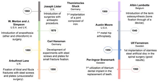
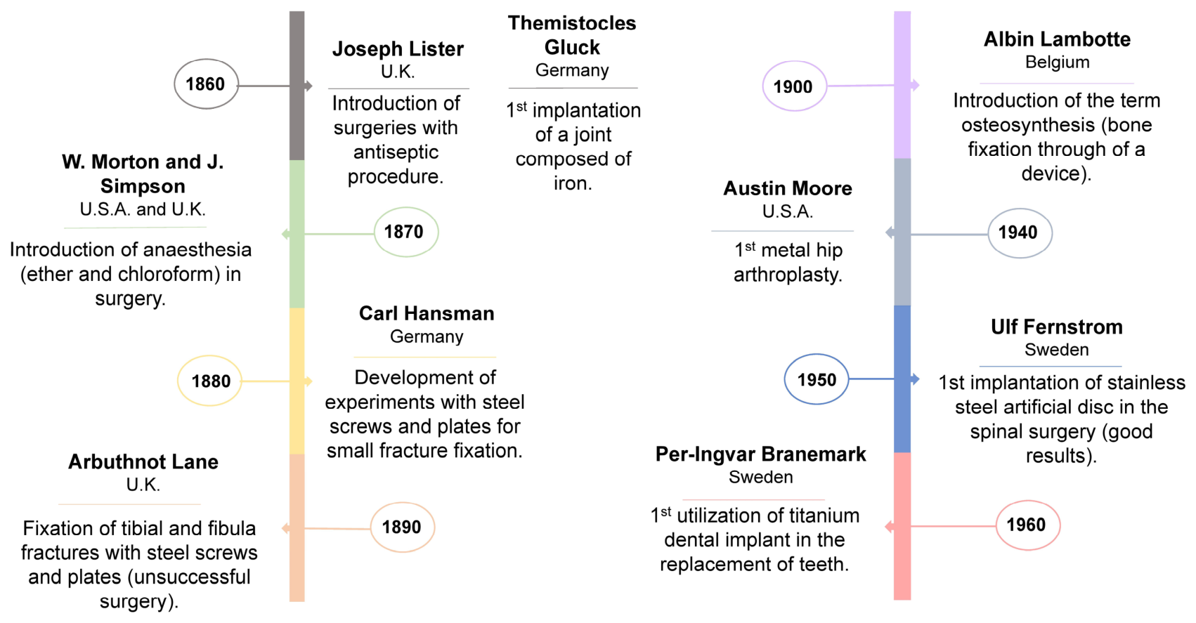
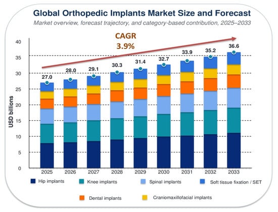
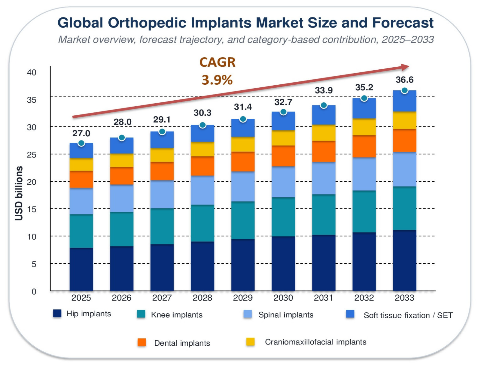
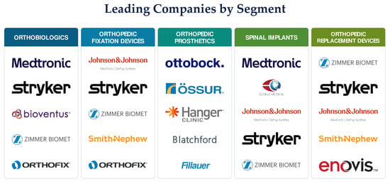
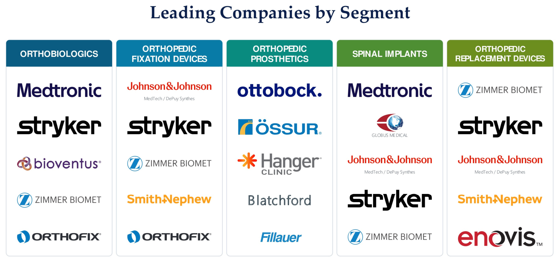
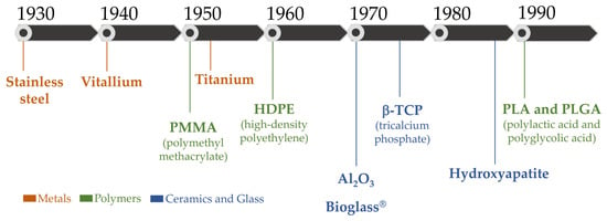
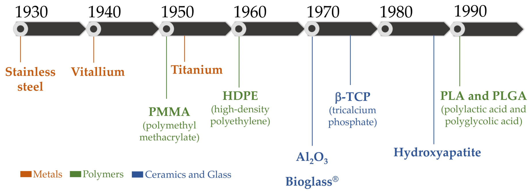
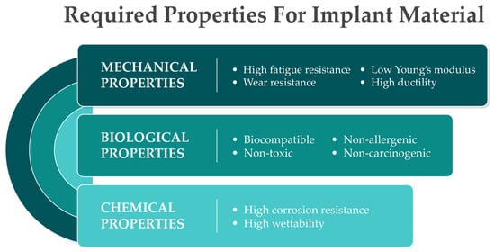
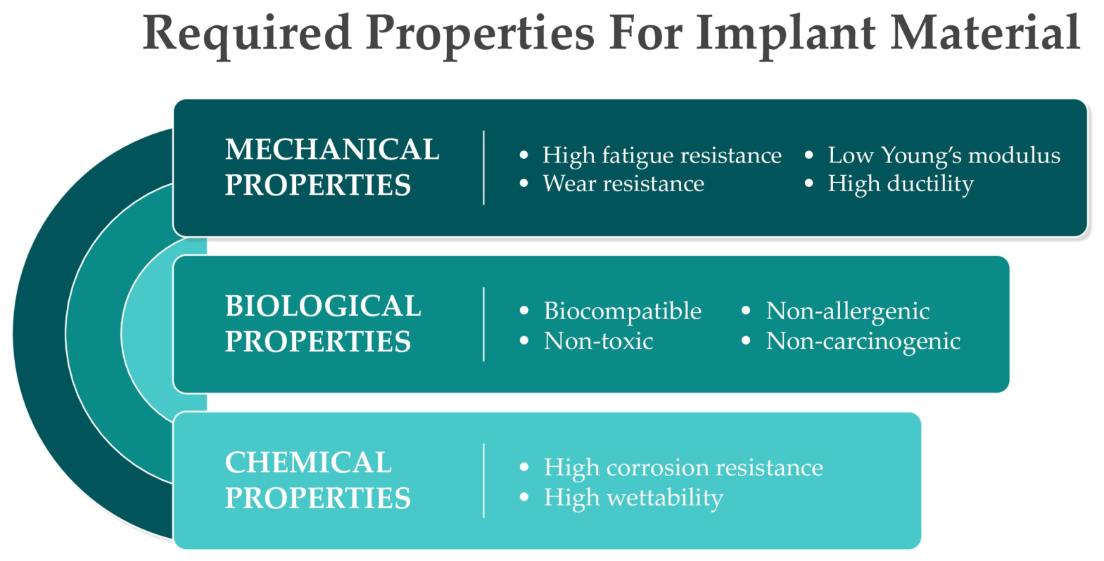
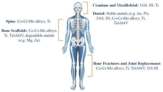
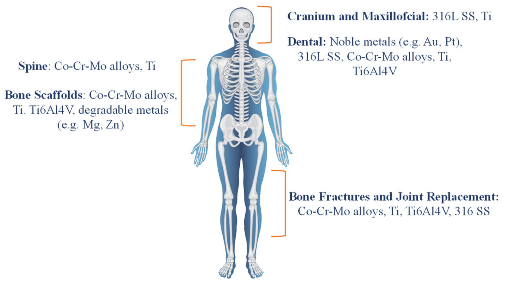
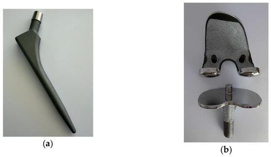
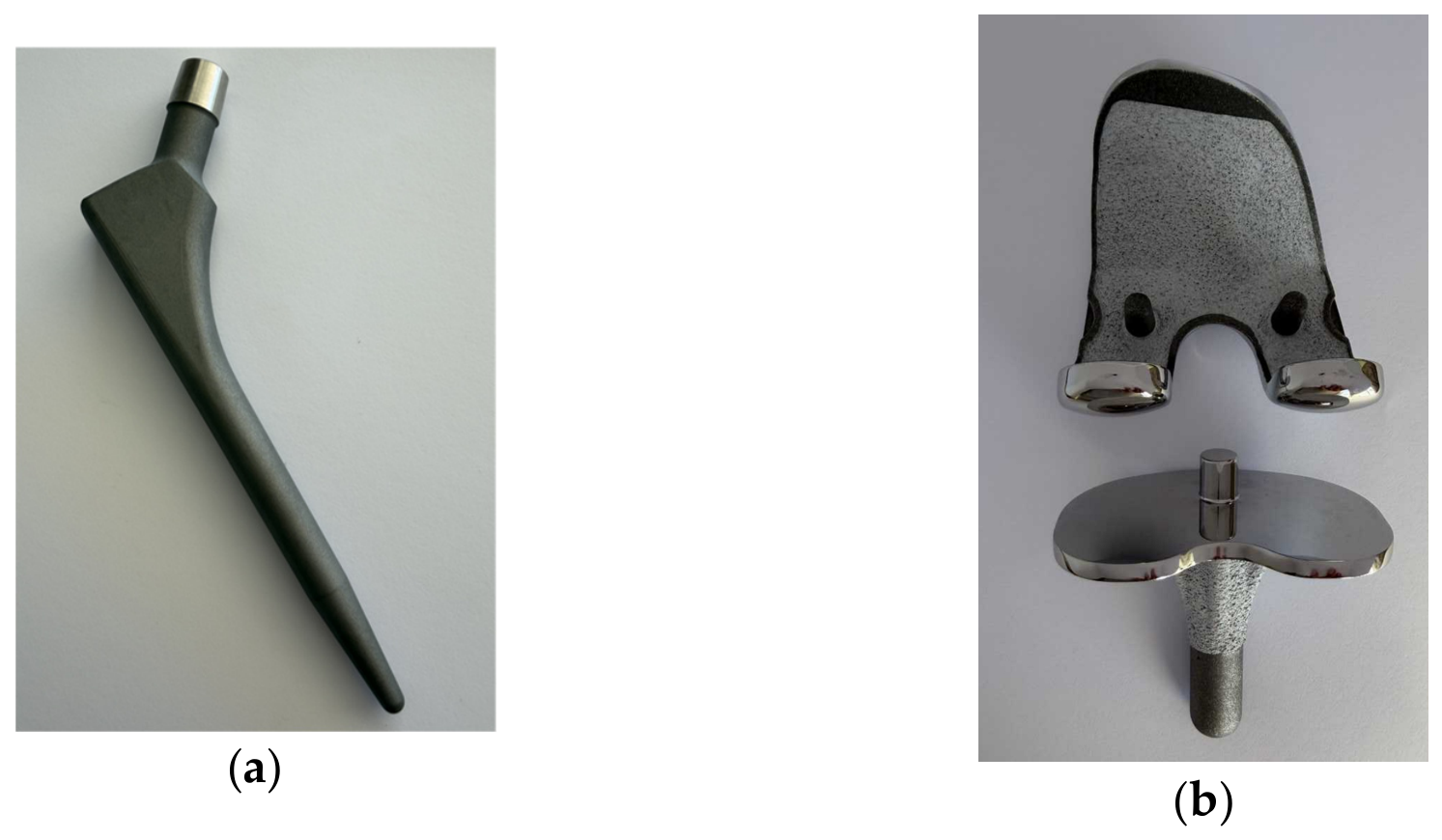
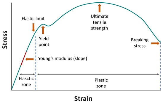
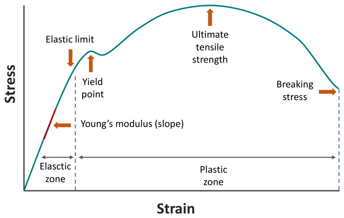
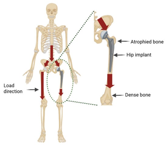
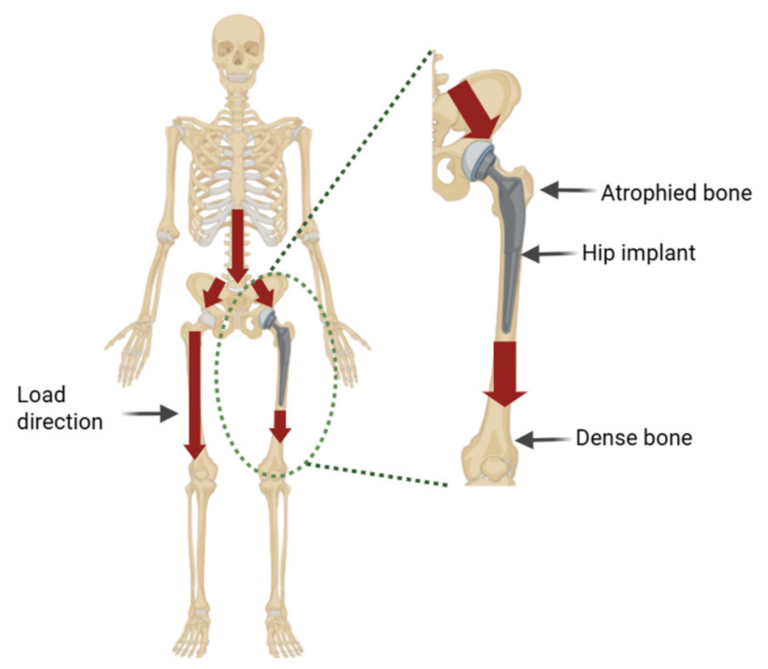
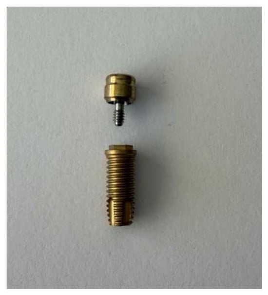
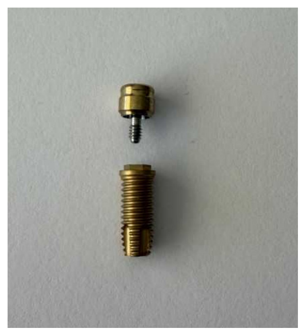
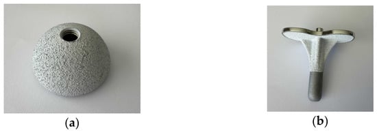
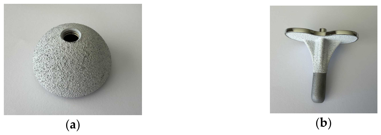
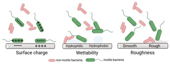
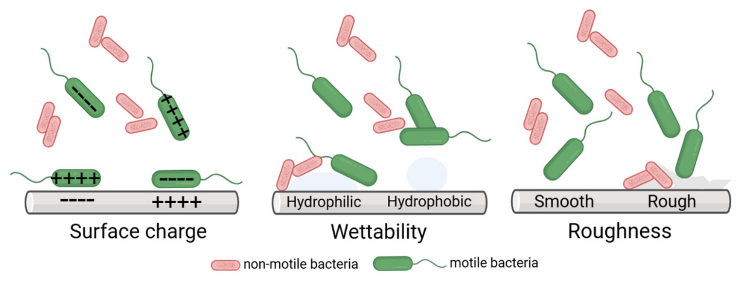
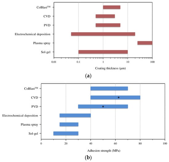
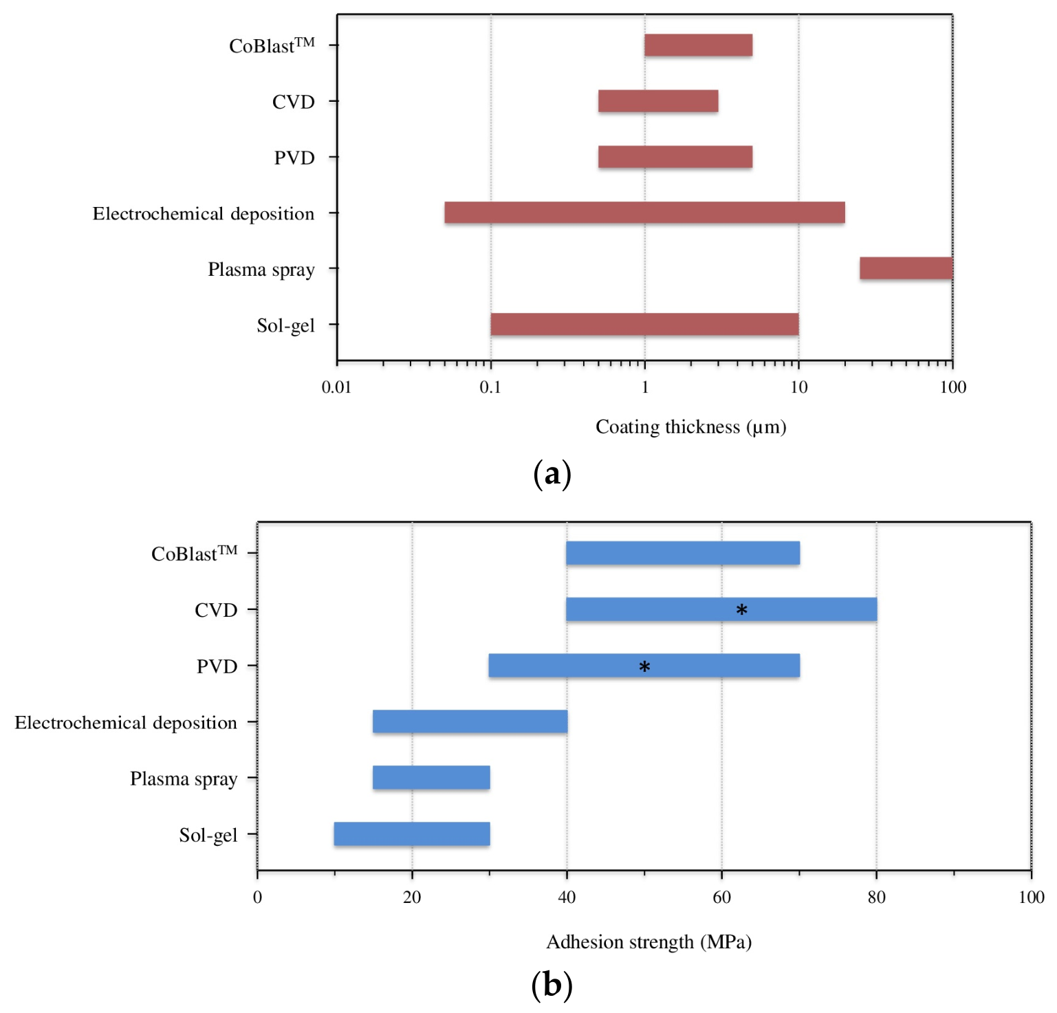
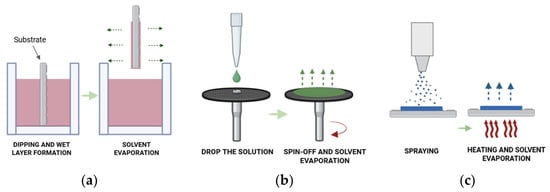
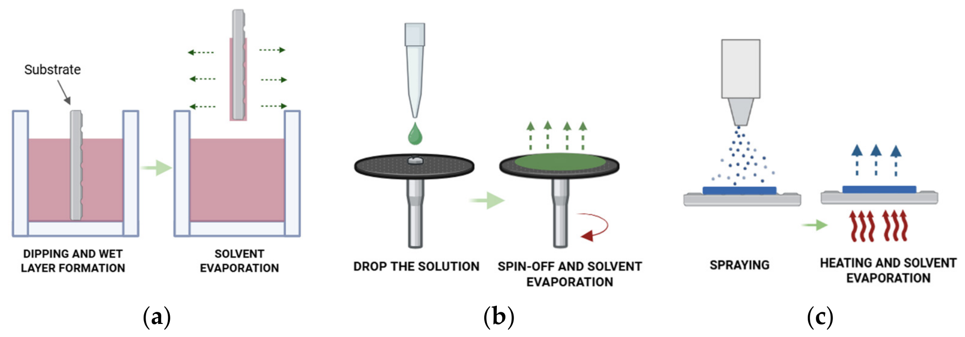
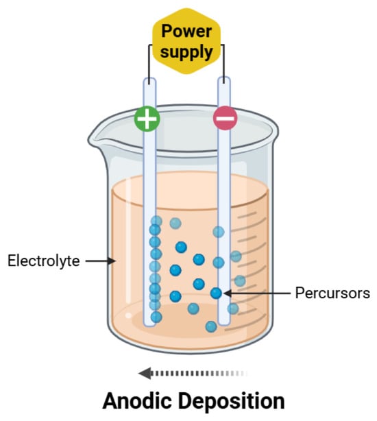
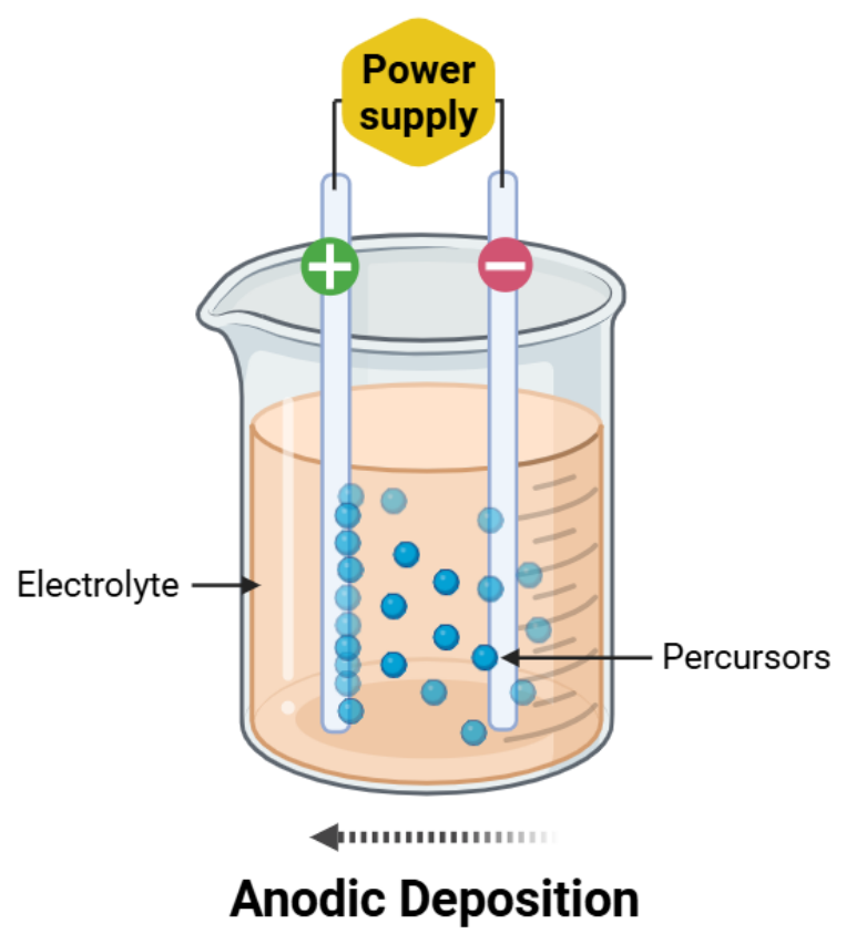
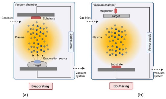
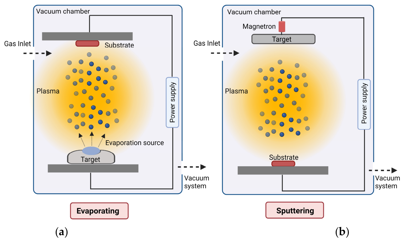
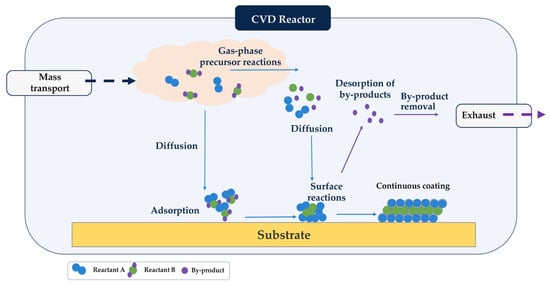
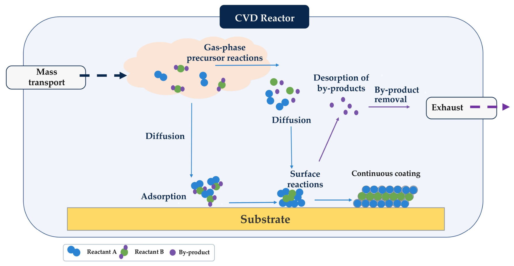
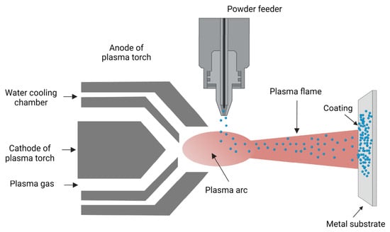
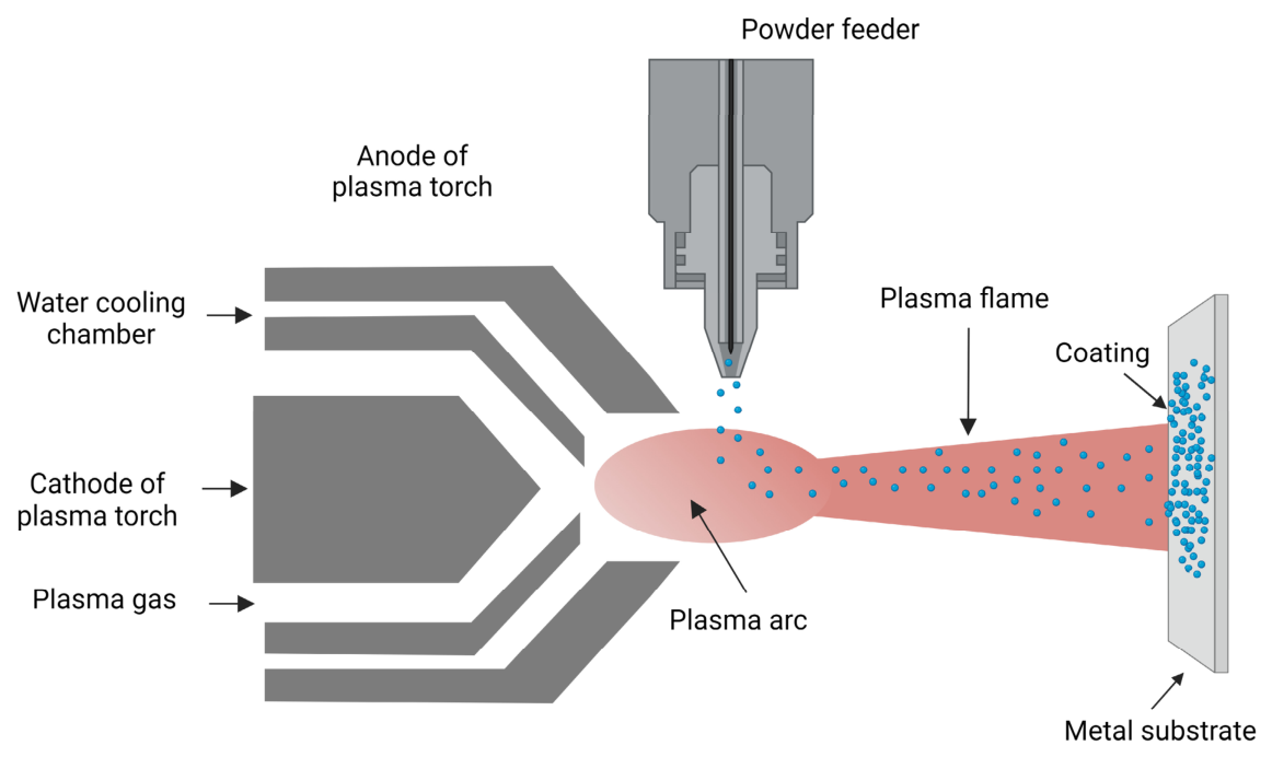
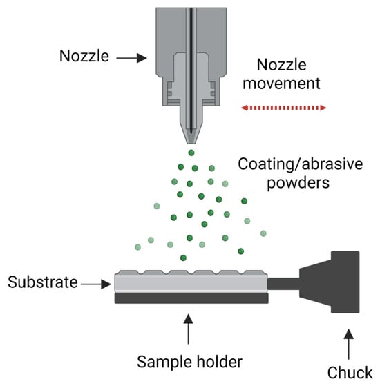
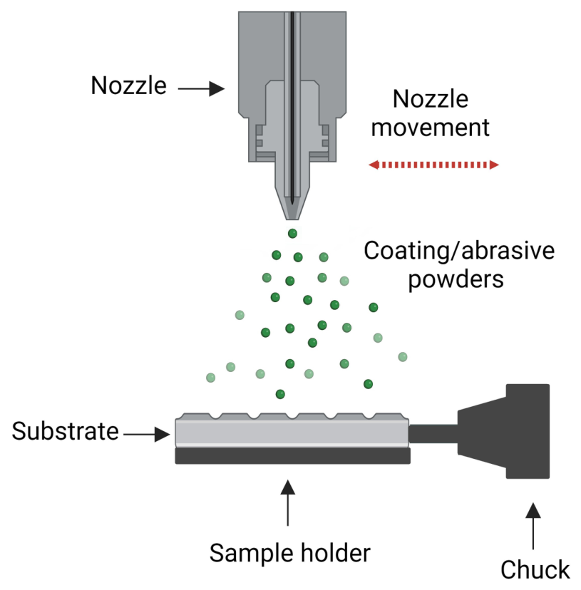
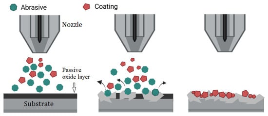
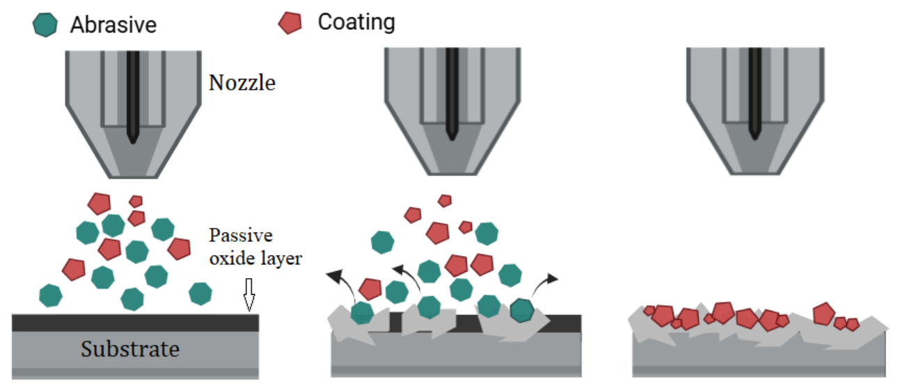
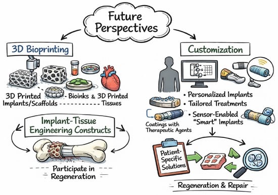
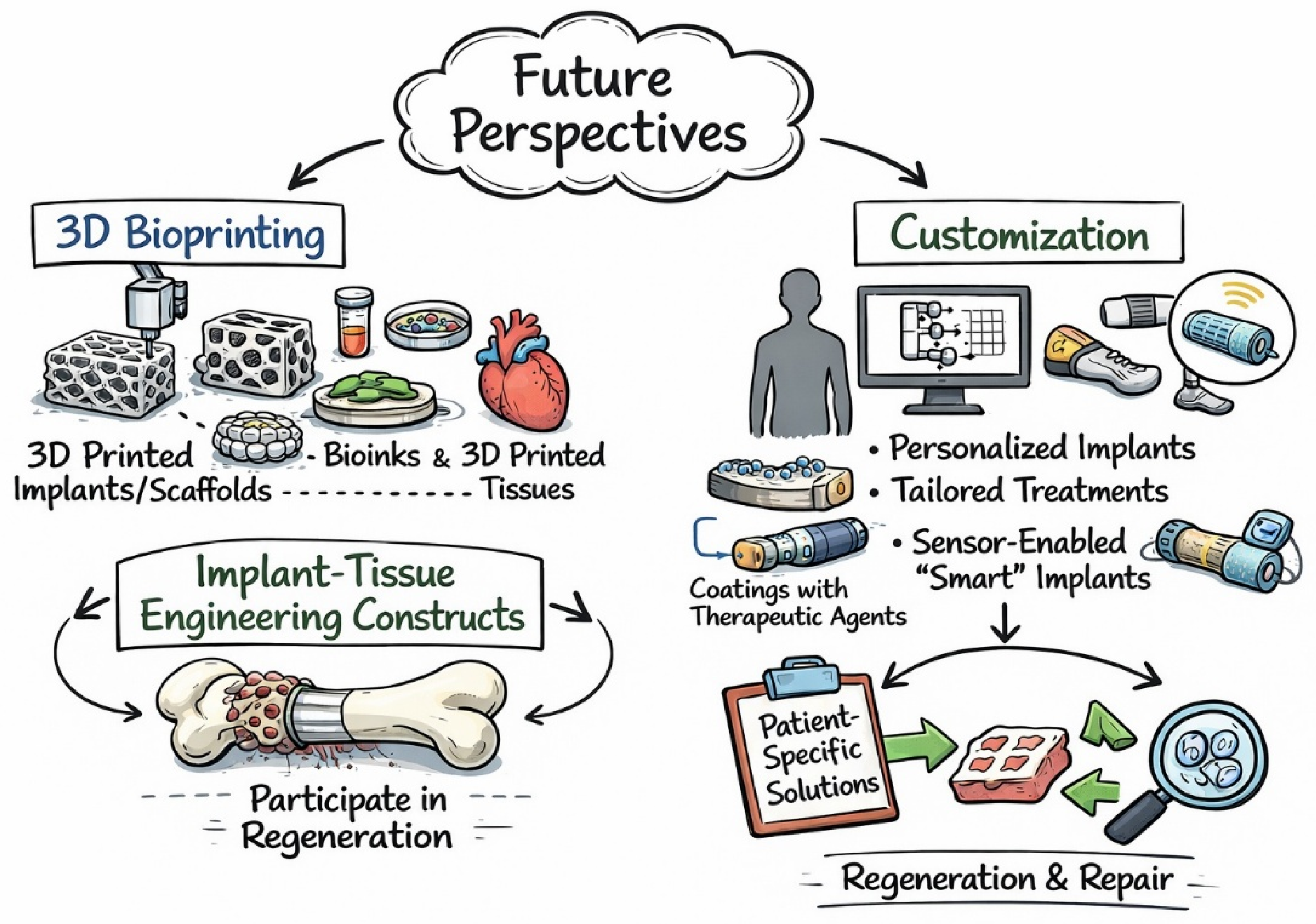

Disclaimer/Publisher’s Note: The statements, opinions and data contained in all publications are solely those of the individual author(s) and contributor(s) and not of MDPI and/or the editor(s). MDPI and/or the editor(s) disclaim responsibility for any injury to people or property resulting from any ideas, methods, instructions or products referred to in the content. |
© 2026 by the authors. Licensee MDPI, Basel, Switzerland. This article is an open access article distributed under the terms and conditions of the Creative Commons Attribution (CC BY) license.