


Abstract
Background: Oxidative stress plays an important role in the development and progression of prediabetes (PD) and type 2 diabetes mellitus (T2DM). Since metabolic disturbances may alter the functional properties of erythrocyte antioxidant systems, we hypothesised that erythrocyte antioxidant enzymes would exhibit differential responses to phytochemical exposure depending on metabolic status. Therefore, this ex vivo study compared the responses of erythrocyte superoxide dismutase (SOD), catalase (CAT), and glutathione peroxidase (GPx) to selected phytochemicals in healthy individuals, subjects with PD, and patients with T2DM. Methods: Erythrocyte hemolysates collected from healthy controls (CG), individuals with PD (PDG), and T2DM (T2DMG) were incubated with curcumin, resveratrol, fisetin, and cinnamon extract. The activities of SOD, CAT, and GPx were determined spectrophotometrically. Statistical analyses included a Kruskal–Wallis test, Friedman ANOVA and a Spearman rank correlation coefficient. For significant results, post hoc tests were performed with Benjamini–Hochberg correction for multiple comparisons. Results: Baseline (vehicle control) SOD activity was significantly lower in T2DMG and PDG compared with CG (p = 0.0035). SOD activity was higher following incubation with curcumin in T2DMG and CG (p < 0.05), while resveratrol showed a significant effect only in CG (p = 0.0326). Cinnamon extract increased CAT activity, particularly in CG (p = 0.0169). Among the analysed enzymes, GPx exhibited the highest responsiveness to phytochemical exposure. Curcumin and fisetin increased GPx activity in PDG and T2DMG, while cinnamon extract improved activity in all groups (p < 0.05). Additionally, baseline GPx activity was negatively correlated with body weight (q = 0.0127) and body mass index (q = 0.0260). Conclusions: These findings suggest that metabolic status influences the responsiveness of erythrocyte antioxidant enzymes to phytochemical exposure. While the tested compounds exerted direct effects on antioxidant enzyme activities, the magnitude and pattern of these responses differed between healthy, prediabetic, and T2DM individuals. This suggests that alterations associated with metabolic dysfunction may influence erythrocyte antioxidant defence mechanisms and their responsiveness to bioactive compounds. These observations provide a rationale for further in vivo and clinical studies addressing the interaction between metabolic status and phytochemical-mediated antioxidant effects.
Keywords:
curcumin; resveratrol; fisetin; cinnamon extract; SOD; CAT; GPx; glucose metabolism disorders; oxidative stress 1. Introduction
Oxidative stress is one of the main factors behind many chronic diseases. It is defined as an imbalance between reactive oxygen/nitrogen species (ROS/RNS) and the body’s defence mechanisms, which interferes with redox signalling and/or causes cellular damage [1,2,3]. There are endogenous (naturally occurring by-products of metabolic processes) and exogenous ROS (radiation, pollution, cigarette smoke, certain drugs, heavy metals, and a high-fat, high-sugar diet) [3,4]. Under physiological conditions, they are captured and converted into non-reactive forms by various intracellular enzymatic antioxidant systems, such as catalase (CAT), glutathione peroxidase (GPx), and superoxide dismutase (SOD), as well as non-enzymatic systems, such as vitamins and reduced glutathione (GSH). However, the overproduction of ROS and their ineffective elimination damages DNA, proteins and lipids, leading to cell apoptosis and dysfunction [5,6].
Type 2 diabetes mellitus (T2DM), characterised by hyperglycemia due to impaired insulin secretion and insulin resistance, is one of the diseases in which oxidative stress plays an important role. High blood glucose levels lead to excessive ROS production, which damages proteins, lipids, and DNA and impairs insulin signalling and β-cell function. These processes exacerbate insulin resistance. In addition, oxidative stress activates inflammatory pathways, thereby increasing the production of inflammatory cytokines and ROS [7,8,9,10]. Similar phenomena are observed during the development of T2DM, a prediabetes (PD) that can occur up to 10 years before the diagnosis of T2DM. Long-term, low-grade inflammation and hyperglycemia lead to complications, including retinopathy, nephropathy, neuropathy, and micro- and macroangiopathy [11,12,13].
Natural polyphenolic compounds have attracted considerable attention due to their potential to modulate oxidative stress and related metabolic disturbances. Curcumin is an active chemical compound present in the rhizome of the plant Curcuma longa L., also known as turmeric. It has antioxidant, anti-inflammatory, antimicrobial, anticancer, antirheumatic, and immunomodulatory effects [14,15,16]. Resveratrol (3,4′,5-trihydroxystilbene) is found in red wine, grapes, berries, peanuts, and chocolate, among others [17]. It positively affects the oxidative-antioxidant balance by increasing the expression of antioxidant enzymes and regulating signalling pathways, including sirtuin 1 (SIRT1), nuclear erythroid factor 2 (Nrf2), and nuclear factor kappa-light-chain-enhancer of activated B cells (NF-κB), thereby increasing GSH levels and maintaining cellular redox balance [18,19]. Fisetin (3,3′,4′,7-tetrahydroxyflavone) is a bioactive polyphenolic compound present in grapes, apples, onions, strawberries, and cucumbers (<160 μg/g). It exhibits several beneficial properties, including antioxidant, anti-inflammatory, anticancer, and neuroprotective [20,21]. Fisetin restores proper mitochondrial function, improves cellular metabolism, and reduces inflammation, oxidative stress, and lipid peroxidation, while increasing intracellular antioxidant levels [22,23,24,25]. Cinnamon extract is a concentrated extract from the bark of the cinnamon tree (Cinnamomum). It contains active substances such as cinnamaldehyde, eugenol, polyphenols, proanthocyanidins, and flavonoids. It has beneficial effects on glycemia and is antioxidant, anti-inflammatory, antibacterial, and antifungal [26,27,28]. Many studies have shown that curcumin, resveratrol, fisetin, and cinnamon extract increase antioxidant enzyme levels and lower inflammatory markers and markers of oxidative damage in various disease entities [14,26,29,30,31,32,33,34,35,36,37].
Erythrocytes contain heme iron and polyunsaturated membrane lipids, which is why they are exposed to ROS and oxidative damage. These changes may include erythrocyte deformities, effects on their elasticity and viability, and interactions with blood vessels, which may contribute to the development of many diseases [38,39]. Therefore, erythrocytes constitute a well-established and biologically relevant model for studying oxidative damage and antioxidant interventions under ex vivo conditions [40]. However, it remains unclear whether erythrocytes derived from individuals with different metabolic backgrounds exhibit a similar capacity to respond to antioxidant phytochemicals and whether metabolic status influences erythrocyte antioxidant responsiveness.
Based on the above, we hypothesised that erythrocytes derived from individuals at different stages of metabolic disturbance (healthy, prediabetes, and T2DM) differ in their baseline redox status and antioxidant defence capacity. Consequently, we expected that curcumin, resveratrol, fisetin, and cinnamon extract would modulate antioxidant enzyme activity in a concentration-dependent manner, with differential responses depending on the metabolic status of the donors. These compounds were selected for their complementary mechanisms of action that target key pathways involved in oxidative stress, inflammation, and mitochondrial function, which are critically impaired in T2DM and PD.
The aim of this study was to evaluate the concentration-dependent effects of these phytochemicals on antioxidant enzymes (SOD, CAT, and GPx) in erythrocytes from type 2 diabetes, prediabetes, and healthy individuals under ex vivo conditions. We also investigated whether erythrocyte antioxidant responsiveness to phytochemicals differs according to metabolic status.
2. Materials and Methods
2.1. Chemicals
A solution of phosphate-buffered saline (PBS; pH 7.4; 137 mM NaCl, 2.7 mM KCl, 10 mM Na2HPO4, 1.8 mM KH2PO4) and dimethyl sulfoxide (DMSO, ≥99.7%, 34869, Sigma-Aldrich Company, Saint Louis, MO, USA) was used to prepare the biological material. Enzymatic activities were determined according to the procedures of the manufacturers: Cayman Chemical Company (Ann Arbor, MI, USA), Superoxide Dismutase Assay Kit 706002, Catalase Assay Kit 707002 and Randox Reagents (Crumlin, County Antrim, Northern Ireland, UK), Glutathione Peroxidase (Ransel) Assay Kit RS505.
2.2. Phytochemicals
Curcumin (Turmeric Root Extract 95% Curcuminoids ~25:1 Powder (Curcuma longa L.); NIGEHER038491), resveratrol (Japanese Knotweed Extract 98% Trans Resveratrol (Reynoutria japonica/Polygonum cuspidatum); NIGEHER033401), fisetin (Smoke Tree Extract 50% Fisetin (Cotinus coggygria Scop.); NIGEHER014752) and cinnamon extract (Ceylon Cinnamon Bark Extract 20:1 (Cinnamomum zeylanicum; NIGEHER010328) were acquired from Nutraceuticals Group Europe (Merstham, Surrey, UK). All phytochemicals were safe for human consumption and non-toxic.
2.3. Characterisation of Cinnamon Extract
The chemical composition of the cinnamon extract used in this study was determined prior to incubation experiments. The major identified compounds included cinnamaldehyde (66.2 mg/g), gallic acid (10.2 mg/g), p-coumaric acid (5.8 mg/g), eugenol (4.1 mg/g), and coumarin (4.1 mg/g), along with smaller amounts of flavonoids (catechins and epicatechins) and terpenoids (e.g., β-caryophyllene, limonene, and pinene derivatives). The detailed composition of the extract is presented in Table S1.
Phenolic compounds in the cinnamon extract, including flavonoids, proanthocyanidins, and phenolic acids, were determined using an Acquity H-Class UPLC system (Waters, Milford, MA, USA) equipped with a PDA detector, with additional LC-MS analysis for structural confirmation. Flavonoids and their glycosides were analysed after acid and/or alkaline hydrolysis to obtain aglycones. Proanthocyanidins were extracted using sonication-assisted methanol–water (70:30, v/v) extraction. Phenolic acids were determined following extraction combined with acid-base hydrolysis. Chromatographic separation was performed on an Acquity UPLC® BEH C18 column (100 × 2.1 mm, 1.7 μm; Waters, Wexford, Ireland) using gradient elution with mobile phases consisting of acetonitrile and water, both containing 0.1% formic acid. Detection wavelengths were set at 280 nm for proanthocyanidins and 320 nm for phenolic acids. Compound identification was based on retention times and spectral data compared to reference standards, and quantification was performed using external calibration curves. The limit of quantification was 1 μg/g.
2.4. Materials
The biological material for the study was obtained from healthy participants (n = 18; aged 66.47 ± 4.95) and with T2DM (n = 19; diagnosed based on medical records and according to the WHO criteria, including fasting plasma glucose ≥ 126 mg/dL and/or HbA1c ≥ 6.5%; T2DM duration time: 10.59 ± 6.90, aged 64.11 ± 7.33, treated with a single antidiabetic medication (90% metformin, 10% sulfonylurea), free from diabetic complications) and PD (n = 14; diagnosed based on fasting glucose measurements within the range of 100–125 mg/dL; aged 63.25 ± 4.33, no hypoglycaemic medications or insulin therapy) (n = 49). An abbreviated description of the participants was presented in Supplementary Materials (Table S2) and an earlier scientific report [41]. The same exclusion criteria applied to each group: treatment with insulin or multiple oral antidiabetic medications (in the case of the T2DMG), as well as cancer, autoimmune diseases, and alcohol/drug addiction. Participants were recruited for the study during the “Biała Sobota” preventive campaign at the District Hospital in Czarnków, held between January and July 2025. Participation in the research was voluntary. Before entering the experiment, all participants signed an informed consent to participate in the study, in accordance with the principles of the Declaration of Helsinki [42]. All studies were conducted with the consent of the Bioethics Committee of the Medical University of Poznań—No. 854/22.
The study material was venous blood collected from participants in the morning, on an empty stomach (at least 12 h after the last meal), using vacuum sets containing lithium heparin. The samples were taken by qualified medical personnel. Blood samples were stored at 4 °C and further processed immediately after collection. Erythrocytes were isolated from whole blood by centrifugation at 3000 rpm (approximately 725× g, based on the rotor radius (r ≈ 7.2 cm)) for 10 min at 4 °C. After removal of plasma and buffy coat, the isolated erythrocytes were washed three times with cold PBS. The erythrocytes were then hemolysed by adding four times the volume of cold deionised water relative to the volume of erythrocyte sediment and centrifuged at 10,000 rpm (approximately 8040× g, based on the rotor radius (r ≈ 7.2 cm)) for 10 min. The resulting supernatant was collected and used as an erythrocyte hemolysate for further determinations. The hemolysate was stored at −80 °C until analysis. Each batch was thawed a maximum of two times.
2.5. Protein Determination
Total protein concentration in erythrocyte hemolysates was determined using an automated biochemical analyser (Accent 220S, Cormay, Łomianki, Poland) based on the Biuret method, according to the manufacturer’s protocol using dedicated reagents (ACCENT-200 TOTAL PROTEIN, Cormay, Łomianki, Poland). This colourimetric assay relies on the formation of a violet complex between copper ions and peptide bonds under alkaline conditions, with absorbance measured spectrophotometrically (λ = 546 nm). Equal volumes of hemolysate were used for all assays, ensuring comparable protein input across samples.
No significant differences in total protein concentration were observed between the study groups (p > 0.05), indicating comparable protein content across samples. Data are presented in g/dL in the Supplementary Materials (Table S7).
2.6. Incubation of Erythrocyte Hemolysate with Phytochemicals
Resveratrol stock solutions were prepared in DMSO at a concentration of 10 mM and subsequently diluted to obtain working solutions of 20 and 200 μM. Curcumin (95% curcuminoids), fisetin extract (50% standardised fisetin extract) and cinnamon extract were prepared as botanical preparations based on their mass concentrations. The stock solutions were prepared by dissolving 36.8 mg of curcumin, 28.6 mg of fisetin extract, and 39.8 mg of cinnamon extract in 10 mL of DMSO, resulting in extract concentrations of 3.47 mg/mL, 2.86 mg/mL, and 3.98 mg/mL, respectively. Subsequent dilutions were performed according to the same experimental scheme, and the obtained concentrations are referred to as nominal extract concentrations, rather than molar concentrations of individual compounds. For consistency across all tested compounds, the lower and higher concentrations were designated as low (L) and high (H), respectively.
To ensure identical environmental conditions for control trials, a vehicle control containing DMSO diluted in PBS, without phytochemicals, was prepared. The final concentration of DMSO did not exceed 0.1% in any sample, which eliminates its potential effect on enzymatic activity.
Erythrocyte hemolysate was mixed with the respective phytochemical working solutions at low (L) and high (H) concentrations or a vehicle control (DMSO + PBS) in a 1:1 volume ratio. To ensure a suitable environment for the reaction, 480 µL of PBS was added to the prepared solutions. For resveratrol, the corresponding final molar concentrations after the incubation dilution were 0.4 and 4 μM. For botanical preparations, including curcumin, fisetin, and cinnamon extract, concentrations were expressed as nominal extract concentrations and were not interpreted as molar concentrations of individual compounds.
Subsequent assay-specific dilution steps further reduced phytochemical concentrations in the reaction mixtures for SOD, CAT, and GPx measurements.
The mixtures were incubated at 37 °C for 60 min under physiological conditions, and then centrifuged at 10,000 rpm (approximately 8040× g, based on the rotor radius (r ≈ 7.2 cm)) for 10 min. The obtained supernatant was used directly for spectrophotometric determination of enzyme activity associated with oxidative stress. Each determination was carried out in duplicate to ensure the reliability and repeatability of the measurements.
This ex vivo model allows the assessment of short-term interactions between bioactive compounds and antioxidant systems in a biologically relevant cellular matrix.
2.7. Enzyme Activity Determinations
The SOD (Cu/Zn-, Mn-, and FeSOD) determination kit is based on the reduction of the tetrazole salt to formazan by superoxide radicals generated by xanthine oxidase. SOD catalyses the dismutation of superoxide radicals, so the degree of inhibition of the reaction is directly proportional to the enzyme’s activity. Absorbance was measured at 450 nm. The enzyme activity was expressed in units of U/mL, where one unit (U) corresponds to the amount of enzyme causing 50% dismutation of the superoxide radical.
Catalase activity (CAT) was determined by a method based on the enzymatic reaction of methanol with hydrogen peroxide (H2O2). The resulting product is formaldehyde, which reacts colourimetrically with chromogen Purpald (4-amino-3-hydrazino-5-mercapto-1,2,4-triazole). Absorbance was measured at 540 nm. One unit of CAT activity is defined as the amount of enzyme that produces 1 nmol of formaldehyde per minute at 25 °C (nmol/min/mL). The enzyme activity is expressed in U/mL.
Determination of GPx activity is based on the oxidation of GSH by cumene hydroxide in the presence of glutathione reductase and NADPH. Then, oxidised glutathione (GSSG) is reduced with simultaneous oxidation of NADPH to NADP+. The decrease in absorbance at 340 nm is proportional to GPx activity in the sample. The enzyme activity is expressed in U/L according to the manufacturer’s recommendations.
Control measurements were performed in the absence of erythrocyte hemolysates (compound-only controls). The tested phytochemicals were incubated with assay reagents under identical conditions as experimental samples. The resulting signals were negligible and close to zero, suggesting no substantial direct interference with the detection system. However, due to the redox-active nature of the tested compounds, potential interactions with assay components, including substrates or coupled reactions, cannot be fully excluded.
2.8. Statistical Analysis
Statistical analysis was performed using PQStat v.1.8.4 (PQStat Software, Poznań, Poland). The normality of the data distribution was assessed using the Shapiro–Wilk test. Due to the lack of normality in most of the variables analysed, the results were presented as medians (Me) and interquartile ranges (IQR). Enzyme activities were primarily expressed according to the original assay output (per volume of erythrocyte lysate). The Supplementary Data additionally present the values expressed as total protein content for descriptive purposes (U/g protein) (Tables S3–S5). Between-group comparisons of vehicle control values (CG—healthy individuals, PDG—individuals with prediabetes, T2DMG—individuals with T2DM) were performed using the Kruskal–Wallis test followed by post hoc tests with Benjamini–Hochberg correction. Differences in antioxidant enzyme activities (SOD, CAT, and GPx) between incubation conditions (vehicle control and low [L] and high [H] phytochemical concentrations) within each study group were assessed using the Friedman test for repeated measures (within-group comparisons). For resveratrol, L and H corresponded to final concentrations of 0.4 and 4 μM, respectively, whereas for botanical preparations, concentrations were expressed as nominal mass concentrations. For significant Friedman test results, post hoc pairwise comparisons were performed with Benjamini–Hochberg correction for multiple comparisons. Detailed results of the Friedman test, including Kendall’s W effect sizes, are presented in Supplementary Table S6. Relative changes (%) in antioxidant enzyme activity after phytochemical incubation compared with vehicle control are presented as median (Q1; Q3) in Supplementary Table S8. Statistical significance was assessed using the exact Wilcoxon signed-rank test. Representative results are presented in the main manuscript, while complete box plots for incubation with other phytocompounds are provided in the Supplementary Materials (Figures S1–S3). The relationships between baseline enzyme activity and participants’ anthropometric parameters were assessed using the Spearman rank correlation coefficient. To account for multiple testing in correlation analyses, the p-values were further corrected using the Benjamini–Hochberg False Discovery Rate (FDR) procedure [43].
The results were considered statistically significant at p < 0.05.
3. Results
Enzyme activities in vehicle control were compared between T2DMG, PDG, and CG (Table 1). The highest SOD and GPx activity was observed in CG, and CAT in T2DMG. Significant differences were observed only in SOD activity (p = 0.0034). The T2DM and PD groups showed lower SOD activity than the CG group (p = 0.0341 and p = 0.0035, respectively). No significant differences were observed between the groups in CAT and GPx activity.
Table 1.
Antioxidant enzyme activities in solvent control samples from the study groups.
No significant differences in total protein concentration were observed between the study groups (p > 0.05), indicating that the samples were comparable in terms of protein content (Table S6).
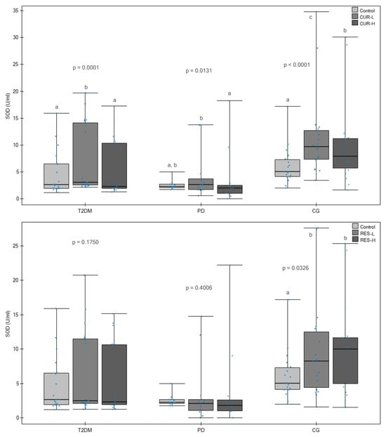
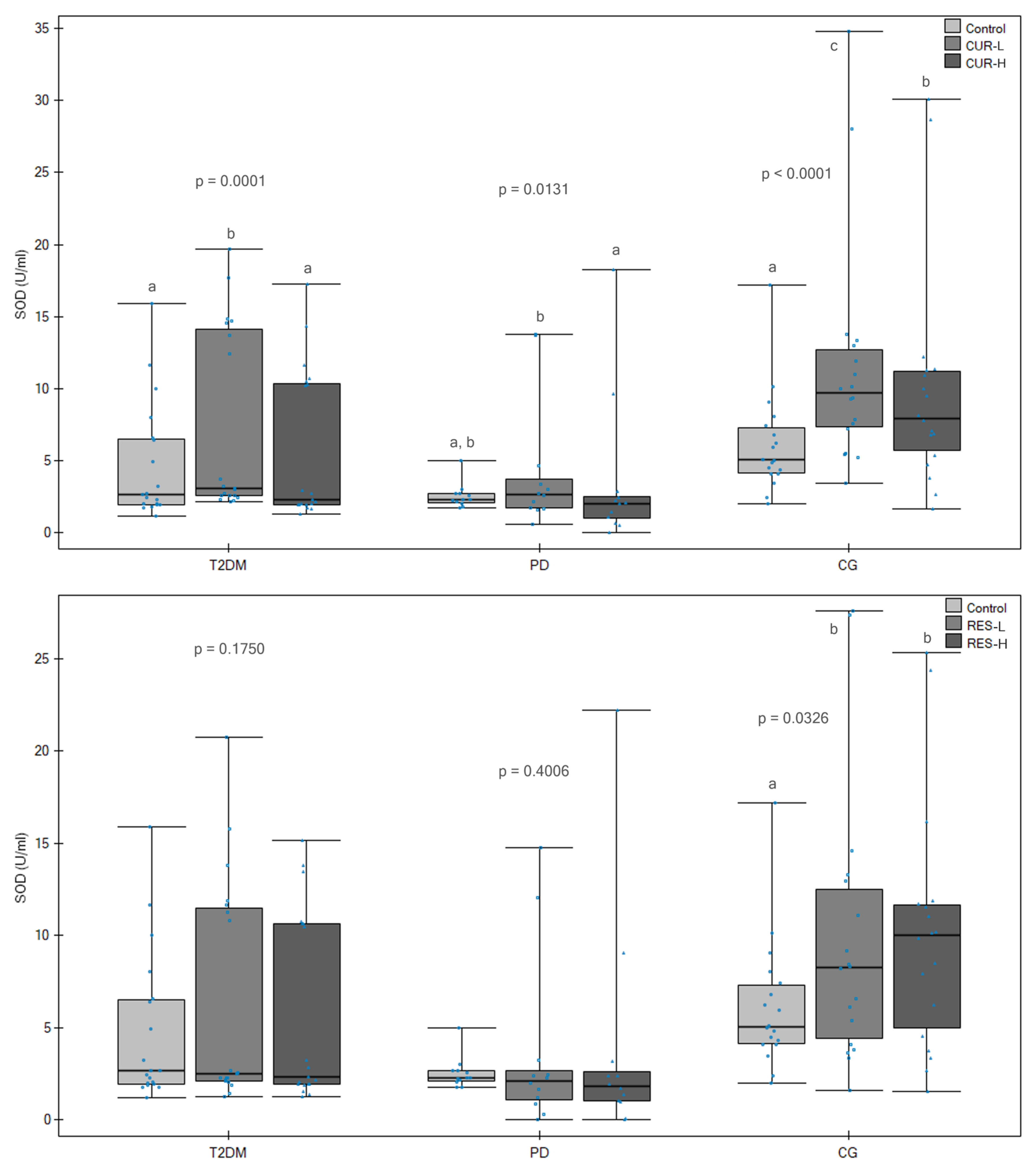
Incubation of erythrocyte hemolysates with curcumin significantly affected SOD activity in all study groups (p = 0.0001 for T2DMG, p = 0.0131 for PDG, and p < 0.0001 for CG, respectively). Post hoc analysis showed that lower curcumin concentration had a significant effect in T2DMG (p = 0.0002) and CG (p < 0.0001), compared to vehicle control activity. In addition, SOD activity was also significantly higher after incubation with a lower concentration of curcumin than a higher concentration in T2DMG and PDG (p = 0.0026 and p = 0.0128) (Figure 1).
Figure 1.
Effects of curcumin and resveratrol on superoxide dismutase (SOD U/mL) activity in erythrocyte hemolysates. Box-and-whisker plots showing SOD activity in erythrocyte hemolysates obtained from patients with type 2 diabetes mellitus (T2DM), individuals with prediabetes (PD) and healthy control subjects (CG) following incubation without phytochemical (Control) or with curcumin (CUR) and resveratrol (RES) at low (L) and high (H) concentrations. For resveratrol, L and H corresponded to final concentrations of 0.4 and 4 μM, respectively; for curcumin, concentrations were expressed as nominal mass concentrations. Boxes represent the interquartile range (IQR), horizontal lines indicate medians, whiskers denote minimum and maximum values, and dots represent individual observations. Different letters indicate statistically significant differences between incubation conditions within the same study group (Friedman ANOVA followed by pairwise post hoc comparisons with Benjamini–Hochberg correction, p < 0.05).
A significant overall effect of resveratrol on SOD activity in CG was observed (p = 0.0326). Post hoc showed that both lower (p = 0.0367) and higher (p = 0.0367) concentrations of phytochemicals modulated SOD, compared to vehicle control. (Figure 1). Fisetin and cinnamon extract did not significantly modify SOD activity in either study group (Table 2).
Table 2.
Effects of phytochemical incubation on superoxide dismutase (SOD U/mL) activity in erythrocyte hemolysates from type 2 diabetes mellitus, prediabetes, and control groups.
Overall, curcumin exerted the strongest effect on SOD activity across all groups (moderate effect size) (Table S6).
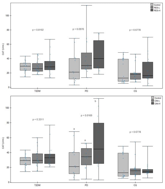
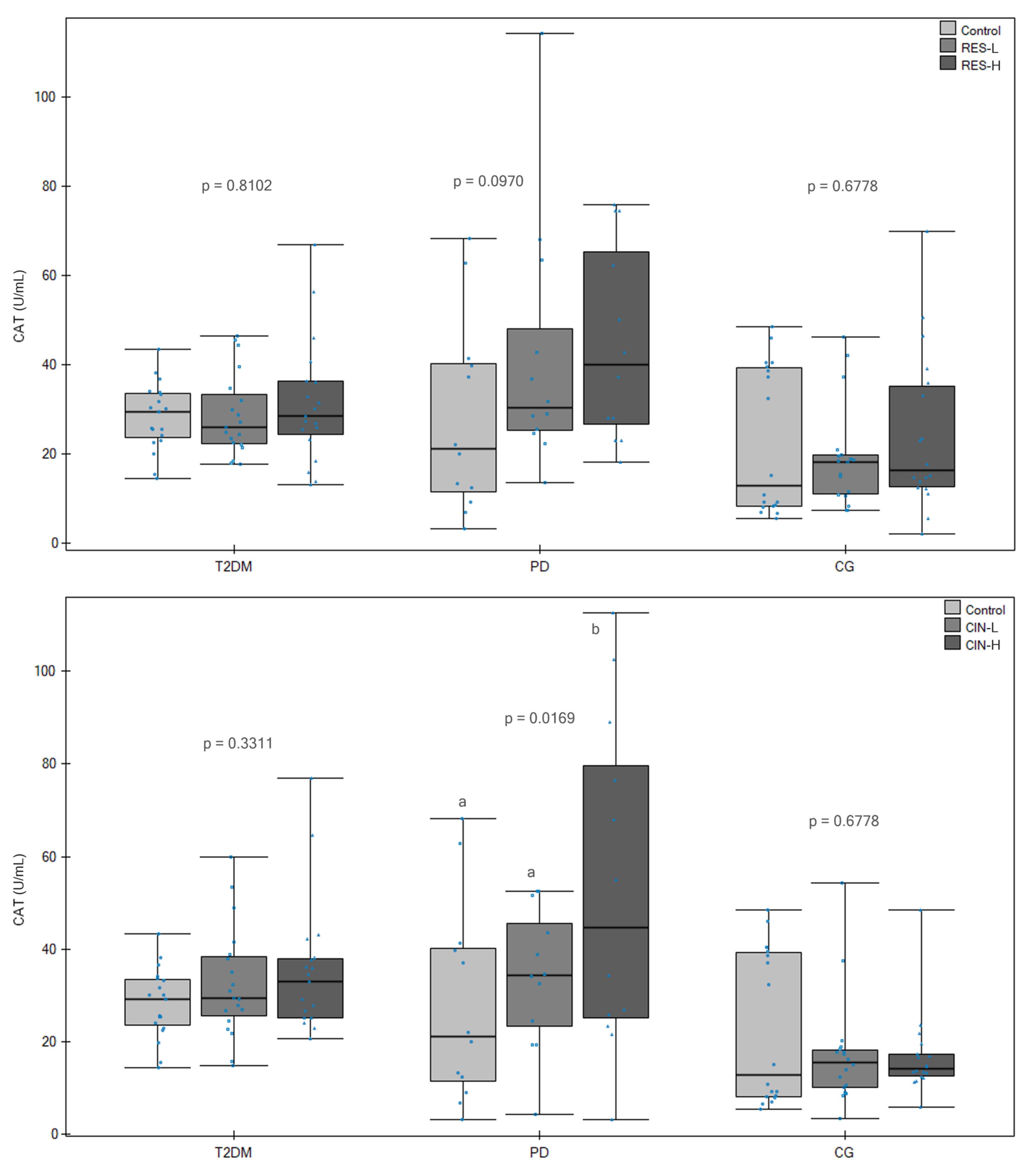
Analysis of CAT activity showed that only cinnamon extract achieved significant results (Table 3). This effect was observed only in PDG, where CAT activity gradually increased with increasing cinnamon concentration (p = 0.0169). Post hoc analysis showed significantly increased CAT activity after incubation with higher cinnamon extract concentration compared to vehicle control (p = 0.0239) (Figure 2). No statistically significant effect of curcumin, resveratrol or fisetin on CAT activity was observed in any of the study groups. Calculated effect sizes were consistently negligible to small, indicating minimal biological impact of the tested compounds on CAT activity under the experimental conditions (Table S6).
Table 3.
Effects of phytochemical incubation on catalase (CAT U/mL) activity in erythrocyte hemolysates from type 2 diabetes mellitus, prediabetes, and control groups.
Figure 2.
Effects of resveratrol and cinnamon extract on catalase (CAT U/mL) activity in erythrocyte hemolysates. Box-and-whisker plots showing CAT activity in erythrocyte hemolysates obtained from patients with type 2 diabetes mellitus (T2DM), individuals with prediabetes (PD) and healthy control subjects (CG) following incubation without phytochemical (Control) or with resveratrol (RES) and cinnamon extract (CIN) at low (L) and high (H) concentrations. For resveratrol, L and H corresponded to final concentrations of 0.4 and 4 μM, respectively; for cinnamon extract, concentrations were expressed as nominal mass concentrations. Boxes represent the interquartile range (IQR), horizontal lines indicate medians, whiskers denote minimum and maximum values, and dots represent individual observations. Different letters indicate statistically significant differences between incubation conditions within the same study group (Friedman ANOVA followed by pairwise post hoc comparisons with Benjamini–Hochberg correction, p < 0.05).
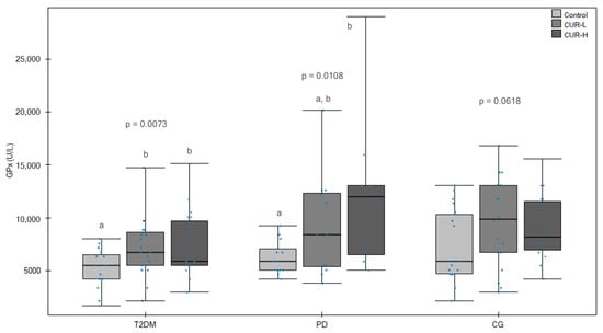
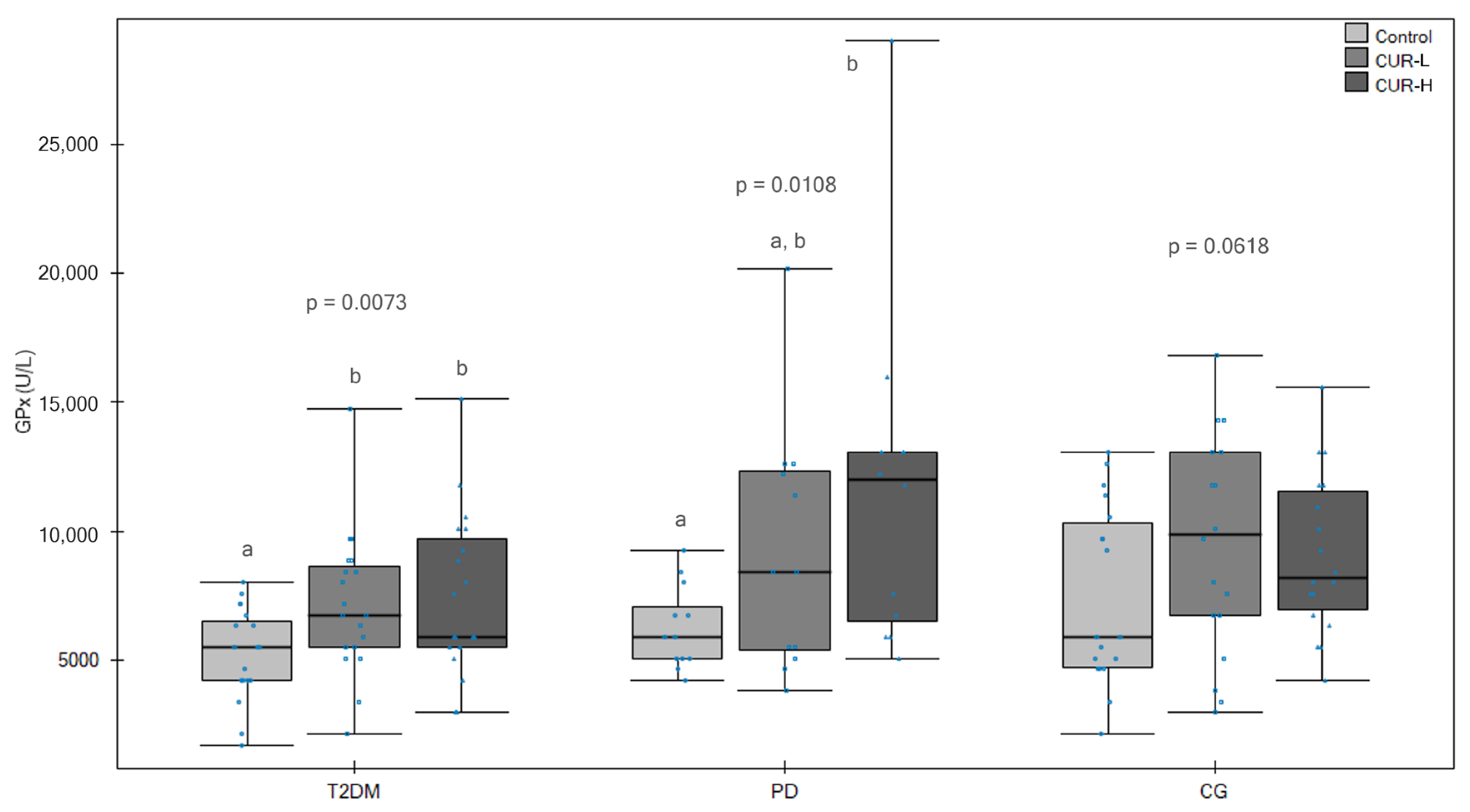
GPx was the most susceptible to phytochemicals (Table 4). Curcumin significantly increased GPx activity in both T2DMG (p = 0.0073) and PDG (p = 0.0108). In individuals with T2DM, both curcumin concentrations increased GPx activity compared with vehicle control (p = 0.0142). The highest activity in PDG was observed after incubation with a higher curcumin concentration (p = 0.0239 compared to vehicle control). CG did not obtain statistical significance.
Table 4.
Effects of phytochemical incubation on glutathione peroxidase (GPx U/L) activity in erythrocyte hemolysates from type 2 diabetes mellitus, prediabetes, and control groups.
Resveratrol significantly increased GPx activity in T2DMG (p = 0.0136), with the highest values observed after incubation with 4 μM resveratrol (p = 0.0175 compared to vehicle control). However, no significant effects were observed in PDG and CG.
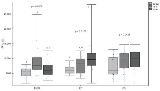
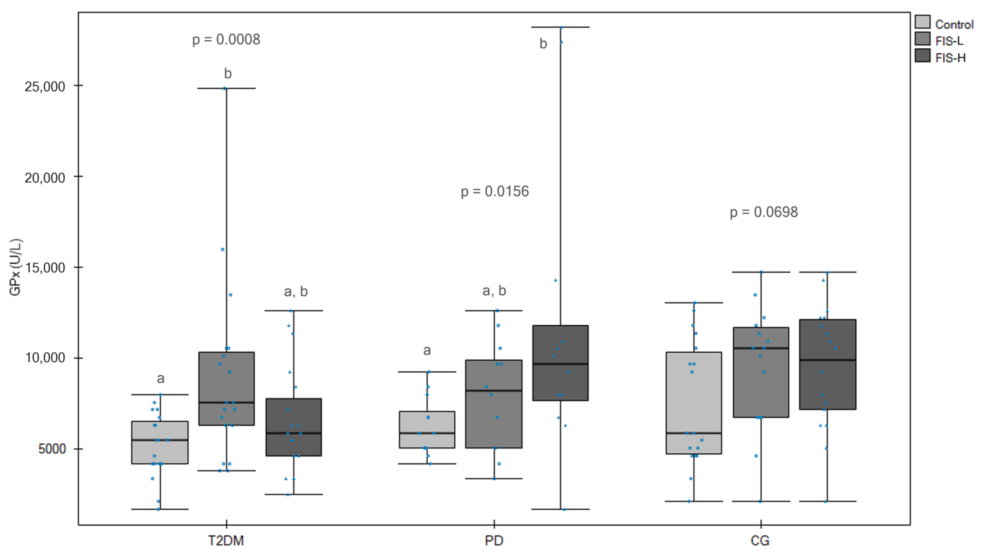
Fisetin significantly affected GPx activity in both T2DMG (p = 0.0008) and prediabetes (p = 0.0156). Post hoc analysis showed that in the T2DM group, GPx activity was significantly higher after incubation with a lower fisetin concentration than in the vehicle control (p = 0.0006). In the PD group, GPx activity increased significantly only after incubation with a higher fisetin concentration compared with the vehicle control (p = 0.0176). CG activity did not change significantly after fisetin incubation (Figure 3).
Figure 3.
Effects of curcumin and fisetin on glutathione peroxidase (GPx U/L) activity in erythrocyte hemolysates. Box-and-whisker plots showing GPx activity in erythrocyte hemolysates obtained from patients with type 2 diabetes mellitus (T2DM), individuals with prediabetes (PD) and healthy control subjects (CG) following incubation without phytochemical (Control) or with curcumin (CUR) and fisetin (FIS) at low (L) and high (H) concentrations. L and H corresponded to the final concentrations of curcumin and fisetin, which were expressed as nominal mass concentrations. Boxes represent the interquartile range (IQR), horizontal lines indicate medians, whiskers denote minimum and maximum values, and dots represent individual observations. Different letters indicate statistically significant differences between incubation conditions within the same study group (Friedman ANOVA followed by pairwise post hoc comparisons with Benjamini–Hochberg correction, p < 0.05).
Cinnamon extract significantly increased GPx activity in all study groups, including T2DMG (p = 0.0392), PDG (p = 0.0219), and CG (p = 0.0057). The strongest response was observed in CG, where both cinnamon concentrations resulted in significantly higher GPx activity than in the vehicle control (p = 0.0081 for the lower and p = 0.0373 for the higher). The modulation was the same for T2DMG (p = 0.0428) compared with vehicle control. For PDG, post hoc analysis showed a significant increase in GPx activity after incubation with higher cinnamon concentration compared to vehicle control (p = 0.0239) (Figure 3).
In conclusion, changes in GPx activity were more pronounced, particularly for fisetin and curcumin, although effect sizes were generally small to moderate (Table S6).
To further characterise the biochemical response to phytochemical exposure, enzyme activity changes were additionally evaluated relative to vehicle control values. The magnitude of response differed between study groups and depended on both the antioxidant enzyme and the phytochemical applied. In general, the control group showed the most pronounced increase in SOD activity after incubation with the tested phytochemicals, whereas erythrocytes from T2DMG and PDG exhibited weaker or no increases. A similar tendency was observed for GPx activity, with the control group frequently demonstrating the highest relative response. In contrast, CAT activity showed a different pattern, with the most pronounced increases observed mainly in PDG after incubation with several phytochemicals, particularly fisetin and cinnamon extract. Statistical analysis of relative changes confirmed that the observed differences between groups followed similar patterns to absolute activity values (Table S8).
In addition, a correlation analysis was performed between baseline antioxidant enzyme activity and anthropometric parameters (Table 5). GPx activity was negatively correlated with body weight (r = −0.429, p = 0.0021) and body mass index (BMI) (r = −0.371, p = 0.0087). Both relationships remained statistically significant after the Benjamini–Hochberg correction for multiple tests (q = 0.0127 and q = 0.0260, respectively).
Table 5.
Associations between vehicle control erythrocyte antioxidant enzyme activities and anthropometric parameters.
A weak negative relationship was observed between SOD activity and BMI (r = −0.2804, p = 0.0510); however, this relationship did not reach statistical significance and did not persist after the FDR correction. No significant correlations were observed between CAT activity and other anthropometric parameters (Table S9).
4. Discussion
The most important finding of this study is that erythrocyte responsiveness to phytochemicals is influenced by metabolic status. Although the tested compounds differed in their effects, a consistent pattern emerged, indicating altered or attenuated responsiveness in PD and T2DM erythrocytes compared with healthy controls. Available literature indicates that this is one of the first studies to compare the direct ex vivo effects of curcumin, resveratrol, fisetin, and cinnamon extract on the activity of antioxidant enzymes in erythrocytes from T2DM, PD, and healthy subjects. This study demonstrates that erythrocyte responses to phytochemicals are not uniform and depend on metabolic status.
4.1. Vehicle Control Samples Antioxidant Status
Vehicle control SOD activity was reduced in T2DM and PD subjects compared with the CG. This finding may suggest that alterations in antioxidant defence mechanisms can already occur during early metabolic disturbances [44,45,46,47,48]. However, differences between the PD and T2DM groups should be interpreted with caution, as they may reflect not only disease-related metabolic changes but also the effects of pharmacological treatment and other clinical factors associated with T2DM progression. In the present study, all participants with T2DM received metformin, whereas none of the participants with PD received antidiabetic medication. Metformin has been reported to exert antioxidant and anti-inflammatory effects, mainly through AMPK activation, inhibition of NF-κB signalling, and reduction in oxidative stress and pro-inflammatory cytokine production. Metformin may partially explain the observed differences in enzyme activity, particularly in the T2DM group. Its known antioxidant effects could contribute to modulation of baseline enzyme activity and potentially influence the magnitude of response to phytochemical exposure. Therefore, the attenuated responsiveness observed in T2DM erythrocytes may reflect a combination of disease-related alterations and pharmacological effects [49,50,51].
Although CAT activity was generally higher in participants with T2DM than in the control group, these differences were not statistically significant. Increased CAT activity in individuals with metabolic disorders has previously been interpreted as a compensatory response to elevated oxidative stress [52]. However, significant inter-individual variability in CAT activity was observed, particularly in the control group. Therefore, CAT results should be interpreted with caution, as erythrocyte CAT activity may be influenced by multiple biological and methodological factors.
T2DM participants obtained the lowest GPx values among the studied groups. The results for GPx activity, although not statistically significant, are consistent with the available scientific literature [53].
The observed differences in baseline enzyme activities support the concept that erythrocytes retain biochemical characteristics associated with systemic metabolic status. Consequently, metabolic background may influence not only antioxidant capacity under resting conditions but also the subsequent responsiveness of erythrocyte antioxidant systems to exogenous bioactive compounds [54].
4.2. Incubation of Phytochemicals with Erythrocyte Hemolysates at Different Concentrations
The applied ex vivo model reflects short-term interactions within erythrocyte-derived systems and does not account for pharmacokinetics, metabolism, or long-term regulation of enzyme expression observed in vivo. Since mature erythrocytes lack nuclei and protein synthesis capacity, the observed changes most likely reflect modulation of existing antioxidant systems rather than altered enzyme expression. Effects of the investigated phytochemicals should be interpreted primarily in terms of direct biochemical interactions within the redox environment.
A notable finding of the present study is that exposure to the same phytochemicals under identical experimental conditions resulted in distinct response patterns depending on the metabolic status of the donor. Since all erythrocyte hemolysates were treated using the same protocol, the observed differences are likely attributable to intrinsic alterations in erythrocyte antioxidant systems associated with prediabetes and T2DM. This finding supports the hypothesis that metabolic status modulates erythrocyte responsiveness to exogenous antioxidant compounds.
Curcumin is a powerful antioxidant with a high potential to reduce cellular damage induced by ROS. Due to the presence of phenolic groups in its chemical structure, curcumin exhibits strong antioxidant activity [14,55]. Curcumin significantly modulated SOD and GPx activity in most study groups. Studies show that it can protect red blood cells from oxidative damage. Curcumin increased CAT and GPx concentrations and decreased lipid peroxidation in erythrocytes exposed to diazinone [56]. In human erythrocytes, curcumin reversed the suppression of antioxidant enzymes, reduced hemolysis, and normalised cell volume [57]. Erythrocyte incubation under stress protected erythrocytes from lipid peroxidation (by lowering malondialdehyde concentrations) and decreased GSH levels. The effect of curcumin was dose-dependent, with a maximum at 10 μM [58]. In addition to its effects on oxidative stress markers, curcumin also alters erythrocyte structure, improving deformability and elasticity [59]. It has also been studied that curcumin can be used as an additive to blood solutions for storage. It modulated antioxidant enzymes, including SOD levels, which increased and then decreased during storage [60].
Although resveratrol has been associated with SIRT1-related pathways in nucleated cells, in the present model, its effects are more likely related to direct redox interactions [61,62]. Resveratrol increased SOD activity in CG. In T2DMG, resveratrol significantly increased GPx activity, with the highest values observed after incubation with 4 μM resveratrol. In human erythrocytes exposed to hypoxanthine and xanthine oxidase, resveratrol retained SOD, CAT, and GPx activity, maintained reduced GSH levels, and lowered reactive oxygen species and hemolysis [63]. Resveratrol reduces erythrocyte susceptibility to oxidation and preserves antioxidant capacity by preserving the activity of CAT, GPx, GSH, and carbonic anhydrase [64,65]. Interestingly, very high doses (≥100 μM) showed signs of prooxidative activity [66].
Fisetin may interact with various signalling pathways, resulting in strengthening intracellular enzymatic and non-enzymatic antioxidants, chelating transition metal ions, and serving as a substrate for oxidoreductase activity [67]. In the present study, fisetin significantly affected GPx activity in both T2DMG and PDG. Post hoc analysis showed that in the T2DMG, GPx activity was significantly higher after incubation with a lower fisetin concentration than in the vehicle control. In the PDG, GPx activity increased significantly only after incubation with a lower fisetin concentration compared to vehicle control. The differential response observed between T2DMG and PDG may suggest that fisetin’s efficacy depends on baseline erythrocyte redox status. This differential response may indicate that the effectiveness of fisetin depends on the baseline redox status of erythrocytes. Cells from individuals with different metabolic conditions may exhibit distinct sensitivity thresholds to phytochemical exposure.
There are a few studies on the effects of fisetin on oxidative stress. Mainly, research concerns oral supplementation, not erythrocyte incubation. These studies indicate that fisetin reduces reactive oxygen species while increasing antioxidant levels and activating the plasma membrane redox system (rats; 15 mg/kg b.w.; for 6 weeks) [68] and reduces erythrocyte osmotic fragility, lipoperoxidation, and increases SOD activity (broiler chickens; 5 mg/kg; for 7 days) [69].
GPx appeared to be the antioxidant enzyme most sensitive to incubation by phytochemicals. Significant changes were observed after incubation with curcumin, fisetin, resveratrol, and cinnamon, while CAT and SOD remained largely unchanged. This may be related to the key role of GPx in the detoxification of hydrogen peroxide and lipid hydroperoxides in erythrocytes, where glutathione-dependent antioxidant defence is a major mechanism protecting cells from oxidative damage. Since erythrocytes do not possess nuclei and mitochondria, rapid changes in GPx activity may be a particularly sensitive response to changes in the redox environment [70,71].
Interestingly, the strongest phytochemical-associated effects were observed for GPx, whereas responses of SOD and CAT were generally less pronounced. This pattern was observed across metabolic groups, although the magnitude of GPx responses differed between them. These findings further support the notion that metabolic status influences not only basal antioxidant capacity but also the responsiveness of specific antioxidant defence mechanisms.
The observed effects of cinnamon extract on enzyme activity may be related to its high content of cinnamaldehyde and phenolic compounds, such as gallic and p-coumaric acids, which are known to exhibit redox activity. In this study, cinnamon extract increased CAT activity in PDG. It increased gradually with increasing cinnamon concentration, and post hoc analysis showed that CAT activity was significantly higher after incubation with a higher cinnamon extract concentration than in the vehicle control. In addition, cinnamon extract significantly modulated GPx activity in all study groups. The strongest response was observed in CG, where both cinnamon concentrations resulted in significantly higher GPx activity than in the vehicle control. In the case of PDG, post hoc analysis showed a significant increase in GPx activity after incubation with a higher cinnamon concentration compared to vehicle control. At the same time, in T2DMG, it revealed no significant differences between the specific incubation conditions.
Studies indicate that cinnamon extract increases the activity of SOD, CAT, and GPx in erythrocytes exposed to oxidative stressors. Patel et al. [72] observed that it significantly reduced hemolysis and increased SOD activity in human erythrocytes under hyperglycemic conditions, and Farag et al. [73] reversed the cyadox-induced decrease in SOD and CAT activity in rabbit erythrocytes. Additionally, when combined with resveratrol (synergistic effect), they increased SOD and CAT activity and improved GSH concentration compared to the control group in rabbit erythrocytes [74].
Differences in responsiveness observed between healthy individuals, subjects with PD, and patients with T2DM may reflect metabolic status-related alterations in erythrocyte redox homeostasis. Variations in oxidative balance, antioxidant reserve, enzyme regulation, or cumulative oxidative damage may contribute to the distinct response patterns observed following phytochemical exposure. Collectively, these findings suggest that metabolic status is an important determinant of erythrocyte antioxidant enzyme responsiveness.
Correlation analysis indicated that GPx activity was negatively correlated with body weight and BMI (statistically significant after Benjamini–Hochberg correction for multiple tests), and a weak negative relationship between SOD activity and BMI (no significance after Benjamini–Hochberg correction for multiple tests). In addition, these findings support the hypothesis that increased obesity is associated with impaired antioxidant defences, even at the erythrocyte level. This is consistent with previous studies showing that excess body weight significantly increases oxidative stress and reduces the activity of antioxidant enzymes [75,76,77].
The concentrations of the tested compounds were selected based on previously reported biologically active ranges, ensuring both physiological relevance and avoidance of cytotoxic effects. Using two concentrations allowed for the assessment of potential dose-dependent effects on antioxidant enzyme activity, which is essential for evaluating the efficacy and safety of these bioactive compounds [58,59,65,66].
Although this study was conducted ex vivo, the findings indicate that metabolic status influences erythrocyte antioxidant enzyme responses to phytochemical exposure. Healthy individuals, subjects with PD, and patients with T2DM exhibited distinct patterns of responsiveness despite identical incubation conditions, suggesting that metabolic background modulates the interaction between phytochemicals and erythrocyte antioxidant defence systems. These observations provide a rationale for further in vivo and clinical studies investigating how metabolic status shapes the biological effects of antioxidant interventions.
4.3. Limitations
This study has a few limitations that need to be taken into account. First, the sample size was relatively small, especially in the prediabetes group, which may have limited the statistical power of detecting subtle differences between experimental conditions. Due to the exploratory nature of this ex vivo study, a formal sample size calculation was not performed. The sample size was based on the availability of eligible participants and the biological material required for the planned analyses. Second, the study was conducted using an ex vivo erythrocyte hemolysate model, which does not fully reflect the complexity of in vivo antioxidant responses and does not account for the absorption, metabolism or bioavailability of phytocompounds. Third, only selected antioxidant enzymes (SOD, CAT, and GPx) were evaluated, whereas other markers of oxidative stress and antioxidant defence were not. Finally, the short-term incubation model enabled assessment of the direct impact of phytocompounds on erythrocyte antioxidant activity, but did not allow for the study of long-term adaptive responses involving gene expression and enzyme synthesis.
Despite these limitations, the use of human erythrocytes from individuals with different metabolic statuses enabled a direct comparison of phytochemical effects under physiologically relevant conditions. It should be emphasised that the comprehensive study design, involving four phytochemicals, two concentration variants, three study groups, and repeated enzyme assays, required significant analytical effort and resources, despite the relatively limited size of the cohort.
5. Conclusions
The principal finding of this study is that erythrocyte responsiveness to phytochemicals depends on metabolic status. Erythrocytes from healthy individuals generally exhibited greater responsiveness to phytochemical exposure, whereas cells obtained from individuals with prediabetes and type 2 diabetes mellitus showed attenuated or altered responses. The observed differences suggest that baseline redox status may influence the susceptibility of erythrocyte antioxidant systems to exogenous modulation.
These differences were reflected in the enzyme activity profiles, with vehicle control SOD activity being lower in T2DM and PD individuals than in healthy subjects. An increase in measured SOD activity was observed following incubation with curcumin in T2DMG and CG, while resveratrol was associated with increased SOD activity only in the CG. CAT activity showed an increase following incubation with cinnamon extract in the PD group. GPx exhibited the most pronounced changes in measured activity in response to phytochemical incubation across the tested conditions. Curcumin, fisetin, and cinnamon extract showed more consistent changes in enzyme activity compared with resveratrol. Particularly notable was the high response of GPx to phytochemical incubation, suggesting that this enzyme may be a sensitive marker of antioxidant modulation in erythrocytes.
GPx activity was inversely correlated with body weight and BMI, suggesting that higher body weight and BMI may be associated with increased oxidative stress and weakened antioxidant defences.
Taken together, these findings indicate that metabolic status is an important determinant of erythrocyte antioxidant responsiveness and should be considered when interpreting the effects of phytochemicals on red blood cell antioxidant systems.
The observed changes reflect compound-dependent modulation of enzyme activity under ex vivo conditions. Due to the experimental design, these findings should not be interpreted as direct evidence of in vivo antioxidant effects, but rather as indicators of potential direct interactions between phytochemicals and erythrocyte enzymatic systems.
Supplementary Materials
The following supporting information can be downloaded at: https://www.mdpi.com/article/10.3390/app16178690/s1, Table S1: Chemical composition of the cinnamon extract used in the study; Table S2: Characteristics and comparison of the studied groups; Table S3: Effects of phytochemical incubation on superoxide dismutase activity, expressed as total protein content (SOD U/g protein), in erythrocyte hemolysates from type 2 diabetes mellitus, prediabetes and control groups; Table S4: Effects of phytochemical incubation on catalase activity, expressed as total protein content (CAT U/g protein), in erythrocyte hemolysates from type 2 diabetes mellitus, prediabetes and control groups; Table S5: Effects of phytochemical incubation on glutathione peroxidase activity, expressed as total protein content (GPx U/g protein), in erythrocyte hemolysates from type 2 diabetes mellitus, prediabetes and control groups; Table S6: Results of the Friedman test for within-group comparisons of enzyme activity following incubation with different phytochemicals; Table S7: Total protein and albumin content in study groups; Table S8: Relative changes (%) in antioxidant enzyme activity after phytochemical incubation compared with vehicle control, presented as median (Q1; Q3); Table S9: Non-significant associations between baseline erythrocyte antioxidant enzyme activities and anthropometric and clinical parameters; Figure S1: Effects of fisetin and cinnamon extract on superoxide dismutase (SOD U/mL) activity in erythrocyte hemolysates; Figure S2: Effects of curcumin and fisetin on catalase (CAT U/mL) activity in erythrocyte hemolysates; Figure S3: Effects of resveratrol and cinnamon extract on glutathione peroxidase (GPx U/L) activity in erythrocyte hemolysates.
Author Contributions
Conceptualisation, M.B., M.K., I.G. and S.D.-C.; data curation, M.B., M.K., P.M., D.K. and K.A.K.-K.; formal analysis, M.B., M.K. and P.M.; funding acquisition, M.B.; investigation, M.B., M.K. and P.M.; methodology, M.B., M.K., P.M., K.D., T.P., K.S.-S., I.G. and S.D.-C.; project administration, S.D.-C.; resources, M.B., M.K., P.M. and I.G.; supervision, K.D. and S.D.-C.; visualisation, M.B.; writing—original draft, M.B.; writing—review and editing, M.B., M.K., I.G. and S.D.-C. All authors have read and agreed to the published version of the manuscript.
Funding
This research was funded by a large research grant from the statutory subsidy for young scientists—doctoral students of the PUMS doctoral school for the year 2024–2026, grant number No. NMN0000113.
Institutional Review Board Statement
This study was conducted in accordance with the Declaration of Helsinki and approved by the Bioethics Committee of the Poznan University of Medical Sciences—Resolution (protocol code No. 854/22 and approval date: 3 November 2022).
Informed Consent Statement
Informed consent was obtained from all subjects involved in this study.
Data Availability Statement
The datasets presented in this article are not readily available because the data are part of an ongoing study. Requests to access the datasets should be directed to the corresponding author.
Acknowledgments
The authors thank Krzysztof Dziedzic for providing laboratory facilities. The authors would like to thank Karol Jakubowski for laboratory and technical assistance.
Conflicts of Interest
The authors declare no conflicts of interest.
Abbreviations
The following abbreviations are used in this manuscript:
| AMPK | AMP-activated protein kinase |
| BMI | Body mass index |
| CAT | Catalase |
| CG | Control group |
| CIN | Cinnamon extract |
| Cu/ZnSOD | Copper/Zinc superoxide dismutase |
| CUR | Curcumin |
| DMSO | Dimethyl sulfoxide |
| DNA | Deoxyribonucleic acid |
| FIS | Fisetin |
| FDR | False Discovery Rate |
| FeSOD | Iron superoxide dismutase |
| GPx | Glutathione peroxidase |
| GSH | Glutathione |
| GSSG | Oxidised glutathione |
| H2O2 | Hydrogen peroxide |
| Hsp70 | 70-kilodalton heat shock proteins |
| IQR | Interquartile ranges |
| MAPK | Mitogen-Activated Protein Kinase |
| Me | Median |
| MnSOD | Manganese superoxide dismutase |
| NADPH | Nicotinamide adenine dinucleotide phosphate (reduced form) |
| NADP+ | Nicotinamide adenine dinucleotide phosphate (oxidised form) |
| NF-κB | Nuclear factor kappa-light-chain-enhancer of activated B cells |
| Nrf2 | Nuclear erythroid factor 2 |
| PBS; | Phosphate-buffered saline |
| PD | Prediabetes |
| PDG | Prediabetes group |
| PI3K/Akt | Phosphoinositide 3-kinase/Akt pathway |
| Q | Quartile |
| RES | Resveratrol |
| RNS | Reactive nitrogen species |
| ROS | Reactive oxygen species |
| SD | Standard deviation |
| SIRT1 | Sirtuin 1 |
| SOD | Superoxide dismutase |
| T2DM | Type 2 diabetes mellitus |
| T2DMG | Type 2 diabetes mellitus group |
| U | Unit |
| X | Mean |
References
- Forman, H.J.; Zhang, H. Targeting Oxidative Stress in Disease: Promise and Limitations of Antioxidant Therapy. Nat. Rev. Drug Discov. 2021, 20, 689–709. [Google Scholar] [CrossRef] [Scilit] [PubMed]
- Liguori, I.; Russo, G.; Curcio, F.; Bulli, G.; Aran, L.; Della-Morte, D.; Gargiulo, G.; Testa, G.; Cacciatore, F.; Bonaduce, D.; et al. Oxidative Stress, Aging, and Diseases. Clin. Interv. Aging 2018, 13, 757–772. [Google Scholar] [CrossRef] [Scilit] [PubMed]
- Jomova, K.; Raptova, R.; Alomar, S.Y.; Alwasel, S.H.; Nepovimova, E.; Kuca, K.; Valko, M. Reactive Oxygen Species, Toxicity, Oxidative Stress, and Antioxidants: Chronic Diseases and Aging. Arch. Toxicol. 2023, 97, 2499–2574. [Google Scholar] [CrossRef] [Scilit] [PubMed]
- Kowalczyk, P.; Sulejczak, D.; Kleczkowska, P.; Bukowska-Ośko, I.; Kucia, M.; Popiel, M.; Wietrak, E.; Kramkowski, K.; Wrzosek, K.; Kaczyńska, K. Mitochondrial Oxidative Stress—A Causative Factor and Therapeutic Target in Many Diseases. Int. J. Mol. Sci. 2021, 22, 13384. [Google Scholar] [CrossRef] [Scilit] [PubMed]
- Reddy, V.P. Oxidative Stress in Health and Disease. Biomedicines 2023, 11, 2925. [Google Scholar] [CrossRef] [Scilit] [PubMed]
- Tejchman, K.; Kotfis, K.; Sieńko, J. Biomarkers and Mechanisms of Oxidative Stress—Last 20 Years of Research with an Emphasis on Kidney Damage and Renal Transplantation. Int. J. Mol. Sci. 2021, 22, 8010. [Google Scholar] [CrossRef] [Scilit] [PubMed]
- Singh, A.; Kukreti, R.; Saso, L.; Kukreti, S. Mechanistic Insight into Oxidative Stress-Triggered Signaling Pathways and Type 2 Diabetes. Molecules 2022, 27, 950. [Google Scholar] [CrossRef] [Scilit] [PubMed]
- Burgos-Morón, E.; Abad-Jiménez, Z.; de Marañón, A.M.; Iannantuoni, F.; Escribano-López, I.; López-Domènech, S.; Salom, C.; Jover, A.; Mora, V.; Roldan, I.; et al. Relationship Between Oxidative Stress, ER Stress, and Inflammation in Type 2 Diabetes: The Battle Continues. J. Clin. Med. 2019, 8, 1385. [Google Scholar] [CrossRef] [Scilit] [PubMed]
- Bhatti, J.S.; Sehrawat, A.; Mishra, J.; Sidhu, I.S.; Navik, U.; Khullar, N.; Kumar, S.; Bhatti, G.K.; Reddy, P.H. Oxidative Stress in the Pathophysiology of Type 2 Diabetes and Related Complications: Current Therapeutics Strategies and Future Perspectives. Free Radic. Biol. Med. 2022, 184, 114–134. [Google Scholar] [CrossRef] [Scilit] [PubMed]
- Caturano, A.; Rocco, M.; Tagliaferri, G.; Piacevole, A.; Nilo, D.; Di Lorenzo, G.; Iadicicco, I.; Donnarumma, M.; Galiero, R.; Acierno, C.; et al. Oxidative Stress and Cardiovascular Complications in Type 2 Diabetes: From Pathophysiology to Lifestyle Modifications. Antioxidants 2025, 14, 72. [Google Scholar] [CrossRef] [Scilit] [PubMed]
- Araszkiewicz, A.; Borys, S.; Broncel, M.; Budzyński, A.; Cyganek, K.; Cypryk, K.; Cyranka, K.; Czupryniak, L.; Dąbrowski, M.; Dzida, G.; et al. Standards of Care in Diabetes. The position of Diabetes Poland—2025. Curr. Top. Diabetes 2025, 5, 1–158. [Google Scholar] [CrossRef] [Scilit]
- Sagesaka, H.; Sato, Y.; Someya, Y.; Tamura, Y.; Shimodaira, M.; Miyakoshi, T.; Hirabayashi, K.; Koike, H.; Yamashita, K.; Watada, H.; et al. Type 2 Diabetes: When Does It Start? J. Endocr. Soc. 2018, 2, 476–484. [Google Scholar] [CrossRef] [Scilit] [PubMed]
- Nicolaisen, S.K.; Pedersen, L.; Witte, D.R.; Sørensen, H.T.; Thomsen, R.W. HbA1c-Defined Prediabetes and Progression to Type 2 Diabetes in Denmark: A Population-Based Study Based on Routine Clinical Care Laboratory Data. Diabetes Res. Clin. Pract. 2023, 203, 110829. [Google Scholar] [CrossRef] [Scilit] [PubMed]
- Banaszak, M.; Górna, I.; Woźniak, D.; Przysławski, J.; Drzymała-Czyż, S. The Impact of Curcumin, Resveratrol, and Cinnamon on Modulating Oxidative Stress and Antioxidant Activity in Type 2 Diabetes: Moving beyond an Anti-Hyperglycaemic Evaluation. Antioxidants 2024, 13, 510. [Google Scholar] [CrossRef] [Scilit] [PubMed]
- Nishiyama, T.; Mae, T.; Kishida, H.; Tsukagawa, M.; Mimaki, Y.; Kuroda, M.; Sashida, Y.; Takahashi, K.; Kawada, T.; Nakagawa, K.; et al. Curcuminoids and Sesquiterpenoids in Turmeric (Curcuma longa L.) Suppress an Increase in Blood Glucose Level in Type 2 Diabetic KK-Ay Mice. J. Agric. Food Chem. 2005, 53, 959–963. [Google Scholar] [CrossRef] [Scilit] [PubMed]
- Dehzad, M.J.; Ghalandari, H.; Nouri, M.; Askarpour, M. Antioxidant and Anti-Inflammatory Effects of Curcumin/Turmeric Supplementation in Adults: A GRADE-Assessed Systematic Review and Dose-Response Meta-Analysis of Randomized Controlled Trials. Cytokine 2023, 164, 156144. [Google Scholar] [CrossRef] [Scilit] [PubMed]
- Basli, A.; Soulet, S.; Chaher, N.; Mérillon, J.-M.; Chibane, M.; Monti, J.-P.; Richard, T. Wine Polyphenols: Potential Agents in Neuroprotection. Oxidative Med. Cell. Longev. 2012, 2012, 805762. [Google Scholar] [CrossRef] [Scilit] [PubMed]
- Truong, V.-L.; Jun, M.; Jeong, W.-S. Role of Resveratrol in Regulation of Cellular Defense Systems against Oxidative Stress. BioFactors 2018, 44, 36–49. [Google Scholar] [CrossRef] [Scilit] [PubMed]
- Timmers, S.; Hesselink, M.K.C.; Schrauwen, P. Therapeutic Potential of Resveratrol in Obesity and Type 2 Diabetes: New Avenues for Health Benefits? Ann. N. Y. Acad. Sci. 2013, 1290, 83–89. [Google Scholar] [CrossRef] [Scilit] [PubMed]
- Ul Hassan, S.S.; Samanta, S.; Dash, R.; Karpiński, T.M.; Habibi, E.; Sadiq, A.; Ahmadi, A.; Bungau, S. The Neuroprotective Effects of Fisetin, a Natural Flavonoid in Neurodegenerative Diseases: Focus on the Role of Oxidative Stress. Front. Pharmacol. 2022, 13, 1015835. [Google Scholar] [CrossRef] [Scilit] [PubMed]
- Khan, N.; Syed, D.N.; Ahmad, N.; Mukhtar, H. Fisetin: A Dietary Antioxidant for Health Promotion. Antioxid. Redox Signal. 2013, 19, 151–162. [Google Scholar] [CrossRef] [Scilit] [PubMed]
- Naeimi, A.F.; Alizadeh, M. Antioxidant Properties of the Flavonoid Fisetin: An Updated Review of in Vivo and in Vitro Studies. Trends Food Sci. Technol. 2017, 70, 34–44. [Google Scholar] [CrossRef] [Scilit]
- Ishige, K.; Schubert, D.; Sagara, Y. Flavonoids Protect Neuronal Cells from Oxidative Stress by Three Distinct Mechanisms. Free Radic. Biol. Med. 2001, 30, 433–446. [Google Scholar] [CrossRef] [Scilit] [PubMed]
- Sivandzade, F.; Prasad, S.; Bhalerao, A.; Cucullo, L. NRF2 and NF-қB Interplay in Cerebrovascular and Neurodegenerative Disorders: Molecular Mechanisms and Possible Therapeutic Approaches. Redox Biol. 2019, 21, 101059. [Google Scholar] [CrossRef] [Scilit] [PubMed]
- Banaszak, M.; Górna, I.; Drzymała-Czyż, S. Fisetin as a Modulator of Oxidative Stress—A Current Review of Mechanisms of Action and Potential Therapeutic Applications (Fisetyna Jako Modulator Stresu Oksydacyjnego—Aktualny Przegląd Mechanizmów Działania i Potencjalnych Zastosowań Terapeutycznych). In Young Scientists 2.0 (Młodzi Naukowcy 2.0); Centre of Sociological Research: Szczecin, Poland, 2026; Volume V, ISBN 978-83-979051-2-2. [Google Scholar]
- Sahib, A.S. Antidiabetic and Antioxidant Effect of Cinnamon in Poorly Controlled Type-2 Diabetic Iraqi Patients: A Randomised, Placebo-Controlled Clinical Trial. J. Intercult. Ethnopharmacol. 2016, 5, 108–113. [Google Scholar] [CrossRef] [Scilit] [PubMed]
- Matacchione, G.; Gurău, F.; Baldoni, S.; Prattichizzo, F.; Silvestrini, A.; Giuliani, A.; Pugnaloni, A.; Espinosa, E.; Amenta, F.; Bonafè, M.; et al. Pleiotropic Effects of Polyphenols on Glucose and Lipid Metabolism: Focus on Clinical Trials. Ageing Res. Rev. 2020, 61, 101074. [Google Scholar] [CrossRef] [Scilit] [PubMed]
- Ahda, Y.; Rahma, M.; Nilasari, R.; Lyrawati, D. Enhancement of Superoxide Dismutase Gene Expression in Mice Following the Administration of Cinnamon Bark Extract. BIO Web Conf. 2025, 154, 03007. [Google Scholar] [CrossRef] [Scilit]
- Alizadeh, M.; Kheirouri, S. Curcumin Reduces Malondialdehyde and Improves Antioxidants in Humans with Diseased Conditions: A Comprehensive Meta-Analysis of Randomized Controlled Trials. BioMedicine 2019, 9, 23. [Google Scholar] [CrossRef] [Scilit] [PubMed]
- Kavyani, Z.; Najafi, K.; Naghsh, N.; Karvane, H.B.; Musazadeh, V. The Effects of Curcumin Supplementation on Biomarkers of Inflammation, Oxidative Stress, and Endothelial Function: A Meta-Analysis of Meta-Analyses. Prostaglandins Other Lipid Mediat. 2024, 174, 106867. [Google Scholar] [CrossRef] [Scilit] [PubMed]
- AlBasher, G.; Abdel-Daim, M.M.; Almeer, R.; Ibrahim, K.A.; Hamza, R.Z.; Bungau, S.; Aleya, L. Synergistic Antioxidant Effects of Resveratrol and Curcumin against Fipronil-Triggered Oxidative Damage in Male Albino Rats. Environ. Sci. Pollut. Res. 2020, 27, 6505–6514. [Google Scholar] [CrossRef] [Scilit] [PubMed]
- Shafabakhsh, R.; Mobini, M.; Raygan, F.; Aghadavod, E.; Ostadmohammadi, V.; Amirani, E.; Mansournia, M.A.; Asemi, Z. Curcumin Administration and the Effects on Psychological Status and Markers of Inflammation and Oxidative Damage in Patients with Type 2 Diabetes and Coronary Heart Disease. Clin. Nutr. ESPEN 2020, 40, 77–82. [Google Scholar] [CrossRef] [Scilit] [PubMed]
- García-Martínez, B.I.; Ruiz-Ramos, M.; Pedraza-Chaverri, J.; Santiago-Osorio, E.; Mendoza-Núñez, V.M. Effect of Resveratrol on Markers of Oxidative Stress and Sirtuin 1 in Elderly Adults with Type 2 Diabetes. Int. J. Mol. Sci. 2023, 24, 7422. [Google Scholar] [CrossRef] [Scilit] [PubMed]
- Mirmiran, P.; Davari, M.; Hashemi, R.; Hedayati, M.; Sahranavard, S.; Bahreini, S.; Tavakoly, R.; Talaei, B. A Randomized Controlled Trial to Determining the Effect of Cinnamon on the Plasma Levels of Soluble Forms of Vascular Adhesion Molecules in Type 2 Diabetes Mellitus. Eur. J. Clin. Nutr. 2019, 73, 1605–1612. [Google Scholar] [CrossRef] [Scilit] [PubMed]
- Dong, B.; Liu, C.; Xue, R.; Wang, Y.; Sun, Y.; Liang, Z.; Fan, W.; Jiang, J.; Zhao, J.; Su, Q.; et al. Fisetin Inhibits Cardiac Hypertrophy by Suppressing Oxidative Stress. J. Nutr. Biochem. 2018, 62, 221–229. [Google Scholar] [CrossRef] [Scilit] [PubMed]
- Jiang, X.; Lei, Y.; Yin, Y.; Ma, F.; Zheng, M.; Liu, G. Fisetin Suppresses Atherosclerosis by Inhibiting Ferroptosis-Related Oxidative Stress in Apolipoprotein E Knockout Mice. Pharmacology 2024, 109, 169–179. [Google Scholar] [CrossRef] [Scilit] [PubMed]
- Prem, P.N.; Kurian, G.A. Fisetin Attenuates Renal Ischemia/Reperfusion Injury by Improving Mitochondrial Quality, Reducing Apoptosis and Oxidative Stress. Naunyn-Schmiedeberg’s Arch. Pharmacol. 2022, 395, 547–561. [Google Scholar] [CrossRef] [Scilit] [PubMed]
- Obeagu, E.I.; Igwe, M.C.; Obeagu, G.U. Oxidative Stress’s Impact on Red Blood Cells: Unveiling Implications for Health and Disease. Medicine 2024, 103, e37360. [Google Scholar] [CrossRef] [Scilit] [PubMed]
- Turpin, C.; Catan, A.; Guerin-Dubourg, A.; Debussche, X.; Bravo, S.B.; Álvarez, E.; Elsen, J.V.D.; Meilhac, O.; Rondeau, P.; Bourdon, E. Enhanced Oxidative Stress and Damage in Glycated Erythrocytes. PLoS ONE 2020, 15, e0235335. [Google Scholar] [CrossRef] [Scilit] [PubMed]
- Pandey, K.B.; Rizvi, S.I. Biomarkers of Oxidative Stress in Red Blood Cells. Biomed. Pap. Med. Fac. Univ. Palacky Olomouc Czechoslov. 2011, 155, 131–136. [Google Scholar] [CrossRef] [Scilit] [PubMed]
- Banaszak, M.; Kosewski, G.; Górna, I.; Drzymała-Czyż, S. Evaluation of Serum Antioxidant Activity in Type 2 Diabetes and Prediabetes: Links with Nutritional and Anthropometric Factors—Preliminary Studies. Curr. Issues Mol. Biol. 2025, 47, 1017. [Google Scholar] [CrossRef] [Scilit] [PubMed]
- Sawicka-Gutaj, N.; Gruszczyński, D.; Guzik, P.; Mostowska, A.; Walkowiak, J. Publication Ethics of Human Studies in the Light of the Declaration of Helsinki—A Mini-Review. J. Med. Sci. 2022, 91, e700. [Google Scholar] [CrossRef] [Scilit]
- Guzik, P.; Więckowska, B. Data Distribution Analysis—A Preliminary Approach to Quantitative Data in Biomedical Research. J. Med. Sci. 2023, 92, e869. [Google Scholar] [CrossRef] [Scilit]
- Nwose, E.U.; Jelinek, H.F.; Richards, R.S.; Kerr, P.G. Changes in the Erythrocyte Glutathione Concentration in the Course of Diabetes Mellitus. Redox Rep. 2006, 11, 99–104. [Google Scholar] [CrossRef] [Scilit] [PubMed]
- Agarwal, A.; Hegde, A.; Yadav, C.; Ahmad, A.; Manjrekar, P.A.; Srikantiah, R.M. Assessment of Oxidative Stress and Inflammation in Prediabetes—A Hospital Based Cross-Sectional Study. Diabetes Metab. Syndr. Clin. Res. Rev. 2016, 10, S123–S126. [Google Scholar] [CrossRef] [Scilit] [PubMed]
- Maschirow, L.; Khalaf, K.; Al-Aubaidy, H.A.; Jelinek, H.F. Inflammation, Coagulation, Endothelial Dysfunction and Oxidative Stress in Prediabetes—Biomarkers as a Possible Tool for Early Disease Detection for Rural Screening. Clin. Biochem. 2015, 48, 581–585. [Google Scholar] [CrossRef] [Scilit] [PubMed]
- Yesupatham, A.; Saraswathy, R. Role of Oxidative Stress in Prediabetes Development. Biochem. Biophys. Rep. 2025, 43, 102069. [Google Scholar] [CrossRef] [Scilit] [PubMed]
- Włodarski, A.; Szymczak-Pajor, I.; Kasznicki, J.; Antanaviciute, E.M.; Szymańska, B.; Śliwińska, A. Dysregulated miR-21/SOD3, but Not miR-30b/CAT, Profile in Elderly Patients with Carbohydrate Metabolism Disorders: A Link to Oxidative Stress and Metabolic Dysfunction. Int. J. Mol. Sci. 2025, 26, 4127. [Google Scholar] [CrossRef] [Scilit] [PubMed]
- Buczyńska, A.; Malinowski, P.; Żbikowski, A.; Krętowski, A.J.; Zbucka-Krętowska, M. Metformin Modulates Oxidative Stress via Activation of AMPK/NF-κB Signaling in Trisomy 21 Fibroblasts: An in Vitro Study. Front. Mol. Biosci. 2025, 12, 1577044. [Google Scholar] [CrossRef] [Scilit] [PubMed]
- Chhunchha, B.; Kubo, E.; Singh, D.P. Obligatory Role of AMPK Activation and Antioxidant Defense Pathway in the Regulatory Effects of Metformin on Cellular Protection and Prevention of Lens Opacity. Cells 2022, 11, 3021. [Google Scholar] [CrossRef] [Scilit] [PubMed]
- Buczyńska, A.; Sidorkiewicz, I.; Krętowski, A.J.; Adamska, A. Examining the Clinical Relevance of Metformin as an Antioxidant Intervention. Front. Pharmacol. 2024, 15, 1330797. [Google Scholar] [CrossRef] [Scilit] [PubMed]
- Spanidis, Y.; Mpesios, A.; Stagos, D.; Goutzourelas, N.; Bar-Or, D.; Karapetsa, M.; Zakynthinos, E.; Spandidos, D.A.; Tsatsakis, A.M.; Leon, G.; et al. Assessment of the redox status in patients with metabolic syndrome and type 2 diabetes reveals great variations. Exp. Ther. Med. 2016, 11, 895–903. [Google Scholar] [CrossRef] [Scilit] [PubMed]
- Gawlik, K.; Naskalski, J.W.; Fedak, D.; Pawlica-Gosiewska, D.; Grudzień, U.; Dumnicka, P.; Małecki, M.T.; Solnica, B. Markers of Antioxidant Defense in Patients with Type 2 Diabetes. Oxidative Med. Cell. Longev. 2016, 2016, 2352361. [Google Scholar] [CrossRef] [Scilit] [PubMed]
- Nwose, E.U.; Jelinek, H.F.; Richards, R.S.; Kerr, P.G. Erythrocyte Oxidative Stress in Clinical Management of Diabetes and Its Cardiovascular Complications. Br. J. Biomed. Sci. 2007, 64, 35–43. [Google Scholar] [CrossRef] [Scilit] [PubMed]
- Zia, A.; Farkhondeh, T.; Pourbagher-Shahri, A.M.; Samarghandian, S. The Role of Curcumin in Aging and Senescence: Molecular Mechanisms. Biomed. Pharmacother. 2021, 134, 111119. [Google Scholar] [CrossRef] [Scilit] [PubMed]
- Messarah, M.; Amamra, W.; Boumendjel, A.; Barkat, L.; Bouasla, I.; Abdennour, C.; Boulakoud, M.S.; Feki, A.E. Ameliorating Effects of Curcumin and Vitamin E on Diazinon-Induced Oxidative Damage in Rat Liver and Erythrocytes. Toxicol. Ind. Health 2013, 29, 77–88. [Google Scholar] [CrossRef] [Scilit] [PubMed]
- Butt, N.; Nasir, S.; Ahmad, U.; Haider, M.; Haider, I.; Murtaza, A.; Javaid, Z.; Hassan, M.N.; Jilani, K. Curcumin Attenuates Hippuric Acid-Induced Eryptosis by Enhancing Antioxidant Enzyme Activity. Indus J. Biosci. Res. 2025, 3, 139–147. [Google Scholar] [CrossRef] [Scilit]
- Singh, P.; Ibrahim Rizvi, S. Anti-Oxidative Effect of Curcumin Against Tert-Butylhydroperoxide Induced Oxidative Stress in Human Erythrocytes. Nat. Prod. J. 2012, 2, 69–73. [Google Scholar] [CrossRef] [Scilit]
- Yang, Q.; Noviana, M.; Zhao, Y.; Chen, D.; Wang, X. Effect of Curcumin Extract against Oxidative Stress on Both Structure and Deformation Capability of Red Blood Cell. J. Biomech. 2019, 95, 109301. [Google Scholar] [CrossRef] [Scilit] [PubMed]
- Ravikumar, S.; Hsieh, C.; Rajashekharaiah, V. Prospects of Curcumin as an Additive in Storage Solutions: A Study on Erythrocytes. Turk. J. Med. Sci. 2016, 46, 825–833. [Google Scholar] [CrossRef] [Scilit] [PubMed]
- Albuquerque, R.V.; Malcher, N.S.; Amado, L.L.; Coleman, M.D.; Dos Santos, D.C.; Borges, R.S.; Valente, S.A.S.; Valente, V.C.; Monteiro, M.C. In Vitro Protective Effect and Antioxidant Mechanism of Resveratrol Induced by Dapsone Hydroxylamine in Human Cells. PLoS ONE 2015, 10, e0134768. [Google Scholar] [CrossRef] [Scilit] [PubMed]
- Miguel, C.A.; Noya-Riobó, M.V.; Mazzone, G.L.; Villar, M.J.; Coronel, M.F. Antioxidant, Anti-Inflammatory and Neuroprotective Actions of Resveratrol after Experimental Nervous System Insults. Special Focus on the Molecular Mechanisms Involved. Neurochem. Int. 2021, 150, 105188. [Google Scholar] [CrossRef] [Scilit] [PubMed]
- Wang, G.; Zhang, M.; Zhong, Q.; Lei, Z.; Wu, H.; Lai, F. Protective Effects of Resveratrol against Hypoxanthine-Xanthine Oxidase-Induced Toxicity on Human Erythrocytes. J. Funct. Foods 2016, 23, 144–153. [Google Scholar] [CrossRef] [Scilit]
- Huyut, Z.; Şekeroğlu, M.R.; Balahoroğlu, R.; Huyut, M.T. Characteristics of Resveratrol and Serotonin on Antioxidant Capacity and Susceptibility to Oxidation of Red Blood Cells in Stored Human Blood in a Time-Dependent Manner. J. Int. Med. Res. 2018, 46, 272–283. [Google Scholar] [CrossRef] [Scilit] [PubMed]
- Pandey, K.B.; Rizvi, S.I. Protective Effect of Resveratrol on Markers of Oxidative Stress in Human Erythrocytes Subjected to in Vitro Oxidative Insult. Phytother. Res. 2010, 24, S11–S14. [Google Scholar] [CrossRef] [Scilit] [PubMed]
- Yang, Q.; Chen, D.; Li, C.; Liu, R.; Wang, X. Mechanism of Hypoxia-Induced Damage to the Mechanical Property in Human Erythrocytes—Band 3 Phosphorylation and Sulfhydryl Oxidation of Membrane Proteins. Front. Physiol. 2024, 15, 1399154. [Google Scholar] [CrossRef] [Scilit] [PubMed]
- Ahmad, A.; Ali, T.; Rehman, S.U.; Kim, M.O. Phytomedicine-Based Potent Antioxidant, Fisetin Protects CNS-Insult LPS-Induced Oxidative Stress-Mediated Neurodegeneration and Memory Impairment. J. Clin. Med. 2019, 8, 850. [Google Scholar] [CrossRef] [Scilit] [PubMed]
- Singh, S.; Garg, G.; Singh, A.K.; Bissoyi, A.; Rizvi, S.I. Fisetin, a Potential Caloric Restriction Mimetic, Attenuates Senescence Biomarkers in Rat Erythrocytes. Biochem. Cell Biol. 2019, 97, 480–487. [Google Scholar] [CrossRef] [Scilit] [PubMed]
- Ogbuagu, N.E.; Aluwong, T.; Ayo, J.O.; Sumanu, V.O. Effect of Fisetin and Probiotic Supplementation on Erythrocyte Osmotic Fragility, Malondialdehyde Concentration and Superoxide Dismutase Activity in Broiler Chickens Exposed to Heat Stress. J. Vet. Med. Sci. 2018, 80, 1895–1900. [Google Scholar] [CrossRef] [Scilit] [PubMed]
- Wondrak, G.T.; Villeneuve, N.F.; Lamore, S.D.; Bause, A.S.; Jiang, T.; Zhang, D.D. The Cinnamon-Derived Dietary Factor Cinnamic Aldehyde Activates the Nrf2-Dependent Antioxidant Response in Human Epithelial Colon Cells. Molecules 2010, 15, 3338–3355. [Google Scholar] [CrossRef] [Scilit] [PubMed]
- Tuzcu, Z.; Orhan, C.; Sahin, N.; Juturu, V.; Sahin, K. Cinnamon Polyphenol Extract Inhibits Hyperlipidemia and Inflammation by Modulation of Transcription Factors in High-Fat Diet-Fed Rats. Oxidative Med. Cell. Longev. 2017, 2017, 1583098. [Google Scholar] [CrossRef] [Scilit] [PubMed]
- Patel, V.; Nupur Mehrotra, N.M.; Khan, S.A. Ex Vivo Study of Cinnamomum Zeylanicum Blume on Antioxidative System and Structural Modifications of Erythrocytes under Hyperglycemic Conditions. Ann. Phytomed. Int. J. 2019, 8, 150–155. [Google Scholar] [CrossRef] [Scilit]
- Farag, M.R.; Alagawany, M.; Tufarelli, V. In Vitro Antioxidant Activities of Resveratrol, Cinnamaldehyde and Their Synergistic Effect against Cyadox-Induced Cytotoxicity in Rabbit Erythrocytes. Drug Chem. Toxicol. 2017, 40, 196–205. [Google Scholar] [CrossRef] [Scilit] [PubMed]
- Alagawany, M.; Farag, M.R.; El-Kholy, M.S.; El-Sayed, S.A.A.; Dhama, K. Effect of Resveratrol, Cinnamaldehyde and Their Combinations on the Antioxidant Defense System and ATP Release of Rabbit Erythrocytes: In Vitro Study. Asian J. Anim. Vet. Adv. 2016, 12, 1–9. [Google Scholar] [CrossRef] [Scilit]
- Cecerska-Heryć, E.; Engwert, W.; Michałów, J.; Marciniak, J.; Birger, R.; Serwin, N.; Heryć, R.; Polikowska, A.; Goszka, M.; Wojciuk, B.; et al. Oxidative Stress Markers and Inflammation in Type 1 and 2 Diabetes Are Affected by BMI, Treatment Type, and Complications. Sci. Rep. 2025, 15, 23605. [Google Scholar] [CrossRef] [Scilit] [PubMed]
- Amirkhizi, F.; Siassi, F.; Djalali, M.; Shahraki, S.H. Impaired Enzymatic Antioxidant Defense in Erythrocytes of Women with General and Abdominal Obesity. Obes. Res. Clin. Pract. 2014, 8, e26–e34. [Google Scholar] [CrossRef] [Scilit] [PubMed]
- Adil, S.A.; Mohammed, L.Y. Evaluation of Oxidative Stress, Antioxidant Defenses, and Biochemical Dysregulation in Obese vs. Non-Obese Adult Males. Sci. J. Univ. Zakho 2025, 13, 408–415. [Google Scholar] [CrossRef] [Scilit]

Disclaimer/Publisher’s Note: The statements, opinions and data contained in all publications are solely those of the individual author(s) and contributor(s) and not of MDPI and/or the editor(s). MDPI and/or the editor(s) disclaim responsibility for any injury to people or property resulting from any ideas, methods, instructions or products referred to in the content. |
© 2026 by the authors. Licensee MDPI, Basel, Switzerland. This article is an open access article distributed under the terms and conditions of the Creative Commons Attribution (CC BY) license.