


Abstract
This study evaluated whether medication-derived electromagnetic waves (MEMW) generated from a combination of clinically used cardiovascular drugs could produce early cardiomyogenic responses comparable to direct medication exposure in human adipose-derived stem cells (ADSCs). ADSCs were assigned to untreated control, direct-medication (salbutamol, ivabradine, valsartan, and sacubitril), or corresponding MEMW exposure groups. After 21 days, cellular morphology, crystal violet staining, and quantitative reverse-transcription PCR were assessed. Both intervention groups showed modest changes from randomly oriented fibroblast-like morphology toward more elongated and aligned arrangements. POU5F1 (OCT4), SOX2, and NANOG expression decreased, whereas the early cardiac-associated markers GATA4, NKX2-5, and NPPA increased. Changes in the late markers TNNI3 and MYH6 were smaller, indicating early cardiomyogenic commitment rather than mature cardiomyocyte differentiation. MEMW exposure therefore produced selected biological responses that broadly paralleled direct medication treatment under the conditions tested. Independent signal characterization, sham controls, protein-level validation, and functional assays are required before mechanistic or translational conclusions can be drawn.
1. Introduction
Cardiovascular disease continues to account for a major share of global mortality, and myocardial infarction is a central cause of permanent cardiomyocyte depletion, adverse ventricular remodeling, progressive heart failure, and reduced functional capacity. Existing medical, interventional, and surgical treatments improve survival and symptom burden, but they do not restore the large pool of contractile cells lost after ischemic injury. This unmet need has kept cardiac regeneration at the center of experimental strategies designed to support, replace, or rebuild damaged myocardium [1,2,3,4,5].
Adipose-derived stem cells are useful candidates for cardiac repair research because adipose tissue is accessible, yields a comparatively high number of cells and supports expansion under routine culture conditions. Beyond multilineage potential, ADSCs secrete paracrine mediators involved in angiogenesis, cytoprotection, immune regulation and matrix remodeling. A persistent barrier, however, is that many in vitro protocols generate early cardiac marker expression without demonstrating complete electrophysiological, contractile and structural maturation [6,7,8,9,10,11,12,13].
Approaches used to drive cardiomyocyte-like differentiation include developmental pathway modulation, epigenetic regulators, small molecules, growth factors, extracellular matrix systems, mechanical loading, three-dimensional culture and other biophysical cues. Pharmacological induction remains attractive when candidate compounds have defined clinical uses and known molecular targets, but prolonged exposure can introduce cytotoxicity, off-target activity, inconsistent response and added complexity during cell-manufacturing workflows [14,15,16,17].
Biological responses to non-ionizing electromagnetic fields have been investigated in relation to cell behavior, morphogenesis, and therapeutic biophysics [18,19,20,21]. The medication-derived electromagnetic waves (MEMW) concept proposes that electromagnetic signatures associated with a drug preparation may be delivered to cells without continuous direct chemical exposure. Previous reports using antibiotic- and anticancer-derived electromagnetic spectra described measurable biological responses in bacterial and cancer-cell models, providing the basis for testing this approach in regenerative biology [22,23].
Salbutamol, ivabradine, valsartan, and sacubitril were selected based on prior work with ADSCs and their established cardiovascular actions [24]. The combination was used to compare direct chemical exposure with the corresponding MEMW signal while holding the medication composition constant. Detailed interpretation of the potentially interacting pathways is provided in the Discussion.
This study compared untreated ADSCs with cells exposed directly to the four-drug combination and cells exposed to the corresponding recorded MEMW signal. The signal-acquisition process itself was outside the experimental scope; the present work evaluated the biological response to playback of the combined recording. Outcomes included qualitative morphology, crystal violet staining, downregulation of stemness-associated transcripts, and expression of early and late cardiac-associated genes. The aim was to determine whether MEMW could serve as a non-contact conditioning cue while avoiding claims of mature cardiomyocyte generation [24].
2. Materials and Methods
2.1. Experimental Framework
Human adipose-derived stem cells (ADSCs) were allocated to three independent experimental conditions to investigate the biological effects of pharmacological exposure and medication-associated electromagnetic signaling. The first condition served as the untreated reference group and was maintained exclusively in standard culture medium throughout the study period. The second condition consisted of cells exposed directly to a combination of cardiovascular agents. The third condition received electromagnetic wave exposure generated from the identical drug formulation without direct chemical contact. All experimental groups were cultured under equivalent environmental parameters, including temperature, humidity, atmospheric composition, medium formulation, and incubation duration. Following a 21-day treatment period, cellular responses were evaluated through microscopic examination, crystal violet staining, quantitative reverse-transcription polymerase chain reaction (qRT-PCR), and subsequent statistical analyses.
2.2. Collection and Processing of Adipose Tissue Samples
Subcutaneous adipose tissue was obtained from six healthy female volunteers undergoing elective liposuction at the cosmetic clinic of King Khaled University Hospital. Inclusion criteria were age ≥18 years, elective liposuction with sufficient tissue available for research, ability to provide written informed consent, and absence of medical conditions expected to substantially affect ADSC biology. Exclusion criteria were active infection, a history of malignancy, chronic disease, or inability or unwillingness to provide consent. All participants provided written informed consent before tissue collection. Immediately after collection, specimens were transferred into sterile containers containing phosphate-buffered saline (PBS) supplemented with 1% penicillin-streptomycin. Samples were delivered to the laboratory within four hours to preserve tissue integrity and reduce variability associated with prolonged storage.
2.3. Isolation and Expansion of Adipose-Derived Stem Cells
Harvested adipose tissue underwent repeated washing with sterile PBS to eliminate residual blood components, lipid contaminants, and surgical debris. Following the washing procedure, samples were subjected to enzymatic dissociation using Collagenase Type II (500 U/mL; Gibco™, Thermo Fisher Scientific, Waltham, MA, USA; Cat. No. 17101-015). Digestion was performed overnight at 37 °C under continuous gentle agitation to facilitate efficient separation of stromal vascular components from mature adipocytes and extracellular matrix structures.
At completion of digestion, enzyme activity was terminated through the addition of an equal volume of Dulbecco’s Modified Eagle Medium (DMEM) supplemented with 10% fetal bovine serum (FBS). The resulting suspension was sequentially filtered through sterile 100 μm and 40 μm mesh strainers to remove undigested fragments and obtain a homogeneous cellular preparation. Filtrates were centrifuged at 300× g for 5 min at room temperature, producing a stromal vascular fraction pellet enriched with progenitor and mesenchymal stem cell populations.
The recovered pellet was resuspended in complete culture medium consisting of DMEM supplemented with 10% FBS and 1% penicillin-streptomycin. Cells were transferred into tissue-culture-treated flasks and maintained under standard growth conditions. Only cells between passages 2 and 4 were included in subsequent experiments to ensure preservation of stem-cell characteristics and minimize culture-induced phenotypic drift, consistent with established ADSC isolation protocols reported in previous studies [6,7,8,9,10,11,12,13].
2.4. Maintenance of Cell Cultures
Expanded ADSCs were cultured in low-glucose DMEM supplemented with 10% fetal bovine serum, 1% penicillin-streptomycin, and 2 mM L-glutamine. Cell cultures were maintained at 37 °C within a humidified incubator containing 5% carbon dioxide. Growth medium was replenished every 48–72 h to maintain nutrient availability and remove accumulated metabolic waste products. Upon reaching approximately 80–90% confluence, cells were detached using 0.25% Trypsin-EDTA (Gibco™, Thermo Fisher Scientific, Waltham, MA, USA; Cat. No. 25200-056) and subcultured for continued expansion or experimental use.
2.5. Verification of Mesenchymal Stem Cell Phenotype
To confirm the mesenchymal identity of isolated ADSCs, immunophenotypic characterization was performed by flow cytometry at passages 2 and 3 and again following establishment of confluent cultures. Cell monolayers were detached using 0.05% trypsin-EDTA, washed with buffer solution, and incubated with fluorochrome-labeled monoclonal antibodies targeting established mesenchymal and hematopoietic markers.
The antibody panel included the positive markers CD13-APC, CD29-PE, and CD73-PE (clone AD2; BD Biosciences, San Jose, CA, USA; Cat. No. 550257), together with the negative markers CD14-PE (clone M5E2; BD Pharmingen™/BD Biosciences, San Jose, CA, USA; Cat. No. 555398), CD31-FITC (clone WM59; BD Pharmingen™/BD Biosciences, San Jose, CA, USA; Cat. No. 555445), and CD45-FITC (clone HI30; BD Pharmingen™/BD Biosciences, San Jose, CA, USA; Cat. No. 555482). Parallel samples stained with matched isotype controls were used to determine nonspecific background fluorescence and establish gating thresholds. Data were acquired using a BD FACS Canto II flow cytometer, with at least 10,000 cellular events recorded for each sample. Phenotypic assessment followed approaches used in prior ADSC differentiation and drug-response work [24].
2.6. Cardiomyogenic Differentiation Strategy
For induction experiments, ADSCs were seeded at an initial density of 2000 cells/cm2. Untreated control cultures continued to receive standard growth medium without additional interventions. Cells assigned to the pharmacological treatment arm were exposed to a combination of Valsartan (10 μM; HY-18204), sacubitril (20 μM; HY-15407), ivabradine (10 μM; HY-B0162A), and salbutamol (10 μM; HY-B1037) purchased from MedChemExpress (MCE; Monmouth Junction, NJ, USA). Selected concentrations were based on published in vitro investigations demonstrating measurable biological activity while avoiding substantial cytotoxic effects [25,26,27].
A separate experimental arm was exposed to the electromagnetic recording generated from the same medication combination. Treatment conditions were maintained for 21 consecutive days. This culture period was selected to allow sequential activation of cardiac-lineage transcriptional programs and to remain consistent with the prior ADSC medication-induction protocol [24]. The 21-day period was intended to assess early lineage commitment and was not assumed to produce mature cardiomyocytes. At the end of the induction period, cultures were examined morphologically and processed for crystal violet staining and gene-expression analysis.
2.7. Electromagnetic Wave Exposure Procedure
Signal acquisition and playback were performed as a two-stage process. Electromagnetic spectral signatures associated with the medication preparations were recorded using a DigiScan device (Vitatec, Baar, Switzerland) and stored in proprietary software without manual spectral modification. The combined recording was then transferred to a DigiConPro VITATEC system (Model D131001), which reproduced the signal through the manufacturer-provided antenna assembly. According to the system configuration used in this study, the composite signal covered an approximate bandwidth from 1 Hz to 4 GHz. The device operated in calibrated automatic mode. Field strength, waveform decomposition, spatial uniformity, and signal stability were not independently measured in this experiment.
The exposure apparatus was housed in a cell-culture incubator maintained at 37 °C and 5% CO2. Antenna units were kept in fixed positions above and below the six-well culture plates to provide a consistent exposure geometry. Control and chemically treated cultures were maintained in a separate incubator under the same environmental settings to minimize unintended exposure. MEMW was applied for 5 h per day over 21 consecutive days, following previously described medication-spectrum electromagnetic methods [22,23].
2.8. RNA Extraction and Quantitative Gene Expression Analysis
Total cellular RNA was isolated from untreated, medication-treated, and electromagnetic-wave-exposed cultures using the InnuPREP RNA Mini Kit (Analytik Jena, Jena, Germany) according to the manufacturer’s instructions (Table 1). RNA yield and purity were assessed spectrophotometrically using a NanoDrop™ 2000 spectrophotometer (Thermo Fisher Scientific, Waltham, MA, USA) by measuring absorbance ratios at 260/280 nm.
Table 1.
Primer panel used for qRT-PCR assessment of stemness-associated and cardiomyogenic gene expression. Targets are grouped by analytical purpose. Stemness-associated markers were used to monitor the shift away from an undifferentiated state, early cardiac genes were used to assess lineage specification, late cardiac transcripts were used to evaluate progression toward maturation, and GAPDH was used for normalization.
For complementary DNA synthesis, 100 ng of purified RNA from each sample was reverse transcribed using the High-Capacity cDNA Reverse Transcription Kit (Applied Biosystems, Foster City, CA, USA; Cat. No. 00709629) on a ProFlex thermal cycler, following the manufacturer’s instructions. Quantitative real-time PCR (qRT-PCR) was performed using Power SYBR™ Green PCR Master Mix (Applied Biosystems, Foster City, CA, USA; Cat. No. 4367659) on a QuantStudio™ 3 Real-Time PCR System (Applied Biosystems, Foster City, CA, USA; Cat. No. A28567). Each run included appropriate negative controls to exclude contamination.
All qRT-PCR reactions were performed in technical triplicate. Relative transcript abundance was calculated using the comparative 2−ΔΔCt method. Expression was normalized to glyceraldehyde-3-phosphate dehydrogenase (GAPDH), the sole endogenous reference gene, and reported as fold change relative to the untreated control group.
2.9. Crystal Violet Staining and Morphological Evaluation
Cell morphology was assessed at the completion of the 21-day experimental period using crystal violet staining. Following removal of culture medium, cell monolayers were gently washed with phosphate-buffered saline to eliminate residual serum proteins and non-adherent cellular material. Cultures were subsequently fixed with 4% paraformaldehyde for 10 min at room temperature to preserve cellular architecture and attachment patterns.
After fixation, samples were rinsed thoroughly with PBS and incubated with 0.1% crystal violet solution for 20 min under ambient laboratory conditions. Crystal violet binds to cellular proteins and nucleic acids, allowing visualization of adherent cell populations and overall monolayer organization. Excess stain was removed by repeated washing with distilled water until background staining was minimized and clear cellular contrast was achieved.
The stained cultures were allowed to air dry before microscopic examination. Representative fields from each experimental group were documented using an inverted phase-contrast microscope under identical imaging parameters. Qualitative assessment focused on cell distribution, adherence, orientation, alignment, and treatment-associated morphology. No cell counting, cell-density measurement, or quantitative image analysis was performed.
2.10. Statistical Analysis
All experimental procedures were conducted using three independent biological replicates to ensure reproducibility and minimize random variation. Quantitative results were compiled and analyzed using GraphPad Prism software (Version 9.0; GraphPad Software, San Diego, CA, USA). Data are presented as mean ± standard deviation (SD) unless otherwise specified.
Data distribution was assessed using the Shapiro–Wilk normality test before group comparisons. For datasets meeting parametric assumptions, the three groups were compared using one-way ANOVA with multiple-comparison testing. When parametric assumptions were not met, the Kruskal–Wallis test followed by Dunn’s post hoc multiple-comparison procedure was used.
All statistical tests were two-tailed, and a probability value below 0.05 was considered indicative of statistical significance. Statistical outcomes were interpreted within the context of biological relevance and experimental consistency across replicates.
3. Results
3.1. Isolation and Characterization of ADSCs
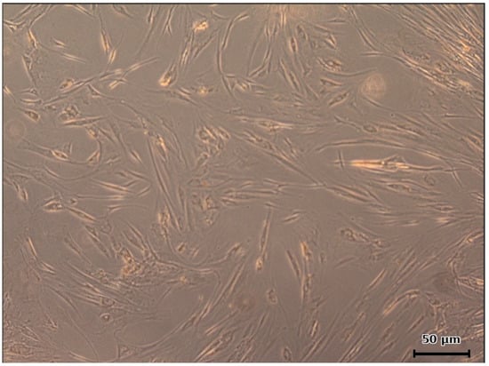
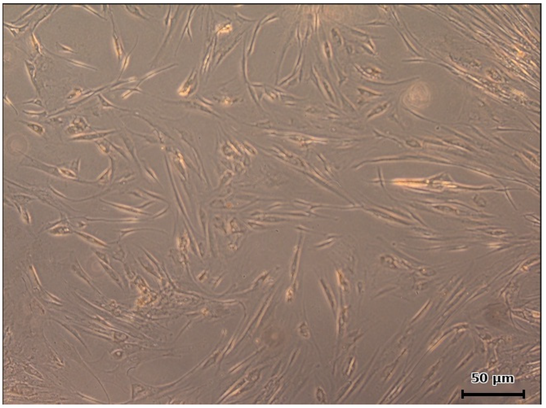
ADSCs were successfully isolated from adipose tissue using collagenase-based enzymatic digestion. After culture expansion, the cells adhered to plastic and showed robust proliferative behavior. Under phase-contrast microscopy, the majority of cells displayed the expected spindle-shaped, fibroblast-like morphology with tapered processes. The cultures showed variable cell size and cytoplasmic texture, but there was no evidence of extensive detachment, vacuolization or degenerative morphology, supporting successful ADSC expansion under standard conditions (Figure 1).
Figure 1.
Morphology of cultured ADSCs on day 7 after isolation. Representative phase-contrast image showing plastic-adherent ADSCs with fibroblast-like, spindle-shaped morphology and elongated cellular processes at 40× magnification. Scale bar = 50 μm.
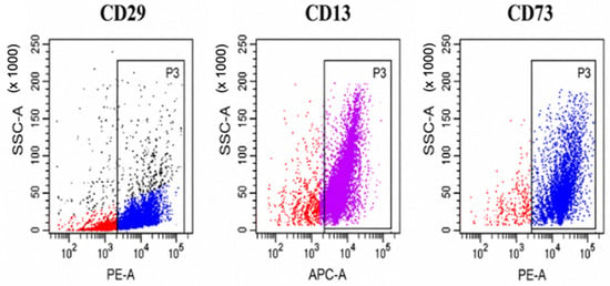
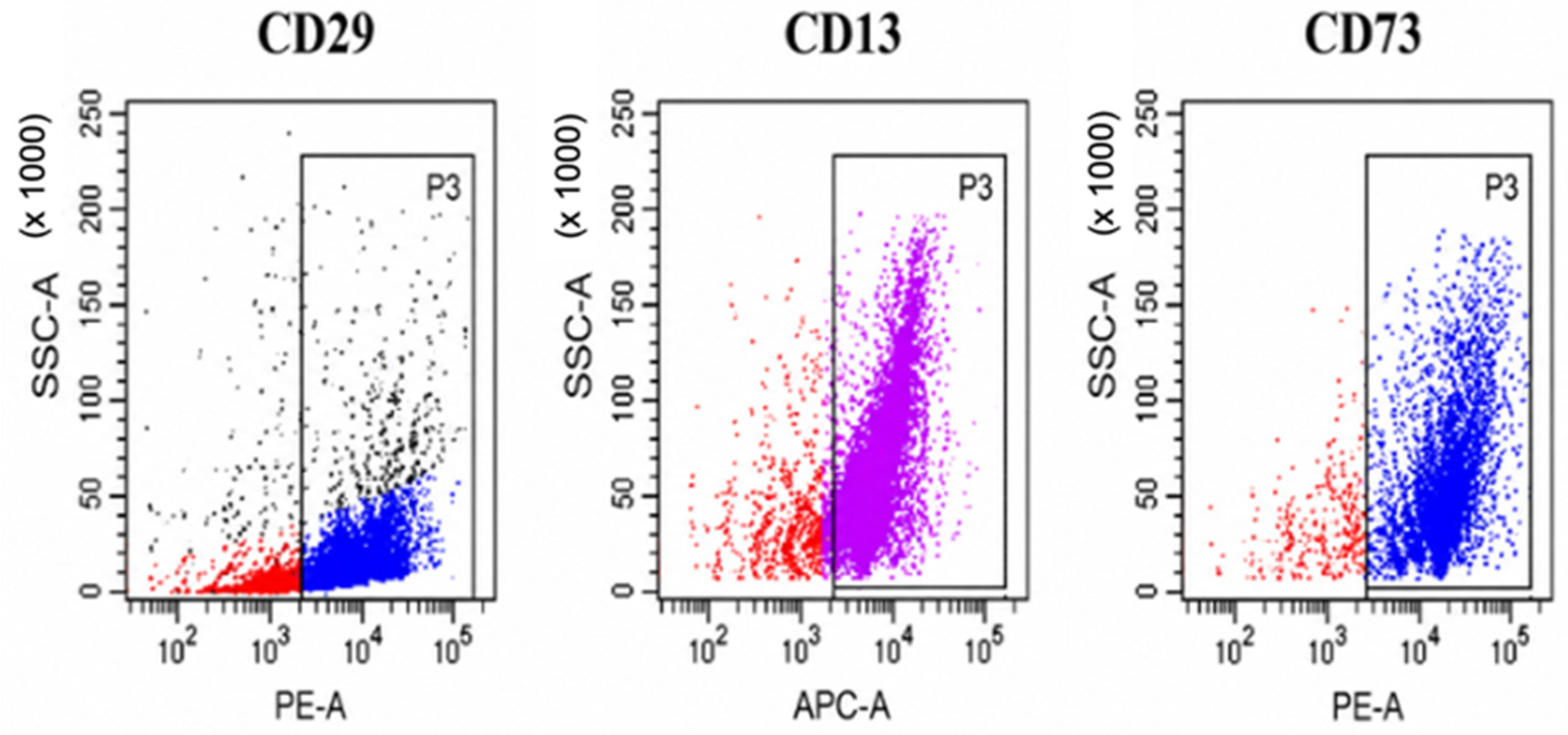
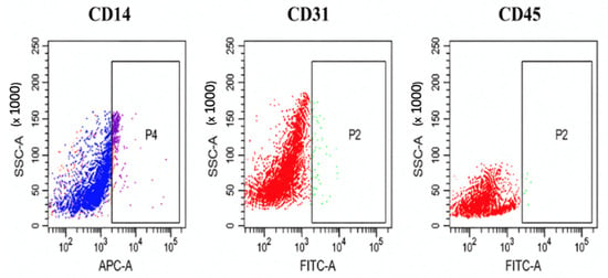
Flow cytometric analysis confirmed a mesenchymal phenotype. ADSCs showed strong expression of CD73, CD13 and CD29, whereas hematopoietic and endothelial markers were minimally expressed. Mean positive expression values were 97.82 ± 1.2% for CD73, 94.32 ± 2.1% for CD13 and 96.53 ± 1.8% for CD29 (Figure 2). In contrast, CD14, CD31 and CD45 expression remained very low at 0.81 ± 0.3%, 1.21 ± 0.4% and 0.91 ± 0.3%, respectively (Figure 3). These data confirmed that the expanded cells met the expected ADSC phenotype and were not substantially contaminated by hematopoietic, monocytic or endothelial populations.
Figure 2.
Positive surface marker profile of ADSCs. Representative flow cytometry plots demonstrate high expression of CD29, CD13, and CD73, confirming the mesenchymal phenotype of the isolated ADSCs. Colors are used for visualization only and have no biological significance. Positive staining was determined exclusively by the rectangular gate.
Figure 3.
Negative lineage marker profile of ADSCs. Representative flow cytometry plots demonstrate minimal expression of CD14, CD31, and CD45, indicating negligible contamination by monocytes, endothelial cells, or hematopoietic cells. Colors are used for visualization only and have no biological significance. Positive staining was determined exclusively by the rectangular gate.
3.2. Morphological Response to Medication Treatment and MEMW Exposure
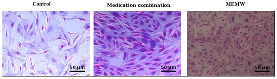
Untreated ADSCs retained a typical fibroblast-like appearance, with randomly distributed polygonal-to-spindle-shaped cells. The medication and MEMW groups showed modest increases in cellular elongation, parallel orientation, and alignment relative to the untreated control. Apparent differences in the number of cells per field were not quantified and are therefore not interpreted as changes in cell density or proliferation. Crystal violet staining was used only for qualitative morphological visualization. Representative images are shown in Figure 4.
Figure 4.
Morphological comparison of ADSCs under control, medication-treated, and MEMW-exposed conditions. All images were acquired at 40× magnification under identical imaging settings. Treated cultures showed modest differences in cellular elongation and alignment. Crystal violet staining was used for qualitative visualization only and not for cell-density quantification. Scale bars = 50 μm.
3.3. Regulation of Stemness-Associated Genes
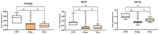
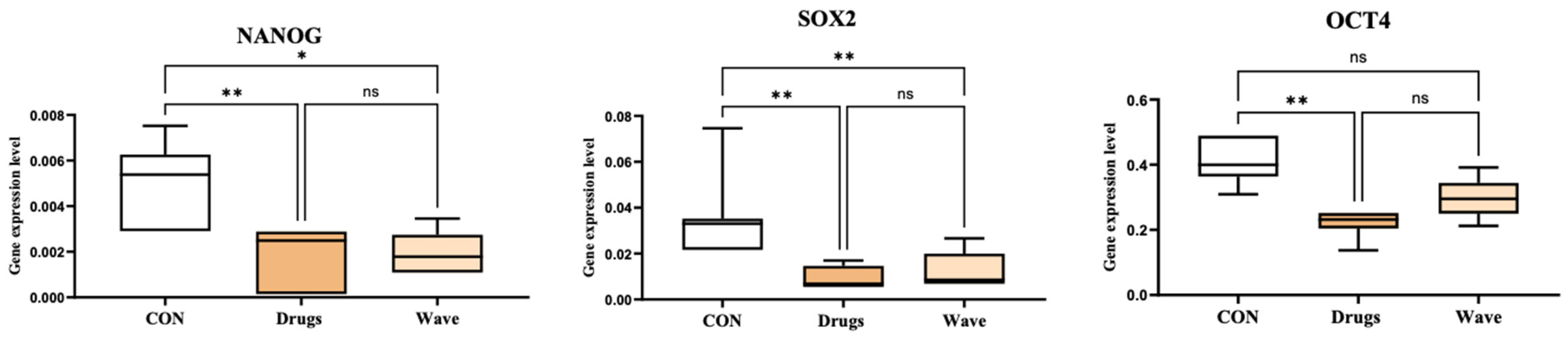
qRT-PCR analysis showed reduced expression of the stemness-associated transcripts POU5F1 (OCT4), SOX2, and NANOG in medication-treated and MEMW-exposed ADSCs relative to untreated controls (Figure 5). This pattern is consistent with a shift away from an undifferentiated state but does not, by itself, establish a specific differentiated phenotype.
Figure 5.
Expression of stemness-associated genes after treatment. Relative expression of NANOG, SOX2, and POU5F1 (OCT4) was measured by qRT-PCR in control, medication-treated, and MEMW-exposed cells. Values were normalized to GAPDH and expressed relative to untreated controls. Statistical comparisons used one-way ANOVA with multiple-comparison testing for parametric data or the Kruskal–Wallis test with Dunn’s post hoc test when parametric assumptions were not met. * p < 0.05; ** p < 0.01; ns, not significant.
3.4. Expression of Early and Late Cardiac Markers
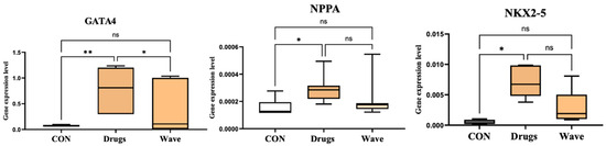
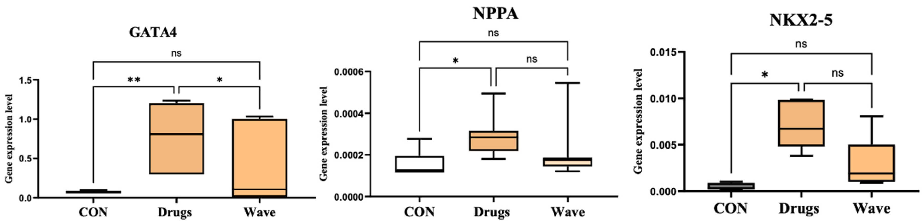
Early cardiac-associated genes increased after both forms of treatment. GATA4, NKX2-5 and NPPA were upregulated in medication-treated and MEMW-exposed ADSCs relative to untreated controls, indicating activation of cardiogenic transcriptional programs (Figure 6). These markers are associated with early cardiac development and lineage specification and their induction supports cardiomyogenic commitment rather than maintenance of a naïve mesenchymal phenotype.
Figure 6.
Early cardiomyogenic marker expression in ADSCs. Relative mRNA levels of GATA4, NPPA, and NKX2-5 were quantified by qRT-PCR and normalized to GAPDH. Statistical comparisons used one-way ANOVA with multiple-comparison testing for parametric data or the Kruskal–Wallis test with Dunn’s post hoc test when parametric assumptions were not met. * p < 0.05; ** p < 0.01; ns, not significant.
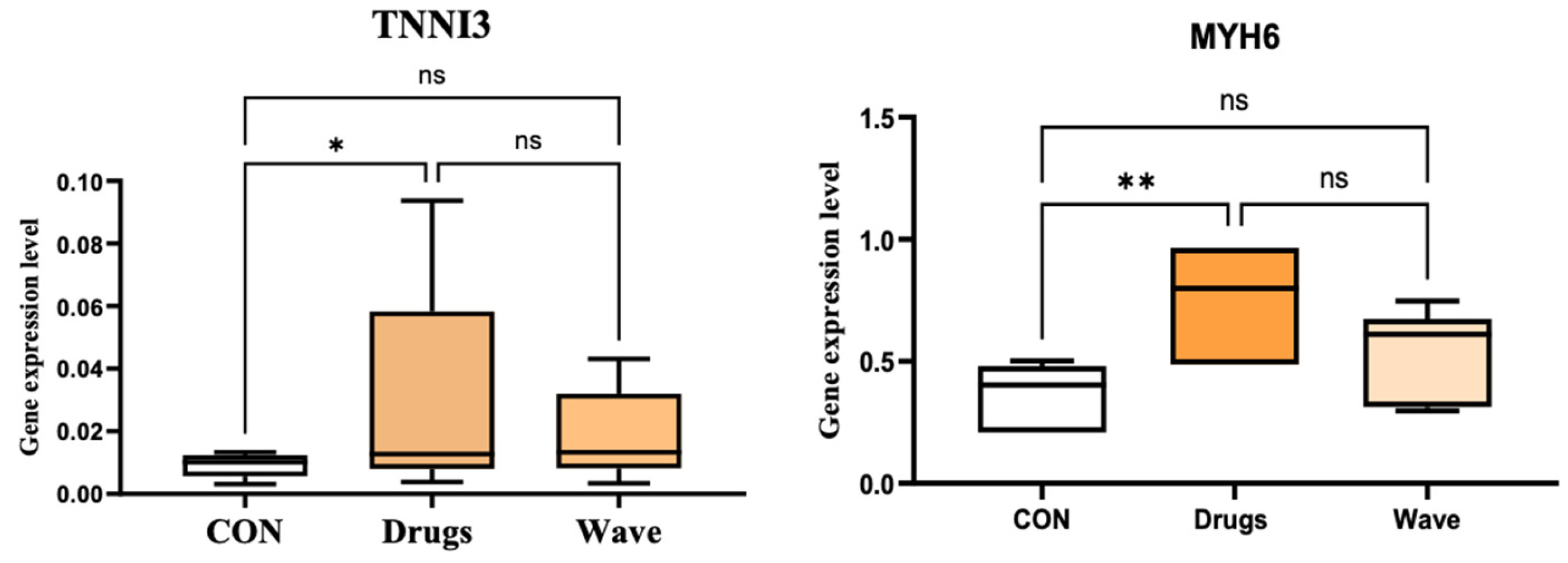
Changes in late cardiomyocyte-associated transcripts were more modest. TNNI3 and MYH6 showed limited increases in the medication-treated group, but the overall magnitude of late marker induction was lower than that observed for early cardiac genes (Figure 7). This pattern indicates that the 21-day protocol promoted early cardiomyocyte-like commitment but may not have been sufficient to achieve complete structural and functional maturation.
Figure 7.
Late cardiomyocyte-associated gene expression after treatment. Relative expression of TNNI3 and MYH6 was assessed in control, medication-treated, and MEMW-exposed ADSCs and normalized to GAPDH. Statistical comparisons used one-way ANOVA with multiple-comparison testing for parametric data or the Kruskal–Wallis test with Dunn’s post hoc test when parametric assumptions were not met. Late-marker changes were smaller than early-marker responses, supporting partial commitment rather than mature cardiomyocyte differentiation. * p < 0.05; ** p < 0.01; ns, not significant.
4. Discussion
This study examined whether medication-derived electromagnetic waves could influence ADSCs toward an early cardiomyocyte-like state. Direct medication treatment and MEMW exposure produced broadly convergent changes: modest increases in elongation and alignment, lower expression of stemness-associated genes, and higher expression of early cardiac-associated markers. These findings show that MEMW produced selected biological responses comparable to the medication mixture under the conditions tested, but they do not establish mature cardiomyocyte differentiation or a drug-specific electromagnetic mechanism in vitro [24].
Initial ADSC characterization was necessary before interpreting the differentiation response. The isolated cells showed plastic adherence, spindle-shaped morphology, strong CD73/CD13/CD29 expression and negligible CD14/CD31/CD45 expression. This phenotype supports an ADSC-enriched mesenchymal population and makes it less likely that the observed cardiac marker profile was driven by hematopoietic or endothelial contamination [6,7,8,9,10,11,12,13].
Morphological organization differed modestly among the groups. Untreated ADSCs remained randomly oriented and fibroblast-like, whereas both intervention groups showed greater elongation and alignment. Because the images were representative and the analysis was qualitative, no conclusion can be drawn about cell density, proliferation, or treatment-specific changes in cell size. The observed alignment is compatible with cytoskeletal reorganization during early lineage commitment, but no direct cytoskeletal assay was performed.
The molecular findings further support a shift away from an undifferentiated state. Reduced POU5F1 (OCT4), SOX2, and NANOG expression indicates lower stemness-associated transcriptional activity. ADSCs are adult mesenchymal cells rather than pluripotent stem cells; therefore, these markers are interpreted as indicators of progenitor-state change rather than proof of pluripotency loss. Their reduction after both interventions is consistent with increased lineage commitment.
The upregulation of GATA4, NKX2-5, and NPPA is consistent with activation of early cardiac transcriptional programs. GATA4 and NKX2-5 participate in cardiac lineage specification, whereas NPPA is associated with fetal cardiac gene expression and cardiomyogenic differentiation [17,28]. These changes support early commitment when interpreted together with the decrease in stemness-associated transcripts.
The four-agent combination was selected from prior ADSC work [24] and was intended to engage complementary cardiovascular pathways rather than a single target. Salbutamol can influence β-adrenergic/cAMP–MAPK signaling; ivabradine is linked to HCN-current biology, calcium handling, and remodeling; and sacubitril/valsartan modifies natriuretic-peptide and renin–angiotensin signaling with downstream effects on inflammation, fibrosis, and AMPK-related responses [29,30,31,32,33,34,35,36,37,38,39,40,41,42]. These actions could converge on stress adaptation, calcium homeostasis, cytoskeletal organization, and cardiac transcriptional networks. Because the agents were not tested in a factorial design, the present study cannot determine whether their combined effects were synergistic, additive, or antagonistic.
The modest response of the late markers TNNI3 and MYH6 is a central limitation. Mature cardiomyocytes require organized sarcomeres, functional calcium handling, electrical excitability, intercellular coupling, and contractile activity. The present evidence therefore supports early cardiomyogenic commitment rather than structural or functional maturation. Protein-level validation and functional assays are required before mature cardiomyocyte differentiation can be claimed.
The mechanism underlying the MEMW-associated response remains unresolved. A physical signal could, in principle, be transduced through membrane polarization, ion-channel activity, calcium flux, redox signaling, mitochondrial responses, and cytoskeletal remodeling, with downstream effects on cardiac transcriptional networks [18,19,20,21,22,23]. Conventional electromagnetic stimulation is generally defined by selected frequencies, waveforms, and field strengths, whereas the present approach uses playback of a proprietary broadband recording associated with a medication preparation. Whether that recording retains drug-specific biological information is a working hypothesis, not an established mechanism.
Several limitations constrain interpretation. Field strength, waveform components, signal stability, and spatial uniformity were not independently characterized. The study did not include sham or nonspecific electromagnetic exposure, so the observed effects cannot be attributed exclusively to medication-specific information. Morphology was assessed qualitatively, the donor cohort was limited to women, and the biological conclusions rely on mRNA rather than protein or function. Blinded image analysis, independent replication, protein-level validation, and functional assays are needed to determine whether the transcriptional changes are stable and biologically meaningful.
5. Future Directions and Potential Applications
Future work should begin by standardizing the MEMW exposure system. Reproducible interpretation will require detailed characterization of the signal source, antenna position, field intensity, exposure frequency range, treatment duration, distance from cultures and possible environmental interference. A sham-exposed control group should also be incorporated to distinguish true MEMW-related effects from incubator handling, plate positioning or nonspecific physical stimulation.
Mechanistic studies should examine how MEMW exposure affects cellular signaling during cardiomyogenic commitment. Priority areas include membrane polarization, intracellular calcium dynamics, reactive oxygen species balance, mitochondrial metabolism, cytoskeletal remodeling and pathways such as MAPK/ERK, Notch, NF-kB, AMPK, GATA4 and NKX2-5. These analyses would help determine whether MEMW acts as a broad biophysical stimulus or whether it activates more specific signaling programs associated with the parent medications.
The evidence should also be expanded beyond morphology and mRNA expression. Protein-level confirmation is needed using immunocytochemistry, Western blotting or flow cytometry for cardiac troponin T, alpha-actinin, connexin-43, MYH6, MYH7 and related markers. Functional maturation should be assessed through calcium transient imaging, electrophysiological analysis, contractility assays, sarcomere organization, mitochondrial respiration and gap-junction formation. These tests are necessary to separate early lineage commitment from genuine cardiomyocyte maturation.
Optimization studies should evaluate different exposure schedules, treatment windows, donor sources, cell densities, passage numbers and culture formats. Three-dimensional scaffolds, extracellular matrix systems, mechanical stimulation and biochemical cardiogenic cues may be combined with MEMW to improve tissue organization and maturation. This is especially important for cardiac tissue engineering, where aligned architecture and synchronized function are essential.
From a translational perspective, MEMW may be explored as a non-contact preconditioning strategy for ADSCs before transplantation or tissue-engineered construct formation. If independently validated, the approach could reduce prolonged drug exposure during cell manufacturing and provide a scalable conditioning platform. In the long term, medication-derived electromagnetic signaling might support controlled biophysical conditioning of patient-derived cells, but this possibility remains experimental.
Before clinical translation, safety and efficacy must be tested rigorously. Future experiments should include independent replication, genomic stability assessment, long-term culture monitoring, arrhythmogenic risk evaluation and in vivo studies using myocardial infarction or cardiac injury models. Outcomes should include cell survival, retention, vascular integration, immune response, scar remodeling, ventricular function and electrophysiological safety. Without these data, MEMW should remain an experimental platform rather than a clinical intervention.
6. Conclusions
This study provides preliminary evidence that medication-derived electromagnetic waves generated from a four-drug cardiovascular combination can influence human ADSCs toward an early cardiomyogenic state. MEMW exposure was associated with modest morphological organization, reduced stemness-associated gene expression, and increased early cardiac-associated transcripts in a pattern broadly comparable to direct medication treatment. The limited late-marker response does not demonstrate mature cardiomyocyte differentiation. Independent signal characterization, sham controls, protein validation, functional testing, and in vivo safety studies are required before mechanistic or translational claims can be made.
Author Contributions
S.A.A.-S. conceived and designed the study, developed the methodology, supervised the project, performed data analysis and interpretation, wrote the original manuscript draft, revised the manuscript critically for important intellectual content, and approved the final version. H.A. contributed to the study design, performed the experimental work and data acquisition, assisted with data analysis and interpretation, reviewed and edited the manuscript, and approved the final version. F.H.A.-G. contributed to the experimental design, methodology, data interpretation, critical revision of the manuscript, and approved the final version. All authors have read and agreed to the published version of the manuscript.
Funding
Financial support was provided by the Ongoing Research Funding Program (ORF-2026-1798), King Saud University, Riyadh, Saudi Arabia.
Institutional Review Board Statement
The project was approved by the Institutional Review Board of King Saud University Medical City (KSU-[E-24-8857]; 12 March 2023). Written informed consent was obtained from every donor before adipose tissue collection and all procedures followed institutional requirements and the Declaration of Helsinki.
Informed Consent Statement
Permission to publish anonymized study information was secured from each donor before enrollment.
Data Availability Statement
Data generated during this work can be provided by the corresponding author after a reasonable request and institutional approval, where applicable.
Acknowledgments
AI-based language tools were used solely to improve wording, organization, and clarity. The authors verified all scientific content, interpretations, and final text.
Conflicts of Interest
Author Fuad H. Al Ghamdi was employed by the company Johns Hopkins Aramco healthcare. The remaining authors declare that the research was conducted in the absence of any commercial or financial relationships that could be construed as a potential conflict of interest.
References
- Beliën, H.; Evens, L.; Hendrikx, M.; Bito, V.; Bronckaers, A. Combining stem cells in myocardial infarction: The road to superior repair? Med. Res. Rev. 2022, 42, 343–373. [Google Scholar] [CrossRef] [Scilit] [PubMed]
- Arjmand, B.; Abedi, M.; Arabi, M.; Alavi-Moghadam, S.; Rezaei-Tavirani, M.; Hadavandkhani, M.; Tayanloo-Beik, A.; Kordi, R.; Parhizkar Roudsari, P.; Larijani, B. Regenerative medicine for the treatment of ischemic heart disease: Status and future perspectives. Front. Cell Dev. Biol. 2021, 9, 704903. [Google Scholar] [CrossRef] [Scilit] [PubMed]
- Chang, M.-L.; Chiu, Y.-J.; Li, J.-S.; Cheah, K.-P.; Lin, H.-H. Analyzing impetus of regenerative cellular therapeutics in myocardial infarction. J. Clin. Med. 2020, 9, 1277. [Google Scholar] [CrossRef] [Scilit] [PubMed]
- Demurtas, J.; Fanelli, G.N.; Romano, S.L.; Solari, M.; Yang, L.; Soysal, P.; López Sánchez, G.F.; Grabovac, I.; Smith, L.; Zorzi, A.; et al. Stem cells for treatment of cardiovascular diseases: An umbrella review of randomized controlled trials. Ageing Res. Rev. 2021, 67, 101257. [Google Scholar] [CrossRef] [Scilit] [PubMed]
- Ali, S.A.; Mahmood, Z.; Mubarak, Z.; Asad, M.; Chaudhri, M.T.S.; Bilal, L.; Ashraf, T.; Khalifa, T.N.; Ashraf, T.; Saleem, F.; et al. Assessing the potential benefits of stem cell therapy in cardiac regeneration for patients with ischemic heart disease. Cureus 2025, 17, e76770. [Google Scholar] [CrossRef] [Scilit] [PubMed]
- Bunnell, B.A.; Flaat, M.; Gagliardi, C.; Patel, B.; Ripoll, C. Adipose-derived stem cells: Isolation, expansion and differentiation. Methods 2008, 45, 115–120. [Google Scholar] [CrossRef] [Scilit] [PubMed]
- Ghiasi, M.; Tabatabaei Qomi, R.; Kalhor, N.; Sheykhhasan, M. Adipose-derived stem cells: An optimized protocol for isolation and proliferation. Acta Med. Int. 2016, 3, 116–121. [Google Scholar] [CrossRef] [Scilit]
- Ferroni, L.; De Francesco, F.; Pinton, P.; Gardin, C.; Zavan, B. Methods to isolate adipose tissue-derived stem cells. Methods Cell Biol. 2022, 171, 215–228. [Google Scholar] [CrossRef] [Scilit] [PubMed]
- Sherman, L.S.; Condé-Green, A.; Naaldijk, Y.; Lee, E.S.; Rameshwar, P. An enzyme-free method for isolation and expansion of human adipose-derived mesenchymal stem cells. J. Vis. Exp. 2019, 154, e59419. [Google Scholar] [CrossRef] [Scilit] [PubMed]
- Alstrup, T.; Eijken, M.; Bohn, A.B.; Møller, B.; Damsgaard, T.E. Isolation of adipose tissue-derived stem cells: Enzymatic digestion in combination with mechanical distortion to increase adipose tissue-derived stem cell yield from human aspirated fat. Curr. Protoc. Stem Cell Biol. 2019, 48, e68. [Google Scholar] [CrossRef] [Scilit] [PubMed]
- Eva, K.; Miloslav, M.; Michal, M. Use of collagenase to isolate adipose tissue-derived stem cells—Substantial manipulation or not? J. Stem Cell Ther. Transplant. 2022, 6, 8–18. [Google Scholar] [CrossRef] [Scilit]
- Markarian, C.F.; Frey, G.Z.; Silveira, M.D.; Chem, E.M.; Milani, A.R.; Ely, P.B.; Nardi, N.B. Isolation of adipose-derived stem cells: A comparison among different methods. Biotechnol. Lett. 2014, 36, 693–702. [Google Scholar] [CrossRef] [Scilit] [PubMed]
- Krylova, T.A.; Bystrova, O.A.; Jakovleva, T.K.; Zenin, V.V.; Moiseeva, O.M.; Parfenov, V.N.; Martynova, M.G. Characteristics of human subepicardial adipose tissue-derived stem cells. Stem Cell Stud. 2011, 1, e18. [Google Scholar] [CrossRef] [Scilit]
- Boheler, K.R.; Czyz, J.; Tweedie, D.; Yang, H.-T.; Anisimov, S.V. Differentiation of pluripotent embryonic stem cells into cardiomyocytes. Circ. Res. 2002, 91, 189–201. [Google Scholar] [CrossRef] [Scilit] [PubMed]
- Laco, F.; Low, J.-L.; Seow, J.; Woo, T.L.; Zhong, Q.; Seayad, J.; Liu, Z.; Wei, H.; Reuveny, S.; Elliott, D.A.; et al. Cardiomyocyte differentiation of pluripotent stem cells with SB203580 analogues correlates with Wnt pathway CK1 inhibition independent of p38 MAPK signaling. J. Mol. Cell. Cardiol. 2015, 80, 56–70. [Google Scholar] [CrossRef] [Scilit] [PubMed]
- Gartz, M.; Strande, J.L. Optimizing the differentiation of cardiomyocytes from human induced pluripotent-derived stem cells. In Cardiovascular Development: Methods and Protocols; Wang, Q., Ed.; Springer: New York, NY, USA, 2021; pp. 51–60. [Google Scholar] [CrossRef] [Scilit] [PubMed]
- Mensah, I.K.; Gowher, H. Signaling Pathways Governing Cardiomyocyte Differentiation. Genes 2024, 15, 798. [Google Scholar] [CrossRef] [Scilit] [PubMed]
- Sun, G.; Li, J.; Zhou, W.; Hoyle, R.G.; Zhao, Y. Electromagnetic Interactions in Regulations of Cell Behaviors and Morphogenesis. Front. Cell Dev. Biol. 2022, 10, 1014030. [Google Scholar] [CrossRef] [Scilit] [PubMed]
- Balzano, Q.; Sheppard, A. RF Nonlinear interactions in living cells-I: Nonequilibrium thermodynamic theory. Bioelectromagnetics 2003, 24, 473–482. [Google Scholar] [CrossRef] [Scilit] [PubMed]
- Ignatavičienė, I.; Vyšniauskienė, R.; Rančelienė, V.; Petrošius, R.; Grauda, D.; Butkauskas, D. The Effects of Electromagnetic Field Radiation of Extremely Low Frequency on Growth Parameters and Nucleotide Substitutions in L. minor Clones. Acta Physiol. Plant. 2024, 46, 47. [Google Scholar] [CrossRef] [Scilit]
- Brabant, C.; Geerinck, A.; Beaudart, C.; Tirelli, E.; Geuzaine, C.; Bruyère, O. Exposure to Magnetic Fields and Childhood Leukemia: A Systematic Review and Meta-Analysis of Case-Control and Cohort Studies. Rev. Environ. Health 2023, 38, 229–253. [Google Scholar] [CrossRef] [Scilit] [PubMed]
- Al-Ghamdi, F.H.; Al-Qaaneh, A.M.; Al-Shehri, B.A. Effect of antibiotics’ electromagnetic spectrums on bacterial growths: Beyond drug-receptor interaction. J. Med. Pharm. Allied Sci. 2023, 12, 6241–6246. [Google Scholar] [CrossRef] [Scilit]
- Al-Ghamdi, F.H.; Al-Qaaneh, A.M.; Almofty, S. Effect of Anticancers’ Electromagnetic Spectrums on Cancer Cell Line Growths. J. Med. Pharm. Allied Sci. 2024, 13, 6603–6607. [Google Scholar] [CrossRef] [Scilit]
- Alshammari, H.; Abuhaimed, J.; Alshehri, S.; Siyal, A.; Shaheen, S.; Abou Al-Saud, S. Cardiac Drug Potential: Exploring Individual and Combined Cardiac Drugs to Promote Differentiation of Adipose-Derived Stem Cells into Cardiomyocytes. Mol. Biol. Rep. 2026, 53, 434. [Google Scholar] [CrossRef] [Scilit] [PubMed]
- Tomlinson, P.R.; Wilson, J.W.; Stewart, A.G. Salbutamol Inhibits the Proliferation of Human Airway Smooth Muscle Cells Grown in Culture: Relationship to Elevated cAMP Levels. Biochem. Pharmacol. 1995, 49, 1809–1819. [Google Scholar] [CrossRef] [Scilit]
- Devalia, J.L.; Sapsford, R.J.; Wells, C.W.; Richman, P.; Davies, R.J. The Effects of Salmeterol and Salbutamol on Ciliary Beat Frequency of Cultured Human Bronchial Epithelial Cells, in Vitro. Pulm. Pharmacol. 1992, 5, 257–263. [Google Scholar] [CrossRef] [Scilit]
- Niehoff, J.; Hirt, M.N.; Staudt, A.; Bittner, A.; Riemer, T.; Fiedler, J.; Just, A.; Eschenhagen, T. The Effect of Antiarrhythmic Drugs on Beat Rate Variability of Human Embryonic and Human Induced Pluripotent Stem Cell-Derived Cardiomyocytes. Sci. Rep. 2019, 9, 1410. [Google Scholar] [CrossRef] [Scilit] [PubMed]
- Valimaki, M.; Ojala, M.; Kaartinen, T.; Kinnunen, S.; Tuuri, T.; Kallio, P. GATA-Targeted Compounds Modulate Cardiac Subtype Cell Differentiation in Dual Reporter Stem Cell Line. Stem Cell Res. Ther. 2021, 12, 190. [Google Scholar] [CrossRef] [Scilit]
- Bourdillon, P.D.V.; Dawson, J.R.; Foale, R.A.; Timmis, A.D.; Poole-Wilson, P.A.; Sutton, G.C. Salbutamol in Treatment of Heart Failure. Heart 1980, 43, 206–210. [Google Scholar] [CrossRef] [Scilit] [PubMed]
- Emilien, G.; Maloteaux, J.M.; Emilien, G. Current Therapeutic Uses and Potential of Beta-Adrenoceptor Agonists and Antagonists. Eur. J. Clin. Pharmacol. 1998, 53, 389–404. [Google Scholar] [CrossRef] [Scilit] [PubMed]
- Stewart, A.F.; Rokosh, D.G.; Bailey, B.A.; Karns, L.R.; Long, C.S.; Brown, J.H. Transcription factor RTEF-1 mediates alpha1-adrenergic reactivation of the fetal gene program in cardiac myocytes. Circ. Res. 1998, 83, 43–49. [Google Scholar] [CrossRef] [Scilit] [PubMed]
- Yan, L.; Lee, J.H.; Lee, J.; Li, Y.; Yang, X. Beta-Adrenergic Signals Regulate Cardiac Differentiation of Mouse Embryonic Stem Cells via Mitogen-Activated Protein Kinase Pathways. Dev. Growth Differ. 2011, 53, 772–779. [Google Scholar] [CrossRef] [Scilit] [PubMed]
- Li, X.L.; Wang, X.; Wang, J.; Li, X.; Chen, Y. Role of Alpha- and Beta-Adrenergic Receptors in Cardiomyocyte Differentiation from Murine-Induced Pluripotent Stem Cells. Cell Prolif. 2017, 50, e12310. [Google Scholar] [CrossRef] [Scilit] [PubMed]
- Heusch, G.; Kleinbongard, P. Ivabradine: Cardioprotection by and Beyond Heart Rate Reduction. Drugs 2016, 76, 733–740. [Google Scholar] [CrossRef] [Scilit]
- Navaratnarajah, M.; Reil, J.C.; Herring, N.; Kacprzak, M.; Zhang, Y.; Watkins, H. Ivabradine Reduces Interstitial Fibrosis and Enhances the Recovery of Cardiomyocyte Excitation-Contraction Coupling Produced by Mechanical Unloading. Circulation 2011, 124, A9342. [Google Scholar] [CrossRef] [Scilit]
- Chen, S.L.; Hu, Z.Y.; Zuo, G.F.; Li, M.H.; Li, B. I(f) Current Channel Inhibitor (Ivabradine) Deserves Cardioprotective Effect via Down-Regulating the Expression of Matrix Metalloproteinase (MMP)-2 and Attenuating Apoptosis in Diabetic Mice. BMC Cardiovasc. Disord. 2014, 14, 150. [Google Scholar] [CrossRef] [Scilit] [PubMed]
- Simko, F.; Baka, T.; Repova, K.; Aziriova, S.; Krajcirovicova, K.; Paulis, L.; Adamcova, M. Ivabradine Improves Survival and Attenuates Cardiac Remodeling in Isoproterenol-Induced Myocardial Injury. Fundam. Clin. Pharmacol. 2021, 35, 744–748. [Google Scholar] [CrossRef] [Scilit] [PubMed]
- Wu, T.Y.; Wu, H.; Chen, C. Effects of Ivabradine on Notch and NF-Kappa B Signaling Pathway in Myocardial Cells of Rats with Myocardial Infarction. J. Hainan Med. Univ. 2019, 25, 810–814. [Google Scholar] [CrossRef]
- Mustafa, N.H.; Jalil, J.; Zainalabidin, S.; Saleh, M.S.M.; Asmadi, A.Y.; Kamisah, Y. Molecular Mechanisms of Sacubitril/Valsartan in Cardiac Remodeling. Front. Pharmacol. 2022, 13, 892460. [Google Scholar] [CrossRef] [Scilit] [PubMed]
- Iborra-Egea, O.; Gálvez-Montón, C.; Roura, S.; Perea-Gil, I.; Prat-Vidal, C.; Soler-Botija, C.; Bayes-Genis, A. Mechanisms of Action of Sacubitril/Valsartan on Cardiac Remodeling: A Systems Biology Approach. npj Syst. Biol. Appl. 2017, 3, 12. [Google Scholar] [CrossRef] [Scilit] [PubMed]
- Hu, F.; Li, Y.; Chen, Y.; Wang, J.; Zhang, Y.; Li, X.; Wang, L. Sacubitril/Valsartan Attenuated Myocardial Inflammation, Fibrosis, Apoptosis and Promoted Autophagy in Doxorubicin-Induced Cardiotoxicity Mice via Regulating the AMPKα-mTORC1 Signaling Pathway. Mol. Cell. Biochem. 2025, 480, 1891–1908. [Google Scholar] [CrossRef] [Scilit]
- Sarikhani, M.; Ebrahimi, M.; Khosravi, M.; Moghaddam, M.M.; Ghahremani, M.H.; Baharvand, H.; Salekdeh, G.H. Sustained Activation of AMPK Enhances Differentiation of Human iPSC-Derived Cardiomyocytes via Sirtuin Activation. Stem Cell Rep. 2020, 15, 498–514. [Google Scholar] [CrossRef] [Scilit] [PubMed]

Disclaimer/Publisher’s Note: The statements, opinions and data contained in all publications are solely those of the individual author(s) and contributor(s) and not of MDPI and/or the editor(s). MDPI and/or the editor(s) disclaim responsibility for any injury to people or property resulting from any ideas, methods, instructions or products referred to in the content. |
© 2026 by the authors. Licensee MDPI, Basel, Switzerland. This article is an open access article distributed under the terms and conditions of the Creative Commons Attribution (CC BY) license.