


Abstract
Akkermansia muciniphila (AM) is a Gram-negative commensal bacteria that has been found in both the oral cavity and gastrointestinal tract and has the ability to maintain homeostasis in the intestinal tract by inducing anti-inflammatory responses. The purpose of this study was to evaluate the presence of AM in the oral microbiome of patients with and without orthodontic appliances to determine any associations with other oral bacteria. This retrospective study of orthodontic and non-orthodontic saliva samples (n = 209) was screened by qPCR analysis for AM, Scardovia wiggsiae (SW), Selenomonas noxia (SN), Streptococcus mutans (SM), Porphyromonas gingivalis (PG), Treponema denticola (TD), and Fusobacterium nucleatum (FN). These data revealed oral microbial prevalence between 29.8% (AM) and 54.3% (SM)—with orthodontic samples revealing decreased AM prevalence (−29.5%), compared with non-orthodontic samples and corresponding increased prevalence of PG (+2.7%), TD (+13.7%), SM (+22.2%) and SN (+26.0%). AM-positive samples exhibited decreased prevalence of SN (−16.0%), SM (−1.6%), PG (−10.2%), TD (−4.2%), and FN (−14.3%). These findings may suggest that orthodontic appliances may be associated with decreased AM prevalence, which may be associated with increased prevalence of other oral pathogens.
1. Introduction
The microbiome of the human oral cavity and gastrointestinal tract has the ability to significantly influence the systemic health of the host [1,2,3]. Many studies confirm the interconnected relationship between the gut microbiota and the maintenance of homeostasis or the development of systemic metabolic and inflammatory diseases, including metabolic syndrome and diabetes mellitus [4,5,6]. Similarly, the oral microbiome has been demonstrated to play a significant role not only in maintaining balance within the gut microbiome but may also directly contribute to the overall health of the individual or the potential development of chronic inflammation or systemic diseases [7,8,9].
Significant research has elucidated the dynamic role of pathogenic oral bacteria in the development of periodontal disease and the relationship with other systemic diseases and chronic inflammation, with a major emphasis on the periodontal pathogens Porphyromonas gingivalis, Treponema denticola, Tannerella forsythia, and Fusobacterium nucleatum [10,11,12]. However, growing interest has more recently focused on the role of other species of commensal bacteria that have the potential to limit oral and gastrointestinal inflammation and may be significant mediators of oral and systemic diseases [13,14,15]. Although many studies have focused on commensal Gram-positive oral streptococci, recent studies have also demonstrated that Gram-negative commensal bacteria present within the mouth and digestive tract may significantly influence homeostasis and the potential for disease development [16,17,18,19,20]. One such organism, Akkermansia muciniphila (AM), is a Gram-negative commensal bacterium that has been found in both the oral cavity and gastrointestinal tract, with recent evidence suggesting multiple roles in mediating or modulating oral and systemic health and disease [21,22]. More specifically, there is significant evidence to demonstrate AM may have the ability to modulate and maintain homeostasis in the intestinal tract through a significant role in inducing multiple types of anti-inflammatory responses [23,24,25].
In fact, evidence has accumulated that may suggest AM might be useful as a probiotic as a preventive measure to limit or prevent the development of many types of systemic and gastrointestinal disease [26,27]. Moreover, the use of AM as a probiotic or supplement may also be useful in the active treatment of existing chronic illnesses, including cancer, obesity, adult-onset diabetes, cardiometabolic disease, and many other types of illnesses and diseases [28,29,30]. However, much of this evidence centers on the role of AM in the gastrointestinal tract and interactions with other microorganisms and the host immune system [31,32,33].
Fewer studies have focused more specifically on the presence and effect of AM more specifically in the oral cavity [34,35]. For instance, some evidence has shown that AM may enhance mucosal immunity and also directly reduce the prevalence of Porphyromonas gingivalis in the oral cavity [36,37]. Furthermore, another recent study found evidence that AM may also function to directly inhibit the growth and effects of Fusobacterium nucleatum within the oral biofilm [38].
However, few studies have focused on the prevalence of AM in the oral cavity—although some studies postulate that this microbe may be influenced by the presence or absence of orthodontic appliances or the other microbial constituents influenced by this treatment [39]. In fact, many studies suggest that fixed orthodontic appliances create multiple new complex surfaces and oral topography that can alter and mediate bacterial adhesion and biofilm formation, thereby influencing changes in the oral microbiome during treatment [40,41,42]. Many of these microbial changes are strongly associated with an increase in gingivitis and periodontal disease, mainly due to the increased presence of Gram-negative pathogens that colonize the newly created biofilm on and surrounding the surfaces and interface between the teeth and the orthodontic brackets [43,44,45]. Moreover, the presence of orthodontic brackets not only creates new adhesive surfaces, but may also hinder the primary mechanisms for biofilm and plaque removal such as toothbrushing and interdental flossing [46,47,48,49].
Based upon this information, the purpose of this study is to further evaluate the presence of AM in samples of the oral microbiome from patients with and without orthodontic appliances to determine what potential associations this may have upon the prevalence of other AM, as well as other bacteria in the oral cavity. The primary hypothesis for this project was that orthodontic appliances may be associated with changes to the prevalence of AM within the samples screened, with the secondary hypothesis being that prevalence of AM may then be associated with differences in the prevalence of other oral microbes within these samples.
2. Materials and Methods
2.1. Study Protocol
This project was a retrospective observational study of existing biorepository samples that was performed using the guidelines and principles as outlined in the Declaration of Helsinki. This project received approval as Research Exempt on 10 June 2022 through the Office for the Protection of Research Subjects (OPRS) and the Institutional Review Board (IRB) at the University of Nevada, Las Vegas (UNLV) under protocol #1619329-I titled “Retrospective analysis of Oral Health Status of Dental Population”. In brief, this study analyzed existing biorepository samples that did not have any corresponding personal identifying information and that did not involve the prospective collection of any data or biologic specimens from patients, which can be considered Research Exempt (does not require Informed Consent) according to Code of Federal Regulations (45 CFR 36) published by the United States Department of Health and Human Services (US-DHHS).
2.2. Biorepository Samples
The original sample collection protocol for the biorepository received approval through the UNLV-OPRS and UNLV-IRB protocol #1305-4466M titled “The Prevalence of Oral Microbes in Saliva from the University of Nevada, Las Vegas School of Dental Medicine (UNLV-SDM) Pediatric and Adult Clinical Population”, as previously described [39,50]. In brief, UNLV-SDM clinic patients who volunteered to participate were subsequently asked to provide Informed Consent (or Pediatric Assent with parental/guardian Informed Consent for patients under the age of 18 years old). Sampling days (n = 50) were chosen at random over a period of two years and all patients within a given time block (morning or afternoon clinic) were asked for voluntary participation and Informed Consent. Each patient who agreed and provided Informed Consent (or Pediatric Assent with parental/guardian Informed Consent) was then asked to provide basic demographic information (age, sex, race or ethnicity with no patient-specific or chart-related identifying information) and an unstimulated saliva sample of up to 5.0 mL. Each collected sample was assigned a randomly generated, non-sequential number prior to being transferred to a biomedical laboratory for storage and further processing. The inclusion criteria were set to include any UNLV-SDM official patient of record that agreed to voluntary participation and who agreed to provide Informed Consent or Pediatric Assent with Informed Consent of the parent or guardian, without any compensation or remuneration. The exclusion criteria were set to exclude anyone who was not a patient of record, any person that refused to participate, and any person that did not provide Informed Consent/Pediatric Assent.
Due to the specifications of the original collection protocol, only basic demographic information was collected (age, sex, race or ethnicity) with no other health information or patient-specific identifiers, such as oral health status, periodontal condition, caries status, oral hygiene habits, orthodontic appliance type, duration of orthodontic treatment, recent antibiotic use, probiotic use, systemic conditions, or smoking and tobacco usage.
2.3. DNA Isolation and Analysis
A total of n = 209 samples were identified from the existing biorepository, comprising n = 117 samples from orthodontic patients with fixed orthodontic appliances and n = 92 samples from non-orthodontic patients. The isolation of DNA biorepository samples was accomplished with the TRIzol (phenol:chloroform) extraction method from Fisher Scientific (Fair Lawn, NJ, USA), as previously described [39,50]. Briefly, samples were thawed and 0.5 mL was transferred to a sterile microcentrifuge tube and mixed with an equal volume of Trizol reagent. An additional 0.2 mL of chloroform was added prior to centrifugation at 10,000 revolutions per minute (RPM) using the 5425R Microcentrifuge from Eppendorf (Hamburg, Germany) for fifteen minutes, as previously described [39,50]. Transfer of the upper aqueous phase to a sterile microcentrifuge tube was performed prior to mixing with an equal volume of isopropanol for precipitation before centrifugation. Removal of supernatant allowed the DNA pellet to be washed with ethanol and then resuspended using sterile nuclease-free water (100 µL). Quantitative measurements of DNA quality and concentration were assessed with a NanoDrop 2000 spectrophotometer from Fisher Scientific (Fair Lawn, NJ, USA) using absorbances of A260 nm and A280 nm. All samples were stored at −80 °C, as previously described [39,50].
2.4. Quantitative Polymerase Chain Reaction (qPCR) Screening
Each sample was screened using the positive control standards glyceraldehyde phosphate dehydrogenase (GAPDH) and beta actin for human DNA, as well as the positive control standard 16S rRNA for bacterial DNA, as previously outlined [39,40]. Amplification of DNA was performed using the Power Track SYBR green qPCR kit from Applied Biosystems (Waltham, MA, USA) using the recommended procedures from the manufacturer. In brief, each reaction consisted of SYBR Green PowerTrack Master Mix (25 µL), nuclease-free water (7.5 µL), forward and reverse primers (1.5 µL each) and study sample DNA (2.0 µL) for screening with the QuantStudio Real-Time Polymerase Chain Reaction system from Applied Biosciences (Waltham, MA, USA). Each reaction was set with enzyme activation (95 °C, 15 min), followed by the standard recommended protocol of 40 cycles (95 °C denaturation for 15 s, primer pair-specific temperature annealing for 30 s, and 72 °C final extension for 30 s). All qPCR reactions were subjected to a final melt-curve analysis to verify the positive sample amplicons exhibited the correct Tm for the specific primer pairs.
All screenings were done in triplicate and the cycle threshold (Ct) cutoff was set at cycle 40 (C40), which provides maximum sensitivity to detect DNA amplification at low levels, as recommended by the manufacturer. Standard curves were established using known concentrations of human DNA with the positive controls GAPDH and beta actin, with standard curves assessed using the bacterial positive control 16S rRNA from a known standard of Mixed Bacteria #55644 from American Type Culture Collection (ATCC; Manassas, VA, USA). Phosphate-buffered saline (PBS) at 1× concentration was used instead of sample DNA as the negative control. The following validated primers (with melting temperatures) were used for this screening [39,50]:
- Positive controls
- GAPDH forward primer: 5′-ATC TTC CAG GAG CGA GAT CC-3′; Tm = 66 °C
- GAPDH reverse primer: 5′-ACC ACT GAC ACG TTG GCA GT-3′; Tm = 70 °C
- Beta actin forward primer: 5′-GTG GGG TCC TGT GGT GTG-3′; Tm = 69 °C
- Beta actin reverse primer: 5′-GAA GGG GAC AGG CAG TGA-3′; Tm = 67 °C
- 16S rRNA forward primer: 5′-ACG CGT CGA CAG AGT TTG ATC CTG GCT-3′; Tm = 76 °C
- 16S rRNA reverse primer: 5′-GGG ACT ACC AGG GTA TCT AAT-3′; Tm = 62 °C
- Screening primers (bacterial)
- Akkermansia muciniphila (AM)
- Forward AM primer: 5′-CAG CAC GTG AAG GTG GGG-3′; Tm = 69 °C
- Reverse AM primer: 5′-CCT TGG GGT TGG CTT CAG AT-3′; Tm = 68 °C
- Fusobacterium nucleatum (FN)
- Forward primer FN: 5′-CGC AGA AGG TGA AAG TCC TGT AT-3′; Tm = 67 °C
- Reverse primer FN: 5′-TGG TCC TCA CTG ATT CAC ACA GA-3′; Tm = 68 °C
- Porphyromonas gingivalis (PG)
- Forward primer PG: 5′-TAC CCA TCG TCG CCT TGG T-3′; Tm = 69 °C
- Reverse primer PG: 5′-CGG ACT AAA ACC GCA TAC ACT TG-3′; Tm = 66 °C
- Scardovia wiggsiae (SW)
- Forward primer SW: 5′-GTG GAC TTT ATG AAT AAG C-3′; Tm = 55 °C
- Reverse primer SW: 5′-CTA CCG TTA AGC AGT AAG-3′; Tm = 56 °C
- Selenomonas noxia (SN)
- Forward primer SN: 5′-TCT GGG CTA CAC ACG TAC TAC AAT G-3′; Tm = 68 °C
- Reverse primer SN: 5′-GCC TGC AAT CCG AAC TGA GA-3′; Tm = 68 °C
- Streptococcus mutans (SM)
- Forward primer SM: 5′-GCC TAC AGC TCA GAG ATG CTA TTC T-3′; Tm = 68 °C
- Reverse primer SM: 5′-GCC ATA CAC CAC TCA TGA ATT GA-3′; Tm = 65 °C
- Treponema denticola (TD)
- Forward primer TD: 5′-TAA TAC CGA ATG TGC TCA TTT ACA T-3′; Tm = 61 °C
- Reverse primer TD: 5′-CTG CCA TAT CTC TTG TCA TTG CTC TT-3′; Tm = 67 °C
2.5. Statistical Analysis
Calculation for the minimum sample size for retrospective studies may be performed utilizing the rate-limiting step estimate for DNA extraction and recovery from these biological samples. The manufacturer estimate of 90–95% efficiency of recovery allows for an expected maximum difference of 10% or 0.1, which was used to estimate the appropriate sample size with a power (p) of 0.9 and a significance level of alpha = 0.05 for a minimum sample size of n = 50 needed for the current analysis.
Demographic data were compiled and descriptive statistics (total number and percentage) were calculated. Analysis of continuous data (DNA concentration, DNA purity) was performed using two-tailed Student’s t-tests with significance levels set at alpha = 0.05. All p-values were reported. Positive and negative results from the qPCR sample screening were compiled and analyzed with chi square statistics with significance levels set at alpha = 0.05), as previously described [39,50]. The chi square statistic, degrees of freedom, and p-values for each calculation were provided. Finally, these data were analyzed to determine the odds ratio (OR) and the relative risk (RR) using a 95% confidence interval by utilizing the GraphPad Prism Version 9 online software program (San Diego, CA, USA).
3. Results
From the biorepository, n = 209 samples were available for screening in this study (Table 1). The demographic data from this sample were analyzed to reveal that approximately half of the study samples were derived from females (49.8%) and half from males (50.2%), which was similar to the percentage of males and females of the overall clinic population from which these samples were originally taken, p = 0.8414. Furthermore, the analysis of race or ethnicity data revealed the percentage of minority patients (including Hispanic, Black, Asian, and Other) within the study sample was 66.9%, which was also similar to the overall clinic patient population (65.4%), p = 0.6750. Finally, analysis of patient age revealed an average of 25.3 years, which was similar to the overall average age in the clinic population of 26.4 years, p = 0.221.
Table 1.
Analysis of demographic data from study samples.
Sorting of samples from orthodontic and non-orthodontic patients also revealed no differences between females and males, p = 0.1602 or between minorities and non-minorities, p = 0.5166. However, significant differences were found among the age range among the samples from patients in the non-orthodontic group compared with the orthodontic group, p = 0.00054 due to the large age range (7 to 69 years versus 11 to 34 years, respectively) rather than significant differences in the overall average age (24.6 years versus 25.7 years).
To determine the suitability of DNA isolated from each biorepository sample for qPCR screening, spectrophotometric analysis was performed to assess both the quality and quantity (Table 2). These data demonstrated an average DNA concentration from the biorepository samples derived from pediatric patients was 392.9 ng/µL, which was similar to the average derived from the samples from adult patients of 363.1 ng/µL, p = 0.2350. In addition, DNA quality assessed using the absorbance ratio of A260 nm to A280 nm averaged 1.74 for the pediatric samples and 1.77 for the adult samples, which was also not significantly different (p = 0.4759). These values correspond to the range of acceptable concentrations (>10 ng/µL) and purity (A260:A280 > 1.70) for the qPCR screening application, as demonstrated in previous studies [39,50].
Table 2.
DNA analysis of study samples.
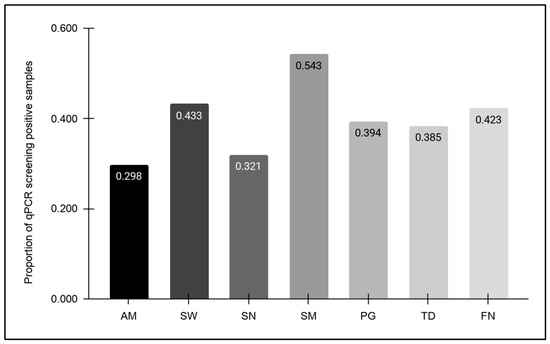
Each of the biorepository samples meeting the standards for DNA quality and quantity was screened using highly specific, previously validated primers for the positive controls, as well as specific oral microbes (Figure 1). Nearly all samples (n = 208/209 or 99.5%) yielded positive results for the bacterial and human DNA-positive controls). Of these samples, approximately 40% harbored one or more of these organisms. More specifically, the lowest prevalence was observed among the Gram-negative bacteria, with 29.8% testing positive for Akkermansia muciniphila (AM), followed by increasing prevalence of Selenomonas noxia (SN) at 32.2%, Treponema denticola (TD) at 38.5%, Porphyromonas gingivalis (PG) at 39.4% and Fusobacterium nucleatum (FN) at 42.3%. Higher prevalence was observed among the Gram-positive cariogenic pathogens Scardovia wiggsiae (SW) at 43.3% and Streptococcus mutans (SM) at 54.3%.
Figure 1.
Biorepository sample screening using qPCR. Prevalence of Gram-negative oral microbes was lower at 29.8% for Akkermansia muciniphila (AM), 32.2% for Selenomonas noxia (SN), 38.5% for Treponema denticola (TD), 39.4% for Porphyromonas gingivalis (PG) and 42.3% for Fusobacterium nucleatum (FN) than for the Gram-positive cariogenic pathogens Scardovia wiggsiae (SW) at 43.3% and Streptococcus mutans (SM) at 54.3%. Key: Akkermansia muciniphila (AM), Selenomonas noxia (SN), Treponema denticola (TD), Porphyromonas gingivalis (PG), Fusobacterium nucleatum (FN), Scardovia wiggsiae (SW), Streptococcus mutans (SM).
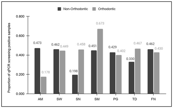
Analysis of the qPCR screening of samples sorted by orthodontic status (patients with or without fixed orthodontic appliances) revealed significant differences in prevalence among several of the oral microbes evaluated (Figure 2). More specifically, a significantly higher proportion of AM-positive results were found among the non-orthodontic samples (47.3%) compared with the orthodontic samples (17.8%), p = 0.0001. In addition, although higher proportions of SW-positive, PG-positive, and FN-positive results were also observed among the non-orthodontic (46.2%, 42.9%, and 46.2, respectively) compared with the orthodontic (44.9%, 40.2%, and 43.0%, respectively) samples, these differences were not statistically significant (p = 0.462, p = 0.591, p = 0.540). In contrast, much lower prevalence was observed for SN-, SM-, and TD-positive samples among the non-orthodontic samples (19.8%, 45.1%, and 33.0%, respectively) compared with the orthodontic samples (45.8%, 67.3%, and 46.7%, respectively), which were statistically significant (p = 0.0004, p = 0.018, p = 0.017).
Figure 2.
qPCR screening results sorted by orthodontic status. Higher prevalence was observed among non-orthodontic versus orthodontic samples with AM-positive (47.3% and 17.8%, p = 0.0001), SW-positive (46.2% and 44.9%, p = 0.462), PG-positive (42.9% and 40.2%, p = 0.591), and FN-positive (46.2% and 43.0%, p = 0.540) results. Lower prevalence was observed among non-orthodontic versus orthodontic samples for SN-positive (19.8% and 45.8%, p = 0.0004), SM-positive (45.1% and 67.3%, p = 0.018), and TD-positive (33.0% and 46.7%, p = 0.017) results. Key: Akkermansia muciniphila (AM), Selenomonas noxia (SN), Treponema denticola (TD), Porphyromonas gingivalis (PG), Fusobacterium nucleatum (FN), Scardovia wiggsiae (SW), Streptococcus mutans (SM).
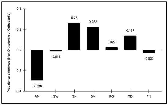
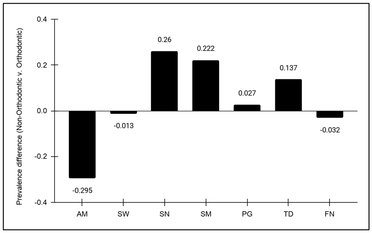
Further analysis revealed differences and changes in prevalence observed between non-orthodontic and orthodontic samples (Figure 3). These results revealed a significant decrease in the prevalence of AM (−29.5%) among the samples from orthodontic patients compared to those from non-orthodontic patients, which was much greater in magnitude than either the decrease in SW (−1.3%) or FN (−3.2%). Although a small increase in prevalence was observed with PG (+2.7%) among the orthodontic-derived samples versus the non-orthodontic samples, much larger changes were found among TD (+13.7%), SM (+22.2%) and SN (+26.0%).
Figure 3.
Change in prevalence between non-orthodontic and orthodontic samples. Decreases in prevalence among orthodontic samples were observed with AM (−29.5%), SW (−1.3%) and FN (−3.2%), with increases in prevalence observed with PG (+2.7%), TD (+13.7%), SM (+22.2%) and SN (+26.0%). Key: Akkermansia muciniphila (AM), Selenomonas noxia (SN), Treponema denticola (TD), Porphyromonas gingivalis (PG), Fusobacterium nucleatum (FN), Scardovia wiggsiae (SW), Streptococcus mutans (SM).
To provide a more detailed assessment of the qPCR screening data, these results were sorted by orthodontic status to determine how this might relate to the presence or absence of AM in the study samples (Table 3). This analysis revealed an odds ratio (OR) of 4.15, which represents an increased odds of an AM-positive result within the non-orthodontic samples that was statistically significant, p < 0.0001. In addition, the calculation of relative risk (RR), which represents an estimation of the risk in one group relative to another (orthodontic versus non-orthodontic) also revealed an RR = 1.56, which also demonstrates a statistically significant likelihood ratio of an AM-positive result within the non-orthodontic samples, p < 0.0001.
Table 3.
Analysis of orthodontic versus non-orthodontic study samples.
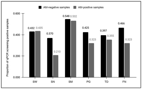
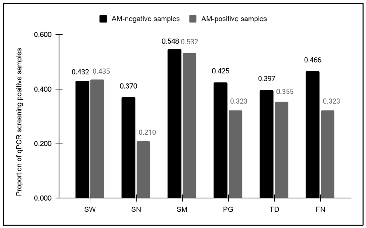
An analysis of the AM-negative and AM-positive samples was performed to determine any association between AM sample status and oral microbial prevalence (Figure 4). This analysis revealed no significant differences were found in the prevalence of SW (43.2%, 43.5%, p = 0.958), SM (54.8%, 53.2%, p = 0.837), PG (42.5%, 32.3%, p = 0.122) and TD (39.7%, 35.5%, p = 0.475) between AM-negative and AM-positive samples. However, significant reductions in the prevalence of SN (37%, 21%, p = 0.016) and FN (46.6%, 32.3%, p = 0.034) were observed between AM-negative and AM-positive samples.
Figure 4.
Analysis of microbial prevalence among AM-negative and AM-positive samples. Significant differences in the prevalence of SN (37%, 21%, p = 0.016) and FN (46.6%, 32.3%, p = 0.034) were observed between AM-negative and AM-positive samples, although no significant differences were observed in the prevalence of SW (43.2%, 43.5%, p = 0.958), SM (54.8%, 53.2%, p = 0.837), PG (42.5%, 32.3%, p = 0.122) or TD (39.7%, 35.5%, p = 0.475). Key: Akkermansia muciniphila (AM), Selenomonas noxia (SN), Treponema denticola (TD), Porphyromonas gingivalis (PG), Fusobacterium nucleatum (FN), Scardovia wiggsiae (SW), Streptococcus mutans (SM).
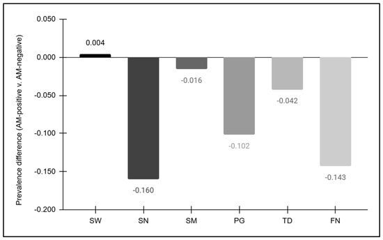
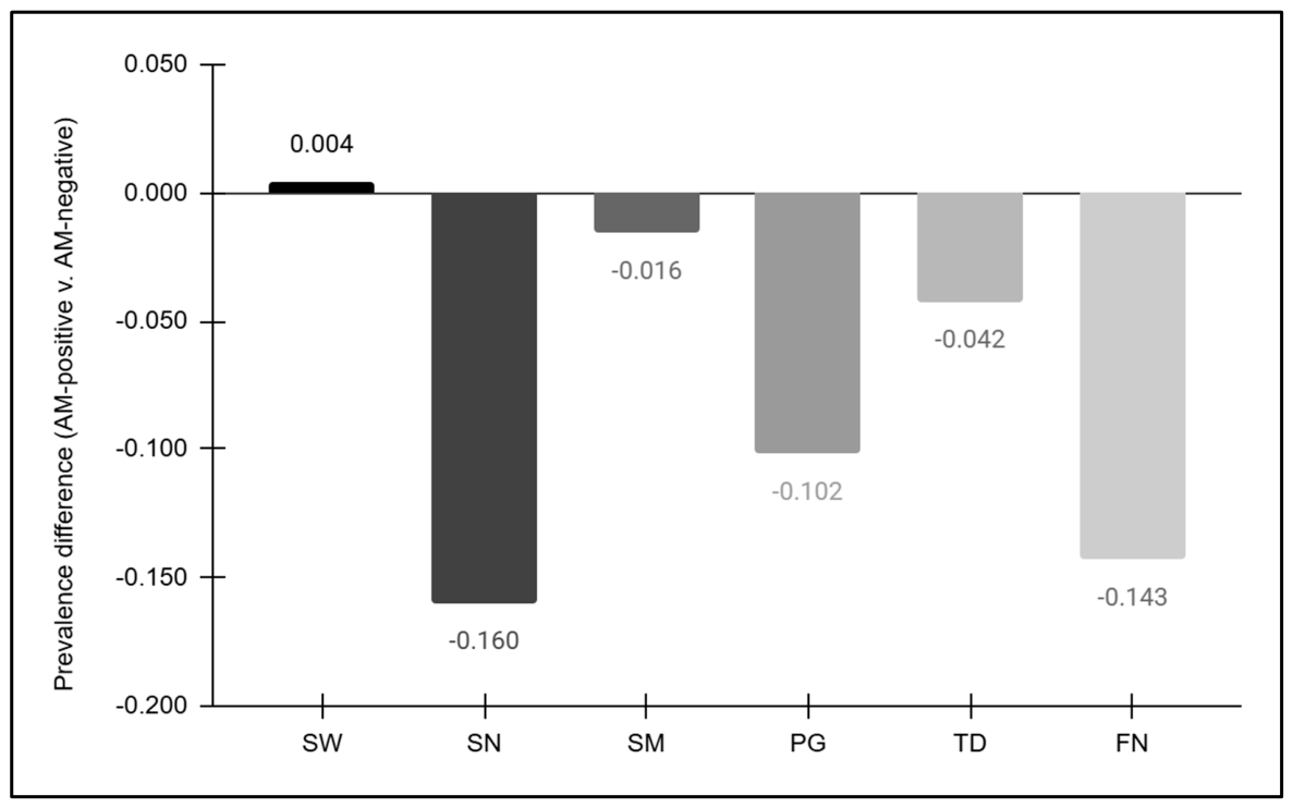
To more fully elucidate any potential associations based upon these findings, the percent differences in microbial prevalence were evaluated between samples found to be AM-positive and AM-negative (Figure 5). This evaluation found that most of oral pathogens evaluated demonstrated decreases in prevalence among AM-positive samples, including SN (−16.0%), SM (−1.6%), PG (−10.2%), TD (−4.2%), and FN (−14.3%). These data also demonstrated that only SW (0.4%) was found to exhibit a small, but positive, increase in prevalence among AM-positive samples.
Figure 5.
Change in prevalence between AM-negative and AM-positive samples. Decreases in prevalence among AM-positive samples were observed with SN (−16.0%), SM (−1.6%), PG (−10.2%), TD (−4.2%), and FN (−14.3%), with only SW (+0.4) demonstrating a slight increase. Key: Akkermansia muciniphila (AM), Selenomonas noxia (SN), Treponema denticola (TD), Porphyromonas gingivalis (PG), Fusobacterium nucleatum (FN), Scardovia wiggsiae (SW), Streptococcus mutans (SM).
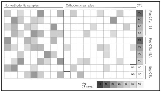
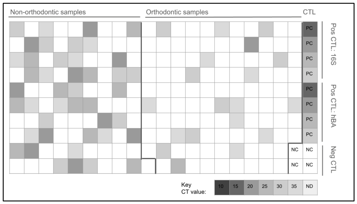
To provide a more detailed assessment of these observations the qPCR results for the AM screening were analyzed using a heat map (Figure 6). Overall, the AM-positive non-orthodontic samples (n = 43) exhibited an average cycle threshold (Ct) detection level of 27.41, with 23.2% detected within the high range (C20 to C24), 27.9% among the moderate–high range (C25 to C29), 25.6% among the moderate range (C30 to C34) and 23.2% among the lowest range (C35 to C40). In contrast the AM-positive orthodontic samples (n = 19) exhibited an average Ct of 31.57 with only 5.3% among the high range (C20 to C24), 31.6% among the moderate range (C30 to C34) and the majority 63.2% among the lowest range (C35 to C30). These data are outlined in detail in Table 4.
Figure 6.
Heat map of qPCR screening for AM, Non-orthodontic AM-positive samples (n = 43) exhibited an average cycle threshold (Ct) detection level of Ct = 27.41 compared with Ct = 31.57 among the orthodontic samples. Non-orthodontic AM positive results were found within the high range (C20 to C24; 23.2% or n = 10/43), the moderate-high range (C25 to C29; 27.9% or n = 12/43), the moderate range (C30 to C34; 25.6% or n = 11/43), and the lowest range (C35 to C40; 23.2% or n = 10/43). However, orthodontic AM-positive samples were mostly observed among the lowest range C35 to C40; 63.2% or n-12/19), with fewer samples among the moderate range (C30 to C35; 31.6% or n = 6/19), with only 5.3% (n = 1/19) among the high range (C20 to C24), Key: CT (cycle threshold), Pos CTL (positive control), Neg CTL (negative control), PC (positive control), NC (negative control), 16S (bacterial 16S rRNA), hBA (human beta actin).
Table 4.
Analysis of qPCR cycle threshold (CT) screening results.
4. Discussion
The overall aim of this study was to more closely examine the association of orthodontic appliances with the prevalence of oral microbes, with the specific objective of determining whether this influenced the prevalence of AM or other oral organisms. These findings revealed that orthodontic appliances may have significant and specific associations with oral microbial prevalence, with observed increases among most of the oral microbial species evaluated supporting the findings of many previous studies of a similar nature [51,52]. However, these data also revealed that the presence of orthodontic brackets had an opposing, negative association, with observed decreases in AM prevalence. This may represent a significant finding, as only two other previous studies have ever evaluated AM prevalence in the presence or absence of orthodontic appliances [39,53].
Review and analysis of prior studies has confirmed that previous research also found a similar decrease in AM prevalence among orthodontic samples (−21%, n = 141), which was comparable with these current results (−29.5%)—although the second, smaller study did not observe this dramatic shift in AM prevalence (n = 36) [39,53]. One potential explanation for the difference in these findings is that the current study includes a much more robust sample size (n = 209), which was greater than either of the other two previous studies of AM prevalence in saliva derived from patients with or without orthodontic appliances and thus more likely to uncover these effects [39,53]. In fact, the findings of this study provide more evidence that orthodontic appliances may not be uniformly associated with increased oral microbial prevalence, but rather may be associated with both increased and decreased oral prevalence of specific key microbial species, such as AM [54,55].
In addition, these results may be the first to reveal that AM-negative and AM-positive saliva samples are strongly associated with increased or decreased prevalence of other oral pathogens. Although some previous studies have evaluated the overall prevalence of AM with either SN or SM, none of these studies evaluated whether these samples harbored them alone or in combination with one another [56,57]. Moreover, the few studies that included screening for AM and either PG of FN did not provide this type of detailed analysis [58,59,60].
Other studies have clearly demonstrated the role of AM in shaping intestinal microbial ecosystems, with the ability to modulate and mediate not only the host biochemical and immune responses but also can influence the activity and prevalence of other microbial species directly [61,62]. Although many studies confirm the role of AM as a gatekeeper of intestinal mucosa and a key regulator of other microbial species, few studies to date have provided evidence that AM may be associated with changes in the biofilm or gingival crevices of the oral cavity [63,64]. This study may be among the first to provide preliminary data to suggest that AM may be associated with changes in microbial prevalence of other key oral pathogens, although more research is needed to determine the factors and variables that may be responsible for these observations.
Despite the significance of these data, there are some limitations of this study that should also be considered when evaluating these findings. First, and most importantly, this was a retrospective study and therefore provided only observational data regarding AM prevalence along with other oral microbes. In addition, this screening was performed using an existing biorepository and therefore does not have important longitudinal data that could be assessed if multiple samples had been taken from the same individuals over time. Although these findings may reveal important associations and correlations that may offer clues as to how AM might function in the oral cavity, future experimental studies will be needed to determine whether the introduction of AM into the oral cavity, oral biofilm, or gingival crevice may be sufficient to alter the prevalence or burden of oral pathogens, such as SM, SN, FN, TD or PG. Furthermore, these studies were based upon samples taken from a single recruitment site, which limits the overall generalizability of these results to a broader population. Moreover, these samples were also taken from a dental school patient population, which was composed mainly of ethnic minorities and was predominantly low-income, which may also present significant and potentially confounding variables on the results obtained due to the challenges and barriers that face these patient populations [65,66].
Additional variables that influence the oral microbiome may include temporal factors and behavioral patterns that were also not available for this analysis due to the retrospective nature of this study. For example, no data were originally collected or available regarding the length of time the orthodontic patients had been in treatment, which may be a significant variable in the assessment of oral microbial burden and biofilm formation over time [67,68]. In addition, analysis of oral hygiene behaviors, including the use of electric versus manual toothbrushes, may be a significant factor that affects plaque index and biofilm formation among orthodontic and non-orthodontic patients, although no information regarding this was available within this data set [69,70]. Other oral hygiene habits, such as the use of antimicrobial mouthwashes, may also significantly influence the prevalence of species like SN and SW. However, data regarding mouthwash usage were not collected from this patient population [71,72]. Finally, no information was available regarding additional behavioral risk factors, such as smoking status or diet, which may have the potential to significantly influence the range and burden of oral bacteria present among this patient population [73,74,75].
5. Conclusions
These data may be among the first to reveal the association between the presence of orthodontic appliances with a significant decrease in the oral prevalence of AM. In addition, the increased prevalence of other oral pathogens among the AM-negative samples and the decreased prevalence of these same pathogens among the AM-positive samples may suggest AM prevalence may be one factor (among many) in the overall changes observed in the prevalence of other oral pathogens.
Author Contributions
Conceptualization, K.K. and K.M.H.; methodology, K.K. and K.M.H.; formal analysis, K.K. and J.S.; investigation, J.S., F.P., S.C.R. and A.B.; resources, K.M.H. and K.K.; data curation, J.S. and K.K.; writing—original draft preparation, J.S. and K.K.; writing—review and editing, J.S., F.P., S.C.R., A.B., K.M.H. and K.K. All authors have read and agreed to the published version of the manuscript.
Funding
This research received no external funding.
Institutional Review Board Statement
The study was conducted in accordance with the Declaration of Helsinki and was approved by the University of Nevada Las Vegas (UNLV) Institutional Review Board under protocol #1717625-1 titled “Retrospective analysis of microbial prevalence from DNA isolated from saliva samples originally obtained from the University of Nevada, Las Vegas (UNLV) School of Dental Medicine (SDM) pediatric and clinical population” as research exempt on 10 June 2022.
Informed Consent Statement
This study was a retrospective analysis of DNA samples using an existing saliva biorepository, but did not involve prospective sample collection or any patient interactions with the current study authors. The original approved protocol “The Prevalence of Oral Microbes in Saliva from the UNLV School of Dental Medicine Pediatric and Adult Clinical Population” required pediatric assent from patients over the age of five years and informed consent from adults, as well as any parents or guardians.
Data Availability Statement
Due to the protocol approval specifications, any additional data from this study may only be made available directly from the study authors.
Acknowledgments
The authors wish to acknowledge the Department of Advanced Education in Orthodontics for their support of this project.
Conflicts of Interest
The authors declare no conflicts of interest.
Abbreviations
The following abbreviations are used in this manuscript:
| AM | Akkermansia muciniphila |
| FN | Fusobacterium nucleatum |
| PG | Porphyromonas gingivalis |
| SW | Scardovia wiggsiae |
| SN | Selenomonas noxia |
| SM | Streptococcus mutans |
| TD | Treponema denticola |
| qPCR | Quantitative polymerase chain reaction |
| IRB | Institutional review board |
| OPRS | Office for the Protection of Research Subjects |
| UNLV | University of Nevada, Las Vegas |
| SDM | School of Dental Medicine |
| GAPDH | glyceraldehyde phosphate dehydrogenase |
References
- Singh, H.; Torralba, M.G.; Moncera, K.J.; DiLello, L.; Petrini, J.; Nelson, K.E.; Pieper, R. Gastro-intestinal and oral microbiome signatures associated with healthy aging. Geroscience 2019, 41, 907–921. [Google Scholar] [CrossRef] [Scilit] [PubMed]
- Katagiri, S.; Shiba, T.; Tohara, H.; Yamaguchi, K.; Hara, K.; Nakagawa, K.; Komatsu, K.; Watanabe, K.; Ohsugi, Y.; Maekawa, S.; et al. Re-initiation of Oral Food Intake Following Enteral Nutrition Alters Oral and Gut Microbiota Communities. Front. Cell Infect. Microbiol. 2019, 9, 434. [Google Scholar] [CrossRef] [Scilit] [PubMed]
- Badal, V.D.; Vaccariello, E.D.; Murray, E.R.; Yu, K.E.; Knight, R.; Jeste, D.V.; Nguyen, T.T. The Gut Microbiome, Aging, and Longevity: A Systematic Review. Nutrients 2020, 12, 3759. [Google Scholar] [CrossRef] [Scilit] [PubMed]
- Agus, A.; Clément, K.; Sokol, H. Gut microbiota-derived metabolites as central regulators in metabolic disorders. Gut 2021, 70, 1174–1182. [Google Scholar] [CrossRef] [Scilit] [PubMed]
- Perler, B.K.; Friedman, E.S.; Wu, G.D. The Role of the Gut Microbiota in the Relationship Between Diet and Human Health. Annu. Rev. Physiol. 2023, 85, 449–468. [Google Scholar] [CrossRef] [Scilit] [PubMed]
- Qi, X.; Yun, C.; Pang, Y.; Qiao, J. The impact of the gut microbiota on the reproductive and metabolic endocrine system. Gut Microbes 2021, 13, 1894070. [Google Scholar] [CrossRef] [Scilit] [PubMed]
- Peng, X.; Cheng, L.; You, Y.; Tang, C.; Ren, B.; Li, Y.; Xu, X.; Zhou, X. Oral microbiota in human systematic diseases. Int. J. Oral Sci. 2022, 14, 14. [Google Scholar] [CrossRef] [Scilit] [PubMed]
- Rosier, B.T.; Marsh, P.D.; Mira, A. Resilience of the Oral Microbiota in Health: Mechanisms That Prevent Dysbiosis. J. Dent. Res. 2018, 97, 371–380. [Google Scholar] [CrossRef] [Scilit] [PubMed]
- Li, Y.; Cui, J.; Liu, Y.; Chen, K.; Huang, L.; Liu, Y. Oral, Tongue-Coating Microbiota, and Metabolic Disorders: A Novel Area of Interactive Research. Front. Cardiovasc. Med. 2021, 8, 730203. [Google Scholar] [CrossRef] [Scilit] [PubMed]
- Lamont, R.J.; Koo, H.; Hajishengallis, G. The oral microbiota: Dynamic communities and host interactions. Nat. Rev. Microbiol. 2018, 16, 745–759. [Google Scholar] [CrossRef] [Scilit] [PubMed]
- Yamazaki, K.; Kamada, N. Exploring the oral-gut linkage: Interrelationship between oral and systemic diseases. Mucosal Immunol. 2024, 17, 147–153. [Google Scholar] [CrossRef] [Scilit] [PubMed]
- Abusleme, L.; Hoare, A.; Hong, B.Y.; Diaz, P.I. Microbial signatures of health, gingivitis, and periodontitis. Periodontology 2000 2021, 86, 57–78. [Google Scholar] [CrossRef] [Scilit] [PubMed]
- Kayama, H.; Okumura, R.; Takeda, K. Interaction Between the Microbiota, Epithelia, and Immune Cells in the Intestine. Annu. Rev. Immunol. 2020, 38, 23–48. [Google Scholar] [CrossRef] [Scilit] [PubMed]
- Mosaddad, S.A.; Tahmasebi, E.; Yazdanian, A.; Rezvani, M.B.; Seifalian, A.; Yazdanian, M.; Tebyanian, H. Oral microbial biofilms: An update. Eur. J. Clin. Microbiol. Infect. Dis. 2019, 38, 2005–2019. [Google Scholar] [CrossRef] [Scilit] [PubMed]
- Miller, D.P.; Lamont, R.J. Signaling Systems in Oral Bacteria. Adv. Exp. Med. Biol. 2019, 1197, 27–43. [Google Scholar] [CrossRef] [Scilit] [PubMed]
- Baty, J.J.; Stoner, S.N.; Scoffield, J.A. Oral Commensal Streptococci: Gatekeepers of the Oral Cavity. J. Bacteriol. 2022, 204, e0025722. [Google Scholar] [CrossRef] [Scilit] [PubMed]
- Bloch, S.; Hager-Mair, F.F.; Andrukhov, O.; Schäffer, C. Oral streptococci: Modulators of health and disease. Front. Cell. Infect. Microbiol. 2024, 14, 1357631. [Google Scholar] [CrossRef] [Scilit] [PubMed]
- Choi, A.; Dong, K.; Williams, E.; Pia, L.; Batagower, J.; Bending, P.; Shin, I.; Peters, D.I.; Kaspar, J.R. Human saliva modifies growth, biofilm architecture, and competitive behaviors of oral streptococci. mSphere 2024, 9, e0077123, Erratum in mSphere 2024, 9, e0086824. https://doi.org/10.1128/msphere.00868-24. [Google Scholar] [CrossRef] [Scilit] [PubMed]
- Tlaskalová-Hogenová, H.; Stepánková, R.; Hudcovic, T.; Tucková, L.; Cukrowska, B.; Lodinová-Zádníková, R.; Kozáková, H.; Rossmann, P.; Bártová, J.; Sokol, D.; et al. Commensal bacteria (normal microflora), mucosal immunity and chronic inflammatory and autoimmune diseases. Immunol. Lett. 2004, 93, 97–108. [Google Scholar] [CrossRef] [PubMed]
- Breivik, T.J.; Gjermo, P.; Gundersen, Y.; Opstad, P.K.; Murison, R.; Hugoson, A.; von Hörsten, S.; Fristad, I. Microbiota-immune-brain interactions: A new vision in the understanding of periodontal health and disease. Periodontology 2000 2024, 96, 20–41. [Google Scholar] [CrossRef] [Scilit] [PubMed]
- Cani, P.D.; Depommier, C.; Derrien, M.; Everard, A.; de Vos, W.M. Akkermansia muciniphila: Paradigm for next-generation beneficial microorganisms. Nat. Rev. Gastroenterol. Hepatol. 2022, 19, 625–637, Erratum in Nat. Rev. Gastroenterol. Hepatol. 2022, 19, 682. https://doi.org/10.1038/s41575-022-00650-6. [Google Scholar] [CrossRef] [Scilit] [PubMed]
- Rodrigues, V.F.; Elias-Oliveira, J.; Pereira, Í.S.; Pereira, J.A.; Barbosa, S.C.; Machado, M.S.G.; Carlos, D. Akkermansia muciniphila and Gut Immune System: A Good Friendship That Attenuates Inflammatory Bowel Disease, Obesity, and Diabetes. Front. Immunol. 2022, 13, 934695. [Google Scholar] [CrossRef] [Scilit] [PubMed]
- Niu, H.; Zhou, M.; Zogona, D.; Xing, Z.; Wu, T.; Chen, R.; Cui, D.; Liang, F.; Xu, X. Akkermansia muciniphila: A potential candidate for ameliorating metabolic diseases. Front. Immunol. 2024, 15, 1370658. [Google Scholar] [CrossRef] [Scilit] [PubMed]
- Jian, H.; Liu, Y.; Wang, X.; Dong, X.; Zou, X. Akkermansia muciniphila as a Next-Generation Probiotic in Modulating Human Metabolic Homeostasis and Disease Progression: A Role Mediated by Gut-Liver-Brain Axes? Int. J. Mol. Sci. 2023, 24, 3900. [Google Scholar] [CrossRef] [Scilit] [PubMed]
- Zhao, Y.; Yang, H.; Wu, P.; Yang, S.; Xue, W.; Xu, B.; Zhang, S.; Tang, B.; Xu, D. Akkermansia muciniphila: A promising probiotic against inflammation and metabolic disorders. Virulence 2024, 15, 2375555. [Google Scholar] [CrossRef] [Scilit] [PubMed]
- Zhang, T.; Li, Q.; Cheng, L.; Buch, H.; Zhang, F. Akkermansia muciniphila is a promising probiotic. Microb. Biotechnol. 2019, 12, 1109–1125. [Google Scholar] [CrossRef] [Scilit] [PubMed]
- Zhai, Q.; Feng, S.; Arjan, N.; Chen, W. A next generation probiotic, Akkermansia muciniphila. Crit. Rev. Food Sci. Nutr. 2019, 59, 3227–3236. [Google Scholar] [CrossRef] [Scilit] [PubMed]
- Ioannou, A.; Berkhout, M.D.; Geerlings, S.Y.; Belzer, C. Akkermansia muciniphila: Biology, microbial ecology, host interactions and therapeutic potential. Nat. Rev. Microbiol. 2025, 23, 162–177. [Google Scholar] [CrossRef] [Scilit] [PubMed]
- Zhao, Q.; Yu, J.; Hao, Y.; Zhou, H.; Hu, Y.; Zhang, C.; Zheng, H.; Wang, X.; Zeng, F.; Hu, J.; et al. Akkermansia muciniphila plays in host health. Crit. Rev. Microbiol. 2023, 49, 82–100. [Google Scholar] [CrossRef] [Scilit] [PubMed]
- Kobyliak, N.; Falalyeyeva, T.; Kyriachenko, Y.; Tseyslyer, Y.; Kovalchuk, O.; Hadiliia, O.; Eslami, M.; Yousefi, B.; Abenavoli, L.; Fagoonee, S.; et al. Akkermansia muciniphila as a novel powerful bacterial player in the treatment of metabolic disorders. Minerva Endocrinol. 2022, 47, 242–252. [Google Scholar] [CrossRef] [Scilit] [PubMed]
- Karamzin, A.M.; Ropot, A.V.; Sergeyev, O.V.; Khalturina, E.O. Akkermansia muciniphila and host interaction within the intestinal tract. Anaerobe 2021, 72, 102472. [Google Scholar] [CrossRef] [Scilit] [PubMed]
- Geerlings, S.Y.; Kostopoulos, I.; de Vos, W.M.; Belzer, C. Akkermansia muciniphila in the Human Gastrointestinal Tract: When, Where, and How? Microorganisms 2018, 6, 75. [Google Scholar] [CrossRef] [Scilit] [PubMed]
- Mirhosseini, S.M.; Mahdavi, A.; Yarmohammadi, H.; Razavi, A.; Rezaei, M.; Soltanipur, M.; Karimi Nemch, M.; Jafari Naeini, S.; Siadat, S.D. What is the link between the dietary inflammatory index and the gut microbiome? A systematic review. Eur. J. Nutr. 2024, 63, 2407–2419. [Google Scholar] [CrossRef] [Scilit] [PubMed]
- Catalan, E.A.; Seguel-Fuentes, E.; Fuentes, B.; Aranguiz-Varela, F.; Castillo-Godoy, D.P.; Rivera-Asin, E.; Bocaz, E.; Fuentes, J.A.; Bravo, D.; Schinnerling, K.; et al. Oral Pathobiont-Derived Outer Membrane Vesicles in the Oral-Gut Axis. Int. J. Mol. Sci. 2024, 25, 11141. [Google Scholar] [CrossRef] [Scilit] [PubMed]
- Minty, M.; Canceil, T.; Serino, M.; Burcelin, R.; Tercé, F.; Blasco-Baque, V. Oral microbiota-induced periodontitis: A new risk factor of metabolic diseases. Rev. Endocr. Metab. Disord. 2019, 20, 449–459. [Google Scholar] [CrossRef] [Scilit] [PubMed]
- Hu, Q.; Leung, W.K.; Acharya, A.; Pelekos, G. Akkermansia muciniphila enhances mucosal immunity against porphyromonas gingivalis. Arch. Oral Biol. 2025, 174, 106240. [Google Scholar] [CrossRef] [Scilit] [PubMed]
- Huck, O.; Mulhall, H.; Rubin, G.; Kizelnik, Z.; Iyer, R.; Perpich, J.D.; Haque, N.; Cani, P.D.; de Vos, W.M.; Amar, S. Akkermansia muciniphila reduces Porphyromonas gingivalis-induced inflammation and periodontal bone destruction. J. Clin. Periodontol. 2020, 47, 202–212, Erratum in J. Clin. Periodontol. 2024, 51, 802–803. https://doi.org/10.1111/jcpe.13978. [Google Scholar] [CrossRef] [Scilit] [PubMed]
- Song, B.; Xian, W.; Sun, Y.; Gou, L.; Guo, Q.; Zhou, X.; Ren, B.; Cheng, L. Akkermansia muciniphila inhibited the periodontitis caused by Fusobacterium nucleatum. npj Biofilms Microbiomes 2023, 9, 49. [Google Scholar] [CrossRef] [Scilit] [PubMed]
- Shen, C.; Clawson, J.B.; Simpson, J.; Kingsley, K. Oral Prevalence of Akkermansia muciniphila Differs among Pediatric and Adult Orthodontic and Non-Orthodontic Patients. Microorganisms 2023, 11, 112. [Google Scholar] [CrossRef] [Scilit] [PubMed]
- Santonocito, S.; Polizzi, A. Oral Microbiota Changes during Orthodontic Treatment. Front. Biosc. 2022, 14, 19. [Google Scholar] [CrossRef] [Scilit] [PubMed]
- Radovic, R.; Begic, G.; Blagojevic, S.L.; Karleusa, L.; Spalj, S.; Gobin, I. Temporal dynamics of adhesion of oral bacteria to orthodontic appliances. Dent. Mater. J. 2023, 42, 469–477. [Google Scholar] [CrossRef] [Scilit] [PubMed]
- Marincak Vrankova, Z.; Rousi, M.; Cvanova, M.; Gachova, D.; Ruzicka, F.; Hola, V.; Lochman, J.; Izakovicova Holla, L.; Brysova, A.; Borilova Linhartova, P. Effect of fixed orthodontic appliances on gingival status and oral microbiota: A pilot study. BMC Oral Health 2022, 22, 455. [Google Scholar] [CrossRef] [Scilit] [PubMed]
- Nemec, M.; Ringl, P.; Spettel, K.; Schneider, L.; Kriz, R.; Galazka, S.; Sedlak, M.; Jonke, E.; Andrukhov, O.; Makristathis, A. Exploring the impact of orthodontic appliances on the oral microbiome and inflammatory parameters. Prog. Orthod. 2025, 26, 13, Erratum in Prog. Orthod. 2025, 26, 29. https://doi.org/10.1186/s40510-025-00577-z. [Google Scholar] [CrossRef] [Scilit] [PubMed]
- Freitas, A.O.; Marquezan, M.; Nojima Mda, C.; Alviano, D.S.; Maia, L.C. The influence of orthodontic fixed appliances on the oral microbiota: A systematic review. Dent. Press J. Orthod. 2014, 19, 46–55. [Google Scholar] [CrossRef] [Scilit] [PubMed]
- Müller, L.K.; Jungbauer, G.; Jungbauer, R.; Wolf, M.; Deschner, J. Biofilm and Orthodontic Therapy. Monogr. Oral Sci. 2021, 29, 201–213. [Google Scholar] [CrossRef] [Scilit] [PubMed]
- Topaloglu-Ak, A.; Ertugrul, F.; Eden, E.; Ates, M.; Bulut, H. Effect of orthodontic appliances on oral microbiota—6 month follow-up. J. Clin. Pediatr. Dent. 2011, 35, 433–436. [Google Scholar] [CrossRef] [Scilit] [PubMed]
- Perkowski, K.; Baltaza, W.; Conn, D.B.; Marczyńska-Stolarek, M.; Chomicz, L. Examination of oral biofilm microbiota in patients using fixed orthodontic appliances in order to prevent risk factors for health complications. Ann. Agric. Environ. Med. 2019, 26, 231–235. [Google Scholar] [CrossRef] [Scilit] [PubMed]
- Reichardt, E.; Geraci, J.; Sachse, S.; Rödel, J.; Pfister, W.; Löffler, B.; Wagner, Y.; Eigenthaler, M.; Wolf, M. Qualitative and quantitative changes in the oral bacterial flora occur shortly after implementation of fixed orthodontic appliances. Am. J. Orthod. Dentofac. Orthop. 2019, 156, 735–744. [Google Scholar] [CrossRef] [Scilit] [PubMed]
- Zhao, M.; Yu, C.; Su, C.; Wang, H.; Wang, X.; Weir, M.D.; Xu, H.H.K.; Liu, M.; Bai, Y.; Zhang, N. Dynamic effects of fixed orthodontic treatment on oral health and oral microbiota: A prospective study. BMC Oral Health 2024, 24, 1537. [Google Scholar] [CrossRef] [Scilit] [PubMed]
- Hodges, K.; Famuliner, P.; Kingsley, K.; Howard, K.M. Oral Prevalence of Selenomonas noxia Differs among Orthodontic Patients Compared to Non-Orthodontic Controls: A Retrospective Biorepository Analysis. Pathogens 2024, 13, 670. [Google Scholar] [CrossRef] [Scilit] [PubMed]
- Kouvelis, G.; Papadimitriou, A.; Merakou, K.; Doulis, I.; Karapsias, S.; Kloukos, D. A Prospective Cohort Study Assessing the Impact of Fixed Orthodontic Appliances on Saliva Properties and Oral Microbial Flora. Oral Health Prev. Dent. 2021, 19, 67. [Google Scholar] [CrossRef] [Scilit] [PubMed]
- Sun, F.; Ahmed, A.; Wang, L.; Dong, M.; Niu, W. Comparison of oral microbiota in orthodontic patients and healthy individuals. Microb. Pathog. 2018, 123, 473–477. [Google Scholar] [CrossRef] [Scilit] [PubMed]
- Wirth, R.; Maróti, G.; Lipták, L.; Mester, M.; Al Ayoubi, A.; Pap, B.; Madléna, M.; Minárovits, J.; Kovács, K.L. Microbiomes in supragingival biofilms and saliva of adolescents with gingivitis and gingival health. Oral Dis. 2022, 28, 2000–2014. [Google Scholar] [CrossRef] [Scilit] [PubMed]
- Iqbal, M.; Yu, Q.; Tang, J.; Xiang, J. Unraveling the gut microbiota’s role in obesity: Key metabolites, microbial species, and therapeutic insights. J. Bacteriol. 2025, 207, e0047924. [Google Scholar] [CrossRef] [Scilit] [PubMed]
- Ottman, N.; Geerlings, S.Y.; Aalvink, S.; de Vos, W.M.; Belzer, C. Action and function of Akkermansia muciniphila in microbiome ecology, health and disease. Best Pract. Res. Clin. Gastroenterol. 2017, 31, 637–642. [Google Scholar] [CrossRef] [Scilit] [PubMed]
- Fakruddin, M.; Amin, T.; Shishir, M.A.; Jameel, R.M.; Bari, M.M.; Shameem, N.F.; Hossain, A.; Jerin, N.; Bin Mannan, S.; Jime, J.S.; et al. Early-Life Microbiome and Neurodevelopmental Disorders: A Systematic Review and Meta-Analysis. Curr. Neuropharmacol. 2026, 24, 213–229. [Google Scholar] [CrossRef] [Scilit] [PubMed]
- Yamamoto, K.; Honda, T.; Ito, T.; Ishizu, Y.; Kuzuya, T.; Nakamura, M.; Miyahara, R.; Kawashima, H.; Ishigami, M.; Fujishiro, M. The relationship between oral-origin bacteria in the fecal microbiome and albumin-bilirubin grade in patients with hepatitis C. J. Gastroenterol. Hepatol. 2021, 36, 790–799. [Google Scholar] [CrossRef] [Scilit] [PubMed]
- Lin, J.; Xu, Q.; Bi, W.; Yu, Y.; Wang, Q. Oral and gut microbiota in gingivitis subtypes: A causal inference study using Mendelian randomization. BMC Oral Health 2025, 25, 1270. [Google Scholar] [CrossRef] [Scilit] [PubMed]
- Soltani-Moghadam, F.; Kafil, H.S.; Sefidan, F.Y.; Nezhadi, J.; Saedi, S.; Mohammadzadeh-Asl, Y.; Sattarpour, S.; Rezaee, M.A. Gut Bacteria: A Beneficial Symbiosis or a Hidden Threat? Investigating the Dual Role of Bacteria in Gastrointestinal Diseases. Curr. Microbiol. 2026, 83, 414. [Google Scholar] [CrossRef] [Scilit] [PubMed]
- Petrukhina, N.B.; Zorina, O.A.; Shikh, E.V.; Shibaeva, A.V.; Shevelev, A.B. Study of mutual dependence of periodontal and colonic microbiome in health and pathology using NSG-sequencing. Stomatology 2016, 95, 8–13. (In Russian) [Google Scholar] [CrossRef] [Scilit] [PubMed]
- Tiffany, E.; Kim, K.S.; Sittipo, P.; Lee, D.W.; Lee, Y.K. Mucin-degrading gut bacteria: Context-dependent roles in intestinal homeostasis and disease. Gut Microbes 2026, 18, 2614054. [Google Scholar] [CrossRef] [Scilit] [PubMed]
- Berkhout, M.D.; de Ram, C.; van Gaal, M.P.; Schols, H.; Belzer, C.; Plugge, C.M. Mucin Type Drives Composition and Mucin Glycan Degradation of an In Vitro Synthetic Microbial Community. Environ. Microbiol. 2026, 28, e70288. [Google Scholar] [CrossRef] [Scilit] [PubMed]
- de Vos, W.M. Microbe Profile: Akkermansia muciniphila: A conserved intestinal symbiont that acts as the gatekeeper of our mucosa. Microbiology 2017, 163, 646–648. [Google Scholar] [CrossRef] [Scilit] [PubMed]
- Ghotaslou, R.; Nabizadeh, E.; Memar, M.Y.; Law, W.M.H.; Ozma, M.A.; Abdi, M.; Yekani, M.; Kadkhoda, H.; Hosseinpour, R.; Bafadam, S.; et al. The metabolic, protective, and immune functions of Akkermansia muciniphila. Microbiol. Res. 2023, 266, 127245. [Google Scholar] [CrossRef] [Scilit] [PubMed]
- Mavi, J.; Kingsley, K. Analysis of a Pediatric Dental School Patient Population Revealed Increasing Trends of Limited English Proficiency (LEP) Patients: Implications for Pediatric Dental Public Health and Access to Care. Pediatr. Rep. 2022, 14, 276–287. [Google Scholar] [CrossRef] [Scilit] [PubMed]
- Hinton, H.; Coleman, S.; Salem, J.R.; Kingsley, K. Screening for High-Risk Oral Human Papillomavirus (HPV31, HPV33, HPV35) in a Multi-Racial Pediatric and Adult Clinic Patient Population. Cancers 2023, 15, 4501. [Google Scholar] [CrossRef] [Scilit] [PubMed]
- Alhuwaizi, M.A.; Saloom, H.F.; Cobourne, M. Impact of clear aligners and conventional fixed orthodontic appliances on oral hygiene and periodontal health: A systematic review with GRADE assessment of the certainty of evidence. Eur. J. Orthod. 2026, 48, cjaf100. [Google Scholar] [CrossRef] [Scilit] [PubMed]
- Nibali, L.; Lu, E.M.; Paz, H.E.S.; Kang, J.; Bostanci, N.; Belibasakis, G.N. Risk Factors and Management of Gingival Enlargement: A Systematic Review and Meta-Analysis. J. Clin. Periodontol. 2026. [Google Scholar] [CrossRef] [Scilit] [PubMed]
- Graves, A.; Grahl, T.; Keiserman, M.; Kingsley, K. Systematic Review and Meta Analysis of the Relative Effect on Plaque Index among Pediatric Patients Using Powered (Electric) versus Manual Toothbrushes. Dent. J. 2023, 11, 46. [Google Scholar] [CrossRef] [Scilit] [PubMed]
- Mylonopoulou, I.M.; Pepelassi, E.; Madianos, P.; Halazonetis, D.J. A randomized, 3-month, parallel-group clinical trial to compare the efficacy of electric 3-dimensional toothbrushes vs manual toothbrushes in maintaining oral health in patients with fixed orthodontic appliances. Am. J. Orthod. Dentofac. Orthop. 2021, 160, 648–658. [Google Scholar] [CrossRef] [Scilit] [PubMed]
- Shayegh, M.; Sorenson, C.; Downey, J.; Lin, S.; Jiang, Y.; Sodhi, P.; Sullivan, V.; Howard, K.M.; Kingsley, K. Assessment of SARS-CoV-2 (COVID-19) Clinical Mouthwash Protocol and Prevalence of the Oral Pathogen Scardovia wiggsiae: A Pilot Study of Antibacterial Effects. Methods Protoc. 2023, 6, 65. [Google Scholar] [CrossRef] [Scilit] [PubMed]
- Sodhi, P.; Jiang, Y.; Lin, S.; Downey, J.; Sorenson, C.; Shayegh, M.; Sullivan, V.; Kingsley, K.; Howard, K.M. Administration of Clinical COVID-19 Mouthwashing Protocol and Potential Modulation of Pediatric Oral Bacterial Prevalence of Selenomonas noxia: A Pilot Study. Pediatr. Rep. 2023, 15, 414–425. [Google Scholar] [CrossRef] [Scilit] [PubMed]
- La Rosa, G.R.M.; Samaranayake, L.P.; Zaura, E.; Sun, A.; Fuochi, V.; Furneri, P.M.; Kowalski, J.; Myszel, K.; Chapple, I.; Polosa, R. Impact of Electronic Cigarette Use on the Oral Microbiota: A Systematic Review. J. Clin. Periodontol. 2026, 53, 956–985. [Google Scholar] [CrossRef] [Scilit] [PubMed]
- Senaratne, N.L.M.; Yung On, C.; Shetty, N.Y.; Gopinath, D. Effect of different forms of tobacco on the oral microbiome in healthy adults: A systematic review. Front. Oral Health 2024, 5, 1310334. [Google Scholar] [CrossRef] [Scilit] [PubMed]
- Chiesa, A.; Generali, L.; Butera, A.; Filippini, T.; Lanteri, V.; Veneri, F. Oral Microbiota Characteristics in Relation to Different Dietary Patterns: A Systematic Review. Nutrients 2026, 18, 1717. [Google Scholar] [CrossRef] [Scilit] [PubMed]

Disclaimer/Publisher’s Note: The statements, opinions and data contained in all publications are solely those of the individual author(s) and contributor(s) and not of MDPI and/or the editor(s). MDPI and/or the editor(s) disclaim responsibility for any injury to people or property resulting from any ideas, methods, instructions or products referred to in the content. |
© 2026 by the authors. Licensee MDPI, Basel, Switzerland. This article is an open access article distributed under the terms and conditions of the Creative Commons Attribution (CC BY) license.