


Abstract
Exopolysaccharides (EPS) from microalgae have emerged as promising natural anticancer agents owing to their selective cytotoxicity toward tumor cells and low toxicity to healthy tissues. However, the molecular mechanisms and targets underlying their anticancer activity remain poorly understood. In the present study, EPS isolated from Coelastrella sp. BGV was evaluated for cytotoxicity and antiproliferative activity against a panel of human cancer cell lines (HepG2, HCT116, A549, and MDA-MB-231). The effects of EPS on mitochondrial function, cell cycle progression, apoptosis, and autophagy were investigated using flow cytometry and fluorescence microscopy. To gain mechanistic insight, molecular docking of a representative EPS disaccharide, α-L-Fucp-(1→6)-β-D-Galp, was performed against DC-SIGN and galectin-3 using the CB-Dock2 platform with the AutoDock Vina engine. The EPS exhibited minimal cytotoxicity toward non-tumor cells, and induced significant concentration-dependent antiproliferative activity in all tested cancer cell lines. MDA-MB-231 breast cancer cells were the most sensitive, with an IC50 value of 57.5 ± 10 µg/mL and a selectivity index of 5.2 relative to non-tumor cells. Mechanistically, EPS treatment induced mitochondrial membrane depolarization, S-phase cell cycle arrest, autophagy, and apoptosis, as demonstrated by nuclear fragmentation, chromatin condensation, and increased Annexin V positivity. Molecular docking identified DC-SIGN and galectin-3 as plausible molecular targets, suggesting potential anticancer activity of Coelastrella sp. BGV EPS that may involve both direct pro-apoptotic effects and modulation of carbohydrate-mediated signaling pathways. Collectively, these findings provide new mechanistic insight into the anticancer activity of Coelastrella sp. BGV EPS and support its further investigation as a promising natural candidate for adjunctive cancer therapy.
1. Introduction
Malignant diseases are increasing worldwide every year and have significant socioeconomic importance [1,2]. Despite considerable progress in modern anticancer therapy leading to improvements in disease-free survival and quality of life, drug resistance continues to be a key barrier to long-term remission [3]. Current drugs are often ineffective and expensive and have a number of side effects, including nausea, vomiting, hair loss alopecia, cardio- and neurotoxicity, myelosuppression, etc. A promising strategy for cancer prevention and therapy involves the use of natural plant-derived products for chemoprophylaxis, chemoprevention and as adjuvant therapies [4].
The increased resistance and side effects of synthetic pharmaceutical drugs have led to the exploration of bioactive compounds from alternative sources. Algae represent a diverse group of photosynthetic organisms found in marine and freshwater environments. Bioactive metabolites of microalgal origin are of special interest in the development of new products for medical, pharmaceutical, cosmetic, food and feed industries and bioenergy. Typically, these compounds are synthesized by algae in response to extreme habitat conditions [4,5,6,7]. They possess a unique structure, exhibit diverse biological properties, and demonstrate selective cytotoxicity against tumor cells and microbial pathogens, and minimal toxicity toward healthy cells. Algal-derived compounds are highly toxic to cancer cells and exert anticancer activity through multiple mechanisms, including inhibition of cell proliferation and viability, induction of apoptosis, suppression of angiogenesis and metastasis, disruption of microtubule dynamics, modulation of antioxidant enzymes, and alterations in cancer cell membrane lipid composition [4,8,9,10].
Among microalgal metabolites, extracellular polysaccharides (EPS) have attracted considerable interest due to their unique structural properties, diverse biological activities, favorable safety profile, biocompatibility, biodegradability, and stable production. Algae-derived polysaccharides have numerous applications, including tissue engineering, biomedicine, drug delivery, nutrition, food packaging, pharmaceuticals, nutraceuticals, biostimulants and bioethanol production and many more [11]. These natural macromolecules exhibit considerable structural heterogeneity and are mainly composed of monosaccharides, including glucose, xylose, galactose, fucose, rhamnose, mannose, and arabinose. They exhibit a variety of bioactive properties, including anti-inflammatory, immunomodulatory, antibacterial, anti-tumor, antiviral, antiparasitic, antioxidant, antihyperlipidemic, anti-obesity, antidiabetic, and gastroprotective effects. These activities are influenced by factors such as the microalgae species, molecular weight, degree of sulfation and structural complexity, the test assay used, selected targets, etc. [12,13]. Numerous studies have highlighted the health-promoting potential of algal EPS, reporting additional properties such as, anticoagulant, antimutagenic, radioprotective, and anti-ulcer effects [13,14,15]. These findings underscore the significant potential of microalgal EPS as a versatile and promising source of natural bioactive compounds.
A diverse range of microalgae and cyanobacteria, including Chlorella, Scenedesmus, Nostoc, Anabaena, Porphyridium, and Haematococcus species, have been identified as producers of bioactive EPS [16,17,18]. In vitro studies have demonstrated their antiproliferative effects against multiple cancer cell lines, including cervical (HeLa), lung (A549), colon (HCT116, HCT8), breast (MCF-7, 4T1), and leukemia (HL60, MOLT-4, K562) models, often in a concentration-dependent manner and closely linked to structural features of the polysaccharides [19,20,21,22]. In several cases, EPS fractions also exhibit antioxidant and immunomodulatory properties, supporting a multi-target mode of action [13,23]. Mechanistic studies suggest that these anticancer effects are mediated through, reactive oxygen species (ROS) generation, and mitochondrial dysfunction [24,25], induction of apoptosis, modulation of cell cycle progression, and regulation of MAPK, PI3K-Akt, and TNF signaling pathways, as well as inhibition of angiogenesis and metastasis [26,27,28]. In addition, immunomodulatory effects involving stimulation of immune cell activity and cytokine regulation have been reported [18].
Among the numerous microalgal taxa reported to exhibit anticancer activity, the genus Coelastrella of the Chlorophyta phylum has attracted increasing scientific interest due to its stress tolerance, high biomass productivity, and rich profile of bioactive secondary metabolites [29,30,31]. The green/red-stage cell extracts from novel green alga Coelastrella sp. D3–1 showed high antioxidant and anti-inflammatory activities via NO production induced by LPS [31,32]. A microalgal extract from thermotolerant Coelastrella sp. F50 exhibited notable antitumor activity against hepatocellular carcinoma (HCC) by specifically targeting cancer stem cells (CSC), without affecting cell proliferation and cell death of in vitro cultured HCC cells. It selectively suppressed CSC markers (CD133 and ABCG2), which is associated with downregulation of the COX-2/PGE2 pathway. In vivo, prophylactic administration of the extract reduced tumor progression and improved liver function in HCC-bearing rats [33]. Other Coelastrella species have also demonstrated anticancer potential. For example, the carotenoid extract of Coelastrella oocystiformis inhibited the growth of DU-145 human prostate cancer cell line by 5% [34]. The bioactivity evaluation of crude polysaccharides (CP), methanol extracts (ME) and crude lipids (CL) from the soil microalga Coelastrella sp. SDEC-28 showed strong antioxidant activity of the CP fraction and the potent antiproliferative effects of the methanol extract on A375 melanoma cancer cells, with the inhibition efficiency up to 99.62% [35]. Additionally, aqueous and oil extracts, along with fatty acids from Coelastrella sp. BGV suppressed cell proliferation and triggered programmed cell death in HeLa cancer cells [36,37,38,39,40]. Ethanol extracts from Coelastrella sp. BGV grown under different LED light regimes showed increased antioxidant, antibacterial, selective antitumor and proapoptotic activities, with the strongest effect observed under blue light illumination [41]. Despite these promising findings, the molecular mechanisms underlying the anticancer effects of Coelastrella-derived polysaccharides remain insufficiently understood.
In our previous investigations, EPS isolated from Coelastrella sp. BGV was comprehensively characterized by chromatographic and spectroscopic analyses [40]. The isolated EPS showed a dry mass yield of 65.2 mg/L of culture medium and consisted of three polysaccharide fractions with molecular masses of 11.5 × 10⁴, 30.7 × 10⁴, and 72.4 × 10⁴ Da. The lowest molecular mass fraction was the predominant component (72.5%), whereas the fractions with higher molecular masses represented 11.8% and 5.7% of the total polysaccharide content, respectively. Chemical analyses revealed that the EPS was composed mainly of carbohydrate structures containing galactose and fucose, with a minor protein component, and exhibited functional groups characteristic of polysaccharides. In addition, the EPS demonstrated anticancer potential in HeLa and MCF-7 cells by reducing cell viability, inhibiting migration, affecting cell cycle progression, and inducing apoptosis, accompanied by altered expression of apoptosis- and proliferation-associated proteins [40].
The monosaccharide composition of the Coelastrella sp. BGV EPS, particularly the presence of galactose and fucose residues, suggests potential interactions with carbohydrate-binding proteins implicated in cancer progression. Among these, galectin-3, a β-galactoside-binding protein, and DC-SIGN (dendritic cell-specific ICAM-3-grabbing non-integrin), a C-type lectin receptor with affinity for fucosylated glycans, represent biologically relevant candidates due to their involvement in tumor growth, metastasis, and modulation of the tumor immune microenvironment.
Galectin-3 is known to be overexpressed in many cancers and to promote tumor cell survival, resistance to apoptosis, cancer progression, and metastasis, making it a clinically relevant anticancer target [42,43]. Given its specific affinity for β-galactosides, galectin-3 has become a target for therapeutic inhibitors, including small-molecule carbohydrates, natural polysaccharides, and their derivatives. These inhibitors exert antitumor effects by disrupting galectin-3-mediated processes such as adhesion, proliferation, and migration. Notably, carbohydrate-based inhibitors have entered clinical trials (e.g., GB1211, a small-molecule galectin-3 inhibitor in Phase II studies for solid tumors) [44], and natural glycans rich in β-galactosidic bonds have been shown to inhibit galectin-3 function, making it a clinically relevant target for anticancer therapy [45].
DC-SIGN is a glycan-binding receptor expressed on dendritic cells and macrophages that recognizes specific carbohydrate structures, including fucosylated glycans, and participates in antigen uptake and presentation. The potential role of DC-SIGN in cancer progression and immune evasion is supported by emerging evidence, although no clinical inhibitor has yet been approved [46]. In colorectal cancer, DC-SIGN is expressed on tumor-associated macrophages (TAMs), where it drives PD-L1-mediated immune evasion and is associated with worse prognosis [47]. Its interaction with colorectal cancer cells also promotes an immunosuppressive cytokine profile in dendritic cells, hindering the development of effective anti-tumor immunity [48]. Similarly, DC-SIGN expression has been detected in lung cancer tissues and can be exploited to promote cancer cell migration [49]. In breast cancer, particularly the aggressive triple-negative MDA-MB-231 subtype, tumor-derived factors can induce DC-SIGN expression on dendritic cells [50]. Together, these findings from relevant cancer models provide a strong rationale for investigating galectin-3 and DC-SIGN as potential targets for Coelastrella EPS.
The aim of this study was to assess the cytotoxicity of EPS derived from Coelastrella sp. BGV and its anticancer potential in a panel of human carcinoma cell lines, with a focus on cell viability, mitochondrial membrane potential, apoptosis, and autophagy. In addition, in silico approaches were employed to identify potential molecular targets of the EPS.
2. Materials and Methods
2.1. Materials and Chemicals
The microalgal strain Coelastrella sp. BGV used in this study was provided by the Algae Culture Collection of the Institute of Plant Physiology and Genetics, Bulgarian Academy of Sciences. Cell culture reagents, including Dulbecco’s Modified Eagle Medium (DMEM) and fetal bovine serum (FBS), were purchased from Gibco-Invitrogen (Leicestershire, UK), while the penicillin–streptomycin antibiotic solution was purchased from Lonza (Verviers, Belgium). Phosphate-buffered saline (PBS), Trypsin EDTA solution (2.5 g/L trypsin and 0.2 g/L EDTA) and Dimethyl sulfoxide (DMSO) were provided from AppliChem (Darmstadt, Germany). Neutral red dye was purchased from Sigma-Aldrich Chemie GmbH (Darmstadt, Germany). Acridine Orange (AO), and ethidium bromide (EtBr), and 4′,6-diamidino-2-phenylindole (DAPI) were delivered from Merck (Darmstadt, Germany). The Annexin V-FITC Apoptosis Detection Kit: sc-4252 AK was the product of Santa Cruz Biotechnology, Inc., Dallas, TX, USA. RNase A was supplied by Roche Diagnostics GmbH (Mannheim, Germany). All sterile plastic laboratory consumables were sourced from Orange Scientific (Braine-l’Alleud, Belgium).
2.2. Cell Lines and Culture Conditions
Human cancer cell lines used in this study included A549 (lung carcinoma), HCT-8 (colorectal carcinoma), HepG2 (hepatocellular carcinoma), and MDA-MB-231 (triple-negative, hormone-independent breast adenocarcinoma). BALB/3T3 clone A31 mouse embryonic fibroblasts and the human keratinocyte cell line HaCaT were used as non-malignant reference cell lines. HaCaT cells were obtained from CLS Cell Lines Service (Eppelheim, Germany), whereas all other cell lines were purchased from the American Type Culture Collection (ATCC, Manassas, VA, USA). The cells were routinely cultured as monolayers in Dulbecco’s modified Eagle’s medium (DMEM) supplemented with 10% heat-inactivated fetal bovine serum, 2 mM L-glutamine and antibiotics (50 U/mL penicillin, and 50 mg/mL streptomycin) at 37 °C in a humidified 5% CO2 incubator and were subcultured every 3–4 days to maintain exponential growth. At 60–80% confluency, the cells were detached with 0.25% trypsin-EDTA in PBS (pH 7.4), counted, resuspended in fresh medium and placed into sterile 24-well or 96-well plates at appropriate densities required for the respective assays to form a monolayer. Sterile coverslips were placed in 24-well plates prior to seeding for subsequent assays. Then the plates were incubated in a CO2 incubator under the specified conditions.
2.3. Microalgal Strain and Cultivation
The green microalga Coelastrella sp. BGV, originally isolated from stagnant water near Varvara village, Bulgaria [51] was maintained as a non-axenic monoculture in Šetlik modified medium. Cultivation was carried out at 28 °C under continuous illumination provided by cool-white fluorescent lamps (132 μmol m−2s−1) in 200 mL flat glass bottles, continuously supplied with 2–3% CO2-enriched air, and manually agitated daily.
2.4. Polysaccharides Sample Preparation
Extracellular polysaccharides (EPS) were obtained from 20-day algal cultures following centrifugation at 5000× g for 20 min. The cell-free supernatant (500 mL) was precipitated with chilled 99% ethanol (1:2, v/v), and the resulting pellet was collected by centrifugation (5000× g, 20 min). The precipitate was washed three times with 65% ethanol and then subjected to dialysis against distilled water for 48 h at 4 °C. The resulting purified EPS was recovered by lyophilization following the previously reported procedure [40].
2.5. Cytotoxicity Evaluation (BALB/3T3 Neutral Red Uptake)
The in vitro toxicological evaluation of the EPS was performed using the validated BALB/3T3 Neutral Red Uptake (NRU) assay. Briefly, cells were plated in 96-well plates at a density of 1 × 10⁴ cells per well/100 µL/and were incubated for 24 h. EPS was applied in concentrations ranging from 4 to 1000 µg/m for 72 h. Untreated cells were used as negative controls. At the completion of the treatment, Neutral Red medium was added to each well and the plates were further incubated for 3 h at 37 °C. After that, the cells were washed and ethanol/acetic acid solution was added to extract the dye. The absorption was measured at 540 nm wavelength on a TECAN microplate reader (TECAN, Grödig, Austria). Data are presented as percentages of values detected in control cells cultured under the same conditions in the absence of exopolysaccharide sample. The concentrations required for 50% inhibition of cell growth were calculated using nonlinear regression analysis (GraphPad Prism 4 Software).
2.6. Evaluation of In Vitro Antitumor Activity
2.6.1. Cell Viability Assay
Antiproliferative activity of EPS was evaluated on HaCaT (non-tumorigenic keratinocytes), MDA-MB-231 (triple-negative breast cancer), A549 (lung adenocarcinoma), HCT (colorectal adenocarcinoma), and HepG2 (hepatocellular carcinoma) cells by the standard NRU assay. The cell lines were routinely grown as monolayers in 75 cm2 tissue culture flasks under standard conditions. Cells were seeded in 96-well plates (1 × 10³ cells/well) and incubated 24 h before treatment with EPS. EPS was applied at concentrations ranging from 7.8 to 1000 µg/mL for HaCaT cells and from 15.6 to 2000 µg/mL for tumor cell lines, followed by incubation for 72 h. Each concentration was tested in five replicates. The respective cells cultured only in medium were used as controls. After the treatment period, the culture medium was discarded and the NRU solution was added to all wells and plates were additionally incubated for 3 h at 37 °C. Finally, the ethanol/acetic acid solution were added to extract the dye. Absorbance was measured at 540 nm (TECAN, SunriseTM, Grodig/Salzburg, Austria), and cell viability was expressed as a percentage relative to untreated controls for three independent examinations. The half-maximal inhibitory concentration (IC50) values were calculated via nonlinear regression analysis using GraphPad Prism 4 Software.
2.6.2. Fluorescent Microscopy Analysis for Cell Death
Cellular and nuclear morphological alterations in EPS-treated MDA-MB-231 cancer cells were analyzed using fluorescent microscopy after staining with acridine orange/ethidum bromide and DAPI to determine the type of cell death.
- Dual Fluorescent Staining with acridine orange/ethidum bromide
Morphological analysis of cell apoptosis was performed after acridine or-ange/etidium bromide (AO/EtBr) dual staining. Briefly MDA-MB-231 tumor cells (1 × 10⁵ cells/well) were cultured on 13 mm-diameter cover glasses placed in 24-well plates overnight. After confluence, they were treated for 72 h with exopolysaccharide sample (60 µg/mL) equal to half the maximal inhibitory concentration (IC50) established by the NRU assay. Cells cultured in medium alone served as controls. After incubation, the coverslips were removed, rinsed with PBS and stained with fluorescent dyes AO and EtBr (5 µg/mL each in PBS) for 5 min. The stained cells were observed (examined) immediately with a fluorescence microscope (Leica DM 5000B, Wetzlar, Germany) using excitation filter BP 340–380, dichromatic mirror 400 and suppression filter LP 425.
- DAPI Fluorescent Staining
Staining with DNA-binding dye 4′,6-Diamidine-2′-phenylindole dihydrochloride (DAPI) was employed to discriminate apoptotic cells from normal cells, which reflects the nuclear changes during apoptosis. DAPI effectively visualizes nuclear DNA in both live and fixed cells because it can penetrate intact cytoplasmic membranes. Cells were cultured on glass coverslips and treated with EPS sample as described in the previous section. When the incubation is finished, the cells were fixed with 3% paraformaldehyde for 10 min, washed with PBS and incubated in 1 µg/mL DAPI solution for 15 min at room temperature in the dark. Samples were coated with Mowiol® (Carl Roth, Karlsruhe, Germany) and mounted on slides. The apoptotic nuclei were observed under a fluorescence microscope (Leica DM 5000B, Wetzlar, Germany) using excitation filter BP 450–490, dichromatic mirror 510 and suppression filter LP 515. Apoptotic nuclei were identified based on characteristic chromatin condensation and nuclear fragmentation.
2.6.3. Flow Cytometry Analyses
Prior to flow cytometric analyses, MDA-MB-231 tumor cells were seeded in 6-well plates at a density of 5 × 10⁵ cells/well and cultured for 24 h until reaching approximately 80% confluence. Cells were then treated with EPS at a concentration of 60 µg/mL. After overnight incubation, cells were washed twice with cold phosphate-buffered saline (PBS), detached using Trypsin-EDTA (AppliChem, Darmstadt, Germany), and collected by centrifugation at 1000 rpm for 10 min. The resulting cell pellets were used for subsequent cell cycle and apoptosis analyses.
- FACS analysis of cell cycle
Cell pellets from control and EPS-treated groups were fixed in ice-cold 70% ethanol and incubated overnight at −20 °C. Following fixation, cells were washed with PBS and treated with RNase A (20 µg/mL; Roche Diagnostics GmbH, Mannheim, Germany) at 37 °C for 30 min. DNA staining was performed with propidium iodide (20 μg/mL; Sigma) for 30 min in the dark at room temperature.
- FACS analysis of apoptosis
For apoptosis analysis, phosphatidylserine (PS) externalization was assessed using the Annexin V-FITC/PI Apoptosis Detection Kit (sc-4252 AK; Santa Cruz Biotechnology, Inc., Dallas, TX, USA) according to the manufacturer’s instructions. After washing with the assay buffer, the cells were incubated for 10 min in the dark with a solution containing Annexin V-FITC (5 μg/mL) and PI (5 μg/mL).
For both analyses, a total of 10,000 cells per sample were acquired using a BD FACSCalibur™ flow cytometer (Becton Dickinson, Mountain View, CA, USA) and data were analyzed using FlowJo™ v10.8 software (BD Biosciences, San Jose, CA, USA). All samples were analyzed in triplicate.
2.6.4. Evaluation of Mitochondrial Membrane Potential
The mitochondrial membrane potential (ΔΨm) was evaluated using two different fluorescent probes 5,5′,6,6′-Tetrachloro-1,1′,3,3′-tetraethylbenzimidazolylcarbocyanine iodide (JC-1) and Tetramethylrhodamine methyl ester (TMRM). MDA-MB-231 cells were plated at a density of 1 × 10⁵ cells/well, cultured on 13 mm-diameter cover glasses in 24-well plates, and incubated for 24 h to allow for good cell adhesion and spreading. Following 72 h exposure to EPS at a concentration of 60 μg/mL, cells were washed with PBS and stained with the fluorescent probes. Untreated cells and cells treated with the standard cytostatic drug 5-fluor uracil (5-FU) at a concentration of 50 μM were used as negative and positive controls.
- JC-1 staining
JC-1 was applied to the control and treated cancer cells at a concentration of 5 μg/mL for 20 min at 37 °C under light-protected conditions. Fluorescence signals were visualized by fluorescence microscopy. Changes in ΔΨm were estimated from the ratio between red fluorescence emitted by JC-1 aggregates and green fluorescence emitted by JC-1 monomers. Image analysis was performed with ImageJ software, version 1.50i on 50 cells per experimental group. Relative fluorescence intensities were calculated as the integrated density normalized to the corresponding cellular area. The red/green fluorescence ratio was used as an indicator of mitochondrial polarization and is presented as the mean value obtained from three independent experiments.
- TMRM staining
Following treatment, cells were incubated with Hoechst 33,342 (1 µg/mL) for 30 min and subsequently stained with TMRM (25 nM) for 15 min at 37 °C in the dark. TMRM is a cell-permeant cationic fluorescent dye that accumulates within polarized mitochondria in a membrane potential-dependent manner, allowing for the assessment of mitochondrial membrane potential.
Fluorescence images of JC-1 and TMRM-stained cells were acquired using a fluorescence microscope (Leica DM 5000B, Wetzlar, Germany).
2.6.5. Evaluation of Autophagy
For autophagy analysis, cells were cultured and treated as described for the mitochondrial membrane potential assay before being stained with monodansylcadaverine (MDC, 50 nM) and LysoTracker Deep Red (10 μM) and examined by fluorescence microscopy. MDC selectively accumulates within autophagic vacuoles through its interaction with autophagic membranes, whereas LysoTracker Deep Red labels acidic intracellular compartments, including lysosomes and autolysosomes. Combined staining enabled the visualization of autophagy-related vesicular structures together with lysosomal compartments involved in the autophagic process. Fluorescence images of control and treated cells were acquired using a Leica DM 5000B fluorescence microscope (Leica Microsystems, Wetzlar, Germany).
2.6.6. Molecular Docking
To identify potential molecular targets of the Coelastrella EPS, molecular docking was performed using the online server CB-Dock2 (https://cadd.labshare.cn/cb-dock2/, accessed on 2 June 2026) [52,53], which employs AutoDock Vina as the docking engine. The server automatically handles grid generation, cavity detection, and ligand flexibility.
- Ligand preparation
A representative disaccharide (α-L-fucopyranose-(1→6)-β-D-galactopyranose) was selected as a minimal model for the EPS repeating unit, based on its monosaccharide composition (galactose and fucose). The three-dimensional structure of the disaccharide was generated using the SMILES-to-3D converter at the National Cancer Institute’s Chemical Structure Translator (https://cactus.nci.nih.gov/translate/, accessed on 2 June 2026). The output was obtained in PDB format and used directly for docking.
- Protein preparation
The crystal structures of galectin-3 (PDB ID: 1A3K) and DC-SIGN (PDB ID: 1SL5) were downloaded from the Protein Data Bank. For each structure, the co-crystallized ligand was removed. Additional preparation steps (e.g., addition of hydrogens, assignment of charges, and generation of molecular surfaces) were performed with AutoDock Vina (v4.2.6) and MGLTools (v1.5.7) [54,55]. The proteins were saved as a clean PDB file using BIOVIA Discovery Studio Visualizer 2025 (v25.1.0.0). The final protein structures were saved in PDB format, as CB-Dock2 processes them automatically.
- Docking protocol and analysis
For each target, docking was performed with a custom cavity centered on the known carbohydrate recognition domain. For galectin-3, the cavity was centered on residues Arg144, His158, and Asn160. Lactose (CID: 6134) was used as a positive control for galectin-3. For DC-SIGN, the custom cavity was defined using residues that coordinate the structural calcium ion (Glu347, Asn349, Asp366) and the fucose-binding site. The disaccharide ligand was uploaded in PDB format, and the docking calculation was run with default CB-Dock2 parameters, performing 5 docking runs. The server outputs binding energies in kcal/mol and lists the top-ranked poses with corresponding contact residues. For comparison, the clinical galectin-3 inhibitor GB1211 (Selvigaltin) was retrieved from PubChem and docked into both galectin-3 (1A3K) and DC-SIGN (1SL5) using the same CB-Dock2 protocol with identical custom cavity definitions and 5 docking runs. The pose with the lowest binding energy (most negative) was selected as the best predicted binding mode. Hydrogen bonds and hydrophobic interactions were analyzed from the contact residues reported by CB-Dock2. Figures were prepared using BIOVIA Discovery Studio Visualizer 2025 (v25.1.0.0) and PyMOL (The PyMOL Molecular Graphics System, Version 2.5).
2.7. Statistical Analysis
Data are presented as mean ± SD from three independent experiments (n = 6). Statistical significance was analyzed for significance by one-way ANOVA followed by Bonferroni’s post hoc test, using GraphPad Prism Software, Version 3.00 (San Diego, CA, USA). p < 0.05 was accepted as the lowest level of statistical significance.
3. Results
3.1. Cytotoxicity Evaluation
Prior to in vitro anticancer activity trials, a preliminary cytotoxicity assessment of EPS was performed using the 3T3 Neutral Red Uptake test. BALB/3T3 mouse fibroblasts were exposed to a wide range of EPS for a period of 72 h. The cytotoxicity results, expressed as a percentage relative to the negative control, are summarized in Figure 1.
Figure 1.
Concentration–response curve for cytotoxicity of EPS determined in BALB/3T3 cells. Values are means ± standard deviation from three independent experiments, n = 6.
At lower and moderate concentrations, the EPS exhibited negligible effects on cell viability. As the concentration increased, a moderate dose-dependent response was observed, with values of 12.57 ± 2.357% at 250 µg/mL and reaching 50.14 ± 2.479% and 81.00 ± 2.029% at the highest concentrations of 500 and 1000 µg/mL, respectively. Based on these findings, the EPS is considered non-toxic and biocompatible within the tested therapeutic range. The low cytotoxicity levels observed, particularly at concentrations up to 250 µg/mL, support the safety of this compound and highlight its significant potential for further development in therapeutic and biomedical applications.
3.2. In Vitro Anticancer Activity
3.2.1. Antiproliferative Activity
The antitumor activity of the EPS sample was tested in vitro using the NRU assay on four human cancer cell lines and one noncancerous human cell line. The NRU assay is a spectrophotometric method based on the ability of neutral red dye to penetrate the cell membrane by passive diffusion and selectively accumulate in the lysosomes of viable cells. The amount of accumulated dye is directly proportional to the number of viable cells. The results demonstrated that the EPS derived from Coelastrella sp. BGV reduced cell viability in all tested tumor cell lines in a dose-dependent manner (Figure 2).
Figure 2.
Cell viability of A549, HCT-8, HepG2, MDA-MB-231 human cancer and HaCaT noncancerous cell lines after treatment with polysaccharide from the Coelastrella sp. BGV assessed by NRU assay at 72 h. The data are expressed as the mean ± SD of five samples from each treatment group. * p < 0.05, ** p < 0.01, and *** p < 0.001 indicate significant differences compared to the negative control.
A concentration-dependent reduction in cell viability was observed in all tested cancer cell lines following EPS treatment. Lung cancer cells A549 exhibited a gradual decline in viability, with pronounced cytotoxicity observed at concentrations of 125 µg/mL or higher and almost complete loss of viability at 500–2000 µg/mL. HCT-8 colon cancer cells showed a decrease in viability after treatment at moderate concentrations of EPS and a strong inhibitory effect at ≥250 µg/mL, and near-complete growth inhibition at the highest doses. HepG2 cells displayed a clear dose-dependent decrease in viability, with moderate reductions at 62.5–125 µg/mL, followed by marked suppression at ≥250 µg/mL and very low viability at higher concentrations. MDA-MB-231 cells were highly sensitive to EPS treatment. A significant decline in viability of MDA-MB-231 cells was observed at 62.5–125 µg/mL, with a dramatic reduction at 250 µg/mL and almost complete inhibition at higher concentrations. In contrast, the nontumorigenic HaCaT cell line maintained comparatively higher viability at equivalent concentrations up to 125 μg/mL with significant reductions only at higher concentrations (≥250 μg/mL), indicating lower susceptibility to the polysaccharide treatment. It is evident that the exopolysaccharide exerted significant, concentration-dependent antiproliferative effects on tumor cells, with varying sensitivity among the different cell lines.
For comparing tumor cell sensitivity, the half-maximal inhibitory concentrations (IC50) of EPS were determined. Low values of IC50 indicate high sensitivity to EPS and conversely—high values indicate low sensitivity. The values of IC50 cytotoxic activity of EPS was subsequently used to calculate the selectivity index and to assess its therapeutic potential. Noncancerous human cells HaCaT were used as a control in the selectivity analysis. SI values indicate the specific (selective) action of the test substance (EPS) on tumor cells. The estimated IC50 values and selective index (SI) of EPS analyzed in this study on the cancer cell lines A549, MDA-MB-231, HCT and HepG2, and on the non-tumorigenic cell line HaCaT are summarized in Table 1.
Table 1.
IC50 values (µg/mL) and selectivity indices (SI) of extracellular polysaccharides (EPS) derived from Coelastrella sp. BGV in cancerous and non-cancerous cell lines after 72 h exposure.
As can be seen, the EPS showed the highest IC50 (296.7 ± 40 µg/mL) for non-tumorigenic HaCaT, indicating a relatively low sensitivity to the treatment. Among the tumor cell lines, MDA-MB-231 cells were the most sensitive, with the lowest IC50 value of 57.5 ± 10 µg/mL, corresponding to the highest selectivity index (SI = 5.2). This suggests strong cytotoxicity toward this breast cancer cell line with good selectivity over non-tumor cells. HepG2 cells also demonstrated high sensitivity, with an IC50 of 95 ± 6 µg/mL and an SI of 3.1, followed by HCT cells (IC50 = 115 ± 10 µg/mL, SI = 2.6) and A549 cells (IC50 = 145 ± 6 µg/mL, SI = 2.1). Different criteria are used to determine selectivity of anticancer action. A compound is considered to be selective against cancer cells if SI ≥ 2 [56,57]. Compounds are classified as highly selective if the SI value is >3 and less selective if the SI value is <3 [57]. Thus, it can be concluded that EPS exhibits moderate to strong antiproliferative activity with acceptable selectivity against tumor cells.
MDA-MB-231 cell line was selected for further experiments because it is a well-characterized and widely used triple-negative breast cancer line that exhibits an aggressive phenotype and higher invasive potential as well as the greatest sensitivity to the tested EPS.
3.2.2. Cytomorphological Analysis
The pronounced and selective antiproliferative activity of EPS against MDA-MB-231 tumor cells in vitro, together with its effect on cell cycle distribution, prompted us to investigate the induced cytomorphological changes in order to confirm its pro-apoptotic mechanism of action in tumor cells. The morphology of apoptotic cells was examined using AO/EtBr double staining. Acridine orange (AO) is a membrane-permeable dye that stains cell nuclei green, whereas ethidium bromide (EtBr) is a DNA-binding dye, that primarily penetrates cells when membrane integrity is compromised, resulting in red nuclear staining. This staining allows for the differentiation of live, early- and late-apoptotic cells. Live cells have green nuclei, while cells undergoing early apoptosis have intense green fluorescence observed via fluorescence microscopy. Condensed chromatin is observed as green patches, while late apoptotic cells are regarded as red-to-orange-colored cells.
The morphological changes that occurred in MDA-MB-231 breast cancer cells after they were treated with EPS at a concentration of 60 µg/mL for 72 h were analyzed using AO/EB and DAPI staining (Figure 3).
Figure 3.
Morphological changes in the MD-MB-231 cancer cells treated with Coelastrella sp. BGV exopolysaccharides (60 µg/mL) for 72 h. (a,b) control cells; (c,d) cells treated with EPS 60 µg/mL; (a,c) AO/EB staining; (b,d) DAPI staining; white arrow—mitotic cell; yellow arrow—early apoptotic cell; red arrow—late apoptotic cell. Scale bar = 20 µm.
MDA-MB-231 control cells had normal morphology, intact cell membranes, typical spindle or polygonal shape and good cell density and adherence. Under AO/EB staining, most cells showed uniform green fluorescence, indicating viable cells (Figure 3a). DAPI staining revealed round to oval nuclei, uniformly stained with homogeneous chromatin distribution with no evidence of condensation or fragmentation (Figure 3b).
Following EPS treatment, the cells exhibited evident morphological alterations, including reduced cell density, mild cell shrinkage, and loss of their typical elongated morphology. AO/EB staining revealed an increased number of bright green to yellowish nuclei, indicating the onset of early apoptotic changes. Moreover, cells with orange/red fluorescence, indicating late apoptotic and cell dead were also evident. DAPI staining further confirmed these changes, demonstrating typical apoptotic alterations, including visible nuclear condensation and progressive shrinkage, chromatin condensation and early nuclear fragmentation (Figure 3d). These observations provide morphological evidence that EPS treatment triggers apoptotic cell death in MDA-MB-231 cells.
3.2.3. FACS Analysis of the Cell Cycle
Given the strong cytotoxicity of EPS against MDA-MB-231 cells, its effect on cell cycle progression was investigated by flow cytometry analysis (Figure 4).
Figure 4.
Effects of EPS from Coelastrella sp. BGV on cell cycle distribution in MDA-MB-231 cells, evaluated by flow cytometry. (A) Representing histogram of control cells and after treatment with EPS in a concentration of 60 µg/mL; G1-phase cells—blue; S-phase cells—dark green; G2/M-phase cells—light green; Black line—experimental DNA-content distribution; Purple line—fitted cell-cycle model. (B) Bar graph showing the percentages of each cell cycle phase’s distributions obtained from three independent experiments; *** p < 0.001 compared to the control.
Flow cytometry analysis demonstrated that EPS treatment altered cell cycle progression in MDA-MB-231 cells (Figure 4). Untreated (control) cells exhibited a typical cell cycle profile, with the majority of cells distributed in the G0/G1 phase, followed by the S phase and a smaller fraction in the G2/M phase, reflecting normal proliferative activity. In contrast, treatment with EPS (60 µg/mL) resulted in a significant redistribution of the cell population. The percentage of cells in the G0/G1 phase decreased significantly, whereas the proportion of cells in the S phase increased markedly, accompanied by a slight reduction in the G2/M population. These findings indicate that EPS interferes with normal cell cycle progression, leading to S-phase arrest and consequently inhibiting cell proliferation.
3.2.4. FACS Analysis of Apoptosis
The induction of apoptosis was evaluated using dual staining with Annexin V-FITC and propidium iodide (PI), followed by flow cytometry analysis. The annexin V protein linked to FITC recognized phosphatidylserine externalization as a marker of early-stage apoptosis. The binding of PI to nuclear DNA revealed membrane damage. Representative dot plots are shown in Figure 5.
Figure 5.
Annexin V-FITC/PI analysis of MDA-MB-231 breast cancer cells treated with 60 μM EPS for 72 h. (A) Representative flow cytometry dot plots of cells for Annexin V-FITC/PI assay; Quadrants showing Annexin V/PI fluorescence are numbered as follows: Q4—viable cells (Annexin V−/PI−); Q3—early apoptotic cells (Annexin V+/PI−); Q2—late apoptotic cells (Annexin V+/PI+); Q1—necrotic cells (Annexin V−/PI+). (B) Quantitative assessment of proportions of different cell population in control and EPS-treated cells; Data are expressed as the mean ± SD from three independent experiments; *** p < 0.001 compared to the control.
In control MCF-7 cells, the majority of the population remained viable (Annexin V−/PI−), accounting for 80.2% of total cells. Early apoptotic cells (Annexin V+/PI−) represented only 0.65%, whereas late apoptotic cells (Annexin V+/PI+) constituted 10.4%. Necrotic cells (Annexin V−/PI+) accounted for 8.81%. In contrast, treatment with EPS markedly altered the apoptotic profile. The percentage of viable cells decreased to 62.9%. A substantial increase in early apoptotic cells (Annexin V+/PI) was observed (17.5%), representing a ~27-fold elevation compared to control. Late apoptotic (Annexin V+/PI+) cells also increased to 17.5%. Notably, the proportion of necrotic cells (Annexin V−/PI+) decreased to 2.18%. The decrease in viable cells, along with the prevalence of apoptosis (early + late apoptosis up to 35.0%) over necrosis following EPS treatment, suggests (indicates) that EPS triggers regulated apoptotic pathways rather than nonspecific membrane disruption.
3.2.5. Assessment of Mitochondrial Membrane Potential
Alterations in mitochondrial membrane potential (ΔΨm) in MDA-MB-231 cells after 72 h exposure to EPS and were examined using two complementary fluorescent probes, JC-1 and TMRM, and compared to that in cells treated with the cytostatic 5-FU (Figure 6). JC-1 staining enables the visualization of mitochondrial polarization through the formation of red-fluorescent aggregates in polarized mitochondria and green-fluorescent monomers in depolarized mitochondria. In addition, TMRM accumulates in active mitochondria in a membrane potential-dependent manner, allowing for the visualization of mitochondrial integrity and polarization status.
Figure 6.
Representative fluorescence images of mitochondrial membrane potential (ΔΨm) changes in MDA-MB-231 cells after 72 h treatment with EPS (60 μg/mL) and 5-FU (50 μM). JC-1 staining. Scale bar = 20 μm.
Fluorescence microscopy analysis revealed treatment-induced alterations in mitochondrial membrane potential (ΔΨm) in MDA-MB-231 cells following 72 h exposure to EPS. JC-1 staining of control cells showed prominent red fluorescence, indicative of polarized mitochondria with preserved membrane potential. In contrast, cells treated with EPS exhibited a noticeable reduction in red fluorescence, suggesting mitochondrial depolarization. This effect was even more pronounced in the 5-FU-treated group. The observed changes were further supported by TMRM staining. Control cells displayed strong mitochondrial fluorescence, whereas both EPS and 5-FU treatment resulted in decreased TMRM signal intensity, indicating a loss of mitochondrial membrane potential. The merged images present an integrated view of mitochondrial and nuclear staining, providing additional visual evidence supporting mitochondrial membrane depolarization in the treated cells and facilitating comparison of signal intensity among the experimental groups. The reduction in fluorescence was more evident in 5-FU-treated cells, consistent with a stronger impairment of mitochondrial function. These findings indicate that EPS induces mitochondrial depolarization in MDA-MB-231 cells, although to a lesser extent than the reference cytostatic agent 5-FU.
3.2.6. Assessment of Autophagy
Autophagic activity in MDA-MB-231 breast carcinoma cells exposed to EPS for 72 h was investigated by fluorescence microscopy following dual staining with monodansylcadaverine (MDC) and LysoTracker Deep Red (DR) (Figure 7). MDC was employed to visualize autophagic vacuoles through its accumulation in autophagy-related vesicular structures, whereas LysoTracker Deep Red was used to detect acidic intracellular compartments, including lysosomes and autolysosomes. The combined staining approach enabled the assessment of both autophagosome formation and lysosomal involvement in the autophagic process.
Figure 7.
Detection of autophagy in MDA-MB-231 breast carcinoma cells after treatment with EPS (60 μg/mL) and 5-FU (50 μM). Autophagic vesicles were identified by MDC fluorescence (green), while acidic lysosomal compartments were visualized using LysoTracker Deep Red (red); Scale bar = 20 µm.
Fluorescence microscopy analysis using MDC and LysoTracker Deep Red staining revealed marked differences in autophagy-associated vesicular structures among the experimental groups. Control cells displayed predominantly diffuse MDC fluorescence with only occasional punctate structures, indicating basal autophagic activity. LysoTracker Deep Red staining was weak, and limited overlap between the two fluorescent signals was observed. Treatment with EPS resulted in a pronounced accumulation of MDC-positive vesicles, appearing as numerous bright cytoplasmic puncta. In parallel, LysoTracker Deep Red fluorescence was markedly increased, indicating an expansion of acidic intracellular compartments. Merged images demonstrated extensive colocalization of MDC and LysoTracker signals, suggesting the formation of autophagic vesicles undergoing lysosomal maturation. In contrast, cells treated with 5-fluorouracil also displayed increased MDC and LysoTracker fluorescence compared with untreated controls; however, the vesicular structures appeared smaller and less abundant than those observed following EPS treatment. Colocalization between MDC-positive vesicles and acidic compartments was present but less pronounced than in the EPS-treated group. Among all experimental groups, EPS-treated cells exhibited the strongest fluorescence intensity and the highest degree of signal overlap. The fluorescence staining pattern indicates that EPS induces a stronger autophagy-associated response in MDA-MB-231 cells than 5-FU, characterized by increased accumulation of autophagic vesicles, expansion of acidic compartments, and enhanced colocalization between autophagic and lysosomal markers.
3.2.7. Molecular Docking
To gain mechanistic insight into the antitumor activity of Coelastrella EPS, the representative disaccharide (α-L-fucp-(1→6)-β-D-galp) was docked into the carbohydrate recognition domains of galectin-3 (PDB: 1A3K) and DC-SIGN (PDB: 1SL5) using CB-Dock2.
- Docking to galectin-3
For galectin-3, the representative disaccharide (alpha-L-Fucp-(1→6)-D-Galp) was docked into its carbohydrate recognition domain (PDB: 1A3K) and compared with the positive control lactose and the clinical galectin-3 inhibitor GB1211 (Selvigaltin) (Table 2, Figure 8). The top-ranked pose of lactose yielded a binding energy of −4.1 kcal/mol, with key contacts involving Arg144, His158, Asn160, and Trp181—residues essential for β-galactoside recognition. The disaccharide bound with an identical affinity of −4.1 kcal/mol, sharing nearly the same contact residues (Arg144, His158, Asn160, Trp181, Glu184). GB1211 bound with a slightly higher predicted affinity of −4.8 kcal/mol, engaging the same core residues (Arg144, His158, Asn160, Trp181) plus additional contacts (Ala146, Asp148, Ser237). These results indicate that the EPS-derived disaccharide engages galectin-3 with an affinity comparable to a natural ligand and only moderately weaker than a clinical-stage inhibitor, supporting a direct pro-apoptotic mechanism.
Table 2.
Predicted binding energies and key contact residues for galectin-3 (PDB: 1A3K).
Figure 8.
Comparison of predicted binding modes of the Coelastrella EPS-derived disaccharide, lactose (positive control), and the clinical inhibitor GB1211 in galectin-3 (PDB: 1A3K). (a,b) Disaccharide (alpha-L-Fucp-(1→6)-D-Galp); (a) 2D interaction diagram. Key residues: Arg144, His158, Asn160, Trp181, Glu184; (b) Close-up 3D view of the binding pocket showing hydrogen bonds between the galactose moiety and Arg144, His158, Asn160, and hydrophobic contact with Trp181. Binding energy: −4.1 kcal/mol; (c,d) Lactose (positive control): (c) 2D interaction diagram. Key residues: Arg144, His158, Asn160, Trp181; (d) Close-up 3D view of the binding pocket. Interactions similar to the disaccharide. Binding energy: −4.1 kcal/mol; (e,f) GB1211 (Selvigaltin, clinical galectin-3 inhibitor): (e) 2D interaction diagram. Key residues: Arg144, Ala146, Asp148, His158, Asn160, Arg162, Trp181, Glu184, Ser237; (f) Close-up 3D view of the binding pocket showing additional contacts (Ala146, Asp148, Ser237). Binding energy: −4.8 kcal/mol. The figure was prepared using BIOVIA Discovery Studio and PyMOL.
- Docking to DC-SIGN
To explore potential immune-mediated mechanisms of the antitumor activity of Coelastrella EPS, the representative alpha-L-Fucp-(1→6)-D-Galp disaccharide was docked into the carbohydrate recognition domain (CRD) of DC-SIGN (PDB: 1SL5). The top-ranked binding pose yielded a predicted affinity of −5.5 kcal/mol. The fucose residue coordinated a structural calcium ion via hydrogen bonds with Glu347, Asn349, and Asp366, while additional contacts were formed with Gln323, Trp343, Asn344, Pro348, Lys340, Gln341, and Glu354 (Figure 9). Several other high-scoring poses showed binding energies ranging from −5.3 to −4.5 kcal/mol, all localizing within the same CRD region and involving overlapping residues (e.g., Asp320, Gln323, Glu324, Asn350, Glu353, Glu354). The full list of residues for each pose is presentd in Table 3. This consistent pattern across multiple poses supports a specific and favorable interaction between the EPS-derived disaccharide and the fucose-binding site of DC-SIGN.
Figure 9.
Predicted binding modes of the Coelastrella EPS-derived representative disaccharide (alpha-L-Fucp-(1→6)-D-Galp) and the clinical inhibitor GB1211 in DC-SIGN (PDB: 1SL5). (a,b) Disaccharide (alpha-L-Fucp-(1→6)-D-Galp): (a) 2D interaction diagram. Key residues: Gln323, Trp327, Pro334, Pro337, Lys340, Trp343, Glu347, Pro348, Asn349; (b) Close-up 3D view showing fucose coordination to calcium ion (Ca2+) via Glu347, Asn349, Asp366. Binding energy: −5.5 kcal/mol; (c,d) GB1211 (Selvigaltin)—pose 2 (the most relevant); (c) 2D interaction diagram. Key residues: Phe313, Asn344, Arg345, Glu347, Asn349, Glu358, Ser360, Gly361, Asn362, Gly363, Asn365, Asp367, Leu371, Lys373; (d) Close-up 3D view showing overlapping interactions with the calcium-coordinating residues Glu347, Asn349, Asp366. Binding energy: −7.1 kcal/mol. The figure was prepared using BIOVIA Discovery Studio and PyMOL.
Table 3.
Predicted binding energies and contact residues of the alpha-L-Fucp-(1→6)-D-Galp disaccharide docked into DC-SIGN (PDB: 1SL5).
To benchmark the sensitivity of our docking protocol, we also docked the clinical galectin-3 inhibitor GB1211 (Selvigaltin) into the same DC-SIGN model. Although GB1211 is not a known DC-SIGN antagonist, its strong affinity for galectin-3 makes it a useful positive control for validating our computational setup. The top-scoring pose of GB1211 yielded a binding energy of −8.0 kcal/mol; however, its binding mode did not overlap with the disaccharide or the canonical fucose-binding pocket. A second high-scoring pose (−7.1 kcal/mol) engaged the same calcium-coordinating residues (Glu347, Asn349, Asp366) and shared additional contacts with Phe313, Asn344, Arg345, and Glu358. This pose was selected for comparison (Figure 9) as it confirms that the fucose-binding pocket of DC-SIGN can accommodate both a natural glycan and a synthetic small molecule, albeit with different preferred orientations.
4. Discussion
Algae-derived polysaccharides have attracted increasing attention owing to their diverse biological activities and broad range of applications in tissue engineering, regenerative medicine, drug delivery systems, pharmaceuticals, nutraceuticals, food technology, agriculture, and biofuel production [11,13,16,58]. Among these applications, their potential as anticancer agents has emerged as one of the most intensively investigated research areas. Unlike many conventional chemotherapeutics, algal polysaccharides often exhibit selective cytotoxicity toward malignant cells while exerting minimal toxicity against normal tissues, making them promising candidates for the development of safer anticancer therapeutics [59,60,61,62].
Our previous study identified the extracellular polysaccharide (EPS) from Coelastrella sp. BGV as a promising bioactive compound with selective antiproliferative and pro-apoptotic effects against human cancer cells, supporting its further pharmacological evaluation [40]. Building upon these findings, the present study was designed to comprehensively evaluate the cytotoxicity Coelastrella sp. BGV EPS and its antiproliferative activity against a panel of human cancer cell lines and to elucidate underlying molecular mechanisms using complementary in vitro and in silico approaches.
The cytotoxicity assessment of EPS was performed using the standard BALB/3T3 Neutral Red Uptake (NRU) assay, a well-established and validated in vitro method for evaluating basal cytotoxicity with good predictive relevance for potential human acute toxicity. The results indicated that Coelastrella sp. BGV EPS is largely non-toxic to BALB/3T3 fibroblasts at low and moderate concentrations, with significant viability reduction observed only at high doses (≥500 µg/mL). Importantly, concentrations up to 250 µg/mL did not substantially affect cell viability, supporting the biocompatibility of EPS within a therapeutically relevant range. Similar low cytotoxicity toward normal fibroblast models has been reported for microalgal and cyanobacterial EPS, which are generally characterized by high molecular weight, limited membrane permeability, and minimal interaction with non-transformed cells [11,14]. This property is considered a key advantage of algal-derived biopolymers in biomedical applications [60].
The anticancer activity of EPS was evaluated against a panel of human cancer cell lines, including A549 (lung adenocarcinoma), HCT-8 (colorectal adenocarcinoma), HepG2 (hepatocellular carcinoma), and MDA-MB-231 (triple-negative breast adenocarcinoma). For the assessment of selectivity, the non-tumorigenic human keratinocyte cell line HaCaT was used as a normal control model. The obtained results showed pronounced concentration-dependent antiproliferative activity of Coelastrella EPS against all tested tumor cell lines. Among them, MDA-MB-231 breast cancer cells were the most sensitive, indicating a particularly strong vulnerability of the triple-negative breast cancer phenotype to EPS treatment. Selectivity index (SI) analysis confirmed that EPS exhibits moderate to strong selectivity toward cancer cells, particularly MDA-MB-231 (SI = 5.2). SI values above 2 are generally considered indicative of selective anticancer potential [56]. These findings reinforce the concept that microalgal EPS can selectively target tumor cells while sparing non-tumorigenic cells. This selective toxicity is essential for therapeutic applications and is consistent with previous findings demonstrating low toxicity of EPS toward normal cells compared to cancer cells [14,61,62].
Mechanistically, the cytotoxic activity of EPS appears to be mediated through the coordinated regulation of multiple cellular processes. Morphological analysis using AO/EB and DAPI staining revealed clear evidence of apoptosis in EPS-treated MDA-MB-231 cells. Fluorescence microscopy demonstrated classical apoptotic hallmarks, including chromatin condensation, nuclear shrinkage, and nuclear fragmentation, which are characteristic of progressive nuclear dismantling during programmed cell death. To further quantify this response, Annexin V-FITC/PI analysis was performed and demonstrated a significant increase in both early and late apoptotic populations following EPS exposure, with apoptosis clearly predominating over necrosis. This result indicates that EPS primarily activates regulated cell death pathways rather than causing nonspecific membrane disruption. This is consistent with previous findings showing that algal polysaccharides preferentially activate programmed cell death pathways rather than inducing membrane lysis or necrotic injury [60,61,62]. The shift toward apoptosis is particularly relevant for anticancer applications, as it minimizes inflammatory responses in surrounding tissues.
Flow cytometry analysis demonstrated that EPS treatment significantly altered cell cycle progression in MDA-MB-231 cells by decreasing the proportion of cells in the G0/G1 phase and increasing the percentage of cells in the S phase, indicating interference with DNA synthesis and cell proliferation. Cell cycle arrest is one of the principal mechanisms underlying the anticancer activity of algal polysaccharides and has been reported in numerous studies, although the affected phase may vary depending on the polysaccharide structure and the cancer cell type [14,61,62].
Mitochondrial membrane potential assays using JC-1 and TMRM staining indicated that EPS induces mitochondrial depolarization in MDA-MB-231 cells. The observed reduction in red fluorescence (JC-1 aggregates) and decreased TMRM signal intensity suggest disruption of mitochondrial integrity, a hallmark of intrinsic apoptosis. Since mitochondrial dysfunction is closely associated with cytochrome C release and caspase activation, these findings imply that EPS may trigger apoptosis through the intrinsic (mitochondria-mediated) pathway.
In addition to apoptosis and cell cycle arrest, EPS also significantly modulated autophagic activity. MDC and LysoTracker staining revealed enhanced formation of autophagic vesicles and increased lysosomal acidification, with strong colocalization between autophagosomes and lysosomes in EPS-treated cells. This indicates active autophagic flux rather than simple accumulation of autophagic vesicles. The relationship between autophagy and apoptosis is complex and context-dependent; autophagy may act as a survival mechanism under stress conditions or contribute to cell death when excessively activated. In this study, the strong autophagic response observed alongside apoptosis suggests that EPS may induce autophagy-associated cell death or that autophagy acts as a failed protective mechanism preceding apoptotic commitment.
Induction of apoptosis and disruption of cell cycle progression are among the best-established mechanisms underlying the anticancer activity of algal polysaccharides and have been consistently reported in numerous in vitro studies involving both marine and microalgal polysaccharides. These effects are generally associated with suppression of cancer cell proliferation and activation of programmed cell death through the modulation of cell cycle regulators and apoptosis-related signaling pathways [14,61,62]. In agreement with these observations, EPS from Coelastrella sp. BGV induced S-phase arrest and apoptosis in MDA-MB-231 cells, indicating that inhibition of cell cycle progression and activation of apoptotic pathways contribute substantially to its antiproliferative activity. In contrast, the involvement of autophagy in the anticancer activity of microalgal polysaccharides has been less frequently reported. Nevertheless, recent evidence suggests that autophagy may represent an additional mechanism contributing to their antitumor effects. A recent study demonstrated that polysaccharide isolated from Chlorella pyrenoidosa suppress cancer progression by inducing autophagy and impairing mitochondrial bioenergetics through the HSP90/AKT signaling pathway [63]. Consistent with these findings, the pronounced accumulation of autophagic vesicles and enhanced lysosomal activity observed in the present study suggest that activation of autophagy may also contribute to the anticancer effects of Coelastrella sp. BGV EPS. Together with the observed mitochondrial depolarization, cell cycle arrest, and apoptosis induction, these results support the hypothesis that EPS triggers a coordinated cellular stress response involving multiple interconnected pathways.
To gain further molecular insight into the mechanisms underlying these effects, we employed molecular docking. The monosaccharide composition of our EPS (galactose and fucose) suggests potential interactions with carbohydrate-recognizing receptors involved in cancer progression. Based on literature evidence and structural considerations, we selected two targets: first, galectin-3, a β-galactoside-binding protein overexpressed in many cancers that promotes survival and resistance to apoptosis [42]; and second, DC-SIGN (dendritic cell-specific ICAM-3-grabbing non-integrin), a C-type lectin that binds fucosylated glycans and modulates immune responses in the tumour microenvironment. Using a representative disaccharide (alpha-L-Fucp-(1→6)-D-Galp) as a minimal model of the EPS repeating unit, we performed docking simulations with CB-Dock2 (AutoDock Vina engine) to predict binding modes and affinities.
The disaccharide bound to galectin-3 with an affinity of −4.1 kcal/mol, comparable to the positive control lactose (−4.1 kcal/mol), and engaged key residues Arg144, His158, Asn160, and Trp181—the canonical β-galactoside recognition site. Galectin-3 is overexpressed in many cancers, including breast cancer, where it promotes survival, drug resistance, and metastasis. Elevated galectin-3 expression is strongly associated with cancer progression. Given its specific affinity for β-galactosides, galectin-3 has become a target for therapeutic inhibitors, including small-molecule carbohydrates and natural polysaccharides. These inhibitors exert antitumor effects by disrupting galectin-3-mediated processes such as adhesion, proliferation, and migration. Notably, carbohydrate-based inhibitors have entered clinical trials (e.g., β-pectin combined with anti-PD-1, and GB1211 for advanced liver cancer), and natural glycans rich in β-galactosidic bonds have been shown to inhibit galectin-3 function [44]. Therefore, our docking results support a direct pro-apoptotic mechanism whereby Coelastrella EPS antagonises galectin-3, leading to the observed cell cycle arrest and apoptosis.
To benchmark our docking results against a clinically relevant compound, we docked the galectin-3 inhibitor GB1211 (Selvigaltin), which has entered Phase II clinical trials for solid tumors, into the same galectin-3 model (1A3K) using an identical protocol. GB1211 bound with a predicted affinity of −4.8 kcal/mol, slightly stronger than our disaccharide (−4.1 kcal/mol) and lactose. Importantly, the binding mode overlapped with the canonical carbohydrate recognition domain, engaging the same key residues: Arg144, His158, Asn160, and Trp181, with additional contacts (Ala146, Asp148, Ser237). This consistency supports the biological relevance of our disaccharide-galectin-3 interaction and suggests that Coelastrella EPS may serve as a natural glycan-based lead comparable to a synthetic clinical candidate.
Our docking results align with a recent study on a sulfated oligosaccharide (DP9) from the red alga Gracilaria lemaneiformis, which was shown to be a potent galectin-3 inhibitor with anti-pancreatic cancer activity [45]. Similar to our findings, DP9 bound to galectin-3’s carbohydrate recognition domain through hydrogen bonds involving His158, Arg162, and Lys176, and hydrophobic contacts with Trp181—residues that also participated in the binding of our EPS oligosaccharide (His158, Asn160, Arg144). Moreover, DP9 induced S-phase cell cycle arrest, apoptosis, and suppressed migration, effects that closely mirror the antitumor activities we observed for Coelastrella EPS. The reported binding energy for DP9 (−6.7 kcal/mol) is stronger than our calculated values, which is consistent with DP9 being a synthetically optimized, sulfated oligosaccharide designed for enhanced inhibitory activity. This comparison supports the notion that naturally occurring algal oligosaccharides can effectively antagonize galectin-3, and that structural modifications (e.g., sulfation, oligomer length) could further improve the affinity of Coelastrella EPS-derived glycans.
The disaccharide also bound to DC-SIGN with a predicted affinity of −5.5 kcal/mol, coordinating a structural calcium ion via Glu347, Asn349, and Asp366—residues essential for fucose recognition. Additional high-scoring poses (−5.3 to −4.5 kcal/mol) localized within the same binding pocket, confirming specificity. This interaction is biologically plausible, as DC-SIGN is a C-type lectin expressed on dendritic cells and macrophages in the tumor microenvironment, and its engagement by fucosylated glycans can trigger immunosuppressive signaling. Based on our docking data, it could be hypothesized that EPS may act as a potential DC-SIGN antagonist and contribute to the modulation of immune evasion mechanisms.
To define the sensitivity of our docking protocol, we also docked the clinical galectin-3 inhibitor GB1211 (Selvigaltin) into the same DC-SIGN model. Although GB1211 is not a known DC-SIGN antagonist, its strong affinity for galectin-3 makes it a useful positive control for validating our computational setup. The top-scoring pose of GB1211 yielded a binding energy of −8.0 kcal/mol; however, its binding mode did not overlap with the disaccharide or the canonical fucose-binding pocket. A second high-scoring pose (−7.1 kcal/mol) engaged the same calcium-coordinating residues (Glu347, Asn349, Asp366) and shared additional contacts with Phe313, Asn344, Arg345, and Glu358. This confirms that the fucose-binding pocket of DC-SIGN can accommodate both a natural glycan and a synthetic small molecule, albeit with different preferred orientations.
The therapeutic potential of targeting DC-SIGN in oncology is further supported by the recent development of UI-102, a first-in-class therapeutic agent that employs a cholesteryl pullulan nanoparticle to selectively deliver a TLR7/8 agonist to DC-SIGN-expressing M2-like tumor-associated macrophages (TAMs) [64,65]. Upon binding to DC-SIGN on these immunosuppressive cells, UI-102 triggers their re-polarization into immunostimulatory M1-like TAMs, thereby reversing tumor immune suppression and eliciting potent anti-tumor immunity. Preclinical studies have demonstrated the efficacy of this approach, including synergy with immune checkpoint inhibitors, and UI-102 is now progressing towards first-in-human clinical trials [65]. Although UI-102 and the natural EPS investigated in the present study differ fundamentally in their molecular architecture and mechanisms of action, the clinical development of UI-102 highlights the therapeutic relevance of DC-SIGN as an immunomodulatory target. In this context, our in silico findings indicate that the Coelastrella EPS-derived disaccharide may interact with DC-SIGN, providing a rationale for further investigation of its potential immunomodulatory activity.
To date, no DC-SIGN antagonist has advanced to clinical trials for cancer therapy, and the receptor remains a promising but unvalidated target in oncology. The field has primarily focused on DC-SIGN as a tool for antigen delivery in vaccine settings, with little emphasis on direct antagonism for therapeutic benefit. Thus, our molecular docking results, suggesting that the Coelastrella EPS-derived disaccharide binds specifically to the fucose-binding pocket of DC-SIGN (−5.5 kcal/mol), represent, to our knowledge, the first report of a natural algal polysaccharide engaging this receptor. These findings support the hypothesis that Coelastrella EPS may modulate DC-SIGN-mediated immune responses within the tumor microenvironment, warranting further experimental validation in appropriate immune cell models and in vivo studies.
While our docking studies employed a representative disaccharide (alpha-L-Fucp-(1→6)-D-Galp) as a minimal model of the Coelastrella EPS repeating unit, the native EPS is a high-molecular-weight polysaccharide with multiple repeating units. This structural complexity makes full-length docking computationally prohibitive. However, the moderate binding affinities observed for the disaccharide (−4.1 kcal/mol for galectin-3 and −5.5 kcal/mol for DC-SIGN) are likely to be significantly enhanced in the context of the native polymer, which can engage multiple receptors simultaneously through multivalent interactions (avidity). This is consistent with our in vitro data showing potent biological activity at low concentrations (IC50 = 57.5 µg/mL), which would be difficult to achieve with a simple disaccharide alone. Therefore, the disaccharide represents a valid minimal epitope, while the full EPS is expected to exhibit substantially stronger binding and functional effects.
Beyond DC-SIGN and galectin-3, other studies have demonstrated that fucose-rich polysaccharides can interact with multiple cancer-related targets. For instance, fucoidan from Sepia pharaonis ink polysaccharides (SIP) exhibited strong binding affinities to AKT (−14.54 kcal/mol) and BCL-2 (−8.98 kcal/mol) in AutoDock Vina simulations, and induced apoptosis and reduced tumor burden in a hepatocellular carcinoma model [66]. Although our EPS is not sulfated to the same extent as fucoidan, its fucose and galactose content may similarly allow for interactions with a panel of receptors beyond those we tested. The multi-targeted mechanism proposed for SIP—involving inhibition of AKT, NF-κB, and VEGF—aligns with our observation that Coelastrella EPS triggers both apoptosis and cell cycle arrest, suggesting that polysaccharides from diverse marine sources may converge on shared signalling pathways. Future studies should explore whether our EPS also modulates AKT, BCL-2, or NF-κB, as suggested by these comparative in silico and in vivo findings.
In silico approaches similar to ours have been applied to other marine algal saccharides. For example, disaccharides derived from carrageenans—sulfated galactans from red algae—were docked using AutoDock Vina into carbonic anhydrases, with binding energies comparable to or better than existing oncology drugs [67].
Together, these in silico findings suggest that Coelastrella EPS may exert its anticancer activity through a dual mechanism involving direct interaction with galectin-3 on cancer cells, thereby promoting apoptosis and cell cycle arrest, as well as binding to DC-SIGN on immune cells, potentially counteracting tumor-induced immunosuppression. This dual activity—direct cytotoxicity plus immune modulation—is particularly attractive for treating aggressive cancers such as triple-negative breast cancer, which currently lack targeted therapies. Integrated with our experimental evidence of apoptosis and cell cycle arrest, these in silico insights position Coelastrella EPS as a promising microalgal glycan for further anticancer drug development.
5. Conclusions
The present study indicates that exopolysaccharides from Coelastrella sp. BGV exhibit significant antiproliferative activity against all human cancer cell lines tested, while demonstrating lower toxicity towards non-cancerous cells. The strongest effect and highest selectivity was found in MDA-MB-231 breast cancer cells. The anticancer activity was associated with mitochondrial dysfunction, S-phase cell cycle arrest, induction of autophagy, and apoptosis. In silico molecular docking identified DC-SIGN and galectin-3 as plausible molecular targets, providing mechanistic insight into the biological activity of the EPS. These findings highlight the potential of Coelastrella sp. BGV EPS as a promising candidate for further development in biomedical applications, particularly as an adjunct in cancer therapy, and provide a foundation for further mechanistic and in vivo studies.
Author Contributions
Conceptualization, R.T. and A.G.; methodology, I.S., T.T.-Y., A.G., R.N. and V.D.; software, I.S., A.G. and R.N.; validation, R.T. and A.G.; formal analysis, I.S., R.N., A.G. and V.D.; investigation, I.S., A.G., R.T., V.D. and I.I.; resources, T.T.-Y. and I.I.; data curation, R.T., A.G. and I.S.; writing—original draft preparation, I.S., R.T., A.G. and R.N.; writing—review and editing, A.G. and R.T.; visualization, A.G., I.S. and R.N.; supervision R.T. and A.G.; project administration, I.S.; funding acquisition, I.S. All authors have read and agreed to the published version of the manuscript.
Funding
This research was supported by the Bulgarian Ministry of Education and Science under the National Research Program “Young scientists and postdoctoral students-2” approved by DCM 206/07.04.2022.
Institutional Review Board Statement
Not applicable.
Informed Consent Statement
Not applicable.
Data Availability Statement
The original contributions presented in this study are included in the article. Further inquiries can be directed to the corresponding authors.
Conflicts of Interest
The authors declare no conflicts of interest.
References
- Bray, F.; Ferlay, J.; Soerjomataram, I.; Siegel, R.L.; Torre, L.A.; Jemal, A. Global cancer statistics 2018: GLOBOCAN estimates of incidence and mortality worldwide for 36 cancers in 185 countries. CA Cancer J. Clin. 2018, 68, 394–424. [Google Scholar] [CrossRef] [Scilit]
- Jokhadze, N.; Das, A.; Dizon, D.S. Global cancer statistics: A healthy population relies on population health. CA Cancer J. Clin. 2024, 74, 224–226. [Google Scholar] [CrossRef] [Scilit] [PubMed]
- Nikolaou, M.; Pavlopoulou, A.; Georgakilas, A.G. The challenge of drug resistance in cancer treatment: A current overview. Clin. Exp. Metastasis 2018, 35, 309–318. [Google Scholar] [CrossRef] [Scilit] [PubMed]
- El-Hack, M.E.A.; Abdelnour, S.; Alagawany, M.; Abdo, M.; Sakr, M.A.; Khafaga, A.F.; Mahgoub, S.A.; Elnesr, S.S.; Gebriel, M.G. Micro-algae in modern cancer therapy: Current knowledge. Biomed. Pharmacother. 2019, 111, 42–50. [Google Scholar] [CrossRef] [Scilit]
- Mizuno, Y.; Sato, A.; Watanabe, K.; Hirata, A.; Takeshita, T.; Ota, S.; Sato, N.; Zachleder, V.; Tsuzuki, M.; Kawano, S. Sequential accumulation of starch and lipid induced by sulfur deficiency in Chlorella and Parachlorella species. Bioresour. Technol. 2013, 129, 150–155. [Google Scholar] [CrossRef] [Scilit] [PubMed]
- Abe, K.; Hattori, H.; Hirano, M. Accumulation and antioxidant activity of secondary carotenoids in the aerial microalga Coelastrella striolata var. multistriata. Food Chem. 2007, 100, 656–661. [Google Scholar] [CrossRef] [Scilit]
- Barsanti, L.; Gualtieri, P. Algae: Anatomy, Biochemistry, and Biotechnology; CRC Press: Boca Raton, FL, USA, 2022. [Google Scholar]
- Naeem, A.; Hu, P.; Yang, M.; Zhang, J.; Liu, Y.; Zhu, W.; Zheng, Q. Natural products as anticancer agents: Current status and future perspectives. Molecules 2022, 27, 8367. [Google Scholar] [CrossRef] [Scilit] [PubMed]
- Adzahar, N.S.; Basri, D.F.; Latif, E.S.; Sallehudin, N.J. In vitro and in vivo cytotoxic effects of Chlorella against various types of cancer. IIUM Med. J. Malays. 2021, 20, 149–158. [Google Scholar] [CrossRef] [Scilit]
- Sharma, R.; Mondal, A.S.; Trivedi, N. Anticancer potential of algae-derived metabolites: Recent updates and breakthroughs. Future J. Pharm. Sci. 2023, 9, 44. [Google Scholar] [CrossRef] [Scilit]
- Patel, A.K.; Vadrale, A.P.; Singhania, R.R.; Michaud, P.; Pandey, A.; Chen, S.-J.; Chen, C.-W.; Dong, C.-D. Algal polysaccharides: Current status and future prospects. Phytochem. Rev. 2023, 22, 1167–1196. [Google Scholar]
- Zhang, J.; Liu, L.; Ren, Y.; Chen, F. Characterization of exopolysaccharides produced by microalgae with antitumor activity on human colon cancer cells. Int. J. Biol. Macromol. 2019, 128, 761–767. [Google Scholar] [CrossRef] [Scilit] [PubMed]
- Magnabosco, C.; Santaniello, G.; Romano, G. Microalgae: A promising source of bioactive polysaccharides for biotechnological applications. Molecules 2025, 30, 2055. [Google Scholar] [CrossRef] [Scilit] [PubMed]
- Raposo, M.F.P.J.; Morais, R.M.S.C.; Morais, A.M.M.B. Marine polysaccharides from algae with potential biomedical applications. Mar. Drugs 2015, 13, 2967–3028. [Google Scholar] [CrossRef] [Scilit]
- Abedini, F.; Ebrahimi, M.; Roozbehani, A.H.; Domb, A.J.; Hosseinkhani, H. Overview on natural hydrophilic polysaccharide polymers in drug delivery. Polym. Adv. Technol. 2018, 29, 2564–2573. [Google Scholar] [CrossRef] [Scilit]
- Costa, J.A.V.; Lucas, B.F.; Alvarenga, A.G.P.; Moreira, J.B.; de Morais, M.G. Microalgae polysaccharides: An overview of production, characterization, and potential applications. Polysaccharides 2021, 2, 759–772. [Google Scholar] [CrossRef] [Scilit]
- Sathasivam, R.; Radhakrishnan, R.; Hashem, A.; Abd Allah, E.F. Microalgae metabolites: A rich source for food and medicine. Saudi J. Biol. Sci. 2019, 26, 709–722. [Google Scholar] [CrossRef] [Scilit] [PubMed]
- Park, G.-T.; Go, R.-E.; Lee, H.-M.; Lee, G.-A.; Kim, C.-W.; Seo, J.-W.; Hong, W.-K.; Choi, K.-C.; Hwang, K.-A. Potential anti-proliferative and immunomodulatory effects of marine microalgal exopolysaccharide on various human cancer cells and lymphocytes in vitro. Mar. Biotechnol. 2017, 19, 136–146. [Google Scholar] [CrossRef] [Scilit]
- Gigova, L.; Toshkova, R.; Gardeva, E.; Gacheva, G.; Ivanova, N.; Yossifova, L.; Petkov, G.D. Growth inhibitory activity of selected microalgae and cyanobacteria towards human cervical carcinoma cells (HeLa). J. Pharm. Res. 2011, 4, 4702–4707. [Google Scholar]
- Li, H.; Li, Z.; Xiong, S.; Zhang, H.; Li, N.; Zhou, S.; Liu, Y.; Huang, Z. Pilot-scale isolation of bioactive extracellular polymeric substances from cell-free media of mass microalgal cultures using tangential-flow ultrafiltration. Process Biochem. 2011, 46, 1104–1109. [Google Scholar] [CrossRef] [Scilit]
- Sheng, J.C.; Yu, F.; Xin, Z.H.; Zhao, L.Y.; Zhu, X.J.; Hu, Q.H. Preparation, identification and antitumor activities in vitro of polysaccharides from Chlorella pyrenoidosa. Food Chem. 2007, 105, 533–539. [Google Scholar] [CrossRef] [Scilit]
- Sulikovska, I.; Toshkova-Yotova, T.; Hristova, R.; Djeliova, V.; Toshkova, R.; Georgieva, A. Antiproliferative and cytotoxic effects of exopolisaccharides from Arthronema africanum on panel of human cell lines. C. R. Acad. Bulg. Sci. 2025, 78, 340–347. [Google Scholar] [CrossRef] [Scilit]
- Zhang, J.; Liu, L.; Chen, F. Production and characterization of exopolysaccharides from Chlorella zofingiensis and Chlorella vulgaris with anti-colorectal cancer activity. Int. J. Biol. Macromol. 2019, 134, 976–983. [Google Scholar] [CrossRef] [Scilit] [PubMed]
- Vishchuk, O.S.; Sun, H.; Wang, Z.; Ermakova, S.P.; Xiao, J.; Lu, T.; Xue, P.; Zvyagintseva, T.N.; Xiong, H.; Shao, C.; et al. PDZ-binding kinase/T-LAK cell-originated protein kinase is a target of fucoidan from brown alga Fucus evanescens in prevention of EGF-induced neoplastic transformation and colon cancer growth. Oncotarget 2016, 7, 18763–18773. [Google Scholar] [CrossRef] [Scilit] [PubMed]
- Narayani, S.S.; Saravanan, S.; Ravindran, J.; Ramasamy, M.S.; Chitra, J. In vitro anticancer activity of fucoidan extracted from Sargassum cinereum against Caco-2 cells. Int. J. Biol. Macromol. 2019, 138, 618–628. [Google Scholar] [CrossRef] [Scilit] [PubMed]
- Zhong, R.; Li, J.Q.; Wu, S.W.; He, X.M.; Xuan, J.C.; Long, H.; Liu, H.Q. Transcriptome analysis reveals possible antitumor mechanism of Chlorella exopolysaccharide. Gene 2021, 779, 145494. [Google Scholar] [CrossRef] [Scilit] [PubMed]
- Guo, R.; Chen, M.; Ding, Y.; Yang, P.; Wang, M.; Zhang, H.; He, Y.; Ma, H. Polysaccharides as potential anti-tumor biomacromolecules—A review. Front. Nutr. 2022, 9, 838179. [Google Scholar] [CrossRef] [Scilit] [PubMed]
- Li, N.; Wang, C.; Georgiev, M.I.; Bajpai, V.K.; Tundis, R.; Simal-Gandara, J.; Lu, X.; Xiao, J.; Tang, X.; Qiao, X. Advances in dietary polysaccharides as anticancer agents: Structure–activity relationship. Trends Food Sci. Technol. 2021, 111, 360–377. [Google Scholar] [CrossRef] [Scilit]
- Dimitrova, P.; Marinova, G.; Alexandrov, S.; Iliev, I.; Pilarski, P. Biochemical characteristics of a newly isolated strain Coelastrella sp. BGV cultivated at different temperatures and light intensities. Annu. Sofia Univ. St. Kliment Ohridski Fac. Biol. 2017, 102, 139–146. [Google Scholar]
- Nayana, K.; Sudhakar, M.P.; Arunkumar, K. Biorefinery potential of Coelastrella biomass for fuel and bioproducts—A review. Biomass Convers. Biorefinery 2025, 15, 29613–29626. [Google Scholar] [CrossRef] [Scilit]
- Saito, M.; Watanabe, H.; Sasaki, M.; Ookubo, M.; Yarita, T.; Shiraiwa, M.; Asayama, M. Coproduction of lipids and carotenoids by the novel green alga Coelastrella sp. depending on cultivation conditions. Biotechnol. Rep. 2023, 37, e00769. [Google Scholar] [CrossRef] [Scilit]
- Minhas, A.K.; Hodgson, P.; Barrow, C.J.; Adholeya, A. Two-phase method of cultivating Coelastrella species for increased production of lipids and carotenoids. Bioresour. Technol. Rep. 2020, 9, 100366. [Google Scholar] [CrossRef] [Scilit]
- Chang, Y.; Chu, T.; Yu, P.; Wang, E.; Huang, C.; Hu, T.; Wen, Z.; Ko, C.; Chen, C.N.; Tai, M. Microalgal extract from thermotolerant Coelastrella sp. F50 retards liver tumor progression by targeting hepatic cancer stem cells. Phytother. Res. 2021, 35, 3954–3967. [Google Scholar] [CrossRef] [Scilit] [PubMed]
- Iyer, G.; Nagle, V.; Gupte, Y.V.; Desai, S.; Iyer, M.; Moramkar, N.; Sawant, V. Characterization of high carotenoid producing Coelastrella oocystiformis and its anticancer potential. Int. J. Curr. Microbiol. Appl. Sci. 2015, 4, 527–536. [Google Scholar]
- Xie, Z.; Meng, X.; Yu, S.; Jiang, L.; Pei, H. Continuous extraction and application potential of value-added products from a promising microalga Coelastrella sp. SDEC-28 for green microalgae-based industry. J. Clean. Prod. 2023, 428, 139364. [Google Scholar] [CrossRef] [Scilit]
- Toshkova-Yotova, T.; Georgieva, A.; Iliev, I.; Alexandrov, S.; Ivanova, A.; Pilarski, P.; Toshkova, R. Antitumor and antimicrobial activity of fatty acids from green microalga Coelastrella sp. BGV. S. Afr. J. Bot. 2022, 151, 394–402. [Google Scholar] [CrossRef] [Scilit]
- Toshkova-Yotova, T.A.; Georgieva, A.; Pilarski, P.; Toshkova, R. Aqueous extracts of green microalga Coelastrella sp. BGV display antiproliferative and proapoptotic activity in vitro against HeLa tumor cells. Comptes Rendus l’Académie Bulg. Sci. 2021, 74, 696–705. [Google Scholar]
- Toshkova-Yotova, T.; Georgieva, A.; Todorova, K.; Pilarski, P.; Toshkova, R. Antitumor properties of vegetable oil extract from green microalga Coelastrella sp. J. Microbiol. Biotechnol. Food Sci. 2021, 11, e2744. [Google Scholar] [CrossRef] [Scilit]
- Toshkova-Yotova, T.; Pilarski, P.; Yocheva, L.; Petrova, D.; Chaneva, G. Screening of antimicrobial and antioxidant properties of green microalga Coelastrella sp. BGV. Oxid. Commun. 2020, 43, 265–279. [Google Scholar]
- Toshkova-Yotova, T.; Sulikovska, I.; Djeliova, V.; Petrova, Z.; Ognyanov, M.; Denev, P.; Toshkova, R.; Georgieva, A. Exopolysaccharides from the green microalga strain Coelastrella sp. BGV—Isolation, characterization, and assessment of anticancer potential. Curr. Issues Mol. Biol. 2024, 46, 10312–10334. [Google Scholar] [CrossRef] [Scilit] [PubMed]
- Georgieva, Z.; Karcheva, Z.; Toshkova-Yotova, T.; Georgieva, A.; Toshkova, R.; Petrova, D.; Zhiponova, M.; Chaneva, G. Blue light enhances the antioxidant, antimicrobial, and antitumor potential of the green microalga Coelastrella sp. BGV. Plants 2024, 13, 3295. [Google Scholar] [CrossRef] [Scilit] [PubMed]
- Seetharaman, J.; Kanigsberg, A.; Slaaby, R.; Leffler, H.; Barondes, S.H.; Rini, J.M. X-ray crystal structure of the human galectin-3 carbohydrate recognition domain at 2.1 Å resolution. J. Biol. Chem. 1998, 273, 13047–13052. [Google Scholar] [CrossRef] [Scilit] [PubMed]
- Lau, L.S.; Mohammed, N.B.B.; Dimitroff, C.J. Decoding strategies to evade immunoregulators galectin-1, -3, and -9 and their ligands as novel therapeutics in cancer immunotherapy. Int. J. Mol. Sci. 2022, 23, 15554. [Google Scholar] [CrossRef] [Scilit] [PubMed]
- Aslanis, V.; Slack, R.J.; MacKinnon, A.C.; McClinton, C.; Tantawi, S.; Gravelle, L.; Nilsson, U.J.; Leffler, H.; Brooks, A.; Khindri, S.K.; et al. Safety and pharmacokinetics of GB1211, an oral galectin-3 inhibitor: A single- and multiple-dose first-in-human study in healthy participants. Cancer Chemother. Pharmacol. 2023, 91, 267–280. [Google Scholar] [CrossRef] [Scilit] [PubMed]
- Liu, P.; Li, F.; Liu, Z.; Liu, Y. Novel sulfated oligosaccharide DP9 from marine algae Gracilaria lemaneiformis: A potent galectin-3 inhibitor for pancreatic cancer therapy. Mar. Drugs 2025, 23, 423. [Google Scholar] [CrossRef] [Scilit] [PubMed]
- Hossain, M.K.; Wall, K.A. Use of dendritic cell receptors as targets for enhancing anti-cancer immune responses. Cancers 2019, 11, 418. [Google Scholar] [CrossRef] [Scilit] [PubMed]
- Zhang, J.; Zhao, Y.; Wang, X.; Miao, C.; Xu, W.; Wan, C.; Hu, B.; Qian, F. A critical role of DC-SIGN+ tumor-associated macrophages in colorectal cancer immune evasion and progression via BCL-3-mediated PD-L1 expression. ImmunoTargets Ther. 2025, 14, 1395–1410. [Google Scholar] [CrossRef] [Scilit] [PubMed]
- Nonaka, M.; Ma, B.Y.; Kawasaki, T. Colorectal carcinoma-associated carbohydrate recognition domain and tumor-immunological regulation by C-type lectin DC-SIGN. Trends Glycosci. Glycotechnol. 2009, 21, 228–236. [Google Scholar] [CrossRef] [Scilit]
- Li, Q.; Hasan, N.; Zhao, F.; Xue, Y.; Zhu, S.; Lv, Y.; Jiang, L.-Y.; Yang, K.; Li, W.; Zhang, Y.; et al. DC-SIGN (CD209)-mediated interactions between bacteria, lung cancer tissues, and macrophages promote cancer metastasis. Infect. Agents Cancer 2025, 20, 40. [Google Scholar] [CrossRef] [Scilit]
- Kötter, B.; Frey, B.; Winderl, M.; Rubner, Y.; Scheithauer, H.; Sieber, R.; Fietkau, R.; Gaipl, U. The in vitro immunogenic potential of caspase-3 proficient breast cancer cells with basal low immunogenicity is increased by hypofractionated irradiation. Radiat. Oncol. 2015, 10, 197. [Google Scholar] [CrossRef] [Scilit] [PubMed]
- Dimitrova, P.; Marinova, G.; Pilarski, P. Preliminary studies on the growth and biochemical composition of a promising carotenoid-producing strain Coelastrella sp. Nat. Math. Sci. 2016, 6, 139–149. [Google Scholar]
- Liu, Y.; Yang, X.; Gan, J.; Chen, S.; Xiao, Z.X.; Cao, Y. CB-Dock2: Improved protein–ligand blind docking by integrating cavity detection, docking and homologous template fitting. Nucleic Acids Res. 2022, 50, 159–164. [Google Scholar] [CrossRef] [Scilit]
- Yang, X.; Liu, Y.; Gan, J.; Xiao, Z.X.; Cao, Y. FitDock: Protein–ligand docking by template fitting. Brief. Bioinform. 2022, 23, bbac087. [Google Scholar] [CrossRef] [Scilit]
- Eberhardt, J.; Santos-Martins, D.; Tillack, A.F.; Forli, S. AutoDock Vina 1.2.0: New docking methods, expanded force field, and Python bindings. J. Chem. Inf. Model. 2021, 61, 3891–3898. [Google Scholar] [CrossRef] [Scilit]
- Trott, O.; Olson, A.J. AutoDock Vina: Improving the speed and accuracy of docking with a new scoring function, efficient optimization and multithreading. J. Comput. Chem. 2010, 31, 455–461. [Google Scholar] [PubMed]
- Demirgan, R.; Karagöz, A.; Pekmez, M.; Önay-Uçar, E.; Artun, F.T.; Gürer, Ç.; Mat, A. In vitro anticancer activity and cytotoxicity of some papaver alkaloids on cancer and normal cell lines. Afr. J. Tradit. Complement. Altern. Med. 2016, 13, 22–26. [Google Scholar] [CrossRef] [Scilit]
- Ambali, O.A.; Ajaiyeoba, E.O.; Ogbole, O.O.; Adeniji, J.A. Ethnobotanical survey of plants used for cancer treatment in Akinyele Local Government of Ibadan, Nigeria and preliminary cytotoxic activity of selected plants. Niger. J. Pharm. Res. 2021, 17, 27–37. [Google Scholar] [CrossRef] [Scilit]
- Laroche, C. Exopolysaccharides from microalgae and cyanobacteria: Diversity of strains, production strategies, and applications. Mar. Drugs 2022, 20, 336. [Google Scholar] [CrossRef] [Scilit] [PubMed]
- Ivanova, J.G.; Toshkova-Yotova, T.S.; Toshkova, R.A.; Deleva, V.R.; Georgieva, A.K.; Gigova, L.G. Antioxidant and anticancer potential of extracellular polysaccharide from Porphyridium aerugineum (Rhodophyta). Fermentation 2024, 10, 259. [Google Scholar] [CrossRef] [Scilit]
- Neethirajan, V.; Lakshmanan, D.K.; Singarayar, M.S.; Balasundaram, D.; Veerasamy, V.; Thilagar, S. Therapeutic potential of microalgal exopolysaccharides: Advancing bio-based health applications. Bioresour. Technol. Rep. 2026, 33, 102574. [Google Scholar] [CrossRef] [Scilit]
- Yao, W.; Qiu, H.M.; Cheong, K.L.; Zhong, S. Advances in anti-cancer effects and underlying mechanisms of marine algae polysaccharides. Int. J. Biol. Macromol. 2022, 221, 472–485. [Google Scholar] [CrossRef] [Scilit] [PubMed]
- Liu, T.; Li, Q.; Xu, X.; Li, G.; Tian, C.; Zhang, T. Molecular mechanisms of anti-cancer bioactivities of seaweed polysaccharides. Chin. Herb. Med. 2022, 14, 528–534. [Google Scholar] [CrossRef] [Scilit] [PubMed]
- Sun, L.; Ji, M.; Liu, Y.; Zhang, M.; Zheng, C.; Wang, P. XQZ3, a Chlorella pyrenoidosa polysaccharide suppresses cancer progression by restraining mitochondrial bioenergetics via HSP90/AKT signaling pathway. Int. J. Biol. Macromol. 2024, 264, 130705. [Google Scholar] [CrossRef] [Scilit] [PubMed]
- United Immunity UI-102. Preclinical Study Results on UI-102; United Immunity Announces FDA Clearance of IND Application for UI-102. Available online: https://unitedimmunity.co.jp/en/news/detail/preclinical-study-results-on-ui-102-will-be-presented-at-the-5th-macrophage-directed-therapies-summit (accessed on 20 June 2026).
- United Immunity Announces FDA Clearance of IND Application for UI-102, a TLR7/8 Agonist Encapsulated in a Pullulan Na-Noparticle for the Treatment of Solid Tumors. Available online: https://unitedimmunity.co.jp/en/news/detail/united-immunity-announces-fda-clearance-of-ind-application-for-ui-102-a-tlr7-8-agonist-encapsulated-in-a-pullulan-nanoparticle-for-the-treatment-of-solid-tumors (accessed on 20 June 2026).
- Kuppusamy, S.; Bhattachajee, B.; Alagiri, A.; Mohan, S.; Sahu, R.K.; Bhattacharjee, A.; Vellapandian, C. Quantification, antioxidant, in silico molecular docking and anti-hepatocellular carcinoma activity of Sepia pharaonis ink polysaccharides. Future J. Pharm. Sci. 2025, 11, 138. [Google Scholar] [CrossRef] [Scilit]
- Silva, O.L.T.; Alves, M.G.d.C.F.; Rocha, H.A.O. Exploring the pharmacological potential of carrageenan disaccharides as antitumor agents: An in silico approach. Mar. Drugs 2025, 23, 6. [Google Scholar]
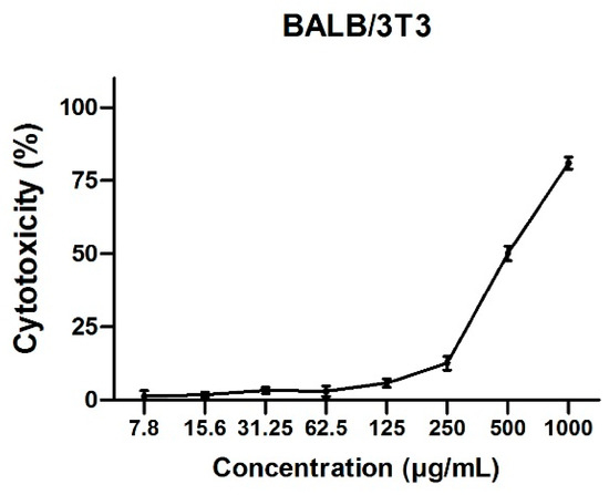
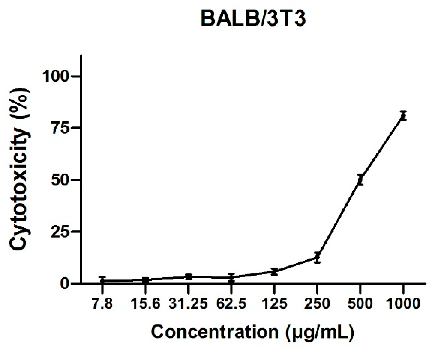
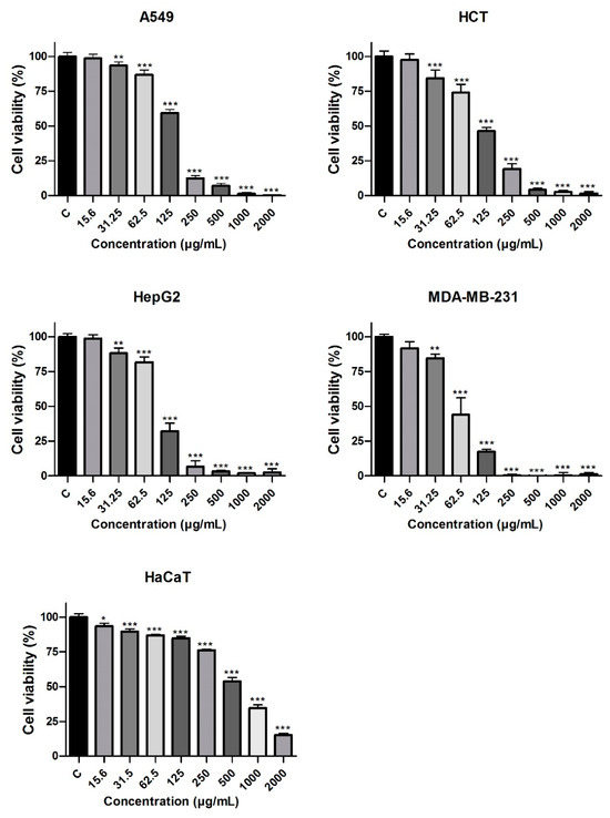
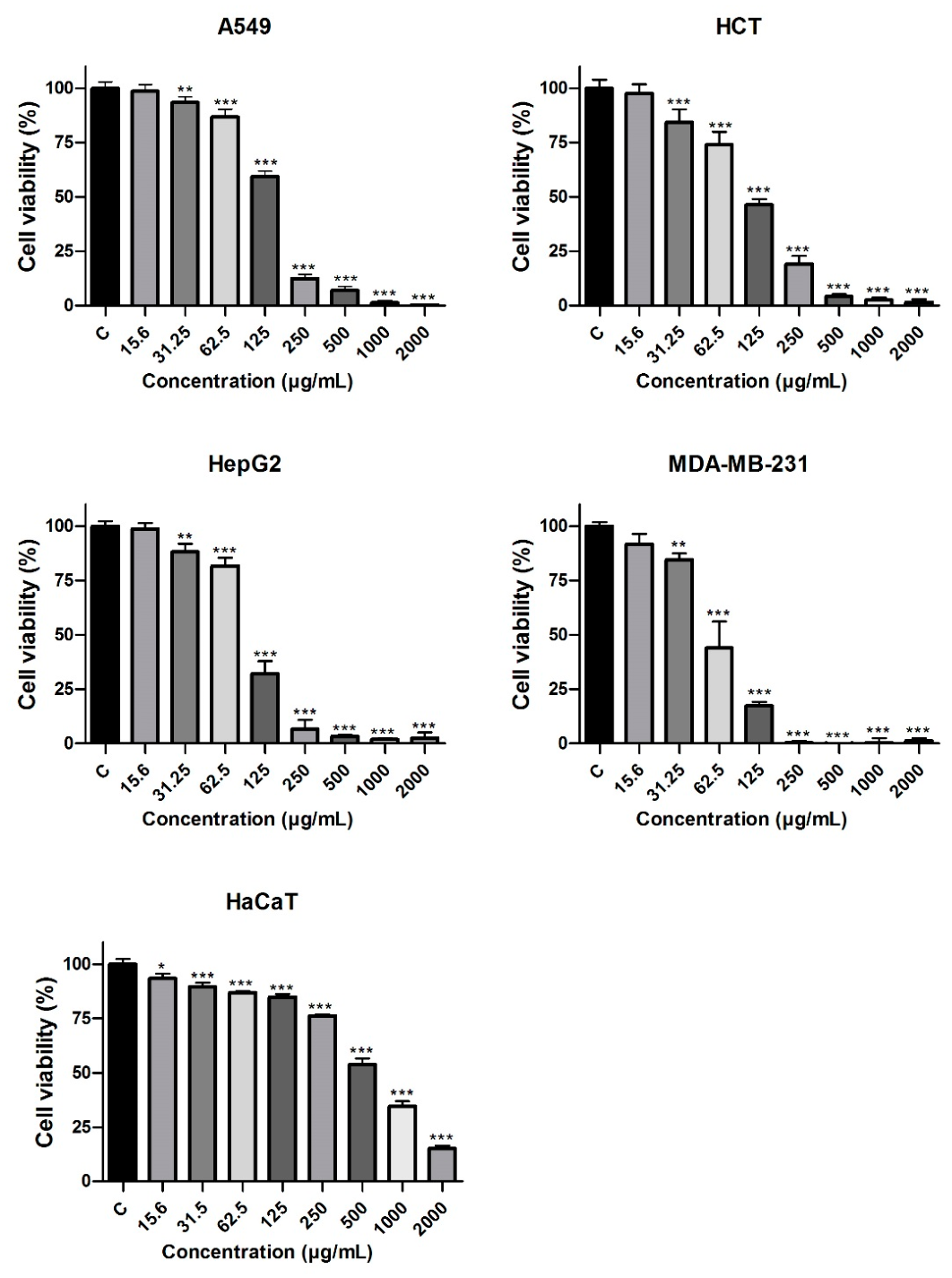
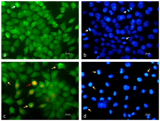
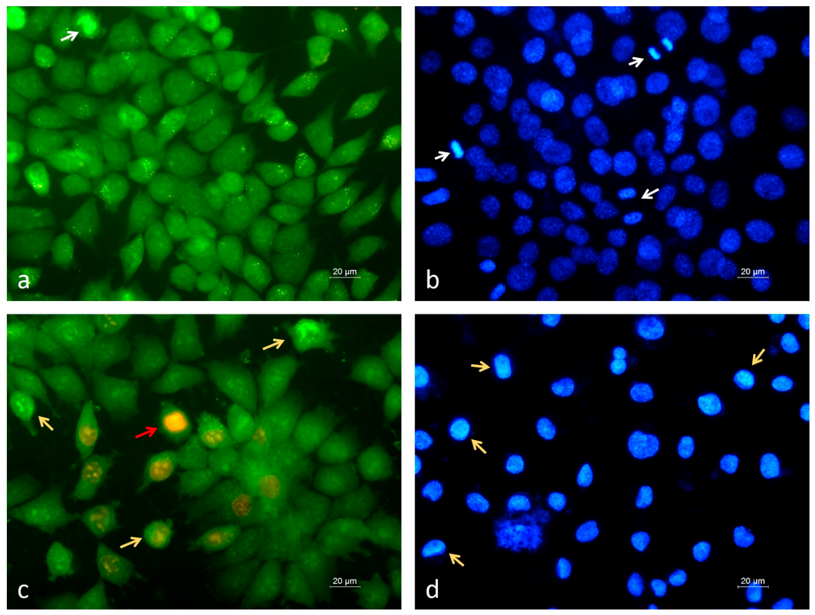
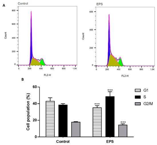
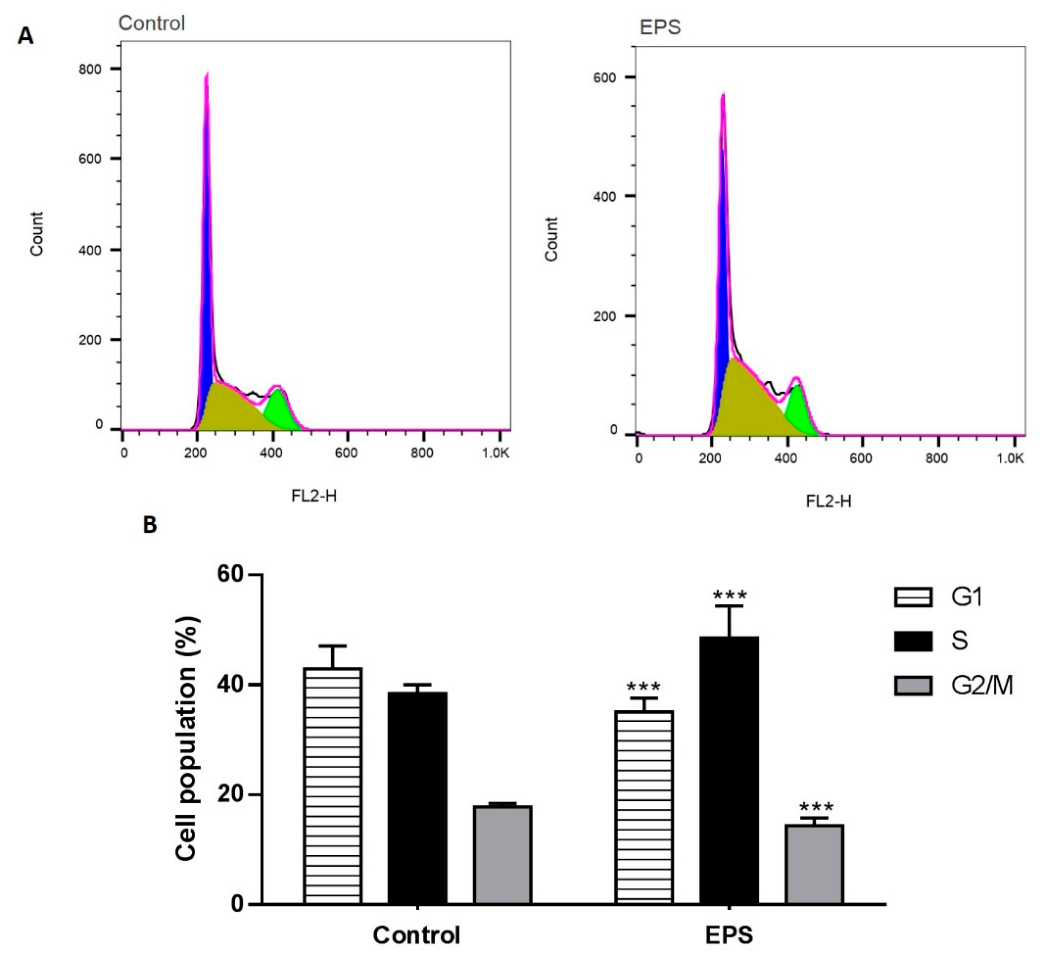
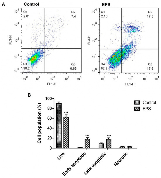
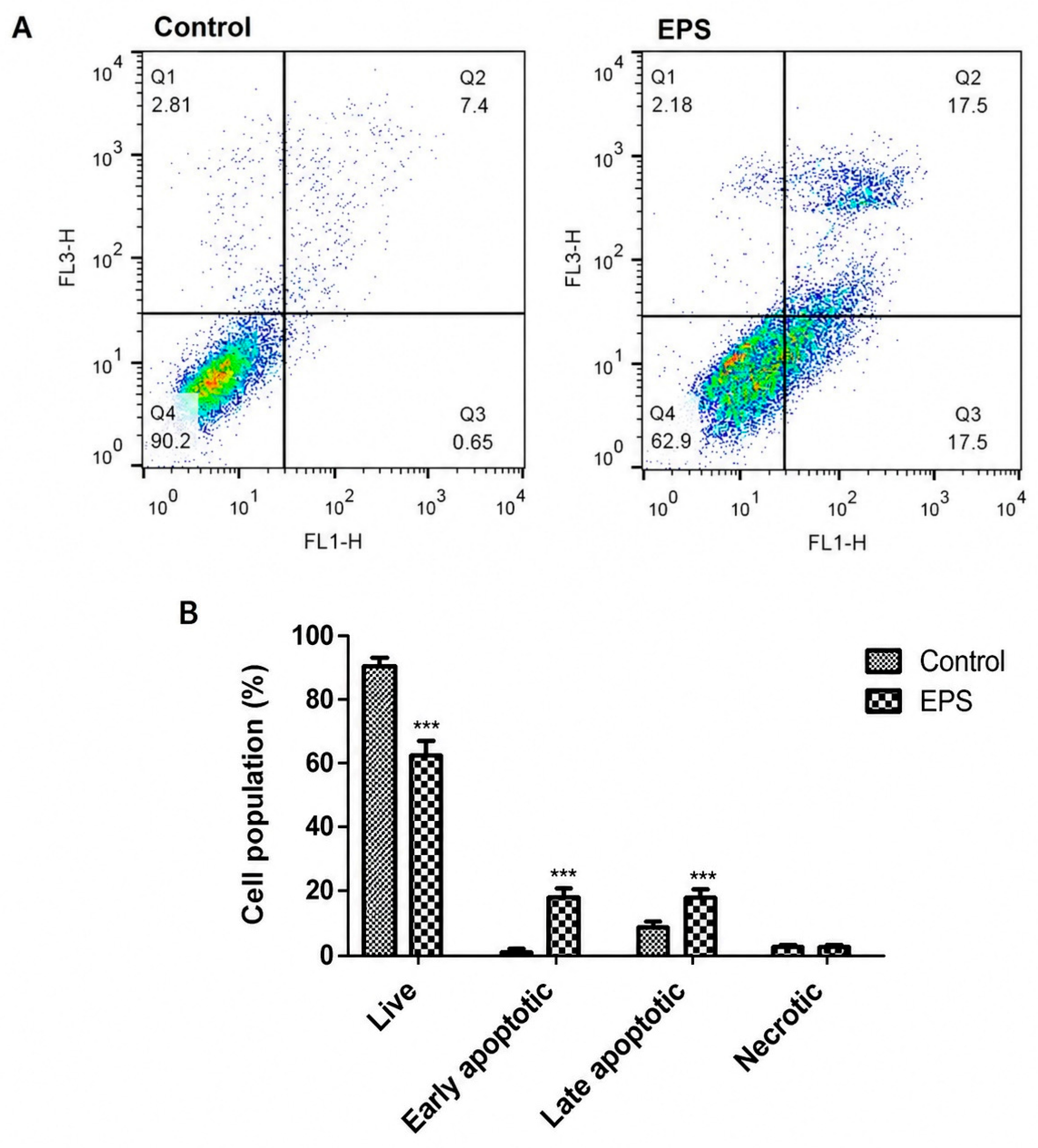
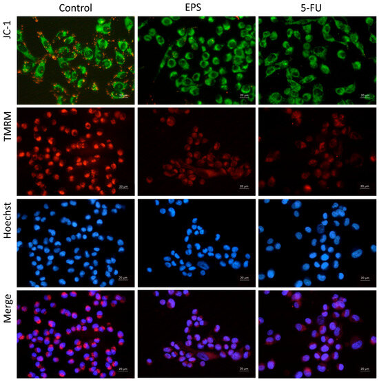
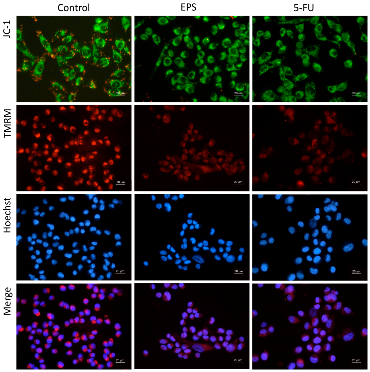
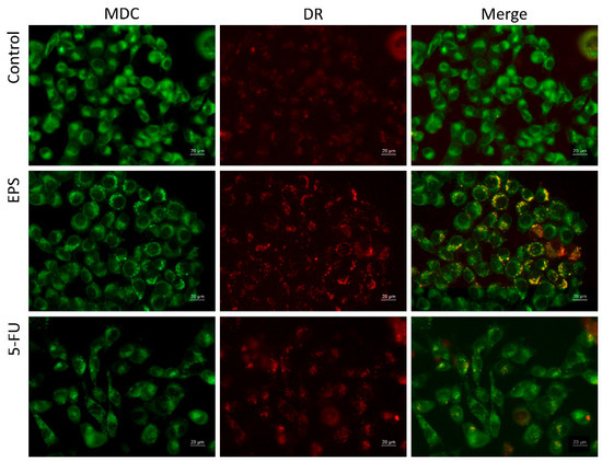
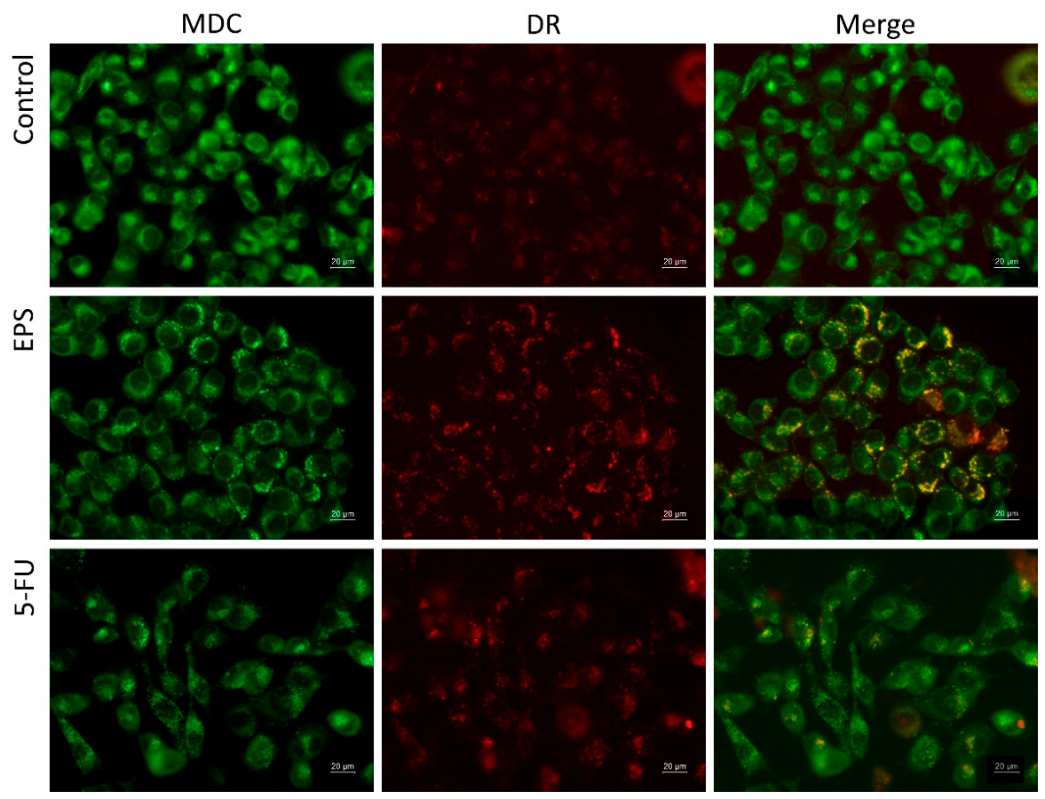
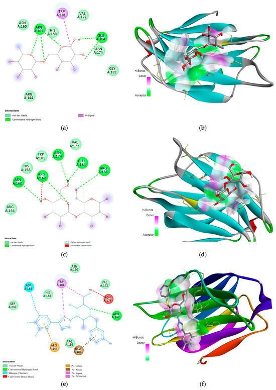
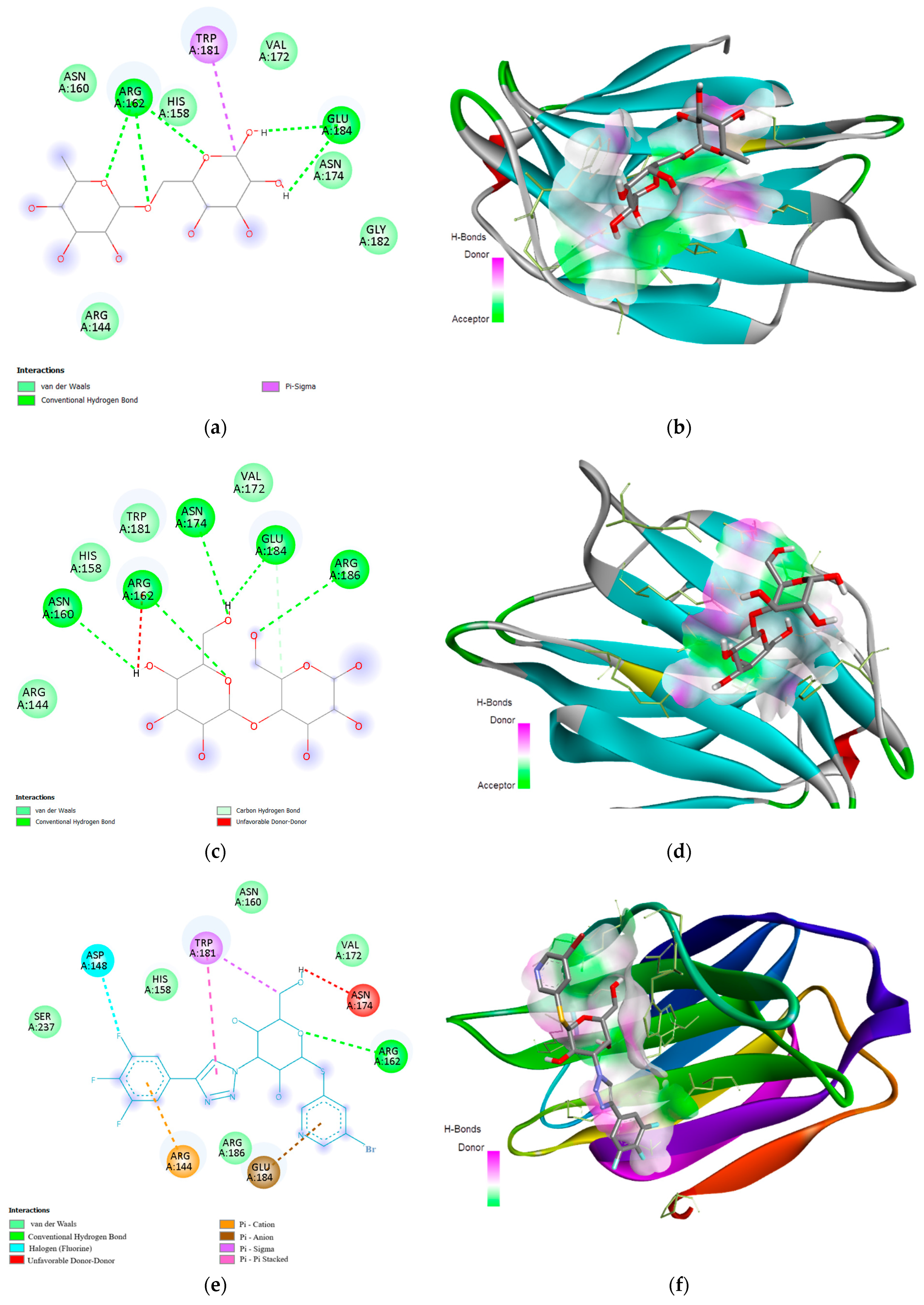
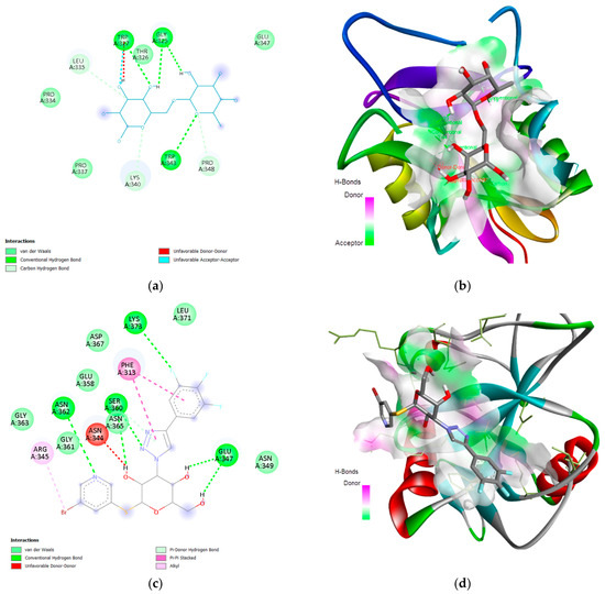
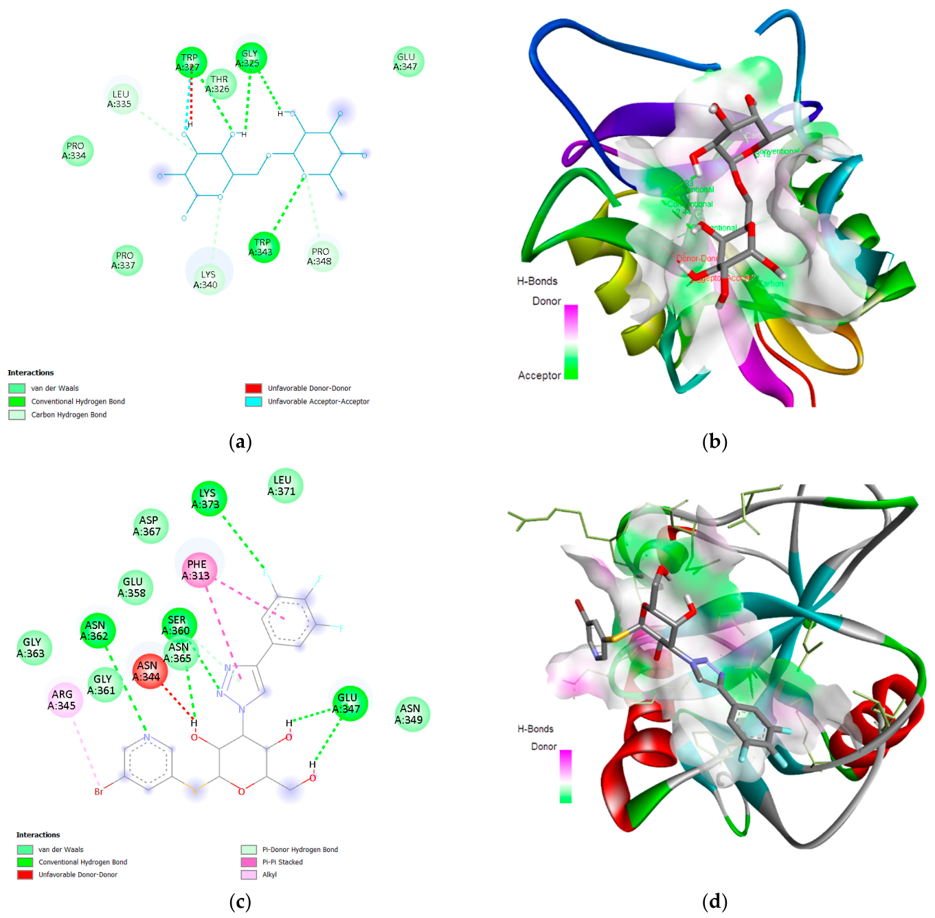

Disclaimer/Publisher’s Note: The statements, opinions and data contained in all publications are solely those of the individual author(s) and contributor(s) and not of MDPI and/or the editor(s). MDPI and/or the editor(s) disclaim responsibility for any injury to people or property resulting from any ideas, methods, instructions or products referred to in the content. |
© 2026 by the authors. Licensee MDPI, Basel, Switzerland. This article is an open access article distributed under the terms and conditions of the Creative Commons Attribution (CC BY) license.