


Featured Application
The aim of this study was to examine the effects of motor imagery (MI) training, performed either alone or in combination with muscle contractions, on quadriceps performance in junior soccer players. The results demonstrated the effectiveness of both training programs in improving isometric strength and vertical jump performance; these findings may help soccer coaches and fitness trainers to improve their training methods. Motor imagery could be a useful tool for enhancing athletic performance and supporting physical conditioning in soccer players.
Abstract
This study examined the effects of motor imagery (MI) training, performed alone or in combination with muscle contractions, on quadriceps performance in junior soccer players. Forty-five players (14.1 ± 0.8 years) were randomly assigned to MI alone (EXP-1; n = 15), based on the Physical, Environment, Task, Timing, Learning, Emotion, Perspective (PETTLEP) model (3 sessions/week, 20–25 min/session), MI combined with muscle contractions (EXP-2; n = 15; 3 session/week, 35–45 min/session), and a control group (CG; n = 15). Physical performance was assessed via vertical jumps (countermovement jump [CMJ], squat jump [SJ], drop jump [DJ]) and maximal isometric knee extension strength. A two-way repeated-measures general linear model (time × group) was used to assess mean differences. If necessary, we used a Mixed ANCOVA (Analysis of Covariance) to control for the influence of important parameters after significant mean differences were observed at the first examination. After 12 weeks, relevant-significant (p < 0.05 and ηp2 > 0.15) interaction effects were observed for all variables. The interaction effects ranged from ηp2 = 0.40 (maximum strength left) to ηp2 = 0.77 (DJ). Regarding DJ, we found only a partial group effect between CG and EXP-2 (p = 0.044). Significant partial effects for strength parameters were calculated for the average strength (CG vs. EXP-2: left: p = 0.008; right: p = 0.018) and for the maximal strength (CG vs. EXP-2: left: p = 0.046). These findings suggest that motor imagery training may improve neuromuscular performance (maximal strength and vertical jump performance) in junior soccer players, whether performed alone or combined with muscle contractions.
1. Introduction
Soccer, due to its technical and intermittent nature, is a contact sport that requires well-developed and balanced muscular strength to withstand the high mechanical and neuromuscular demands associated with repeated accelerations, decelerations, jumps, and physical duels [1,2]. It is a sport in which maximal muscle power and the ability to maintain it throughout the match are essential. In addition, appropriate general and specific muscle strengthening is essential for playing at the highest level. Moreover, soccer players require sufficient strength and power to perform key match actions, such as sprinting, jumping, and physical duels with opponents, which are critical determinants of performance during match play [3,4]. Therefore, optimal conditioning programs should include strength training (ST).
Quadriceps isometric strength and vertical jump performance were selected as the primary outcomes because they are widely recognized indicators of lower-limb neuromuscular function in soccer players. Quadriceps strength contributes to sprinting, kicking, change-of-direction ability, and knee joint stability, whereas vertical jump performance reflects lower-limb explosive power, a key determinant of soccer-specific performance [5,6].
Isometric training is widely used in athletic conditioning and rehabilitation and has been shown to induce substantial strength adaptations when performed at high intensities [7]. Dynamic movements involving stretch–shortening cycle actions (eccentric–concentric contractions) are widely used in resistance-training programs to enhance strength, power, and athletic performance. Moreover, concentric, eccentric, and isometric contractions each elicit distinct neuromuscular and musculoskeletal adaptations, underscoring their complementary roles in athletic development [8]. Furthermore, recent systematic reviews and meta-analyses have demonstrated that motor imagery is an effective strategy for improving motor performance, strength development, and motor learning across a wide range of populations [9,10,11]. However, uncertainty remains regarding the optimal integration of motor imagery with physical training modalities, particularly in youth athletes and strength-oriented interventions.
In junior athletes, puberty is characterized by increased neuromuscular plasticity and accelerated development of strength and power capacities, with neural adaptations playing a major role in performance improvements during this period [12,13,14]. This developmental phase is a sensitive window during which inter- and intramuscular coordination abilities can be greatly optimized [12]. Previous studies have suggested that combining motor imagery with physical practice may enhance performance outcomes. However, the mechanisms underlying these potential benefits remain incompletely understood, particularly in young athletes. Moreover, MI does not appear to induce substantial neuromuscular fatigue or muscle soreness, making it a potentially valuable tool for athletes, especially in-season competition.
Motor imagery and actual muscle contractions may exert complementary effects on the neuromuscular system. While motor imagery primarily enhances central neural processes by increasing corticospinal excitability and motor planning, and by activating neural networks involved in movement execution, actual muscle contractions provide peripheral neuromuscular adaptations through motor unit recruitment and mechanical loading. Combining these approaches may therefore promote additive or synergistic adaptations by simultaneously targeting central and peripheral mechanisms involved in strength development and motor performance [2,5,15].
Thus, the objective of this study was to evaluate the impact of an MI training program, compared with an MI training program combined with isometric, concentric, and eccentric muscle contractions, on the development of quadriceps strength and vertical jump performance in junior soccer players. We expected that both MI training alone and MI combined with muscle contractions would improve quadriceps strength and vertical jump performance in junior soccer players. Given the limited evidence available in youth populations, no specific hypothesis was formulated regarding the magnitude of differences between MI alone and MI combined with muscle contractions.
2. Materials and Methods
2.1. Experimental Approach to the Problem
This study used a randomized controlled design to examine the effects of motor imagery (MI) training, performed alone or combined with muscle strengthening, on neuromuscular performance in youth soccer players. Forty-five male players (age range: 13–15 years), competing at the national level, were randomly assigned to one of three groups (n = 15 per group): a motor imagery group (EXP-1), a combined motor imagery and strength training group (EXP-2), and a non-training control group (CG).
The EXP-1 group performed a structured motor imagery training program, whereas the EXP-2 group completed the same MI program combined with isometric, concentric, and eccentric muscle contractions. The control group continued their usual soccer training regimen, including technical, tactical, and general conditioning exercises, without any MI practice or targeted strength interventions.
To evaluate the effects of the interventions, all participants underwent pre- and post-intervention testing over a 12-week training period. Neuromuscular performance was assessed through vertical jump tests: Countermovement jump (CMJ), squat jump (SJ), and drop jump (DJ). Intraclass correlation coefficients (ICCs) and coefficients of variation (CVs) were computed to assess inter-session reliability. ICC values were 0.96 (95% CI: 0.93–0.98), 0.96 (95% CI: 0.92–0.98), and 0.91 (95% CI: 0.78–0.96) for the CMJ, SJ, and DJ, respectively, indicating excellent reliability. Correspondingly, CVs were 7.6% (95% CI: 5.3–15.7%), 4.2% (95% CI: 2.9–8.4%), and 7.4% (95% CI: 5.1–15.3%) for the CMJ, SJ, and DJ, respectively.
Jump performance was recorded using the Optojump system (Microgate, Bolzano, Italy), a validated optical device. In contrast, maximal strength was assessed using an isometric knee extension test with a Kinvent Physio dynamometer (K-PULL, Kinvent, Montpellier, France).
2.2. Participants
Forty-five male soccer players participating in the national championship took part in the study. Although participants were of similar chronological age (13–15 years), biological maturation status was not formally assessed.
The study design was approved by the Ethics Committee of the Martin Luther University of Halle-Wittenberg (reference number: 2022-11; Date of approval: 18 March 2022), in accordance with the Declaration of Helsinki [16], and met the ethical standards in Sport and Exercise Science Research [17]. All parents gave their written consent. None of the participants had any musculoskeletal injuries or pain.
The players were randomly divided into three groups of 15 subjects each. Participants were randomly assigned to the experimental and control groups using a computer-generated randomization sequence with a 1:1:1 allocation ratio. Allocation was concealed using sealed, opaque envelopes prepared before enrolment. The randomization procedure was carried out by an independent researcher who was not involved in participant recruitment, assessment, or intervention delivery.
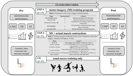
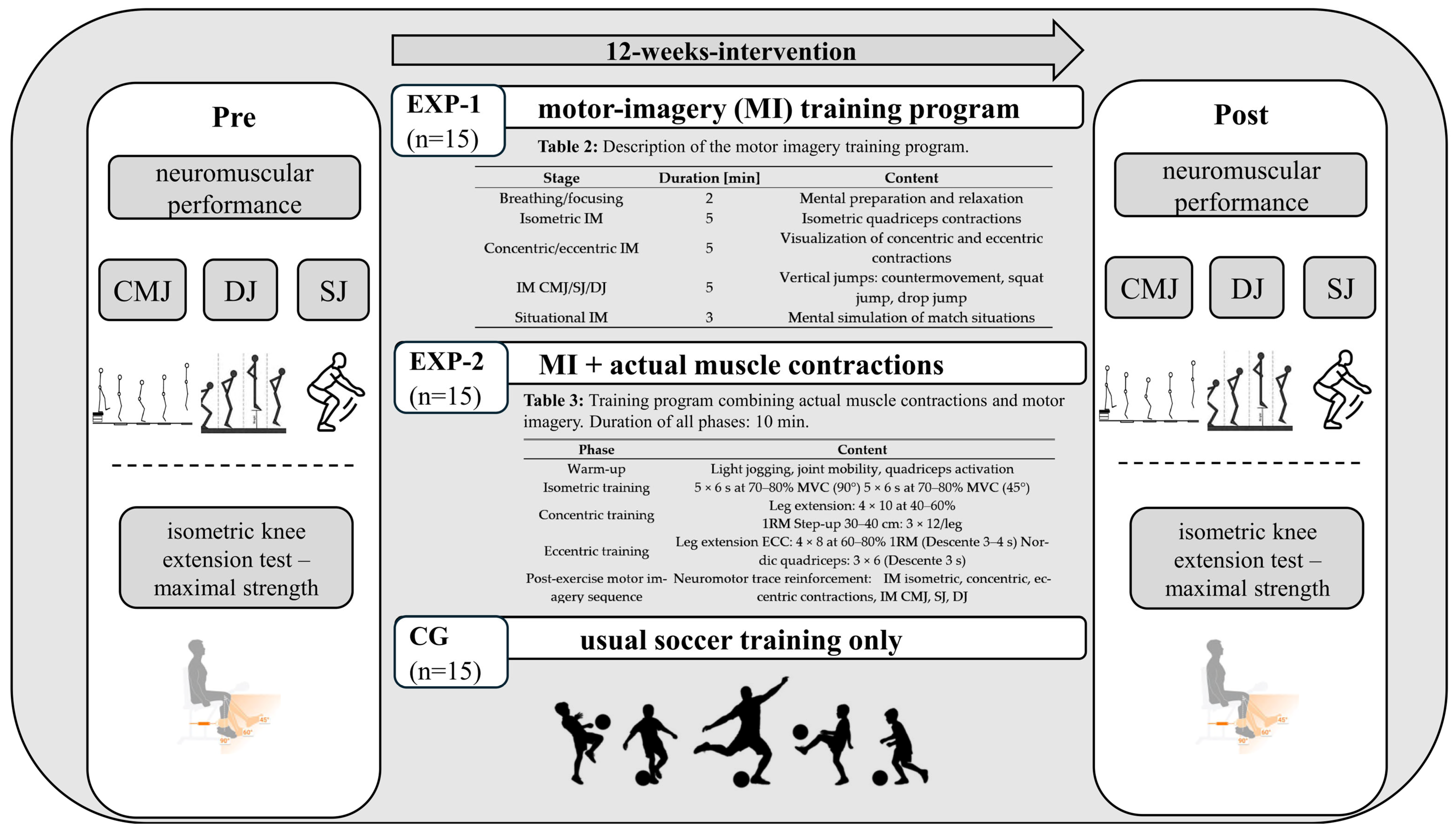
The first experimental group (EXP-1) followed a mental imagery training program. The second group (EXP-2) underwent an MI training program incorporating isometric, concentric, and eccentric muscle contractions. The third and final group served as the control group (CG), which followed only the usual soccer training program (Table 1; Figure 1).
Table 1.
Physical characteristics of interventions and control groups (mean ± SD). Relevant-significant (p < 0.05 and ηp2 > 0.15) mean differences marked in bold.
Figure 1.
Flow diagram of the study design including time line, measurements and interventions.
2.3. Procedures and Evaluation
All participants underwent experimental measurements of vertical jump height (CMJ, SJ, DJ) and isometric knee extension strength of both limbs twice. The Optojump System (Microgate SARL, Bolzano, Italy) was used to measure jump height. The first measurements were taken before the training programs began, and the second series was taken immediately after the 12-week programs ended.
All testing sessions were conducted at the same time of day to minimize potential circadian influences on performance. Participants were instructed to refrain from strenuous physical activity for 24 h before testing, to maintain their usual sleep schedule the night before assessments, and to avoid caffeine for at least 12 h before testing. They were also advised to maintain their normal dietary habits and arrive well hydrated. The testing order was standardized for all participants to ensure consistency across measurements. All participants completed the same sequence of tests under identical conditions. Before performance assessments, a standardized warm-up protocol was conducted, consisting of a 5–7 min light jog followed by dynamic stretching of the lower limbs. This was followed by 5–8 min of sport-specific activation exercises, including submaximal squats, lunges, and progressively increasing vertical jumps. The warm-up was designed to gradually increase intensity and prepare participants for maximal effort in both strength and jump performance testing. These procedures were implemented to minimize inter- and intra-subject variability.
The CMJ test was performed to assess participants’ vertical jump performance. The subjects stood with their feet shoulder-width apart and their hands on their hips to eliminate upper-limb use. From this position, they performed rapid flexion of the lower limbs, followed immediately by maximum extension, to produce a vertical jump. Three trials were performed, with a 1 min recovery period between them. The best performance was selected for analysis. The SJ was used to assess the vertical jump performance without using the stretch-shortening cycle. Participants started the test in a squatting position, with knees bent to approximately 90° and hands on their hips. After holding the static position for 2 s, they performed a maximum extension and a vertical jump. Three attempts were made, and the best performance was recorded. The DJ was used to assess the lower-limb reactive abilities. Participants jumped from a 30 cm-high platform and, upon landing, performed a maximum vertical jump, minimizing ground contact time. Three attempts were made, and the best performance was retained for analysis.
Maximum isometric quadriceps strength was assessed using a Kinvent Physio dynamometer (K-PULL, Kinvent, Montpellier, France). Participants were seated on the edge of a table or chair with their feet unsupported and their knees flexed at 45°, as quadriceps force production is influenced by knee joint angle [18]. A strap connected to the dynamometer was secured around the ankle, and participants were instructed to exert maximal force in knee extension. Kinvent dynamometry is a reliable tool for isometric strength assessment in sport populations [19,20]. The testing protocol included a 15 s familiarization period, one set per leg, and three maximal isometric contractions per leg. Each contraction was maintained for 5 s, with 15 s of recovery between repetitions.
2.4. Training Programs
2.4.1. Motor Imagery Training Program
The intervention lasted 12 weeks, with three supervised sessions per week performed on non-consecutive days. Training load was progressively increased across the intervention period by manipulating the number of sets and repetitions according to a structured overload principle, while maintaining the same exercise selection throughout the program. No reassessment of one-repetition maximum (1RM) was conducted during the intervention; therefore, training intensity was based on baseline values. Standardized rest intervals were used to ensure consistency across participants: 60–90 s between sets and 2–3 min between exercises. The research team fully supervised all sessions to ensure protocol adherence and standardization of training execution. Attendance was recorded for each session, and overall compliance with the intervention was high across all groups. Any missed sessions were documented for transparency and considered in the final analysis. These details enhance the reproducibility of the intervention and ensure transparency in the progression of the training load.
For the first experimental group (EXP-1), the motor imagery program was designed according to the PETTLEP model described by Holmes and Collins [21], aiming to reinforce the functional equivalence between imagery and actual movement execution. This program was delivered at a rate of 3 sessions per week for 12 weeks, with each session lasting 20 to 25 min. The “Physical” component was implemented using a seated position with the knees bent to 90°, while the “Environment” component was based on a visualization of a typical training room. The Task component consisted of motor imagery of isometric, concentric, and eccentric muscle contractions, as well as vertical jumps (counter movement jump, squat jump, and drop jump). The imagined movements were performed in real time, and the Learning component accounted for a gradual progression of content over the 12-week program. The Emotion component incorporated a competitive imagery context to strengthen participants’ emotional engagement, with imagery predominantly internal (80%) and supplemented by an external perspective (20%). Each session began with a breathing and attention focus phase (2 min), followed by a phase of imagery of isometric contractions (5 min), then concentric and eccentric contractions (5 min), imagery of CMJ, SJ, and DJs (5 min), and ended with a phase of situational imagery in a match context (3 min) (Table 2).
The imagined scenarios were selected according to the PETTLEP model to maximize functional equivalence between imagery and actual soccer performance. Although the primary outcomes were quadriceps strength and jump performance, the imagery scripts included soccer-specific actions requiring high levels of lower-limb force production, such as jumping for headers, explosive accelerations, rapid changes in direction, and powerful ball striking. These sport-specific movements were intended to reinforce the neural processes underlying the neuromuscular performance assessed in the present study.
No formal assessment of motor imagery ability was conducted before the intervention. However, all participants received standardized instructions and were supervised throughout the intervention to ensure adherence to the imagery protocol.
Table 2.
Description of the motor imagery (MI) training program.
2.4.2. Motor Imagery Combined with Muscle Strengthening
The second experimental group (EXP-2) completed the program’s content. It consisted of combining actual muscle contractions with motor imagery and was conducted three times per week, with each session lasting 35–45 min. The maximum repetition value (1RM) for the leg extension exercise was determined before the start of the training program. Each session began with a standardized 10 min warm-up consisting of light jogging, joint mobility exercises, and specific quadriceps activation. Isometric work was then performed using five 6-second contractions at 70–80% of maximum voluntary force at knee flexion angles of 90° and 45° (Table 3). Concentric work consisted of leg extension exercises (4 sets of 10 repetitions at 40–60% of 1RM) and step-ups on a 30–40 cm height (3 sets of 12 repetitions per lower limb). Eccentric work included eccentric leg extensions (4 sets of 8 repetitions at 60–80% ofs 1RM, with a controlled descent extension time of 3 to 4 s) as well as Nordic quadriceps exercises (Performed by slowly lowering the body under control, resisting gravity to maximize eccentric loading) (3 sets of 6 repetitions with a 3 s eccentric phase). Each session ended with a 10 min post-exercise motor imagery sequence aimed at reinforcing the neuromotor trace through imagery of isometric, concentric, and eccentric contractions, as well as jumping actions.
Table 3.
Training program combining actual muscle contractions and motor imagery. Duration of all phases: 10 min.
2.5. Control Group
The CG did not receive any training beyond the usual soccer training (technique, tactics, general strengthening) that each experimental group also participated in.
Participants in the control group continued their usual soccer training, including regular technical, tactical, and strength training sessions as well as match play. Training frequency typically ranged from 3 to 5 sessions per week, with each session lasting approximately 60–90 min, depending on the club schedule. No additional experimental intervention (motor imagery or resistance training) was performed. Training load was not formally monitored in the control condition; however, participants were instructed to maintain their habitual training routines throughout the intervention period.
The control group completed only their regular soccer training, as the study was designed to compare practical training applications rather than a strictly time-matched laboratory-based protocol (Figure 1).
2.6. Statistical Analyses
All statistical analyses were performed using SPSS Statistics version 31.0 (IBM, Armonk, NY, USA). All variables were examined for normal distribution (Shapiro–Wilk Test), and variance homogeneity (Levene Test for Equal Variances) before the variance analysis was performed. Means and SDs were calculated to describe metric-scaled and normally distributed data.
A priori, a sample size calculation (nQuery Advisor 4.0; Statistical Solutions, Saugus, MA, USA) was performed to determine the appropriate sample size (primary outcome: CMJ). The calculation is based on the results of Bouteraa et al. [22] and the following assumptions: a two-sided hypothesis, alpha-level: p < 0.05, power 1-β: 0.8, mean difference (SD) intervention groups: 28.8 (3.5) cm, mean difference (SD) control group: 24.4 (3.5) cm. Based on a calculated effect size of 0.58, the analysis revealed that 11 athletes per group were necessary to detect relevant differences between groups over time.
Mean differences between tests (pre and post) and groups (CG, EXP-1, EXP-2) were tested using a two-factorial (time and group) univariate general linear model. Based on the significant group effect concerning body mass, we used the parameter body mass as a covariate (except the normalized parameter DJ-power) in a Mixed ANCOVA (Analysis of Covariance). Initially, the significance and relevance levels were defined as p < 0.05 and partial eta-squared (ηp2 > 0.15) [23,24]. Based on this conservative approach, we intended to avoid overestimating mean differences.
Effect sizes were calculated using Cohen’s d and were classified as small (0.00 ≤ d ≤ 0.49), medium (0.50 ≤ d ≤ 0.79), and large (d ≥ 0.80) as suggested by Cohen [25].
Intrarater reliability was assessed using intra-class correlation coefficients (ICC) [26] as an indicator of relative intrarater reliability, and the coefficient of variation (CV). The CV was derived from log-transformed data [27] and used to assess absolute intrarater reliability (an indicator of measurement variability and random error) across consecutive pairs of intra-subject trials [28]. The reliability thresholds were defined as ICC ≥ 0.75 and CV < 10%, based on published guidelines [29,30,31,32,33].
3. Results
3.1. Normal Distribution and Variance Homogeneity
Seven variables (age: p < 0.001; DJ-power: p <0.001; maximum strength left: p = 0.043; maximum strength right: p = 0.001; average strength right: p = 0.005) were not normally distributed at the first examination. Regarding variance homogeneity, all parameters had homogeneous variances.
3.2. Vertical Jumps and Drop Jump Power
The experimental groups showed significant improvements (time effects) in all jumping parameters (p < 0.001). In contrast, the control group displayed a significant performance reduction concerning the parameter DJ (p = 0.003). The interaction effects ranged from ηp2 = 0.55 (SJ) to ηp2 = 0.77 (DJ; Table 4). Within the experimental groups, we found a significant partial effect concerning DJ (p = 0.044). The effect size in the EXP-2 was larger than in the EXP-1 (d = 0.90 vs. d = 0.84). Regarding SJ, the improvement in the EXP-1 was larger than in EXP-2 (d = 0.85 vs. d = 0.71). In relation to body weight, the EXP-1 displayed a larger effect from pre to post testing (d = 0.77) than the EXP-2 (d = 0.67).
Table 4.
Descriptive comparison and analysis of Covariance (covariate: body mass) for jump performance parameters depending on groups before and after the 12-week intervention. Significantly relevant differences (p < 0.05 and ηp2 > 0.15) are marked in bold.
Within the Mixed ANCOVA, the covariate body mass did not show any relevant influence regarding the non-normalized lumping parameters (CMJ: p = 0.761, ηp2 = 0; SJ: p = 0.606, ηp2 = 0.01; DJ: p = 0.386, ηp2 = 0.02).
3.3. Quadriceps Muscle Strength
Quadriceps strength showed greater changes in the average compared with the maximum performance level. The interaction effects moved between ηp2 = 0.40 (maximum left) to ηp2 = 0.60 (average left). Group effects were only calculated for the average strength on both sides (left and right: ηp2 = 0.17–0.21). The control group showed a markedly lower performance level (average strength left) at the first examination, especially compared with EXP-2 on the left side (EXP-2: 20.9 ± 6.45 kg vs. CG: 16.8 ± 6.99 kg). Partial effects between groups were only found for the CG compared with EXP-2 across three of four parameters (Table 5). In contrast to the CG, the experimental groups showed significant time effects in all strength parameters. The largest difference was calculated for average strength left (EXP-1: d = 0.61 vs. EXP-2: d = 0.81; Table 5).
Table 5.
Descriptive comparison and analysis of Covariance (covariate: body mass) for strength performance parameters depending on groups before and after the 12-week intervention. Significantly relevant differences (p < 0.05 and ηp2 > 0.15) are marked in bold.
In the Mixed ANCOVA, the covariate body mass did not show any relevant influence regarding the strength parameters (range ηp2 = 0.03–0.09).
In summary, in 5 of 8 parameters (63%), the athletes of EXP-2 achieved greater improvements than the soccer players of EXP-1.
4. Discussion
This study aimed to examine the effects of combining motor imagery according to the PETTLEP model with actual muscle contractions (isometric, concentric, and eccentric) on the development of quadriceps femoris strength in junior soccer players. The main results showed that the systematic integration of MI into a specific training program leads to significant strength gains, exceeding those observed in conditions without MI, thus confirming the benefits of this approach for developing athletes’ performance. Indeed, both experimental groups showed relevant (d > 0.5) improvements in the four parameters assessed during vertical jump tests (CMJ, SJ, and DJ height and power). Furthermore, a comparison between the experimental groups revealed that EXP-2, which received more intensive combined training, showed greater CMJ progress than EXP-1.
The effects observed for EXP-2 were d = 0.68 for CMJ, d = 0.71 for SJ, and d = 0.90 for DJ height, while EXP-1 recorded improvements of d = 0.52, d = 0.85, and d = 0.84, respectively. There was also a significantly relevant interaction effect observed in DJ power (p < 0.001; ηp2 = 0.57) with slight differences between the intervention groups (EXP-1: d = 0.77; EXP-2: d = 0.67). These results are consistent with previous evidence suggesting that motor imagery can enhance the effects of physical training on strength and performance. Similar improvements have been reported following mental practice alone and when motor imagery is combined with physical exercise [10,34,35]. Furthermore, the training interventions elicited marked increases in maximal and mean isometric strength in both experimental groups. Nevertheless, post hoc analyses indicated that only EXP-2 achieved significantly greater gains in mean isometric strength than the control group, whereas no significant between-group differences were detected for maximal isometric strength. In this context, it should be considered that the training duration for the EXP-2 group (35–45 min per session) was markedly higher compared with the EXP-1 group (20–25 min per session). Consequently, the total training volume of the two groups was not matched.
Our results also confirm the positive relationship between muscle strength and vertical jump performance, as reported in previous investigations [8,36,37], suggesting that maximal strength is a key determinant of jump height in athletes. This relationship is particularly relevant in sports requiring repeated high-intensity efforts. Improvements in strength are associated with enhanced ability to generate vertical impulse, which may explain the gains observed in our jump performance tests, particularly in the combined training group. These adaptations may be related to mechanisms previously proposed in the literature. However, the present study did not directly assess the underlying neurophysiological or morphological adaptations. These improvements may reflect neural and morphological mechanisms previously proposed to underlie strength development [37,38]. For example, motor imagery has been shown to attenuate strength loss during periods of immobilization. During a 4-week wrist flexor immobilization protocol, MI reduced the decline in maximal strength by approximately 50% compared with a control group (23.8 ± 5.6% reduction in strength versus 45.1 ± 5.0% for the control group) [9,15]. Furthermore, mental contraction repetition does not appear to induce substantial fatigue, allowing for increased training volume without compromising maximum force production [11,35].
Although EXP-2 generally demonstrated greater percentage improvements than EXP-1, the limited number of statistically significant between-group differences suggests that the superiority of the combined intervention should be interpreted cautiously. Furthermore, because EXP-2 completed a greater overall training volume than EXP-1, the observed advantages may have been attributable, at least in part, to the higher training dose rather than exclusively to the combination of motor imagery and actual muscle contractions. Therefore, the present findings cannot fully disentangle the respective contributions of training dose and intervention characteristics. Future studies should control for total training volume by matching session duration and overall workload across groups, allowing a clearer distinction between dose-related effects and the specific effects of combining motor imagery with actual muscle contractions.
The results of this study are consistent with those reported by Saumur et al. [39], who showed that a motor imagery training program targeting the quadriceps could significantly increase peak muscle torque after only three weeks of intervention. This latest study highlights that MI can produce strength gains even in proximal muscles, which are traditionally considered less sensitive to mental interventions than distal muscles. Our findings are also consistent with previous intervention studies conducted specifically in youth soccer players. For example, Aloui et al. [40] reported that an 8-week combined plyometric and short-sprint training program significantly improved vertical jump performance, sprint speed, agility, and balance in elite adolescent soccer players. Similarly, Aloui et al. [41] demonstrated significant improvements in countermovement jump, squat jump, sprint, change-of-direction ability, repeated sprint ability, and dynamic balance following a comparable training intervention in U-15 male soccer players. Although these studies investigated physical training rather than motor imagery, the improvements in jumping performance are consistent with the present findings. Together, these results suggest that motor imagery, particularly when combined with muscle contractions, may represent an effective complementary strategy for enhancing neuromuscular performance in young soccer players while imposing a substantially lower mechanical load than conventional strength- and power-oriented training.
These observations suggest that motor imagery may involve central neural processes previously described in the literature, although these mechanisms were not directly assessed in the present study. In our study, the significant gains in quadriceps strength and jump performance observed in both experimental groups may reflect mechanisms previously suggested in the literature. However, any additional contribution of combining motor imagery with muscle contractions remains speculative [42,43]. The improvements observed in the present study are likely related to central neural mechanisms previously reported in the literature. Previous studies have suggested that strength improvements following motor imagery may involve increased corticospinal excitability, improved motor planning, and enhanced motor unit recruitment, although these mechanisms were not directly evaluated in the present study [11]. These effects are further supported by evidence indicating modulation of corticospinal drive and intermuscular coordination during motor imagery tasks [44,45]. Classic findings also demonstrate that mental practice alone can induce measurable strength gains, reinforcing the role of central adaptations in force production [15,44]. However, these mechanisms remain indirect in behavioral training studies and were not directly assessed in the present work. Therefore, while neural adaptations provide a plausible explanation for the observed performance improvements, they remain inferential in the current experimental design.
Our results are inconsistent with those reported by Reiser et al. [46], who studied the effect of different proportions of motor imagery and isometric physical exercise on elbow flexor muscle strength. In their study, the group performing only physical exercise (100% maximum voluntary contraction) showed the greatest gain in maximum isometric strength (+5%). In contrast, the groups combining motor imagery with physical exercise recorded slightly lower, yet significant, gains (between +3% and +4%). Although these gains are lower than those observed with full physical training, they remain higher than those in no-training conditions, highlighting the potential contribution of the central neural processes proposed in previous studies. Conversely, in our study, EXP-2 generally demonstrated larger improvements in isometric strength than EXP-1. However, no significant differences were found between the two experimental groups. Consequently, while the combination of motor imagery and physical training may offer additional benefits, the present results do not conclusively demonstrate a synergistic effect beyond that achieved through motor imagery alone. In comparison, the CG showed no improvement in isometric strength. Methodological differences, including the type of exercise, training intensity, study population, or intervention duration, could explain this discrepancy. Nevertheless, all of these results converge towards the idea that motor imagery can be an effective complement to physical training, particularly for multi-joint and high-intensity exercises, without ignoring the effects of actual practice [2,10,11].
The use of the PETTLEP model is a central element of our protocol. By integrating the physical, environmental, task, timing, learning, emotional, and perspective dimensions, motor imagery was performed under conditions that closely resembled actual training situations. This improves functional equivalence between imagined and executed movement. Recent systematic reviews confirm that PETTLEP-based interventions enhance imagery effectiveness and motor performance outcomes [21,47,48]. In junior soccer players aged 13 to 15, a period characterized by rapid neuromuscular maturation, motor imagery may be particularly well suited to support neural processes involved in motor performance, without excessively increasing mechanical load. Previous investigations [9,46,49] have proposed that neural adaptations may contribute to the effects of motor imagery training, although these mechanisms were not directly assessed in the present study. Thus, combining motor imagery with specific quadriceps training may facilitate the transfer of strength gains to soccer-specific tasks such as vertical jumping [5]. In practical terms, integrating motor imagery sessions into regular training may reduce mechanical load while maintaining or enhancing neuromuscular performance, particularly during periods of high training density or active recovery [2,10,11]. This strategy is especially relevant for junior athletes, whose strength development is strongly dependent on neural factors [2,12,13]. The effectiveness of motor imagery in the present study may be particularly relevant during adolescence, a period in which strength development is strongly influenced by neuromuscular performance [2,12].
Finally, incorporating MI at the beginning and end of training sessions appears to be optimal for facilitating motor learning, improving movement perception, and maximizing strength gains [47,48]. In the context of junior soccer players, MI may also contribute to the development of explosive abilities without increasing muscle fatigue, possibly through central neural processes previously proposed in the literature that are essential for athletic performance [2,9,11]. In summary, the present study suggests that combining motor imagery with physical training is an effective strategy for improving quadriceps strength and jump performance in junior soccer players. However, the physiological mechanisms underlying these improvements remain to be elucidated by future studies combining motor imagery with direct neurophysiological assessments, such as surface electromyography, voluntary activation techniques, rate of force development analyses, and other measures of neural drive, together with morphological assessments.
It should be noted that the total intervention duration was greater in the motor imagery plus actual contraction group (EXP-2) than in the motor imagery-only group (EXP-1), as the execution of actual muscle contractions required additional training time. Therefore, the greater improvements observed in EXP-2 may have been influenced not only by the combined intervention but also by the higher training volume. Consequently, the present findings should be interpreted with caution, and future studies should match intervention duration and total training load across groups to isolate the specific effect of combining motor imagery with actual muscle contractions.
Limitations and Future Research Directions
Several limitations should be acknowledged. First, the total training volume was not strictly matched between the two experimental groups, limiting the ability to determine whether the additional improvements observed in EXP-2 were attributable to the combined motor imagery and actual contraction protocol or, at least in part, to the greater training volume. Future studies should control for total training exposure by matching session duration across intervention groups. This could be achieved by incorporating a neutral, non-physical sport-related activity (e.g., watching tactical video footage) into the motor imagery-only condition, thereby enabling a more accurate evaluation of the specific contribution of muscle contractions.
The control group continued their regular soccer training without receiving an additional time- or attention-matched intervention. Therefore, the observed differences between groups may partly reflect the effect of the additional training stimulus rather than the specific effects of motor imagery alone. Future studies should consider including an active, time-matched control condition to better isolate the specific contribution of motor imagery-based training.
Second, participants’ motor imagery ability, imagery vividness, and adherence to the imagery protocol were not formally assessed, which may have affected the intervention’s effectiveness. Third, the particular soccer training experience and biological maturation were not assessed. Given that the participants were 13–15 years old, differences in maturity status may have influenced muscle strength, jumping performance, and the response to training. Although participants were randomly assigned to the study groups and exhibited comparable baseline characteristics, the potential influence of biological maturation status was not monitored and cannot be excluded. Biological maturation is a known confounding variable that significantly influences training-induced strength gains and vertical jump kinetics in this specific age cohort.
Fourth, no neurophysiological measurements (e.g., EMG, EEG, or TMS) were collected, preventing direct investigation of the mechanisms underlying the observed performance improvements. Finally, please note that no assessment of imagery ability (e.g., MIQ, KVIQ, or a similar questionnaire) was conducted before the intervention. However, imagery ability substantially influences training effectiveness.
Future studies should assess baseline motor imagery ability using validated instruments, such as the Movement Imagery Questionnaire-3 (MIQ-3) or the Vividness of Movement Imagery Questionnaire-2 (VMIQ-2), to improve baseline homogeneity and strengthen the interpretation of intervention effects. A final limitation is that training load in the control group was not objectively monitored.
5. Conclusions
This study demonstrates that combined training, combining motor imagery and muscle strengthening, could be an effective approach to improve quadriceps strength and maximum performance in junior soccer players aged 13 to 15. The significant gains observed in maximum, average, and vertical jump strength confirm that motor imagery is a useful complementary tool to traditional physical training.
The present findings suggest that incorporating motor imagery into regular training programs may improve strength and jump performance in young soccer players. However, the mechanisms responsible for these adaptations remain to be clarified through future investigations. These effects are particularly interesting in junior athletes, for whom motor learning, movement perception, and explosive strength are still maturing. In practice, incorporating motor imagery sessions at the beginning and end of physical training sessions not only optimizes performance but also supplements training volume without risking muscle overload. These results support the use of motor imagery as a complementary training strategy to enhance strength development and optimize athletic performance in junior soccer players. Both interventions displayed a similar effectiveness, whereas evidence supporting the superiority of the combined intervention remains limited.
Future studies tracking youth motor imagery interventions should incorporate non-invasive maturation estimates (e.g., Mirwald maturity offset equations, Tanner stage, or maturity offset) to ensure complete group baseline homogeneity. That would be a valuable contribution regarding the estimation of its potential impact on training adaptations.
In conclusion, it is recommended that future research should combine physical and mental training with surface electromyography or neural drive metrics to clarify the underlying pathways in a more valid manner.
Author Contributions
Conceptualization, N.S. and M.S.C.; methodology, N.S. and M.S.C.; software, N.S.; validation, N.S. and M.S.C.; formal analysis, N.S., R.S., and M.S.C.; investigation, N.S.; resources, N.S. and M.S.C.; data curation, N.S.; writing—original draft preparation, N.S.; writing—review and editing, D.J.O., R.P.M., R.S., and M.S.C.; visualization, R.S. and R.P.M.; supervision, D.J.O., R.S., and R.P.M.; project administration, N.S. and M.S.C.; funding acquisition, N.S. and M.S.C. All authors have read and agreed to the published version of the manuscript.
Funding
This research received no external funding. The publication of the study was supported by the Open Access Publication Fund of the Martin Luther University Halle-Wittenberg.
Institutional Review Board Statement
The study was conducted in accordance with the Declaration of Helsinki and approved by the Martin Luther University Halle-Wittenberg (reference number: 2022-11; date of approval: 18 March 2022).
Informed Consent Statement
Informed consent was obtained from all subjects involved in the study. Written informed consent has been obtained from the athletes to publish this paper.
Data Availability Statement
The data are not publicly available due to ethical restrictions. The anonymized data are available upon request from the first or corresponding author.
Acknowledgments
We would like to express our sincere thanks to all those responsible for the soccer team for the opportunity to conduct the ongoing investigation.
Conflicts of Interest
The authors declare that the research was conducted in the absence of any commercial or financial relationships that could be construed as a potential conflict of interest.
References
- Silva, J.R.; Nassis, G.P.; Rebelo, A. Strength training in soccer with a specific focus on highly trained players. Sports Med. Open 2015, 1, 17. [Google Scholar] [CrossRef] [Scilit] [PubMed]
- Doungchan, N.; Supalak, R.; Masodsai, K.; Miyashita, M.; Jirapongsatorn, B. Effects of a Six-Week Functional Training Program on Physical Performance and Passing Accuracy in Young Adult Male Soccer Players. Phys. Act. Health 2026, 10, 54–65. [Google Scholar] [CrossRef] [Scilit]
- Chelly, M.S.; Fathloun, M.; Cherif, N.; Ben Amar, M.; Tabka, Z.; Van Praagh, E. Effects of a back squat training program on leg power, jump, and sprint performances in junior soccer players. J. Strength Cond. Res. 2009, 23, 2241–2249. [Google Scholar] [CrossRef] [Scilit] [PubMed]
- Oliver, J.L.; Ramachandran, A.K.; Singh, U.; Ramirez-Campillo, R.; Lloyd, R.S. The effects of strength, plyometric and combined training on strength, power and speed characteristics in high-level, highly trained male youth soccer players: A systematic review and meta-analysis. Sports Med. 2024, 54, 623–643. [Google Scholar] [CrossRef] [Scilit] [PubMed]
- Suchomel, T.J.; Nimphius, S.; Stone, M.H. The importance of muscular strength in athletic performance. Sports Med. 2016, 46, 1419–1449. [Google Scholar] [CrossRef] [Scilit] [PubMed]
- Wisløff, C.; Castagna, C.; Helgerud, J.; Jones, R.; Hoff, J. Strong correlation of maximal squat strength with sprint performance and vertical jump height in elite soccer players. Br. J. Sports Med. 2004, 38, 285–288. [Google Scholar] [CrossRef] [Scilit] [PubMed]
- Oranchuk, D.J.; Storey, A.G.; Nelson, A.R.; Cronin, J.B. Isometric training and long-term adaptations: Effects of muscle length, intensity, and intent: A systematic review. Scand. J. Med. Sci. Sports 2019, 29, 484–503. [Google Scholar] [CrossRef] [Scilit] [PubMed]
- Tomalka, A. Eccentric muscle contractions: From single muscle fibre to whole muscle mechanics. Pflug. Arch. 2023, 475, 421–435. [Google Scholar] [CrossRef] [Scilit] [PubMed]
- Slimani, M.; Tod, D.; Chaabene, H.; Miarka, B.; Chamari, K. Effects of mental imagery on muscular strength in healthy and patient participants: A systematic review. J. Sports Sci. Med. 2016, 15, 434–450. [Google Scholar] [PubMed]
- Paravlic, A.H.; Slimani, M.; Tod, D.; Marusic, U.; Milanovic, Z.; Pisot, R. Effects and dose-response relationships of motor imagery practice on strength development in healthy adult populations: A systematic review and meta-analysis. Sports Med. 2018, 48, 1165–1187. [Google Scholar] [CrossRef] [Scilit] [PubMed]
- Simonsmeier, B.A.; Andronie, M.; Buecker, S.; Frank, C. The effects of imagery interventions in sports: A meta-analysis. Int. Rev. Sport Exerc. Psychol. 2021, 14, 186–207. [Google Scholar] [CrossRef] [Scilit]
- Lloyd, R.S.; Oliver, J.L. The youth physical development model. Strength Cond. J. 2012, 34, 61–72. [Google Scholar] [CrossRef] [Scilit]
- Lloyd, R.S.; Faigenbaum, A.D.; Stone, M.H.; Oliver, J.L.; Jeffreys, I.; Moody, J.A.; Brewer, C.; Pierce, K.C.; McCambridge, T.M.; Howard, R.; et al. Position statement on youth resistance training: The 2014 International Consensus. Br. J. Sports Med. 2014, 48, 498–505. [Google Scholar] [CrossRef] [Scilit] [PubMed]
- Hammami, M.; Negra, Y.; Billaut, F.; Hermassi, S.; Shephard, R.J.; Chelly, M.S. Effects of lower-limb strength training on agility, repeated sprinting with changes of direction, leg peak power, and neuromuscular adaptations of soccer players. J. Strength Cond. Res. 2018, 32, 37–47. [Google Scholar] [CrossRef] [Scilit] [PubMed]
- Ranganathan, V.K.; Siemionow, V.; Liu, J.Z.; Sahgal, V.; Yue, G.H. From mental power to muscle power—Gaining strength by using the mind. Neuropsychologia 2004, 42, 944–956. [Google Scholar] [CrossRef] [Scilit] [PubMed]
- Millum, J.; Wendler, D.; Emanuel, E.J. The 50th anniversary of the Declaration of Helsinki: Progress but many remaining challenges. JAMA 2013, 310, 2143–2144. [Google Scholar] [CrossRef] [Scilit] [PubMed]
- Harriss, D.J.; Macsween, A.; Atkinson, G. Standards for ethics in sport and exercise science research: 2018 update. Int. J. Sports Med. 2017, 38, 1126–1131. [Google Scholar] [CrossRef] [Scilit] [PubMed]
- Hahn, D.; Herzog, W.; Schwirtz, A. Interdependence of torque, joint angle, angular velocity and muscle action during human multi-joint leg extension. Eur. J. Appl. Physiol. 2014, 114, 1691–1702. [Google Scholar] [CrossRef] [Scilit] [PubMed]
- Trybulski, R.; Więckowski, J.; Muracki, J.; Matuszczyk, F.; Gałęziok, K.; Wilk, M.; Kuzdzal, A. Reliability and reproducibility of the Kinvent K-push dynamometer for assessing quadriceps strength and force development in athletes and untrained individuals. Front. Physiol. 2025, 16, 1573748. [Google Scholar] [CrossRef] [Scilit] [PubMed]
- Rebhi, S.; Cherni, Y.; Hammami, M.; Oranchuk, D.J.; Schwesig, R.; Chelly, M.S. Plyometrics with Reactive Agility or Isometrics: Divergent Effects on Change-Of-Direction Ability, Sprint, and Strength Adaptations in Junior Female Handball Players. Front. Bioeng. Biotechnol. 2026, 14, 1765655. [Google Scholar] [CrossRef] [Scilit] [PubMed]
- Holmes, P.S.; Collins, D.J. The PETTLEP approach to motor imagery: A functional equivalence model for sport psychologists. J. Appl. Sport Psychol. 2001, 13, 60–83. [Google Scholar] [CrossRef] [Scilit]
- Bouteraa, I.; Negra, Y.; Shephard, R.J.; Chelly, M.S. Effects of Combined Balance and Plyometric Training on Athletic Performance in Female Basketball Players. J. Strength Cond. Res. 2020, 34, 1967–1973. [Google Scholar] [CrossRef] [Scilit] [PubMed]
- Hopkins, W.G.; Marshall, S.W.; Batterham, A.M.; Hanin, J. Progressive statistics for studies in sports medicine and exercise science. Med. Sci. Sports Exerc. 2009, 41, 3–13. [Google Scholar] [CrossRef] [Scilit] [PubMed]
- Richardson, J.T.E. Eta squared and partial eta squared as measures of effect size in educational research. Educ. Res. Rev. 2011, 6, 135–147. [Google Scholar] [CrossRef] [Scilit]
- Cohen, J. Statistical Power Analysis for the Behavioural Sciences; Lawrence Erlbaum Associates: Hillsdale, NJ, USA, 1988; pp. 278–280. [Google Scholar]
- Vincent, W. Statistics in Kinesiology; Human Kinetics: Champaign, IL, USA, 1995. [Google Scholar]
- Hopker, J.; Myers, S.; Jobson, S.A.; Bruce, W.; Passfield, L. Validity and reliability of the Wattbike cycle ergometer. Int. J. Sports Med. 2010, 31, 731–736. [Google Scholar] [CrossRef] [Scilit] [PubMed]
- Schabort, E.J.; Hopkins, W.G.; Hawley, J.A. Reproducibility of selfpaced treadmill performance of trained endurance runners. Int. J. Sports Med. 1998, 19, 48–51. [Google Scholar] [CrossRef] [Scilit] [PubMed]
- Shrout, P.E.; Fleiss, J.L. Intraclass correlations: Uses in assessing rater reliability. Psychol. Bull. 1979, 86, 420–428. [Google Scholar] [CrossRef] [Scilit] [PubMed]
- Hopkins, W.G. Measures of reliability in sports medicine and science. Sports Med. 2000, 30, 1–15. [Google Scholar] [CrossRef] [Scilit] [PubMed]
- Cormack, S.J.; Newton, R.U.; McGuigan, M.R.; Doyle, T.L. Reliability of measures obtained during single and repeated countermovement jumps. Int. J. Sports Physiol. Perform. 2008, 3, 131–144. [Google Scholar] [CrossRef] [Scilit] [PubMed]
- Portney, L.G.; Watkins, M.P. Foundations of Clinical Research: Applications to Practice, 2nd ed.; Prentice-Hall: Upper Saddle River, NJ, USA, 2009. [Google Scholar]
- Brughelli, M.; Van Leemputte, M. Reliability of power output during eccentric sprint cycling. J. Strength Cond. Res. 2013, 27, 76–82. [Google Scholar] [CrossRef] [Scilit] [PubMed]
- Sidaway, B.; Trzaska, A.R. Can mental practice increase ankle dorsiflexor torque? Phys. Ther. 2005, 85, 1053–1060. [Google Scholar] [CrossRef] [Scilit] [PubMed]
- Lebon, F.; Collet, C.; Guillot, A. Benefits of motor imagery training on muscle strength. J. Strength Cond. Res. 2010, 24, 1680–1687. [Google Scholar] [CrossRef] [Scilit] [PubMed]
- Markovic, G.; Jaric, S. Is vertical jump height a body size-independent measure of muscle power? J. Sports Sci. 2007, 25, 1355–1363. [Google Scholar] [CrossRef] [Scilit] [PubMed]
- Cormie, P.; McGuigan, M.R.; Newton, R.U. Developing maximal neuromuscular power: Part 1—Biological basis of maximal power production. Sports Med. 2011, 41, 17–38. [Google Scholar] [CrossRef] [Scilit] [PubMed]
- Folland, J.P.; Williams, A.G. The adaptations to strength training: Morphological and neurological contributions to increased strength. Sports Med. 2007, 37, 145–168. [Google Scholar] [CrossRef] [Scilit] [PubMed]
- Saumur, T.M.; Perry, S.D. Using motor imagery training to increase quadriceps strength: A pilot study. Eur. Neurol. 2018, 80, 87–92. [Google Scholar] [CrossRef] [Scilit] [PubMed]
- Aloui, G.; Hermassi, S.; Hayes, L.D.; Sanal Hayes, N.E.M.; Bouhafs, E.G.; Chelly, M.S.; Schwesig, R. Effects of Plyometric and Short Sprint with Change-of-Direction Training in Male U17 Soccer Players. Appl. Sci. 2021, 11, 4767. [Google Scholar] [CrossRef] [Scilit]
- Aloui, G.; Hermassi, S.; Bartels, T.; Hayes, L.D.; Bouhafs, E.G.; Chelly, M.S.; Schwesig, R. Combined Plyometric and Short Sprint Training in U-15 Male Soccer Players: Effects on Measures of Jump, Speed, Change of Direction, Repeated Sprint, and Balance. Front. Physiol. 2022, 13, 757663. [Google Scholar] [CrossRef] [Scilit] [PubMed]
- Munzert, J.; Lorey, B.; Zentgraf, K. Cognitive motor processes: The role of motor imagery in the study of motor representations. Brain Res. Rev. 2009, 60, 306–326. [Google Scholar] [CrossRef] [Scilit] [PubMed]
- Di Rienzo, F.; Blache, Y.; Kanthack, T.F.; Monteil, K.; Collet, C.; Guillot, A. Short-term effects of integrated motor imagery practice on muscle activation and force performance. Neuroscience 2015, 305, 146–156. [Google Scholar] [CrossRef] [Scilit] [PubMed]
- Yue, G.; Cole, K.J. Strength increases from the motor program: Comparison of training with maximal voluntary and imagined muscle contractions. J. Neurophysiol. 1992, 67, 1114–1123. [Google Scholar] [CrossRef] [Scilit] [PubMed]
- Meers, R.; Nuttall, H.E.; Vogt, S. Motor imagery alone drives corticospinal excitability during concurrent action observation and motor imagery. Cortex 2020, 126, 322–333. [Google Scholar] [CrossRef] [Scilit] [PubMed]
- Reiser, M.; Büsch, D.; Munzert, J. Strength gains by motor imagery with different ratios of physical to mental practice. Front. Psychol. 2011, 2, 194. [Google Scholar] [CrossRef] [Scilit] [PubMed]
- Morone, G.; Ghanbari Ghooshchy, S.; Pulcini, C.; Spagnul, E.; Zoccolotti, P.; Martelli, M.; Spitoni, G.F.; Russo, V.; Ciancarelli, I.; Paolucci, S. Motor imagery and sport performance: A systematic review on the PETTLEP model. Int. J. Environ. Res. Public Health 2022, 19, 9753. [Google Scholar] [CrossRef] [Scilit]
- Scott, M.W.; Wright, D.J.; Smith, D.; Holmes, P.S. Twenty years of PETTLEP imagery: An update and new direction for simulation-based training. Asian J. Sport Exerc. Psychol. 2022, 2, 70–79. [Google Scholar] [CrossRef] [Scilit]
- Schuster, C.; Hilfiker, R.; Amft, O.; Scheidhauer, A.; Andrews, B.; Butler, J.; Kischka, U.; Ettlin, T. Best practice for motor imagery: A systematic literature review on motor imagery training elements in five different disciplines. BMC Med. 2011, 9, 75. [Google Scholar] [CrossRef] [Scilit] [PubMed]

Disclaimer/Publisher’s Note: The statements, opinions and data contained in all publications are solely those of the individual author(s) and contributor(s) and not of MDPI and/or the editor(s). MDPI and/or the editor(s) disclaim responsibility for any injury to people or property resulting from any ideas, methods, instructions or products referred to in the content. |
© 2026 by the authors. Licensee MDPI, Basel, Switzerland. This article is an open access article distributed under the terms and conditions of the Creative Commons Attribution (CC BY) license.