


Abstract
Iron deficiency is a major global health concern, primarily attributable to inadequate dietary intake and limited absorption of iron. Foliar fertilization of vegetables like bell pepper (Capsicum annuum) may increase their iron content. When especially rich in ascorbic acid, intestinal iron absorption might be improved even further. However, iron content in foods and/or bioaccessibility after in vitro digestion alone would be unreliable predictors of iron bioavailability. Consequently, it seems to be necessary to reconsider digestion models and bioavailability evaluation. The objective of this study was to establish a mucin-enhanced in vitro digestion model to assess the bioavailability of non-heme iron from food matrices in combination with the widely utilized Caco-2 model of intestinal iron absorption. Compared with Fe(III), incubation with 20 µM and 200 µM Fe(II) sulfate increased ferritin formation (normalized to total protein (TP)) in differentiated Caco-2 cells by 80% and 130%, respectively. Furthermore, no loss of cellular viability was observed across the tested Fe(II) and Fe(III) concentrations (20–2000 µM). Three in vitro digestion models (DIN, DIN-lite, and DIN+G), differing mainly in digestive enzyme content, were evaluated for iron bioaccessibility and bioavailability. Only DIN-lite and DIN+G were compatible with Caco-2 cells. Although DIN-lite yielded 25% higher bioaccessible iron than DIN+G, both models resulted in comparable ferritin formation in Caco-2 cells. The DIN+G/Caco-2 model was applied to bell pepper cultivars (‘Ferrari’, ‘Morbidelli’, and ‘Jack Miller’), treated with foliar Fe(II) sprays during cultivation, achieving up to 3.9-fold increased iron content. However, this increase did not translate into enhanced in vitro iron bioavailability in the bell pepper pericarp. Consistent with previous studies, these findings indicate that iron content and bioaccessibility alone are insufficient predictors of iron bioavailability in plant-based foods. At the same time, the mucin-enhanced DIN+G/Caco-2 model proved to be a suitable approach for investigating iron bioavailability in plants.
1. Introduction
According to the most recent Global Burden of Disease (GBD) study, the most prevalent micronutrient deficiency worldwide, with a global prevalence of 16.4–16.9%, is dietary iron deficiency (DID) [1]. The World Health Organization (WHO) recognizes iron deficiency (ID), largely attributable to iron malnutrition, as the primary cause of anemia [2]. GBD projections from 2021 estimate that the global prevalence of anemia across all age groups was approximately 24.3%, affecting more than 1.92 billion individuals worldwide, with approximately two-thirds of cases attributable to ID [1]. Reproductive-aged women and girls are particularly vulnerable to anemia, with 605 million afflicted in 2023, representing 30.7% of women aged 15–49 y [3].
The main strategies to alleviate dietary iron deficiency anemia (DIDA) include iron supplementation [4], food fortification [5], and biofortification [6]. Agronomic biofortification using iron fertilizers aims at addressing the inherently pronounced risk of DID associated with plant-based diets by increasing the intrinsic iron content of crops and, consequently, dietary iron intake [7]. Although plant-rich or vegetarian diets are generally recommended as part of a healthy lifestyle, diets must be carefully planned to ensure adequate absorption of essential trace minerals.
There is broad consensus that the bioavailability of the predominantly non-heme iron in vegetarian and vegan diets is low [8,9]. In biological systems, non-heme iron primarily exists in the oxidation states Fe(II) or Fe(III), while Fe(IV) and possibly Fe(V) occur only as transient intermediates in oxidases and oxygenases [10]. Among these, Fe(II) is more bioavailable than Fe(III), as it represents the iron species that can be directly absorbed by enterocytes via the divalent metal transporter 1 (DMT1) [11,12,13]. Due to the absence of a protective heme chelation, the bioavailability of non-heme iron for enterocytes is strongly influenced by promoting factors such as ascorbic acid [14], potentially other organic acids such as citric acid and malic acid [15], and low duodenal pH [16,17] as well as inhibiting factors, such as inositol phosphates (i.e., phytate) [18], Ca2+ [19], and polyphenols [20]. Strategies to improve non-heme iron absorption from foods and supplements may therefore include increasing total iron content, reducing inhibitory factors, using Fe(II) species, and enhancing the presence of absorption-promoting factors.
When assessing iron bioavailability and bioaccessibility of foods in vitro, enterocytes inherently lack the capacity for gastrointestinal digestion. This necessitates artificial digestion models to simulate the breakdown of food occurring in vivo prior to contact with the epithelium in the duodenum and upper jejunum [21]. The most common artificial digestion methods for subsequent iron bioavailability assessment are performed in a stepwise manner, with each step altering the chemical and enzymatic environment surrounding the food sample over defined time periods [22]. Static in vitro digestion approaches typically comprise three distinct phases (oral, gastric, duodenal), each characterized by different organic and inorganic substances, biologically derived compounds, and pH, aiming to mimic the release and transformation of (micro)nutrients, contaminants, and toxins in saliva, gastric fluid, and duodenal fluid [23].
The mucus layer covering the intestinal epithelium is important for mineral absorption, particularly for maintaining metal ions in solution. This is critical for metals such as iron and copper, which are prone to precipitation via hydroxy polymerization at neutral to basic pH [24,25,26]. Although previous studies showed that both a mucus layer [24] and the addition of exogenous mucins [27] can differentially impact iron bioavailability of various iron species and food matrices in vitro, mucus is rarely incorporated into models for iron bioavailability testing [22]. Consequently, it seems to be necessary to reconsider digestion models and bioavailability evaluation. Therefore, we aimed to develop a static in vitro digestion model with added mucin that remains compatible with Caco-2 cells despite the presence of potentially cytotoxic digestive enzymes. Additionally, this combined in vitro digestion/Caco-2 cell model was subsequently applied to assess iron bioavailability of iron-biofortified bell pepper cultivars as a model food-related research question in the context of DID alleviation.
2. Materials and Methods
2.1. Chemicals
3-(4,5-Dimethylthiazol-2-yl)-2,5-diphenyltetrazolium bromide (MTT), calcium chloride dihydrate (≥99%), di-potassium hydrogen phosphate (≥99%, anhydrous), hydrogen peroxide (30%, synthesis grade), nitric acid (65%, p.a.), l(+)-ascorbic acid (≥99%), sodium chloride (≥99.5%), sodium hydrogen carbonate (≥99%), sodium thiocyanate (≥98%), tri-sodium citrate dihydrate (≥99%), urea (≥99.5%), and uric acid (≥99%) (Carl Roth GmbH & Co KG, Karlsruhe, Germany), Fe(III) sulfate (anhydrous, p.a., Riedel-de Haën AG, Seelze, Germany), Amino Ultra®Fe-20 (AU-Fe, containing ferrous bisglycinate) (INTERMAG sp. z o.o., Olkusz, Poland), In standard for ICP (inductively coupled plasma) (1 g/L, stock keeping unit (SKU) code 06734), Rh standard for ICP (1 g/L, SKU code 04736), Transition metal mix 1 for ICP (100 mg/L, SKU code 04330) (Merck KGaA, Burlington, VT, USA), Dulbecco’s phosphate-buffered saline (DPBS) (w/o: Ca and Mg), Dulbecco’s modified Eagles medium (DMEM) (w: 4.5 g/L glucose, w: stable glutamine, w/o: sodium pyruvate, w: 3.7 g/L NaHCO3), DMEM (w: 4.5 g/L glucose, w: stable glutamine, w/o: sodium pyruvate, w: 3.7 g/L NaHCO3, phenol red), Eagle’s minimum essential medium (MEM) (w: Earle’s balanced salt solution (EBSS), w: L-glutamine, w: 2.2 g/L NaHCO3, phenol red) Penicillin-Streptomycin (10,000 U/mL penicillin, 10 mg/mL streptomycin), and Trypsin/EDTA (ethylenediaminetetraacetic acid) (0.05%/0.02% in DPBS (w/o: Ca and Mg)) (PAN-Biotech GmbH, Aidenbach, Germany), fetal bovine serum (FBS) (Fetal+, Serana Europe GmbH, Pessin, Germany), red bell pepper (lyophilized pericarp referred to as in-house control) (REWE Bio, REWE Markt GmbH, Cologne, Germany), pepper seeds of ‘Morbidelli’, ‘Jack Miller’, and ‘Ferrari’ cultivars (Rijk Zwaan, De Lier, Netherlands; Enza Zaden, Enkhuizen, The Netherlands), trypsin 1:250 from porcine pancreas (314 USP U/mg, AppliChem GmbH, Darmstadt, Germany), α-amylase from porcine pancreas (Type VI-B, 10 units/mg), bile extract (porcine), bovine serum albumin (BSA) (≥98%), mucin from porcine stomach (Type II), pancreatin from porcine pancreas (≥3xUSP), pepsin from porcine gastric mucosa (≥400 units/mg), magnesium chloride-hexahydrate (p.a.), potassium chloride (≥99.5%), sodium sulfate-decahydrate (p.a.), Fe(II) sulfate-heptahydrate (99.0%), and Ethanol (p.a.) (Merck KGaA, Darmstadt, Germany), NIST SRM 1547 (Peach Leaves) (National Institute of Standards and Technology, Gaithersburg, MD, USA), Squall® wetting agent (GreenA B.V., Amsterdam, The Netherlands), hydrogen peroxide (35%, p.a., Acros Organics, Antwerpen, Belgium). All other chemicals and materials were purchased from standard sources. All nitric acid solutions were prepared with nitric acid (65%, p.a., Carl Roth GmbH & Co. KG, Karlsruhe, Germany) and ultrapure water (18.2 MΩ·cm, Ultra Clear UV plus, SG Wasseraufbereitung und Regenerierstation GmbH, Barsbüttel, Germany), unless otherwise specified.
2.2. Bell Pepper Cultivation, Iron Fertilization, and Sample Preparation
Three pepper cultivars (‘Morbidelli’, ‘Jack Miller’, and ‘Ferrari’) were grown on rockwool slabs (100 × 15 × 7.5 cm, Grodan® Vital, Rockwool B.V., Roermond, The Netherlands) in a greenhouse at Osnabrück University of Applied Sciences (Germany), using a randomized block design and supplied with a nutrient solution. The nutrient solution contained macro- and micronutrients required for plant growth, including c(N) = 152.3 mg/L, c(P) = 26.1 mg/L, c(K) = 195.8 mg/L, and c(Fe) = 0.7 mg/L. The electrical conductivity and pH were adjusted to 2.5 mS/cm and 5.8, respectively. Seeds were sown, transplanted into larger rockwool cubes after 11 days, and transplanted into final rockwool slabs after a further 31 days. The greenhouse was maintained at a heating setpoint of 21 °C and a ventilation setpoint of 23 °C. As part of the block design, plants were iron-biofortified via foliar fertilization, with one group receiving FeSO4 and another group receiving Amino Ultra®Fe-20 (AU-Fe) [28]. One week before the peppers were harvested, a total of 15 mL of fertilizer solution (3.90 g Fe/L + Squall® wetting agent) was applied once per 1 m of plant height with a hand-held PET spray bottle (0.5 L, Packari GmbH, Kirchbichl, Germany), corresponding to 2 mL per pepper fruit.
After harvest, peppers were washed with deionized water and cut into approximately 3 cm2 pericarp pieces, with other tissues removed. Two types of samples were prepared: (a) ‘Morbidelli’, ‘Jack Miller’, and ‘Ferrari’ samples (referred to as cultivar samples) were air-dried at 60 °C for determination of water content and iron content, and (b) samples were directly frozen at −20 °C or −196 °C (cultivar samples) and at −80 °C (bell pepper in-house control) for the iron bioavailability analysis and ascorbic acid quantification. The frozen samples were lyophilized to minimize constituent degradation and subsequently stored at ≤−26 °C until homogenization into a fine powder using liquid N2 cooling and finally stored at −30 °C under an N2 atmosphere to prevent oxidation.
2.3. Caco-2 Cell Culture
Caco-2 cells (human colon adenocarcinoma cell line, obtained from ECACC, Porton Down, UK; refs. [29,30]) were cultured at 37 °C in a humidified atmosphere containing 5% CO2 (Heracell 150i and 150 incubators, Heraeus, Hanau, Germany) in DMEM supplemented with 10% FBS, 100 U/mL penicillin, and 100 µg/mL streptomycin.
Cells were maintained at a confluency below 80% in 25 and 75 cm2 tissue culture flasks (TPP AG, Trasadingen, Switzerland) and passaged every 2–3 d using 2 mL trypsin/EDTA (3 min, 37 °C, 5% CO2). Cell concentration and viability (minimum 75%) were assessed using a CASY TT cell counter (Roche Innovatis AG, Basel, Switzerland).
Cells between passages 5 and 20 were used and differentiated over a period of 21 d, with medium changes every 2–3 d. The differentiation status, morphology, and barrier integrity of Caco-2 cells exhibiting an enterocyte-like phenotype have been characterized previously in our laboratory [31].
2.4. Viability Assay
Cells were seeded at an initial density of 5000 cells/well (14,000 cells/cm2) in 96-well plates (F-base, TPP AG, Trasadingen, Switzerland) and cultured for 21 d. For assessment of Fe toxicity, differentiated cells were incubated in parallel with 0–2000 μM Fe(II) or Fe(III) sulfate for 4, 8, 16, and 24 h in MEM without FBS. Subsequently, cells were washed with DPBS and viability was determined by incubation with 1 mg/mL MTT in phenol red-free DMEM for 90 min. Cells were lysed with Na dodecyl sulfate solution (0.7% in isopropanol) under shaking for 30 min. Absorbance was measured using a microplate reader (Infinite M200, Tecan Group AG, Männedorf, Switzerland) at 570 nm with background correction at 630 nm. A final concentration of 0.01% Triton-X-100 was used as a positive control.
2.5. In Vitro Digestion
A static in vitro digestion model, slightly modified from the literature [32,33], was further adapted into three variants, DIN, DIN-lite, and DIN+G [34], which differ in their content of pepsin, trypsin, pancreatin, and bile extract (Table 1) [35]. Stock solutions for each digestion phase (oral: pH = 6.4, gastric: pH = 2, duodenal: pH = 7.5) were prepared using ultrapure water (18.2 MΩ·cm) and stored at 4 °C (Table 2). Artificial saliva, gastric fluid, and duodenal fluid were freshly prepared prior to each digestion, and the pH was adjusted using HCl (conc.) or NaHCO3 prior to addition of mucin, enzymes, and bile extract.
Table 1.
Content of biologically derived components in the in vitro digestion models.
Table 2.
In-/organic components of the stock solutions used for the in vitro digestion model phases.
Aliquots of lyophilized bell pepper pericarp powder (cultivar samples and in-house control, approximately 0.50 g) were equilibrated to 37 °C in a stirring block thermostat (15.5, HP Labortechnik GmbH, Oberschleißheim, Germany). Subsequently, 3.75 mL artificial saliva was added to initiate the oral phase (5 min, 37 °C). The pH was then adjusted to 2.0 using 1 mol/L HCl, followed by addition of 8.75 mL artificial gastric fluid to initiate the gastric phase (2 h, 37 °C). The duodenal phase (2 h, 37 °C) was initiated by adjusting the pH to 7.5 using NaHCO3 and adding 12.5 mL artificial duodenal fluid. Digestion was terminated by cooling to 4 °C, followed by centrifugation (30 min, ≥1860× g). The resulting supernatant (bioaccessible fraction) was stored at −20 °C until determination of iron bioaccessibility and bioavailability.
2.6. Iron Bioavailability Assay
Iron bioavailability was assessed using 6-well plates (TPP AG, Trasadingen, Switzerland) with 21-day differentiated Caco-2 monolayers. After aspiration of the culture medium and rinsing with 1 mL DPBS (37 °C), test solutions were applied.
In preliminary experiments, model solutions of Fe(II) and Fe(III) sulfate (20 or 200 µM in 2 mL MEM, 1% P/S) were applied in duplicate (technical replicates) per concentration and incubated for 24 h (37 °C, 5% CO2) across three independent biological replicates.
For assessment of bell pepper iron bioavailability, bioaccessible fractions obtained after in vitro digestion were centrifuged (45 min, 1860× g) using 10 kDa filters (4 mL Amicon, Merck KGaA, Darmstadt, Germany). Aliquots of 0.75 or 1.5 mL filtrate were added to Caco-2 cells in 1 mL MEM and incubated for 2 h, followed by the addition of 1 mL MEM and further incubation for 22 h.
Subsequently, plates were placed on ice, monolayers rinsed with 1 mL DPBS (4 °C), and cells scraped twice, each using 500 µL DPBS (4 °C). Cell suspension aliquots were subjected to a freeze-thaw cycle prior to ferritin quantification (Ferritin Assay, ELISA, DRG Instruments GmbH, Marburg, Germany) and total protein determination (Pierce™ BCA Protein Assay Kit, Fisher Scientific GmbH, Schwerte, Germany), both measured using a microplate reader (M200, Tecan Group AG, Männedorf, Switzerland).
2.7. Ascorbic Acid Quantification
Extraction and quantification of ascorbic acid from lyophilized bell pepper, cultivar samples, and in-house control were done according to Shekarchi et al. [36] with slight modifications. Lyophilized bell pepper powders (25 mg) were extracted twice with ice-cold extraction solution (10 % acetic acid, 2 % meta-phosphoric acid, 0.3 N H2SO4, 1 mM EDTA), using 1.5 mL (15 min shaking) and 1.0 mL (10 min shaking), each followed by centrifugation (6200× g, 5 min); Ice-cold supernatants were pooled, filtered (0.22 µm, PTFE syringe membrane), and directly used for ascorbic acid quantification with HPLC-DAD (high performance liquid chromatography-diode-array detector). The following HPLC-DAD setup was used (Agilent Technologies Deutschland GmbH, Waldbronn, Germany): pump, G1312A; degasser, G1322A; autosampler, G1313A; column oven, G1316A; detector, G1313A; column, Nucleosil™ 120-5 C18 EC 5 μm 250 × 4.6 mm (MACHEREY-NAGEL GmbH & Co. KG, Düren, Germany); software, MassHunter Workstation 10.1, MassHunter Qualitative Analysis Navigator 8.00, MassHunter Quantitative Analysis 8.0. The following settings were used: column temperature, 30 °C; flow rate, 0.7 mL/min; eluent A, ortho-phosphoric acid in water (pH = 2.2) with 50 or 100 mg/L EDTA; eluent B, ortho-phosphoric acid in methanol:water (50:50 V/V, pH = 2.2); eluent gradient, 0 min 100% A; 3 min 100% A; 5 min 90% A, 10% B; 8 min 70% A, 30% B; 9 min 100% A; 14 min 100% A, post time, 1 min; wavelength for quantitation, 254 nm.
For external calibration, a stock solution of l-ascorbic acid (1 mg/mL) was diluted with extraction solution and mixed (1:1 V/V) with tris(2-carboxyethyl)phosphine (TCEP, 10 mM) in extraction solution to stabilize l-ascorbic acid standards (0–200 µg/L, 5 mM TCEP) [37].
2.8. Bell Pepper Iron Quantification
Iron content of air-dried, fertilized bell pepper (cultivar samples) was determined to assess iron biofortification. Samples were homogenized using an ultra-centrifugal mill (14,000 rpm; Retsch ZM 200, Retsch GmbH, Haan-Gruiten, Germany) with a Ti-rotor head, a 0.5 mm Ti-sieve, and a collection cassette coated with Nb-Ti alloy. Samples were again air-dried at 60 °C prior to microwave acid digestion (ETHOS.lab with Rotor MAXI-44, MLS Mikrowellen-Labor-Systeme GmbH, Leutkirch im Allgäu, Germany). Digestion was performed using 7 mL HNO3 (65%, VWR International, LLC., Radnor, PA, USA) and 2 mL H2O2 (35%) at 170 °C for 15 min followed by 200 °C for 25 min. The clear acid digests were diluted to a final volume of 15 mL with ultrapure water and analyzed by ICP-OES (iCAP7400 Duo, Thermo Fisher Scientific Inc., Waltham, MA, USA) at 238.20 nm.
2.9. Statistical Analysis
Statistical analyses were performed using GraphPad Prism (version 8.0.2, GraphPad Software Inc., San Diego, CA, USA). As indicated in the respective figure legends, statistically significant differences were determined using one- or two-way analyses of variance (ANOVA) followed by Bonferroni multiple comparison post hoc tests (p < 0.05). Error bars represent the standard error of the mean (SEM), and the number of replicates is indicated for each figure. Box plots represent three or four independent plant replicates.
3. Results
3.1. Viability of Caco-2 Cells
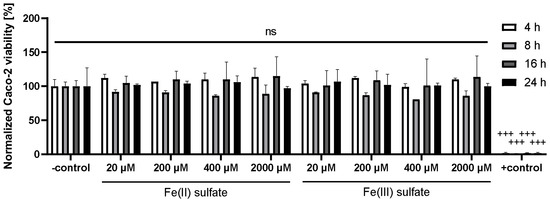
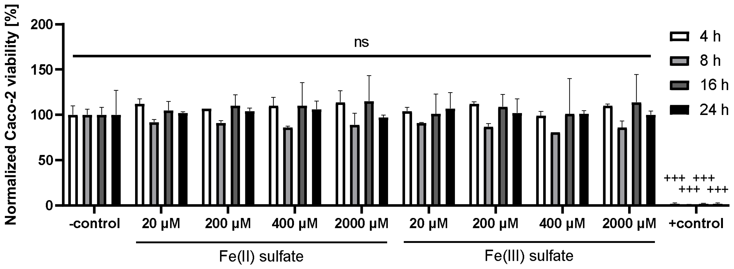
The MTT assay was used to assess the effects of Fe(II) and Fe(III) sulfate on differentiated Caco-2 cells. No significant differences in Caco-2 viability were observed between Fe(II) and Fe(III), or across iron concentrations ranging from 20 to 2000 µM, when normalized to the untreated control (Figure 1).
Figure 1.
Toxicity of Fe(II) and Fe(III) in Caco-2 cells. Viability was analyzed using MTT assay [31] and normalized to control (mean ± SEM of biological replicates (nBIO) = 2, technical replicates (nTECH) = 3). Caco-2 cells were incubated with 0–2000 μM Fe(II) or Fe(III) sulfate for 4 h, 8 h, 16 h, and 24 h in MEM (phenol red, P/S 1%). Statistical significance is indicated (+++ p < 0.001; two-way ANOVA with Bonferroni post hoc test).
Incubation with iron for 4–24 h did not result in statistically significant differences (p > 0.05) compared to the control. Overall, iron exposure in MEM did not affect Caco-2 viability.
3.2. Iron Bioavailability of Fe(II) and Fe(III) Sulfate
Ferritin expression in Caco-2 cells was measured following incubation with Fe(II) and Fe(III) sulfate to evaluate iron bioavailability under model conditions.
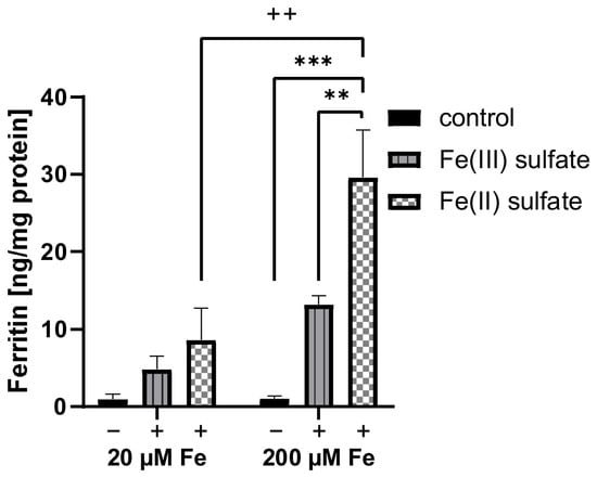
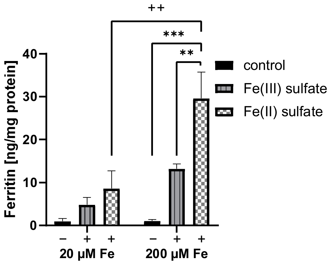
At 20 µM iron concentrations, Fe(II) induced an 80% higher ferritin expression after 24 h, with a mean value of 9 ± 4 ng ferritin/mg TP compared to 5 ± 2 ng ferritin/mg TP for Fe(III). A significantly higher iron bioavailability was observed at 200 µM supplementation, with ferritin levels of 30 ± 6 and 13 ± 1 ng ferritin/mg TP for Fe(II) and Fe(III), respectively, corresponding to a 130% increase for Fe(II) relative to Fe(III). These results demonstrate consistently higher iron bioavailability of Fe(II) compared to Fe(III) at both low and high iron concentrations (Figure 2).
Figure 2.
Bioavailability of Fe(II) and Fe(III) in Caco-2 cells. Ferritin formation (ng/mg TP) after incubation of Caco-2 cells with low (20 µM) and high (200 µM) iron concentrations in MEM (phenol red, P/S 1%) (mean ± SEM of nBIO = 3, nTECH = 2, nFerritin Assay = 2, nBCA Assay = 3). Statistically significant differences are indicated (**/++ p < 0.01, *** p < 0.001; two-way ANOVA with Bonferroni post hoc test).
3.3. Iron Bioaccessibility and Bioavailability of Bell Pepper After In Vitro Digestion
The compatibility of different mucin-enhanced in vitro digestion models with the Caco-2 cells was investigated to establish a combined system for assessing iron bioavailability from a dry bell pepper matrix. For this, three digestion models, DIN [32,33], DIN-lite (derived from [32,33]), and DIN+G (derived from [32,33,34]), given in Table 1, were applied to the bell pepper in-house control (matrix-matched reference in in vitro digestion).
Iron bioaccessibility of bell pepper pericarp (in-house control) differed between the digestion models. Soluble iron content in the bioaccessible fraction was quantified by microwave-assisted acid digestion followed by analysis of the iron content via DRC-Q-ICP-MS. Bioaccessibility was calculated by subtracting the iron content of the corresponding sample-omitted digest (blank digest) and normalizing to the iron content of the dry bell pepper in-house control.
ICP-MS results were validated using the NIST 1547 standard reference material, yielding an iron content of 206 ± 14 mg/kg consistent with the certified values [38]. The in-house control contained 57 ± 3 mg Fe/kg dry mass (DM), corresponding to 0.3 mg Fe/100 g wet mass (WM) based on a water content of the in-house control of 95.2 ± 0.6% (HR73 Moisture Analyzer, Mettler-Toledo GmbH, Gießen, Germany). These values are consistent with literature reports [39,40].
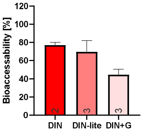
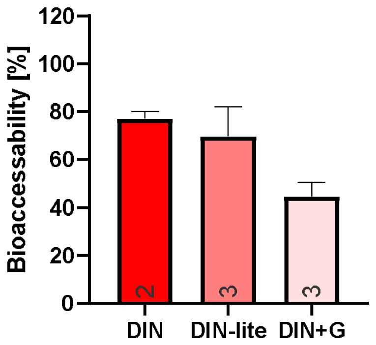
Iron bioaccessibility of the in-house control was 45 ± 8%, 70 ± 16%, and 77 ± 3% for DIN+G, DIN-lite, and DIN, respectively (Figure 3). Although not statistically significant, the 25% lower bioaccessibility observed for DIN+G (which utilizes fewer digestive enzymes) compared to DIN-lite is noteworthy (Figure 3).
Figure 3.
Iron bioaccessibility of bell pepper following different digestion protocols. Soluble iron fraction released from bell pepper (in-house control) after digestion with DIN, DIN-lite, and DIN+G, corrected for matching blank digests (mean ± SEM, nTECH indicated in the figure). No statistically significant differences were observed (one-way ANOVA with Bonferroni post hoc test).
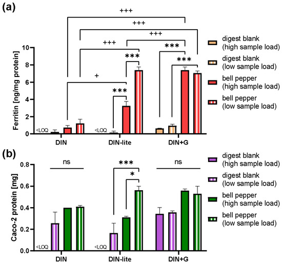
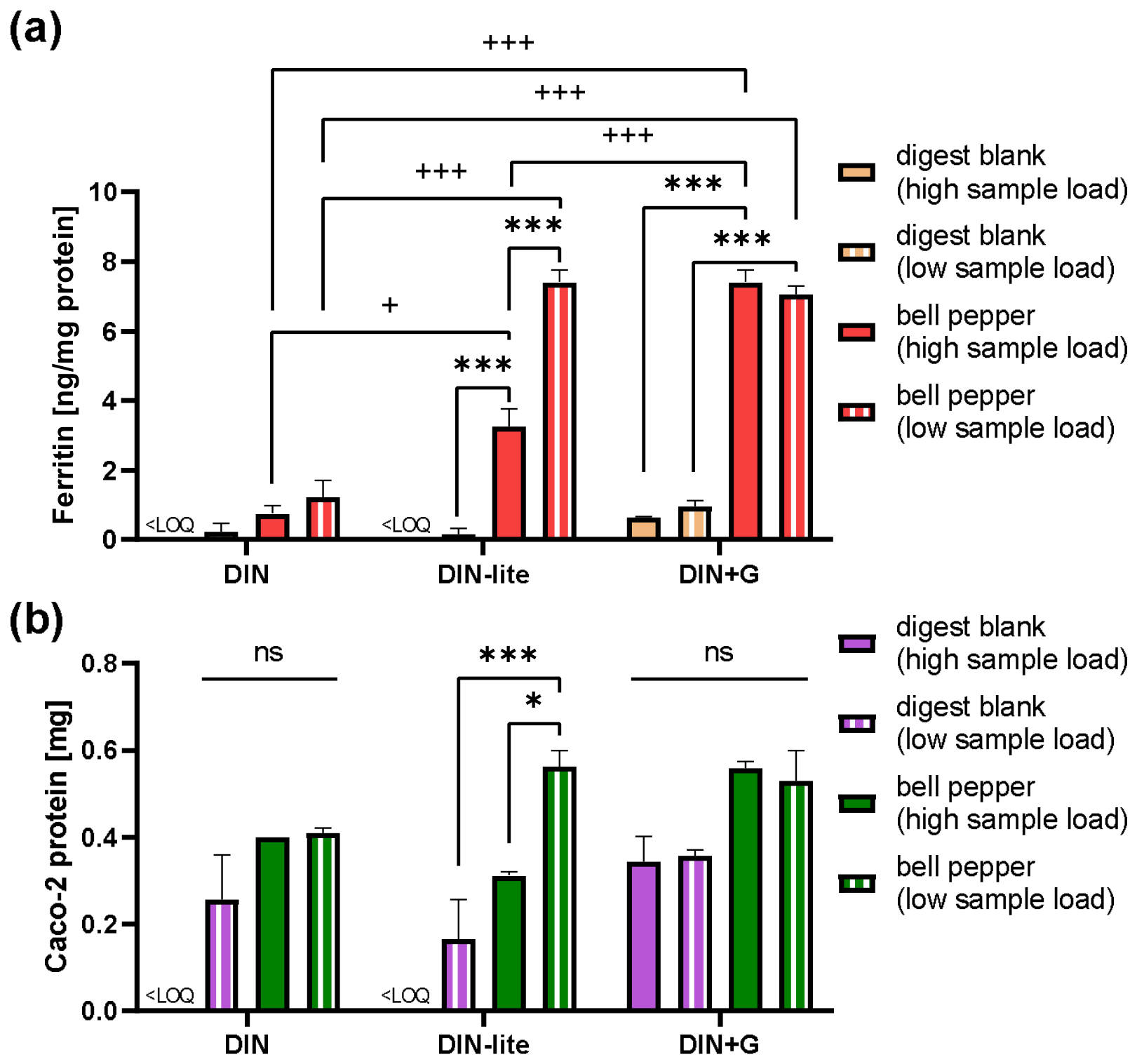
The impact of the three in vitro digestion models on ferritin expression and TP content of Caco-2 cells was assessed after 24 h incubation (Figure 4). For DIN, low ferritin levels were observed at both high (43% V/V in MEM) and low (27% V/V in MEM) sample loads, with values of 0.8 ± 0.2 and 1.2 ± 0.5 ng ferritin/mg TP, respectively. Blank digests induced even lower (low-load) or non-quantifiable (high-load) ferritin levels (Figure 4). A strong reduction in TP content was also observed, indicating substantial loss of Caco-2 cells, likely due to the high content of proteolytic enzymes such as trypsin. This was further confirmed by visual inspection.
Figure 4.
Iron bioavailability of bell pepper after different digestion protocols. (a) Ferritin formation by Caco-2 cells after incubation with DIN, DIN-lite, and DIN+G in-house control digests at low (27% V/V) and high (43% V/V) sample load (mean ± SEM of nBIO = 2, nTECH = 2, nFerritin Assay = 3). (b) Total cellular protein after incubation (mean ± SEM of nBIO = 2, nTECH = 2, nBCA Assay = 3). Statistical significance is indicated (ns not significant, */+ p < 0.05, ***/+++ p < 0.001; two-way ANOVA with Bonferroni post hoc test).
Omission of trypsin in DIN-lite (Table 1) resulted in an increased ferritin expression to 3.3 ± 0.5 and 7.4 ± 0.4 ng ferritin/mg TP for high and low sample load, respectively. However, significant cell loss and reduced TP content were still observed at high sample load (Figure 4).
To further enhance cellular compatibility and assay reproducibility, the concentrations of digestive enzymes and bile extract were reduced compared to DIN-lite, in accordance with a model described by Glahn in 2022 [34]; this modified model was designated DIN+G (Table 1). Although iron bioaccessibility of DIN+G was 25% lower with 45 ± 8% than for DIN-lite (Figure 3), at low sample load bioavailability was reduced by only 4% (Figure 4). The slight, non-significant decrease in iron bioavailability of DIN+G despite a markedly lower bioaccessibility implies the existence of factors other than the soluble iron content impacting its bioavailability.
Notably, increasing the DIN+G digest sample load by 59% (high vs. low sample load) resulted in only a 10% increase in ferritin expression (7.8 ± 0.6 ng ferritin/mg TP), indicating a limited effect of higher substrate concentration on cellular iron uptake (Figure 4).
For the incubation (24 h) of Caco-2 cells with DIN+G digests, the cells responded with a relatively high ferritin formation considering the reduced apical sample content of 27% V/V (low-load) compared to 43% V/V (high-load). The DIN+G digests of bell pepper on a differentiated Caco-2 monolayer resulted in a non-significantly lower ferritin expression at low apical sample load (7.1 ± 0.2 ng ferritin/mg TP) compared to the most responsive (DIN+G, high-load). As the DIN+G model was cell-compatible for in vitro digestion of bell pepper, it was therefore selected for the investigation of biofortified bell pepper cultivars.
3.4. Iron Bioavailability of Biofortified Bell Pepper
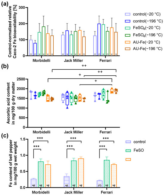
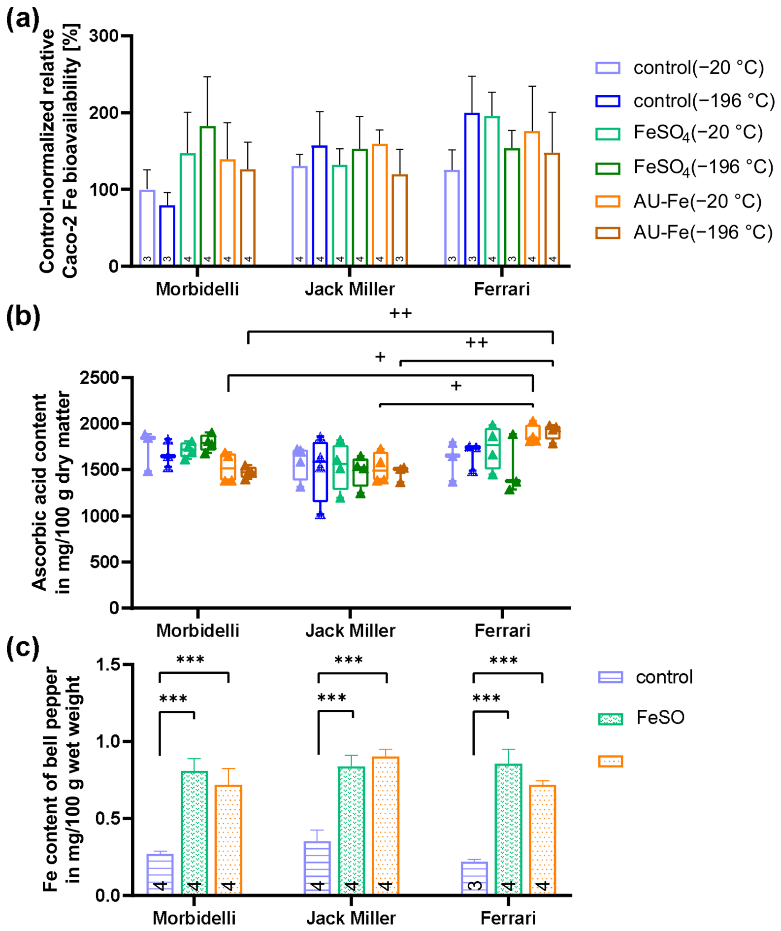
Three bell pepper cultivars (‘Morbidelli’, ‘Jack Miller’, and ‘Ferrari’) were foliar-fertilized with Fe(II) sulfate or AU-Fe to investigate the effect of iron biofortification on iron bioavailability, as well as the influence of post-harvest freezing temperature (−20 °C and −196 °C) on iron bioavailability and ascorbic acid content of the pericarp (Figure 5).
Figure 5.
The effect of foliar fertilization on iron bioavailability and content in bell pepper. (a) Iron bioavailability, assessed as ferritin content in Caco-2 cells (nBIO = 3), after incubation with DIN+G in vitro digests (27% V/V) of foliar iron-fertilized bell pepper cultivars frozen at −20 °C and −196 °C. Ferritin values (n = 2) were normalized to total protein (n = 3) and sample weight (approximately 0.50 g). These ferritin results were then normalized to ferritin levels induced by a co-incubated and co-digested in-house control as matrix-matched reference. Normalized results are expressed relative to the mean of the ‘Morbidelli’ control (−20 °C, set to 100%). Bars show mean ± SEM of plant replicates (nPLT, indicated in the figure). (b) Ascorbic acid content of iron-fertilized bell pepper (box plots of nPLT (indicated in 4a), nTECH = 2). (c) Iron content after fertilization (mean ± SEM of nPLT). Statistical significance is indicated (+ p < 0.05, ++ p < 0.01, *** p < 0.001; two-way ANOVA with Bonferroni post hoc test).
Unfertilized controls showed similar iron content across cultivars with 0.27 ± 0.02, 0.35 ± 0.07, and 0.22 ± 0.02 mg Fe/100 g WM for ‘Morbidelli’, ‘Jack Miller’, and ‘Ferrari’, respectively. Both fertilizer treatments increased iron content to comparable levels: Fe(II) sulfate to 0.81–0.86 mg Fe/100 g WM (maximum biofortification factor 3.9× for ‘Ferrari’) and AU-Fe to 0.72–0.90 mg Fe/100 g WM (Figure 5c).
However, the elevated iron content did not consistently translate into increased iron bioavailability (Figure 5a). A tendency for improved bioavailability was observed only in ‘Morbidelli’, with relative bioavailability increases of up to 2.0× for Fe(II) sulfate and 1.4× for AU-Fe. No statistically significant effect on iron bioavailability after biofortification was observed for any of the tested cultivars (Figure 5a).
Freezing temperature (−20 °C and −196 °C) had no detectable impact on iron bioavailability or ascorbic acid content of bell pepper pericarp (Figure 5b). Ascorbic acid content was highest for ‘Ferrari’ in response to AU-Fe treatment (1786–2031 mg/100 g DM), significantly exceeding levels in ‘Morbidelli’ (1383–1688 mg/100 g DM) and ‘Jack Miller’ (1365–1728 mg/100 g DM). Otherwise, ascorbic acid levels were comparable across samples (Figure 5b).
4. Discussion
Agronomic iron biofortification using foliar fertilization has been put forward as an economical, environmentally friendly, and feasible approach to reduce DID(A) in vulnerable groups of the population [41,42]. In bell pepper, the present study demonstrated that iron biofortification of the pericarp can be achieved by foliar spraying, with biofortification factors of up to 3.9-fold (Figure 5c), although the application resulted in visible fruit damage, rendering the fruits unmarketable. Wala et al. recently reported that iron fertilization by soil application of iron chelates increased the iron content of bell pepper fruits by up to 1.7-fold, with no mention of phytotoxic effects [39]. Although agronomic approaches to increase the iron content have also shown promise in other vegetables, such as members of the Brassicaceae family [43], as well as in staple crops such as wheat [44] and maize [45], assessment of total iron content alone is not sufficient for evaluating the nutritional value. Consequently, three in vitro digestion models for bell pepper pericarp, assessing their effect on iron bioaccessibility and bioavailability, were evaluated, with potential implications for future iron bioavailability studies in other vegetable matrices.
First, the ferritin response of differentiated Caco-2 cell monolayers following incubation with Fe(II) and Fe(III) model solutions was characterized. Supplementation with Fe(II) at 20 µM and 200 µM resulted in 80% and 130% increased ferritin expression, respectively, compared to Fe(III). These results are consistent with previous reports showing that Fe(II) is more bioavailable than Fe(III) [12,13].
Next, the iron bioaccessibility and bioavailability obtained with the DIN, DIN-lite, and DIN+G digestion protocols were compared. Iron bioaccessibility of the in-house control was 45 ± 8%, 70 ± 16%, and 77 ± 3% for DIN+G, DIN-lite, and DIN, respectively (Figure 3). These findings indicate that, within the range of digestion conditions tested here, increasing concentrations of digestive enzymes and bile salts were associated with greater release of iron from the bell pepper pericarp matrix into the bioaccessible fraction.
For comparison, Suliburska et al. [46] reported bioaccessible iron fractions of 5.8–32.1% for cereal groats, 14.2–21.4% for rice, 9.4–32.6% for pulses, and 2.0–51.1% for nuts after enzymatic in vitro digestion adhering to [47]. Performing in vitro digestion (adhering to [48]), similar to our DIN+G model, bioaccessible iron values of 56%/42%, 19%/38%, and 45%/36% were reported for mature/microgreen fenugreek, rocket, and broccoli, respectively [49]. For raw green bell pepper, 12.2% bioaccessible iron was reported by Rodriguez-Ramiro et al. [22] after in vitro digestion adhering to [48]. This value is lower than the 45 ± 8% observed here for raw red bell pepper with the similar model DIN+G. Possible reasons include differences in cultivar, ripening stage, tissue composition, and digestion protocol. At present, however, the available data are too limited to draw firm conclusions, and additional studies are needed to better define the range of bioaccessible iron released from bell pepper pericarp.
When iron bioavailability was assessed after digestion, DIN+G resulted in only a non-significantly lower ferritin response than DIN-lite, despite its 25% lower iron bioaccessibility. Thus, the lower bioaccessible iron content of DIN+G digests did not translate into a substantial reduction of cellular iron uptake, as measured by ferritin formation in Caco-2 cells (Figure 4). This observation supports the view that iron bioaccessibility alone is not a sufficient predictor of iron bioavailability. Rather, iron uptake appears to depend on the overall composition of the digested matrix, particularly the balance of absorption-promoting and absorption-inhibiting constituents [50].
A plausible explanation is that relevant matrix components affecting iron uptake, such as ascorbic acid [14,50,51] and polyphenols [20,39,51], were released from the plant matrix to a similar extent by DIN-lite and DIN+G, thereby limiting differences in bioavailability despite differences in soluble iron concentration. The present data suggest that increasing the bioaccessible non-heme iron fraction does not necessarily increase bioavailability if the relative abundance of promoters and inhibitors remains unchanged. Further work is therefore needed to clarify how matrix composition influences the relationship between iron bioaccessibility and iron bioavailability.
Previous studies showed that neither total iron content nor iron bioaccessibility reliably predicts iron bioavailability in vegetables [22,49]. For example, measuring ferritin formation in Caco-2 cells, Khoja et al. reported similar iron bioavailability for in vitro digested broccoli and rocket, although iron content was higher in rocket, both as a microgreen (33%) and as a mature plant (140%), compared to the broccoli equivalent [49]. In the same study, mature fenugreek exhibited 1.4-fold higher iron bioaccessibility than microgreen fenugreek, whereas the microgreen fenugreek digests showed approximately 4-fold higher iron bioavailability in Caco-2 cells [49]. Furthermore, Rodriguez-Ramiro et al. showed that iron bioaccessibility (11.8%) of boiled broccoli and boiled curly kale was similar to iron bioaccessibility (9.7%), while iron bioavailability from boiled broccoli was 2.9-fold higher than from boiled kale [22]. While the overall plant composition with respect to promoters and inhibitors of iron bioavailability certainly influenced Caco-2 cell iron uptake and ferritin formation in the study by Rodriguez-Ramiro et al. [22], polyphenols are likely a major group of inhibitory compounds in kale and broccoli and have been reported to occur at similar levels in both vegetables in another study [52]. It is plausible that a greater loss of organic acids (which promote iron absorption) as well as ascorbic acid oxidation in particular, contributed to the lower bioavailability of kale compared to broccoli after boiling [22]. This may be explained by the porous leaf structure of kale, which likely facilitated greater leaching, in contrast to the more robust, stem-like structure of broccoli.
In the present study, it was specifically investigated whether agronomic biofortification via foliar iron fertilization increases the in vitro iron bioavailability of bell pepper pericarp. Fruits from multiple and randomly distributed pepper plants grown in a greenhouse setting were evaluated for each cultivar and treatment. Among the three cultivars tested, only the pericarp of the bell pepper cultivar ‘Morbidelli’ showed increased iron bioavailability after biofortification with Fe(II) sulfate and AU-Fe. However, this increase was modest compared to the increase in total iron content and was not statistically significant. Moreover, the relative iron bioavailability of the biofortified ‘Morbidelli’ bell peppers remained within the range observed for the untreated ‘Ferrari’ and ‘Jack Miller’ cultivars. Previous studies on the iron bioavailability in biofortified edible plants are limited to cereal crops such as pearl millet and wheat [53], and legumes, including beans, peas, and lentils [53,54,55,56]. Increased iron bioavailability has been reported for pearl millet, with or without high-β-carotene sweet potato, and for cowpeas. To our knowledge, no previous study has characterized the iron bioavailability of iron-biofortified bell pepper.
The ascorbic acid concentrations measured in the three cultivars correspond to roughly 50–100 mg/100 g WM, at a water content of approximately 95%, which is in line with previous studies [57,58,59]. The present findings demonstrate that in vitro iron bioavailability in bell pepper depends not only on iron content, but also on matrix composition. Because ascorbic acid varied only modestly between cultivars, it is unlikely to fully explain the different cultivar-dependent tendencies in bioavailability observed here. Nevertheless, ascorbic acid performs multiple and vital functions in non-heme iron absorption. Firstly, it reduces Fe(III) to Fe(II), thereby facilitating DMT1-mediated uptake into enterocytes [60], where it triggers the cytosolic IRE-IRP (iron responsive element-iron regulatory protein) system. In turn, this leads to mRNA translation of both ferritin light and heavy chains by freeing the IRE in their 5′-untranslated region (UTR) from the IRPs through formation of iron-sulfur clusters in the IRPs [61]. Ascorbate has also been implicated in the enzymatic reduction of Fe(III) via duodenal cytochrome b at the brush border prior to uptake by DMT1 [14]. Finally, direct uptake of Fe(III) ascorbate complexes has been discussed, although a defined uptake mechanism remains to be established [62].
5. Conclusions
In this study, we showed that the mucin-enhanced DIN+G in vitro digestion model is a viable approach for iron bioavailability testing of bell pepper. Furthermore, a more pronounced iron bioaccessibility after in vitro digestion does not necessarily translate into improved iron bioavailability.
Overall, little is known about the relationship between agronomic iron biofortification and the bioavailability of non-heme iron in vegetables. In the present study, successful agronomic biofortification of bell pepper did not lead to a corresponding increase in iron uptake in vitro. These findings call into question whether increasing iron content alone is sufficient to improve the nutritional value of bell pepper with respect to iron supply. Further studies are required to confirm these findings in iron-biofortified bell pepper, and to evaluate agronomic iron biofortification across different vegetable species for alleviating iron deficiency.
Author Contributions
Conceptualization, H.H., D.D. and S.R.; methodology, B.T. and M.K.; formal analysis, B.T.; investigation, B.T., M.K., A.K. and N.U.; resources, H.H., D.D. and S.R.; data curation, B.T.; writing—original draft preparation, B.T.; writing—review and editing, H.H., C.K. and S.R.; visualization, B.T.; supervision, C.K.; project administration, B.T. and E.S.; funding acquisition, H.H., D.D. and S.R. All authors have read and agreed to the published version of the manuscript.
Funding
This research was funded by the German Federal Ministry of Research, Technology and Space (BMFTR; grant number: 031B1380A/B) and was conducted within the project EiBiG (“Enhancing the health value of vegetables by increasing the bioavailable iron content”). H.H. thanks the Foundation for Food Safety and Consumer Protection (Stiftung für Lebensmittelsicherheit und Verbraucherschutz) for their generous financial support.
Institutional Review Board Statement
Not applicable.
Informed Consent Statement
Not applicable.
Data Availability Statement
The original contributions presented in this study are included in the article. Further inquiries can be directed to the corresponding author.
Acknowledgments
During the preparation of this study, the author(s) used deepl.com, consensus.app, and ChatGPT Edu (version 5.5) for the purposes of correcting grammatical errors and identifying relevant literature. The authors have reviewed and edited the output and take full responsibility for the content of this publication.
Conflicts of Interest
The authors declare no conflicts of interest.
Abbreviations
The following abbreviations are used in this manuscript:
| AU-Fe | Amino Ultra®Fe-20 |
| BSA | bovine serum albumin |
| DAD | diode-array detector |
| DID | dietary iron deficiency |
| DIDA | dietary iron deficiency anemia |
| DM | dry mass |
| DMEM | Dulbecco’s modified Eagles medium |
| DMT1 | divalent metal transporter 1 |
| DPBS | Dulbecco’s phosphate-buffered saline |
| DRC | dynamic reaction cell |
| EBSS | Earle’s balanced salt solution |
| EDTA | ethylenediaminetetraacetic acid |
| FBS | fetal bovine serum |
| GBD | Global Burden of Disease |
| HPLC | high performance liquid chromatography |
| ICP | inductively coupled plasma |
| ID | iron deficiency |
| IRE | iron responsive element |
| IRP | iron regulatory protein |
| MEM | minimum essential medium |
| MS | mass spectrometry |
| MTT | 3-(4,5-Dimethylthiazol-2-yl)-2,5-diphenyltetrazolium bromide |
| nBIO | biological replicate |
| nPLT | plant replicate |
| nTECH | technical replicate |
| SKU | stock keeping unit |
| TCEP | tris(2-carboxyethyl)phosphine |
| TP | total protein |
| UTR | untranslated region |
| WM | wet mass |
References
- Lee, S.; Son, Y.; Hwang, J.; Kim, M.S.; Il Shin, J.; Yon, D.K.; Kassebaum, N.J. Global, regional and national burden of dietary iron deficiency from 1990 to 2021: A Global Burden of Disease study. Nat. Med. 2025, 31, 1809–1829. [Google Scholar] [CrossRef] [Scilit]
- WHO. Anaemia. Available online: https://www.who.int/news-room/fact-sheets/detail/anaemia (accessed on 30 October 2025).
- Monitoring Surveillance Nutrition and Food Safety. WHO Global Anaemia Estimates: Key Findings, 2025. World Health Organization, 15 August 2025. Available online: https://www.who.int/publications/i/item/9789240113930 (accessed on 9 February 2026).
- Lewis, E.D.; Ortega, E.F.; Dao, M.C.; Barger, K.; Mason, J.B.; Leong, J.M.; Osburne, M.S.; Magoun, L.; Nepveux, V.F.J.; Chishti, A.H.; et al. Safe and effective delivery of supplemental iron to healthy adults: A two-phase, randomized, double-blind trial—The safe iron study. Front. Nutr. 2023, 10, 1230061. [Google Scholar] [CrossRef] [Scilit] [PubMed]
- Jalal, C.S.; De-Regil, L.M.; Pike, V.; Mithra, P. Fortification of condiments and seasonings with iron for preventing anaemia and improving health. Cochrane Database Syst. Rev. 2023, 9, CD009604. [Google Scholar] [CrossRef] [Scilit] [PubMed]
- Buturi, C.V.; Mauro, R.P.; Fogliano, V.; Leonardi, C.; Giuffrida, F. Mineral Biofortification of Vegetables as a Tool to Improve Human Diet. Foods 2021, 10, 223. [Google Scholar] [CrossRef] [Scilit] [PubMed]
- Benvenuti, L.; Sette, S.; de Santis, A.; Riso, P.; Petroni, K.; Crosatti, C.; Losa, A.; Martone, D.; Martini, D.; Cattivelli, L.; et al. Simulation of Daily Iron Intake by Actual Diet Considering Future Trends in Wheat and Rice Biofortification, Environmental, and Dietary Factors: An Italian Case Study. Nutrients 2024, 16, 4097. [Google Scholar] [CrossRef] [Scilit] [PubMed]
- Haider, L.M.; Schwingshackl, L.; Hoffmann, G.; Ekmekcioglu, C. The effect of vegetarian diets on iron status in adults: A systematic review and meta-analysis. Crit. Rev. Food Sci. Nutr. 2018, 58, 1359–1374. [Google Scholar] [CrossRef] [Scilit] [PubMed]
- Neufingerl, N.; Eilander, A. Nutrient Intake and Status in Adults Consuming Plant-Based Diets Compared to Meat-Eaters: A Systematic Review. Nutrients 2021, 14, 29. [Google Scholar] [CrossRef] [Scilit] [PubMed]
- Kovaleva, E.G.; Lipscomb, J.D. Versatility of biological non-heme Fe(II) centers in oxygen activation reactions. Nat. Chem. Biol. 2008, 4, 186–193. [Google Scholar] [CrossRef] [Scilit] [PubMed]
- Alvarez-Hernandez, X.; Nichols, G.M.; Glass, J. Caco-2 cell line: A system for studying intestinal iron transport across epithelial cell monolayers. Biochim. Biophys. Acta 1991, 1070, 205–208. [Google Scholar] [CrossRef] [Scilit] [PubMed]
- He, W.; Feng, Y.; Li, X.; Wei, Y.; Yang, X. Availability and toxicity of Fe(II) and Fe(III) in Caco-2 cells. J. Zhejiang Univ. Sci. B 2008, 9, 707–712. [Google Scholar] [CrossRef] [Scilit] [PubMed]
- He, W.; Li, X.; Ding, K.; Li, Y.; Li, W. Ascorbic Acid can Reverse the Inhibition of Phytic Acid, Sodium Oxalate and Sodium Silicate on Iron Absorption in Caco-2 cells. Int. J. Vitam. Nutr. Res. 2018, 88, 65–72. [Google Scholar] [CrossRef] [Scilit] [PubMed]
- Ganasen, M.; Togashi, H.; Takeda, H.; Asakura, H.; Tosha, T.; Yamashita, K.; Hirata, K.; Nariai, Y.; Urano, T.; Yuan, X.; et al. Structural basis for promotion of duodenal iron absorption by enteric ferric reductase with ascorbate. Commun. Biol. 2018, 1, 120. [Google Scholar] [CrossRef] [Scilit] [PubMed]
- Salovaara, S.; Sandberg, A.-S.; Andlid, T. Organic acids influence iron uptake in the human epithelial cell line Caco-2. J. Agric. Food Chem. 2002, 50, 6233–6238. [Google Scholar] [CrossRef] [Scilit] [PubMed]
- Liziczai, M.; Fuchs, A.; Manatschal, C.; Dutzler, R. Structural basis for metal ion transport by the human SLC11 proteins DMT1 and NRAMP1. Nat. Commun. 2025, 16, 761. [Google Scholar] [CrossRef] [Scilit] [PubMed]
- Shawki, A.; Engevik, M.A.; Kim, R.S.; Knight, P.B.; Baik, R.A.; Anthony, S.R.; Worrell, R.T.; Shull, G.E.; Mackenzie, B. Intestinal brush-border Na+/H+ exchanger-3 drives H+-coupled iron absorption in the mouse. Am. J. Physiol. Gastrointest. Liver Physiol. 2016, 311, G423–G430. [Google Scholar] [CrossRef] [Scilit] [PubMed]
- Bretti, C.; Cigala, R.M.; Lando, G.; Milea, D.; Sammartano, S. Sequestering ability of phytate toward biologically and environmentally relevant trivalent metal cations. J. Agric. Food Chem. 2012, 60, 8075–8082. [Google Scholar] [CrossRef] [Scilit] [PubMed]
- Abioye, A.I.; Okuneye, T.A.; Odesanya, A.-M.O.; Adisa, O.; Abioye, A.I.; Soipe, A.I.; Ismail, K.A.; Yang, J.F.; Fasehun, L.-K.; Omotayo, M.O. Calcium Intake and Iron Status in Human Studies: A Systematic Review and Dose-Response Meta-Analysis of Randomized Trials and Crossover Studies. J. Nutr. 2021, 151, 1084–1101. [Google Scholar] [CrossRef] [Scilit] [PubMed]
- McGee, E.J.T.; Diosady, L.L. Prevention of iron-polyphenol complex formation by chelation in black tea. LWT 2018, 89, 756–762. [Google Scholar] [CrossRef] [Scilit]
- Bohn, T.; Carriere, F.; Day, L.; Deglaire, A.; Egger, L.; Freitas, D.; Golding, M.; Le Feunteun, S.; Macierzanka, A.; Menard, O.; et al. Correlation between in vitro and in vivo data on food digestion. What can we predict with static in vitro digestion models? Crit. Rev. Food Sci. Nutr. 2018, 58, 2239–2261. [Google Scholar] [CrossRef] [Scilit] [PubMed]
- Rodriguez-Ramiro, I.; Dell’Aquila, C.; Ward, J.L.; Neal, A.L.; Bruggraber, S.F.A.; Shewry, P.R.; Fairweather-Tait, S. Estimation of the iron bioavailability in green vegetables using an in vitro digestion/Caco-2 cell model. Food Chem. 2019, 301, 125292. [Google Scholar] [CrossRef] [Scilit] [PubMed]
- Brodkorb, A.; Egger, L.; Alminger, M.; Alvito, P.; Assunção, R.; Ballance, S.; Bohn, T.; Bourlieu-Lacanal, C.; Boutrou, R.; Carrière, F.; et al. INFOGEST static in vitro simulation of gastrointestinal food digestion. Nat. Protoc. 2019, 14, 991–1014. [Google Scholar] [CrossRef] [Scilit] [PubMed]
- Mahler, G.J.; Shuler, M.L.; Glahn, R.P. Characterization of Caco-2 and HT29-MTX cocultures in an in vitro digestion/cell culture model used to predict iron bioavailability. J. Nutr. Biochem. 2009, 20, 494–502. [Google Scholar] [CrossRef] [Scilit] [PubMed]
- Einhorn, V.; Haase, H.; Maares, M. Interaction and competition for intestinal absorption by zinc, iron, copper, and manganese at the intestinal mucus layer. J. Trace Elem. Med. Biol. 2024, 84, 127459. [Google Scholar] [CrossRef] [Scilit] [PubMed]
- Maares, M.; Einhorn, V.; Behrendt, J.; Marczynski, M.; Schüßler, C.; Lieleg, O.; Haase, H. Investigation of competitive binding of the essential trace elements zinc, iron, copper, and manganese by gastrointestinal mucins and the effect on their absorption in vitro. J. Nutr. Biochem. 2025, 144, 109983. [Google Scholar] [CrossRef] [Scilit] [PubMed]
- Jin, F.; Welch, R.; Glahn, R. Moving toward a more physiological model: Application of mucin to refine the in vitro digestion/Caco-2 cell culture system. J. Agric. Food Chem. 2006, 54, 8962–8967. [Google Scholar] [CrossRef] [Scilit] [PubMed]
- INTERMAG sp. z o.o. AMINO ULTRA Fertilizers for New Challenges. Available online: https://pdf.agriexpo.online/de/pdf-en/intermag-sp-z-oo/amino-ultra-leaflet/171917-31692.html#open130143 (accessed on 2 October 2023).
- Sambuy, Y.; de Angelis, I.; Ranaldi, G.; Scarino, M.L.; Stammati, A.; Zucco, F. The Caco-2 cell line as a model of the intestinal barrier: Influence of cell and culture-related factors on Caco-2 cell functional characteristics. Cell Biol. Toxicol. 2005, 21, 1–26. [Google Scholar] [CrossRef] [Scilit] [PubMed]
- Fogh, J.; Fogh, J.M.; Orfeo, T. One hundred and twenty-seven cultured human tumor cell lines producing tumors in nude mice. J. Natl. Cancer Inst. 1977, 59, 221–226. [Google Scholar] [CrossRef] [Scilit] [PubMed]
- Maares, M.; Keil, C.; Thomsen, S.; Günzel, D.; Wiesner, B.; Haase, H. Characterization of Caco-2 cells stably expressing the protein-based zinc probe eCalwy-5 as a model system for investigating intestinal zinc transport. J. Trace Elem. Med. Biol. 2018, 49, 296–304. [Google Scholar] [CrossRef] [Scilit] [PubMed]
- DIN 19738:2004-07; Soil Quality-Absorption Availability of Organic and Inorganic Pollutants from Contaminated Soil Material. DIN Media GmbH: Berlin, Germany, 2004. Available online: https://www.dinmedia.de/de/norm/din-19738/72163151 (accessed on 10 December 2025).
- Versantvoort, C.H.M.; Oomen, A.G.; van de Kamp, E.; Rompelberg, C.J.M.; Sips, A.J.A.M. Applicability of an in vitro digestion model in assessing the bioaccessibility of mycotoxins from food. Food Chem. Toxicol. 2005, 43, 31–40. [Google Scholar] [CrossRef] [Scilit] [PubMed]
- Glahn, R.P. The Caco-2 Cell Bioassay for Measurement of Food Iron Bioavailability. J. Vis. Exp. 2022, 182, e63859. [Google Scholar] [CrossRef] [Scilit] [PubMed]
- Maares, M.; Keil, C.; Pallasdies, L.; Schmacht, M.; Senz, M.; Nissen, J.; Kieserling, H.; Drusch, S.; Haase, H. Zinc availability from zinc-enriched yeast studied with an in vitro digestion/Caco-2 cell culture model. J. Trace Elem. Med. Biol. 2022, 71, 126934. [Google Scholar] [CrossRef] [Scilit] [PubMed]
- Shekarchi, M.; Khalili, F.E.; Mostofi, Y.; Hajimehdipoor, H. High Performance Liquid Chromatographic Determination of Ascorbic Acid in Brassica oleracea L. Var. Italica Plenck. Biosci. Biotechnol. Res. Asia 2011, 8, 95–100. [Google Scholar] [CrossRef] [Scilit]
- Wechtersbach, L.; Cigić, B. Reduction of dehydroascorbic acid at low pH. J. Biochem. Biophys. Methods 2007, 70, 767–772. [Google Scholar] [CrossRef] [Scilit] [PubMed]
- NIST. Standard Reference Material® 1547 Peach Leaves: Certificate of Analysis. Available online: https://tsapps.nist.gov/srmext/certificates/archives/1547.pdf (accessed on 27 January 2026).
- Wala, M.; Kołodziejek, J.; Sieczyńska, K.; Lasoń-Rydel, M.; Krępska, M. Iron biofortification in four non-heterotic red sweet cultivars of bell pepper. Food Res. Int. 2024, 196, 115050. [Google Scholar] [CrossRef] [Scilit] [PubMed]
- USDA. Peppers, Sweet, Green, Raw (FDC ID: 170427)-Nutrients. Available online: https://fdc.nal.usda.gov/food-details/170427/nutrients (accessed on 27 January 2026).
- Welk, A.-K.; Mehlhose, C.; Daum, D.; Enneking, U. Vegetables with Enhanced Iron Bioavailability-German Consumers’ Perceptions of a New Approach to Improve Dietary Iron Supply. Nutrients 2023, 15, 2291. [Google Scholar] [CrossRef] [Scilit] [PubMed]
- Sharma, S.; Anand, N.; Bindraban, P.S.; Pandey, R. Foliar Application of Humic Acid with Fe Supplement Improved Rice, Soybean, and Lettuce Iron Fortification. Agriculture 2023, 13, 132. [Google Scholar] [CrossRef] [Scilit]
- Di Gioia, F.; Petropoulos, S.A.; Ozores-Hampton, M.; Morgan, K.; Rosskopf, E.N. Zinc and Iron Agronomic Biofortification of Brassicaceae Microgreens. Agronomy 2019, 9, 677. [Google Scholar] [CrossRef] [Scilit]
- Zhou, C.-X.; Zhang, C.-C.; Zhao, Q.-Y.; Yu, B.-G.; Zhang, W.; Chen, X.-P.; Zou, C.-Q. Iron biofortification and yield of wheat grain in response to Fe fertilization and its driving variables: A meta-analysis. Glob. Food Secur. 2024, 40, 100737. [Google Scholar] [CrossRef] [Scilit]
- Kihara, J.; Sileshi, G.W.; Bolo, P.; Mutambu, D.; Senthilkumar, K.; Sila, A.; Devkota, M.; Saito, K. Maize-grain zinc and iron concentrations as influenced by agronomic management and biophysical factors: A meta-analysis. Food Sec. 2024, 16, 1147–1173. [Google Scholar] [CrossRef] [Scilit]
- Suliburska, J.; Krejpcio, Z. Evaluation of the content and bioaccessibility of iron, zinc, calcium and magnesium from groats, rice, leguminous grains and nuts. J. Food Sci. Technol. 2014, 51, 589–594. [Google Scholar] [CrossRef] [Scilit] [PubMed]
- Skibniewska, K.A.; Kozirok, W.; Fornal, L.; Markiewicz, K. In vitro availability of minerals from oat products. J. Sci. Food Agric. 2002, 82, 1676–1681. [Google Scholar] [CrossRef] [Scilit]
- Glahn, R.P.; Lee, O.A.; Yeung, A.; Goldman, M.I.; Miller, D.D. Caco-2 cell ferritin formation predicts nonradiolabeled food iron availability in an in vitro digestion/Caco-2 cell culture model. J. Nutr. 1998, 128, 1555–1561. [Google Scholar] [CrossRef] [Scilit] [PubMed]
- Khoja, K.K.; Buckley, A.; Aslam, M.F.; Sharp, P.A.; Latunde-Dada, G.O. In Vitro Bioaccessibility and Bioavailability of Iron from Mature and Microgreen Fenugreek, Rocket and Broccoli. Nutrients 2020, 12, 1057. [Google Scholar] [CrossRef] [Scilit] [PubMed]
- Piskin, E.; Cianciosi, D.; Gulec, S.; Tomas, M.; Capanoglu, E. Iron Absorption: Factors, Limitations, and Improvement Methods. ACS Omega 2022, 7, 20441–20456. [Google Scholar] [CrossRef] [Scilit] [PubMed]
- Hamed, M.; Kalita, D.; Bartolo, M.E.; Jayanty, S.S. Capsaicinoids, Polyphenols and Antioxidant Activities of Capsicum annuum: Comparative Study of the Effect of Ripening Stage and Cooking Methods. Antioxidants 2019, 8, 364. [Google Scholar] [CrossRef] [Scilit] [PubMed]
- La Fuente, B.d.; López-García, G.; Máñez, V.; Alegría, A.; Barberá, R.; Cilla, A. Antiproliferative Effect of Bioaccessible Fractions of Four Brassicaceae Microgreens on Human Colon Cancer Cells Linked to Their Phytochemical Composition. Antioxidants 2020, 9, 368. [Google Scholar] [CrossRef] [Scilit] [PubMed]
- Gannon, B.M.; Glahn, R.P.; Mehta, S. Iron Bioavailability from Multiple Biofortified Foods Using an In Vitro Digestion, Caco-2 Assay for Optimizing a Cyclical Menu for a Randomized Efficacy Trial. Curr. Dev. Nutr. 2021, 5, nzab111. [Google Scholar] [CrossRef] [Scilit] [PubMed]
- Glahn, R.P.; Wiesinger, J.A.; Lung’aho, M.G. Iron Concentrations in Biofortified Beans and Nonbiofortified Marketplace Varieties in East Africa Are Similar. J. Nutr. 2020, 150, 3013–3023. [Google Scholar] [CrossRef] [Scilit] [PubMed]
- Corrêa, S.R.; Brigide, P.; Vaz-Tostes, M.d.G.; Costa, N.M.B. Cultivars of biofortified cowpea and sweet potato: Bioavailability of iron and interaction with vitamin A in vivo and in vitro. J. Food Sci. 2020, 85, 816–823. [Google Scholar] [CrossRef] [Scilit] [PubMed]
- De Souza Scherrer Medeiros, L.; Grancieri, M.; Sant’Ana, C.T.; de Araujo Santiago, M.C.P.; Da Silva, L.B.; Glahn, R.P.; Costa, N.M.B. The lower content of mineral-complexing compounds favored the in vitro and in vivo iron bioavailability of biofortified cowpeas. J. Funct. Foods 2024, 123, 106601. [Google Scholar] [CrossRef] [Scilit]
- Ignat, T.; Schmilovitch, Z.; Fefoldi, J.; Steiner, B.; Alkalai-Tuvia, S. Non-destructive measurement of ascorbic acid content in bell peppers by VIS-NIR and SWIR spectrometry. Postharvest Biol. Technol. 2012, 74, 91–99. [Google Scholar] [CrossRef] [Scilit]
- Nerdy, N. Determination of Vitamin C in Various Colours of Bell Pepper (Capsicum annuum L.) by Titration Method. Alchemy J. Pen. Kim. 2018, 14, 164. [Google Scholar] [CrossRef] [Scilit]
- Omar, S.A.; Fadala, L.T. Effect of Organic Nutrients and Biofertilizers on Vegetative and Chemo-Physiological Growth of Sweet Pepper (Capsicum annuum L.). IOP Conf. Ser. Earth Environ. Sci. 2025, 1538, 12032. [Google Scholar] [CrossRef] [Scilit]
- Han, O.; Failla, M.L.; Hill, A.D.; Morris, E.R.; Smith, J.C. Reduction of Fe(III) is required for uptake of nonheme iron by Caco-2 cells. J. Nutr. 1995, 125, 1291–1299. [Google Scholar] [CrossRef] [Scilit] [PubMed]
- Galy, B.; Conrad, M.; Muckenthaler, M. Mechanisms controlling cellular and systemic iron homeostasis. Nat. Rev. Mol. Cell Biol. 2024, 25, 133–155. [Google Scholar] [CrossRef] [Scilit] [PubMed]
- Thumser, A.E.; Rashed, A.A.; Sharp, P.A.; Lodge, J.K. Ascorbate enhances iron uptake into intestinal cells through formation of a FeCl3–ascorbate complex. Food Chem. 2010, 123, 281–285. [Google Scholar] [CrossRef] [Scilit]

Disclaimer/Publisher’s Note: The statements, opinions and data contained in all publications are solely those of the individual author(s) and contributor(s) and not of MDPI and/or the editor(s). MDPI and/or the editor(s) disclaim responsibility for any injury to people or property resulting from any ideas, methods, instructions or products referred to in the content. |
© 2026 by the authors. Licensee MDPI, Basel, Switzerland. This article is an open access article distributed under the terms and conditions of the Creative Commons Attribution (CC BY) license.