


Abstract
Organ failure remains one of the foremost medical and socioeconomic challenges of the twenty-first century, with global transplant waiting lists far exceeding the supply of donor organs. Chronic supportive therapies sustain life but do not restore organ function, underscoring an urgent need for curative alternatives. Bioartificial organs represent a major frontier in organ replacement, driven by converging advances in cell biology, biomaterials science, and bioengineering. By integrating living cells or biologically derived matrices with engineered devices or scaffolds, these systems aim to restore functions that purely mechanical supports cannot reproduce. This review examines the principal technological platforms underpinning the field, including cell encapsulation, decellularization and recellularization, three-dimensional bioprinting, organoids, organ-on-chip systems, and xenotransplantation, and discusses their application to kidney, liver, heart, pancreas, and lung replacement. Across organ systems, progress is advancing from experimental proof-of-concept toward modular and increasingly translational platforms, although whole-organ bioengineering remains largely preclinical for the most structurally complex targets. The major unresolved barriers include vascularization, immune compatibility, scalable cell manufacturing, durable function, and stable integration between biological and engineered components. Overall, bioartificial organ engineering is evolving toward clinically relevant therapeutic strategies capable of complementing, bridging, or eventually reducing dependence on donor-organ transplantation.
1. Introduction
Organ failure constitutes one of the most significant medical and socioeconomic challenges of the twenty-first century. The persistent shortage of transplantable organs leaves millions of patients dependent on chronic supportive therapies such as dialysis, mechanical ventilation, and insulin administration, which are life-sustaining but do not restore full organ function [1,2,3,4,5]. According to the Organ Procurement and Transplantation Network (OPTN), as of April 2026, there are more than 121,200 registrations on the U.S. transplant waiting list [6]. Kidney alone accounts for 103,655 registrations (≈85%), followed by liver with 8973 (≈7%), heart with 4100 (≈3%), lung with 1018 (≈1%), pancreas with 834 (≈1%), and combined kidney–pancreas with 2504 (≈2%) [6]. Although these figures derive from the U.S. transplant system, they effectively illustrate the magnitude and organ distribution of unmet transplant need in high-resource healthcare settings and reflect a broader global imbalance between organ demand and supply.
Bioartificial organs seek to address this gap by integrating living biological components, including cells, tissues, or extracellular matrix, with engineered devices or biomaterial scaffolds in order to restore or supplement organ-specific function [7,8,9,10].
Unlike purely mechanical artificial organs, these systems are intended to reproduce complex metabolic, endocrine, and biosynthetic activities that cannot be fully replicated by engineering alone [7,8,9,10].
Their development depends on the convergence of biomaterials science, stem cell biology, physiology, anatomy, and biomedical engineering, and is fundamentally constrained by three interdependent requirements: sustained cell viability and function, biocompatibility, and efficient mass transport of oxygen, nutrients, and metabolic waste [11,12,13]. Over the past decades, the field has expanded from early extracorporeal support systems to increasingly sophisticated strategies that include wearable devices, encapsulated cell therapies, decellularized organ scaffolds, and stem cell-based tissue engineering [14,15,16,17,18,19,20]. This review provides an overview of the principal bioartificial organ technologies and examines their application to the organs that currently dominate transplant demand, namely the kidney, liver, heart, pancreas, and lung (Figure 1). The literature search underlying this work was conducted primarily in PubMed/MEDLINE and Scopus, supplemented by targeted searches in Web of Science and ClinicalTrials.gov. Searches were performed between February 2026 and May 2026, with no lower time limit applied in order to capture foundational studies alongside recent developments. Search terms included combinations of the following: “bioartificial organ”, “bioengineered organ”, “organ-on-chip”, “decellularization”, “recellularization”, “cell encapsulation”, “3D bioprinting”, “organoids”, “xenotransplantation”, “bioartificial kidney”, “bioartificial liver”, “bioartificial pancreas”, “bioartificial heart”, and “bioartificial lung”. Priority was given to peer-reviewed original research articles, systematic reviews, and clinical trial reports published in English. Conference abstracts and non-peer-reviewed materials were excluded. The selection of cited works was guided by clinical relevance, methodological rigor, recency, and representativeness of the main research directions within each topic area.
Figure 1.
Schematic overview of the current landscape of bioartificial organ technologies and their translational evolution in regenerative medicine. Representative strategies targeting the heart, liver, kidney, pancreas, and lung are arranged around a central human silhouette, highlighting the organ-specific application of extracorporeal support systems, hybrid devices, scaffold-based constructs, and tissue-engineering platforms. The figure illustrates the progression from early functional support approaches toward advanced bioengineered systems integrating living cells, biomaterials, bioreactors, and organ-mimetic design principles. This visual framework emphasizes the convergence of regenerative medicine, biomaterials science, and biofabrication in the development of next-generation bioartificial organs. Clinically, these platforms may provide complementary strategies to bridge organ failure, support recovery, or reduce dependence on donor-organ transplantation.
2. Foundational Technologies and Emerging Frontiers
Foundational bioengineering technologies underpin the development of all bioartificial organ systems, providing core platforms that recur across different organ targets. These include cell encapsulation, decellularization and recellularization, three-dimensional bioprinting, organoids, organ-on-chip systems, and xenotransplantation, each of which offers distinct biological and engineering advantages while introducing specific translational limitations (Table 1).
Cell encapsulation involves surrounding living cells with a semipermeable membrane that permits the diffusion of small molecules—including oxygen, glucose, insulin, and metabolic waste—while shielding the encapsulated cells from host immune recognition. This strategy is central to the development of bioartificial endocrine organs, particularly the bioartificial pancreas, in which insulin-producing islet cells must be protected from autoimmune destruction without requiring systemic immunosuppression [21,22,23,24,25]. Three principal encapsulation architectures have been developed: microencapsulation (individual islets in hydrogel capsules of 100–400 µm diameter), macroencapsulation (larger planar or cylindrical devices), and nanoencapsulation (conformal coatings applied directly to the cell surface), each presenting distinct trade-offs between mass transport efficiency, immunoprotection, biocompatibility, and retrievability [21,22,23,24,25]. Hydrogel-based biomaterials—particularly alginate, polyethylene glycol (PEG), and zwitterionic polymers—have been the primary candidates for encapsulation membranes due to their biocompatibility and tunable permeability [26,27,28]. A major research focus has been the engineering of encapsulation materials that minimize the foreign body response, which remains a primary mechanism of device failure in vivo. In this regard, chemically modified alginates and immunomodulatory formulations have shown encouraging results in minimizing fibrotic overgrowth and improving long-term glycemic control in preclinical models [29,30,31].
A complementary scaffold-based strategy is decellularization, defined as the removal of cellular material from a donor organ while preserving the native three-dimensional extracellular matrix (ECM) architecture [32]. The resulting acellular scaffold retains organ-specific structural cues—vascular channels, basement membranes, bile canaliculi, glomerular structures, alveolar compartments—that provide the spatial template for subsequent repopulation with recipient-derived cells [17,32,33,34]. Perfusion decellularization has emerged as the most consistent method for whole-organ scaffold preparation, delivering detergent solutions through the native vasculature to achieve uniform cell removal while preserving the hierarchical vascular architecture [17,34,35,36]. A critical determinant of success is the balance between adequate cellular clearance and preservation of ECM composition and mechanics, as excessive detergent exposure can damage matrix proteins and compromise subsequent recellularization efficiency [37]. Although this approach has been applied successfully to heart, liver, lung, kidney, and pancreas scaffolds in preclinical settings, recellularization remains the principal bottleneck, particularly with respect to the spatial organization of multiple cell types, the generation of sufficient cell numbers for human-scale organs, and the induction of mature functional phenotypes [38,39,40,41,42,43,44,45,46].
An alternative bottom-up approach is three-dimensional bioprinting, which constructs tissue architectures by the layer-by-layer deposition of bioinks composed of cells, biomaterials, and signaling factors [47,48]. Unlike decellularization-based approaches, 3D bioprinting is, in principle, entirely donor-independent and can be personalised to match patient anatomy and cell biology [49,50,51,52]. Major modalities include extrusion-based printing, inkjet printing, stereolithography (SLA/DLP), and laser-assisted bioprinting, each offering different trade-offs between resolution, cell viability, printing speed, and material compatibility [53,54,55]. The fundamental unsolved challenge in 3D bioprinting of solid organs is vascularization, because tissues thicker than about 100–200 µm exceed the diffusion limit for oxygen and nutrients and therefore require an internal perfusable vascular network to remain viable [56,57]. This limitation has been demonstrated in vascularized bioprinting studies showing that thick constructs rapidly lose viability without embedded vessels. In contrast, bioprinted tissues with perfusable channels or prevascular networks can be maintained for prolonged perfusion and support endothelialization and angiogenesis. Recent progress in vascularized bioprinting has been achieved through smart bioinks and sacrificial printing strategies. Notable examples include SWIFT (Sacrificial Writing Into Functional Tissue), in which organ building blocks are embedded in ECM matrices and sacrificial inks are used to create perfusable channel networks, as well as coaxial extrusion and embedded bioprinting approaches that enable endothelialized tubular structures and multiscale vascular architectures [58,59,60,61]. These advances are increasingly supported by artificial intelligence tools for bioink optimization and vascular design, further enhancing the precision and scalability of printed constructs [62].
Complementary biological platforms include organoids and organ-on-chip devices. Organoids are three-dimensional tissue structures generated from pluripotent or adult stem cells that self-organize to recapitulate important aspects of organ-specific cellular composition, architecture, and function [63,64]. They preserve patient-specific genetic and pathophysiological features with significantly higher fidelity than typical two-dimensional cultures, and hence provide enormous advantages for disease modeling and regenerative applications [65,66,67]. They are also of particular interest in bioartificial organ development, as they represent scalable sources of patient-derived functional cells for tissue assembly [68]. In contrast, organ-on-chip systems incorporate living cells into microfluidic devices designed to replicate the chemical, physical, and mechanical milieu of native tissues [69,70,71]. These models have gained renewed relevance with recent regulatory and institutional developments, including increasing FDA (Food and Drug Administration) openness to alternative preclinical platforms and broader support for the validation of organoid- and organ-on-chip-based approaches, which may influence future translational and regulatory pathways [72]. The emergent convergence of these two approaches in organoid-on-chip systems merges the self-organizing complexity of organoids with the engineering control of microfluidic platforms, allowing for improved vascularization, immune-cell interactions, and inter-organ connectivity [69,71,73,74].
While conceptually distinct from scaffold-based bioengineering, xenotransplantation has become increasingly relevant to bioartificial organ development because both fields must solve common immunological bottlenecks—complement regulation, coagulation compatibility, and endothelial protection—that determine the long-term viability of any biological construct in vivo. The transplantation of genetically engineered animal organs into human recipients has demonstrated that large-scale physiological support is feasible, but also highlighted the significant hurdles of innate immunity, complement activation, coagulation incompatibility, and endothelial injury [75,76,77,78,79]. Gene-edited pig kidney and liver grafts have provided measurable functional support in highly selected human settings [75,76,77,78,79,80,81], while genetically modified swine lungs have shown maintained viability without hyperacute rejection in cross-species transplantation models [82]. The results are very informative for bioartificial organ engineering, as the same strategies employed in xenotransplantation, particularly xenoantigen deletion, insertion of human complement-regulatory genes, and modulation of thromboregulatory pathways, can conceptually be applied to decrease the immunogenicity of biological components of hybrid organ constructs. Itoh et al. developed an immunodeficient porcine model that was able to incorporate artificial human vascular grafts [83], and Cravedi et al. elucidated broader immunological mechanisms of biomaterial-host interactions, such as complement activation, macrophage polarization, T-cell recruitment, and humoral immunity [84,85]. Collectively, these data position xenotransplantation as a translationally instructive platform for understanding and engineering immunological compatibility in next-generation bioartificial organs.
Beyond these basic platforms, several emerging areas are likely to further expand bioartificial organ engineering. Four-dimensional bioprinting is an extension of traditional 3D bioprinting that incorporates stimuli-responsive materials that respond to environmental cues such as temperature, pH, light, or moisture to change their form, stiffness, or functionality after production [86,87,88]. This opens the door to dynamic constructs that respond to physiological cues, potentially enabling self-assembling vascular networks or valve-like structures responding to haemodynamic stresses. Concurrently, in situ bioprinting enables direct deposition of bioink into the wound bed or surgical area, enabling tissue creation in vivo rather than ex vivo [89]. The approach is particularly useful for partial liver or heart repair but introduces further issues of sterility, gelation kinetics, and image-guided precision during implantation [90]. Artificial intelligence and machine learning are also developing as enabling technologies across the full design pipeline of bioartificial systems [91,92]. These technologies are being used to optimize construct performance through scaffold design, vascular network configuration, bioink formulation, and bioreactor settings, decreasing empirical trial and error [47,93]. Computational methods have been shown to substantially reduce bioink optimization time, and fluid dynamics-based design algorithms are increasingly used to maximize nutrient delivery and minimize hypoxic regions within engineered tissues [94,95,96]. At the same time, cell viability may be monitored in real time, and automated construct quality control can be performed using image analysis based on neural networks [97,98]. Another challenge is represented by the creation of multi-organ bioartificial systems able to reproduce inter-organ metabolic interactions. This idea is particularly important in patients with complex or multiorgan failure because consecutive single-organ support may be biologically inadequate. Organoid-on-chip platforms integrating liver, kidney, and gut tissues through common microfluidic circuits are the most advanced preclinical models of inter-organ crosstalk to date, and could represent a conceptual foundation for future extracorporeal systems offering combined, biologically accurate support across different organ functions [11,66].
Table 1.
Foundational technologies in bioartificial organ engineering: comparative principles, advantages, and translational limitations.
3. Organ-Specific Bioartificial Platforms
Bioartificial organ development has progressed unevenly across organ systems, reflecting substantial differences in physiological complexity, clinical need, engineering feasibility, and regulatory maturity. Whereas some platforms primarily aim to replace filtration or detoxification functions, others must reproduce highly integrated endocrine, contractile, or gas-exchange activities, thereby requiring distinct combinations of living cells, biomaterials, extracorporeal circuits, and tissue-engineering strategies. To provide a coherent comparative framework, the following sections examine kidney, liver, heart, pancreas, and lung bioartificial systems with emphasis on their biological rationale, principal cell sources, bioreactor or device configurations, major functional achievements, and the main barriers to clinical implementation (Figure 2).
Figure 2.
The figure summarizes, for kidney, liver, heart, pancreas and lung, the principal physiological functions targeted, the predominant cell sources employed, and the main bioreactor or device architectures developed to date, ranging from extracorporeal hollow-fibre cartridges and encapsulation systems to scaffold-based constructs and emerging implantable configurations. This integrated overview highlights organ-specific engineering strategies and the main technical constraints that continue to shape progress in bioartificial organ development. From a clinical perspective, these organ-specific configurations highlight how different bioartificial platforms are being tailored to address the functional deficits, translational constraints, and therapeutic priorities of each target organ. Abbreviations: WAK, wearable artificial kidney; iBAK, implantable bioartificial kidney; RAD, renal assist device; BAL, bioartificial liver; PSC, pluripotent stem cells; iPSC, induced pluripotent stem cells; EVLP, ex vivo lung perfusion; ALI, air-liquid interface.
Figure 3 provides a complementary translational overview of these platforms, highlighting their relative maturity, near-term clinical applications, major safety and regulatory challenges, and next development priorities.
Figure 3.
Translational landscape of organ-specific bioartificial replacement strategies. The figure compares kidney, liver, heart, pancreas, and lung bioartificial platforms according to translational maturity, near-term clinical application, major safety and regulatory barriers, and next development priorities. It emphasizes the more advanced modular trajectories of renal and hepatic systems, the early clinical evaluation of selected cardiac and pancreatic strategies, and the predominantly preclinical status of whole-organ heart and lung bioengineering. Clinically, this comparative overview helps identify which platforms are closest to implementation and which biological, safety, and regulatory barriers still limit broader therapeutic application. Abbreviations: BAK, bioartificial kidney; BAL, bioartificial liver; WAK, wearable artificial kidney; iBAK, implantable bioartificial kidney; hiHeps, human induced hepatocytes; GMP, good manufacturing practice; QC, quality control.
Conventional haemodialysis and peritoneal dialysis remove uraemic solutes and control fluid balance but cannot replicate the multidimensional endocrine, metabolic, and homeostatic functions of the native kidney, including erythropoietin production, calcitriol synthesis, renin secretion, prostaglandin- and kinin-mediated blood pressure regulation, and acid-base homeostasis at the cellular level [148,149]. Haemodialysis is based on diffusion and ultrafiltration across a semipermeable membrane, allowing for efficient removal of unwanted solutes from the blood. However, the endocrine and biosynthetic functions of the kidney are not reproduced by the device and must therefore be supplemented pharmacologically. Furthermore, the intermittent nature of conventional thrice-weekly sessions leads to cyclic fluctuations in uraemic toxin concentrations, which contribute to persistent cardiovascular morbidity [148,149,150].
3.1. The Bioartificial Kidney
The Bioartificial Kidney (BAK) concept emerged from the recognition that cell-based renal replacement could combine the metabolic and endocrine activities of living renal tubular epithelial cells with membrane-based clearance, thereby more closely approximating a complete functional analogue of the native organ [151]. The central design premise is that biomembrane filtration and cellular metabolic support are not merely additive components but functionally interdependent pillars. While membrane separation addresses the accumulation of small uraemic toxins and extracellular volume derangement, only living renal epithelium can reconstitute the homeostatic, endocrine, and biosynthetic repertoire required for physiological organ replacement [152].
One of the first BAK platforms to show clinical feasibility was the Renal Assist Device (RAD) [153]. It consisted of a hollow-fibre cartridge containing approximately one billion human renal proximal tubule cells. The earliest designs used primary porcine renal cells and LLC-PK1 cells, but these were later replaced with human renal epithelial cells for clinical safety reasons. The device was placed in series with a standard haemofilter. In a Phase I/II study approved by the FDA, 10 critically ill patients with acute renal failure and a mean predicted hospital mortality risk of 86% were treated with the RAD. The study confirmed safety and suggested improved outcomes, with six patients surviving beyond 30 days [153].
A subsequent Phase II randomized controlled trial comparing continuous venovenous haemofiltration (CVVH) alone with CVVH plus RAD in 58 patients reported a 28-day mortality of 33% in the RAD group versus 61% in the control group, as well as a 52% reduction in the adjusted risk of death at 180 days. However, the study was underpowered for definitive conclusions, and a later Stage IIb trial failed to reproduce the same benefit. The programme was therefore not translated further into clinical practice, owing in part to major barriers in manufacturing, cell sourcing, quality control, and logistics that are intrinsic to bioartificial renal cell therapy [154,155].
Modern BAK research has diverged into two parallel trajectories: (1) wearable artificial kidney (WAK) systems, which aim to replace conventional dialysis apparatus with portable devices; and (2) implantable bioartificial kidney (iBAK) systems, designed as permanent internal replacements. An FDA-approved human feasibility trial of the WAK showed effective uraemic solute clearance, fluid balance control, and electrolyte maintenance, but it was stopped after seven subjects because of device-related technical problems, particularly excessive carbon dioxide generation and variable blood flow [156]. These findings provided proof of concept while also highlighting key engineering limitations [156]. continuous-flow peritoneal dialysis with a sorbent-based dialysate regeneration system, reducing daily peritoneal exchanges from 4 to 6 to one. A first-in-human safety trial (NCT06314503; CORDIAL project, EU Horizon 2020) in 12 patients has evaluated the short-term clinical safety and performance of this system in a multicentre European study [157].
The most advanced iBAK programme is the Kidney Project, which aims to develop an implantable device combining a silicon nanopore membrane (SNM) haemofilter, manufactured using microelectromechanical systems (MEMS) technology, with a bioreactor containing human renal tubular epithelial cells [158,159]. A major engineering advantage of this device is that it relies solely on the patient’s cardiac output to drive blood flow and therefore does not require an external pump. The SNM has pore sizes of approximately 7–11 nm, small enough to reject IgG and albumin while still allowing passage of water and small uraemic toxins. This design is intended to eliminate the need for systemic anticoagulation or immunosuppression. Key components have already been validated in vitro, including laboratory demonstration of urine formation, and the programme is advancing through preclinical development with the long-term goal of clinical feasibility testing.
Alongside membrane-based and cell-seeded implantable platforms, kidney organoids are emerging as a complementary biological strategy to provide more physiologically relevant renal cellular components for future bioartificial kidney devices. Derived mainly from pluripotent stem cells, these three-dimensional self-organizing structures recapitulate key aspects of renal development and generate nephron-like domains resembling glomerular and tubular compartments [119,120,121]. Their generation relies on stepwise differentiation protocols that reproduce the major signalling pathways of nephrogenesis, guiding cells through intermediate mesoderm and nephron progenitor stages toward the formation of organized renal structures in vitro [121,160]. In parallel, adult renal stem or progenitor cells have also been used to generate tubular organoid-like structures through defined growth factor-based protocols, providing an additional and experimentally practical platform for nephrotoxicity testing and preclinical investigation [161]. Beyond their established role in disease modelling and drug screening, kidney organoids are increasingly being investigated as a patient-derived and potentially scalable cell source for bioartificial kidney applications. Transplantation studies have shown that permissive extracellular environments can promote vascularization and maturation of implanted organoids, while bioengineering strategies have improved structural organization and differentiation fidelity [122,123,124,125]. Nevertheless, major limitations remain, including incomplete nephron maturation, inter-batch variability, insufficient perfusion, and the persistence of a fetal-like molecular phenotype that does not yet fully reproduce the adult kidney [119,122]. Further advances in vascular integration, immune compatibility, scalable manufacturing, and robust functional validation will therefore be required before kidney organoids can serve as reliable cellular components of clinically deployable bioartificial kidney devices [123,126,127].
In the related field of xenotransplantation, the pig has become the preferred donor species because of its morphological and physiological similarities to humans, rapid reproductive rate, and amenability to genetic modification [136,137,138]. The major immunological barriers [139,140,141,142], including hyperacute rejection mediated by preformed natural antibodies targeting three porcine carbohydrate xenoantigens, acute humoral and cellular rejection, coagulation dysregulation, and the risk of porcine endogenous retrovirus transmission, have been progressively addressed through combinatorial CRISPR/Cas9-based gene-editing strategies [142,162,163]. Deletion of the three principal xenoantigen enzymes (α1,3-galactosyltransferase, CMP-N-acetylneuraminic acid hydroxylase, and β1,4-N-acetylgalactosaminyltransferase 2), together with expression of human complement-regulatory, anticoagulant, and anti-inflammatory proteins, has enabled consistent graft survival of more than one year in pig-to-non-human primate models, with a maximum reported survival of 758 days under co-stimulation blockade [143,144]. Subsequent compassionate-use cases in living recipients with end-stage kidney disease, including one patient who remained dialysis-independent for up to eight months (271 days), have provided important proof of concept that multiplex gene-edited porcine kidneys can sustain renal function in humans and may help inform the design of future structured clinical trials [138,145,146]. These advances in xenotransplantation also inform BAK development through shared immunoprotective strategies, particularly complement regulation, coagulation stabilization, anti-phagocytic signalling via CD47, and PERV biosafety approaches that are increasingly being integrated into BAK device engineering. In addition, long-term graft function data help define the biological requirements for sustained renal replacement in immunocompromised recipients.
3.2. The Bioartificial Liver
Acute liver failure (ALF) and acute-on-chronic liver failure (ACLF) are life-threatening conditions defined by the rapid loss of hepatic biosynthetic, detoxifying, metabolic, and biotransformation functions, which present as a clinical syndrome of jaundice, coagulopathy, hepatic encephalopathy, hepatorenal syndrome, and rapidly progressive multiorgan failure [164,165,166]. In the most severe grade of ACLF, 28-day mortality can exceed 50% [164,166,167]. In ALF, a double-hit mechanism underlies disease progression: primary hepatocellular apoptosis or necrosis induced by hepatotropic viruses, drugs, toxins or metabolic insults is augmented by immune-mediated injury via activation of damage-associated and pathogen-associated molecular patterns [164,165,166,167]. In ACLF, a vicious cycle of systemic inflammation, immune dysfunction, and metabolic derangements sustains progressive organ failure [164,165,166,167,168]. Currently, liver transplantation is the sole final therapy, but it is severely limited by donor organ scarcity and the danger of chronic rejection. Patients with grade 3 ACLF have higher 14-day waiting-list mortality than Status 1a patients, underscoring the need for effective bridging therapies [164,169].
Non-bioartificial liver (NBAL) systems, such as plasma exchange, plasma perfusion, albumin dialysis, haemofiltration, and haemodiafiltration, can temporarily substitute hepatic detoxification, ameliorate endotoxaemia, and relieve hepatic encephalopathy but cannot replicate the biosynthetic, metabolic, and biotransformation functions of native hepatocytes [169,170,171,172]. Bioartificial Liver (BAL) devices were developed to combine blood purification and living hepatocytes in an extracorporeal bioreactor to support temporary hepatic function as a bridge to transplantation or spontaneous recovery. Since their inception—beginning with Matsumura et al. in 1987, using a modified Kiil dialyser loaded with rabbit hepatocytes—the field has seen over 40 years of continuous technological evolution [173]. Despite this progress, no BAL system has been licensed for clinical use in Western markets, nor has it shown a reproducible survival benefit in large randomized trials, mainly because of challenges in sourcing enough functional cell mass, sustaining hepatocyte viability under clinical flow conditions, and ensuring robust biosafety.
Several BAL devices have progressed to advanced clinical evaluation [174,175]. Early clinical application of bioartificial liver support began in 1987, when a BAL loaded with rabbit hepatocytes was used to treat a single patient with acute liver failure, and this was followed by the first clinical experience with the ELAD system in 1991 [174,175]. However, the first large prospective, randomized, multicentre controlled efficacy trial in the field evaluated HepatAssist, an extracorporeal BAL system based on porcine hepatocytes housed in hollow-fibre cartridges [20,176]. In that study, 30-day survival in the overall cohort was 71% in the BAL group versus 62% in the control group, a difference that did not reach statistical significance. Nevertheless, the system was shown to be safe, improved biochemical parameters such as bilirubin and ammonia, and an adjusted analysis suggested a survival benefit in the subgroup with fulminant/subfulminant hepatic failure [20,176].
The ELAD system, derived from the human C3A hepatoblastoma cell line, has been evaluated in three phase III trials. VTIC-301 enrolled Chinese patients with hepatitis B-associated ACLF [177], whereas VTI-208 and VTL-308 focused on severe alcoholic hepatitis in Western populations; the more encouraging signal observed in the former, compared with the negative results of the latter studies, may reflect both aetiological differences between the enrolled populations and intrinsic functional limitations of the C3A cell line [178]. This interpretation is biologically plausible because C3A cells can produce urea but do not provide effective ammonia detoxification, highlighting a major limitation of tumour-derived BAL cell sources. These limitations have contributed to the shift toward next-generation cell sources [179,180], although recent studies have explored genetic restoration of urea-cycle and bilirubin-handling pathways in HepG2/C3A derivatives, suggesting that engineered tumour lines might still serve as transitional BAL cell sources [181,182]. In parallel, newer BAL platforms based on both biological and non-biological principles of liver support have continued to emerge [183,184]. Nevertheless, recent reviews still describe a limited clinical evidence base, with relatively few informative studies and conflicting survival data, indicating that clinical translation remains incomplete [185].
The source of hepatocytes is of utmost importance for the biological potency of any BAL system. Primary human hepatocytes (PHHs) represent the functional gold standard, but are constrained by rivalry with liver transplantation, donor scarcity, and the necessity for high-quality cells for each treatment [185]. Most PHHs are, in practice, isolated from marginal livers, which are not suitable for transplantation [128,129,186]. Porcine hepatocytes are broadly available and less costly but have risks of xenogeneic immune responses and PERV transmission. Active development of strategies for targeted deletion of immunodominant carbohydrate antigens and inactivation of PERV using CRISPR–Cas9 is underway to enable safer sources [147,187]. In addition to cell identification, practical deployment of BALs also requires strong expansion, 3D culture, microencapsulation, and cryopreservation procedures that can provide off-the-shelf hepatocytes at scale [188,189]. Tumour-derived cell lines such as HepG2 and C3A are easy to expand and readily available, but they do not reproduce the full metabolic repertoire of native hepatocytes; notably, although C3A cells produce urea, they do not provide effective ammonia detoxification, a limitation that is thought to have contributed to the restricted clinical performance of the ELAD programme [178,190,191].
The field is therefore moving toward directly reprogrammed and stem cell-derived hepatocytes. GMP-grade human induced hepatocytes (hiHeps), generated by direct fibroblast transdifferentiation, have shown several therapeutic advantages in preclinical models, including improvement of liver function, reduction in ammonia and bilirubin levels, and stimulation of liver regeneration [192,193,194]. Furthermore, hiHeps-based BAL platforms have advanced toward early clinical translation [192,194]. In the SRBAL (spheroid reservoir BAL) system, human iPSC-derived hepatic spheroids rescued all pigs in a randomized porcine post-hepatectomy ALF model at a cell dose of approximately 109, with reduced ammonia and bilirubin levels, earlier liver regeneration, and no evidence of PERV transmission [195,196]. Another scalable strategy derived from PHHs is represented by expandable primary human hepatocyte lines, such as ProliHH, which have also shown therapeutic efficacy in mouse models of liver failure [130,189].
The most conceptually novel recent advance is the UTOpiA system, which combines HLA-A/B/CIITA triple-knockout iPSC-derived hepatocyte-like cells (iHLCs) with a granulocyte-monocyte apheresis (GMA) column in a tandem whole blood extracorporeal circuit for simultaneous hepatic support and direct immunomodulation [131]. This approach tackles the two pillars of ACLF pathogenesis, hepatic failure and systemic inflammation. In a rat ACLF model, this strategy was associated with an 88.9% survival rate after a single 2 h treatment, although validation in larger animal models remains necessary. However, functional immaturity of iHLCs compared to PHHs, especially in urea production, and the necessity for validation in large animal models still pose major translational challenges [131].
The second main pillar of BAL design is bioreactor engineering. Major topologies include hollow-fibre bioreactors, fluidised-bed systems (including choanoid arrangements for increased mass exchange), scaffold-based constructs and microfluidic liver-on-chip platforms [197]. These bioreactors separate blood or plasma from the cell compartment by semipermeable membranes, which permit the management of shear stress [174]. The development of bioreactor design is trending toward dynamic ecological systems with the integration of 3D bioprinting, biomimetic scaffolds, and liver chips that more closely resemble the hepatic milieu and promote intercellular mass transfer [198].
Future clinical trials should include next-generation GMP cell sources, biomimetic reactor designs and prospectively stratified patient enrolment by aetiology and residual liver function, as well as composite endpoints that include organ failure reversal, amelioration of hepatic encephalopathy and intensive-care-unit-free days, to demonstrate the survival benefit that has so far proved elusive for the field.
3.3. The Bioartificial Heart
Heart failure is a progressive and debilitating syndrome affecting more than 64 million people worldwide and is associated with a mortality of approximately 50% within five years of diagnosis [199]. In advanced stages, it may progress to irreversible biventricular dysfunction, leaving heart transplantation or durable mechanical circulatory support as the only life-saving therapeutic options [200]. This unmet clinical need has driven the development of total artificial hearts and, more recently, bioartificial and biohybrid cardiac platforms designed to improve haemocompatibility, physiological integration, and long-term haemodynamic support.
The SynCardia Total Artificial Heart (TAH) is a durable, pneumatically driven biventricular mechanical support device that replaces the native ventricles and is primarily used in patients with end-stage biventricular heart failure who are not suitable for conventional left ventricular assist device support [201,202,203]. It remains the only approved total artificial heart for clinical use and is most commonly implanted as a bridge-to-transplant, particularly in patients with severe right ventricular dysfunction or complex conditions such as ventricular thrombosis, restrictive cardiomyopathy, or congenital heart disease [201,202,203]. Although fully implantable, the system requires percutaneous drivelines connected to an external pneumatic driver, which may adversely affect quality of life, and device failure can result in immediate haemodynamic collapse [201,202,203].
The CARMAT Aeson bioprosthetic artificial heart represents a more explicitly bioartificial configuration because it combines bovine pericardium blood-contacting surfaces with self-regulating haemodynamic control through embedded sensors [204,205]. It received CE Mark approval in Europe and is being evaluated in an FDA-approved early feasibility study in the United States, illustrating the gradual clinical translation of selected biohybrid cardiac replacement systems [206].
Beyond durable mechanical replacement, the concept of the bioartificial heart has progressively expanded toward tissue-engineered constructs designed to reproduce not only pump function but also the structural, vascular, and biological complexity of native myocardium. Decellularised whole-heart scaffolds preserve native extracellular matrix architecture and can be recellularised with cardiomyocytes to generate coordinated contractile activity, establishing the proof of principle for whole-heart bioengineering [106,107,108,207,208]. Whole-heart decellularization also retains key anatomical features, including chamber geometry, valvular structures, and the coronary vascular tree, making these scaffolds attractive templates for repopulation with cardiomyocytes, endothelial cells, and supportive stromal populations [106,107,108,207,208].
However, functional performance remains far from clinical applicability, as early recellularised rat hearts generated only about 25% of neonatal systolic pressure, corresponding to approximately 15 mmHg [209,210,211]. Bioartificial hearts and large-scale cardiac constructs, therefore, remain confined to preclinical development, with in vitro studies showing very limited haemodynamic output and unstable electrophysiological behaviour. Heterotopic transplantation experiments in large animals have further highlighted persistent barriers, including thrombosis, vascular occlusion, and inflammatory infiltration after implantation [210,212,213].
Cardiac patches currently represent the most clinically advanced branch of bioartificial cardiac research because they aim to reinforce injured ventricular regions, deliver reparative cells or bioactive factors, and stimulate endogenous myocardial regeneration without requiring replacement of the whole organ [214,215,216]. Most published work in this area has focused on cardiac patches, whereas only a small minority of studies have addressed whole-heart constructs or biohybrid ventricular assist strategies, underscoring the translational preference for modular and surgically manageable approaches [215,216,217,218,219]. Most current preclinical and early clinical evidence suggest that the main therapeutic effect of these constructs is paracrine rather than directly contractile, mediated through the release of growth factors, cytokines, extracellular vesicles, and other regenerative signals [220,221,222].
Engineered heart muscle allografts derived from iPSC-cardiomyocytes and stromal cells achieved remuscularisation in non-human primates and subsequently supported transition toward first-in-human clinical evaluation [223]. Early clinical experience with allogeneic iPSC-derived cardiomyocyte patches in ischemic cardiomyopathy has also suggested an acceptable safety profile, with no tumorigenesis or severe arrhythmia reported during follow-up, although efficacy data remain preliminary and based on very small cohorts [224,225,226].
Despite this progress, several critical barriers still hinder the transition from experimental constructs to a clinically effective bioartificial heart. These include the need to generate a sufficient mass of mature cardiomyocytes for human-scale support, establish stable electromechanical synchronization across the construct, and ensure rapid and durable vascular integration capable of sustaining the high metabolic demands of cardiac tissue [227,228,229]. Bioartificial heart technologies are also likely to face more complex translational pathways than conventional mechanical circulatory support devices because they combine high-risk implantable hardware with biologically active materials, engineered tissues, or living cells.
Future development will therefore require integrated evidence on mechanical reliability, haemodynamic efficacy, hemocompatibility, thrombogenicity, immunogenicity, sterility, long-term scaffold stability, and, where applicable, arrhythmogenic and tumorigenic risk. For this reason, the field is likely to progress stepwise, from acellular or minimally cellular biomaterial patches to vascularised and electrically integrated myocardial grafts, and only later toward chamber-scale or whole-heart bioengineering strategies. In this translational continuum, cardiac patches currently represent the most realistic intermediate platform bridging conventional mechanical circulatory support and the long-term goal of a fully implantable bioartificial heart.
3.4. The Bioartificial Pancreas
Type 1 diabetes mellitus is an autoimmune disease characterized by selective destruction of pancreatic beta cells, resulting in absolute insulin deficiency and lifelong dependence on exogenous insulin therapy [230,231,232]. Existing treatments like multiple daily injections, continuous subcutaneous insulin infusion and closed-loop artificial pancreas systems improve glycaemic control but do not eradicate long-term complications or risk of hypoglycaemia and impose a continuous daily burden of management [233,234]. In addition to these established complications, chronic hyperglycaemia contributes to diabetic nephropathy, neuropathy, and retinopathy, substantially increasing both patient morbidity and the socioeconomic burden of the disease [235].
Pancreatic islet transplantation via the Edmonton Protocol has demonstrated that beta-cell replacement can achieve insulin independence, but is severely limited by donor availability and the need for lifelong immunosuppression, which carries substantial toxicity, including increased risk of infection and malignancy [236,237,238]. Since the landmark Edmonton Protocol was reported in 2000, approximately 400 pancreatic islet transplantations were performed worldwide between 1999 and 2005, confirming clinical reproducibility while also underscoring the persistent shortage of donor pancreases [238].
The bioartificial pancreas (BAP) is designed to address both limitations by encapsulating insulin-producing cells within an immunoprotective device, thereby providing physiological glucose-stimulated insulin secretion without systemic immunosuppression [239]. A BAP device consists of a semipermeable membrane that allows the diffusion of oxygen, nutrients, and glucose to the encapsulated cells and permits insulin release in response to glucose levels, while physically blocking immune cells, antibodies, and complement components that would otherwise destroy the graft [100,101,135,240]. The islets of Langerhans are complex multicellular micro-organs (70–250 µm in diameter), supplied by two or three afferent arterioles and composed of beta, alpha, delta, and PP cells [241]. This intricate architecture and the highly dynamic blood supply make precise replication within an encapsulation device exceptionally challenging [100]. Three principal encapsulation architectures are under active development [100,132,242]. Macroencapsulation devices are larger planar, cylindrical, or disc-shaped constructs, often several centimetres in diameter, that house hundreds to thousands of islets. They are retrievable and can incorporate ports for cell refilling, but the central cells are vulnerable to hypoxia because diffusion distances may exceed 200 µm, and these devices are also prone to a brisk foreign body response, resulting in fibrotic encapsulation [102,103,243]. In animal models, macroencapsulated devices have maintained blood glucose control for more than three years in allogeneic canine transplants [103,244], but progressive fibroblast overgrowth on the device surface reduces oxygen and nutrient delivery and ultimately impairs glucose responsiveness [103,244].
Microencapsulation involves the individual encapsulation of islets within hydrogel beads of 300–800 µm, providing a high surface-area-to-volume ratio and enabling minimally invasive intraperitoneal delivery. Using alginate-poly-L-lysine microcapsules, barium-cross-linked alginate, and agarose hydrogel formulations, sustained blood glucose control has been demonstrated for more than 350 days with allogeneic islets and more than 140 days with fetal porcine islets in preclinical models [99,239,245]. In clinical studies using human pancreatic islets encapsulated in alginate-poly-L-lysine, some patients achieved insulin independence for up to nine months, with improved quality of life and no device-related complications [246,247,248]. However, consistency of performance, pericapsular fibrosis, and long-term cell survival remain major challenges [249,250].
Nanoencapsulation and conformal coating apply ultrathin protective layers directly onto the cell surface, minimizing diffusion distances and enabling transplantation into highly vascularised sites such as the liver via portal vein infusion [27,104,251].
For the bioartificial pancreas, oxygen supply and fibrosis prevention remain the two principal unresolved challenges for clinical translation [252,253]. The blood-perfusion type of BAP, in which the device is connected to an arteriovenous shunt and insulin-secreting cells are cultured within the device septum, offers favourable glucose responsiveness because blood flows directly through the device, facilitating rapid oxygen and nutrient exchange [254]. However, this design carries an increased risk of embolism due to blood clot formation and requires invasive surgical connection, thereby limiting long-term use [254]. Subcutaneous implantation is preferred for patient quality of life, but subcutaneous sites lack adequate blood flow; strategies involving pre-vascularization with basic fibroblast growth factor (bFGF) have demonstrated excellent induction of new blood vessels around implanted devices, enabling sustained islet function in animal models The choice of implantation site, including intraperitoneal, epididymal, intramuscular, subcutaneous, and intermuscular locations, remains an active area of investigation, with intramuscular prevascularization emerging as a particularly promising strategy [254,255].
Alongside implantation-site optimization, decellularised pancreatic ECM has also emerged as a relevant proof-of-concept for ECM-based bioartificial pancreas development, given its ability to support beta-cell function and insulin secretion [109]. Incorporation of CXCL12 into purified alginate microcapsules enhanced glucose-stimulated insulin secretion and beta-cell function while preventing pericapsular fibrosis, enabling long-term glycaemic control for up to 174 days in diabetic mice without immunosuppression [256]. The βAir device (Betalin Therapeutics) is a subcutaneous macroencapsulation device with an integrated oxygen supply chamber, which is refuelled via subdermally implanted access ports, and provides a direct approach to the oxygen supply problem [257]. In an open-label pilot investigation (NCT02064309), it showed safety and partial islet function over 6 months, but with low C-peptide levels and limited efficacy due to inflammatory and fibrotic responses [257].
Among the cell sources, the most transformative innovation is the use of iPSC-derived beta cells [258,259]. Reprogramming of patient-derived somatic cells yields glucose-responsive, insulin-producing cells that theoretically overcome donor scarcity and reduce immune rejection [260,261,262]. Embryonic and adult stem cells are being actively studied. Although differentiation into insulin-secreting cells has been achieved in vitro, the generation of efficient and tightly regulated human pancreatic beta-cell lines remains an unresolved challenge [263,264,265]. Zimislecel (VX-880) has shown promising first-in-class clinical results, although longer follow-up, broader patient experience, and the ongoing need for immunosuppression remain important limitations for wider implementation [266]. In a Phase 1/2 trial (NCT04786262), all 12 recipients of a full-dose single infusion of hESC-derived islets reached ADA-recommended HbA1c (<7.0%) and time-in-range (>70%) targets at one year; 10 of 12 (83%) achieved insulin independence, and all remained free from severe hypoglycaemic events from day 90 onward [266]. These findings supported continued development, with zimislecel entering Phase III in November 2024 as the first stem cell-derived islet therapy to reach this milestone [267,268].
The need for ongoing immunosuppression is a critical restriction [267,268]. Attempts to abrogate immunosuppression by macroencapsulation have met with limited success to date. VX-264, a macroencapsulated allogeneic stem cell-derived islet therapy, was terminated in early 2025 after failing to meet its efficacy endpoint, highlighting that encapsulation-mediated immune evasion without functional compromise is yet to be achieved [25,269]. In parallel, CRISPR-based hypoimmune engineering has emerged as a promising encapsulation-free strategy for allogeneic beta-cell transplantation by suppressing HLA class I and II expression via B2M and CIITA knockout and limiting NK cell-mediated cytotoxicity via CD47 overexpression [270]. This approach achieved graft survival and glycaemic control without immunosuppression in immunocompetent non-human primates in 2024 and has encouraged further clinical development, including ongoing early-phase evaluation of VCTX211 (NCT05565248) [271,272,273,274].
Complex regulatory issues at the intersection of combination products have persisted for cell treatment and medical devices. Constructs of bioartificial pancreas in Europe are classed as Advanced Therapy Medicinal Products (ATMPs) in accordance with Regulation (EC) No 1394/2007 [275]. Hybrid bioartificial products that combine the mode of action of cell therapy with the functionality of a medical device highlight a basic regulatory gap, as they do not satisfactorily fit into ATMP or medical device regulatory models. This has led to calls for updated, product-specific guidance from the EMA and national competent authorities [276]. The European Commission-funded VANGUARD project developed a vascularised and immune-protected bioartificial pancreas for transplantation in type 1 diabetes, with the aim of restoring glucose control without lifelong immunosuppression [277,278].
3.5. The Bioartificial Lung
The lung remains one of the most challenging targets in bioartificial organ engineering because effective replacement requires not only structural reconstruction, but also coordinated restoration of ventilation, perfusion, and an ultra-thin gas-exchange interface [134,279,280,281,282,283]. This challenge is particularly relevant in light of the persistent limitations of conventional lung transplantation: donor shortage remains severe, only a minority of donated lungs are suitable for clinical use, and long-term outcomes are still constrained by chronic rejection and the adverse effects of immunosuppressive therapy [6].
Decellularization of whole lungs has therefore emerged as a central strategy in lung bioengineering, because perfusion-based approaches can remove cellular components while preserving much of the native extracellular matrix, the airway branching pattern, and the vascular framework, thereby providing a biologically instructive scaffold that remains difficult to reproduce with fully synthetic systems [110,111,112,113,284]. However, successful lung bioengineering depends not only on scaffold preservation but also on effective and anatomically appropriate recellularization with the main pulmonary cell compartments, including alveolar epithelial cells, endothelial cells, and mesenchymal support cells, whose reciprocal interactions are essential for tissue organization, repair, and functional integration [105,285,286,287,288,289].
In particular, alveolar type I pneumocytes are required to re-establish the extremely thin air-blood interface necessary for oxygen and carbon dioxide exchange, whereas alveolar type II pneumocytes are essential for surfactant production, epithelial repair, and maintenance of alveolar homeostasis, also acting as progenitor cells in the adult lung [290,291]. Pioneering proof-of-concept studies demonstrated that decellularized rat lungs could be recellularized, orthotopically transplanted, and transiently support in vivo gas exchange, thereby establishing the feasibility of whole-lung bioengineering at the experimental level [292,293,294]. Subsequent studies extended decellularization protocols to porcine and human lungs, bringing the field closer to a clinically relevant scale and confirming that large-animal and human scaffolds can retain structural and matrix characteristics suitable for recellularization and ex vivo investigation of lung regeneration [114,115,116,295].
Despite this progress, clinical translation remains limited by incomplete endothelialization, thrombogenicity, and insufficient functional maturation after implantation [117,296]. As a complementary strategy, organ-specific extracellular matrix-derived scaffolds and acellular lung matrices have been used to support lung cell attachment and differentiation, although the generation of a fully functional organ-scale graft remains a major challenge [118,295]. Accordingly, major priorities include preservation of matrix properties during decellularization, more controlled epithelial and endothelial repopulation, improved bioreactor systems, and the use of pluripotent patient-specific cell sources [297,298].
In parallel with scaffold-based approaches, extracorporeal and device-driven strategies represent a clinically more immediate dimension of lung bioengineering. Although not strictly classifiable as bioartificial organs, these extracorporeal support strategies are considered here for completeness, given their direct relevance to the translational landscape of lung replacement. Extracorporeal membrane oxygenation (ECMO) currently provides the most advanced level of temporary gas-exchange support available in clinical practice, serving as a bridge to transplantation or recovery in patients with refractory respiratory failure [299,300,301,302]. However, ECMO carries substantial risks of bleeding, thromboembolism, infection, and end-organ damage with prolonged use, and does not replicate the metabolic or immunological functions of the native lung [299,300,301,302,303]. Next-generation artificial lung devices aim to address these limitations by reducing circuit complexity, improving hemocompatibility, and enabling longer-duration ambulatory support [304,305]. Biohybrid approaches represent a promising intermediate strategy: by seeding gas-exchange hollow-fiber membranes with autologous endothelial cells, these devices aim to reduce thrombogenicity and inflammatory activation at the blood-membrane interface, potentially extending the safe duration of extracorporeal respiratory support [299,303,304,305]. Lung-on-chip platforms also provide a relevant complementary perspective in this field [306,307]. By recreating key features of the alveolar-capillary interface within microfluidic systems lined with human lung epithelial and endothelial cells, these models can reproduce important aspects of pulmonary mechanics, inflammatory signalling, and drug transport with greater physiological relevance than conventional two-dimensional cultures [306,307,308]. Although they are not intended as implantable replacement platforms, their growing translational value as advanced preclinical models has increased in parallel with broader regulatory and scientific interest in organ-on-chip technologies. In the same vein, pulmonary organoid models contribute complementary insight by offering patient-derived three-dimensional representations of airway and alveolar biology, thereby supporting disease modelling, mechanistic investigation, and future cell-source development for lung bioengineering [309,310].
Collectively, lung bioengineering is progressing through a combination of complementary approaches, with the most direct translational advances currently arising from scaffold-based strategies and extracorporeal or biohybrid support systems, while microfluidic lung models continue to provide important preclinical and mechanistic insight. In the near term, the clinical impact of the field is most likely to emerge from incremental improvements in extracorporeal support and biohybrid devices, whereas whole-lung recellularization remains a longer-term objective that still depends on overcoming major challenges in vascularization, innervation, and immunological integration.
4. Conclusions
Bioartificial organ research has evolved into a broad translational field in which progress differs substantially among target organs. The most advanced opportunities currently lie in modular or hybrid platforms, including extracorporeal liver support systems, wearable or implantable renal devices, stem cell-based endocrine replacement strategies, and tissue-engineered cardiac repair constructs. By contrast, whole-organ engineering of highly vascularized and structurally complex organs, particularly the heart and lung, remains largely preclinical. Across all organ systems, durable clinical translation will depend on solving a common set of challenges, especially vascularization, immune protection, long-term functional maturation, scalable cell manufacturing, and regulatory validation. In addition, meaningful clinical impact will require GMP-compliant scale-up, robust preservation and supply-chain strategies for living cell-based products, and clearer regulatory and reimbursement pathways to support adoption in real-world healthcare systems. The near-term future of the field is therefore more likely to be defined by stepwise and indication-specific clinical applications than by the immediate replacement of fully bioengineered solid organs.
Author Contributions
Conceptualization, S.P. (Salvatore Pezzino); writing—original draft preparation, S.P. (Salvatore Pezzino); writing—review and editing, S.P. (Stefano Puleo), C.C., T.L., D.T. and S.C.; supervision, S.P. (Salvatore Pezzino) and S.C. All authors have read and agreed to the published version of the manuscript.
Funding
This research received no external funding.
Data Availability Statement
Not applicable.
Acknowledgments
During the preparation of this manuscript/study, the author(s) used FigureLabs in creating illustrative figures. The authors have reviewed and edited the output and take full responsibility for the content of this publication.
Conflicts of Interest
The authors declare no conflicts of interest.
Abbreviations
The following abbreviations are used in this manuscript:
| ACLF | Acute-on-chronic liver failure |
| ATMP | Advanced Therapy Medicinal Product |
| AI | Artificial intelligence |
| ALF | Acute liver failure |
| BAK | Bioartificial kidney |
| BAL | Bioartificial liver |
| CD47 | Cluster of differentiation 47 |
| bFGF | Basic fibroblast growth factor |
| CO2 | Carbon dioxide |
| CORDIS | Community Research and Development Information Service |
| CORDIAL | Chronic Online peritoneal dialysis tReatment with a wearable Intelligent Automated Loop device |
| CVVH | Continuous venovenous haemofiltration |
| DLP | Digital light processing |
| ECM | Extracellular matrix |
| ECMO | Extracorporeal membrane oxygenation |
| ELAD | Extracorporeal Liver Assist Device |
| EU | European Union |
| FDA | Food and Drug Administration |
| GMP | Good manufacturing practice |
| hiHeps | Human induced hepatocytes |
| IgG | Immunoglobulin G |
| iBAK | Implantable bioartificial kidney |
| MEMS | Microelectromechanical systems |
| NBAL | Non-bioartificial liver |
| NIH | National Institutes of Health |
| OOC | Organ-on-chip |
| OPTN | Organ Procurement and Transplantation Network |
| PERV | Porcine endogenous retrovirus |
| PEG | Polyethylene glycol |
| PHHs | Primary human hepatocytes |
| PSC | Pluripotent stem cells |
| QC | Quality control |
| RAD | Renal Assist Device |
| SDS | Sodium dodecyl sulfate |
| SLA | Stereolithography |
| SNM | Silicon nanopore membrane |
| SWIFT | Sacrificial Writing Into Functional Tissue |
| WAK | Wearable artificial kidney |
References
- Weiss, J.; Coslovsky, M.; Keel, I.; Immer, F.F.; Jüni, P.; Comité National du Don d’Organes. Organ Donation in Switzerland—An Analysis of Factors Associated with Consent Rate. PLoS ONE 2014, 9, e106845. [Google Scholar] [CrossRef] [Scilit] [PubMed]
- Wynn, J.J.; Alexander, C.E. Increasing Organ Donation and Transplantation: The U.S. Experience over the Past Decade. Transpl. Int. 2011, 24, 324–332. [Google Scholar] [CrossRef] [Scilit] [PubMed]
- Gunkel, J.; Miller, F.G. Xenotransplantation: Injustice, Harm, and Alternatives for Addressing the Organ Crisis. Hastings Cent. Rep. 2025, 55, 7–17. [Google Scholar] [CrossRef] [Scilit] [PubMed]
- Briggs, A.M.; Shiffman, J.; Shawar, Y.R.; Åkesson, K.; Ali, N.; Woolf, A.D. Global Health Policy in the 21st Century: Challenges and Opportunities to Arrest the Global Disability Burden from Musculoskeletal Health Conditions. Best. Pract. Res. Clin. Rheumatol. 2020, 34, 101549. [Google Scholar] [CrossRef] [Scilit] [PubMed]
- Olawade, D.B.; Marinze, S.; Qureshi, N.; Weerasinghe, K.; Teke, J. Transforming Organ Donation and Transplantation: Strategies for Increasing Donor Participation and System Efficiency. Eur. J. Intern. Med. 2025, 133, 14–24. [Google Scholar] [CrossRef] [Scilit] [PubMed]
- Organ Procurement & Transplantation Network (OPTN)|HRSA. Available online: https://www.hrsa.gov/optn (accessed on 23 May 2026).
- Mirshafiei, M.; Rashedi, H.; Yazdian, F.; Rahdar, A.; Baino, F. Advancements in Tissue and Organ 3D Bioprinting: Current Techniques, Applications, and Future Perspectives. Mater. Des. 2024, 240, 112853. [Google Scholar] [CrossRef] [Scilit]
- Wang, X. Bioartificial Organ Manufacturing Technologies. Cell Transplant. 2019, 28, 5–17. [Google Scholar] [CrossRef] [Scilit] [PubMed]
- Prokop, A. Bioartificial Organs in the Twenty-First Century: Nanobiological Devices. Ann. N. Y. Acad. Sci. 2001, 944, 472–490. [Google Scholar] [CrossRef] [Scilit] [PubMed]
- Tan, X.; Shen, Y.; Li, X.; Han, M. Artificial Cells in Disease Therapy: Structural and Functional Biomimicry, Challenges, and Future Perspectives. Precis. Med. Eng. 2026, 3, 100057. [Google Scholar] [CrossRef] [Scilit]
- Huang, M.; Dissanayaka, W.L.; Yiu, C.K.Y. Artificial Intelligence Driven Innovation: Advancing Mesenchymal Stem Cell Therapies and Intelligent Biomaterials for Regenerative Medicine. Bioengineering 2025, 12, 1302. [Google Scholar] [CrossRef] [Scilit] [PubMed]
- Moreno-Borchart, A. Building Organs Piece by Piece. EMBO Rep. 2004, 5, 1025–1028. [Google Scholar] [CrossRef] [Scilit] [PubMed]
- Sohn, S.; Buskirk, M.V.; Buckenmeyer, M.J.; Londono, R.; Faulk, D. Whole Organ Engineering: Approaches, Challenges, and Future Directions. Appl. Sci. 2020, 10, 4277. [Google Scholar] [CrossRef] [Scilit]
- Khosropanah, M.H.; Torabinavid, P.; Azimzadeh, A.; Tanourlouee, S.B.; Kajbafzadeh, A.-M. Efficient Decellularization of Human Fetal Kidneys through Optimized SDS Exposure. Sci. Rep. 2024, 14, 21545. [Google Scholar] [CrossRef] [Scilit] [PubMed]
- Rana, D.; Zreiqat, H.; Benkirane-Jessel, N.; Ramakrishna, S.; Ramalingam, M. Development of Decellularized Scaffolds for Stem Cell-Driven Tissue Engineering. J. Tissue Eng. Regen. Med. 2017, 11, 942–965. [Google Scholar] [CrossRef] [Scilit] [PubMed]
- He, M.; Callanan, A.; Lagaras, K.; Steele, J.A.M.; Stevens, M.M. Optimization of SDS Exposure on Preservation of ECM Characteristics in Whole Organ Decellularization of Rat Kidneys. J. Biomed. Mater. Res. B Appl. Biomater. 2017, 105, 1352–1360. [Google Scholar] [CrossRef] [Scilit] [PubMed]
- Ott, H.C.; Matthiesen, T.S.; Goh, S.-K.; Black, L.D.; Kren, S.M.; Netoff, T.I.; Taylor, D.A. Perfusion-Decellularized Matrix: Using Nature’s Platform to Engineer a Bioartificial Heart. Nat. Med. 2008, 14, 213–221. [Google Scholar] [CrossRef] [Scilit] [PubMed]
- Keymeulen, B.; De Groot, K.; Jacobs-Tulleneers-Thevissen, D.; Thompson, D.M.; Bellin, M.D.; Kroon, E.J.; Daniels, M.; Wang, R.; Jaiman, M.; Kieffer, T.J.; et al. Encapsulated Stem Cell–Derived β Cells Exert Glucose Control in Patients with Type 1 Diabetes. Nat. Biotechnol. 2024, 42, 1507–1514. [Google Scholar] [CrossRef] [Scilit] [PubMed]
- Sussman, N.L.; Gislason, G.T.; Kelly, J.H. Extracorporeal Liver Support. Application to Fulminant Hepatic Failure. J. Clin. Gastroenterol. 1994, 18, 320–324. [Google Scholar] [CrossRef] [Scilit] [PubMed]
- Demetriou, A.A.; Brown, R.S.; Busuttil, R.W.; Fair, J.; McGuire, B.M.; Rosenthal, P.; Am Esch, J.S.; Lerut, J.; Nyberg, S.L.; Salizzoni, M.; et al. Prospective, Randomized, Multicenter, Controlled Trial of a Bioartificial Liver in Treating Acute Liver Failure. Ann. Surg. 2004, 239, 660–667. [Google Scholar] [CrossRef] [Scilit] [PubMed]
- Photiadis, S.J.; Gologorsky, R.C.; Sarode, D. The Current Status of Bioartificial Pancreas Devices. ASAIO J. 2021, 67, 370–381. [Google Scholar] [CrossRef] [Scilit] [PubMed]
- Orive, G.; Emerich, D.; Khademhosseini, A.; Matsumoto, S.; Hernández, R.M.; Pedraz, J.L.; Desai, T.; Calafiore, R.; de Vos, P. Engineering a Clinically Translatable Bioartificial Pancreas to Treat Type I Diabetes. Trends Biotechnol. 2018, 36, 445–456. [Google Scholar] [CrossRef] [Scilit] [PubMed]
- Zhang, Q.; Gonelle-Gispert, C.; Li, Y.; Geng, Z.; Gerber-Lemaire, S.; Wang, Y.; Buhler, L. Islet Encapsulation: New Developments for the Treatment of Type 1 Diabetes. Front. Immunol. 2022, 13, 869984. [Google Scholar] [CrossRef] [Scilit] [PubMed]
- Li, H.; Shang, Y.; Feng, Q.; Liu, Y.; Chen, J.; Dong, H. A Novel Bioartificial Pancreas Fabricated via Islets Microencapsulation in Anti-Adhesive Core-Shell Microgels and Macroencapsulation in a Hydrogel Scaffold Prevascularized In Vivo. Bioact. Mater. 2023, 27, 362–376. [Google Scholar] [CrossRef] [Scilit] [PubMed]
- Waris, S.; Begam, H.H.; Kumar, M.P.; Abdulrasool, Z.H.I.; Avudaiappan, M.; Butler, A.E.; Nandakumar, M. Stem Cell-Derived Beta-Cell Therapies: Encapsulation Advances and Immunological Hurdles in Diabetes Treatment. Cells 2026, 15, 191. [Google Scholar] [CrossRef] [Scilit] [PubMed]
- Liu, S.; Tang, J.; Ji, F.; Lin, W.; Chen, S. Recent Advances in Zwitterionic Hydrogels: Preparation, Property, and Biomedical Application. Gels 2022, 8, 46. [Google Scholar] [CrossRef] [Scilit] [PubMed]
- Ghasemi, A.; Akbari, E.; Imani, R. An Overview of Engineered Hydrogel-Based Biomaterials for Improved β-Cell Survival and Insulin Secretion. Front. Bioeng. Biotechnol. 2021, 9, 662084. [Google Scholar] [CrossRef] [Scilit] [PubMed]
- Mu, J.; Li, S.; Zhang, Z.; Li, Z.; Li, S.; Li, Z.; Li, H.; Yu, L.; Ren, H.; Ren, J.; et al. Phase-Separated Hydrogels for Advanced Biomedical Engineering: From Material Design to Applications. Mater. Today Bio 2026, 38, 103096. [Google Scholar] [CrossRef] [Scilit] [PubMed]
- White, A.M.; Shamul, J.G.; Xu, J.; Stewart, S.; Bromberg, J.S.; He, X. Engineering Strategies to Improve Islet Transplantation for Type 1 Diabetes Therapy. ACS Biomater. Sci. Eng. 2020, 6, 2543–2562. [Google Scholar] [CrossRef] [Scilit] [PubMed]
- Syanda, A.M.; Kringstad, V.I.; Blackford, S.J.I.; Kjesbu, J.S.; Ng, S.S.; Ma, L.; Xiao, F.; Coron, A.E.; Rokstad, A.M.A.; Modi, S.; et al. Sulfated Alginate Reduces Pericapsular Fibrotic Overgrowth on Encapsulated cGMP-Compliant hPSC-Hepatocytes in Mice. Front. Bioeng. Biotechnol. 2022, 9, 816542. [Google Scholar] [CrossRef] [Scilit] [PubMed]
- Alagpulinsa, D.A.; Cao, J.J.L.; Driscoll, R.K.; Sîrbulescu, R.F.; Penson, M.F.E.; Sremac, M.; Engquist, E.N.; Brauns, T.A.; Markmann, J.F.; Melton, D.A.; et al. Alginate-Microencapsulation of Human Stem Cell-Derived β Cells with CXCL12 Prolongs Their Survival and Function in Immunocompetent Mice without Systemic Immunosuppression. Am. J. Transplant. 2019, 19, 1930–1940. [Google Scholar] [CrossRef] [Scilit] [PubMed]
- Golebiowska, A.A.; Intravaia, J.T.; Sathe, V.M.; Kumbar, S.G.; Nukavarapu, S.P. Decellularized Extracellular Matrix Biomaterials for Regenerative Therapies: Advances, Challenges and Clinical Prospects. Bioact. Mater. 2023, 32, 98–123. [Google Scholar] [CrossRef] [Scilit] [PubMed]
- Ko, I.K.; Lee, S.J.; Atala, A.; Yoo, J.J. In Situ Tissue Regeneration through Host Stem Cell Recruitment. Exp. Mol. Med. 2013, 45, e57. [Google Scholar] [CrossRef] [Scilit] [PubMed]
- He, M.; Callanan, A. Comparison of Methods for Whole-Organ Decellularization in Tissue Engineering of Bioartificial Organs. Tissue Eng. Part B Rev. 2013, 19, 194–208. [Google Scholar] [CrossRef] [Scilit] [PubMed]
- Ramírez-Marín, Y.; Abad-Contreras, D.E.; Ustarroz-Cano, M.; Pérez-Gallardo, N.S.; Villafuerte-García, L.; Puente-Guzmán, D.M.; Villar-Velasco, J.L.d.; Rodríguez-López, L.A.; Torres-Villalobos, G.; Mercado, M.Á.; et al. Perfusion Decellularization of Extrahepatic Bile Duct Allows Tissue-Engineered Scaffold Generation by Preserving Matrix Architecture and Cytocompatibility. Materials 2021, 14, 3099. [Google Scholar] [CrossRef] [Scilit] [PubMed]
- Gerli, M.F.M.; Guyette, J.P.; Evangelista-Leite, D.; Ghoshhajra, B.B.; Ott, H.C. Perfusion Decellularization of a Human Limb: A Novel Platform for Composite Tissue Engineering and Reconstructive Surgery. PLoS ONE 2018, 13, e0191497. [Google Scholar] [CrossRef] [Scilit] [PubMed]
- Caralt, M.; Uzarski, J.S.; Iacob, S.; Obergfell, K.P.; Berg, N.; Bijonowski, B.M.; Kiefer, K.M.; Ward, H.H.; Wandinger-Ness, A.; Miller, W.M.; et al. Optimization and Critical Evaluation of Decellularization Strategies to Develop Renal Extracellular Matrix Scaffolds as Biological Templates for Organ Engineering and Transplantation. Am. J. Transplant. 2015, 15, 64–75. [Google Scholar] [CrossRef] [Scilit] [PubMed]
- Scarritt, M.E.; Pashos, N.C.; Bunnell, B.A. A Review of Cellularization Strategies for Tissue Engineering of Whole Organs. Front. Bioeng. Biotechnol. 2015, 3, 43. [Google Scholar] [CrossRef] [Scilit] [PubMed]
- Ahmed, E.; Saleh, T.; Xu, M. Recellularization of Native Tissue Derived Acellular Scaffolds with Mesenchymal Stem Cells. Cells 2021, 10, 1787. [Google Scholar] [CrossRef] [Scilit] [PubMed]
- Mangani, S.; Vetoulas, M.; Mineschou, K.; Spanopoulos, K.; Vivanco, M.D.; Piperigkou, Z.; Karamanos, N.K. Design and Applications of Extracellular Matrix Scaffolds in Tissue Engineering and Regeneration. Cells 2025, 14, 1076. [Google Scholar] [CrossRef] [Scilit] [PubMed]
- Liu, J.; Song, Q.; Yin, W.; Li, C.; An, N.; Le, Y.; Wang, Q.; Feng, Y.; Hu, Y.; Wang, Y. Bioactive Scaffolds for Tissue Engineering: A Review of Decellularized Extracellular Matrix Applications and Innovations. Exploration 2025, 5, 20230078. [Google Scholar] [CrossRef] [Scilit] [PubMed]
- van der Pol, A.; Peters, M.C.; Jorba, I.; Smits, A.M.; van der Kaaij, N.P.; Goumans, M.-J.; Wever, K.E.; Bouten, C.V.C. Preclinical Extracellular Matrix-Based Treatment Strategies for Myocardial Infarction: A Systematic Review and Meta-Analysis. Commun. Med. 2025, 5, 95. [Google Scholar] [CrossRef] [Scilit] [PubMed]
- Alevra Sarika, N.; Payen, V.L.; Fléron, M.; Ravau, J.; Brusa, D.; Najimi, M.; De Pauw, E.; Eppe, G.; Mazzucchelli, G.; Sokal, E.M.; et al. Human Liver-Derived Extracellular Matrix for the Culture of Distinct Human Primary Liver Cells. Cells 2020, 9, 1357. [Google Scholar] [CrossRef] [Scilit] [PubMed]
- Song, J.J.; Ott, H.C. Organ Engineering Based on Decellularized Matrix Scaffolds. Trends Mol. Med. 2011, 17, 424–432. [Google Scholar] [CrossRef] [Scilit] [PubMed]
- Tremmel, D.M.; Sackett, S.D.; Feeney, A.K.; Mitchell, S.A.; Schaid, M.D.; Polyak, E.; Chlebeck, P.J.; Gupta, S.; Kimple, M.E.; Fernandez, L.A.; et al. A Human Pancreatic ECM Hydrogel Optimized for 3-D Modeling of the Islet Microenvironment. Sci. Rep. 2022, 12, 7188. [Google Scholar] [CrossRef] [Scilit] [PubMed]
- Tajima, K.; Yagi, H.; Morisaku, T.; Nishi, K.; Kushige, H.; Kojima, H.; Higashi, H.; Kuroda, K.; Kitago, M.; Adachi, S.; et al. An Organ-Derived Extracellular Matrix Triggers in Situ Kidney Regeneration in a Preclinical Model. npj Regen. Med. 2022, 7, 18. [Google Scholar] [CrossRef] [Scilit] [PubMed]
- Castorina, S.; Puleo, S.; Crescimanno, C.; Pezzino, S. Advanced 3D Modeling and Bioprinting of Human Anatomical Structures: A Novel Approach for Medical Education Enhancement. Appl. Sci. 2026, 16, 5. [Google Scholar] [CrossRef] [Scilit]
- Dey, M.; Ozbolat, I.T. 3D Bioprinting of Cells, Tissues and Organs. Sci. Rep. 2020, 10, 14023. [Google Scholar] [CrossRef] [Scilit] [PubMed]
- Javaid, M.; Haleem, A.; Singh, R.P.; Suman, R. 3D Printing Applications for Healthcare Research and Development. Glob. Health J. 2022, 6, 217–226. [Google Scholar] [CrossRef] [Scilit]
- Singh, N.K.; Han, W.; Nam, S.A.; Kim, J.W.; Kim, J.Y.; Kim, Y.K.; Cho, D.-W. Three-Dimensional Cell-Printing of Advanced Renal Tubular Tissue Analogue. Biomaterials 2020, 232, 119734. [Google Scholar] [CrossRef] [Scilit] [PubMed]
- Wang, X. Advanced Polymers for Three-Dimensional (3D) Organ Bioprinting. Micromachines 2019, 10, 814. [Google Scholar] [CrossRef] [Scilit] [PubMed]
- Ji, S.; Guvendiren, M. Recent Advances in Bioink Design for 3D Bioprinting of Tissues and Organs. Front. Bioeng. Biotechnol. 2017, 5, 23. [Google Scholar] [CrossRef] [Scilit] [PubMed]
- Davoodi, E.; Sarikhani, E.; Montazerian, H.; Ahadian, S.; Costantini, M.; Swieszkowski, W.; Willerth, S.M.; Walus, K.; Mofidfar, M.; Toyserkani, E.; et al. Extrusion and Microfluidic-Based Bioprinting to Fabricate Biomimetic Tissues and Organs. Adv. Mater. Technol. 2020, 5, 1901044. [Google Scholar] [CrossRef] [Scilit] [PubMed]
- Walcott, J.C.; Davis, M.E. Bioprinting Organoids for Functional Cardiac Constructs: Progress and Unmet Challenges. Int. J. Bioprint. 2025, 11, 85–114. [Google Scholar] [CrossRef] [Scilit]
- You, S.; Xiang, Y.; Hwang, H.H.; Berry, D.B.; Kiratitanaporn, W.; Guan, J.; Yao, E.; Tang, M.; Zhong, Z.; Ma, X.; et al. High Cell Density and High-Resolution 3D Bioprinting for Fabricating Vascularized Tissues. Sci. Adv. 2023, 9, eade7923. [Google Scholar] [CrossRef] [Scilit] [PubMed]
- Suo, L.; Guo, Y.; Mou, S.; Jin, Y.; Zou, D.; Sun, J.; Wang, Z. Scaffold Internal Network Bioprinting for Vascularized Tissue Regeneration. Compos. Part B Eng. 2025, 299, 112401. [Google Scholar] [CrossRef] [Scilit]
- Jiang, Z.; Jin, B.; Liang, Z.; Wang, Y.; Ren, S.; Huang, Y.; Li, C.; Sun, H.; Li, Y.; Liu, L.; et al. Liver Bioprinting within a Novel Support Medium with Functionalized Spheroids, Hepatic Vein Structures, and Enhanced Post-Transplantation Vascularization. Biomaterials 2024, 311, 122681. [Google Scholar] [CrossRef] [Scilit] [PubMed]
- Gadre, M.; Vasanthan, K.S. Engineering a GelMA–dECM-Based 3D Bioprinted Liver Fibrosis Model: Methotrexate-Induced Functional and Molecular Validation. RSC Adv. 2025, 15, 37012–37026. [Google Scholar] [CrossRef] [Scilit] [PubMed]
- Bertassoni, L.E.; Cardoso, J.C.; Manoharan, V.; Cristino, A.L.; Bhise, N.S.; Araujo, W.A.; Zorlutuna, P.; Vrana, N.E.; Ghaemmaghami, A.M.; Dokmeci, M.R.; et al. Direct-Write Bioprinting of Cell-Laden Methacrylated Gelatin Hydrogels. Biofabrication 2014, 6, 024105. [Google Scholar] [CrossRef] [Scilit] [PubMed]
- Wang, Y.; Kankala, R.K.; Zhu, K.; Wang, S.-B.; Zhang, Y.S.; Chen, A.-Z. Coaxial Extrusion of Tubular Tissue Constructs Using a Gelatin/GelMA Blend Bioink. ACS Biomater. Sci. Eng. 2019, 5, 5514–5524. [Google Scholar] [CrossRef] [Scilit] [PubMed]
- Skylar-Scott, M.A.; Uzel, S.G.M.; Nam, L.L.; Ahrens, J.H.; Truby, R.L.; Damaraju, S.; Lewis, J.A. Biomanufacturing of Organ-Specific Tissues with High Cellular Density and Embedded Vascular Channels. Sci. Adv. 2019, 5, eaaw2459. [Google Scholar] [CrossRef] [Scilit] [PubMed]
- Bukač, M.; Čanić, S.; Muha, B.; Wang, Y. A Computational Algorithm for Optimal Design of a Bioartificial Organ Scaffold Architecture. PLoS Comput. Biol. 2024, 20, e1012079. [Google Scholar] [CrossRef] [Scilit] [PubMed]
- Tang, X.-Y.; Wu, S.; Wang, D.; Chu, C.; Hong, Y.; Tao, M.; Hu, H.; Xu, M.; Guo, X.; Liu, Y. Human Organoids in Basic Research and Clinical Applications. Signal Transduct. Target. Ther. 2022, 7, 168. [Google Scholar] [CrossRef] [Scilit] [PubMed]
- Luca, T.; Pezzino, S.; Puleo, S.; Castorina, S. Lesson on Obesity and Anatomy of Adipose Tissue: New Models of Study in the Era of Clinical and Translational Research. J. Transl. Med. 2024, 22, 764. [Google Scholar] [CrossRef] [Scilit] [PubMed]
- Cheng, X.; Fang, Z.; Sun, J.; Liu, L.; Yang, Y.; Wang, J.; Shuai, J.; Zhou, X.; Lin, P.; Yang, G.; et al. Organoids: Technology Refining, Current Applications and Future Directions. Mol. Biomed. 2026, 7, 31. [Google Scholar] [CrossRef] [Scilit] [PubMed]
- Gao, Q.; Wu, Y.; Mao, Y.; Chen, X.; Geng, Z.; Su, J. Organoids: A New Frontier in Precision Medicine and Engineering. Precis. Med. Eng. 2025, 2, 100049. [Google Scholar] [CrossRef] [Scilit]
- Luce, E.; Duclos-Vallee, J.-C. Stem Cells and Organoids: A Paradigm Shift in Preclinical Models Toward Personalized Medicine. Pharmaceuticals 2025, 18, 992. [Google Scholar] [CrossRef] [Scilit] [PubMed]
- Kumar, D.; Gupta, S.; Gupta, V.; Tanwar, R.; Rani, P.; Bansal, V. Stem Cells to Organoids: Pioneering the Future of Regenerative Therapies. Stem Cell Rev. Rep. 2026, 22, 118–143. [Google Scholar] [CrossRef] [Scilit] [PubMed]
- Delon, L.C.; Busek, M.; Lal, D.L.; Heesakkers, R.C.; Wergeland, T.J.; Stokowiec, J.; Socha, A.J.; Combriat, T.M.; Golovin, A.; Boichuk, Y.; et al. Extracellular Matrix Properties, Interstitial Flow, and VEGF Gradients Shape Trophoblast Behavior in a Pumpless Trophoblast Invasion-on-Chip (TIoC). Biomaterials 2026, 334, 124230. [Google Scholar] [CrossRef] [Scilit] [PubMed]
- Ahn, J.; Mun, S.; Koo, Y.; Lee, S.; Yang, S.-H.; Choi, S.H.; Mok, J.H.; Ahn, J.; Cho, S.K.; Ko, J.; et al. A Monolithic 3D-Printed Platform for Functional Maturation and In Situ Contractility Assessment of 3D Skeletal Muscle. Biomater. Res. 2026, 30, 0363. [Google Scholar] [CrossRef] [Scilit] [PubMed]
- Leung, C.M.; de Haan, P.; Ronaldson-Bouchard, K.; Kim, G.-A.; Ko, J.; Rho, H.S.; Chen, Z.; Habibovic, P.; Jeon, N.L.; Takayama, S.; et al. A Guide to the Organ-on-a-Chip. Nat. Rev. Methods Primers 2022, 2, 33. [Google Scholar] [CrossRef] [Scilit]
- Dzakah, E.E.; Ni, C.; Feng, X.; Hu, W.; Zhao, B.; Tang, S.; Pang, C. Modernizing Drug Development and Ensuring Global Equity in the Organoid Revolution. Commun. Biol. 2026, 9, 540. [Google Scholar] [CrossRef] [Scilit] [PubMed]
- Sun, L.; Wang, P.; Lin, X.; Qiu, Y. Microfluidic Vascular Organoids-on-Chips: A Promising Platform for Advancing Plastic and Reconstructive Surgery. Chin. J. Plast. Reconstr. Surg. 2025, in press. [Google Scholar] [CrossRef] [Scilit]
- Shariati, L.; Esmaeili, Y.; Haghjooy Javanmard, S.; Bidram, E.; Amini, A. Organoid Technology: Current Standing and Future Perspectives. Stem Cells 2021, 39, 1625–1649. [Google Scholar] [CrossRef] [Scilit] [PubMed]
- Riella, L.V.; Elias, N.; Williams, W.; Shah, S.; Kimura, S.; Longchamp, A.; Crisalli, K.; Madsen, J.; Rosales, I.; Colvin, R.; et al. First and Second Genetically Modified Pig Kidney Xenotransplants in Living Human Recipients: Immunological and Physiological Insights. Am. J. Transplant. 2025, 25, S154–S155. [Google Scholar] [CrossRef] [Scilit]
- Ribas, G.T.; Cunha, A.F.; Avila, J.P.; Giarraputo, A.; Morena, L.; Lima, K.; Gassen, R.B.; Chen, J.-Y.; Lin, J.-R.; Santagata, S.; et al. Immune Profiling in a Living Human Recipient of a Gene-Edited Pig Kidney. Nat. Med. 2026, 32, 270–280. [Google Scholar] [CrossRef] [Scilit] [PubMed]
- Ali, A.; Kemter, E.; Wolf, E. Advances in Organ and Tissue Xenotransplantation. Annu. Rev. Anim. Biosci. 2024, 12, 369–390. [Google Scholar] [CrossRef] [Scilit] [PubMed]
- Tao, P.; Zhou, K. Recent Progress in Pig-to-Human Kidney Xenotransplantation. Front. Immunol. 2025, 16, 1735113. [Google Scholar] [CrossRef] [Scilit] [PubMed]
- Shirini, K.; Ladowski, J.M.; Meier, R.P.H. Xenotransplantation Literature Update: January–June 2025. Xenotransplantation 2025, 32, e70072. [Google Scholar] [CrossRef] [Scilit] [PubMed]
- Tao, K.-S.; Yang, Z.-X.; Zhang, X.; Zhang, H.-T.; Yue, S.-Q.; Yang, Y.-L.; Song, W.-J.; Wang, D.-S.; Liu, Z.-C.; Li, H.-M.; et al. Gene-Modified Pig-to-Human Liver Xenotransplantation. Nature 2025, 641, 1029–1036. [Google Scholar] [CrossRef] [Scilit] [PubMed]
- Hara, H.; Wang, Y. Beyond Transplantation: Gene-Edited Pig Liver Supports Function in a Human Host. Cell Stem Cell 2025, 32, 1034–1035. [Google Scholar] [CrossRef] [Scilit] [PubMed]
- Takemoto, S.; Burdorf, L.; Pierson, R.N. Pig Lung Xenotransplantation: Barriers on the Road to Clinical Translation. Transpl. Int. 2026, 38, 15542. [Google Scholar] [CrossRef] [Scilit] [PubMed]
- Itoh, M.; Mukae, Y.; Kitsuka, T.; Arai, K.; Nakamura, A.; Uchihashi, K.; Toda, S.; Matsubayashi, K.; Oyama, J.-I.; Node, K.; et al. Development of an Immunodeficient Pig Model Allowing Long-Term Accommodation of Artificial Human Vascular Tubes. Nat. Commun. 2019, 10, 2244. [Google Scholar] [CrossRef] [Scilit] [PubMed]
- Cravedi, P.; Heeger, P.S. Complement as a Multifaceted Modulator of Kidney Transplant Injury. J. Clin. Investig. 2014, 124, 2348–2354. [Google Scholar] [CrossRef] [Scilit] [PubMed]
- Cravedi, P.; Farouk, S.; Angeletti, A.; Edgar, L.; Tamburrini, R.; Dusuit, J.; Perin, L.; Orlando, G. Regenerative Immunology: The Immunological Reaction to Biomaterials. Transpl. Int. 2017, 30, 1199–1208. [Google Scholar] [CrossRef] [Scilit] [PubMed]
- Yarali, E.; Mirzaali, M.J.; Ghalayaniesfahani, A.; Accardo, A.; Diaz-Payno, P.J.; Zadpoor, A.A. 4D Printing for Biomedical Applications. Adv. Mater. 2024, 36, 2402301. [Google Scholar] [CrossRef] [Scilit] [PubMed]
- Ashammakhi, N.; Ahadian, S.; Zengjie, F.; Suthiwanich, K.; Lorestani, F.; Orive, G.; Ostrovidov, S.; Khademhosseini, A. Advances and Future Perspectives in 4D Bioprinting. Biotechnol. J. 2018, 13, e1800148. [Google Scholar] [CrossRef] [Scilit] [PubMed]
- Aftab, M.; Ikram, S.; Ullah, M.; Khan, S.U.; Wahab, A.; Naeem, M. Advancement of 3D Bioprinting Towards 4D Bioprinting for Sustained Drug Delivery and Tissue Engineering from Biopolymers. J. Manuf. Mater. Process. 2025, 9, 285. [Google Scholar] [CrossRef] [Scilit]
- Jian, Y.; Dehli, F.; Wisbar, M.; Taoum, A.; Duarte Campos, D. In Situ Bioprinting: Bioprinting Methods, Bioinks, Cell Sources & Advanced Bioprinting Strategies. Biofabrication 2026, 18, 012007. [Google Scholar] [CrossRef] [Scilit] [PubMed]
- Samandari, M.; Mostafavi, A.; Quint, J.; Memic, A.; Tamayol, A. In Situ Bioprinting: Intraoperative Implementation of Regenerative Medicine. Trends Biotechnol. 2022, 40, 1229–1247. [Google Scholar] [CrossRef] [Scilit] [PubMed]
- Mozafari, M. How Artificial Intelligence Shapes the Future of Biomaterials? Next Mater. 2025, 7, 100381. [Google Scholar] [CrossRef] [Scilit]
- Han, S.; Wu, J. Artificial Intelligence (AI) Meets Biomaterials and Biomedicine. Smart Mater. Med. 2024, 5, 251–255. [Google Scholar] [CrossRef] [Scilit]
- Morouço, P.; Lattanzi, W.; Alves, N. Four-Dimensional Bioprinting As a New Era for Tissue Engineering and Regenerative Medicine. Front. Bioeng. Biotechnol. 2017, 5, 61. [Google Scholar] [CrossRef] [Scilit] [PubMed]
- Blanco, J.C.G.; Macías-García, A.; Rodríguez-Rego, J.M.; Mendoza-Cerezo, L.; Sánchez-Margallo, F.M.; Marcos-Romero, A.C.; Pagador-Carrasco, J.B. Optimising Bioprinting Nozzles through Computational Modelling and Design of Experiments. Biomimetics 2024, 9, 460. [Google Scholar] [CrossRef] [Scilit] [PubMed]
- Pezzino, S.; Luca, T.; Castorina, M.; Puleo, S.; Castorina, S. Transforming Medical Education Through Intelligent Tools: A Bibliometric Exploration of Digital Anatomy Teaching. Educ. Sci. 2025, 15, 346. [Google Scholar] [CrossRef] [Scilit]
- Selvakumar, M.; Dhanasekar, J.; Nandhakumaran, S.; Sudhamani, T. 3D Bioprinting: Current Status and Future Prospects in Tissue and Organ Regeneration. Future J. Pharm. Sci. 2026, 12, 58. [Google Scholar] [CrossRef] [Scilit]
- Matavosian, A.A.; Bonassar, L.J. Reimagining Bioprinters: Real-Time Monitoring for Quality Control of Bioprinted Constructs and Future Vision. Biofabrication 2025, 17, 042008. [Google Scholar] [CrossRef] [Scilit] [PubMed]
- Pezzino, S.; Luca, T.; Castorina, M.; Puleo, S.; Castorina, S. Current Trends and Emerging Themes in Utilizing Artificial Intelligence to Enhance Anatomical Diagnostic Accuracy and Efficiency in Radiotherapy. Prog. Biomed. Eng. 2025, 7, 032002. [Google Scholar] [CrossRef] [Scilit] [PubMed]
- Shin, S.; Yoo, Y.J. Separation of Empty Microcapsules after Microencapsulation of Porcine Neonatal Islets. Biotechnol. Lett. 2013, 35, 2185–2191. [Google Scholar] [CrossRef] [Scilit] [PubMed]
- Opara, A.; Jost, A.; Dagogo-Jack, S.; Opara, E.C. Islet Cell Encapsulation—Application in Diabetes Treatment. Exp. Biol. Med. 2021, 246, 2570–2578. [Google Scholar] [CrossRef] [Scilit] [PubMed]
- Weaver, J.D.; Headen, D.M.; Coronel, M.M.; Hunckler, M.D.; Shirwan, H.; García, A.J. Synthetic Poly(Ethylene Glycol)-Based Microfluidic Islet Encapsulation Reduces Graft Volume for Delivery to Highly Vascularized and Retrievable Transplant Site. Am. J. Transplant. 2019, 19, 1315–1327. [Google Scholar] [CrossRef] [Scilit] [PubMed]
- Espona-Noguera, A.; Ciriza, J.; Cañibano-Hernández, A.; Villa, R.; Saenz Del Burgo, L.; Alvarez, M.; Pedraz, J.L. 3D Printed Polyamide Macroencapsulation Devices Combined with Alginate Hydrogels for Insulin-Producing Cell-Based Therapies. Int. J. Pharm. 2019, 566, 604–614. [Google Scholar] [CrossRef] [Scilit] [PubMed]
- Bernardi, M.D.L.; Rus, N.; Vernooij, R.W.M.; Verhaar, M.C.; Rookmaaker, M.B. Cell Macroencapsulation Devices in Contemporary Research: A Systematic Review. Regen. Ther. 2025, 30, 144–156. [Google Scholar] [CrossRef] [Scilit] [PubMed]
- Krol, S.; del Guerra, S.; Grupillo, M.; Diaspro, A.; Gliozzi, A.; Marchetti, P. Multilayer Nanoencapsulation. New Approach for Immune Protection of Human Pancreatic Islets. Nano Lett. 2006, 6, 1933–1939. [Google Scholar] [CrossRef] [Scilit] [PubMed]
- Gilpin, S.E.; Ren, X.; Okamoto, T.; Guyette, J.P.; Mou, H.; Rajagopal, J.; Mathisen, D.J.; Vacanti, J.P.; Ott, H.C. Enhanced Lung Epithelial Specification of Human Induced Pluripotent Stem Cells on Decellularized Lung Matrix. Ann. Thorac. Surg. 2014, 98, 1721–1729. [Google Scholar] [CrossRef] [Scilit] [PubMed]
- Barbulescu, G.I.; Bojin, F.M.; Ordodi, V.L.; Goje, I.D.; Barbulescu, A.S.; Paunescu, V. Decellularized Extracellular Matrix Scaffolds for Cardiovascular Tissue Engineering: Current Techniques and Challenges. Int. J. Mol. Sci. 2022, 23, 13040. [Google Scholar] [CrossRef] [Scilit] [PubMed]
- Sharma, N.S.; Karan, A.; Tran, H.Q.; John, J.V.; Andrabi, S.M.; Shatil Shahriar, S.M.; Xie, J. Decellularized Extracellular Matrix-Decorated 3D Nanofiber Scaffolds Enhance Cellular Responses and Tissue Regeneration. Acta Biomater. 2024, 184, 81–97. [Google Scholar] [CrossRef] [Scilit] [PubMed]
- Di Meglio, F.; Nurzynska, D.; Ricci, G.; Sacco, A.M.; Castaldo, C. Decellularized Extracellular Matrix for Modeling Cardiac Extracellular Microenvironment. Methods Mol. Biol. 2024, 2803, 3–12. [Google Scholar] [CrossRef] [Scilit] [PubMed]
- Guruswamy Damodaran, R.; Vermette, P. Decellularized Pancreas as a Native Extracellular Matrix Scaffold for Pancreatic Islet Seeding and Culture. J. Tissue Eng. Regen. Med. 2018, 12, 1230–1237. [Google Scholar] [CrossRef] [Scilit] [PubMed]
- Petersen, T.H.; Calle, E.A.; Colehour, M.B.; Niklason, L.E. Matrix Composition and Mechanics of Decellularized Lung Scaffolds. Cells Tissues Organs 2012, 195, 222–231. [Google Scholar] [CrossRef] [Scilit] [PubMed]
- Crapo, P.M.; Gilbert, T.W.; Badylak, S.F. An Overview of Tissue and Whole Organ Decellularization Processes. Biomaterials 2011, 32, 3233–3243. [Google Scholar] [CrossRef] [Scilit] [PubMed]
- Wagner, D.E.; Bonvillain, R.W.; Jensen, T.J.; Girard, E.D.; Bunnell, B.A.; Finck, C.M.; Hoffman, A.M.; Weiss, D.J. Can Stem Cells Be Used to Generate New Lungs? Ex Vivo Lung Bioengineering with Decellularized Whole Lung Scaffolds. Respirology 2013, 18, 895–911. [Google Scholar] [CrossRef] [Scilit] [PubMed]
- Arenas-Herrera, J.E.; Ko, I.K.; Atala, A.; Yoo, J.J. Decellularization for Whole Organ Bioengineering. Biomed. Mater. 2013, 8, 014106. [Google Scholar] [CrossRef] [Scilit] [PubMed]
- Gilpin, S.E.; Guyette, J.P.; Gonzalez, G.; Ren, X.; Asara, J.M.; Mathisen, D.J.; Vacanti, J.P.; Ott, H.C. Perfusion Decellularization of Human and Porcine Lungs: Bringing the Matrix to Clinical Scale. J. Heart Lung Transplant. 2014, 33, 298–308. [Google Scholar] [CrossRef] [Scilit] [PubMed]
- Nichols, J.E.; Niles, J.; Riddle, M.; Vargas, G.; Schilagard, T.; Ma, L.; Edward, K.; La Francesca, S.; Sakamoto, J.; Vega, S.; et al. Production and Assessment of Decellularized Pig and Human Lung Scaffolds. Tissue Eng. Part A 2013, 19, 2045–2062. [Google Scholar] [CrossRef] [Scilit] [PubMed]
- O’Neill, J.D.; Anfang, R.; Anandappa, A.; Costa, J.; Javidfar, J.; Wobma, H.M.; Singh, G.; Freytes, D.O.; Bacchetta, M.D.; Sonett, J.R.; et al. Decellularization of Human and Porcine Lung Tissues for Pulmonary Tissue Engineering. Ann. Thorac. Surg. 2013, 96, 1046–1055. [Google Scholar] [CrossRef] [Scilit] [PubMed]
- Uhl, F.E.; Zhang, F.; Pouliot, R.A.; Uriarte, J.J.; Rolandsson Enes, S.; Han, X.; Ouyang, Y.; Xia, K.; Westergren-Thorsson, G.; Malmström, A.; et al. Functional Role of Glycosaminoglycans in Decellularized Lung Extracellular Matrix. Acta Biomater. 2020, 102, 231–246. [Google Scholar] [CrossRef] [Scilit] [PubMed]
- Ohata, K.; Ott, H.C. Human-Scale Lung Regeneration Based on Decellularized Matrix Scaffolds as a Biologic Platform. Surg. Today 2020, 50, 633–643. [Google Scholar] [CrossRef] [Scilit] [PubMed]
- Low, J.H.; Li, P.; Chew, E.G.Y.; Zhou, B.; Suzuki, K.; Zhang, T.; Lian, M.M.; Liu, M.; Aizawa, E.; Esteban, C.R.; et al. Generation of Human PSC-Derived Kidney Organoids with Patterned Nephron Segments and a De Novo Vascular Network. Cell Stem Cell 2019, 25, 373–387.e9. [Google Scholar] [CrossRef] [Scilit] [PubMed]
- Garreta, E.; Nauryzgaliyeva, Z.; Montserrat, N. Human Induced Pluripotent Stem Cell-Derived Kidney Organoids toward Clinical Implementations. Curr. Opin. Biomed. Eng. 2021, 20, 100346. [Google Scholar] [CrossRef] [Scilit]
- Morizane, R.; Lam, A.Q.; Freedman, B.S.; Kishi, S.; Valerius, M.T.; Bonventre, J.V. Nephron Organoids Derived from Human Pluripotent Stem Cells Model Kidney Development and Injury. Nat. Biotechnol. 2015, 33, 1193–1200. [Google Scholar] [CrossRef] [Scilit] [PubMed]
- Menzani, B.; Gea, P.D.; Gidrol, X.; Tubbs, E. Vascularizing Organoids-on-Chip for Perfused and Personalized Models. Lab. A Chip 2026, 26, 1798–1819. [Google Scholar] [CrossRef] [Scilit] [PubMed]
- Nwokoye, P.N.; Abilez, O.J. Bioengineering Methods for Vascularizing Organoids. Cell Rep. Methods 2024, 4, 100779. [Google Scholar] [CrossRef] [Scilit] [PubMed]
- Homan, K.A.; Gupta, N.; Kroll, K.T.; Kolesky, D.B.; Skylar-Scott, M.; Miyoshi, T.; Mau, D.; Valerius, M.T.; Ferrante, T.; Bonventre, J.V.; et al. Flow-Enhanced Vascularization and Maturation of Kidney Organoids In Vitro. Nat. Methods 2019, 16, 255–262. [Google Scholar] [CrossRef] [Scilit] [PubMed]
- van den Berg, C.W.; Ritsma, L.; Avramut, M.C.; Wiersma, L.E.; van den Berg, B.M.; Leuning, D.G.; Lievers, E.; Koning, M.; Vanslambrouck, J.M.; Koster, A.J.; et al. Renal Subcapsular Transplantation of PSC-Derived Kidney Organoids Induces Neo-Vasculogenesis and Significant Glomerular and Tubular Maturation In Vivo. Stem Cell Rep. 2018, 10, 751–765. [Google Scholar] [CrossRef] [Scilit] [PubMed]
- Tekguc, M.; Matsumoto, T.; Altenburger, L.M.; Kobayashi, K.; Hiratsuka, K.; Higashi, Y.; Rizki-Safitri, A.; Miyoshi, T.; El-Jouni, W.; Arnaout, M.A.; et al. Engineering Scalable Vascularized Kidney Organoids for In Vivo Glomerular Filtration with Human Endothelial Integration. npj Biomed. Innov. 2026, 3, 10. [Google Scholar] [CrossRef] [Scilit] [PubMed]
- Bantounas, I.; Rooney, K.M.; Lopes, F.M.; Tengku, F.; Woods, S.; Zeef, L.A.H.; Lin, I.-H.; Kuba, S.Y.; Bates, N.; Hummelgaard, S.; et al. Human Pluripotent Stem Cell-Derived Kidney Organoids Reveal Tubular Epithelial Pathobiology of Heterozygous HNF1B-Associated Dysplastic Kidney Malformations. Stem Cell Rep. 2024, 19, 859–876. [Google Scholar] [CrossRef] [Scilit] [PubMed]
- Messina, A.; Luce, E.; Hussein, M.; Dubart-Kupperschmitt, A. Pluripotent-Stem-Cell-Derived Hepatic Cells: Hepatocytes and Organoids for Liver Therapy and Regeneration. Cells 2020, 9, 420. [Google Scholar] [CrossRef] [Scilit] [PubMed]
- Feng, Z.; Zhou, B.; Shuai, Q.; Mu, J.; Xie, J. Applications and Limitations of Pluripotent Stem Cell-Derived Liver Organoids. Chin. Med. J. 2026, 139, 362–375. [Google Scholar] [CrossRef] [Scilit] [PubMed]
- Yuan, X.; Wu, J.; Sun, Z.; Cen, J.; Shu, Y.; Wang, C.; Li, H.; Lin, D.; Zhang, K.; Wu, B.; et al. Preclinical Efficacy and Safety of Encapsulated Proliferating Human Hepatocyte Organoids in Treating Liver Failure. Cell Stem Cell 2024, 31, 484–498.e5. [Google Scholar] [CrossRef] [Scilit] [PubMed]
- Yamaguchi, H.; Yoneyama, Y.; Ichimura, K.; Ohtsu, K.; Soen, M.; Moriya, C.; Kumagai, M.; Myers, R.P.; Subramanian, G.M.; Takebe, T. Reversal of ACLF and ALF Using Whole Blood Extracorporeal System Combining HLA-Depleted Liver Organoids with Granulocyte-Monocyte Apheresis. J. Hepatol. 2026, 84, 293–307. [Google Scholar] [CrossRef] [Scilit] [PubMed]
- Hossain, M.K.; Kim, H.-R. Reconstructing the Islets: Advances in 3D Pancreatic Organoid Models for Functional β-Cell Replacement. Int. J. Mol. Sci. 2026, 27, 1280. [Google Scholar] [CrossRef] [Scilit] [PubMed]
- Roberto de Barros, N.; Wang, C.; Maity, S.; Peirsman, A.; Nasiri, R.; Herland, A.; Ermis, M.; Kawakita, S.; Gregatti Carvalho, B.; Hosseinzadeh Kouchehbaghi, N.; et al. Engineered Organoids for Biomedical Applications. Adv. Drug Deliv. Rev. 2023, 203, 115142. [Google Scholar] [CrossRef] [Scilit] [PubMed]
- Potkay, J.A.; Magnetta, M.; Vinson, A.; Cmolik, B. Bio-Inspired, Efficient, Artificial Lung Employing Air as the Ventilating Gas. Lab. Chip 2011, 11, 2901–2909. [Google Scholar] [CrossRef] [Scilit] [PubMed]
- Krishnan, R.; Ko, D.; Foster, C.E.; Liu, W.; Smink, A.M.; de Haan, B.; De Vos, P.; Lakey, J.R.T. Immunological Challenges Facing Translation of Alginate Encapsulated Porcine Islet Xenotransplantation to Human Clinical Trials. Methods Mol. Biol. 2017, 1479, 305–333. [Google Scholar] [CrossRef] [Scilit] [PubMed]
- Xi, J.; Zheng, W.; Chen, M.; Zou, Q.; Tang, C.; Zhou, X. Genetically Engineered Pigs for Xenotransplantation: Hopes and Challenges. Front. Cell Dev. Biol. 2023, 10, 1093534. [Google Scholar] [CrossRef] [Scilit] [PubMed]
- Dieckhoff, B.; Petersen, B.; Kues, W.A.; Kurth, R.; Niemann, H.; Denner, J. Knockdown of Porcine Endogenous Retrovirus (PERV) Expression by PERV-Specific shRNA in Transgenic Pigs. Xenotransplantation 2008, 15, 36–45. [Google Scholar] [CrossRef] [Scilit] [PubMed]
- Montgomery, R.A.; Stern, J.M.; Lonze, B.E.; Tatapudi, V.S.; Mangiola, M.; Wu, M.; Weldon, E.; Lawson, N.; Deterville, C.; Dieter, R.A.; et al. Results of Two Cases of Pig-to-Human Kidney Xenotransplantation. N. Engl. J. Med. 2022, 386, 1889–1898. [Google Scholar] [CrossRef] [Scilit] [PubMed]
- Brenner, P.; Reichenspurner, H.; Schmoeckel, M.; Wimmer, C.; Rucker, A.; Eder, V.; Meiser, B.; Hinz, M.; Felbinger, T.; Hammer, C.; et al. Prevention of Hyperacute Xenograft Rejection in Orthotopic Xenotransplantation of Pig Hearts into Baboons Using Immunoadsorption of Antibodies and Complement Factors. Transpl. Int. 2000, 13, S508–S517. [Google Scholar] [CrossRef] [Scilit] [PubMed]
- Habibabady, Z.; McGrath, G.; Kinoshita, K.; Maenaka, A.; Ikechukwu, I.; Elias, G.F.; Zaletel, T.; Rosales, I.; Hara, H.; Pierson, R.N.; et al. Antibody-Mediated Rejection in Xenotransplantation: Can It Be Prevented or Reversed? Xenotransplantation 2023, 30, e12816. [Google Scholar] [CrossRef] [Scilit] [PubMed]
- Lin, S.S.; Kooyman, D.L.; Daniels, L.J.; Daggett, C.W.; Parker, W.; Lawson, J.H.; Hoopes, C.W.; Gullotto, C.; Li, L.; Birch, P.; et al. The Role of Natural Anti-Gal Alpha 1-3Gal Antibodies in Hyperacute Rejection of Pig-to-Baboon Cardiac Xenotransplants. Transpl. Immunol. 1997, 5, 212–218. [Google Scholar] [CrossRef] [Scilit] [PubMed]
- Denner, J. Porcine Endogenous Retroviruses and Xenotransplantation, 2021. Viruses 2021, 13, 2156. [Google Scholar] [CrossRef] [Scilit] [PubMed]
- Wang, J.; Xu, K.; Liu, T.; Zhao, H.; Jamal, M.A.; Chen, G.; Huo, X.; Yang, C.; Jiao, D.; Wei, T.; et al. Production and Functional Verification of 8-Gene (GGTA1, CMAH, β4GalNT2, hCD46, hCD55, hCD59, hTBM, hCD39)-Edited Donor Pigs for Xenotransplantation. Cell Prolif. 2025, 58, e70028. [Google Scholar] [CrossRef] [Scilit] [PubMed]
- Eisenson, D.; Hisadome, Y.; Santillan, M.; Iwase, H.; Chen, W.; Shimizu, A.; Schulick, A.; Gu, D.; Akbar, A.; Zhou, A.; et al. Consistent Survival in Consecutive Cases of Life-Supporting Porcine Kidney Xenotransplantation Using 10GE Source Pigs. Nat. Commun. 2024, 15, 3361. [Google Scholar] [CrossRef] [Scilit] [PubMed]
- Cooper, D.K.C. Genetically-Engineered Pig Kidney Transplantation in a Brain-Dead Human Subject. Xenotransplantation 2021, 28, e12718. [Google Scholar] [CrossRef] [Scilit] [PubMed]
- Kawai, T.; Williams, W.W.; Elias, N.; Fishman, J.A.; Crisalli, K.; Longchamp, A.; Rosales, I.A.; Duggan, M.; Kimura, S.; Morena, L.; et al. Xenotransplantation of a Porcine Kidney for End-Stage Kidney Disease. N. Engl. J. Med. 2025, 392, 1933–1940. [Google Scholar] [CrossRef] [Scilit] [PubMed]
- Denner, J. Monitoring for PERV Following Xenotransplantation. Transpl. Int. 2024, 37, 13491. [Google Scholar] [CrossRef] [Scilit] [PubMed]
- van Eck van der Sluijs, A.; Bonenkamp, A.A.; van Wallene, V.A.; Hoekstra, T.; Lissenberg-Witte, B.I.; Dekker, F.W.; van Ittersum, F.J.; Verhaar, M.C.; van Jaarsveld, B.C.; Abrahams, A.C. Differences in Hospitalisation between Peritoneal Dialysis and Haemodialysis Patients. Eur. J. Clin. Investig. 2022, 52, e13758. [Google Scholar] [CrossRef] [Scilit] [PubMed]
- Ethier, I.; Hayat, A.; Pei, J.; Hawley, C.M.; Johnson, D.W.; Francis, R.S.; Wong, G.; Craig, J.C.; Viecelli, A.K.; Htay, H.; et al. Peritoneal Dialysis versus Haemodialysis for People Commencing Dialysis. Cochrane Database Syst. Rev. 2024, 6, CD013800. [Google Scholar] [CrossRef] [Scilit] [PubMed]
- Agarwal, A.; Anjum, F. Hemodialysis. In StatPearls; StatPearls Publishing: Treasure Island, FL, USA, 2026. [Google Scholar]
- Humes, H.D.; MacKay, S.M.; Funke, A.J.; Buffington, D.A. Tissue Engineering of a Bioartificial Renal Tubule Assist Device: In Vitro Transport and Metabolic Characteristics. Kidney Int. 1999, 55, 2502–2514. [Google Scholar] [CrossRef] [Scilit] [PubMed]
- Legallais, C.; Kim, D.; Mihaila, S.M.; Mihajlovic, M.; Figliuzzi, M.; Bonandrini, B.; Salerno, S.; Yousef Yengej, F.A.; Rookmaaker, M.B.; Sanchez Romero, N.; et al. Bioengineering Organs for Blood Detoxification. Adv. Healthc. Mater. 2018, 7, e1800430. [Google Scholar] [CrossRef] [Scilit] [PubMed]
- Humes, D.; Weitzel, W.F.; Bartlett, R.H.; Swaniker, F.C.; Paganini, E.P.; Luderer, J.R.; Sobota, J. Initial Clinical Results of the Bioartificial Kidney Containing Human Cells in ICU Patients with Acute Renal Failure. Kidney Int. 2004, 66, 1578–1588. [Google Scholar] [CrossRef] [Scilit] [PubMed]
- Tumlin, J.; Wali, R.; Williams, W.; Murray, P.; Tolwani, A.J.; Vinnikova, A.K.; Szerlip, H.M.; Ye, J.; Paganini, E.P.; Dworkin, L.; et al. Efficacy and Safety of Renal Tubule Cell Therapy for Acute Renal Failure. J. Am. Soc. Nephrol. 2008, 19, 1034–1040. [Google Scholar] [CrossRef] [Scilit] [PubMed]
- Buffington, D.A.; Westover, A.J.; Johnston, K.A.; Humes, H.D. The Bioartificial Kidney. Transl. Res. 2014, 163, 342–351. [Google Scholar] [CrossRef] [Scilit] [PubMed]
- Gura, V.; Rivara, M.B.; Bieber, S.; Munshi, R.; Smith, N.C.; Linke, L.; Kundzins, J.; Beizai, M.; Ezon, C.; Kessler, L.; et al. A Wearable Artificial Kidney for Patients with End-Stage Renal Disease. JCI Insight 2016, 1, e86397. [Google Scholar] [CrossRef] [Scilit] [PubMed]
- Clinical Validation of a Continuous Flow Peritoneal Dialysis System, with Dialysate Regeneration|H2020|CORDIS|Commissione Europea. Available online: https://cordis.europa.eu/project/id/945207/reporting/it (accessed on 20 May 2026).
- Fissell, W.H.; Dubnisheva, A.; Eldridge, A.N.; Fleischman, A.J.; Zydney, A.L.; Roy, S. High-Performance Silicon Nanopore Hemofiltration Membranes. J. Membr. Sci. 2009, 326, 58–63. [Google Scholar] [CrossRef] [Scilit] [PubMed]
- Fissell, W.H.; Roy, S. Design and Implementation of a Universal Donor Kidney. Kidney360 2026, 7, 909–916. [Google Scholar] [CrossRef] [Scilit] [PubMed]
- Kitamura, S.; Sakurai, H.; Makino, H. Single Adult Kidney Stem/Progenitor Cells Reconstitute Three-Dimensional Nephron Structures In Vitro. Stem Cells 2015, 33, 774–784. [Google Scholar] [CrossRef] [Scilit] [PubMed]
- Giannuzzi, F.; Picerno, A.; Maiullari, S.; Montenegro, F.; Cicirelli, A.; Stasi, A.; De Palma, G.; Di Lorenzo, V.F.; Pertosa, G.B.; Pontrelli, P.; et al. Unveiling Spontaneous Renal Tubule-like Structures from Human Adult Renal Progenitor Cell Spheroids Derived from Urine. Stem Cells Transl. Med. 2025, 14, szaf002. [Google Scholar] [CrossRef] [Scilit] [PubMed]
- Łopata, K.; Wojdas, E.; Nowak, R.; Łopata, P.; Mazurek, U. Porcine Endogenous Retrovirus (PERV)—Molecular Structure and Replication Strategy in the Context of Retroviral Infection Risk of Human Cells. Front. Microbiol. 2018, 9, 730. [Google Scholar] [CrossRef] [Scilit] [PubMed]
- Kim, N.; Choi, J.; Kim, S.; Gwon, Y.-D.; Cho, Y.; Yang, J.M.; Oh, Y.-K.; Kim, Y. bong Transmission of Porcine Endogenous Retrovirus Produced from Different Recipient Cells In Vivo. PLoS ONE 2016, 11, e0165156. [Google Scholar] [CrossRef] [Scilit] [PubMed]
- Guo, H.; Yin, N.; Liu, J.; Li, A. Acute versus Acute-on-Chronic Liver Failure: Comparative Analysis of Clinical Outcomes in Beijing, China. BMC Gastroenterol. 2025, 25, 637. [Google Scholar] [CrossRef] [Scilit] [PubMed]
- Moreau, R.; Tonon, M.; Krag, A.; Angeli, P.; Berenguer, M.; Berzigotti, A.; Fernandez, J.; Francoz, C.; Gustot, T.; Jalan, R.; et al. EASL Clinical Practice Guidelines on Acute-on-Chronic Liver Failure. J. Hepatol. 2023, 79, 461–491. [Google Scholar] [CrossRef] [Scilit] [PubMed]
- Girish, V.; Mousa, O.Y.; Syed, K.; John, S. Acute-on-Chronic Liver Failure. In StatPearls; StatPearls Publishing: Treasure Island, FL, USA, 2026. [Google Scholar]
- Gülcicegi, D.E.; Goeser, T.; Kasper, P. Prognostic Assessment of Liver Cirrhosis and Its Complications: Current Concepts and Future Perspectives. Front. Med. 2023, 10, 1268102. [Google Scholar] [CrossRef] [Scilit] [PubMed]
- Pezzino, S.; Sofia, M.; Faletra, G.; Mazzone, C.; Litrico, G.; La Greca, G.; Latteri, S. Gut-Liver Axis and Non-Alcoholic Fatty Liver Disease: A Vicious Circle of Dysfunctions Orchestrated by the Gut Microbiome. Biolgy 2022, 11, 1622. [Google Scholar] [CrossRef] [Scilit] [PubMed]
- Sundaram, V.; Shah, P.; Wong, R.J.; Karvellas, C.J.; Fortune, B.E.; Mahmud, N.; Kuo, A.; Jalan, R. Patients With Acute on Chronic Liver Failure Grade 3 Have Greater 14-Day Waitlist Mortality Than Status-1a Patients. Hepatology 2019, 70, 334–345. [Google Scholar] [CrossRef] [Scilit] [PubMed]
- Chen, Y.; Han, T.; Duan, Z. Clinical Application of Artificial Liver and Blood Purification: Expert Consensus Recommendations. Hepatol. Int. 2022, 17, 4–17. [Google Scholar] [CrossRef] [Scilit] [PubMed]
- Chen, Y.-Y.; Li, H.; Xu, B.-Y.; Zheng, X.; Li, B.-L.; Wang, X.-B.; Huang, Y.; Gao, Y.-H.; Qian, Z.-P.; Liu, F.; et al. Plasma Exchange-Based Non-Bioartificial Liver Support System Improves the Short-Term Outcomes of Patients With Hepatitis B Virus-Associated Acute-on-Chronic Liver Failure: A Multicenter Prospective Cohort Study. Front. Med. 2021, 8, 779744. [Google Scholar] [CrossRef] [Scilit] [PubMed]
- Agarwal, B.; Cañizares, R.B.; Saliba, F.; Ballester, M.P.; Tomescu, D.R.; Martin, D.; Stadlbauer, V.; Wright, G.; Sheikh, M.; Morgan, C.; et al. Randomized, Controlled Clinical Trial of the DIALIVE Liver Dialysis Device versus Standard of Care in Patients with Acute-on- Chronic Liver Failure. J. Hepatol. 2023, 79, 79–92. [Google Scholar] [CrossRef] [Scilit] [PubMed]
- Matsumura, K.N.; Guevara, G.R.; Huston, H.; Hamilton, W.L.; Rikimaru, M.; Yamasaki, G.; Matsumura, M.S. Hybrid Bioartificial Liver in Hepatic Failure: Preliminary Clinical Report. Surgery 1987, 101, 99–103. [Google Scholar] [PubMed]
- Kumar, A.; Tripathi, A.; Jain, S. Extracorporeal Bioartificial Liver for Treating Acute Liver Diseases. J. Extra Corpor. Technol. 2011, 43, 195–206. [Google Scholar] [CrossRef] [Scilit] [PubMed]
- He, Y.-T.; Qi, Y.-N.; Zhang, B.-Q.; Li, J.-B.; Bao, J. Bioartificial Liver Support Systems for Acute Liver Failure: A Systematic Review and Meta-Analysis of the Clinical and Preclinical Literature. World J. Gastroenterol. 2019, 25, 3634–3648. [Google Scholar] [CrossRef] [Scilit] [PubMed]
- Mullon, C.; Pitkin, Z. The HepatAssist® Bioartificial Liver Support System: Clinical Study and Pig Hepatocyte Process. Expert Opin. Investig. Drugs 1999, 8, 229–235. [Google Scholar] [CrossRef] [Scilit] [PubMed]
- Duan, Z.; Xin, S.; Zhang, J.; You, S.; Chen, Y.; Liu, H.; Zheng, S.; Li, Z.; Ashley, R.; Millis, M. Comparison of Extracorporeal Cellular Therapy (ELAD®) vs Standard of Care in a Randomized Controlled Clinical Trial in Treating Chinese Subjects with Acute-on-Chronic Liver Failure. Hepat. Med. 2018, 10, 139–152. [Google Scholar] [CrossRef] [Scilit] [PubMed]
- Thompson, J.; Jones, N.; Al-Khafaji, A.; Malik, S.; Reich, D.; Munoz, S.; MacNicholas, R.; Hassanein, T.; Teperman, L.; Stein, L.; et al. Extracorporeal Cellular Therapy (ELAD) in Severe Alcoholic Hepatitis: A Multinational, Prospective, Controlled, Randomized Trial. Liver Transpl. 2018, 24, 380–393. [Google Scholar] [CrossRef] [Scilit] [PubMed]
- Lauschke, V.M.; Hendriks, D.F.G.; Bell, C.C.; Andersson, T.B.; Ingelman-Sundberg, M. Novel 3D Culture Systems for Studies of Human Liver Function and Assessments of the Hepatotoxicity of Drugs and Drug Candidates. Chem. Res. Toxicol. 2016, 29, 1936–1955. [Google Scholar] [CrossRef] [Scilit] [PubMed]
- Tricot, T.; De Boeck, J.; Verfaillie, C. Alternative Cell Sources for Liver Parenchyma Repopulation: Where Do We Stand? Cells 2020, 9, 566. [Google Scholar] [CrossRef] [Scilit] [PubMed]
- Pluta, K.D.; Samluk, A.; Wencel, A.; Zakrzewska, K.E.; Gora, M.; Burzynska, B.; Ciezkowska, M.; Motyl, J.; Pijanowska, D.G. Genetically Modified C3A Cells with Restored Urea Cycle for Improved Bioartificial Liver. Biocybern. Biomed. Eng. 2020, 40, 378–387. [Google Scholar] [CrossRef] [Scilit]
- Wang, K.; Zhu, Y.; Li, M.; Yang, Y.; Zuo, D.; Sheng, J.; Zhang, X.; Wang, W.; Zhou, P.; Feng, M. Genetically Modified Hepatocytes Targeting Bilirubin and Ammonia Metabolism for the Construction of Bioartificial Liver System. Biomater. Res. 2024, 28, 0043. [Google Scholar] [CrossRef] [Scilit] [PubMed]
- Zhong, Z.; Liang, W.; Dai, Y.; Xue, C.; Zhou, D.; Xu, Z.; Li, L.; Pan, L.; He, C.; Zhou, X.; et al. HepAssis2® Bioartificial Liver System in Treating Acute-on-Chronic Liver Failure Patients: Findings from a Phase 1 Randomised, Open-Label Clinical Trial. Clin. Transl. Med. 2026, 16, e70620. [Google Scholar] [CrossRef] [Scilit] [PubMed]
- Wang, Y.; Zheng, Q.; Sun, Z.; Wang, C.; Cen, J.; Zhang, X.; Jin, Y.; Wu, B.; Yan, T.; Wang, Z.; et al. Reversal of Liver Failure Using a Bioartificial Liver Device Implanted with Clinical-Grade Human-Induced Hepatocytes. Cell Stem Cell 2023, 30, 617–631.e8. [Google Scholar] [CrossRef] [Scilit] [PubMed]
- Jiang, Q.; Su, Y.; Cao, X.; Zhang, H.; Zhang, J.; Li, L. Bioartificial Liver: Where Lies the Path Ahead-A Review. Hepatol. Commun. 2025, 9, e0788. [Google Scholar] [CrossRef] [Scilit] [PubMed]
- Liu, C.; Wang, Y.; Zhou, X.; Dong, L. Advances in Liver Engineering with Cell, Scaffold, and Vascularization. EngMedicine 2024, 1, 100041. [Google Scholar] [CrossRef] [Scilit]
- Niu, D.; Wei, H.-J.; Lin, L.; George, H.; Wang, T.; Lee, I.-H.; Zhao, H.-Y.; Wang, Y.; Kan, Y.; Shrock, E.; et al. Inactivation of Porcine Endogenous Retrovirus in Pigs Using CRISPR-Cas9. Science 2017, 357, 1303–1307. [Google Scholar] [CrossRef] [Scilit] [PubMed]
- Nath, S.C.; Harper, L.; Rancourt, D.E. Cell-Based Therapy Manufacturing in Stirred Suspension Bioreactor: Thoughts for cGMP Compliance. Front. Bioeng. Biotechnol. 2020, 8, 599674. [Google Scholar] [CrossRef] [Scilit] [PubMed]
- Zhang, K.; Zhang, L.; Liu, W.; Ma, X.; Cen, J.; Sun, Z.; Wang, C.; Feng, S.; Zhang, Z.; Yue, L.; et al. In Vitro Expansion of Primary Human Hepatocytes with Efficient Liver Repopulation Capacity. Cell Stem Cell 2018, 23, 806–819.e4. [Google Scholar] [CrossRef] [Scilit] [PubMed]
- Mavri-Damelin, D.; Damelin, L.H.; Eaton, S.; Rees, M.; Selden, C.; Hodgson, H.J.F. Cells for Bioartificial Liver Devices: The Human Hepatoma-Derived Cell Line C3A Produces Urea but Does Not Detoxify Ammonia. Biotechnol. Bioeng. 2008, 99, 644–651. [Google Scholar] [CrossRef] [Scilit] [PubMed]
- Wisniewska, M.J.; Wencel, A.; Jakubowska, M.; Motyl, J.A.; Dudek, K.; Burzynska, B.; Pijanowska, D.G.; Pluta, K.D. Variable Expression of Hepatic Genes in Different Liver Tumor Cell Lines: Conclusions for Drug Testing. Front. Cell Dev. Biol. 2025, 13, 1646602. [Google Scholar] [CrossRef] [Scilit] [PubMed]
- Huang, P.; Zhang, L.; Gao, Y.; He, Z.; Yao, D.; Wu, Z.; Cen, J.; Chen, X.; Liu, C.; Hu, Y.; et al. Direct Reprogramming of Human Fibroblasts to Functional and Expandable Hepatocytes. Cell Stem Cell 2014, 14, 370–384. [Google Scholar] [CrossRef] [Scilit] [PubMed]
- Choi, J.S.; Han, S.; Ryu, H.A.; Kim, S.-W. Directly Induced Hepatogenic Cells Derived from Human Fibroblast Ameliorate Liver Fibrosis. J. Tissue Eng. Regen. Med. 2020, 14, 1028–1036. [Google Scholar] [CrossRef] [Scilit] [PubMed]
- Ni, X.; Gao, Y.; Wu, Z.; Ma, L.; Chen, C.; Wang, L.; Lin, Y.; Hui, L.; Pan, G. Functional Human Induced Hepatocytes (hiHeps) with Bile Acid Synthesis and Transport Capacities: A Novel In Vitro Cholestatic Model. Sci. Rep. 2016, 6, 38694. [Google Scholar] [CrossRef] [Scilit] [PubMed]
- Chen, H.S.; Joo, D.J.; Shaheen, M.; Li, Y.; Wang, Y.; Yang, J.; Nicolas, C.T.; Predmore, K.; Amiot, B.; Michalak, G.; et al. Randomized Trial of Spheroid Reservoir Bioartificial Liver in Porcine Model of Posthepatectomy Liver Failure. Hepatology 2019, 69, 329–342. [Google Scholar] [CrossRef] [Scilit] [PubMed]
- Chen, S.; Wang, J.; Ren, H.; Liu, Y.; Xiang, C.; Li, C.; Lu, S.; Shi, Y.; Deng, H.; Shi, X. Hepatic Spheroids Derived from Human Induced Pluripotent Stem Cells in Bio-Artificial Liver Rescue Porcine Acute Liver Failure. Cell Res. 2020, 30, 95–97. [Google Scholar] [CrossRef] [Scilit] [PubMed]
- Morelli, S.; Piscioneri, A.; Salerno, S.; De Bartolo, L. Hollow Fiber and Nanofiber Membranes in Bioartificial Liver and Neuronal Tissue Engineering. Cells Tissues Organs 2022, 211, 447–476. [Google Scholar] [CrossRef] [Scilit] [PubMed]
- Zhu, Y.; Yang, W.; Wang, Z.; Chen, D.; Wang, J.; Ren, H. Constructing Biomimetic Microenvironments for Liver Regeneration. J. Nanobiotechnol. 2025, 23, 655. [Google Scholar] [CrossRef] [Scilit] [PubMed]
- Savarese, G.; Becher, P.M.; Lund, L.H.; Seferovic, P.; Rosano, G.M.C.; Coats, A.J.S. Global Burden of Heart Failure: A Comprehensive and Updated Review of Epidemiology. Cardiovasc. Res. 2023, 118, 3272–3287. [Google Scholar] [CrossRef] [Scilit] [PubMed]
- Palazzuoli, A.; Del Buono, M.G.; La Vecchia, G.; Greene, S.J.; Ambrosy, A.P.; Chioncel, O.; Gustafsson, F.; Krim, S.R.; Lavie, C.J.; Adamo, M.; et al. Worsening versus Advanced Heart Failure: Management and Challenges. ESC Heart Fail. 2025, 12, 3856–3868. [Google Scholar] [CrossRef] [Scilit] [PubMed]
- Malas, J.; Chen, Q.; Akhmerov, A.; Tremblay, L.P.; Egorova, N.; Krishnan, A.; Moriguchi, J.; Kobashigawa, J.; Czer, L.; Cole, R.; et al. Experience With SynCardia Total Artificial Heart as a Bridge to Transplantation in 100 Patients. Ann. Thorac. Surg. 2023, 115, 725–732. [Google Scholar] [CrossRef] [Scilit] [PubMed]
- Razumov, A.; Burri, M.; Zittermann, A.; Radakovic, D.; Lauenroth, V.; Rojas, S.V.; Fox, H.; Schramm, R.; Gummert, J.; Deutsch, M.; et al. Outcomes after SynCardia® Temporary Total Artificial Heart Implantation: A 20-year Single-center Experience in 196 Patients. Artif. Organs 2025, 49, 266–275. [Google Scholar] [CrossRef] [Scilit] [PubMed]
- Morshuis, M.; Rojas, S.V.; Hakim-Meibodi, K.; Razumov, A.; Gummert, J.F.; Schramm, R. Heart Transplantation after SynCardia® Total Artificial Heart Implantation. Ann. Cardiothorac. Surg. 2020, 9, 98–103. [Google Scholar] [CrossRef] [Scilit] [PubMed]
- Schroder, J.N.; McCartney, S.L.; Jansen, P.; Plichta, R.; Katz, J.N.; Smadja, D.M.; Dewan, K.C.; Milano, C.A. The First Autoregulated Total Artificial Heart Implant in the United States. Ann. Thorac. Surg. Short. Rep. 2023, 1, 185–187. [Google Scholar] [CrossRef] [Scilit] [PubMed]
- Richez, U.; De Castilla, H.; Guerin, C.L.; Gendron, N.; Luraghi, G.; Grimme, M.; Wu, W.; Taverna, M.; Jansen, P.; Latremouille, C.; et al. Hemocompatibility and Safety of the Carmat Total Artifical Heart Hybrid Membrane. Heliyon 2019, 5, e02914. [Google Scholar] [CrossRef] [Scilit] [PubMed]
- Martin, A.; Cholley, B. The Total Artificial Heart: An Existential Transformation beyond Technology. Eur. J. Heart Fail. 2025, 27, 628–629. [Google Scholar] [CrossRef] [Scilit] [PubMed]
- Taylor, D.A.; Frazier, O.H.; Elgalad, A.; Hochman-Mendez, C.; Sampaio, L.C. Building a Total Bioartificial Heart: Harnessing Nature to Overcome the Current Hurdles. Artif. Organs 2018, 42, 970–982. [Google Scholar] [CrossRef] [Scilit] [PubMed]
- Hof, A.; Raschke, S.; Baier, K.; Nehrenheim, L.; Selig, J.I.; Schomaker, M.; Lichtenberg, A.; Meyer, H.; Akhyari, P. Challenges in Developing a Reseeded, Tissue-Engineered Aortic Valve Prosthesis. Eur. J. Cardiothorac. Surg. 2016, 50, 446–455. [Google Scholar] [CrossRef] [Scilit] [PubMed]
- Wang, H.; Paulsen, M.J.; Hironaka, C.E.; Shin, H.S.; Farry, J.M.; Thakore, A.D.; Jung, J.; Lucian, H.J.; Eskandari, A.; Anilkumar, S.; et al. Natural Heart Regeneration in a Neonatal Rat Myocardial Infarction Model. Cells 2020, 9, 229. [Google Scholar] [CrossRef] [Scilit] [PubMed]
- Weber, C.; Reinhardt, S.; Eghbalzadeh, K.; Wacker, M.; Guschlbauer, M.; Maul, A.; Sterner-Kock, A.; Wahlers, T.; Wippermann, J.; Scherner, M. Patency and In Vivo Compatibility of Bacterial Nanocellulose Grafts as Small-Diameter Vascular Substitute. J. Vasc. Surg. 2018, 68, 177S–187S.e1. [Google Scholar] [CrossRef] [Scilit] [PubMed]
- Sedmera, D.; Thompson, R.P.; Kolar, F. Effect of Increased Pressure Loading on Heart Growth in Neonatal Rats. J. Mol. Cell Cardiol. 2003, 35, 301–309. [Google Scholar] [CrossRef] [Scilit] [PubMed]
- Tschoeke, B.; Flanagan, T.C.; Koch, S.; Harwoko, M.S.; Deichmann, T.; Ellå, V.; Sachweh, J.S.; Kellomåki, M.; Gries, T.; Schmitz-Rode, T.; et al. Tissue-Engineered Small-Caliber Vascular Graft Based on a Novel Biodegradable Composite Fibrin-Polylactide Scaffold. Tissue Eng. Part A 2009, 15, 1909–1918. [Google Scholar] [CrossRef] [Scilit] [PubMed]
- Liu, R.H.; Ong, C.S.; Fukunishi, T.; Ong, K.; Hibino, N. Review of Vascular Graft Studies in Large Animal Models. Tissue Eng. Part B Rev. 2018, 24, 133–143. [Google Scholar] [CrossRef] [Scilit] [PubMed]
- Efraim, Y.; Sarig, H.; Cohen Anavy, N.; Sarig, U.; de Berardinis, E.; Chaw, S.-Y.; Krishnamoorthi, M.; Kalifa, J.; Bogireddi, H.; Duc, T.V.; et al. Biohybrid Cardiac ECM-Based Hydrogels Improve Long Term Cardiac Function Post Myocardial Infarction. Acta Biomater. 2017, 50, 220–233. [Google Scholar] [CrossRef] [Scilit] [PubMed]
- Gao, L.; Gregorich, Z.R.; Zhu, W.; Mattapally, S.; Oduk, Y.; Lou, X.; Kannappan, R.; Borovjagin, A.V.; Walcott, G.P.; Pollard, A.E.; et al. Large Cardiac Muscle Patches Engineered From Human Induced-Pluripotent Stem Cell-Derived Cardiac Cells Improve Recovery From Myocardial Infarction in Swine. Circulation 2018, 137, 1712–1730. [Google Scholar] [CrossRef] [Scilit] [PubMed]
- Liu, T.; Hao, Y.; Zhang, Z.; Zhou, H.; Peng, S.; Zhang, D.; Li, K.; Chen, Y.; Chen, M. Advanced Cardiac Patches for the Treatment of Myocardial Infarction. Circulation 2024, 149, 2002–2020. [Google Scholar] [CrossRef] [Scilit] [PubMed]
- Martínez-Ramos, C.; Rodríguez-Pérez, E.; Garnes, M.P.; Chachques, J.C.; Moratal, D.; Vallés-Lluch, A.; Monleón Pradas, M. Design and Assembly Procedures for Large-Sized Biohybrid Scaffolds as Patches for Myocardial Infarct. Tissue Eng. Part C Methods 2014, 20, 817–827. [Google Scholar] [CrossRef] [Scilit] [PubMed]
- Eliwa, A.; Abbas, M.K.G.; Al-Ejji, M. Advancing Cardiac Patch Viability and Functionality: Innovations in Scaffold Design and Cellular Optimization. J. Mater. Sci. Mater. Med. 2025, 36, 77. [Google Scholar] [CrossRef] [Scilit] [PubMed]
- Akbarzadeh, A.; Sobhani, S.; Soltani Khaboushan, A.; Kajbafzadeh, A.-M. Whole-Heart Tissue Engineering and Cardiac Patches: Challenges and Promises. Bioengineering 2023, 10, 106. [Google Scholar] [CrossRef] [Scilit] [PubMed]
- Ye, L.; Zimmermann, W.-H.; Garry, D.J.; Zhang, J. Patching the Heart: Cardiac Repair from Within and Outside. Circ. Res. 2013, 113, 922–932. [Google Scholar] [CrossRef] [Scilit] [PubMed]
- Balbi, C.; Lodder, K.; Costa, A.; Moimas, S.; Moccia, F.; van Herwaarden, T.; Rosti, V.; Campagnoli, F.; Palmeri, A.; De Biasio, P.; et al. Reactivating Endogenous Mechanisms of Cardiac Regeneration via Paracrine Boosting Using the Human Amniotic Fluid Stem Cell Secretome. Int. J. Cardiol. 2019, 287, 87–95. [Google Scholar] [CrossRef] [Scilit] [PubMed]
- Tilokee, E.L.; Latham, N.; Jackson, R.; Mayfield, A.E.; Ye, B.; Mount, S.; Lam, B.-K.; Suuronen, E.J.; Ruel, M.; Stewart, D.J.; et al. Paracrine Engineering of Human Explant-Derived Cardiac Stem Cells to Over-Express Stromal-Cell Derived Factor 1α Enhances Myocardial Repair. Stem Cells 2016, 34, 1826–1835. [Google Scholar] [CrossRef] [Scilit] [PubMed]
- Jebran, A.-F.; Seidler, T.; Tiburcy, M.; Daskalaki, M.; Kutschka, I.; Fujita, B.; Ensminger, S.; Bremmer, F.; Moussavi, A.; Yang, H.; et al. Engineered Heart Muscle Allografts for Heart Repair in Primates and Humans. Nature 2025, 639, 503–511. [Google Scholar] [CrossRef] [Scilit] [PubMed]
- Miyagawa, S.; Kainuma, S.; Kawamura, T.; Suzuki, K.; Ito, Y.; Iseoka, H.; Ito, E.; Takeda, M.; Sasai, M.; Mochizuki-Oda, N.; et al. Case Report: Transplantation of Human Induced Pluripotent Stem Cell-Derived Cardiomyocyte Patches for Ischemic Cardiomyopathy. Front. Cardiovasc. Med. 2022, 9, 950829. [Google Scholar] [CrossRef] [Scilit] [PubMed]
- Kawaguchi, S.; Soma, Y.; Nakajima, K.; Kanazawa, H.; Tohyama, S.; Tabei, R.; Hirano, A.; Handa, N.; Yamada, Y.; Okuda, S.; et al. Intramyocardial Transplantation of Human iPS Cell-Derived Cardiac Spheroids Improves Cardiac Function in Heart Failure Animals. JACC Basic Transl. Sci. 2021, 6, 239–254. [Google Scholar] [CrossRef] [Scilit] [PubMed]
- Kawamura, T.; Mikami, T.; Suzuki, K.; Nakae, M.; Yoshioka, D.; Kawamura, A.; Yajima, S.; Misumi, Y.; Ito, E.; Takeda, M.; et al. Abstract 4362564: First-in-Human Clinical Application of Induced Pluripotent Stem Cell-Derived Cardiomyocyte Patches for Non-Ischemic Dilated Cardiomyopathy. Circulation 2025, 152, A4362564. [Google Scholar] [CrossRef] [Scilit]
- Karbassi, E.; Fenix, A.; Marchiano, S.; Muraoka, N.; Nakamura, K.; Yang, X.; Murry, C.E. Cardiomyocyte Maturation: Advances in Knowledge and Implications for Regenerative Medicine. Nat. Rev. Cardiol. 2020, 17, 341–359. [Google Scholar] [CrossRef] [Scilit] [PubMed]
- Sun, X.; Nunes, S.S. Biowire Platform for Maturation of Human Pluripotent Stem Cell-Derived Cardiomyocytes. Methods 2016, 101, 21–26. [Google Scholar] [CrossRef] [Scilit] [PubMed]
- Cho, G.-S.; Tampakakis, E.; Andersen, P.; Kwon, C. Use of a Neonatal Rat System as a Bioincubator to Generate Adult-like Mature Cardiomyocytes from Human and Mouse Pluripotent Stem Cells. Nat. Protoc. 2017, 12, 2097–2109. [Google Scholar] [CrossRef] [Scilit] [PubMed]
- Pechhold, K.; Zhu, X.; Harrison, V.S.; Lee, J.; Chakrabarty, S.; Koczwara, K.; Gavrilova, O.; Harlan, D.M. Dynamic Changes in Pancreatic Endocrine Cell Abundance, Distribution, and Function in Antigen-Induced and Spontaneous Autoimmune Diabetes. Diabetes 2009, 58, 1175–1184. [Google Scholar] [CrossRef] [Scilit] [PubMed]
- Roep, B.O.; Thomaidou, S.; van Tienhoven, R.; Zaldumbide, A. Type 1 Diabetes Mellitus as a Disease of the β-Cell (Do Not Blame the Immune System?). Nat. Rev. Endocrinol. 2021, 17, 150–161. [Google Scholar] [CrossRef] [Scilit] [PubMed]
- Kahaly, G.J.; Hansen, M.P. Type 1 Diabetes Associated Autoimmunity. Autoimmun. Rev. 2016, 15, 644–648. [Google Scholar] [CrossRef] [Scilit] [PubMed]
- Templer, S. Closed-Loop Insulin Delivery Systems: Past, Present, and Future Directions. Front. Endocrinol. 2022, 13, 919942. [Google Scholar] [CrossRef] [Scilit] [PubMed]
- Valla, V. Therapeutics of Diabetes Mellitus: Focus on Insulin Analogues and Insulin Pumps. Exp. Diabetes Res. 2010, 2010, 178372. [Google Scholar] [CrossRef] [Scilit] [PubMed]
- Anson, M.; Henney, A.E.; Edwards, H.; Ibarburu, G.H.; Mordi, I.; Jaffar, S.; Garrib, A.; Lip, G.Y.H.; Wang, D.; Nabrdalik, K.; et al. The Rapidly Increasing Incidence of Type 2 Diabetes and Macrovascular and Microvascular Complications Disproportionately Affects Younger Age Groups: A Decade of Evidence from an International Federated Database. Diabetes Res. Clin. Pract. 2025, 228, 112431. [Google Scholar] [CrossRef] [Scilit] [PubMed]
- Shapiro, A.M.J.; Ricordi, C.; Hering, B.J.; Auchincloss, H.; Lindblad, R.; Robertson, R.P.; Secchi, A.; Brendel, M.D.; Berney, T.; Brennan, D.C.; et al. International Trial of the Edmonton Protocol for Islet Transplantation. N. Engl. J. Med. 2006, 355, 1318–1330. [Google Scholar] [CrossRef] [Scilit] [PubMed]
- Ryan, E.A.; Lakey, J.R.; Rajotte, R.V.; Korbutt, G.S.; Kin, T.; Imes, S.; Rabinovitch, A.; Elliott, J.F.; Bigam, D.; Kneteman, N.M.; et al. Clinical Outcomes and Insulin Secretion after Islet Transplantation with the Edmonton Protocol. Diabetes 2001, 50, 710–719. [Google Scholar] [CrossRef] [Scilit] [PubMed]
- Czarnecka, Z.; Dadheech, N.; Razavy, H.; Pawlick, R.; Shapiro, A.M.J. The Current Status of Allogenic Islet Cell Transplantation. Cells 2023, 12, 2423. [Google Scholar] [CrossRef] [Scilit] [PubMed]
- Kobayashi, N. Bioartificial Pancreas for the Treatment of Diabetes. Cell Transplant. 2008, 17, 11–17. [Google Scholar] [CrossRef] [Scilit] [PubMed]
- Davis, N.E.; Beenken-Rothkopf, L.N.; Mirsoian, A.; Kojic, N.; Kaplan, D.L.; Barron, A.E.; Fontaine, M.J. Enhanced Function of Pancreatic Islets Co-Encapsulated with ECM Proteins and Mesenchymal Stromal Cells in a Silk Hydrogel. Biomaterials 2012, 33, 6691–6697. [Google Scholar] [CrossRef] [Scilit] [PubMed]
- In’t Veld, P.; Marichal, M. Microscopic Anatomy of the Human Islet of Langerhans. Adv. Exp. Med. Biol. 2010, 654, 1–19. [Google Scholar] [CrossRef] [Scilit] [PubMed]
- Salg, G.A.; Giese, N.A.; Schenk, M.; Hüttner, F.J.; Felix, K.; Probst, P.; Diener, M.K.; Hackert, T.; Kenngott, H.G. The Emerging Field of Pancreatic Tissue Engineering: A Systematic Review and Evidence Map of Scaffold Materials and Scaffolding Techniques for Insulin-Secreting Cells. J. Tissue Eng. 2019, 10, 2041731419884708. [Google Scholar] [CrossRef] [Scilit] [PubMed]
- McDermott, B.; Robinson, S.; Holcombe, S.; Levey, R.E.; Dockery, P.; Johnson, P.; Wang, S.; Dolan, E.B.; Duffy, G.P. Developing a Morphomics Framework to Optimize Implant Site-Specific Design Parameters for Islet Macroencapsulation Devices. J. R. Soc. Interface 2021, 18, 20210673. [Google Scholar] [CrossRef] [Scilit] [PubMed]
- Harrington, S.; Karanu, F.; Ramachandran, K.; Williams, S.J.; Stehno-Bittel, L. PEGDA Microencapsulated Allogeneic Islets Reverse Canine Diabetes without Immunosuppression. PLoS ONE 2022, 17, e0267814. [Google Scholar] [CrossRef] [Scilit] [PubMed]
- Shimoda, M.; Matsumoto, S. Methods for Microencapsulated Porcine Islet Production. Methods Mol. Biol. 2017, 1479, 347–356. [Google Scholar] [CrossRef] [Scilit] [PubMed]
- Soon-Shiong, P.; Feldman, E.; Nelson, R.; Komtebedde, J.; Smidsrod, O.; Skjak-Braek, G.; Espevik, T.; Heintz, R.; Lee, M. Successful Reversal of Spontaneous Diabetes in Dogs by Intraperitoneal Microencapsulated Islets. Transplantation 1992, 54, 769–774. [Google Scholar] [CrossRef] [Scilit] [PubMed]
- Soon-Shiong, P.; Heintz, R.E.; Merideth, N.; Yao, Q.X.; Yao, Z.; Zheng, T.; Murphy, M.; Moloney, M.K.; Schmehl, M.; Harris, M. Insulin Independence in a Type 1 Diabetic Patient after Encapsulated Islet Transplantation. Lancet 1994, 343, 950–951. [Google Scholar] [CrossRef] [Scilit] [PubMed]
- O’Sullivan, E.S.; Vegas, A.; Anderson, D.G.; Weir, G.C. Islets Transplanted in Immunoisolation Devices: A Review of the Progress and the Challenges That Remain. Endocr. Rev. 2011, 32, 827–844. [Google Scholar] [CrossRef] [Scilit] [PubMed]
- Whyte, W.; Goswami, D.; Wang, S.X.; Fan, Y.; Ward, N.A.; Levey, R.E.; Beatty, R.; Robinson, S.T.; Sheppard, D.; O’Connor, R.; et al. Dynamic Actuation Enhances Transport and Extends Therapeutic Lifespan in an Implantable Drug Delivery Platform. Nat. Commun. 2022, 13, 4496. [Google Scholar] [CrossRef] [Scilit] [PubMed]
- Kim, M.J.; Park, H.-S.; Kim, J.-W.; Lee, E.-Y.; Rhee, M.; You, Y.-H.; Khang, G.; Park, C.-G.; Yoon, K.-H. Suppression of Fibrotic Reactions of Chitosan-Alginate Microcapsules Containing Porcine Islets by Dexamethasone Surface Coating. Endocrinol. Metab. 2021, 36, 146–156. [Google Scholar] [CrossRef] [Scilit] [PubMed]
- Carty, S. Nanoparticles Help the Spleen Support Pancreatic Islet Transplants. Nat. Rev. Endocrinol. 2025, 21, 457. [Google Scholar] [CrossRef] [Scilit] [PubMed]
- Zheng, Y.; Yang, W.; Gao, W.; Zhang, X.; Wu, Z.; Wang, M. A Bioartificial Pancreas with “Immune Stealth” and Continuous Oxygen Supply for Islet Transplantation. Macromol. Rapid Commun. 2023, 44, 2300383. [Google Scholar] [CrossRef] [Scilit] [PubMed]
- Dela Ahator, S.; Franzè, S.; Obuobi, S.; Anderluzzi, G.; Flaten, G.E.; Škalko-Basnet, N. Overcoming Barriers to Delivery of Biologics by Novel Approaches and Advanced Delivery Systems. Adv. Drug Deliv. Rev. 2026, 234, 115875. [Google Scholar] [CrossRef] [Scilit] [PubMed]
- Barkai, U.; Weir, G.C.; Colton, C.K.; Ludwig, B.; Bornstein, S.R.; Brendel, M.D.; Neufeld, T.; Bremer, C.; Leon, A.; Evron, Y.; et al. Enhanced Oxygen Supply Improves Islet Viability in a New Bioartificial Pancreas. Cell Transplant. 2013, 22, 1463–1476. [Google Scholar] [CrossRef] [Scilit] [PubMed]
- Gabriel Silvério Scholl, V.; Todeschini Justus, L.; Girotto, O.S.; Karine Pasqual, K.; Garcia, M.H.H.; da Silva Petronio, F.G.; de Moraes, A.F.; Maria Barbalho, S.; Araújo, A.C.; Fornari Laurindo, L.; et al. Assessing Implantation Sites for Pancreatic Islet Cell Transplantation: Implications for Type 1 Diabetes Mellitus Treatment. Bioengineering 2025, 12, 499. [Google Scholar] [CrossRef] [Scilit] [PubMed]
- Chen, T.; Yuan, J.; Duncanson, S.; Hibert, M.L.; Kodish, B.C.; Mylavaganam, G.; Maker, M.; Li, H.; Sremac, M.; Santosuosso, M.; et al. Alginate Encapsulant Incorporating CXCL12 Supports Long-Term Allo- and Xenoislet Transplantation without Systemic Immune Suppression. Am. J. Transplant. 2015, 15, 618–627. [Google Scholar] [CrossRef] [Scilit] [PubMed]
- Carlsson, P.; Espes, D.; Sedigh, A.; Rotem, A.; Zimerman, B.; Grinberg, H.; Goldman, T.; Barkai, U.; Avni, Y.; Westermark, G.T.; et al. Transplantation of Macroencapsulated Human Islets within the Bioartificial Pancreas βAir to Patients with Type 1 Diabetes Mellitus. Am. J. Transplant. 2018, 18, 1735–1744. [Google Scholar] [CrossRef] [Scilit] [PubMed]
- El Khatib, M.M.; Ohmine, S.; Jacobus, E.J.; Tonne, J.M.; Morsy, S.G.; Holditch, S.J.; Schreiber, C.A.; Uetsuka, K.; Fusaki, N.; Wigle, D.A.; et al. Tumor-Free Transplantation of Patient-Derived Induced Pluripotent Stem Cell Progeny for Customized Islet Regeneration. Stem Cells Transl. Med. 2016, 5, 694–702. [Google Scholar] [CrossRef] [Scilit] [PubMed]
- Zanfrini, E.; Bandral, M.; Jarc, L.; Ramirez-Torres, M.A.; Pezzolla, D.; Kufrin, V.; Rodriguez-Aznar, E.; Avila, A.K.M.; Cohrs, C.; Speier, S.; et al. Generation and Application of Novel hES Cell Reporter Lines for the Differentiation and Maturation of hPS Cell-Derived Islet-like Clusters. Sci. Rep. 2024, 14, 19863. [Google Scholar] [CrossRef] [Scilit] [PubMed]
- Grattoni, A.; Korbutt, G.; Tomei, A.A.; García, A.J.; Pepper, A.R.; Stabler, C.; Brehm, M.; Papas, K.; Citro, A.; Shirwan, H.; et al. Harnessing Cellular Therapeutics for Type 1 Diabetes Mellitus: Progress, Challenges, and the Road Ahead. Nat. Rev. Endocrinol. 2025, 21, 14–30. [Google Scholar] [CrossRef] [Scilit] [PubMed]
- Qin, T.; Smink, A.M.; de Vos, P. Enhancing Longevity of Immunoisolated Pancreatic Islet Grafts by Modifying Both the Intracapsular and Extracapsular Environment. Acta Biomater. 2023, 167, 38–53. [Google Scholar] [CrossRef] [Scilit] [PubMed]
- Westenfelder, C.; Gooch, A.; Hu, Z.; Ahlstrom, J.; Zhang, P. Durable Control of Autoimmune Diabetes in Mice Achieved by Intraperitoneal Transplantation of “Neo-Islets,” Three-Dimensional Aggregates of Allogeneic Islet and “Mesenchymal Stem Cells”. Stem Cells Transl. Med. 2017, 6, 1631–1643. [Google Scholar] [CrossRef] [Scilit] [PubMed]
- Manzar, G.S.; Kim, E.-M.; Zavazava, N. Demethylation of Induced Pluripotent Stem Cells from Type 1 Diabetic Patients Enhances Differentiation into Functional Pancreatic β Cells. J. Biol. Chem. 2017, 292, 14066–14079. [Google Scholar] [CrossRef] [Scilit] [PubMed]
- Blyth, N.J. Mechanisms and Techniques of Reprogramming: Using PDX-1 Homeobox Protein as a Novel Treatment of Insulin Dependent Diabetes Mellitus. Diabetes Metab. Syndr. 2012, 6, 113–119. [Google Scholar] [CrossRef] [Scilit] [PubMed]
- Kalo, E.; Read, S.; Ahlenstiel, G. Reprogramming—Evolving Path to Functional Surrogate β-Cells. Cells 2022, 11, 2813. [Google Scholar] [CrossRef] [Scilit] [PubMed]
- Reichman, T.W.; Markmann, J.F.; Odorico, J.; Witkowski, P.; Fung, J.J.; Wijkstrom, M.; Kandeel, F.; de Koning, E.J.P.; Peters, A.L.; Mathieu, C.; et al. Stem Cell-Derived, Fully Differentiated Islets for Type 1 Diabetes. N. Engl. J. Med. 2025, 393, 858–868. [Google Scholar] [CrossRef] [Scilit] [PubMed]
- Chen, J.T.; Dadheech, N.; Tan, E.H.P.; Ng, N.H.J.; Koh, M.B.C.; Shapiro, J.; Teo, A.K.K. Stem Cell Therapies for Diabetes. Nat. Med. 2025, 31, 2147–2160. [Google Scholar] [CrossRef] [Scilit] [PubMed]
- Yang, Q.; Song, Y.; Markmann, J.F.; Lei, J. Immune Modulation for β-Cell Replacement in Type 1 Diabetes. Front. Immunol. 2026, 17, 1785523. [Google Scholar] [CrossRef] [Scilit] [PubMed]
- Vertex Pharmaceuticals Incorporated. A Phase 1/2 Study to Evaluate the Safety, Tolerability, and Efficacy of VX-264 in Subjects with Type 1 Diabetes Mellitus; 2026, Identifier: NCT05791201. Available online: https://clinicaltrials.gov/study/NCT05791201 (accessed on 10 June 2026).
- Han, J.; Lim, D.; Yang, K. Gene Editing Strategies to Address Current Challenges in Stem Cell-Derived β Cell Therapy for Type 1 Diabetes. J. Tissue Eng. 2025, 16, 20417314251373039. [Google Scholar] [CrossRef] [Scilit] [PubMed]
- Kim, M.; Kim, M.; Jang, J. Engineering Immune-Evasive Islet Replacement: Cell-Intrinsic and Peri-Graft Strategies. Biomater. Sci. 2026, 14, 2805–2823. [Google Scholar] [CrossRef] [Scilit] [PubMed]
- Hu, X.; Gattis, C.; Olroyd, A.G.; Friera, A.M.; White, K.; Young, C.; Basco, R.; Lamba, M.; Wells, F.; Ankala, R.; et al. Human Hypoimmune Primary Pancreatic Islets Avoid Rejection and Autoimmunity and Alleviate Diabetes in Allogeneic Humanized Mice. Sci. Transl. Med. 2023, 15, eadg5794. [Google Scholar] [CrossRef] [Scilit] [PubMed]
- Lin, T.-M.; Chang, H.-F.; Lin, T.-C.; Lin, C.-H.; Sun, Y.-L.; Lin, C.-S. Gene Therapy and Gene Editing in Type 1 Diabetes: CRISPR-Based β-Cell Replacement and Treg Immune Modulation Approaches. Diabetes Obes. Metab. 2026, 28, 5476–5491. [Google Scholar] [CrossRef] [Scilit] [PubMed]
- Brown, M.E.; Barra, J.M.; Pina, M.R.; Proia, J.; Brusko, T.M.; Russ, H.A. Human Stem Cell-Derived β Cells Expressing an Optimized CD155 Reduce Cytotoxic Immune Cell Function for Application in Type 1 Diabetes. Sci. Adv. 2025, 11, eadx9755. [Google Scholar] [CrossRef] [Scilit] [PubMed]
- Iglesias-Lopez, C.; Agustí, A.; Obach, M.; Vallano, A. Regulatory Framework for Advanced Therapy Medicinal Products in Europe and United States. Front. Pharmacol. 2019, 10, 921. [Google Scholar] [CrossRef] [Scilit] [PubMed]
- Thom, R.L.; Cronin, A.J. Legal and Regulatory Challenges for Emerging Regenerative Medicine Solutions for Diabetes. Transplantation 2024, 108, 1072–1079. [Google Scholar] [CrossRef] [Scilit] [PubMed]
- de Jongh, D.; Massey, E.K.; Cronin, A.J.; Schermer, M.H.N.; Bunnik, E.M. Early-Phase Clinical Trials of Bio-Artificial Organ Technology: A Systematic Review of Ethical Issues. Transpl. Int. 2022, 35, 10751. [Google Scholar] [CrossRef] [Scilit] [PubMed]
- Vanguard. Available online: https://vanguard-project.eu/ (accessed on 27 May 2026).
- Lemon, G.; Lim, M.L.; Ajalloueian, F.; Macchiarini, P. The Development of the Bioartificial Lung. Br. Med. Bull. 2014, 110, 35–45. [Google Scholar] [CrossRef] [Scilit] [PubMed]
- Leiby, K.L.; Raredon, M.S.B.; Niklason, L.E. Bioengineering the Blood-Gas Barrier. Compr. Physiol. 2020, 10, 415–452. [Google Scholar] [CrossRef] [Scilit] [PubMed]
- Tanaka, Y.; Takei, T.; Aiba, T.; Masuda, K.; Kiuchi, A.; Fujiwara, T. Development of Synthetic Lung Surfactants. J. Lipid Res. 1986, 27, 475–485. [Google Scholar] [CrossRef] [Scilit] [PubMed]
- Novosel, E.; Borchers, K.; Kluger, P.J.; Mantalaris, A.; Matheis, G.; Pistolesi, M.; Schneider, J.; Wenz, A.; Lelkes, P.I. New Approaches to Respiratory Assist: Bioengineering an Ambulatory, Miniaturized Bioartificial Lung. ASAIO J. 2019, 65, 422–429. [Google Scholar] [CrossRef] [Scilit] [PubMed]
- Suki, B. Assessing the Functional Mechanical Properties of Bioengineered Organs with Emphasis on the Lung. J. Cell Physiol. 2014, 229, 1134–1140. [Google Scholar] [CrossRef] [Scilit] [PubMed]
- Calle, E.A.; Ghaedi, M.; Sundaram, S.; Sivarapatna, A.; Tseng, M.K.; Niklason, L.E. Strategies for Whole Lung Tissue Engineering. IEEE Trans. Biomed. Eng. 2014, 61, 1482–1496. [Google Scholar] [CrossRef] [Scilit] [PubMed]
- Stevens, T. Functional and Molecular Heterogeneity of Pulmonary Endothelial Cells. Proc. Am. Thorac. Soc. 2011, 8, 453–457. [Google Scholar] [CrossRef] [Scilit] [PubMed]
- Akram, K.M.; Samad, S.; Spiteri, M.A.; Forsyth, N.R. Mesenchymal Stem Cells Promote Alveolar Epithelial Cell Wound Repair In Vitro through Distinct Migratory and Paracrine Mechanisms. Respir. Res. 2013, 14, 9. [Google Scholar] [CrossRef] [Scilit] [PubMed]
- Kruk, D.M.L.W.; Wisman, M.; Noordhoek, J.A.; Nizamoglu, M.; Jonker, M.R.; de Bruin, H.G.; Arevalo Gomez, K.; Ten Hacken, N.H.T.; Pouwels, S.D.; Heijink, I.H. Paracrine Regulation of Alveolar Epithelial Damage and Repair Responses by Human Lung-Resident Mesenchymal Stromal Cells. Cells 2021, 10, 2860. [Google Scholar] [CrossRef] [Scilit] [PubMed]
- Shannon, J.M.; Nielsen, L.D.; Gebb, S.A.; Randell, S.H. Mesenchyme Specifies Epithelial Differentiation in Reciprocal Recombinants of Embryonic Lung and Trachea. Dev. Dyn. 1998, 212, 482–494. [Google Scholar] [CrossRef] [Scilit]
- Stone, K.C.; Mercer, R.R.; Gehr, P.; Stockstill, B.; Crapo, J.D. Allometric Relationships of Cell Numbers and Size in the Mammalian Lung. Am. J. Respir. Cell Mol. Biol. 1992, 6, 235–243. [Google Scholar] [CrossRef] [Scilit] [PubMed]
- Ochs, M.; Hegermann, J.; Lopez-Rodriguez, E.; Timm, S.; Nouailles, G.; Matuszak, J.; Simmons, S.; Witzenrath, M.; Kuebler, W.M. On Top of the Alveolar Epithelium: Surfactant and the Glycocalyx. Int. J. Mol. Sci. 2020, 21, 3075. [Google Scholar] [CrossRef] [Scilit] [PubMed]
- Dua, A.; Sharma, S. Physiology, Alveolar Tension. In StatPearls; StatPearls Publishing: Treasure Island, FL, USA, 2026. [Google Scholar]
- Song, J.J.; Kim, S.S.; Liu, Z.; Madsen, J.C.; Mathisen, D.J.; Vacanti, J.P.; Ott, H.C. Enhanced In Vivo Function of Bioartificial Lungs in Rats. Ann. Thorac. Surg. 2011, 92, 998–1005. [Google Scholar] [CrossRef] [Scilit] [PubMed]
- Petersen, T.H.; Calle, E.A.; Zhao, L.; Lee, E.J.; Gui, L.; Raredon, M.B.; Gavrilov, K.; Yi, T.; Zhuang, Z.W.; Breuer, C.; et al. Tissue-Engineered Lungs for In Vivo Implantation. Science 2010, 329, 538–541. [Google Scholar] [CrossRef] [Scilit] [PubMed]
- Ott, H.C.; Clippinger, B.; Conrad, C.; Schuetz, C.; Pomerantseva, I.; Ikonomou, L.; Kotton, D.; Vacanti, J.P. Regeneration and Orthotopic Transplantation of a Bioartificial Lung. Nat. Med. 2010, 16, 927–933. [Google Scholar] [CrossRef] [Scilit] [PubMed]
- Wagner, D.E.; Bonenfant, N.R.; Sokocevic, D.; DeSarno, M.; Borg, Z.; Parsons, C.; Brooks, E.M.; Platz, J.; Khalpey, Z.; Hoganson, D.M.; et al. Three-Dimensional Scaffolds of Acellular Human and Porcine Lungs for High Throughput Studies of Lung Disease and Regeneration. Biomaterials 2014, 35, 2664–2679. [Google Scholar] [CrossRef] [Scilit] [PubMed]
- Dye, B.R.; Miller, A.J.; Spence, J.R. How to Grow a Lung: Applying Principles of Developmental Biology to Generate Lung Lineages from Human Pluripotent Stem Cells. Curr. Pathobiol. Rep. 2016, 4, 47–57. [Google Scholar] [CrossRef] [Scilit] [PubMed]
- Huang, S.X.L.; Islam, M.N.; O’Neill, J.; Hu, Z.; Yang, Y.-G.; Chen, Y.-W.; Mumau, M.; Green, M.D.; Vunjak-Novakovic, G.; Bhattacharya, J.; et al. Efficient Generation of Lung and Airway Epithelial Cells from Human Pluripotent Stem Cells. Nat. Biotechnol. 2014, 32, 84–91. [Google Scholar] [CrossRef] [Scilit] [PubMed]
- Rodrigues Toste de Carvalho, A.L.; Liu, H.-Y.; Chen, Y.-W.; Porotto, M.; Moscona, A.; Snoeck, H.-W. The In Vitro Multilineage Differentiation and Maturation of Lung and Airway Cells from Human Pluripotent Stem Cell-Derived Lung Progenitors in 3D. Nat. Protoc. 2021, 16, 1802–1829. [Google Scholar] [CrossRef] [Scilit] [PubMed]
- Zhou, X.; Wang, D.; Sumpter, R.; Pattison, G.; Croft, C.; Zwischenberger, J.B. Chronic Support with an Ambulatory Percutaneous Paracorporeal Artificial Lung. J. Heart Lung Transplant. 2012, 31, 648–654. [Google Scholar] [CrossRef] [Scilit] [PubMed]
- Zhang, C.; Wang, Q.; Lu, A. ECMO for Bridging Lung Transplantation. Eur. J. Med. Res. 2024, 29, 628. [Google Scholar] [CrossRef] [Scilit] [PubMed]
- Bandari, P.; Alnasiri, Y.M.H.; Ahsan, A.; Manoharan, H.K.K.; Maharaj, S.P.; Maram Reddy, V.Y.; Shahul Hameed, A.S.; Chinthala, S.; Shaikh, C.; Bano, K.; et al. A Narrative Review on ECMO as a Bridge to Transplantation: Optimising Outcomes in Heart and Lung Failure. Cureus 2026, 18, e100650. [Google Scholar] [CrossRef] [Scilit] [PubMed]
- Werner, N.L.; Park, P.K. Extracorporeal Membrane Oxygenation (ECMO)/Extracorporeal Carbon Dioxide Removal (ECCO2R). In Principles of Adult Surgical Critical Care; Springer International Publishing: Cham, Switzerland, 2016; pp. 105–114. [Google Scholar] [CrossRef] [Scilit]
- Szymanski, M.W.; Wang, C.F.; Hafzalah, M. Extracorporeal Membrane Oxygenation Anticoagulation. In StatPearls; StatPearls Publishing: Treasure Island, FL, USA, 2026. [Google Scholar]
- Arens, J.; Grottke, O.; Haverich, A.; Maier, L.S.; Schmitz-Rode, T.; Steinseifer, U.; Wendel, H.P.; Rossaint, R. Toward a Long-Term Artificial Lung. ASAIO J. 2020, 66, 847–854. [Google Scholar] [CrossRef] [Scilit] [PubMed]
- Nguyen Thi, B.P.; Duy Nguyen, B.T.; Jeong, I.-S.; Kim, J.F. Hemocompatibility Challenge of Membrane Oxygenator for Artificial Lung Technology. Acta Biomater. 2022, 152, 19–46. [Google Scholar] [CrossRef] [Scilit] [PubMed]
- Bennet, T.J.; Randhawa, A.; Hua, J.; Cheung, K.C. Airway-On-A-Chip: Designs and Applications for Lung Repair and Disease. Cells 2021, 10, 1602. [Google Scholar] [CrossRef] [Scilit] [PubMed]
- Farhang Doost, N.; Srivastava, S.K. A Comprehensive Review of Organ-on-a-Chip Technology and Its Applications. Biosensors 2024, 14, 225. [Google Scholar] [CrossRef] [Scilit] [PubMed]
- Wang, H.; Yin, F.; Li, Z.; Su, W.; Li, D. Advances of Microfluidic Lung Chips for Assessing Atmospheric Pollutants Exposure. Environ. Int. 2023, 172, 107801. [Google Scholar] [CrossRef] [Scilit] [PubMed]
- Zanetto, L.; Bonadies, L.; Moll-Diaz, R.; Beekman, J.; Muraca, M.; Pozzobon, M.; Baraldi, E. Lung Organoids: A New Frontier in Neonatology and Paediatric Respiratory Medicine. Eur. Respir. Rev. 2025, 34, 240255. [Google Scholar] [CrossRef] [Scilit] [PubMed]
- Xue, Z.; Yang, R.; Liu, Y.; Luo, H. Organoid Models: Revolutionizing Disease Modeling and Personalized Therapeutics. Organoids 2026, 5, 9. [Google Scholar] [CrossRef] [Scilit]
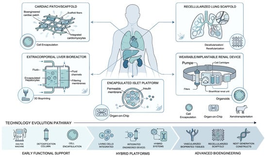
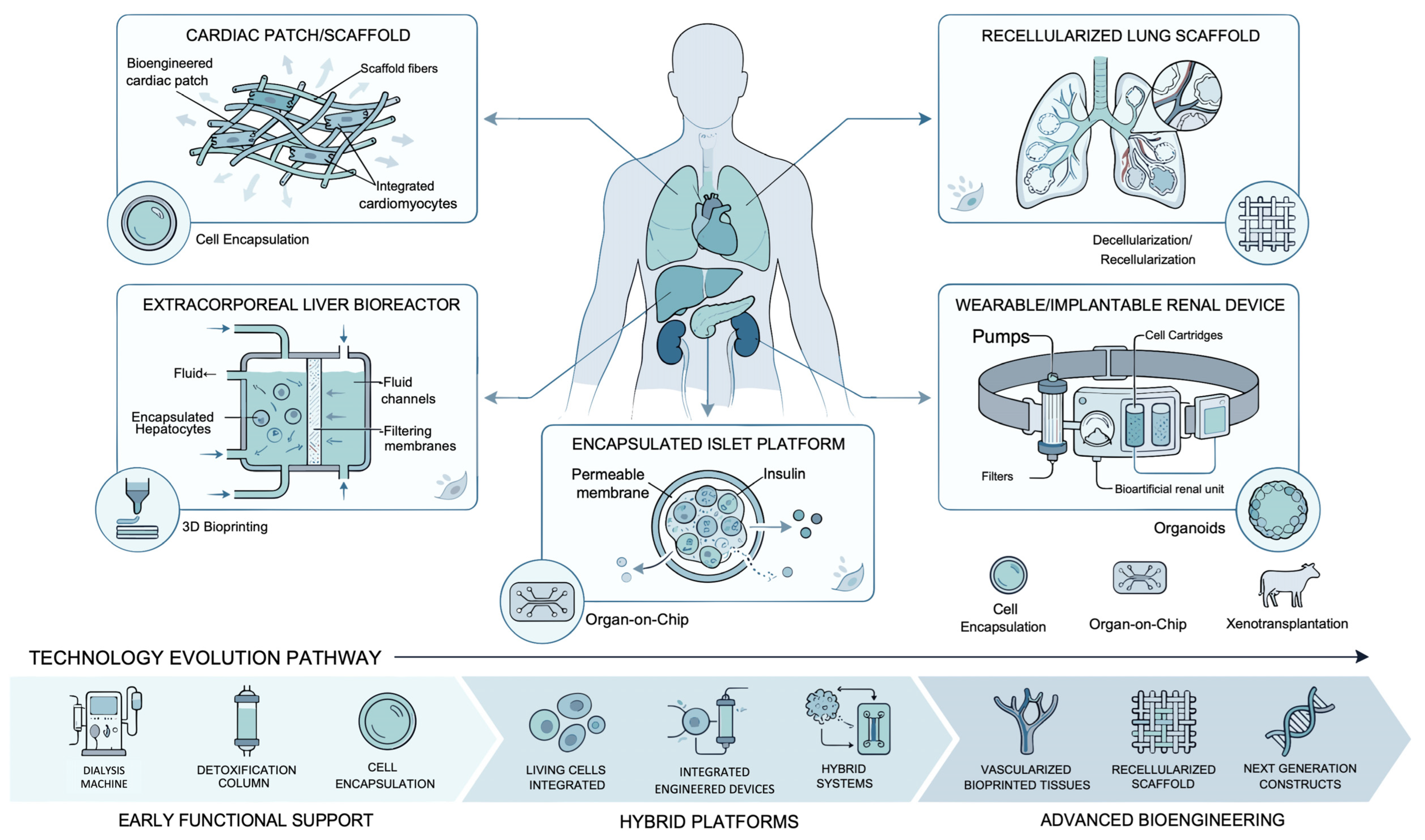
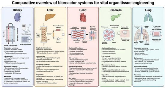
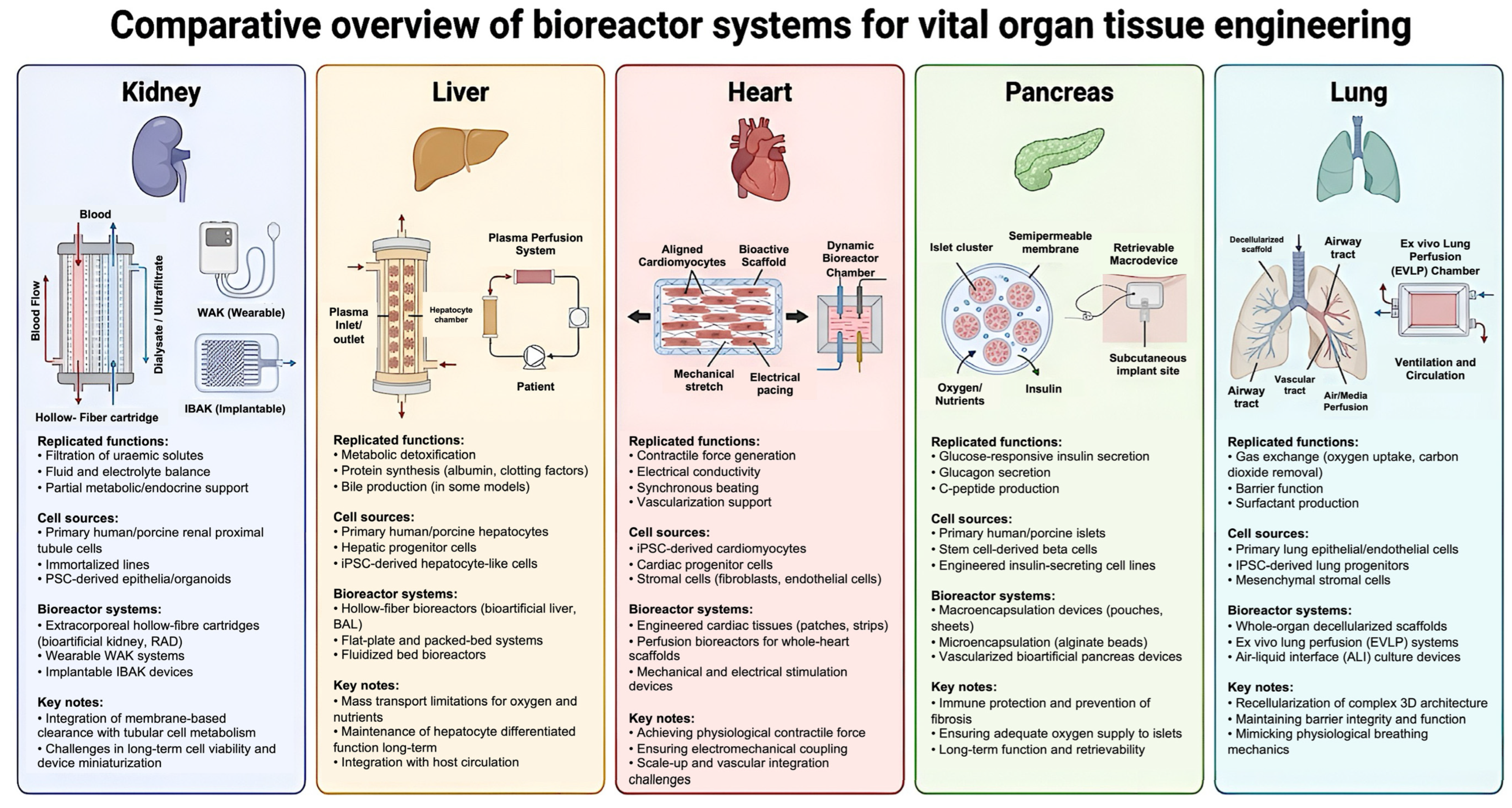
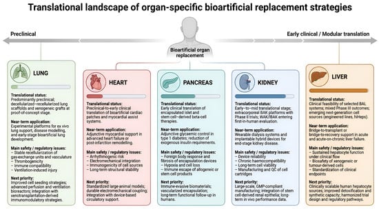
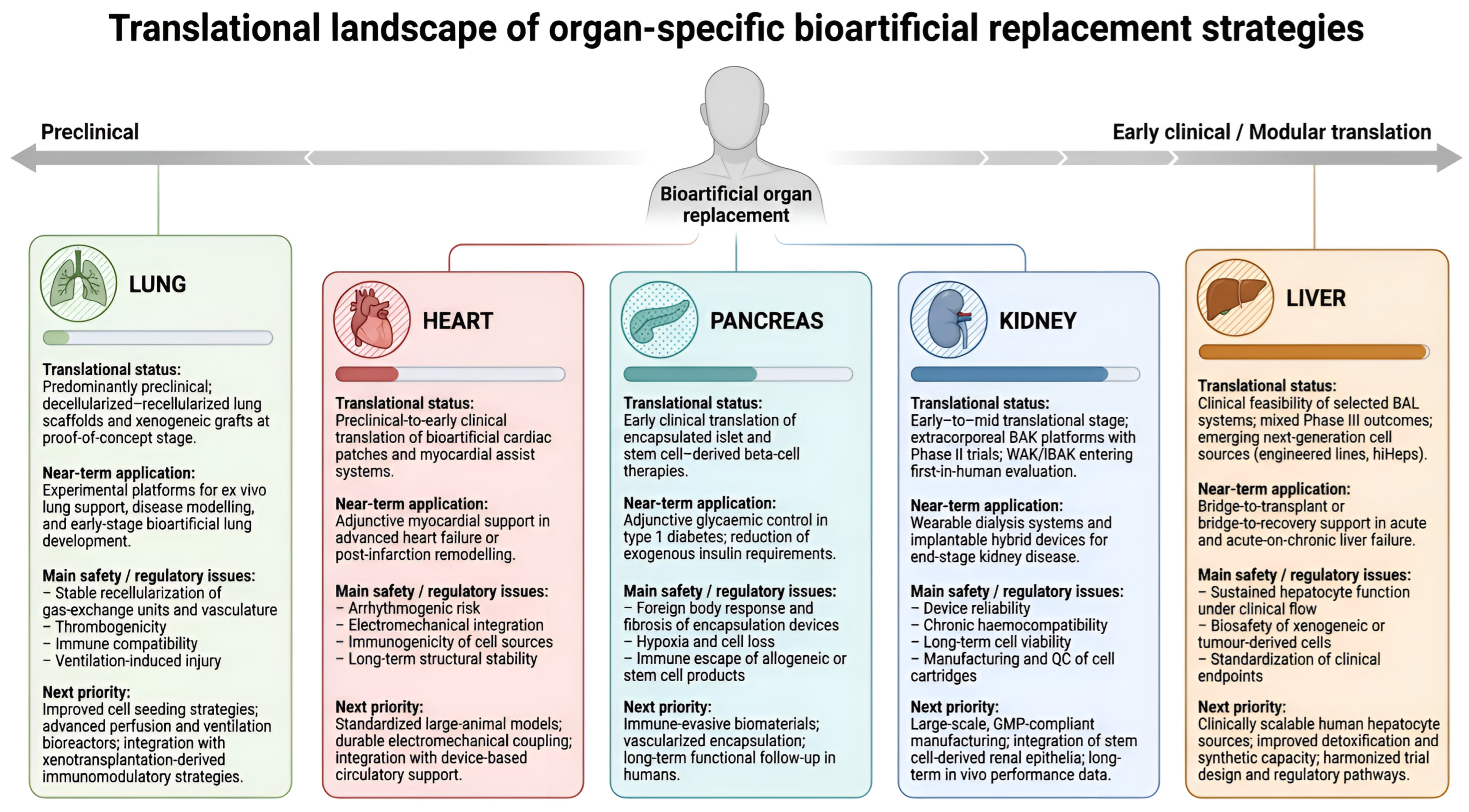
Disclaimer/Publisher’s Note: The statements, opinions and data contained in all publications are solely those of the individual author(s) and contributor(s) and not of MDPI and/or the editor(s). MDPI and/or the editor(s) disclaim responsibility for any injury to people or property resulting from any ideas, methods, instructions or products referred to in the content. |
© 2026 by the authors. Licensee MDPI, Basel, Switzerland. This article is an open access article distributed under the terms and conditions of the Creative Commons Attribution (CC BY) license.