


Abstract
Introduction: Spontaneous and recurrent epileptic seizures cause neuroinflammation, whereas the chronic administration of antiepileptic drugs may lead to hepatotoxicity and nephrotoxicity. Investigating natural compounds such as naringenin, which exhibits antioxidant, anti-inflammatory, and neuroprotective properties, could mitigate these toxic effects. However, whether naringenin delays seizure onset through anti-inflammatory mechanisms remains unclear. Objective: We evaluated the anticonvulsant effect of subchronic naringenin administration and its impact on inflammatory biomarkers in rats. Methods: Thirty-two male Wistar rats were divided into four groups (n = 8 each): vehicle (10% DMSO), naringenin (50 mg/kg), naringenin (100 mg/kg), and diazepam (4 mg/kg). Treatments were administered once daily for ten days through the intraperitoneal route. On day 11, status epilepticus (SE) was induced via the lithium-pilocarpine model. One hour after SE onset, cardiac blood was collected, and the serum was analyzed via ELISA to quantify the following inflammatory markers: superoxide dismutase (SOD), C-reactive protein (CRP), carbonyl protein, and nitrite/nitrate. Results: We found that 100 mg/kg naringenin increased the latency to SE and the first generalized seizure and shortened the duration of generalized seizures. Fifty percent of the rats in the 100 mg/kg naringenin group did not develop SE. The group treated with 100 mg/kg naringenin demonstrated reduced levels of CRP, protein carbonyls, and nitrates; conversely, the inhibition of SOD activity increased. Conclusions: Naringenin exerted anticonvulsant activity associated with a reduction in oxidative stress and inflammatory plasma biomarkers, suggesting its potential utility in the future development of alternative treatments to reduce epilepsy symptoms.
1. Introduction
Epilepsy is among the most common neurological disorders and is characterized by frequent seizures. Several factors implicated in its pathophysiology are related to apoptosis, neuroinflammation, and oxidative stress [1]. Epilepsy affects more than 50 million people worldwide [2]. Its treatment includes anticonvulsant medications that act on neurotransmission systems, such as carbamazepine, phenytoin, oxcarbazepine, ethosuximide, diazepam, clonazepam, and valproic acid. These treatments may alter various organs, resulting in liver and kidney damage and skin, vision, and hearing problems, among other conditions [3,4]. Owing to the potentially adverse effects of antiepileptic drugs (AEDs), a significant number of patients resort to natural products as alternative treatments, and various studies have investigated bioactive secondary metabolites from plants to contribute to the development of effective antiepileptic therapies with fewer side effects [5]. Secondary metabolites include flavonoids, alkaloids, glycosides, coumarins, and terpenoids [6]. For example, the anticonvulsant effects of curcumin and resveratrol (a polyphenolic flavonoid) did not alter hepatic or renal function in a lithium-pilocarpine-induced epilepsy model [7]. Additionally, Prunus domestica L., a plant in the Rosaceae family, yields a lignan known as pinoresinol-4-O-β-D-glucopyranoside (PGu), which has antioxidant and anti-inflammatory activities and can reduce seizures induced by lithium-pilocarpine in Wistar rats [8]. Moreover, cannabidiol (CBD), a phytocannabinoid component of Cannabis sativa, was approved as an anticonvulsant drug by the U.S. FDA in 2018 because of its antiepileptic and anticonvulsant activities [9]. In another study, the flavonoid apigenin decreased the oxidative stress caused by myeloperoxidase and inhibited ferroptosis in a kainic acid (KA) model [10]. Consequently, in vivo and in vitro studies suggest that antioxidants such as flavonoids attenuate cellular degeneration and reduce oxidative stress [11,12]. However, flavonoids may exhibit pro-oxidant activity under certain conditions. Notably, this pro-oxidant activity can be beneficial by inducing mild oxidative stress, thereby enhancing antioxidant defense mechanisms, as seen with naringenin, which, owing to its pro-oxidant potential, can act as an anticancer molecule by inducing apoptosis in cancer cells [13]. Another property of flavonoids is their allosteric modulation of GABAA receptors through binding to the benzodiazepine site, resulting in anti-inflammatory effects on the brain, which could be related to their potential anticonvulsant effects [14]. The additional properties of naringenin include gene expression modulation, metal chelation, radical scavenging, and enzymatic regulation [15,16]. Naringenin has low oral bioavailability; however, experimental models (in vitro and in situ) have demonstrated its ability to effectively cross the blood–brain barrier [17,18]. Pharmacokinetic studies conducted on rodents and humans have confirmed its presence in brain tissue following systemic administration at oral doses ranging from 50 to 900 mg [19,20,21]. Despite this limited exposure, evidence of systemic anti-inflammatory and antioxidant effects has been reported, suggesting that even low concentrations can cross the blood–brain barrier and exert neuroprotective effects [22,23]. These findings further suggest that naringenin may modulate seizure susceptibility through indirect mechanisms. Based on the above, naringenin is considered a compound of interest for antiepileptic therapy. Therefore, the present study evaluates the anticonvulsant effect of subchronic treatment with naringenin through a behavioral assessment of epilepsy induced by a lithium-pilocarpine model in male Wistar rats. Additionally, the effects of naringenin administration on the levels of inflammation markers, superoxide dismutase (SOD), C-reactive protein (CRP), carbonyl protein, and nitrite/nitrate were evaluated via ELISA. Assessing the effect of naringenin on epileptic seizures and biomarkers of inflammation may help identify its potential anticonvulsant properties for the future development of antiepileptic agents.
2. Materials and Methods
2.1. Ethical Approval
All experimental procedures followed the “Guide for the Care and Use of Laboratory Animals” (National Research Council, 2011) [24] and the Mexican Official Standard NOM-062-ZOO-1999 [25]. The protocol was approved by the Internal Committee for the Care and Use of Laboratory Animals at Instituto de Ciencias de la Salud, Universidad Veracruzana (CICUAL-ICS: 2021-02/001; Authorization: 17 February 2022).
2.2. Experimental Subjects
Thirty-two three-month-old male Wistar rats were randomly assigned to four groups (n = 8 per group): vehicle (10% dimethyl sulfoxide), naringenin (50 mg/kg), naringenin (100 mg/kg), and diazepam (4 mg/kg). The rats were maintained under a 12 h/12 h light/dark cycle, with lights on at 7:00 a.m. The ambient temperature was maintained at 22 ± 1 °C, with access to water and food ad libitum. All treatments consisted of an intraperitoneal (i.p.) injection; naringenin and diazepam were administered every 24 h for 10 days. All the experimental groups underwent SE induction the day after the final intraperitoneal administration, and behavioral assessment was conducted via the Racine scale [26]. Upon completion of the behavioral test, blood samples were obtained via a cardiac puncture for enzyme-linked immunosorbent assay (ELISA).
2.3. Drugs
Diazepam was obtained in its injectable 10 mg/kg formulation (brand name: Valium, lot: RJB39, manufactured by Roche Químicos e Farmacéuticos S.A., Estrada dos Bandeirantes 2020, Jacarepaguá, Rio de Janeiro, Brazil). Dimethyl sulfoxide was acquired at a concentration ≤ 100%, purchased from Sigma-Aldrich, Inc. (3050 Spruce St., St. Louis, MO 63103, USA; reference 472301). Naringenin (25 g) was obtained from Sigma-Aldrich®, CAS 67604-48-2, with a purity of ≥95% (3050 Spruce Street, Saint Louis, MO 63103, USA). Lithium chloride (50 g) was obtained from Reactivos Química MEYER®, QUÍMICA SUASTES S.A. DE C.V., Pampano #7, Colonia del Mar, Tlahuac, postcode 13270, Mexico City, Mexico. Pilocarpine (5 g) was obtained from SIGMA-ALDRICH, Co. (3050 Spruce Street, St. Louis, MO 63103, USA, +1-314-771-5765). Sodium pentobarbital (100 mL) solution for injection was obtained from SEDALPHARMA de México (Morelia, Michoacán, Mexico; SAGARPA Registration No. Q-7972-004).
2.4. Dosing
The dosages used were 50 and 100 mg/kg/day naringenin, calculated based on the rats’ body weights, in accordance with previously published studies [27,28]. In a pentylenetetrazol (PTZ, 30 mg/kg) model, intraperitoneal (i.p.) administration of naringenin increased the latency to the onset of seizures [27]. Unlike the pentylenetetrazol seizure model, which acts as a GABAA receptor agonist by reducing neuronal inhibition and generating acute, generalized seizures, the lithium-pilocarpine model stimulates the cholinergic system. It increases neuronal excitability and generates reproducible generalized tonic-clonic seizures, mimicking temporal lobe epilepsy, such as that seen in patients. In the present study, naringenin was i.p. administered every 24 h for 10 consecutive days prior to seizure induction.
2.5. Seizure Status Induction with Lithium-Pilocarpine
Once naringenin and diazepam had been administered on the tenth day, the rats received an i.p. injection of lithium chloride (3 mEq/kg), followed by a subcutaneous (s.c.) injection of pilocarpine hydrochloride (30 mg/kg) to induce SE twenty hours later. Seizures that last for at least 30 min, whether continuous or intermittent, are classified as SEs. Behavioral evaluation of SE was performed according to the Racine scale [19], which consists of the following behavioral stages: stage 0 (behavioral arrest), stage I (facial myoclonus), stage II (stage I behaviors plus head myoclonus), stage III (limb myoclonus), stage IV (all previous behaviors plus kangaroo posture), and stage V (stage IV behaviors plus loss of postural tone and collapse of the experimental subject).
2.6. Cardiac Puncture
At the end of the behavioral test, the rats were deeply anesthetized with sodium pentobarbital (120 mg/kg, i.p.). The absence of palpebral and paw or tail pinch reflexes was confirmed, as was the absence of pain or distress and the presence of an unconscious state. Blood samples were collected via cardiac puncture using a 3 mL syringe fitted with a 22 × 32 mm needle. The blood was then transferred to a tube without an anticoagulant and, once it had clotted, was centrifuged at 3500 rpm for 5 min. Serum was extracted using a Pasteur pipette and transferred to appropriate containers to determine inflammatory markers via ELISA.
2.7. Determination of Inflammatory Markers
The 32 serum samples were divided into two aliquots of 250 µL each. Each aliquot was placed in duplicate into Eppendorf tubes (clear and amber) to prevent sample contamination. As the samples were not used immediately, they were stored at −20 °C in a freezer. For laboratory analyses, a cold chain system was used to prevent the denaturation of the metabolites of interest. Total protein was determined using the EasyKem Pro Vet semiautomatic veterinary chemistry analyzer (KONTROLAB, Florence, Italy), whereas the SOD, CRP, carbonyl protein, and nitrite/nitrate analyses were carried out according to the manufacturers’ instructions for the various kits; their catalog numbers, in order of use, are CS0009-1KT, RAB0097-1KT, MAK094-1KT and 23479-1KT-F (3050 Spruce Street, Saint Louis, MO 63103, USA). The microplates were read using a MULTISKAN microplate reader (THERMO, PerkinElmer; AlphaScreen and AlphaLISA are trademarks of PerkinElmer, Inc. COL121542 1023; Bridgeport, CT, USA).
2.8. Statistical Analysis
The data were statistically analyzed using version 8.0.2c of GraphPad Prism, with a significance level of p ≤ 0.05. The normality of the variables was assessed using the Shapiro-Wilk test. Data with a normal distribution were analyzed using one-way ANOVA, with treatment as the sole factor, and Tukey’s post hoc test. For variables that did not have a normal distribution, Kruskal-Wallis one-way analysis of variance on ranks was applied. The mean and standard error were used to present the results.
3. Results
3.1. Behavioral Evaluation of SE
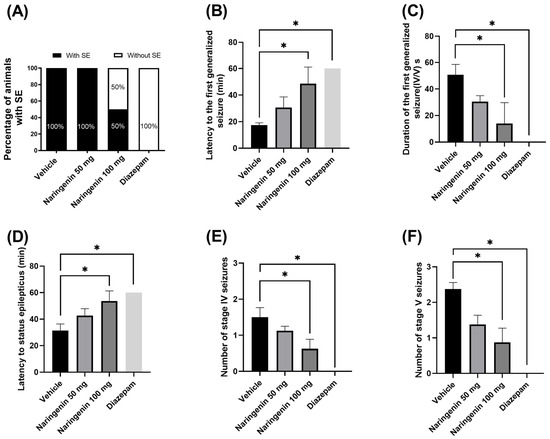
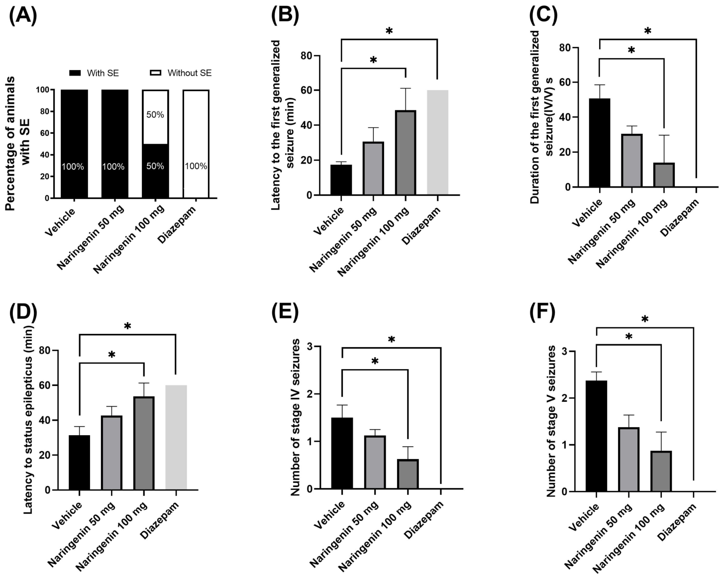
One hundred percent of the individuals in the diazepam group and 50% of the rats in the 100 mg/kg naringenin group did not develop phase IV or V generalized seizures or SE; however, a cutoff time of 60 min was used as the latency value for the first generalized seizure and SE in these cases. With respect to the duration of the first generalized seizure and the number of stage IV and V seizures, these subjects were assigned a value of 0.
The time of onset for the first generalized seizure (stage IV or V) was significantly longer in the diazepam and 100 mg/kg naringenin groups than in the vehicle group [H(3, 24) = 26.50, 3; p < 0.001]. The duration of the first generalized seizure (phase IV or V) was longer in the vehicle group than in the naringenin (100 mg/kg) or diazepam groups [H(3, 24) = 26.69, 3; p < 0.001]. The latency to SE following pilocarpine injection was also longer in the diazepam and 100 mg/kg naringenin groups than in the vehicle group [H(3, 24) = 27.57, 3; p < 0.001]. Rats exhibited generalized seizures following the administration of the pilocarpine dose, which occurred up to 60 min after the onset of SE. The number of generalized seizures (stages IV and V) was significantly lower in the diazepam and 100 mg/kg naringenin groups than in the vehicle group [H(3, 24) = 21.64, 3; p < 0.001] and [H(3, 24) = 23.28, 3; p < 0.001, respectively] (Figure 1).
Figure 1.
(A) Proportion of individuals who developed SE, (B) latency to the first generalized seizure (IV/V), (C) duration of the first generalized seizure, (D) latency to SE following pilocarpine injection, (E) number of phase IV generalized seizures, and (F) number of phase V generalized seizures. The data were analyzed using a Kruskal-Wallis test, followed by a post hoc Dunn test. The data are expressed as the mean ± SEM. * p < 0.001 vs. the vehicle group.
3.2. Evaluation of Inflammatory Markers
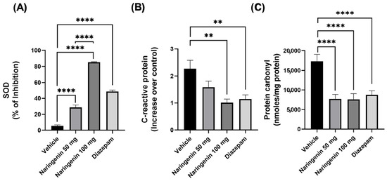
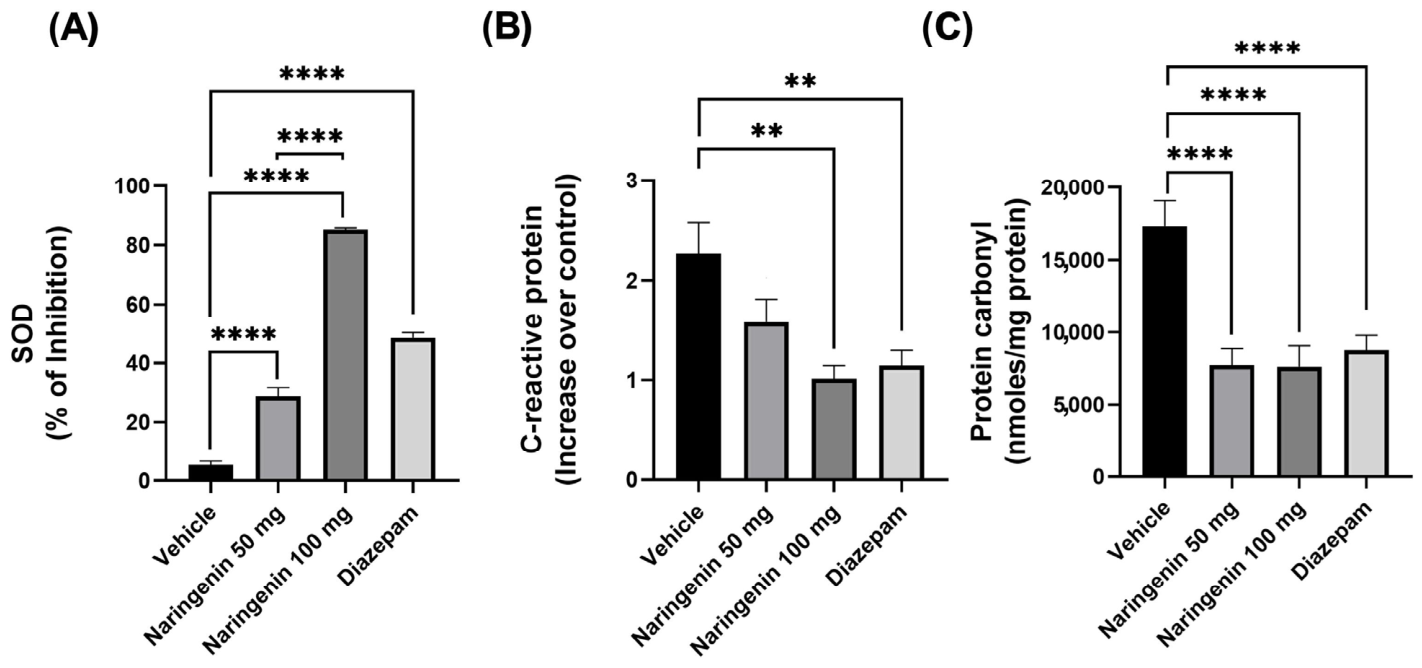
SOD inhibition analysis revealed that, compared with the vehicle group, the groups treated with 50 and 100 mg/kg naringenin and diazepam presented a significantly greater percentage of inhibition [F(3, 28) = 325.3, p < 0.001] (Figure 2). The CRP levels were significantly greater [F(3, 28) = 6.787, p = 0.001] in the vehicle group than in the naringenin 100 mg/kg and diazepam groups. No significant difference was observed between the 50 mg/kg naringenin group and the vehicle group (Figure 2). The picomoles of protein carbonyl per milligram of protein in the experimental groups—naringenin 50 mg/kg, naringenin 100 mg/kg, and diazepam—demonstrated significantly lower protein carbonyl concentrations [F(3, 28) = 17.80, p < 0.001] than those of the vehicle group, which demonstrated the highest concentration (Figure 2).
Figure 2.
(A) Percentage inhibition of superoxide dismutase (SOD), (B) increase in C-reactive protein, and (C) protein carbonyl concentration. The data were analyzed using one-way ANOVA, followed by Tukey’s post hoc test. The data are expressed as the mean ± SEM; ** p < 0.001 vs. the vehicle group; **** p < 0.001 vs. the vehicle group.
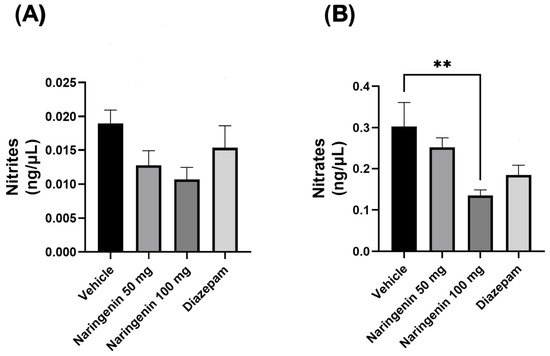
Finally, the nitrite concentration was analyzed. Compared with the vehicle group, the groups treated with 50 and 100 mg/kg naringenin and diazepam did not significantly differ [F(3, 28) = 2.511, p = 0.079]. Regarding the nitrate concentration, the 50 mg/kg naringenin and diazepam groups did not differ significantly from the vehicle group; however, the 100 mg/kg naringenin group was significantly different from the vehicle group [F(3, 28) = 4.598, p = 0.009], with a tendency toward increased nitrate levels observed in the vehicle group (Figure 3).
Figure 3.
(A) Nitrite levels and (B) nitrate levels, expressed in nanograms per microliter. The data were analyzed using one-way ANOVA, followed by Tukey’s post hoc test. The data are expressed as the mean ± SEM. ** p < 0.009 vs. the vehicle group.
4. Discussion
In this study, we evaluated the anticonvulsant effect of naringenin on inflammatory markers in Wistar rats with lithium-pilocarpine-induced SE. The results show that (A) subchronic administration of naringenin (100 mg/kg) exerts an anticonvulsant-like effect against SE, as evidenced by increased latency to the onset of generalized epileptic activity, increased latency to SE, a shorter duration of the first generalized seizure, and a reduction in the number of generalized phase V seizures, and (B) regarding inflammatory markers in the naringenin groups, an antioxidant effect is suggested, given the increase in SOD activity, the inhibition of free radicals, and the reduction in levels of CRP, protein carbonyls, and nitrates. These findings suggest that the anticonvulsant effects produced by naringenin may be associated with a reduction in serum biomarkers of inflammation and oxidative stress, without ruling out other mechanisms of action that were not explored in the present study.
4.1. Effect of Naringenin on Behavior
Some subjects in the diazepam and naringenin 100 mg/kg groups did not experience any phase IV or V generalized seizures or SE. However, in these cases, a 60 min cutoff time was used as the latency value for the first generalized seizure and the SE. Regarding the duration of the first generalized seizure and the number of phase IV and V seizures, these subjects were assigned a value of 0.
Generalized stage IV and V seizures, as well as status epilepticus, were prevented by administering diazepam. Diazepam is a 1,4-benzodiazepine derivative used clinically to suppress SE and generalized tonic-clonic seizures [29], which is why it was used as a pharmacological control. Conversely, naringenin administration modified SE behavior in the following ways: (1) treatment with 100 mg/kg naringenin prolonged the time to the onset of the first generalized seizure, and the rate of generalized seizures (SE) increased; (2) furthermore, the 100 mg/kg dose of naringenin reduced the duration of the first generalized seizure and reduced generalized seizures associated with stages V and VI; and (3) only 50% of the animals in the 100 mg/kg naringenin group developed SE. These findings suggest that naringenin at 100 mg/kg has anticonvulsant effects and attenuates the severity of generalized SE seizures. In other mouse models, such as PTZ, kainic acid, and pilocarpine, naringenin has demonstrated anticonvulsant effects when used without lithium. An increase in the latency to seizures was observed following intraperitoneal administration of naringenin at doses of 50, 100, and 200 mg/kg, and it decreased the duration of myoclonic seizures and tonic extension in male C57 albino mice in a pentylenetetrazole model (PTZ, 30 mg/kg/day) [27]. A 100 mg/kg dose of naringenin delayed seizure onset in male C57BL/6 mice in a kainic acid model in [30], and pretreatment with naringenin (20 and 40 mg/kg, p.o., for 15 days) reduced seizure severity and increased seizure latency in a pilocarpine model (300 mg/kg, i.p.) in [28]. Therefore, based on the present results and those reported by other research groups, naringenin demonstrates anticonvulsant properties in epilepsy.
4.2. Effects of Naringenin on Inflammatory Markers
4.2.1. Effect on Superoxide Dismutase (SOD)
The percentage inhibition of the antioxidant enzyme SOD in our study revealed that administering naringenin (50 and 100 mg/kg, i.p.) for 10 days resulted in a greater percentage of SOD inhibition, indicating increased enzymatic activity. These findings suggest that increased SOD activity and concentration confer greater antioxidant protection, suggesting an antioxidant mechanism and fewer reactive oxygen species (ROS) during SE. These findings are supported by those of other animal models, such as the study by Shakeel et al. (2017) [28], which was conducted on Swiss albino mice with pilocarpine-induced epileptic seizures in the absence of lithium chloride. Doses of 20 and 40 mg/kg naringenin had previously been administered orally for 15 days, increasing SOD activity and thereby confirming their antioxidant effect.
Reduced SOD levels indicate oxidative stress in epilepsy, as observed in the vehicle group, which received no naringenin pretreatment. Wang et al. (2021) [31] reported that patients with epilepsy had lower serum SOD levels than healthy controls did. These findings align with previous reports in which decreased SOD-1 levels in cerebrospinal fluid (CSF) were associated with increased ROS levels [32].
4.2.2. Effect on C-Reactive Protein (CRP)
The results presented in this study revealed that compared with the 100 mg/kg naringenin and diazepam groups, the control group exhibited elevated CRP levels, a greater number of generalized motor seizures, and longer seizure durations. Consistent with these findings, studies have demonstrated that patients with epilepsy who experience generalized motor seizures have elevated levels of CRP and IL-6, which leads to chronic inflammation [33,34,35]. Significantly elevated blood CRP levels have also been detected in patients with generalized motor seizures [34].
Conversely, compared with the control group, the 100 mg/kg naringenin group presented lower CRP levels. By contrast, compared with the control group, the group treated with 100 mg/kg naringenin exhibited lower levels of CRP. Given that naringenin possesses potent anti-inflammatory properties, it reduces the release of inflammatory mediators in microglia following treatment. Although our inflammatory markers were quantified at the serum level, they are consistent with [36,37].
4.2.3. Effect on Protein Carbonylation
The serum levels of protein carbonyls measured in this study were higher in the vehicle group. A study conducted by Zhu et al. (2017) [38] using a PTZ-induced epilepsy model (35 mg/kg, i.p.) demonstrated that elevated levels of protein carbonyls were observed in the hippocampus of C57BL/6J mice; this is associated with increased nNOS activation and, therefore, acts as a marker of oxidation [38]. Our study revealed an increase in serum levels of protein carbonyls, which could be due to the following factors: (a) increased susceptibility of proteins to oxidative attack, (b) increased ROS production, (c) reduced capacity to eliminate oxidized proteins, or (d) impairment of the antioxidant system [39].
In a study assessing toxicity in mice treated with AlCl3 (10 mg/kg bw/day), naringenin (10 mg/kg bw/day) was administered simultaneously. Treatment with naringenin attenuated oxidative stress by regulating serum levels of nitric oxide and protein carbonyls, as well as lipid peroxidation. This finding demonstrates the antioxidant and anti-inflammatory properties of naringenin [40], which is consistent with our study.
4.2.4. Effect on Nitrite/Nitrate
Our results regarding nitrite levels did not reveal any significant differences between the groups. However, the group treated with 100 mg/kg naringenin had lower nitrate levels than the control group did. These findings are consistent with the nitric oxide levels reported in patients with epilepsy, in whom no differences have been compared with controls [41,42].
In a pilocarpine model, male Wistar rats treated with 400 mg/kg pilocarpine exhibited increased lipid peroxidation and nitrite levels, whereas the group pretreated with levetiracetam presented decreases in these parameters [43]. In addition, naringenin was observed to have a protective effect by reducing nitrite and nitrate levels; doses of 50 and 100 mg/kg administered over 4 weeks decreased serum nitric oxide levels in rats with obesity-related hypertension fed a high-fat diet. These findings indicate that naringenin reduces oxidative stress [44].
Finally, in the present study, administering 50 and 100 mg/kg naringenin to male Wistar rats was sufficient to attenuate SE-induced changes, such as the severity and frequency of seizures, as well as the delay in their onset. These effects are probably associated with a reduction in oxidative stress, as reflected by serum levels of inflammatory markers—including SOD inhibition, CRP, and protein carbonyls and nitrates—which are comparable to those produced by diazepam. The limitations of this study include the following: (1) Clinical trials are needed to demonstrate the potential benefits of naringenin in the treatment of epilepsy. (2) Inflammation biomarkers in nervous tissue were not evaluated; therefore, future studies should evaluate central nervous system inflammation markers such as IL-1β and TNF-α, which have been shown to increase during epileptogénesis in specific brain structures, such as the hippocampus and prefrontal cortex. (3) Although naringenin can cross the blood-brain barrier and produce neuroprotective effects, it is important to conduct pharmacokinetic studies in future research. (4) The use of male rats in this study was intended to reduce experimental variability associated with the estrous cycle in females; therefore, this practice may limit the generalizability of the results.
5. Conclusions
Administering naringenin (100 mg/kg) for 10 days to male Wistar rats in a lithium-pilocarpine model had anticonvulsant-like effects. These effects could be associated with a reduction in neuroinflammation, which is supported by low CRP and nitrates in naringenin-treated rats, in addition to an increase in SOD. Therefore, these results add information about the anticonvulsant effects of naringenin, which could be considered in the study and design of complementary anticonvulsant drugs aimed at improving epileptic disorders.
Author Contributions
E.R.-D. and I.Z.-B. were responsible for conceptualization and the methodology; P.M.S.-H. performed the experiments; J.F.-R.-L., E.R.-D., I.Z.-B., O.R.-S. and J.C.-E. analyzed the data; E.R.-D. contributed reagents, materials, and analytical tools; and E.R.-D., J.F.-R.-L., I.Z.-B., O.R.-S. and J.C.-E. wrote the manuscript. All authors have read and agreed to the published version of the manuscript.
Funding
This research was funded by the Academic Group of Biology, Chemistry, and Molecular Functionality of Plant Metabolites (UV-CA-368) of the Universidad Veracruzana and the Sistema Nacional de Investigadores Exp. 171150 (E.R.-D.).
Institutional Review Board Statement
The animal study protocol was approved by the Institutional Review Board (or Ethics Committee) of Instituto de Ciencias de la Salud, Universidad Veracruzana (CICUAL-ICS: 2021-02/001; authorization: 17 February 2022).
Data Availability Statement
The datasets generated and analyzed during this study may be obtained from the corresponding author upon reasonable request.
Acknowledgments
The authors would like to thank Daniel Hernández Baltazar for his technical assistance in the ELISA analysis and the Facultad de Química Farmacéutica Biológica Zona Xalapa-UV for the facilities used to house and maintain the rats included in this study.
Conflicts of Interest
The authors declare that they have no conflicts of interest.
Abbreviations
The following abbreviations are used in this manuscript:
| °C | Degrees Celsius |
| µL | Microliters |
| CBD | Cannabidiol |
| DMSO | Dimethyl sulfoxide |
| E.S.M. | Standard error of the mean |
| SE | Status epilepticus |
| ROS | Reactive oxygen species |
| AED | Antiepileptic drug |
| FDA | Food and Drug Administration |
| GABA | Gamma-aminobutyric acid |
| i.p. | Intraperitoneal |
| IL-1 | Interleukin 1 |
| IL-1β | Interleukin 1 beta |
| IL-6 | Interleukin 6 |
| KA | Kainic acid |
| CSF | Cerebrospinal fluid |
| mEq | Milliequivalent |
| O | Singlet Oxygen |
| CRP | C-reactive protein |
| PGu | Pinoresinol-4-O-β-D-glucopyranoside |
| PTZ | Pentylenetetrazol |
| rpm | Revolutions per minute |
| s.c. | Subcutaneous |
| CNS | Central nervous system |
| SOD | Superoxide dismutase |
| TNF-α | Tumor necrosis factor alpha |
References
- Kegler, A.; Caprara, A.L.F.; Pascotini, E.T.; Arend, J.; Gabbi, P.; Duarte, M.M.M.F.; Furian, A.F.; Oliveira, M.S.; Royes, L.F.F.; Fighera, M.R. Apoptotic Markers Are Increased in Epilepsy Patients: A Relation with Manganese Superoxide Dismutase Ala16Val Polymorphism and Seizure Type through IL-1β and IL-6 Pathways. BioMed Res. Int. 2020, 2020, 6250429. [Google Scholar] [CrossRef] [Scilit]
- World Health Organization. Epilepsy; World Health Organization: Geneva, Switzerland, 2024; Available online: https://www.who.int/es/news-room/fact-sheets/detail/epilepsy (accessed on 17 May 2026).
- Thijs, R.D.; Surges, R.; O’Brien, T.J.; Sander, J.W. Epilepsy in adults. Lancet 2019, 393, 689–701. [Google Scholar] [CrossRef] [Scilit] [PubMed]
- Akyüz, E.; Köklü, B.; Ozenen, C.; Arulsamy, A.; Shaikh, M.F. Elucidating the Potential Side Effects of Current Anti-Seizure Drugs for Epilepsy. Curr. Neuropharmacol. 2021, 19, 1865–1883. [Google Scholar] [CrossRef] [Scilit]
- Lin, C.-H.; Hsieh, C.-L. Chinese Herbal Medicine for Treating Epilepsy. Front. Neurosci. 2021, 15, 682821. [Google Scholar] [CrossRef] [Scilit]
- He, L.-Y.; Hu, M.-B.; Li, R.-L.; Zhao, R.; Fan, L.-H.; He, L.; Lu, F.; Ye, X.; Huang, Y.-L.; Wu, C.-J. Natural Medicines for the Treatment of Epilepsy: Bioactive Components, Pharmacology and Mechanism. Front. Pharmacol. 2021, 12, 604040. [Google Scholar] [CrossRef] [Scilit]
- Zamora-Bello, I.; Rivadeneyra-Domínguez, E.; Rodríguez-Landa, J.F. Anticonvulsant Effect of Turmeric and Resveratrol in Lithium/Pilocarpine-Induced Status Epilepticus in Wistar Rats. Molecules 2022, 27, 3835. [Google Scholar] [CrossRef] [Scilit]
- Youssef, F.S.; Menze, E.T.; Ashour, M.L. A Potent Lignan from Prunes Alleviates Inflammation and Oxidative Stress in Lithium/Pilocarpine-Induced Epileptic Seizures in Rats. Antioxidants 2020, 9, 575. [Google Scholar] [CrossRef] [Scilit]
- Buchanan-Peart, K.-A.R.; Oribhabor, G.I.; Khokale, R.V.; Nelson, M.L.; Cancarevic, I. Cannabis, More Than the Euphoria: Its Therapeutic Use in Drug-Resistant Epilepsy. Cureus 2020, 12, e9299. [Google Scholar] [CrossRef] [Scilit] [PubMed]
- Shao, C.; Yuan, J.; Liu, Y.; Qin, Y.; Wang, X.; Gu, J.; Chen, G.; Zhang, B.; Liu, H.-K.; Zhao, J.; et al. Epileptic Brain Fluorescent Imaging Reveals Apigenin Can Relieve the Myeloperoxidase-Mediated Oxidative Stress and Inhibit Ferroptosis. Proc. Natl. Acad. Sci. USA 2020, 117, 10155–10164. [Google Scholar] [CrossRef] [Scilit] [PubMed]
- Pardo-Peña, K.; Martínez-Gallegos, S.M.; Noriega-Ruiz, M.A. Efecto de los Antioxidantes en la Epilepsia del Lóbulo Temporal. e-CUCBA 2022, 9, 177–191. [Google Scholar] [CrossRef] [Scilit]
- Shen, N.; Wang, T.; Gan, Q.; Liu, S.; Wang, L.; Jin, B. Plant Flavonoids: Classification, Distribution, Biosynthesis, and Antioxidant Activity. Food Chem. 2022, 383, 132531. [Google Scholar] [CrossRef] [Scilit] [PubMed]
- Slika, H.; Mansour, H.; Wehbe, N.; Nasser, S.A.; Iratni, R.; Nasrallah, G.; Shaito, A.; Ghaddar, T.; Kobeissy, F.; Eid, A.H. Therapeutic Potential of Flavonoids in Cancer: ROS-Mediated Mechanisms. Biomed. Pharmacother. 2022, 146, 112442. [Google Scholar] [CrossRef] [Scilit] [PubMed]
- Copmans, D.; Orellana-Paucar, A.M.; Steurs, G.; Zhang, Y.; Ny, A.; Foubert, K.; Exarchou, V.; Siekierska, A.; Kim, Y.; De Borggraeve, W.; et al. Methylated Flavonoids as Anti-Seizure Agents: Naringenin 4′,7-Dimethyl Ether Attenuates Epileptic Seizures in Zebrafish and Mouse Models. Neurochem. Int. 2018, 112, 124–133. [Google Scholar] [CrossRef] [Scilit] [PubMed]
- Prasain, J.; Carlson, S.; Wyss, J. Flavonoids and Age-Related Disease: Risk, Benefits and Critical Windows. Maturitas 2010, 66, 163–171. [Google Scholar] [CrossRef] [Scilit]
- Zaidun, N.H.; Thent, Z.C.; Latiff, A.A. Combating Oxidative Stress Disorders with Citrus Flavonoid: Naringenin. Life Sci. 2018, 208, 111–122. [Google Scholar] [CrossRef] [Scilit]
- Youdim, K.A.; Qaiser, M.Z.; Begley, D.J.; Rice-Evans, C.A.; Abbott, N.J. Flavonoid permeability across an in situ model of the blood-brain barrier. Free Radic. Biol. Med. 2004, 36, 592–604. [Google Scholar] [CrossRef] [Scilit]
- Stasiłowicz-Krzemień, A.; Gołębiewski, M.; Płazińska, A.; Płaziński, W.; Miklaszewski, A.; Żarowski, M.; Adamska-Jernaś, Z.; Cielecka-Piontek, J. The Systems of Naringenin with Solubilizers Expand Its Capability to Prevent Neurodegenerative Diseases. Int. J. Mol. Sci. 2022, 23, 755. [Google Scholar] [CrossRef] [Scilit]
- El Mohsen, M.A.; Marks, J.; Kuhnle, G.; Rice-Evans, C.; Moore, K.; Gibson, G.; Debnam, E.; Srai, S.K. The differential tissue distribution of the citrus flavanone naringenin following gastric instillation. Free Radic. Res. 2004, 38, 1329–1340. [Google Scholar] [CrossRef] [Scilit]
- Rebello, C.J.; Beyl, R.A.; Lertora, J.J.L.; Greenway, F.L.; Ravussin, E.; Ribnicky, D.M.; Poulev, A.; Kennedy, B.J.; Castro, H.F.; Campagna, S.R.; et al. Safety and pharmacokinetics of naringenin: A randomized, controlled, single-ascending-dose clinical trial. Diabetes Obes. Metab. 2020, 22, 91–98. [Google Scholar] [CrossRef] [Scilit]
- Manish, R.; Brindha, D.; Deenathayalan, U. Behavioral rescue: Naringenin’s neuroprotective effects against PTZ-induced seizures by mitigating oxidative stress and neuroinflammation in zebrafish larvae. Pharmacol. Res.-Mod. Chin. Med. 2024, 12, 100459. [Google Scholar] [CrossRef] [Scilit]
- Nouri, Z.; Fakhri, S.; El-Senduny, F.F.; Sanadgol, N.; Abd-ElGhani, G.E.; Farzaei, M.H.; Chen, J.T. On the Neuroprotective Effects of Naringenin: Pharmacological Targets, Signaling Pathways, Molecular Mechanisms, and Clinical Perspective. Biomolecules 2019, 9, 690. [Google Scholar] [CrossRef] [Scilit]
- Lai, Z.; Ke, L.; Zhao, W. Naringenin as a neurotherapeutic agent in Alzheimer’s disease: Epigenetic signatures, gut microbiota alterations, and molecular neuroprotection. Front. Aging Neurosci. 2025, 17, 1647967. [Google Scholar] [CrossRef] [Scilit]
- National Research Council. Guide for the Care and Use of Laboratory Animals: A Report of the Committee for the Update of the Guide for the Care and Use of Laboratory Animals, 8th ed.; Department of Health and Human Services: Washington, DC, USA, 2011. [Google Scholar]
- NOM-ZOO-062; Especificaciones Técnicas para la Producción, Cuidado y Uso de los Animales de Laboratorio. Secretaría de Agricultura, Ganadería, Desarrollo Rural, Pesca y Alimentación: Ciudad de México, Mexico, 1999.
- Racine, R.J. Modificación de la actividad convulsiva mediante estimulación eléctrica: II. Convulsión motora. Electroencefalogr. Electroencephalogr. Clin. Neurophysiol. 1972, 32, 281–294. [Google Scholar] [CrossRef] [Scilit] [PubMed]
- Khodayar, M.J.; Salehi, S.; Rezaei, M.; Siahpoosh, A.; Khazaei, A.; Houshmand, G. Evaluation of the Effect of Naringenin on Pentylenetetrazole and Maximal Electroshock-Induced Convulsions in Mice. Jundishapur J. Nat. Pharm. Prod. 2016, 12, e31384. [Google Scholar] [CrossRef] [Scilit]
- Tabassum, N.; Shakeel, S.; Rehman, M.U.; Amin, U.; Mir, M.U.R. Effect of Naringenin (a Naturally, Occurring Flavanone) Against Pilocarpine-Induced Status Epilepticus and Oxidative Stress in Mice. Pharmacogn. Mag. 2017, 13, 154. [Google Scholar] [CrossRef] [Scilit] [PubMed]
- Vergara, J.P.; Ladino, L.; Castro, C.A.; Gómez, B.; Forero, E.; Vargas, J.; Guio, L. Treatment of status epilepticus, an expert consensus report, Colombian Neurology Association-Epilepsy Committee. Acta Neurológica Colomb. 2019, 35, 74–88. [Google Scholar] [CrossRef] [Scilit]
- Jeong, K.H.; Jung, U.J.; Kim, S.R. Naringin Attenuates Autophagic Stress and Neuroinflammation in Kainic Acid-Treated Hippocampus In Vivo. Evid.-Based Complement. Altern. Med. eCAM 2015, 2015, 354326. [Google Scholar] [CrossRef] [Scilit]
- Wang, M.; Zhang, X.; Jia, W.; Zhang, C.; Boczek, T.; Harding, M.; Liu, Y.; Li, M.; Zhang, S.; Lei, S.; et al. Circulating Glutathione Peroxidase and Superoxide Dismutase Levels in Patients with Epilepsy: A Meta-Analysis. Seizure 2021, 91, 278–286. [Google Scholar] [CrossRef] [Scilit]
- Chen, D.; Lu, Y.; Yu, W.; Luo, J.; Xiao, Z.; Xiao, F.; Wang, X. Clinical Value of Decreased Superoxide Dismutase 1 in Patients with Epilepsy. Seizure 2012, 21, 508–511. [Google Scholar] [CrossRef] [Scilit]
- Alapirtti, T.; Waris, M.; Fallah, M.; Soilu-Hänninen, M.; Mäkinen, R.; Kharazmi, E.; Peltola, J. C-Reactive Protein and Seizures in Focal Epilepsy: A Video-Electroencephalographic Study. Epilepsia 2012, 53, 790–796. [Google Scholar] [CrossRef] [Scilit]
- Ishikawa, N.; Kobayashi, Y.; Fujii, Y.; Kobayashi, M. Increased Interleukin-6 and High-Sensitivity C-Reactive Protein Levels in Pediatric Epilepsy Patients with Frequent, Refractory Generalized Motor Seizures. Seizure 2015, 25, 136–140. [Google Scholar] [CrossRef] [Scilit]
- Urquizo, G.; Arteaga, R. Proteína C reactiva en el Diagnóstico y Pronóstico de Enfermedades Infecciosas en Pacientes Geriátricos. Rev. Méd. La Paz 2017, 23, 69–73. [Google Scholar]
- Park, J.; Jeong, K.H.; Shin, W.H.; Bae, Y.S.; Jung, U.J.; Kim, S.R. Naringenin ameliorates kainic acid-induced morphological alterations in the dentate gyrus in a mouse model of temporal lobe epilepsy. Neuroreport 2016, 27, 1182–1189. [Google Scholar] [CrossRef] [Scilit]
- Jin, L.; Zeng, W.; Zhang, F.; Zhang, C.; Liang, W. Naringenin Ameliorates Acute Inflammation by Regulating Intracellular Cytokine Degradation. J. Immunol. 2017, 199, 3466–3477. [Google Scholar] [CrossRef] [Scilit] [PubMed]
- Zhu, X.; Dong, J.; Han, B.; Huang, R.; Zhang, A.; Xia, Z.; Chang, H.; Chao, J.; Yao, H. Neuronal Nitric Oxide Synthase Contributes to PTZ Kindling Epilepsy-Induced Hippocampal Endoplasmic Reticulum Stress and Oxidative Damage. Front. Cell. Neurosci. 2017, 11, 377. [Google Scholar] [CrossRef] [Scilit]
- Nyström, T. Role of Oxidative Carbonylation in Protein Quality Control and Senescence. EMBO J. 2005, 24, 1311–1317. [Google Scholar] [CrossRef] [Scilit]
- Rai, R.; Ahmad, Z.; Jain, S.K.; Jat, D.; Mishra, S.K. Naringenin suppresses aluminum-induced experimental hepato-nephrotoxicity in mice through modulation of oxidative stress and inflammation. Toxicol. Res. 2023, 40, 97–110. [Google Scholar] [CrossRef] [Scilit]
- Menon, B.; Ramalingam, K.; Kumar, R.V. Oxidative Stress in Patients with Epilepsy is Independent of Antiepileptic Drugs. Seizure 2012, 21, 780–784. [Google Scholar] [CrossRef] [Scilit] [PubMed]
- Ibragic, S.; Sofic, E.; Suljic, E.; Avdagic, N.; Bajraktarevic, A.; Tahirovic, I. Serum Nitric Oxide Concentrations in Patients with Multiple Sclerosis and Patients with Epilepsy. J. Neural Transm. 2012, 119, 7–11. [Google Scholar] [CrossRef] [Scilit] [PubMed]
- Oliveira, A.A.; Almeida, J.P.C.; Freitas, R.M.; Nascimento, V.S.; Aguiar, L.M.V.; Júnior, H.V.N.; Fonseca, F.N.; Viana, G.S.B.; Sousa, F.C.F.; Fonteles, M.M.F. Effects of Levetiracetam in Lipid Peroxidation Level, Nitrite–Nitrate Formation and Antioxidant Enzymatic Activity in Mice Brain After Pilocarpine-Induced Seizures. Cell. Mol. Neurobiol. 2007, 27, 395–406. [Google Scholar] [CrossRef] [Scilit]
- Liu, H.; Zhao, H.; Che, J.; Yao, W. Naringenin Protects against Hypertension by Regulating Lipid Disorder and Oxidative Stress in a Rat Model. Kidney Blood Press. Res. 2022, 47, 423–432. [Google Scholar] [CrossRef] [Scilit]
Disclaimer/Publisher’s Note: The statements, opinions and data contained in all publications are solely those of the individual author(s) and contributor(s) and not of MDPI and/or the editor(s). MDPI and/or the editor(s) disclaim responsibility for any injury to people or property resulting from any ideas, methods, instructions or products referred to in the content. |
© 2026 by the authors. Licensee MDPI, Basel, Switzerland. This article is an open access article distributed under the terms and conditions of the Creative Commons Attribution (CC BY) license.