


Abstract
Halophilic microorganisms are a valuable resource for the bioremediation of saline environments contaminated with heavy metals. This study investigated the microbiota that could be cultured in the hypersaline Laguna Rosa in Torrevieja, Alicante, Spain, by combining an analysis of microbial diversity with an evaluation of tolerance to heavy metals. Water and sediment samples were collected from contrasting microenvironments (salinity 1–40%; pH 7.1–8.1) and subjected to enrichment and selective isolation strategies, yielding 79 isolates: 48 recovered from enrichment cultures and 31 from metal-supplemented media. These isolates represent all three domains of life: Archaea, Bacteria and Eukarya. The dominant taxa included Halorubrum sp., Haloferax sp., Halomonas sp., Marinobacter sp. and Dunaliella sp., as well as cyanobacteria such as Sodalinema sp. and Synechococcus sp. Tolerance assays revealed that haloarchaea exhibited high resistance to lithium, while several bacterial isolates tolerated chromium and arsenite. Dunaliella sp. showed the broadest overall metal tolerance among the photosynthetic isolates. In liquid culture, Halomonas smyrnensis maintained growth at Cr(VI) concentrations up to 25 mM, whereas Halorubrum saccharovorum tolerated up to 4 mM Cr(VI). These findings identify Laguna Rosa as a reservoir of halophilic microorganisms tolerant of heavy metal, with promising potential for the bioremediation of saline and hypersaline environments, including saline industrial wastewater contaminated with toxic metals.
1. Introduction
In recent years, there has been a significant increase in heavy metal pollution due to factors such as climate change, urbanisation, industrialisation, and the intensive use of pesticides and chemical fertilisers containing trace metals [1]. These processes have led to water salinisation, desertification and the accumulation of contaminants, including heavy metals. As these contaminants are non-biodegradable, they persist in soils in the long term and can bioaccumulate through the food chain [2,3]. Emissions from agricultural and industrial sources directly affect surface water and groundwater, posing significant risk to human health and biodiversity [1,4,5].
In the context of heavy metal bioremediation in aquatic environments and sediments, chromium, arsenic, lithium and lead are particularly relevant. Hexavalent chromium (Cr(VI)) is highly toxic and carcinogenic. It primarily originates from industrial activities such as electroplating, tanning and mining. Exposure to arsenic is also hazardous and has been associated with skin cancer, melanosis, keratosis and other physiological disorders. This element can be transported easily through groundwater via geochemical processes and human activities such as mining, pesticide use, and industrial operations. In wastewater, arsenic exists mainly in two oxidation states: arsenate (As(V)) and arsenite (As(III)). Arsenite is considered the more harmful of these due to its greater reactivity and potential to disrupt essential cellular processes [6,7,8]. Although lithium is not traditionally classified as a heavy metal, its accumulation in aquatic systems is a growing concern, largely driven by the expansion of mining activities and inadequate battery disposal practices [9]. Lead is a well-known neurotoxic metal that is particularly harmful to children, and causes kidney and circulatory damage. It is released into the environment through mining, smelting, metal refining, pigment and paint production, electroplating, and the manufacture of glass and ceramics, as well as through the steel industry [10,11,12,13].
Common strategies used to mitigate heavy metal toxicity in contaminated environments include bioleaching, biotransformation, bioaccumulation and biosorption. These approaches can target heavy metals, phenolic compounds and pesticides [14,15]. Although conventional treatments can be rapid and allow metal recovery, they are often only moderately efficient and pose additional challenges, including the generation of large amounts of sludge, high energy consumption, and long-term environmental impacts. Consequently, bioremediation approaches using microorganisms offer a sustainable, efficient, and environmentally friendly alternative [16,17]. However, a significant proportion of metal-contaminated industrial waste consists of saline or hypersaline wastewater, which conventional microorganisms cannot remediate effectively. The high salinity of these effluents poses an additional challenge for bioremediation, as osmotic stress impairs microbial survival and disrupts normal metabolic pathways [18].
In this context, using halotolerant or halophilic microorganisms to remove heavy metals represents an attractive emerging alternative. These microorganisms have evolved strategies to withstand extreme abiotic conditions, making them promising candidates for biotechnological applications in saline or contaminated environments [19,20]. Several studies have reported that halophiles exhibit remarkable remediation capacity and tolerance to heavy metals. Genera such as Haloferax, Halorubrum and Haloarcula belong to the Archaea domain and can tolerate metals including cadmium, lithium, nickel, cobalt, silver and arsenic [21,22,23,24,25,26,27]. Similarly, bacterial taxa such as Halomonas and Marinococcus can tolerate metals including arsenic, chromium, and copper [28,29]. Cyanobacteria are notable for their ecological role as photosynthetic organisms, and for their ability to tolerate and remove heavy metals. Genera such as Synechococcus and Phormidium have demonstrated the capacity to accumulate cadmium and lead [30,31]. Finally, within the Eukarya domain, halophilic microorganisms such as Dunaliella, Tetraselmis, and Chlamydomonas have also demonstrated tolerance to zinc, lead, and mercury, as well as the ability to remove heavy metals from contaminated environments [32,33].
The Laguna Rosa of Torrevieja (Alicante, Spain) is a unique hypersaline ecosystem in the Bajo Segura region of southeastern Spain, covering 1400 hectares and characterised by its arid climate, distinctive geological history and limited freshwater inputs. The prolonged dry season, high temperatures, low and irregular precipitation, and intense evaporation promote the progressive concentration of dissolved salts, with salinity reaching approximately 390 g/L, far exceeding that of conventional seawater [34]. Its marine-sedimentary origin, subsequent isolation from the Mediterranean Sea, and long-term exploitation as an industrial saltworks have further contributed to its extreme physicochemical conditions. These factors impose strong environmental constraints that limit most microorganisms, favouring specialised halophilic and halotolerant communities with potentially distinctive physiological and metabolic adaptations. Despite its ecological and microbiological relevance, the Laguna Rosa is less well-studied than other nearby hypersaline environments, such as the Santa Pola salterns. Therefore, characterising its physicochemical conditions and microbial communities is essential for better understanding microbial adaptation to extreme environments and evaluating its potential as a source of microorganisms with novel biotechnological applications.
This study involved the systematic sampling and characterisation of Laguna Rosa in Torrevieja to provide a comprehensive assessment of its physicochemical properties. The microorganisms isolated from these samples were identified by 16S and 18S rRNA gene sequencing, which enabled the characterisation of the archaeal, bacterial and eukaryotic communities. In addition, their tolerance to heavy metals was evaluated to determine their potential applicability in bioremediation strategies under saline and extremely hypersaline conditions.
2. Materials and Methods
2.1. Sampling and Physicochemical Characterisation of the Laguna Rosa
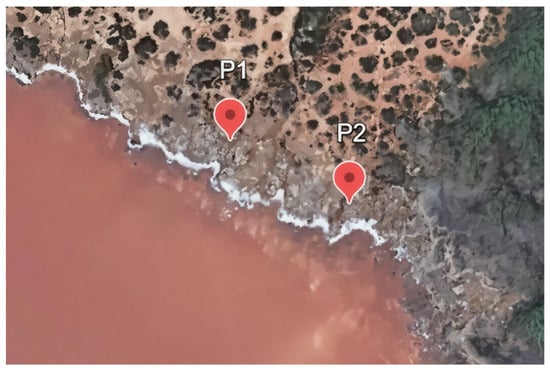
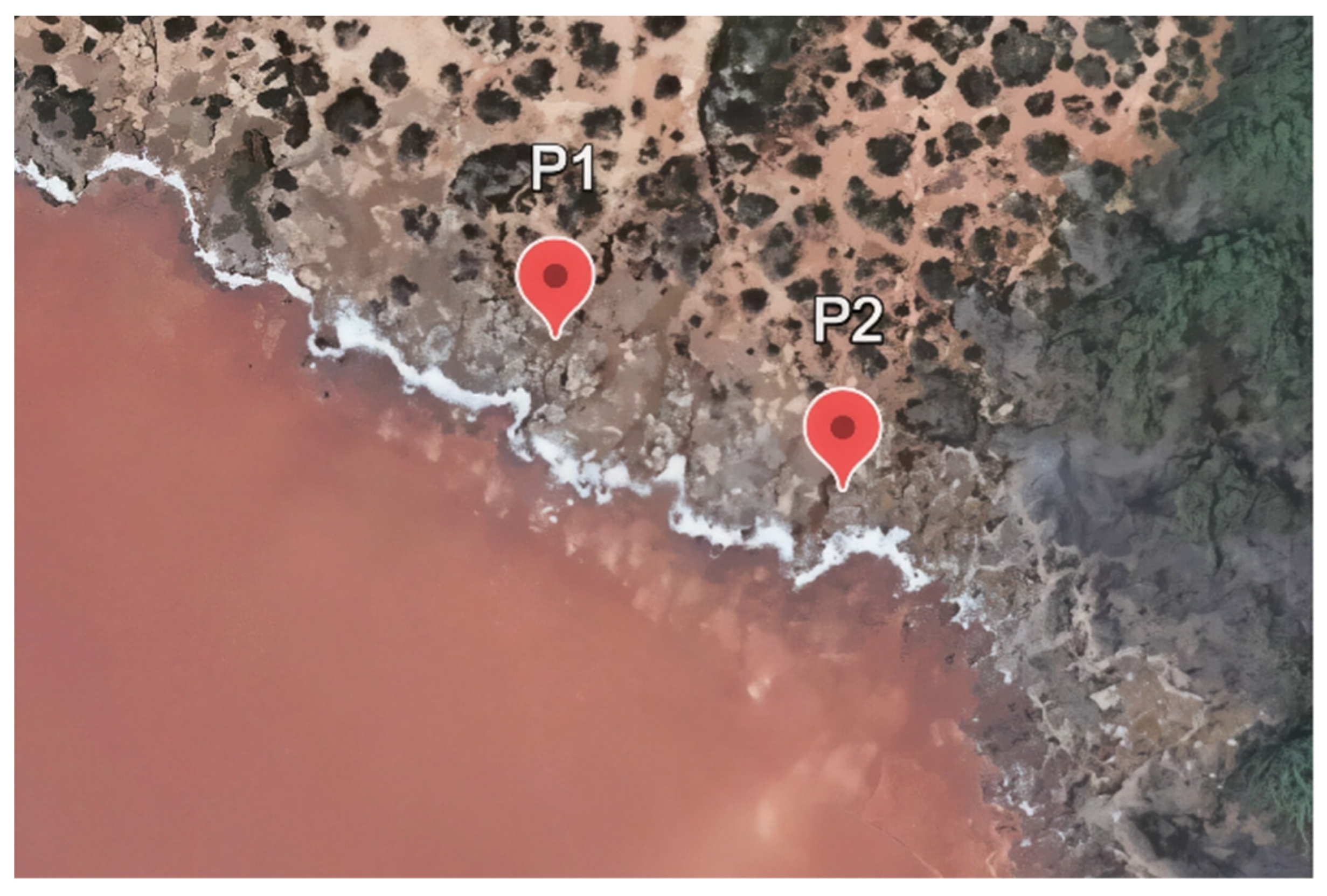
Water and sediment samples were collected from the Laguna Rosa (Torrevieja, Spain) on 18 September 2024. To capture the heterogeneity of the microbial communities present, samples were collected from two designated sampling points, P1 (38.016830, −0.707360) and P2 (38.0160171, −0.7048908) (Figure 1). At P1, three samples were collected from areas exhibiting different physical and chemical properties (P1S1, P1S2 and P1S3). In the P2 area, six samples were collected from two areas, with three replicates from each area (P2S1.1–P2S1.3 and P2S2.1–P2S2.3).
Figure 1.
Sampling locations in the Laguna Rosa, Torrevieja (Alicante, Spain). Google Earth.
The physicochemical properties of the two sampling points were characterised in situ by measuring salinity using an optical refractometer (101 ATC, MS Schippers, Barcelona, Spain) and pH using a PRO waterproof pH/Temp Tester (Milwaukee, WI, USA). Due to the extremely high sodium content in the Laguna Rosa (Torrevieja), it was not possible to determine the Na+ concentration directly. Instead, elemental analysis of water was carried out at the Research Technical Services (SSTTI) of the University of Alicante using inductively coupled plasma mass spectrometry (ICP–MS) and inductively coupled plasma optical emission spectrometry (ICP–OES).
Relative proportions of the major cation (Na+) and the other dominant cations were calculated from ICP–MS and ICP–OES results to formulate a concentrated synthetic seawater (SW) stock solution at 30% (w/v). The salinity of the final SW solution was confirmed using an optical refractometer. The composition of the synthetic seawater was as follows: 260 g/L NaCl, 30 g/L MgCl2·6H2O, 35 g/L MgSO4·7H2O, 7.5 g/L KCl, 0.4 g/L CaCl2·2H2O, 0.2 g/L NaHCO3, 0.8 g/L KBr, 0.35 g/L H3BO3, 0.018 g/L LiCl, and 0.003 g/L RbCl.
2.2. Culture Media
Two media were used to cultivate heterotrophic microorganisms: defined medium (DM) and complex medium (CM), at 18% and 25% SW, respectively. Both media were adjusted to pH 7.2 using Tris-HCl. Alternatively, F2 medium (Guillard’s f/2 medium) was used to cultivate photosynthetic microorganisms at salinities of 3%, 8%, and 18%, with a neutral pH of 7.4–7.8 (Table 1).
Table 1.
Composition of culture media used for microorganism isolation.
2.3. Isolation and Cultivation of Microorganisms
Two complementary strategies were used to isolate microorganisms from the Laguna Rosa samples. To recover photosynthetic and heterotrophic microorganisms from enrichment cultures, sediment samples (P1S1, P1S2, P1S3, P2S1.1, and P2S1.2) were resuspended in synthetic seawater adjusted to match the in situ salinity at the corresponding sampling sites. These suspensions were then spread onto agar plates prepared with SW at salinities ranging from 3 to 25%. In brief, the samples were diluted 1:10, and 100 μL of the resulting suspensions were spread onto the agar plates, which were then incubated for 7–14 days. Photoautotrophic isolates were maintained in a controlled growth chamber under constant light, temperature, and humidity. In contrast, heterotrophic isolates were incubated in humid chambers at either 20 °C or 37 °C when archaeal growth was suspected. Isolates were purified by repeatedly streaking single colonies until phenotypic homogeneity was confirmed by direct microscopic observation.
A second isolation strategy involved culturing heterotrophic microorganisms in selective liquid media containing 18% SW supplemented with various heavy metals. The solid samples P2S1.2 and P2S2.3, as well as the liquid sample P2S1.1, were used as inocula. The cultures were then grown in the presence of increasing concentrations of NaAsO2 (0, 1, 2, 5, or 15 mM), K2CrO4 (0, 1, 2, 5, or 15 mM), or LiCl (0, 0.25, 0.5, 1, or 2 M). The cultures were incubated at 37 °C with shaking at 180 rpm, and cell growth was monitored by measuring the optical density at 600 nm (OD600). Samples with an OD600 value above 0.3 were streaked onto solid media with the same CM and DM compositions but supplemented with 1% (w/v) agar. Plates were supplemented with 2, 3, or 15 mM NaAsO2 or K2CrO4 for arsenite and chromate, and with 2 or 3 M LiCl for lithium. All plates were inoculated with 20 μL of liquid culture and incubated at 37 °C for 7–14 days. All these isolation experiments were performed in triplicate.
2.4. Molecular Identification of Isolates by 16S rRNA Gene Sequencing
Individual colonies were subjected to colony PCR to amplify the 16S or 18S ribosomal RNA genes for sequencing. The following primers were used: BacFor (5′-AGAGTTTGATCATGGCTCAG-3′) and BacRev (5′-CGGCTACCTTGTTACGACTT-3′) for bacteria; ArchFor (5′-ATTCCGGTTGATCCTGC-3′) and ArchRev (5′-ACGGHTACCTTGTTACGACTT-3′) for archaea; and EukaFor (5′-GTCAGAGGTGAAATTCTTGGAT-3′) and EukaRev (5′-AGGGCAGGGACGTAATCA-3′) for eukaryotes. PCR amplifications were performed using NZY Taq DNA polymerase (NZYTech, Lisbon, Portugal) under the following cycling conditions: initial denaturation at 95 °C for 3 min; 36 cycles of 95 °C for 30 s, 57 °C for 45 s, and 72 °C for 1.2 min; followed by a final extension at 72 °C for 8 min.
PCR products were resolved by electrophoresis on 1% (w/v) agarose gels. DNA bands were then purified using the NZYGelpure kit (NZYTech, Lisbon, Portugal), following the manufacturer’s instructions, and were subsequently sequenced by the StabVida Genetics Laboratory (Caparica, Portugal). The obtained sequences were analysed using Chromas v2.6.6 (Technelysium Pty Ltd., South Brisbane, Australia). Sequence identities were determined by comparing them with the non-redundant nucleotide database (nr/nt) using BLASTn v.2.17.0 at the National Center for Biotechnology Information (NCBI, Bethesda, MD, USA).
2.5. Screening of Heavy Metal Tolerance
The heavy metal tolerance of the isolated microorganisms was assessed using agar plates supplemented with varying concentrations of Pb(NO3)2, NaAsO2, K2CrO4 or LiCl, as summarised in Table 2. The plates were incubated for 7–14 days at temperatures of 20 °C for photoautotrophic and bacterial isolates and at 42 °C for archaeal isolates. Growth was evaluated based on the presence or absence of colonies, as well as their size, pigmentation and morphology. All assays were performed in three biological replicates.
Table 2.
Heavy metal concentrations evaluated in tolerance assays of heterotrophic and photoautotrophic isolates.
2.6. Evaluation and Quantification of Heavy Metal Tolerance in Liquid Cultures of Selected Isolates
Promising isolates identified in preliminary screenings were evaluated for their tolerance to heavy metals in liquid cultures. Halorubrum saccharovorum and Haloarcula sinaiiensis were cultivated at 42 °C with constant shaking at 200 rpm in MC medium at 10% SW. In parallel, Marinococcus halophilus, Marinobacter adhaerens, Halomonas sp., Halomonas smyrnensis, and an unclassified halophilic bacterium were cultivated at 37 °C with constant shaking at 200 rpm in CM at 8% SW using a Stratus microplate reader (Cerillo, Charlottesville, VA, USA). To assess metal tolerance, the cultures were exposed to increasing concentrations of Pb(II) (0.5, 0.75, 1.0 and 1.5 mM), As(III) (1, 2, 3 and 4 mM) and Cr(VI) (1, 2, 3, 4, 15, 20 and 25 mM).
Cultures were initiated at an optical density (OD600) of 0.02 using exponential-phase preinocula. Growth was monitored over time by measuring the OD600, and the specific growth rate (μ) and doubling time (td) were calculated as described by Matarredona et al. (2021) [23]. All experiments were performed in triplicate.
Chromium removal by selected isolates Hrr. saccharovorum (2 mM K2CrO4), H. smyrnensis (15 mM K2CrO4), and the unclassified halophilic bacterium (2 mM K2CrO4) were evaluated using ICP-MS. Control medium samples were collected before inoculation (time 0). The cultures were incubated under their respective growth conditions, and aliquots of the culture medium were collected at 24 and 48 h. The cells were removed by centrifugation at 10,000× g for 10 min, and the resulting supernatant was analysed by ICP-MS to determine chromium concentrations. The efficiency of chromium removal by each isolate was expressed as the percentage reduction in chromium content relative to the initial concentration and was calculated using the following formula:
where Co is the chromium concentration in the medium at time 0 (control) and Ct is the chromium remaining after growth of the selected isolates.
2.7. Statistical Analysis
Statistical analyses were performed using GraphPad Prism software (Version 8), according to the methodology described by Matarredona et al. (2021) [23]. All data presented in the figures are expressed as the mean ± standard deviation (SD) of three independent replicates. To evaluate significant differences between each stress treatment and the control group, Brown–Forsythe and Welch one-way ANOVA tests were performed, followed by Dunnett’s T3 multiple comparisons test. Statistical significance was established at p < 0.05.
3. Results
3.1. Characterisation of the Sampling Area
The physicochemical properties and visual appearance of the samples are summarised in Table 3. The results showed significant variations in pH, salinity, and appearance among samples, reflecting the presence of distinct microenvironments within the sampling area. The pH values of the liquid samples (P1S1, P1S2, and P2S2.1–P2S2.3) ranged from neutral to slightly alkaline (7.1–8.1). Alkaline pH was recorded in greenish water and runoff water. In contrast, the pH could not be determined for samples P1S3, P2S1.1, P2S1.2, and P2S1.3 because they were solid.
Table 3.
Physicochemical characteristics and sample types.
Salinity values showed high variability, ranging from 1% to 40%. As with pH measurements, salinity could not be determined in solid samples. Sample P1S1 exhibited the lowest salinity value, consistent with its association with runoff water and a cyanobacterial mat. The highest salinity value was recorded in sample P1S2 (40%), comparable to hypersaline environments where photosynthetic activity is inhibited. Based on their visual appearance, the greenish water samples and cyanobacterial mats differed markedly from the salt crusts and pink-coloured water samples.
The ionic composition of the collected water samples was characterised, and the main cation concentrations were quantified using ICP-OES analysis for the design of artificial seawater. Comparing the two water samples showed that the cationic ratios remained constant, indicating that the synthetic seawater adequately reproduces the ionic composition of natural lagoon water (Table S1, Supplementary Material). ICP-MS analysis revealed the presence of lithium, chromium, arsenic, and lead in the water samples. The concentrations of these elements were 4.947 mg/L for lithium, 0.003 mg/L for chromium, 0.014 mg/L for arsenic, and 0.107 mg/L for lead.
3.2. Isolation, Identification and Screening for Heavy Metal Tolerance
A total of 48 microbial isolates were obtained from enrichment cultures, comprising 13 Archaea, 21 Bacteria, and 14 Eukarya (Table 4). Given the large number of samples and the added complexity of culturing photosynthetic microorganisms, the results shown in Table 4 are based solely on plate isolations. In contrast, liquid cultures were also performed using selective media supplemented with different metal concentrations. Relying exclusively on plate cultures could introduce bias towards microorganisms that only grow on solid media. Therefore, it was important to use liquid cultures to recover microorganisms that might not be detected on plates. This strategy resulted in the isolation of an additional 31 microorganisms, of which 20 belonged to the Bacteria domain and 11 to the Archaea domain (Table 5). High sequence similarity values generally supported confident taxonomic assignment. Photosynthetic isolates were obtained from greenish-coloured samples (P1S1, P1S3, P2S1.3, P2S2.1, P2S2.2 and P2S2.3). These samples were considered suitable for growing microalgae and cyanobacteria due to their lower salinity and alkaline pH. In contrast, haloarchaea and halotolerant bacteria were isolated from the pink, high-salinity samples (P1S1, P1S2, P2S1.1, P2S1.2 and P2S1.3). Enriched media yielded a higher number and diversity of isolates due to the absence of metal restriction. Within the Archaea domain, the most abundant genera recovered by both strategies were Halorubrum and Haloferax, members of the family Halobacteriaceae. Among heterotrophic bacteria, the genera Halomonas, Marinobacter, Salinivibrio, Pontibacillus, and Alkalinibacillus were detected; these are all widely distributed in solar salterns and hypersaline lagoons. Among photosynthetic microorganisms, the genus Dunaliella sp. dominated the eukaryotes. At lower salinities, isolates were obtained from members of the genera Tetraselmis sp. and Chlamydomonas, together with Chrysotila cartae. Filamentous cyanobacteria such as Sodalinema sp. were also isolated.
Table 4.
Isolation, identification, and metal tolerance of isolates obtained using enriched media.
Table 5.
Isolation and identification of microorganisms isolated using selective media.
Four metals of interest were selected to identify the isolates with the greatest bioremediation potential: lithium, lead, chromium and arsenite, all of which are widely documented in environments impacted by human activity. Metal tolerance tests revealed variations among the isolates across domain, genus, and evaluated metal, as shown in Table 4 and Table 5. Haloarchaea, particularly the Halorubrum genus and, to a lesser extent, the Haloferax genus, showed high tolerance to lithium. All isolates were capable of growth in the presence of 2 M lithium, with some, such as Haloferax prahovense, tolerating concentrations of up to 4 M.
The resistance profile of the heterotrophic bacteria differed from that observed in the archaea. In isolates obtained from enriched media (Table 4), genera such as Halomonas sp., Marinobacter sp., and Marinococcus sp. tolerated moderate concentrations of Cr(VI) (up to 5 mM). In contrast, growth was observed at 15 mM chromium in different species of Halomonas sp., Marinobacter sp., Salinivibrio sp., and Alkalinobacillus sp. in selective media (Table 5). Arsenite resistance ranged from 2 to 5 mM, primarily in Halomonas sp. In contrast, bacterial tolerance to lithium was lower than that observed in archaea, with values generally between 1 and 3 M lithium in enriched media (Table 4) and no growth in selective media (Table 5).
No chromium-resistant strains were obtained among the photosynthetic isolates, suggesting that chromium was the most growth-restrictive metal. Cyanobacterial isolates, including Sodalinema sp. and Synechococcus sp., showed moderate tolerance to lithium (0.25–0.5 M), arsenite (2–5 mM), and lead (0.25–1 mM). However, these levels are lower than those observed in halophilic eukaryotes. Among eukaryotes, Dunaliella sp. exhibited the highest tolerance to metals, with a tolerance of up to 15 mM arsenite and 1 M lithium. These characteristics make Dunaliella sp. a promising candidate for treating wastewater in hypersaline conditions.
3.3. Evaluation of Heavy Metal Tolerance in Liquid Culture and Metal Quantification
Of all the isolated microorganisms, five bacterial isolates (M. adhaerens, M. halophilus, Halomonas sp., H. smyrnensis, and an unclassified halophilic bacterium) and two haloarchaeal isolates (Har. sinaiiensis and Hrr. saccharovorum) were selected for testing in a liquid medium. This selection was based on the results of the metal tolerance screening, including the isolates that showed the greatest tolerance and the most reliable taxonomic identification. The analysis focused on the same three metals, previously tested (arsenite, lead, and chromium) due to their toxicological and environmental relevance. Lithium was excluded because it is not a heavy metal and has lower toxicological relevance. The experiments were carried out at reduced salinity levels (8% and 10%) to better simulate industrial effluents. The specific growth rate (μ) and doubling time (td) were determined for each isolate during the exponential phase at different metal concentrations (Table S2, Supplementary Material).
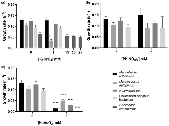
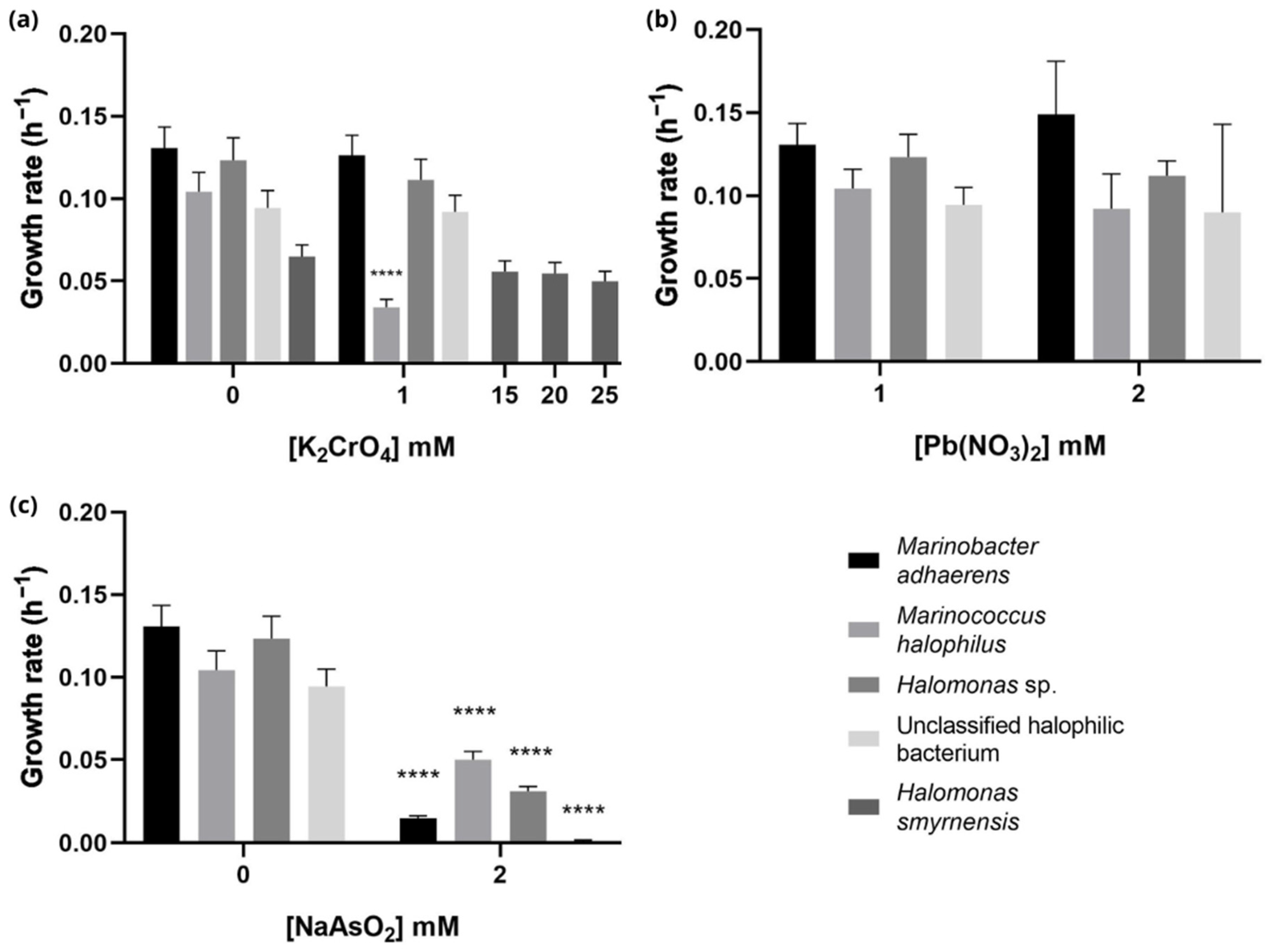
In bacterial assays, each metal had a distinct effect on bacterial growth. At 1 mM Cr(VI), the growth rates of M. adhaerens, M. halophilus, Halomonas sp., and the unclassified halophilic bacterium were similar to those of the control, suggesting high resistance to low concentrations of chromium (Figure 2). This finding is consistent with previous plate analysis, in which these species were able to grow in the presence of 2 mM chromium. In contrast, H. smyrnensis, which was isolated from a medium containing 15 mM Cr(VI), exhibited a remarkable ability to grow in the presence of up to 25 mM chromium, with only minor decreases in its growth rate compared to the control (Figure 2). Arsenite was the most toxic metal evaluated, producing strong growth inhibition in halophilic bacteria and M. adhaerens. M. halophilus proved to be the most tolerant species, with its growth rate falling from 0.104 h−1 in the control to 0.092 h−1. Exposure to 1 mM lead produced effects similar to those of chromium.
Figure 2.
Growth rates of M. adhaerens, M. halophilus, Halomonas sp., an unclassified halophilic bacterium, and H. smyrnensis at different metal concentrations: (a) chromium; (b) lead; and (c) arsenite. Plotted values are the mean of triplicate measurements, and error bars represent ± SD. **** p ≤ 0.0001.
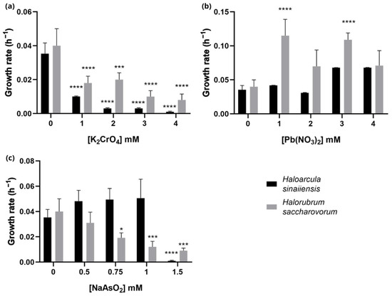
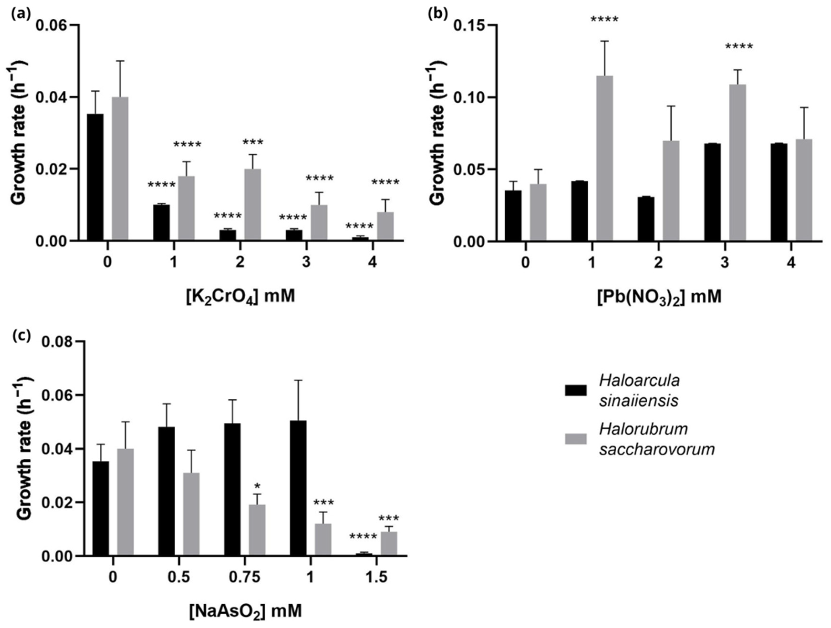
Haloarchaea showed a progressive reduction in growth with increasing chromium concentrations, with near-complete inhibition at 3–4 mM (Figure 3). Exposure to arsenite was highly inhibitory at 1.5 mM in both species. In contrast, exposure to lead had the opposite effect: the growth rate increased relative to the control as the lead concentration increased. Adding lead to the culture medium resulted in the formation of a white precipitate, which may have affected the spectrophotometric measurements of growth. Consequently, the higher OD600 values observed in the presence of lead may partly result from light scattering by suspended precipitates rather than haloarchaeal growth. Therefore, these results should be interpreted with caution, as OD600 measurements may overestimate growth under these conditions.
Figure 3.
Growth rates of Har. sinaiiensis and Hrr. saccharovorum at different metal concentrations: (a) chromium; (b) lead; and (c) arsenite. Plotted values are the mean of triplicate measurements, and error bars represent ± SD. * p ≤ 0.05; *** p ≤ 0.001; **** p ≤ 0.0001.
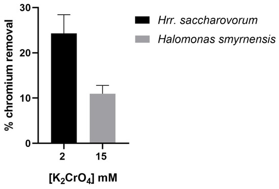
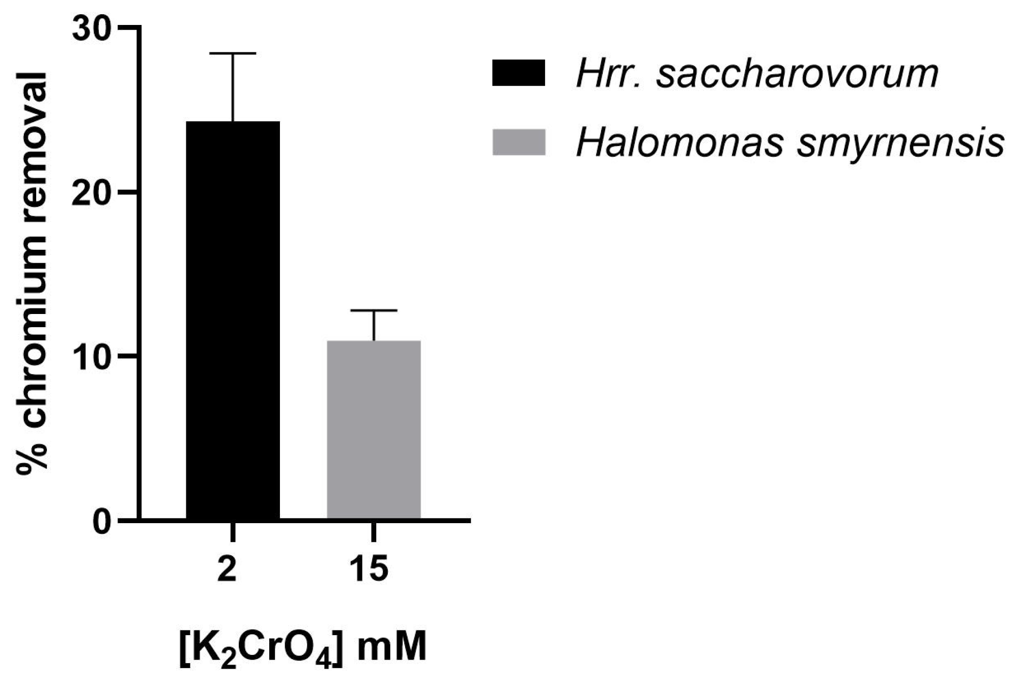
The potential of a microorganism to act as a bioremediation agent depends on its ability to grow in the presence of the contaminant and to remove it from the environment. This study evaluated the ability of H. smyrnensis and Hrr. saccharovorum to remove chromium from the culture medium at initial concentrations of 15 mM and 2 mM, respectively. The results revealed that H. smyrnensis reduced the chromium concentration by 10.96% at 15 mM, whereas Hrr. saccharovorum achieved a 24.29% reduction at 2 mM (Figure 4).
Figure 4.
Chromium removal efficiency by Hrr. saccharovorum and H. smyrnensis at different initial K2CrO4 concentrations. Plotted values are the mean of triplicate measurements, and error bars represent ± SD.
4. Discussion
Physicochemical characterisation of Laguna Rosa revealed significant variation in pH, salinity and sample appearance, reflecting the dynamic nature of this hypersaline ecosystem. Such environmental gradients are characteristic of solar salterns and hypersaline lagoons, where evaporation, freshwater inputs and salt precipitation continuously modify local physicochemical conditions. This generates distinct ecological niches that determine the composition of the microbial community [35,36]. The slightly alkaline pH measured in the greenish water samples is consistent with intense photosynthetic activity. Meanwhile, the wide salinity range observed among sampling points reflects the coexistence of environments favourable to both phototrophic microorganisms and extremely halophilic prokaryotes [37].
The visual appearance of the different samples closely reflected the taxonomic composition of the cultivable microbiota. Photosynthetic microorganisms dominated greenish waters and cyanobacterial mats, whereas pink waters and salt crusts predominantly yielded haloarchaeal and halophilic bacterial isolates. This distribution is consistent with the well-established ecological succession occurring along salinity gradients in hypersaline systems, where moderate salinity favours cyanobacteria and microalgae, while increasing salt concentrations progressively select for haloarchaea and specialised halophilic bacteria [19,38].
The presence of metals in aquatic and terrestrial ecosystems is one of the most significant environmental problems due to their persistence, toxicity, and ability to bioaccumulate. While these metals can occur naturally in the environment through geological processes, anthropogenic activities have intensified their mobilisation and accumulation in bodies of water, posing a risk to human health and ecosystems [39]. Concentrations of arsenic and lead exceeded World Health Organization (WHO) drinking water standards. In contrast, lithium concentrations were considerably higher than those generally reported for freshwater environments but within the range previously described for hypersaline systems [40,41,42,43]. The presence of arsenic in hypersaline environments ranges from tens of µg·L−1 to extremely high levels, highlighting these environments’ ability to accumulate this metal. For instance, hypersaline lakes in Bolivia have been reported to have arsenic levels of 82 mg/L [44]. Furthermore, in saline environments, high salinity and complexation with chlorides can increase the solubility and persistence of lead in the aqueous phase [45]. In the case of chromium, although the reported value is lower than the WHO established value, the risk depends not only on its concentration but also on its oxidation state. Hypersaline environments present redox and pH conditions that favour the conversion of less toxic and less mobile Cr(III) to highly toxic and more soluble Cr(VI) [11,46].
Hypersaline environments are extreme habitats in which the physicochemical conditions, such as high salt concentrations, elevated temperatures, pH fluctuations, and limited nutrient and oxygen availability, severely restrict life. Despite these challenges, these ecosystems harbour a remarkable diversity of microorganisms, encompassing representatives from all three domains of life: Archaea, Bacteria and Eukarya. These microorganisms typically require high salt concentrations to survive and grow [19,38]. Many halophilic and halotolerant microorganisms have evolved physiological and molecular mechanisms that enable them to tolerate not only high salinity, but also high concentrations of metals. Consequently, these microbial communities play a pivotal role in regulating these elements [22]. In this study, heavy metal tolerance was evaluated not only for characterisation but also as a selection criterion for halophilic and halotolerant microorganisms with potential application in bioremediation. Two different strategies were used to isolate microorganisms. First, isolation was performed on selective media supplemented with varying metal concentrations, to facilitate the identification of species with bioremediation potential. However, this approach has limitations, as it can restrict the diversity of microorganisms recovered, selecting for those capable of tolerating metals under culture conditions that may not be representative of those in situ. To overcome these limitations, this strategy was combined with isolation in enrichment media, which allowed greater recovery of microbial diversity. This enabled the isolation of a broad spectrum of microorganisms and yielded a more representative sample of the ecosystem. However, enrichment-based isolation has its own limitations, such as the need for subsequent screening to assess metal tolerance and the isolation of microorganisms that are not particularly relevant to bioremediation. Therefore, combining both strategies maximises the isolation of microbial diversity while identifying metal-tolerant species of interest for bioremediation.
The taxonomic composition of the isolates was consistent with that described in previous studies of microbial communities inhabiting hypersaline ecosystems. The dominant archaeal isolates were members of the genera Halorubrum and Haloferax, in line with their recognised ecological success in saline lakes, salterns and salt crusts worldwide [19,38,47]. Similarly, the bacterial isolates were predominantly from genera commonly associated with hypersaline habitats, such as Halomonas, Marinobacter, Salinivibrio, Pontibacillus and Alkalinibacillus [48,49,50,51]. The predominance of haloarchaea among the isolates is also consistent with culture-independent studies showing that archaeal populations generally dominate microbial communities as salinity approaches saturation [19,52]. Among the photosynthetic microorganisms, Dunaliella was the predominant eukaryotic genus in the lagoon, whereas Tetraselmis, Chlamydomonas, Chrysotila carterae, and filamentous cyanobacteria were mainly found at sampling sites with lower salinity. This distribution reflects the different osmoadaptation strategies developed by these organisms. Dunaliella is particularly well adapted to hypersaline environments because it accumulates intracellular glycerol as its principal compatible solute, enabling growth over an exceptionally broad salinity range [53,54]. In contrast, many cyanobacteria and other microalgae exhibit greater sensitivity to rising salinity, which helps explain their predominance in the less saline microhabitats identified during sampling.
Despite this general trend, clear differences in metal tolerance were observed among microbial groups. Haloarchaea consistently exhibited the highest tolerance to lithium, with several isolates remaining viable at concentrations of up to 4 M. Such remarkable resistance has previously been associated with the exceptional ionic homeostasis characteristic of extremely halophilic archaea. These microorganisms possess highly specialised membrane transport systems and efficient antioxidant defences, which minimise the combined effects of osmotic and metal stress [23,24,55]. In addition, their cell surface architecture may contribute to metal tolerance, as the S-layer and extracellular polymeric substances (EPS) provide metal-binding sites that can immobilise metal ions at the cell surface, potentially limiting their intracellular accumulation. Recent work with the haloarchaeon Haloferax alexandrinus demonstrated efficient metal adsorption by dried biomass and attributed this capacity, in part, to the involvement of S-layer-associated functional groups [56]. The high lithium tolerance observed in the present study may therefore be partly related to the ability of haloarchaea to biosorb lithium at the cell surface, potentially mediated by metal-binding sites associated with the S-layer and EPS. While haloarchaea exhibited exceptional lithium tolerance, bacterial isolates displayed a different resistance profile, showing greater tolerance to chromium and arsenite. Notably, Halomonas sp. maintained growth over a broad range of chromium concentrations and showed consistent resistance to arsenite. This behaviour is consistent with the remarkable metabolic versatility previously observed in this genus, whose members possess multiple mechanisms for metal detoxification, including extracellular biosorption, chromate reduction, active efflux systems, and arsenic resistance operons [18,29,57,58,59,60]. Rather than relying on a single resistance mechanism, these microorganisms likely combine several complementary physiological responses to tolerate simultaneous exposure to high salinity and toxic metals. This versatility explains why members of Halomonas have attracted increasing interest in the treatment of saline industrial wastewaters.
The responses observed in Marinobacter and Marinococcus further demonstrate the functional diversity of halophilic bacteria. Although Marinobacter species are recognised for their role in arsenic biogeochemical cycling through arsenate respiration and arsenite oxidation [61], the marked growth inhibition observed upon arsenite exposure suggests that the presence of arsenic transformation pathways does not necessarily confer high tolerance to As(III). As this element directly interferes with protein function and cellular redox homeostasis, its toxicity may exceed the detoxification capacity of these metabolic pathways at high concentrations. This observation is consistent with the results of the present study, in which arsenite was found to be the most inhibitory metal for the majority of bacterial isolates.
The behaviour of the photosynthetic microorganisms differed from that of haloarchaea and heterotrophic bacteria. None of the phototrophic isolates exhibited significant chromium resistance, suggesting that chromium is among the most restrictive metals for oxygenic photosynthesis under the tested experimental conditions. Chromium toxicity has been linked to the production of reactive oxygen species and the disruption of the photosynthetic apparatus after uptake via sulphate transporters [62], which may explain the absence of chromium-resistant cyanobacteria and microalgae among the isolates. However, cyanobacteria showed moderate tolerance to lithium, arsenite and lead, likely due to the protective role of extracellular polymeric substances that bind metal ions and reduce their bioavailability [31,63].
Among the photosynthetic microorganisms, Dunaliella showed the greatest tolerance to metal exposure, particularly to lithium and arsenite. In addition to its well-established osmoadaptation mechanisms involving intracellular glycerol accumulation, Dunaliella produces extracellular polysaccharides that can bind metal ions. This reduces their toxicity and facilitates metal immobilisation [32,64,65,66,67]. The combination of efficient osmotic regulation and metal-binding mechanisms makes this microalga an attractive candidate for future applications involving the treatment of hypersaline wastewater contaminated with metals.
Industrial wastewaters and landfill leachates often contain high concentrations of salts (up to 10%) and heavy metals. This represents a significant source of environmental pollution, threatening both ecosystems and human health [11,68]. Consequently, it is more realistic to assess microbial performance under moderate salinity in relation to future biotechnological applications.
Analysis of the bacterial isolates revealed that H. smyrnensis exhibited the highest chromium tolerance, maintaining active growth even at concentrations of up to 25 mM Cr(VI). Similar results have been described in Halomonas sp. strains isolated from industrial effluents and contaminated hypersaline environments, where high salinity and metal concentrations select for microorganisms with robust detoxification systems (Table S3, Supplementary Material). Members of this genus can tolerate high concentrations of chromium. They can also biotransform the highly toxic and mutagenic Cr(VI) into the relatively less toxic Cr(III) via enzymatic reduction mediated by chromate reductases [56,57,58,59,69]. However, further studies are required to determine whether the H. smyrnensis strain investigated in the present study possesses a chromate reductase and whether Cr(VI) removal is mediated, at least in part, by enzymatic biotransformation to Cr(III). These findings position H. smyrnensis as a promising candidate for bioremediation strategies in saline environments contaminated with chromium. Arsenite-resistance plasmid operons containing genes for metal sensors, metal chaperones, metal transporters and detoxifying enzymes have also been detected in these microorganisms [22]. Recent studies have shown that species such as Halomonas elongata can tolerate up to 25 mM of arsenate due to complete ars operons [29]. The genus Marinobacter sp. includes metabolically versatile species, some of which participate in arsenic biogeochemical cycles. For example, Marinobacter santoriniensis can respire arsenate and oxidise arsenite, indicating the presence of specific metabolic pathways for arsenic transformation [60]. However, the presence of these pathways does not guarantee arsenite tolerance, as they can affect the redox machinery and ribosomes, thereby exceeding the detoxifying capacity at high concentrations. Exposure to 1 mM lead produced effects similar to those of chromium. Proposed mechanisms of lead tolerance include extracellular precipitation and sequestration by exopolysaccharides [70]. Several studies have investigated microorganisms such as Halomonas sp. for their tolerance of lead and potential to bioremediate it, as they can precipitate or adsorb it [71,72,73]. For instance, Abdel-Razik et al. (2020) [59] reported that a Halomonas sp. Strain removed 94% of lead within 6 h.
In the case of haloarchaea, previous studies by Matarredona et al. (2024) [24] characterised the growth of Har. sinaiiensis in the presence of chromium, determining an MIC (minimum inhibitory concentration) of 2.5 mM. These assays were performed under high-salinity conditions (20% NaCl), whereas the salinity was reduced to 10% in the present study. This difference may explain the lower tolerance observed, given that the conditions were outside the species’ optimal growth range. Halorubrum sp. species, such as Hrr. californiensis and Hrr. litoreum, have been reported to exhibit MIC values of 1 and 2 mM chromium, respectively. In the case of lithium, these values are lower than those observed in the present study, in which growth was still detected at concentrations up to 4 mM (Table S3). Matarredona et al. (2024) [24] demonstrated that Har. sinaiiensis and Halorubrum sp. can grow in the presence of up to 12 mM of arsenate (As(V)). However, given that As(III) is more toxic than As(V), growth inhibition is likely to occur at significantly lower concentrations.
Although resistance to toxic metals is a prerequisite for their potential application in bioremediation, it does not necessarily guarantee efficient contaminant removal. For this reason, chromium removal assays were performed using the isolates with the highest chromium tolerance. Both H. smyrnensis and Hrr. saccharovorum were capable of reducing chromium concentrations in the culture medium. However, the removal efficiencies were lower than those reported for previously described bacterial strains. Previous studies have shown that different Halomonas sp. strains can effectively remove up to 80% of chromium at an initial concentration of 50 mg/L [59], although removal efficiency decreases progressively at higher concentrations. Other studies report reductions of close to 60% even at 50 mg/L, demonstrating significant variability depending on the strain and experimental conditions. Similarly, it has been reported that salinity significantly influences the bioavailability of chromium and the capacity for microbial biosorption. High NaCl concentration increases the ionic strength of the medium, which can weaken the electrostatic interactions between Cr(VI) and the charged functional groups on cell surfaces, thereby reducing chromium biosorption [74]. Furthermore, high salinity could affect cellular metabolic activity and consequently the biological mechanisms involved in Cr(VI) reduction [75]. The relatively low removal efficiency observed in the present study can be partly explained by the high initial chromium concentration, which could have a toxic effect on cell growth and activity [76]. Despite the limited experimental evidence available on the bioremediation of chromium in haloarchaea, these findings provide a basis for further investigation of their potential for chromium removal under hypersaline conditions.
5. Conclusions
Laguna Rosa in Torrevieja is a highly heterogeneous hypersaline ecosystem, with physicochemical conditions that vary significantly between microenvironments. Field measurements and chemical analyses confirmed significant variations in salinity and pH, as well as the presence of environmentally relevant concentrations of lithium, arsenic, and lead.
Culture-dependent screening showed that the lagoon is home to a diverse range of halophilic and halotolerant microorganisms from the three domains of life, including haloarchaea, halophilic and halotolerant bacteria, and photosynthetic microorganisms. Metal tolerance assays revealed distinct response patterns among taxa and contaminants. Haloarchaea showed particularly high tolerance to lithium; several bacterial isolates tolerated chromium and arsenite; and Dunaliella sp. exhibited broad tolerance to the metals tested.
Among the isolates tested in liquid culture, H. smyrnensis was the most chromium-tolerant bacterium, growing at concentrations of up to 25 mM Cr(VI). Hrr. saccharovorum also exhibited notable tolerance and chromium-removal capacity. Although the observed reductions in chromium removal were modest under the tested conditions, these results show that indigenous halophilic microorganisms can contribute to metal attenuation in saline systems. Overall, this work demonstrates that the microbiota of Laguna Rosa includes halophilic and halotolerant microorganisms with remarkable resistance to chromium, arsenite, lead and lithium, highlighting their potential for bioremediation applications in saline and hypersaline environments, including the treatment of industrial wastewater contaminated with heavy metals.
Supplementary Materials
The following supporting information can be downloaded at https://www.mdpi.com/article/10.3390/environments13090512/s1. Table S1. Composition of the artificial saline medium compared with Laguna Rosa water based on ICP analysis. Salt amounts used for medium preparation, resulting ion concentrations in the artificial medium, and the corresponding concentration ratios between Laguna Rosa water and the artificial medium are shown. Table S2. Effect of different metal concentrations on the growth kinetics of the selected isolates. Table S3. Reported metal tolerance and resistance of selected halophilic and halotolerant microorganisms to chromium, arsenic, lithium, and lead.
Author Contributions
Conceptualization, J.E., B.Z. and M.C.; methodology, N.L.-G., A.C.-C., S.P. and B.Z.; validation, N.L.-G., B.Z. and J.E.; formal analysis, J.E. and M.C.; investigation, N.L.-G., A.C.-C., S.P. and B.Z.; resources, M.C. and J.E.; data curation, J.E. and B.Z.; writing—original draft preparation, N.L.-G. and J.E.; writing—review and editing, J.E., N.L.-G. and M.C.; supervision, J.E. and B.Z.; project administration, J.E.; funding acquisition, J.E. All authors have read and agreed to the published version of the manuscript.
Funding
This work was supported by grants from the Valencian Conselleria d’Innovació, Universitats, Ciencia y Societat Digital grant CIAICO/2022/047, Universidad de Alicante (VIGROB-016), Spanish Ministry of Science and Innovation (MCIN) (EQC2018-004065-P) and “ERDF: A Way to Build Europe”.
Data Availability Statement
Data are contained within the article and Supplementary Materials.
Acknowledgments
We want to thank María José Bonete for their valuable assistance and helpful advice throughout this study.
Conflicts of Interest
The authors declare no conflict of interest.
References
- Goswami, R.K.; Agrawal, K.; Shah, M.P.; Verma, P. Bioremediation of heavy metals from wastewater: A current perspective on microalgae-based future. Lett. Appl. Microbiol. 2022, 75, 701–717. [Google Scholar] [CrossRef] [Scilit] [PubMed]
- Manikandan, A.; Suresh Babu, P.; Shyamalagowri, S.; Kamaraj, M.; Muthukumaran, P.; Aravind, J. Emerging role of microalgae in heavy metal bioremediation. J. Basic Microbiol. 2022, 62, 330–347. [Google Scholar] [CrossRef] [Scilit] [PubMed]
- Shah, V.; Daverey, A. Phytoremediation: A multidisciplinary approach to clean up heavy metal contaminated soil. Environ. Technol. Innov. 2020, 18, 100774. [Google Scholar] [CrossRef] [Scilit]
- Antwi, S.O.; Eckert, E.C.; Sabaque, C.V.; Leof, E.R.; Hawthorne, K.M.; Bamlet, W.R.; Chaffee, K.G.; Oberg, A.L.; Petersen, G.M. Exposure to environmental chemicals and heavy metals, and risk of pancreatic cancer. Cancer Causes Control 2015, 26, 1583–1591. [Google Scholar] [CrossRef] [Scilit] [PubMed]
- Capezzone, M.; Tosti Balducci, M.; Morabito, E.M.; Durante, C.; Piacentini, P.; Torregrossa, L.; Materazzi, G.; Giubbolini, G.; Mancini, V.; Rossi, M.; et al. High Incidence of Thyroid Cancer in Southern Tuscany (Grosseto Province, Italy): Potential Role of Environmental Heavy Metal Pollution. Biomedicines 2023, 11, 298. [Google Scholar] [CrossRef] [Scilit] [PubMed]
- Kanel, S.R.; Das, T.K.; Varma, R.S.; Kurwadkar, S.; Chakraborty, S.; Joshi, T.P.; Bezbaruah, A.N.; Nadagouda, M.N. Arsenic Contamination in Groundwater: Geochemical Basis of Treatment Technologies. ACS Environ. Au 2023, 3, 135–152. [Google Scholar] [CrossRef] [Scilit] [PubMed]
- Kuivenhoven, M.; Mason, K. Arsenic Toxicity. In StatPearls [Internet]; StatPearls Publishing: Treasure Island, FL, USA, 2026. [Google Scholar]
- Oziegbe, O.; Oziegbe, E.J.; Ojo-Omoniyi, O. Bioremediation of heavy metals in aquatic environment: A review. Clean. Chem. Eng. 2025, 11, 100193. [Google Scholar] [CrossRef] [Scilit]
- Adeel, M.; Zain, M.; Shakoor, N.; Ahmad, M.A.; Azeem, I.; Aziz, M.A.; Tulcan, R.X.S.; Rathore, A.; Tahir, M.; Horton, R.; et al. Global navigation of Lithium in water bodies and emerging human health crisis. npj Clean Water 2023, 6, 33. [Google Scholar] [CrossRef] [Scilit]
- Generalova, A.; Davidova, S.; Satchanska, G. The Mechanisms of Lead Toxicity in Living Organisms. J. Xenobiot. 2025, 15, 146. [Google Scholar] [CrossRef] [Scilit] [PubMed]
- Oladimeji, T.E.; Oyedemi, M.; Emetere, M.E.; Agboola, O.; Adeoye, J.B.; Odunlami, O.A. Review on the impact of heavy metals from industrial wastewater effluent and removal technologies. Heliyon 2024, 10, e40370. [Google Scholar] [CrossRef] [Scilit] [PubMed]
- Wani, A.L.; Ara, A.; Usmani, J.A. Lead toxicity: A review. Interdiscip. Toxicol. 2015, 8, 55–64. [Google Scholar] [CrossRef] [Scilit] [PubMed]
- Zhang, L.; Zhu, Y.; Gu, H.; Lam, S.S.; Chen, X.; Sonne, C.; Peng, W. A review of phytoremediation of environmental lead (pb) contamination. Chemosphere 2024, 362, 142691. [Google Scholar] [CrossRef] [Scilit] [PubMed]
- Chakdar, H.; Thapa, S.; Srivastava, A.; Shukla, P. Genomic and proteomic insights into the heavy metal bioremediation by cyanobacteria. J. Hazard. Mater. 2022, 424, 127609. [Google Scholar] [CrossRef] [Scilit] [PubMed]
- Ding, C.; Ding, Z.; Liu, Q.; Liu, W.; Chai, L. Advances in mechanism for the microbial transformation of heavy metals: Implications for bioremediation strategies. Chem. Commun. 2024, 60, 12315–12332. [Google Scholar] [CrossRef] [Scilit] [PubMed]
- Cheng, S.Y.; Show, P.L.; Lau, B.F.; Chang, J.S.; Ling, T.C. New Prospects for Modified Algae in Heavy Metal Adsorption. Trends Biotechnol. 2019, 37, 1255–1268. [Google Scholar] [CrossRef] [Scilit] [PubMed]
- Martínez-Espinosa, R.M. Halophilic archaea as tools for bioremediation technologies. Appl. Microbiol. Biotechnol. 2024, 108, 401. [Google Scholar] [CrossRef] [Scilit] [PubMed]
- Tavoosi, N.; Akhavan Sepahi, A.; Kiarostami, V.; Amoozegar, M.A. Arsenite tolerance and removal potential of the indigenous halophilic bacterium, Halomonas elongata SEK2. Biometals 2024, 37, 1393–1409. [Google Scholar] [CrossRef] [Scilit] [PubMed]
- Martínez, G.M.; Pire, C.; Martínez-Espinosa, R.M. Hypersaline environments as natural sources of microbes with potential applications in biotechnology: The case of solar evaporation systems to produce salt in Alicante County (Spain). Curr. Res. Microb. Sci. 2022, 3, 100136. [Google Scholar] [CrossRef] [Scilit] [PubMed]
- Alabssawy, A.N.; Hashem, A.H. Bioremediation of hazardous heavy metals by marine microorganisms: A recent review. Arch. Microbiol. 2024, 206, 103. [Google Scholar] [CrossRef] [Scilit] [PubMed]
- Prakash Williams, G.; Gnanadesigan, M.; Ravikumar, S. Biosorption and bio-kinetic studies of halobacterial strains against Ni2+, Al3+ and Hg2+ metal ions. Bioresour. Technol. 2012, 107, 526–529. [Google Scholar] [CrossRef] [Scilit] [PubMed]
- Voica, D.M.; Bartha, L.; Banciu, H.L.; Oren, A. Heavy metal resistance in halophilic Bacteria and Archaea. FEMS Microbiol. Lett. 2016, 363, fnw146. [Google Scholar] [CrossRef] [Scilit] [PubMed]
- Matarredona, L.; Camacho, M.; Zafrilla, B.; Bravo-Barrales, G.; Esclapez, J.; Bonete, M.J. The survival of Haloferax mediterranei under stressful conditions. Microorganisms 2021, 9, 336. [Google Scholar] [CrossRef] [Scilit] [PubMed]
- Matarredona, L.; Zafrilla, B.; Camacho, M.; Bonete, M.J.; Esclapez, J. Understanding the tolerance of halophilic archaea to stress landscapes. Environ. Microbiol. Rep. 2024, 16, e70039. [Google Scholar] [CrossRef] [Scilit] [PubMed]
- Buda, D.M.; Szekeres, E.; Tudoran, L.B.; Esclapez, J.; Banciu, H.L. Genome-wide transcriptional response to silver stress in extremely halophilic archaeon Haloferax alexandrinus DSM 27206T. BMC Microbiol. 2023, 23, 381. [Google Scholar] [CrossRef] [Scilit] [PubMed]
- Saez-Zamacona, I.; Grindlay, G.; Martínez-Espinosa, R.M. Evaluation of Haloferax mediterranei Strain R4 Capabilities for Cadmium Removal from Brines. Mar. Drugs 2023, 21, 72. [Google Scholar] [CrossRef] [Scilit] [PubMed]
- Tavoosi, N.; Akhavan Sepahi, A.; Amoozegar, M.A.; Kiarostami, V. Toxic heavy metal/oxyanion tolerance in haloarchaea from some saline and hypersaline ecosystems. J. Basic Microbiol. 2023, 63, 558–569. [Google Scholar] [CrossRef] [Scilit] [PubMed]
- Nieto, J.J.; Fernández-Castillo, R.; Márquez, M.C.; Ventosa, A.; Quesada, E.; Ruiz-Berraquero, F. Survey of metal tolerance in moderately halophilic eubacteria. Appl. Environ. Microbiol. 1989, 55, 2385–2390. [Google Scholar] [CrossRef] [Scilit] [PubMed]
- Pila-Lacuta, S.; Pauccar, D.; Rojas-Vargas, J.; Rodríguez-Cruz, U.E.; Sierra, J.L.; Castelán-Sánchez, H.G.; Quispe-Ricalde, M.A. Isolation of a potentially arsenic-resistant Halomonas elongata strain (ml10562) from hypersaline systems in the Peruvian Andes, Cusco. PLoS ONE 2025, 20, e0320639. [Google Scholar] [CrossRef] [Scilit] [PubMed]
- Al-Amin, A.; Parvin, F.; Chakraborty, J.; Kim, Y.I. Cyanobacteria mediated heavy metal removal: A review on mechanism, biosynthesis, and removal capability. Environ. Technol. Rev. 2021, 10, 44–57. [Google Scholar] [CrossRef] [Scilit]
- Kumar, D.; Yadav, A.; Gaur, J.P. Growth, composition and metal removal potential of a Phormidium bigranulatum-dominated mat at elevated levels of cadmium. Aquat. Toxicol. 2012, 116–117, 24–33. [Google Scholar] [CrossRef] [Scilit] [PubMed]
- Gao, M.; Ling, N.; Tian, H.; Guo, C.; Wang, Q. Toxicity, physiological response, and biosorption mechanism of Dunaliella salina to copper, lead, and cadmium. Front. Microbiol. 2024, 15, 1374275. [Google Scholar] [CrossRef] [Scilit] [PubMed]
- Huarachi-Olivera, R.; Mata, M.T.; Valdés, J.; Riquelme, C. Biosorption of Zn(II) from Seawater Solution by the Microalgal Biomass of Tetraselmis marina AC16-MESO. Int. J. Mol. Sci. 2021, 22, 12799. [Google Scholar] [CrossRef] [Scilit] [PubMed]
- Maraver, F.; Carbajo, J.M.; Armijo, F.; Aguilera, L.; Martin-Megias, A.I.; Fernandez-Toran, M.A.; Fernandez-Gonzalez, M.V.; Vela, M.L. Evaluation of climate, water and peloid of the Pink Lagoon of Torrevieja and its possible use in thalassotherapy. Int. J. Biometeorol. 2025, 69, 1943–1956. [Google Scholar] [CrossRef] [Scilit] [PubMed]
- Meier, D.V.; Greve, A.J.; Chennu, A.; van Erk, M.R.; Muthukrishnan, T.; Abed, R.M.M.; Woebken, D.; de Beer, D. Limitation of Microbial Processes at Saturation-Level Salinities in a Microbial Mat Covering a Coastal Salt Flat. Appl. Environ. Microbiol. 2021, 87, e0069821. [Google Scholar] [CrossRef] [Scilit] [PubMed]
- Mirete, S.; Lamprecht-Grandío, M.; de Figueras, C.G.; González-Pastor, J.E. Domain-specific osmoadaptation revealed by metatranscriptomic analysis in hypersaline environments. Sci. Rep. 2025, 15, 23320. [Google Scholar] [CrossRef] [Scilit] [PubMed]
- Zerveas, S.; Mente, M.S.; Tsakiri, D.; Kotzabasis, K. Microalgal photosynthesis induces alkalization of aquatic environment as a result of H+ uptake independently from CO2 concentration—New perspectives for environmental applications. J. Environ. Manag. 2021, 289, 112546. [Google Scholar] [CrossRef] [Scilit] [PubMed]
- Ventosa, A.; de la Haba, R.R.; Sánchez-Porro, C.; Papke, R.T. Microbial diversity of hypersaline environments: A metagenomic approach. Curr. Opin. Microbiol. 2015, 25, 80–87. [Google Scholar] [CrossRef] [Scilit] [PubMed]
- Fulke, A.B.; Ratanpal, S.; Sonker, S. Understanding heavy metal toxicity: Implications on human health, marine ecosystems and bioremediation strategies. Mar. Pollut. Bull. 2024, 206, 116707. [Google Scholar] [CrossRef] [Scilit] [PubMed]
- World Health Organization (WHO). Guidelines for Drinking-Water Quality: Chemical Contaminants II; World Health Organization: Geneva, Switzerland, 2018. [Google Scholar]
- Haferburg, G.; Gröning, J.A.D.; Schmidt, N.; Kummer, N.A.; Erquicia, J.C.; Schlömann, M. Microbial diversity of the hypersaline and lithium-rich Salar de Uyuni, Bolivia. Microbiol. Res. 2017, 199, 19–28. [Google Scholar] [CrossRef] [Scilit] [PubMed]
- Torres, D.; Pérez, K.; Galleguillos Madrid, F.M.; Leiva, W.H.; Gálvez, E.; Salinas-Rodríguez, E.; Gallegos, S.; Jamett, I.; Castillo, J.; Saldana, M.; et al. Salar de Atacama Lithium and Potassium Productive Process. Metals 2024, 14, 1095. [Google Scholar] [CrossRef] [Scilit]
- Duan, C.; Zhao, Y.; Xiao, Y.; Hou, Y.; Gong, W.; Zhang, H.; Wang, Y.; Nie, X. Lithium with environmentally relevant concentrations interferes with mitochondrial function, antioxidant response, and autophagy processes in Daphnia magna, leading to changes in life-history traits and behavior. J. Hazard. Mater. 2025, 488, 137420. [Google Scholar] [CrossRef] [Scilit] [PubMed]
- Guédron, S.; Tolu, J.; Amouroux, D.; Tessier, E.; Molina, C.; Bueno, M.; Mestrot, A.; Tisserand, D.; Acha, D. Arsenic, selenium, and mercury speciation in hypersaline lakes of the Andean Altiplano: Link between extreme levels and biodiversity repartition. J. Geochem. Explor. 2024, 267, 107577. [Google Scholar] [CrossRef] [Scilit]
- Gantayat, R.R.; Elumalai, V. Salinity-Induced Changes in Heavy Metal Behavior and Mobility in Semi-Arid Coastal Aquifers: A Comprehensive Review. Water 2024, 16, 1052. [Google Scholar] [CrossRef] [Scilit]
- Cheng, X.; Chen, J.; Li, H.; Sheng, G. Preparation and evaluation of celite decorated iron nanoparticles for the sequestration performance of hexavalent chromium from aqueous solution. Environ. Sci. Pollut. Res. Int. 2023, 30, 63535–63548. [Google Scholar] [CrossRef] [Scilit] [PubMed]
- Ding, Y.; Ke, J.; Hong, T.; Zhang, A.; Wu, X.; Jiang, X.; Shao, S.; Gong, M.; Zhao, S.; Shen, L.; et al. Microbial diversity and ecological roles of halophilic microorganisms in Dingbian (Shaanxi, China) saline-alkali soils and salt lakes. BMC Microbiol. 2025, 25, 287. [Google Scholar] [CrossRef] [Scilit] [PubMed]
- Cycil, L.M.; DasSarma, S.; Pecher, W.; McDonald, R.; AbdulSalam, M.; Hasan, F. Metagenomic Insights Into the Diversity of Halophilic Microorganisms Indigenous to the Karak Salt Mine, Pakistan. Front. Microbiol. 2020, 11, 1567. [Google Scholar] [CrossRef] [Scilit] [PubMed]
- Luque, R.; Béjar, V.; Quesada, E.; Llamas, I. Diversity of halophilic bacteria isolated from Rambla Salada, Murcia (Spain). Can. J. Microbiol. 2014, 60, 839–846. [Google Scholar] [CrossRef] [Scilit] [PubMed]
- Oueriaghli, N.; González-Domenech, C.M.; Martínez-Checa, F.; Muyzer, G.; Ventosa, A.; Quesada, E.; Béjar, V. Diversity and distribution of Halomonas in Rambla Salada, a hypersaline environment in the southeast of Spain. FEMS Microbiol. Ecol. 2014, 87, 460–474. [Google Scholar] [CrossRef] [Scilit] [PubMed]
- He, Y.; He, L.; Wang, Z.; Liang, T.; Sun, S.; Liu, X. Salinity Shapes the Microbial Communities in Surface Sediments of Salt Lakes on the Tibetan Plateau, China. Water 2022, 14, 4043. [Google Scholar] [CrossRef] [Scilit]
- Heidelberg, K.B.; Nelson, W.C.; Holm, J.B.; Eisenkolb, N.; Andrade, K.; Emerson, J.B. Characterization of eukaryotic microbial diversity in hypersaline Lake Tyrrell, Australia. Front. Microbiol. 2013, 4, 115. [Google Scholar] [CrossRef] [Scilit] [PubMed]
- Ben-Amotz, A.; Polle, J.E.W.; Rao, D.V.S. The Alga Dunaliella: Biodiversity, Physiology, Genomics and Biotechnology; Science Publishers: Enfield, NH, USA, 2019. [Google Scholar]
- Borovkov, A.B.; Gudvilovich, I.N.; Avsiyan, A.L. Scale-up of Dunaliella salina cultivation: From strain selection to open ponds. J. Appl. Phycol. 2020, 32, 1545–1558. [Google Scholar] [CrossRef] [Scilit]
- Straková, D.; Sánchez-Porro, C.; de la Haba, R.R.; Ventosa, A. Strategies of Environmental Adaptation in the Haloarchaeal Genera Haloarcula and Natrinema. Microorganisms 2025, 13, 761. [Google Scholar] [CrossRef] [Scilit] [PubMed]
- Naik-Samant, S.; Gaonkar, S.; Furtado, I. Bio-adsorption of Fe (II) by dry biomass of metal-tolerant haloarchaeon Haloferax alexandrinus GUSF-1. Braz. J. Microbiol. 2024, 55, 3389–3402. [Google Scholar] [CrossRef] [Scilit] [PubMed]
- Focardi, S.; Pepi, M.; Landi, G.; Gasperini, S.; Ruta, M.; Di Biasio, P.; Focardi, S.E. Hexavalent chromium reduction by whole cells and cell free extract of the moderate halophilic bacterial strain Halomonas sp. TA-04. Int. Biodeterior. Biodegrad. 2012, 66, 63–70. [Google Scholar] [CrossRef] [Scilit]
- Biswas, J.; Bose, P.; Mandal, S.; Paul, A.K. Reduction of hexavalent chromium by a moderately halophilic bacterium, Halomonas smyrnensis KS802 under saline environment. Environ. Sustain. 2018, 1, 411–423. [Google Scholar] [CrossRef] [Scilit]
- Abdel-Razik, M.A.; Azmy, A.F.; Khairalla, A.S.; AbdelGhani, S. Metal bioremediation potential of the halophilic bacterium, Halomonas sp. strain WQL9 isolated from Lake Qarun, Egypt. Egypt. J. Aquat. Res. 2020, 46, 19–25. [Google Scholar] [CrossRef] [Scilit]
- Kalola, V.; Desai, C. Biosorption of Cr(VI) by Halomonas sp. DK4, a halotolerant bacterium isolated from chrome electroplating sludge. Environ. Sci. Pollut. Res. Int. 2020, 27, 27330–27344. [Google Scholar] [CrossRef] [Scilit] [PubMed]
- Handley, K.M.; Héry, M.; Lloyd, J.R. Redox cycling of arsenic by the hydrothermal marine bacterium Marinobacter santoriniensis. Environ. Microbiol. 2009, 11, 1601–1611. [Google Scholar] [CrossRef] [Scilit] [PubMed]
- Li, Y.; Fan, D.; Li, D.; Wang, L.; Chen, K.; Che, X. Chromium Removal by Dunaliella salina in High-Salinity Environments: An Investigation Based on Microalgal Cytotoxic Responses and Adsorption Capacity. Separations 2026, 13, 23. [Google Scholar] [CrossRef] [Scilit]
- Chakravorty, M.; Nanda, M.; Bisht, B.; Sharma, R.; Kumar, S.; Mishra, A.; Vlaskin, M.S.; Chauhan, P.K.; Kumar, V. Heavy metal tolerance in microalgae: Detoxification mechanisms and applications. Aquat. Toxicol. 2023, 260, 106555. [Google Scholar] [CrossRef] [Scilit] [PubMed]
- Saçan, M.T.; Oztay, F.; Bolkent, S. Exposure of Dunaliella tertiolecta to lead and aluminum: Toxicity and effects on ultrastructure. Biol. Trace Elem. Res. 2007, 120, 264–272. [Google Scholar] [CrossRef] [Scilit] [PubMed]
- Wang, Y.; Zhang, C.; Zheng, Y.; Ge, Y. Phytochelatin synthesis in Dunaliella salina induced by arsenite and arsenate under various phosphate regimes. Ecotoxicol. Environ. Saf. 2017, 136, 150–160. [Google Scholar] [CrossRef] [Scilit] [PubMed]
- Wang, Y.; Zhang, C.; Zheng, Y.; Ge, Y. Bioaccumulation kinetics of arsenite and arsenate in Dunaliella salina under different phosphate regimes. Environ. Sci. Pollut. Res. Int. 2017, 24, 21213–21221. [Google Scholar] [CrossRef] [Scilit] [PubMed]
- Wang, Y.; Zhang, C.; Yu, X.; Ge, Y. Arsenite Oxidation by Dunaliella salina is Affected by External Phosphate Concentration. Bull. Environ. Contam. Toxicol. 2020, 105, 868–873. [Google Scholar] [CrossRef] [Scilit] [PubMed]
- Chen, S.; Liu, C.; Cao, G.; Li, K.; Huang, J. Effect of salinity on biological nitrogen removal from wastewater and its mechanism. Environ. Sci. Pollut. Res. Int. 2024, 31, 24713–24723. [Google Scholar] [CrossRef] [Scilit] [PubMed]
- Mabrouk, M.E.M.; Arayes, M.A.; Sabry, S.A. Hexavalent chromium reduction by chromate-resistant haloalkaliphilic Halomonas sp. M-Cr newly isolated from tannery effluent. Biotechnol. Biotechnol. Equip. 2014, 28, 659–667. [Google Scholar] [CrossRef] [Scilit] [PubMed]
- Nnaji, N.D.; Anyanwu, C.U.; Miri, T.; Onyeaka, H. Mechanisms of Heavy Metal Tolerance in Bacteria: A Review. Sustainability 2024, 16, 11124. [Google Scholar] [CrossRef] [Scilit]
- Amoozegar, M.A.; Ghazanfari, N.; Didari, M. Lead and Cadmium Bioremoval by Halomonas sp., an Exopolysaccharide-Producing Halophilic Bacterium. Prog. Biol. Sci. 2012, 2, 1–11. [Google Scholar] [CrossRef] [Scilit]
- Diba, H.; Cohan, R.A.; Salimian, M.; Mirjani, R.; Soleimani, M.; Khodabakhsh, F. Isolation and characterization of halophilic bacteria with the ability of heavy metal bioremediation and nanoparticle synthesis from Khara salt lake in Iran. Arch. Microbiol. 2021, 203, 3893–3903. [Google Scholar] [CrossRef] [Scilit] [PubMed]
- Rathakrishnan, D.; Gopalan, A.K. Isolation and characterization of halophilic isolates from Indian salterns and their screening for production of hydrolytic enzymes. Environ. Chall. 2022, 6, 100426. [Google Scholar] [CrossRef] [Scilit]
- Yu, L.; Zhou, Q.; Liang, J. Efficient Bioreduction of Cr(VI) by a halotolerant Acinetobacter sp. ZQ-1 in high-salt environments: Performance and Metabolomic Mechanism. Processes 2025, 13, 3423. [Google Scholar] [CrossRef] [Scilit]
- Van Nguyen, B.; Yang, X.; Hirayama, S.; Wang, J.; Zhao, Z.; Lei, Z.; Shimizu, K.; Zhang, Z.; Le, S.X. Effect of salinity on Cr(VI) bioremediation by algal-bacterial aerobic granular sludge treating synthetic wastewater. Processes 2021, 9, 1400. [Google Scholar] [CrossRef] [Scilit]
- Murugavelh, S.; Mohanty, K. Bioreduction of hexavalent chromium by free cells and cell free extracts of Halomonas sp. Chem. Eng. J. 2012, 203, 415–422. [Google Scholar] [CrossRef] [Scilit]

Disclaimer/Publisher’s Note: The statements, opinions and data contained in all publications are solely those of the individual author(s) and contributor(s) and not of MDPI and/or the editor(s). MDPI and/or the editor(s) disclaim responsibility for any injury to people or property resulting from any ideas, methods, instructions or products referred to in the content. |
© 2026 by the authors. Licensee MDPI, Basel, Switzerland. This article is an open access article distributed under the terms and conditions of the Creative Commons Attribution (CC BY) license.