


Abstract
This study examined how one night of 5 h partial sleep restriction affects mental fatigue and cognitive control and compared the magnitude of change between subjective and objective measures. In a within-subject crossover design, 32 healthy university students completed the Stanford Sleepiness Scale (SSS), a visual analog scale (VAS) of fatigue, the brief Psychomotor Vigilance Task (PVT-B), and a modified Stroop task under a normal-sleep condition and a one-night 5 h sleep-restriction condition. Data were analyzed with linear and generalized linear mixed models. Sleep restriction significantly increased subjective sleepiness and fatigue (both p < 0.001; ηp2 = 0.69 and 0.71, respectively), and these increases were already present before the task. It also produced a small decline in Stroop accuracy for congruent trials (95.94% vs. 93.86%; OR = 0.65, p < 0.001) and special trials (80.40% vs. 76.62%; OR = 0.80, FDR-corrected p = 0.038), with no significant change in incongruent-trial accuracy or reaction time. Objective vigilance and overall reaction time did not change significantly. One night of mild sleep restriction was therefore sufficient to induce subjective mental fatigue; the only objective effect detected was a small decline in accuracy for congruent and special trials, whereas objective vigilance and overall reaction time showed no significant change. The early effects of mild sleep loss appear subjectively sensitive, while the objective cognitive effects, if any, are small and confined to congruent- and special-trial accuracy.
1. Introduction
Mental fatigue refers to the subjective feeling of tiredness, reduced energy, and reduced efficiency of cognitive processing that appear after sustained or intense cognitive activity; it is a state distinct from physical fatigue, characterized by the depletion of cognitive resources and insufficient recovery (Marcora et al., 2009; Tanaka et al., 2014). Mental fatigue impairs sustained attention and task performance and poses a real threat to operators in high-risk occupations who must maintain a high level of alertness (Habay et al., 2021; Meeusen et al., 2021; Sun et al., 2021). It is therefore an important state variable for indexing cognitive and operational safety, and a core outcome measure that has long attracted attention in sleep research. Mental fatigue is commonly assessed through two subjective indicators that capture related but distinct states: sleepiness—the propensity to fall asleep—and fatigue, the subjective feeling of tiredness and reduced energy. Because the two can respond differently to sleep loss, they are reported separately—although both are treated here as subjective measures of mental fatigue.
Mental fatigue is often induced in the laboratory by means of cognitive paradigms and simulated tasks (Alix-Fages et al., 2023; Habay et al., 2021), but such procedures have limited ecological validity. Insufficient sleep is a more common and more ecologically valid cause of mental fatigue, and it broadly impairs cognitive functions such as attention, working memory, and executive control (Goel et al., 2009; Krause et al., 2017; Lowe et al., 2017). Among its forms, partial nocturnal sleep restriction refers to shortening nocturnal sleep below the normal requirement without eliminating it entirely; experimental studies typically restrict sleep to 4 to 6 h (Shlisky et al., 2012; Wu et al., 2025). Compared with total sleep deprivation, sleep restriction more closely resembles the actual sleep schedules of modern adults and therefore constitutes a more natural experimental manipulation for studying mental fatigue (Reynolds & Banks, 2010).
Acute partial sleep restriction is highly prevalent in modern populations: the weighted prevalence of sleeping no more than 6 h per day among Chinese adults rose from 15.3% in 2010 to 18.5% in 2018 (Chen et al., 2026), and it is even more common in occupational groups that require continuous duty or shift work, such as nurses, on-duty physicians, and pilots. The health hazards of insufficient sleep are now widely recognized (Luyster et al., 2012). Nevertheless, exactly what a single night of mild sleep restriction produces remains incompletely answered (Wüst et al., 2024). Meta-analytic evidence indicates that one night of sleep restriction has the largest effect on subjective sleepiness and a moderate effect on vigilance, whereas its effects on choice reaction time, working memory, and inhibitory control are inconsistent because the samples and tasks used across studies differ considerably (Ballesio et al., 2018; Cullen et al., 2019; Gibbings et al., 2022; Lim & Dinges, 2010; Mishra et al., 2020; Wüst et al., 2024). The impact of mild one-night sleep restriction on mental fatigue and cognitive control therefore requires further clarification.
The present study used a within-subject crossover design to examine the effects of one night of 5 h partial sleep restriction on mental fatigue and cognitive control in healthy university students, addressing two questions: first, how subjective mental fatigue (sleepiness and fatigue) changes; and second, whether objective vigilance and cognitive control are impaired. Whereas existing meta-analyses rely mainly on cross-study synthesis, the present within-subject design was intended to reproduce, under strict control, the graded change in subjective and objective measures, and to resolve the effect on cognitive control into the congruent, incongruent, and special trial types of the Stroop task.
2. Materials and Methods
2.1. Participants
Between March and June 2026, 35 participants who met the inclusion criteria were recruited. Three participants were excluded from the final analyses: one because of abnormal behavioral data, one because of inadequate compliance with sleep restriction, and one because of a missing normal-sleep Stroop condition (incomplete crossover). This left 32 participants (26 men and 6 women; mean age 22.19 ± 1.57 years) in the final analyses. All participants provided written informed consent before the experiment. The study was approved by the Ethics Committee of the Naval Medical University.
The inclusion criteria were as follows: (1) age 18 to 27 years; (2) right-handedness (Edinburgh Handedness Inventory); (3) normal or corrected-to-normal vision without red–green color blindness or color weakness; (4) a Pittsburgh Sleep Quality Index (PSQI) (Buysse et al., 1989) total score no greater than 7; (5) an Epworth Sleepiness Scale (ESS) (Johns, 1991) total score no greater than 11; (6) a Self-rating Depression Scale (SDS) (Zung, 1965) total score below 53; (7) a short-form state anxiety scale (STAI-5) (Zsido et al., 2020) score below 14; (8) no shift work, transmeridian travel, or all-night activity within the preceding month; (9) the ability to understand and sign the informed consent form and a willingness to complete the two sessions at least 7 days apart; and (10) habitual nocturnal sleep of 7 to 9 h, with sleep onset before 01:00, wake time after 06:00, and a Morning and Evening Questionnaire-5 (MEQ-5) score above 41, indicating a non-evening chronotype.
The exclusion criteria were a history of psychiatric illness (depression, anxiety, or schizophrenia) or neurological disease (epilepsy, brain injury, or migraine); severe cardiovascular disease, metabolic disease, or recent infection; use within the preceding month of medication affecting the central nervous system, cardiovascular system, or sleep; regular smoking (at least three times per week); and alcohol abuse or daily caffeine intake above 400 mg.
2.2. Experimental Design
A within-subject crossover design was used. Each participant completed two test sessions, one under the normal-sleep condition and one under the nocturnal sleep-restriction condition, separated by at least 7 days to eliminate fatigue carryover and practice effects. Test order was counterbalanced across participants using a Latin square design: approximately half the participants completed the normal-sleep condition first and the sleep-restriction condition second (Order A), and the remaining participants completed the test in the opposite order (Order B).
2.3. Materials and Tasks
Subjective sleepiness was assessed with the Stanford Sleepiness Scale (Hoddes et al., 1973) (SSS), a single-item 7-point scale from 1 (awake and active) to 7 (almost asleep), administered before and after the task. Subjective fatigue was assessed with a visual analog scale (Aitken, 1969) (VAS), on which participants marked their current fatigue on a 100 mm line anchored at “not at all fatigued” and “extremely fatigued”; the VAS was completed before the task and after each task block (seven time points in total).
Objective vigilance was measured with the 3 min brief Psychomotor Vigilance Task (PVT-B) (Basner et al., 2011); mean RT, median RT, RT standard deviation, lapses (RT > 355 ms), and false starts (RT < 100 ms) were recorded.
Cognitive control was measured with a modified Stroop task (Smith et al., 2016). Participants responded to the ink color of color words while ignoring word meaning; congruent trials had matching word and ink color, and incongruent trials had conflicting word and ink color. As a modification, whenever the ink color was red, participants responded to the word meaning instead (special trials). The three trial types were presented at a 1:1:1 ratio across six blocks of 150 trials; keys were D (red), F (blue), J (green), and K (yellow); a 30 s rest with VAS completion followed each block. A practice phase was administered on the first visit only: participants completed practice trials until their accuracy reached at least 83.3%, after which they began the formal task; no practice was given on the second visit.
2.4. Procedure
For 3 days before each test session, participants kept a sleep diary and wore an Actigraph, which was used to verify actual total sleep time and compliance with the schedule. In the normal-sleep condition, participants followed their habitual sleep schedule and woke at their habitual time (habitual sleep onset before 01:00 and habitual wake time after 06:00, per the inclusion criteria). In the sleep-restriction condition, participants stayed awake from 23:30 until 01:00 in their dormitory (compliance monitored by a video check-in every 30 min; studying and entertainment were permitted) and then had a 5 h time in bed (TIB) from 01:00 to 06:00, with the Actigraph recording throughout. In both conditions, participants completed the cognitive task battery at the same clock time (09:00–10:30), comprising the pre-task SSS and VAS, the PVT-B, the modified Stroop task, and the post-task SSS. Caffeine and alcohol were prohibited for 24 h before each session.
2.5. Statistical Analysis
Analyses were performed in R (version 4.6.1; α = 0.05), with multiple comparisons corrected by the Benjamini–Hochberg FDR method within each outcome family. The primary outcomes formed five families: SSS, VAS, PVT-B, Stroop reaction time, and Stroop accuracy; within the Stroop-accuracy family, correction covered the three simple effects (congruent, incongruent, and special). Linear mixed models (LMMs) were fitted with lmer (lme4/lmerTest), and generalized linear mixed models (GLMMs) with glmer (logit link) or glmer.nb (negative binomial) as appropriate. The models were
m_sss < − lmer(value ~ time ∗ condition + (1 | Subject_ID) +
(0 + cond_c | Subject_ID) + (0 + time_c | Subject_ID), data = d)
(0 + cond_c | Subject_ID) + (0 + time_c | Subject_ID), data = d)
m_vas < − lmer(value ~ time ∗ condition + (condition | Subject_ID), data = d)
m_pvt_rt < − lmer(value ~ condition + session + (1 | Subject_ID), data = d)
m_stroop_rt < − lmer(RT ~ trial_type ∗ condition + session + block
+ trial_centered +
+ trial_centered +
(1 | Subject_ID) + (1 | run), data = d)
m_stroop_sd < − lmer(RT_SD ~ trial_type ∗ condition
+ (condition | Subject_ID), data = d)
+ (condition | Subject_ID), data = d)
m_stroop_acc < − glmer(correct ~ trial_type ∗ condition + session + block
+ trial_centered + (condition | Subject_ID) + (1 | run),
data = d, family = binomial)
+ trial_centered + (condition | Subject_ID) + (1 | run),
data = d, family = binomial)
m_pvt_count < − glmer.nb(count ~ condition + session
+ (1 | Subject_ID), data = d)
+ (1 | Subject_ID), data = d)
m_pvt_rate < − glmer(cbind(lapses, trials − lapses) ~ condition + session
+ (1 | Subject_ID), data = d, family = binomial)
+ (1 | Subject_ID), data = d, family = binomial)
In the SSS model, the random slopes used effect-coded numeric versions of the two factors (cond_c, time_c, coded ±0.5) to obtain a non-singular fit, whereas the fixed effects retained the factor coding for interpretable coefficients. Condition and time (pre-task vs. post-task) were within-subject factors for the subjective measures, and condition and session (visit 1 vs. visit 2) were within-subject factors for the PVT-B and Stroop measures; test order was absorbed into Session. Random effects were the maximal structure that converged (Barr et al., 2013), simplified only when singular or non-convergent. LMM effects were tested with Type III F tests (Satterthwaite degrees of freedom) and GLMM effects with Wald chi-square tests, with simple effects from emmeans (z tests). Effect sizes were ηp2 for omnibus effects and paired Cohen’s dz for condition contrasts; null objective findings were examined with TOST equivalence tests and Bayes factors. The ordinal SSS item was additionally checked with a cumulative link mixed model (ordinal package). Only correct trials entered the Stroop RT analyses (no trimming or transformation), with a trial-centered covariate (trial position within block, centered at the block mean) for within-block position and practice. Models used the bobyqa optimizer (2 × 10⁵ iterations) and converged normally.
3. Results
3.1. Sleep Manipulation Check (Actigraphy)
Actigraphy confirmed the intended sleep manipulation. In the normal-sleep condition, total sleep time (TST) averaged 7.92 ± 0.56 h, consistent with the 7–9 h habitual sleep reported at screening. In the sleep-restriction condition, TST was reduced to 5.09 ± 0.42 h (p < 0.001), confirming compliance with the 5 h schedule.
3.2. Subjective Mental Fatigue
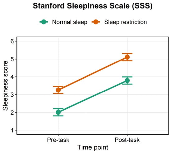
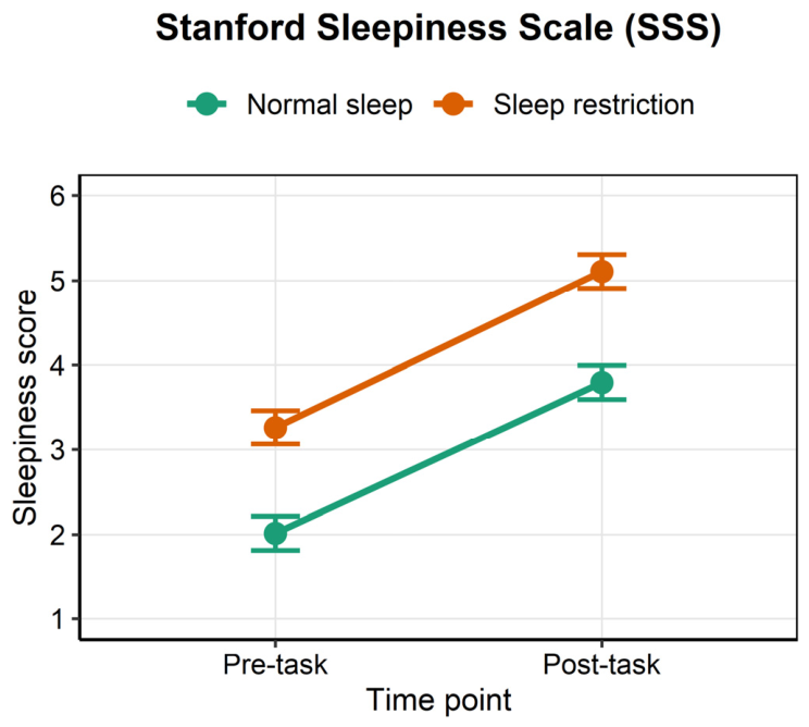
Sleepiness (SSS) was analyzed with a linear mixed model (LMM) that included time (pre-task vs. post-task) and sleep condition (normal sleep vs. sleep restriction) as within-subject factors. The main effect of time was significant, F(1, 31) = 114.85, ηp2 = 0.79; the main effect of sleep condition was significant, F(1, 31) = 67.94, p < 0.001, ηp2 = 0.69; and their interaction was not significant, F(1, 31) = 0.05, p = 0.825 (Table 1). Simple-effects analyses showed that sleepiness was significantly higher in the sleep-restriction condition than in the normal-sleep condition both before the task (3.26 vs. 2.01, p < 0.001, dz = 1.05) and after the task (5.11 vs. 3.80, p < 0.001, dz = 1.12) (Figure 1).
Table 1.
Main results of the Type III tests from the SSS mixed-effects model.
Figure 1.
Stanford Sleepiness Scale (SSS) scores under the two sleep conditions.
As a sensitivity check, the ordinal SSS item was also analyzed with a cumulative link mixed model (ordinal package; logit link; random intercept for participant). The results were equivalent to the LMM: condition z = 5.16, p < 0.001; time z = 6.21, p < 0.001; and the time × condition interaction z = −0.06, p = 0.955.
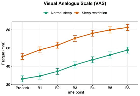
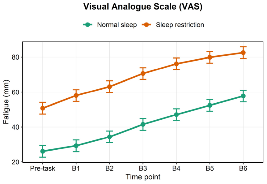
For fatigue (VAS), the main effect of sleep condition was significant, F(1, 31) = 75.66, ηp2 = 0.71, p < 0.001; the main effect of time was significant, F(6, 372) = 72.28, ηp2 = 0.54, p < 0.001; and the interaction was not significant, F(6, 372) = 0.51, p = 0.804 (Table 2 and Table 3). Fatigue was already higher in the sleep-restriction condition than in the normal-sleep condition before the task (50.81 vs. 26.15), and this difference remained stable throughout the task (dz = 0.87 to 1.49 at each time point, all p < 0.001) (Figure 2).
Table 2.
Main results of the Type III tests from the VAS mixed-effects model.
Table 3.
Estimated marginal means (M ± SE) for the subjective measures.
Figure 2.
Visual analog scale (VAS) fatigue scores before the task and across the six blocks under the two sleep conditions.
3.3. Objective Vigilance
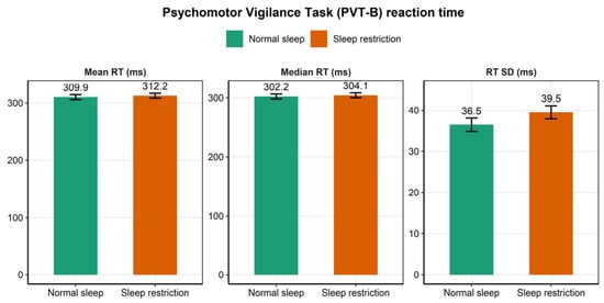
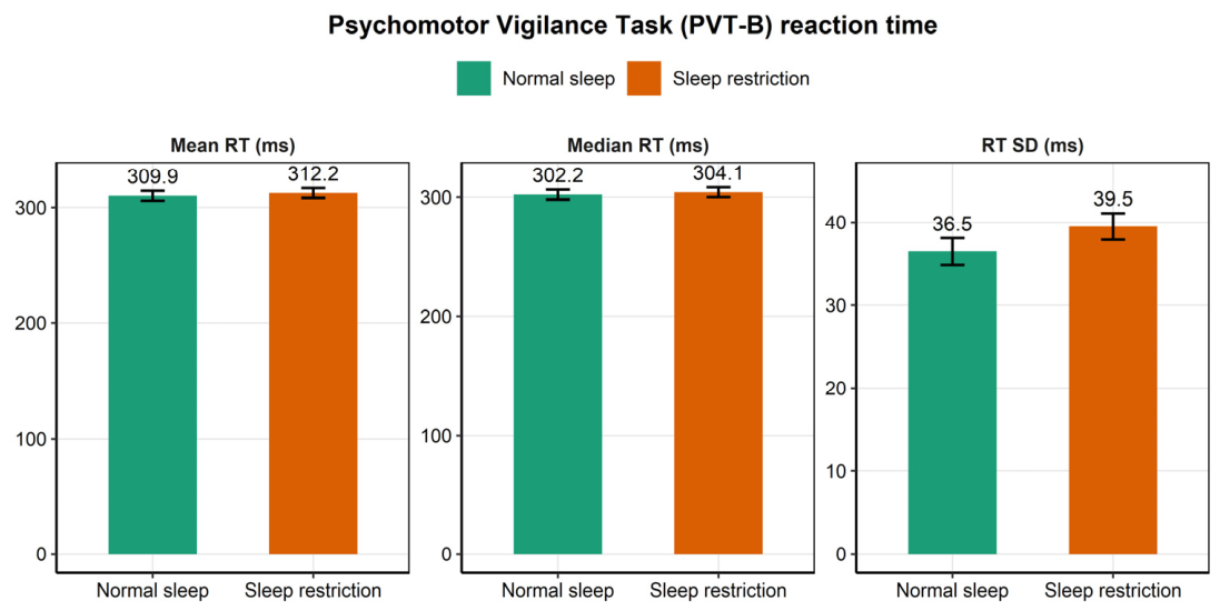
Each PVT-B measure was modeled separately, and none showed a significant effect of sleep condition after false discovery rate (FDR) correction (all p(FDR) > 0.05). The RT measures showed a slight but nonsignificant trend toward slowing, including mean RT (+2.27 ms, F(1, 30) = 0.71, p = 0.405, dz = 0.16), median RT (+1.86 ms, F(1, 30) = 0.56, p = 0.462, dz = 0.14), and RT standard deviation (+2.82 ms, F(1, 30) = 1.98, p = 0.169, dz = 0.26) (Table 4, Figure 3). The order split was 17 normal-first and 15 restricted-first. Session (first versus second testing) was modeled as the within-subject period term; it was not significant for any PVT-B measure (all p > 0.16), and the sleep-condition effects were null.
Table 4.
Estimated marginal means (M ± SE) for the PVT-B measures.
Figure 3.
Brief Psychomotor Vigilance Task (PVT-B) reaction-time measures under the two sleep conditions.
Equivalence testing confirmed that the PVT-B reaction-time null reflected limited power rather than evidence of absence: Bayes factors gave moderate evidence for the null on mean and median RT (BF01 = 3.7 and 4.0), and TOST showed the study could establish equivalence only against large effects (|d| ≥ 0.8).
3.4. Cognitive Control (Stroop Task)
3.4.1. Reaction Time (RT)
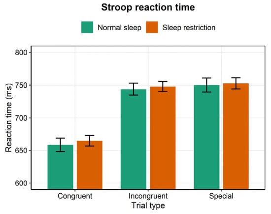
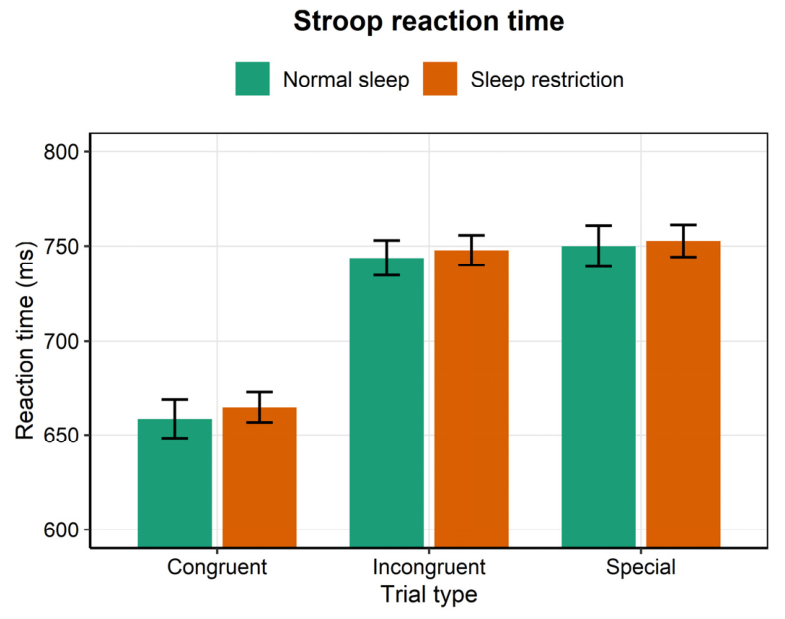
The main effect of trial type was significant, F(2, 47,530) = 3319.97, p < 0.001, ηp2 = 0.12; the main effect of sleep condition was not significant, F(1, 30) = 1.76, p = 0.194; and their interaction was not significant, F(2, 47,538) = 0.98, p = 0.376. The effect of session (first vs. second testing) was significant, F(1, 30) = 38.85, p < 0.001, reflecting faster responses on the second visit. Among the covariates, the effect of block was significant, F(1, 47,527) = 283.14, p < 0.001, as was the trial-centered effect, F (1, 47,525) = 27.01, p < 0.001 (Table 5). Sleep restriction increased RT by 7.80, 5.62, and 4.36 ms for congruent, incongruent, and special trials, respectively, and by 5.99 ms overall (p = 0.180), none of which was significant (Figure 4). Within-participant RT variability (the standard deviation of RT) was 3.23 ms higher overall under restriction (p = 0.096) and 5.92 ms higher for congruent trials (p = 0.050; FDR-corrected p = 0.150); neither effect survived correction. Because restriction made responses slower, more variable, and less accurate, the congruent-trial accuracy decline is more consistent with a general performance deterioration than with a speed–accuracy trade-off.
Table 5.
Main results of the Type III tests from the Stroop reaction-time mixed-effects model.
Figure 4.
Stroop reaction time by trial type under the two sleep conditions.
The random-effects structure of this model comprised by-participant and by-run random intercepts (SD = 43.77 ms and 15.98 ms, respectively) and a by-participant random slope for sleep condition (SD = 4.71 ms), with residual SD = 112.71 ms; the condition slope converged and was non-singular, and its inclusion did not change the condition effect.
The Stroop reaction-time null, by contrast, reflected a genuine absence of effect (5.99 ms; standardized effect 0.04, 90% CI [−0.06, 0.13], within ±0.3).
3.4.2. Accuracy
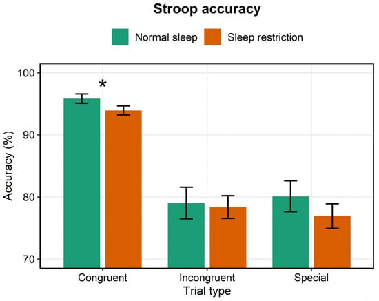
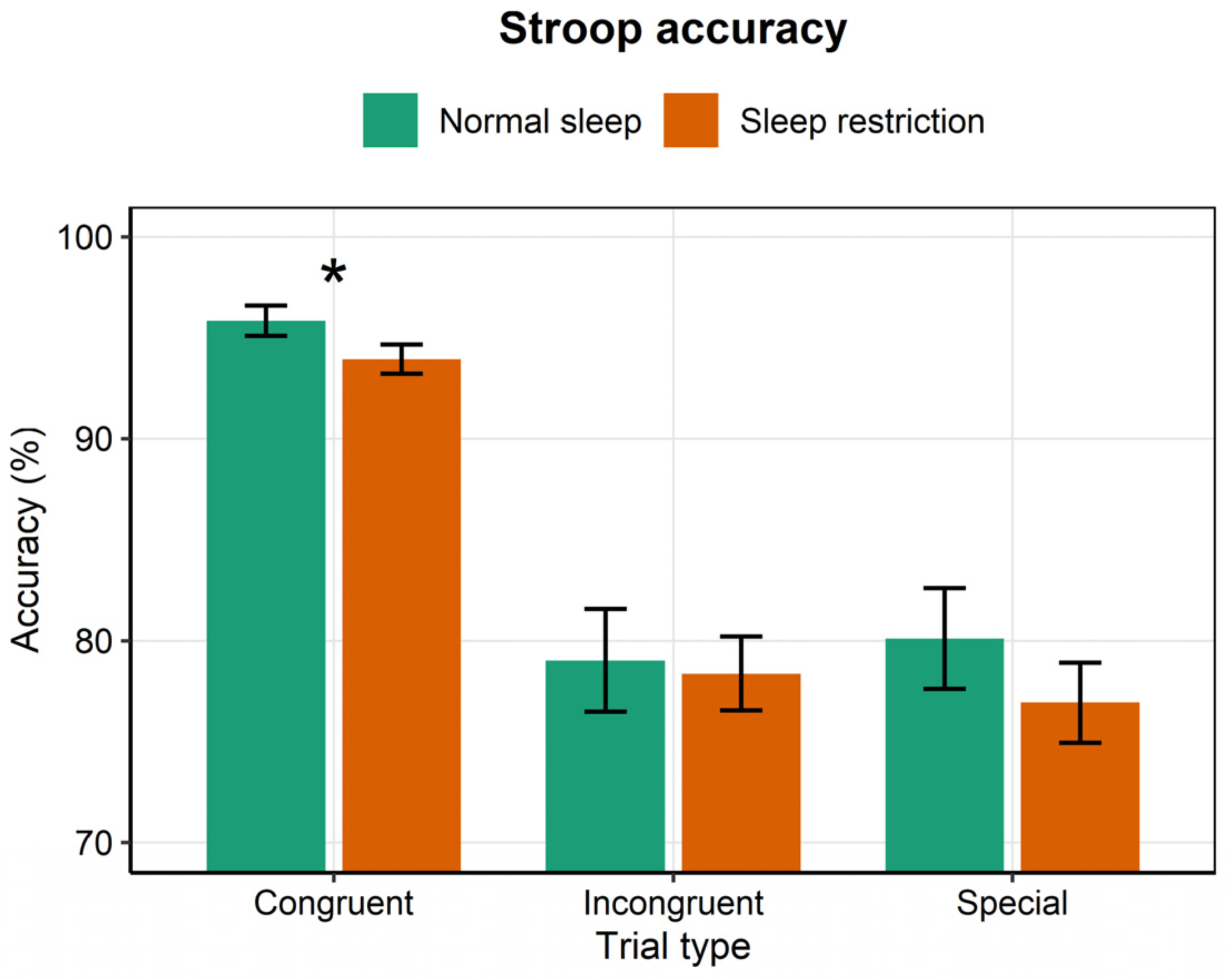
The main effect of trial type was significant, Wald χ2(2) = 2352.89, p < 0.001 (Table 6 and Table 7); the main effect of sleep condition was significant, χ2(1) = 6.24, p = 0.012; and the trial type × sleep condition interaction was significant, χ2(2) = 26.78, p < 0.001. Among the covariates, the effect of block was significant, χ2(1) = 196.50, p < 0.001 and the effect of session was significant, χ2(1) = 32.07, p < 0.001. Simple-effects analyses showed that sleep restriction significantly reduced accuracy for congruent trials (95.94% vs. 93.86%; OR = 0.65, 95% CI [0.52, 0.81], p < 0.001, FDR-corrected p = 0.0004) and special trials (80.40% vs. 76.62%; OR = 0.80, 95% CI [0.66, 0.97], p = 0.025, FDR-corrected p = 0.038). The change for incongruent trials (−1.24 percentage points, p = 0.461) was not significant (Figure 5).
Table 6.
Wald chi-square tests from the Stroop accuracy generalized linear mixed model.
Table 7.
Estimated marginal means (M ± SE) for Stroop reaction time and accuracy.
Figure 5.
Stroop accuracy by trial type under the two sleep conditions. * p < 0.05.
A post hoc sensitivity analysis indicated that, with n = 32 paired observations and two-sided α = 0.05, the study had 80% power to detect a within-subject effect of dz = 0.50 (90% power for dz = 0.57). The observed PVT-B effects (dz = 0.14 to 0.26) are below this threshold. Adding sex as a covariate did not change the condition effects (identical to four decimal places), and sex was not significant for any measure (all p > 0.24); however, with only six women, formal gender-moderation testing is underpowered.
3.4.3. Error-Type Decomposition
To clarify whether the accuracy decline reflected a specific impairment or a general performance deterioration, errors were decomposed into rule errors (responding to the word on incongruent trials or to the ink color on special trials), mapping errors (a wrong key press under the correct rule), and omissions (no response). Restriction increased mapping errors across all three trial types (congruent: 2.8% to 3.5%, p < 0.001; incongruent: 3.1% to 4.2%, p < 0.001; and special: 6.4% to 8.4%, p = 0.002), increased omissions on special trials (10.7% to 12.3%, p = 0.002) and marginally so on congruent trials (2.4% to 3.2%, p = 0.069), and did not change rule errors (incongruent: 9.0% to 7.4%, p = 0.679; special: 4.9% to 3.7%, p = 0.490). The significant mapping-error and omission effects survived Benjamini–Hochberg correction across the eight tests (FDR p ≤ 0.004). These results indicate that the decline reflects a general increase in response-selection errors and attentional lapses rather than a specific impairment of interference control or rule switching.
4. Discussion
Using a within-subject crossover design, this study examined the effects of one night of 5 h sleep restriction on mental fatigue and cognitive control. The most prominent change occurred at the subjective level: under sleep restriction, SSS and VAS scores were already elevated before the task and remained elevated throughout, with large effect sizes, whereas the objective RT measures of the PVT and Stroop tasks showed no detectable change. Subjective fatigue and reduced energy are themselves core components of mental fatigue and the earliest, most direct signals of insufficient sleep (Habay et al., 2021); the high sensitivity of subjective measures to mild restriction is therefore unsurprising. The within-subject design stably reproduced this pattern of subjective sensitivity and no significant change in objective measures, consistent with the graded effects reported by meta-analyses (Wüst et al., 2024): one night of sleep restriction has its strongest effect on subjective measures; a weaker effect on vigilance; and the smallest effect on choice reaction time, working memory, and inhibitory control, suggesting that the early effects of mild sleep loss fall first on subjective experience.
The null time × condition interaction is itself informative. It shows that sleep restriction raised the starting level of sleepiness and fatigue before the task but did not accelerate their accumulation across blocks, consistent with the idea that these measures index a sustained sleep-loss state rather than a within-task process. This distinction, namely that sleep loss sets the baseline whereas time-on-task drives within-task build-up, is the reason the two constructs are reported separately even though both are grouped here under subjective mental fatigue.
The prominence of subjective change in the absence of strong objective impairment can also be read as an early-warning signal: subjective sleepiness and fatigue register the accumulating homeostatic and circadian pressure of sleep loss before that pressure is large enough to measurably degrade vigilance and cognitive control. Under this account, subjective sensitivity to a single night of mild restriction reflects a monitoring function rather than mere response bias, and it yields the testable prediction that subjective reports should precede, rather than merely accompany, objective deterioration under accumulating restriction. Homeostatic and circadian pressure are brain-based signals that become available to consciousness as sleepiness before they measurably degrade the prefrontal and attentional networks supporting vigilance and cognitive control; testing this would require neuroimaging.
An important alternative account for the subjective findings is expectancy or demand characteristics. Because participants were aware of the sleep-restriction manipulation, the pre-task elevation of the SSS and VAS and the constant offset across blocks could reflect either a genuine change in sleepiness and fatigue or the participants’ expectation that restriction should make them feel sleepier. The present design cannot fully separate these accounts, because participants were not blind to condition and their beliefs about the expected effect were not recorded. We therefore regard the subjective–objective dissociation as a pattern that is consistent with a genuine state change but that should be replicated under conditions that control for expectancy, for example by measuring expected impairment or by including an active control condition. This caveat applies equally to the proposal to use the SSS and VAS for screening shift workers, where expectancy could inflate self-report sensitivity.
The null results for the objective measures are related to the relatively light dose. The restriction in this study was only one night of 5 h, constituting mild-to-moderate sleep deprivation, and the impairment caused by insufficient sleep increases as the degree of restriction deepens (Belenky et al., 2003; Dinges et al., 1997; Van Dongen et al., 2003). At this dose, vigilance and RT had not yet shown detectable impairment. Vigilant attention is the function most sensitive to sleep deprivation, and its impairment is characterized by state instability and more frequent lapses; the standard 10 min PVT is a sensitive instrument for detecting such impairment (Goel et al., 2009; Hudson et al., 2020), whereas the 3 min PVT-B used here has relatively lower sensitivity (Basner et al., 2011), and the RT measures—although not significant—still showed a slight trend toward slowing (mean RT dz = 0.16; RT standard deviation dz = 0.26). The null PVT result therefore mainly reflects the light dose and the lower sensitivity of the PVT-B, rather than complete immunity of vigilance. Equivalence testing clarified these null results: the PVT-B null reflects limited power to exclude small-to-medium effects, whereas the Stroop reaction-time null reflects a genuine absence of effect. The absence of clear vigilance impairment under mild restriction is consistent with the observation that impaired vigilant attention partly accounts for impaired inhibitory control (Mao et al., 2021).
In the Stroop task, the effect of restriction was selective. Congruent-trial accuracy fell from 95.94% to 93.86% (OR = 0.65), and special-trial accuracy fell from 80.40% to 76.62% (OR = 0.80), whereas accuracy on incongruent trials and reaction times for all three trial types did not change. This selective decline should be read cautiously for two reasons. First, congruent-trial accuracy was near the ceiling, so the reduction of about 2.1 percentage points for congruent trials and 3.8 percentage points for special trials may partly reflect scale compression rather than a genuine deficit. Second, congruent trials made up only one third of the trials and were interleaved with incongruent and rule-switching “special” trials, so they were unlikely to be processed in a fully automatic mode; the task also taxes rule maintenance and switching (incongruent accuracy was relatively low), and we therefore describe it as a combined interference/rule-switching task. Whether the congruent-trial decline is trial-type-specific or reflects a more general attentional fluctuation remains uncertain and warrants replication (Cunningham et al., 2018). The finding is reported here at the behavioral level, and its neural mechanism awaits further study with neurophysiological measures.
Taken together, the effects of one night of 5 h sleep restriction were not distributed uniformly; they concentrated first on subjective experience, whereas objective vigilance and RT showed no significant change and cognitive control showed a decline in congruent- and special-trial accuracy. This gradient, from subjective to objective measures and from overall RT to specific trial types, outlines the basic profile of early impairment from mild sleep loss: subjective mental fatigue is the earliest and most reliable signal, whereas objective cognitive changes are small and localized. The graded profile itself has been reported previously (Wüst et al., 2024); the present contribution is therefore a within-subject replication with a trial-type decomposition rather than a new claim about the profile. From an applied perspective, subjective scales such as the SSS and VAS are highly sensitive to mild sleep loss and hold potential as instruments for early screening and status monitoring in shift workers; their predictive value still needs to be tested in real shift-work settings and with longer restriction durations. However, under chronic restriction, subjective sleepiness plateaus while performance continues to decline (Van Dongen et al., 2003), so the SSS and VAS may fail to track accumulating impairment during chronic restriction; this limits the screening proposal to acute, mild restriction of the kind studied here.
This study has several limitations. First, the restriction lasted only one night, so the effects of chronic or cumulative restriction cannot be inferred. Second, the protocol delayed sleep onset (a 23:30–01:00 wakeful interval) rather than only shortening sleep, introducing a circadian component that prevents us from attributing the effects to reduced sleep duration alone. Third, the participants were healthy university students aged 18 to 27 years and predominantly male, so generalization to the general population and to shift workers should be made with caution, and whether gender moderates these effects was not tested. Fourth, the PSQI inclusion range (≤7) admits variability in habitual sleep quality; self-reported sleep quality is associated with the functional organization of limbic and fronto-temporo-parietal networks in an age-modulated manner (Federico et al., 2022), suggesting that subjective sleep reports may index a meaningful neural state rather than mere bias, and testing baseline PSQI as a moderator is a natural next step. Fifth, the PVT-B is a brief 3 min version with lower sensitivity than the standard 10 min PVT and may miss subtle changes in vigilance, and the task order was fixed with the PVT-B always administered before the Stroop task. Given the modest sample size (n = 32) and the brief PVT-B, the null objective findings should be interpreted as no detected change rather than as evidence that performance was preserved; subtle effects cannot be excluded. Future studies could extend the restriction duration, include shift-working populations, and add neurophysiological measures.
5. Conclusions
One night of 5 h partial sleep restriction was sufficient to significantly elevate subjective mental fatigue and to produce a small decline in Stroop accuracy that was confined to congruent and special trials, with no significant change in incongruent-trial accuracy or reaction time; objective vigilance and overall RT did not change significantly. These results reveal that the early effects of mild sleep loss are characterized by high sensitivity in subjective experience and, at most, a small and selective reduction in objective performance.
Author Contributions
Conceptualization and methodology, T.S. and J.L.; validation, J.L. and M.D.; formal analysis, J.L. and Q.Y.; investigation, J.L. and S.X.; data curation, J.L., J.X., L.X. and R.Z.; writing—original draft preparation, J.L.; writing—review and editing, T.S. and Q.Y.; supervision, T.S.; funding acquisition, T.S. All authors have read and agreed to the published version of the manuscript.
Funding
This research was funded by National Natural Science Foundation of China, grant number 82371890.
Institutional Review Board Statement
The study was conducted in accordance with the Declaration of Helsinki and approved by the ethic committee of Naval Medical University (date of approval: 10 March 2023). The study is a non-interventional observational study conducted within the approved project period, and the data were collected between March and June 2026.
Informed Consent Statement
Informed consent was obtained from all subjects involved in the study. Written informed consent has been obtained from the volunteers to publish this paper.
Data Availability Statement
The data presented in this study are available on request from the corresponding author. The data are not publicly available due to privacy and ethical restrictions.
Acknowledgments
We acknowledge all project participants and the investigators for collecting data.
Conflicts of Interest
The authors declare no conflicts of interest.
Abbreviations
The following abbreviations are used in this manuscript:
| CI | Confidence Interval |
| ESS | Epworth Sleepiness Scale |
| FDR | False Discovery Rate |
| GLMM | Generalized Linear Mixed Model |
| LMM | Linear Mixed Model |
| MEQ-5 | Morning and Evening Questionnaire-5 |
| OR | Odds Ratio |
| PSQI | Pittsburgh Sleep Quality Index |
| PVT | Psychomotor Vigilance Task |
| PVT-B | Brief Psychomotor Vigilance Task |
| RT | Reaction Time |
| SDS | Self-rating Depression Scale |
| SE | Standard Error |
| SSS | Stanford Sleepiness Scale |
| STAI-5 | Five-item short form of the State-Trait Anxiety Inventory |
| VAS | Visual Analog Scale |
References
- Aitken, R. C. (1969). Measurement of feelings using visual analogue scales. Proceedings of the Royal Society of Medicine, 62(10), 989–993. [Google Scholar] [CrossRef] [Scilit] [PubMed]
- Alix-Fages, C., Cano, H., Baz-Valle, E., & Balsalobre-Fernández, C. (2023). Effects of mental fatigue induced by stroop task and by social media use on resistance training performance, movement velocity, perceived exertion, and repetitions in reserve: A randomized and double-blind crossover trial. Motor Control, 27, 645–659. [Google Scholar] [CrossRef] [PubMed]
- Ballesio, A., Cerolini, S., Ferlazzo, F., Cellini, N., & Lombardo, C. (2018). The effects of one night of partial sleep deprivation on executive functions in individuals reporting chronic insomnia and good sleepers. Journal of Behavior Therapy and Experimental Psychiatry, 60, 42–45. [Google Scholar] [CrossRef] [Scilit] [PubMed]
- Barr, D. J., Levy, R., Scheepers, C., & Tily, H. J. (2013). Random effects structure for confirmatory hypothesis testing: Keep it maximal. Journal of Memory and Language, 68(3), 255–278. [Google Scholar] [CrossRef] [Scilit] [PubMed]
- Basner, M., Mollicone, D., & Dinges, D. F. (2011). Validity and sensitivity of a brief psychomotor vigilance test (PVT-B) to total and partial sleep deprivation. Acta Astronautica, 69(11), 949–959. [Google Scholar] [CrossRef] [Scilit] [PubMed]
- Belenky, G., Wesensten, N. J., Thorne, D. R., Thomas, M. L., Sing, H. C., Redmond, D. P., Russo, M. B., & Balkin, T. J. (2003). Patterns of performance degradation and restoration during sleep restriction and subsequent recovery: A sleep dose-response study. Journal of Sleep Research, 12, 1–12. [Google Scholar] [CrossRef] [Scilit] [PubMed]
- Buysse, D. J., Reynolds, C. F., 3rd, Monk, T. H., Berman, S. R., & Kupfer, D. J. (1989). The Pittsburgh sleep quality index: A new instrument for psychiatric practice and research. Psychiatry Research, 28(2), 193–213. [Google Scholar] [CrossRef] [Scilit] [PubMed]
- Chen, Y., Wang, L., Zhang, M., Hu, S., Shao, Y., Zhang, X., Li, C., Chen, J., Zhao, Z., Dong, Y., Lu, L., Zhou, M., Wang, L., Yuan, J., & Sun, H. (2026). The increasing trends of short and long sleep duration among Chinese adults from 2010 to 2018: A repeated nationally representative cross-sectional survey. Biomedical and Environmental Sciences, 39(1), 46–59. [Google Scholar] [CrossRef] [Scilit] [PubMed]
- Cullen, T., Thomas, G., Wadley, A. J., & Myers, T. (2019). The effects of a single night of complete and partial sleep deprivation on physical and cognitive performance: A Bayesian analysis. Journal of Sports Sciences, 37(23), 2726–2734. [Google Scholar] [CrossRef] [Scilit] [PubMed]
- Cunningham, J. E. A., Jones, S. A. H., Eskes, G. A., & Rusak, B. (2018). Acute sleep restriction has differential effects on components of attention. Frontiers in Psychiatry, 9, 499. [Google Scholar] [CrossRef] [Scilit] [PubMed]
- Dinges, D. F., Pack, F., Williams, K., Gillen, K. A., Powell, J. W., Ott, G. E., Aptowicz, C., & Pack, A. I. (1997). Cumulative sleepiness, mood disturbance, and psychomotor vigilance performance decrements during a week of sleep restricted to 4-5 hours per night. Sleep, 20(4), 267–277. [Google Scholar] [CrossRef] [Scilit]
- Federico, G., Alfano, V., Garramone, F., Mele, G., Salvatore, M., Aiello, M., & Cavaliere, C. (2022). Self-reported sleep quality across age modulates resting-state functional connectivity in limbic and fronto-temporo-parietal networks: An exploratory cross-sectional fMRI study. Frontiers in Aging Neuroscience, 14, 806374. [Google Scholar] [CrossRef] [Scilit] [PubMed]
- Gibbings, A., Ray, L. B., Gagnon, S., Collin, C. A., Robillard, R., & Fogel, S. M. (2022). The EEG correlates and dangerous behavioral consequences of drowsy driving after a single night of mild sleep deprivation. Physiology & Behavior, 252, 113822. [Google Scholar] [CrossRef] [Scilit] [PubMed]
- Goel, N., Rao, H., Durmer, J., & Dinges, D. (2009). Neurocognitive consequences of sleep deprivation. Seminars in Neurology, 29, 320–339. [Google Scholar] [CrossRef] [Scilit] [PubMed]
- Habay, J., Van Cutsem, J., Verschueren, J., De Bock, S., Proost, M., De Wachter, J., Tassignon, B., Meeusen, R., & Roelands, B. (2021). Mental fatigue and sport-specific psychomotor performance: A systematic review. Sports Medicine, 51(7), 1527–1548. [Google Scholar] [CrossRef] [Scilit] [PubMed]
- Hoddes, E., Zarcone, V., Smythe, H., Phillips, R., & Dement, W. C. (1973). Quantification of sleepiness: A new approach. Psychophysiology, 10(4), 431–436. [Google Scholar] [CrossRef] [Scilit] [PubMed]
- Hudson, A. N., Van Dongen, H. P. A., & Honn, K. A. (2020). Sleep deprivation, vigilant attention, and brain function: A review. Neuropsychopharmacology, 45(1), 21–30. [Google Scholar] [CrossRef] [Scilit] [PubMed]
- Johns, M. W. (1991). A new method for measuring daytime sleepiness: The epworth sleepiness scale. Sleep, 14(6), 540–545. [Google Scholar] [CrossRef] [Scilit] [PubMed]
- Krause, A. J., Simon, E. B., Mander, B. A., Greer, S. M., Saletin, J. M., Goldstein-Piekarski, A. N., & Walker, M. P. (2017). The sleep-deprived human brain. Nature Reviews Neuroscience, 18(7), 404–418. [Google Scholar] [CrossRef] [Scilit] [PubMed]
- Lim, J., & Dinges, D. F. (2010). A meta-analysis of the impact of short-term sleep deprivation on cognitive variables. Psychological Bulletin, 136(3), 375–389. [Google Scholar] [CrossRef] [Scilit] [PubMed]
- Lowe, C. J., Safati, A., & Hall, P. A. (2017). The neurocognitive consequences of sleep restriction: A meta-analytic review. Neuroscience & Biobehavioral Reviews, 80, 586–604. [Google Scholar] [CrossRef] [Scilit] [PubMed]
- Luyster, F. S., Strollo, P. J., Zee, P. C., & Walsh, J. K. (2012). Sleep: A health imperative. Sleep, 35(6), 727–734. [Google Scholar] [CrossRef] [Scilit] [PubMed]
- Mao, T., Dinges, D., Deng, Y., Zhao, K., Yang, Z., Lei, H., Fang, Z., Yang, F. N., Galli, O., Goel, N., Basner, M., & Rao, H. (2021). Impaired vigilant attention partly accounts for inhibition control deficits after total sleep deprivation and partial sleep restriction. Nature and Science of Sleep, 13, 1545–1560. [Google Scholar] [CrossRef] [Scilit] [PubMed]
- Marcora, S. M., Staiano, W., & Manning, V. (2009). Mental fatigue impairs physical performance in humans. Journal of Applied Physiology, 106(3), 857–864. [Google Scholar] [CrossRef] [Scilit] [PubMed]
- Meeusen, R., Van Cutsem, J., & Roelands, B. (2021). Endurance exercise-induced and mental fatigue and the brain. Experimental Physiology, 106(12), 2294–2298. [Google Scholar] [CrossRef] [Scilit] [PubMed]
- Mishra, P., Panigrahi, M., & Ankit, D. (2020). Cognition and alertness in medical students: Effects of a single night of partial sleep deprivation. Annals of Neurosciences, 27(2), 57–62. [Google Scholar] [CrossRef] [Scilit] [PubMed]
- Reynolds, A. C., & Banks, S. (2010). Total sleep deprivation, chronic sleep restriction and sleep disruption. In G. A. Kerkhof, & H. P. A. Dongen (Eds.), Progress in brain research (Vol. 185, pp. 91–103). Elsevier. [Google Scholar] [CrossRef] [Scilit] [PubMed]
- Shlisky, J. D., Hartman, T. J., Kris-Etherton, P. M., Rogers, C. J., Sharkey, N. A., & Nickols-Richardson, S. M. (2012). Partial sleep deprivation and energy balance in adults: An emerging issue for consideration by dietetics practitioners. Journal of the Academy of Nutrition and Dietetics, 112(11), 1785–1797. [Google Scholar] [CrossRef] [Scilit] [PubMed]
- Smith, M., Coutts, A., Merlini, M., Deprez, D., Lenoir, M., & Marcora, S. (2016). Mental fatigue impairs soccer-specific physical and technical performance. Medicine & Science in Sports & Exercise, 48(2), 267–276. [Google Scholar] [CrossRef] [Scilit] [PubMed]
- Sun, H. A.-O., Soh, K. G., Roslan, S., Wazir, M., & Soh, K. L. (2021). Does mental fatigue affect skilled performance in athletes? A systematic review. PLoS ONE, 16(10), e0258307. [Google Scholar] [CrossRef] [Scilit] [PubMed]
- Tanaka, M., Ishii, A., & Watanabe, Y. (2014). Neural effect of mental fatigue on physical fatigue: A magnetoencephalography study. Brain Research, 1542, 49–55. [Google Scholar] [CrossRef] [Scilit] [PubMed]
- Van Dongen, H. P., Maislin, G., Mullington, J. M., Dinges, D. F., & New Collective Author. (2003). The cumulative cost of additional wakefulness: Dose-response effects on neurobehavioral functions and sleep physiology from chronic sleep restriction and total sleep deprivation. Sleep, 26(2), 117–126. [Google Scholar] [CrossRef] [Scilit] [PubMed]
- Wu, Y., Fan, L., Chen, W., Su, X., An, S., Yao, N., Zhu, Q., Huang, Z.-G., & Li, Y. (2025). Brain dynamics alterations induced by partial sleep deprivation: An energy landscape study. Neuroimage, 310, 121108. [Google Scholar] [CrossRef] [Scilit] [PubMed]
- Wüst, L. N., Capdevila, N. C., Lane, L. T., Reichert, C. F., & Lasauskaite, R. (2024). Impact of one night of sleep restriction on sleepiness and cognitive function: A systematic review and meta-analysis. Sleep Medicine Reviews, 76, 101940. [Google Scholar] [CrossRef] [Scilit] [PubMed]
- Zsido, A. N., Teleki, S. A., Csokasi, K., Rozsa, S., & Bandi, S. A. (2020). Development of the short version of the spielberger state—Trait anxiety inventory. Psychiatry Research, 291, 113223. [Google Scholar] [CrossRef] [Scilit] [PubMed]
- Zung, W. W. K. (1965). A self-rating depression scale. Archives of General Psychiatry, 12(1), 63–70. [Google Scholar] [CrossRef] [Scilit] [PubMed]

Disclaimer/Publisher’s Note: The statements, opinions and data contained in all publications are solely those of the individual author(s) and contributor(s) and not of MDPI and/or the editor(s). MDPI and/or the editor(s) disclaim responsibility for any injury to people or property resulting from any ideas, methods, instructions or products referred to in the content. |
© 2026 by the authors. Licensee MDPI, Basel, Switzerland. This article is an open access article distributed under the terms and conditions of the Creative Commons Attribution (CC BY) license.