


Abstract
Background: Perfluorooctane sulfonic acid (PFOS) is a ubiquitous industrial chemical linked to cardiovascular toxicity. Quercetin, a plant-derived flavonoid, exhibits known anti-inflammatory properties. While PFOS induces endothelial dysfunctions, the molecular mechanisms underlying quercetin’s protective effects remain unclear. Methods: We integrated transcriptomic analysis and in vitro experiments to investigate these mechanisms in human microvascular endothelial cells (HMEC-1). Results: Five overlapping hub genes between PFOS and quercetin targets (IL6, RELA, VEGFA, CXCL8 and PIM1) were identified. Enrichment and protein–protein interaction network analyses showed the AGE–RAGE signaling pathway and cytokine activity as primary mediators. Furthermore, these five hub genes were significantly up-regulated in human atheromatous plaque tissues (GSE43292). Crucially, an independent external validation using a Logistic Regression machine learning model on an external dataset (GSE100927) demonstrated the strong predictive power of these five overlapping hub genes (AUC = 0.82) in classifying atherosclerotic lesions, effectively mitigating internal validation bias and strongly supporting their clinical diagnostic relevance. To verify these findings, we demonstrated that PFOS exposure induced p65 phosphorylation, up-regulated VEGFA and ICAM1, and down-regulated SIRT1 and SOCS3 expression in HMEC-1 cells. Quercetin co-treatment effectively reversed these molecular alterations, suppressing p65 phosphorylation and VEGFA/ICAM1 expression, while restoring SIRT1 and SOCS3 levels. Conclusions: These findings provide novel insights into how quercetin alleviates PFOS-induced inflammatory responses associated with endothelial activation.
1. Introduction
Perfluorooctane sulfonic acid (PFOS) is a fluorinated chemical substance that is widely used in various industries and household products [1]. Structurally, PFOS is composed of a carbon–fluorine (C–F) backbone attached to a sulfonic acid functional group; consequently, persistence in the environment, drinking water, and foodstuff and presence in human serum has been detected [2]. A previous study reported that there was a human serum half-life of approximately 3.4–5.4 years [3]. Several studies indicated that PFOS exposure is associated with multiple adverse health effects including neurotoxicity, hepatotoxicity, endocrine disruption, gastrointestinal toxicity and cardiovascular disease (CVD) [4,5,6].
Atherosclerosis (AS) is a major pathological condition of cardiovascular disease (CVD), characterized by the formation of lipid-enriched plaques within arterial walls [7]. Epidemiological studies reported that the global burden of CVD caused 18.6 million deaths in 2019 and is expected to reach 20.5 million deaths in 2050 [8,9]. Accumulating evidence has identified several major risk factors associated with atherosclerosis, including arterial hypertension, chronic inflammation, familial hypercholesterolemia, smoking, and endothelial dysfunctions, which may subsequently lead to myocardial infarction, ischemic stroke and mortality [10]. Although several conventional pharmacological agents are currently available for the treatment of AS, these drugs may cause adverse effects such as headache, dizziness, myopathy, nausea, and diarrhea [11]. Therefore, alternative therapeutic approaches, particularly plant-derived traditional herbal medicines such as quercetin, have gained increasing attention for their potential therapeutic benefits against AS [12].
Quercetin is a polyphenolic flavonoid that plays a crucial role in antioxidant, anti-inflammatory, anti-proliferative and anti-atherosclerosis properties [13]. A previous study reported that quercetin attenuated H2O2-induced lipid peroxidation, cytokine secretion and cell-surface adhesion molecules through down-regulating the NF-κB signaling pathway in human hepatocytes [14]. Furthermore, there was evidence showed that quercetin alleviated atherosclerosis through down-regulating the p38 MAPK/p16 signaling pathway in oxidized low-density lipoprotein (ox-LDL)-induced high-fat diet (HFD) mouse models [15]. Unfortunately, the molecular mechanisms of quercetin in PFOS-induced endothelial dysfunction related to atherosclerosis are poorly understood.
In the present study, the integration of transcriptomic analysis and in vitro studies were used to investigate the molecular mechanism of how quercetin alleviates PFOS-induced endothelial dysfunctions in human immortalized endothelial cell 1 (HMEC-1) cells. Overlapped DEGs were retrieved from our previous study and DEGs from the quercetin database. Biological alterations were performed by using the gene ontology (GO) database and Kyoto Encyclopedia of Genes and Genomes (KEGG) pathway analysis. A protein–protein interaction (PPI) network and modular clustering were constructed to identify hub genes. We further verified hub genes by using the GEO database and in vitro studies.
2. Materials and Methods
2.1. Data Collection
Total RNA was extracted from three independent biological replicates of HMEC-1 cells for both the PFOS-treated groups and the vehicle groups [16]. The raw sequencing reads were subjected to quality control and aligned to the human reference genome (GRCh38.p13) using Hisat2 (version 2.2.1). Gene expression quantification was performed using HT-Seq (version 0.6.1). Differential expression analyses were performed using the DESeq2 Bioconductor package 1.52.0. The data in this publication have been deposited in the NCBI BioProject database and are accessible through the BioProject accession number PRJNA1465111. DEGs were defined using a selection threshold of |log2(Fold change)| ≥ 1 and an adjusted p-value (FDR) < 0.05, applying the Benjamini–Hochberg procedure for multiple-testing correction.
2.2. Hub Genes Identification
To identify hub genes from 167 DEGs of PFOS-induced transcriptomic alterations, a Protein–Protein Interaction (PPI) network was constructed. The complete set of the 167 DEGs was inputted directly into the NetworkAnalyst platform (https://www.networkanalyst.ca, accessed on 14 November 2025). The PPI network was generated using the integrated STRING interactome database. Crucially, to strictly reflect our specific experimental conditions, a zero-order network was employed. This approach ensured that the interactome consisted of the 167 original input DEGs, without expanding the network to include any external added interactors. Subsequently, advanced topological analysis was performed within the platform to evaluate the network architecture. The top ten hub genes were extracted and ranked based on their betweenness centrality scores, highlighting their fundamental roles as critical mediators within the PFOS-induced disease network.
2.3. Overlapped Hub Genes Identification
To identify overlapped hub genes, ten hub genes were intersected with the quercetin-associated target genes (retrieved from the SymMap database (http://www.symmap.org/, accessed on 14 November 2025) using Venny 2.1.0 (https://bioinfogp.cnb.csic.es/tools/venny/, accessed on 14 November 2025).
2.4. Biological Alterations
The overlapped hub genes were subsequently used to investigate biological alterations in terms of gene ontology (GO) and enriched pathway analyses using the ShinyGO platform version 0.85, a graphical gene set enrichment analysis platform (https://bioinformatics.sdstate.edu/go85/, accessed on 14 November 2025) [17]. An adjusted p-value (FDR) < 0.05 was considered statistically significant for multiple testing.
2.5. Protein–Protein Interaction (PPI) Network Construction and Modular Clustering Analysis
To evaluate the specific physical and functional connectivity among the five overlapping hub genes (IL-6, RELA, VEGFA, CXCL8 and PIM1), a Protein–Protein Interaction (PPI) network was constructed using the Metascape platform (http://metascape.org, accessed on 14 November 2025). Following the network generation, the Molecular Complex Detection (MCODE) algorithm was applied to identify highly densely connected protein neighborhoods (core complexes) within this specific target set. The isolated MCODE core sub-network was subsequently annotated to determine enriched biological pathways, thereby validating whether these overlapped hub genes physically interact as a coordinated functional unit to drive pathogenesis. The cut-off criteria were defined as p < 0.05, enriched factor ≥ 1.5 and minimum count = 3.
2.6. Hub Genes Verification Using GEO Dataset
The Gene Expression Omnibus GSE43292 dataset was obtained from 32 hypertensive patients containing paired atheromatous plaque tissue and distant macroscopically intact arterial tissue. The dataset was generated using the GPL6244 platform (Affymetrix Human Genome 1.0 ST Array), GEO dataset, PubMed, NCBI (https://www.ncbi.nlm.nih.gov/, accessed on 14 November 2025). Raw microarray data were preprocessed and normalized using the robust multi-array average (RMA) algorithm. Probe-to-gene mapping was systematically performed using the corresponding Affymetrix platform annotation file. To verify clinical relevance, the five overlapping hub genes and the GSE43292 dataset were intersected using Venny 2.1.0 (https://bioinfogp.cnb.csic.es/tools/venny/, accessed on 14 November 2025). The expression data of these hub genes were subsequently extracted. The effect sizes (Log2 Fold Change) and adjusted p-values (FDR < 0.05) between the intact and plaque tissues were evaluated, then the gene expression levels were visualized using frequency dot plots.
2.7. External Validation of Hub Genes Using Machine Learning
To validate the clinical diagnostic relevance and generalizability of the five overlapping hub genes (IL6, RELA, VEGFA, CXCL8 and PIM1), independent external validation was conducted using the external clinical microarray dataset GSE100927, while using the GSE43292 dataset as the training cohort. The external validation dataset GSE100927 comprised a total of 104 clinical peripheral arterial samples, including 69 atherosclerotic plaque lesion samples and 35 normal/control arterial tissue samples obtained from organ donors profiled on the Agilent microarray platform (GPL17077).
For data preprocessing, original platform identifiers from both datasets were systematically mapped to official gene symbols, and the expression values of the five overlapping hub genes were extracted. To eliminate scale bias and prevent data leakage, the expression values were standardized using Z-score normalization fitted exclusively on the training set (GSE43292) and subsequently applied to the external test set (GSE100927).
To determine the most robust and stable classifier for the independent dataset, two distinct machine learning algorithms were trained: a linear Logistic Regression (LR) model and a non-linear Random Forest (RF) classifier (constructed with 100 decision trees). Both models were trained on the GSE43292 cohort and directly evaluated on the unseen GSE100927 cohort without additional tuning. The diagnostic performance and discriminatory power of the models were quantified using the Area Under the Receiver Operating Characteristic (ROC) curve (AUC). All computational procedures were executed in a Python 3.13 environment using the scikit-learn 1.9.1 and matplotlib 3.11.2 packages.
2.8. Chemical Reagents
Perfluorooctane sulfonic acid (PFOS, purity ≥ 95.0%, Cat. No. 321-61582) was purchased from FUJIFILM Wako chemicals (Richmond, VA, USA). Quercetin (purity ≥ 95%, Cat. No. Q4951) and MTT were purchased from Sigma Aldrich (Burlington, Massachusetts, USA). Dimethyl sulfoxide (DMSO), L-glutamine, gentamicin, MCDB131, penicillin/streptomycin and trypsin-EDTA were purchased from Gibco (Grand Island, NY, USA). Human immortalized endothelial cell 1 (HMEC-1) was kindly gifted by Dr. Thomas J. Lawley from the Centers for Disease Control and Prevention to Dr. Tawit Suriyo, Laboratory of Pharmacology, Chulabhorn Research Institute. The primary antibodies included anti-SIRT1 (Cat. No. sc-74504), anti-SOCS3 (Cat. No. sc-73045), anti-phospho-p65 (Ser536) (Cat. No. sc-136548), anti-p65 (Cat. No. sc-8008), anti-ICAM-1 (Cat. No. sc-8439) and anti-VEGFA (Cat. No. sc-7269), which were purchased from Santa Cruz Biotechnology, Inc. (Dallas, TX, USA). The IL-6 ELISA kit (Cat. No. 430504) was purchased from Biolegend (San Diego, CA, USA).
2.9. Cell Culture and Conditions
HMEC-1 cells (used between passages 6 and 18) were cultured in MCDB131 medium supplemented with 100 U/mL of penicillin and 100 μg/mL of streptomycin, 10 mM of L-glutamine, 1 μg/mL of hydrocortisone, 10 ng/mL of epidermal growth factor (EGF), 10% of fetal bovine serum (FBS) and 20 μg/mL of gentamicin. Cells were incubated under a 5% CO2 atmosphere at 37 °C. The cell line was authenticated by routine testing to ensure it was free of mycoplasma contamination.
2.10. Cell Viability
Viable cells were assessed by using the MTT assay following overnight seeding in 96-well plates, 1 × 10⁴ cells/well. Cells were exposed to concentrations of PFOS (1, 10, 15, 20, 40, 60, 80 and 100 μM) or quercetin (1, 10, 25, 50, 75, and 100 μM) for 72 h, and 0.1% DMSO was used as a vehicle control. For all treatments, the final vehicle (DMSO) concentration was adjusted to exactly 0.1% (v/v) across all four experimental groups to eliminate vehicle-induced confounding effects. After 72 h, cells were incubated with MTT solution for 3 h. Following 3 h, crystal formazan was dissolved with DMSO and measured at 570 nm. The percentage of viable cells was plotted against the concentrations of PFOS and quercetin. Data represent three independent biological replicates.
2.11. Enzyme-Linked Immunosorbent Assay (ELISA)
HMEC-1 cells were cultured in 100 mm dishes and incubated to 80% confluence, followed by 72 h exposure to the vehicle control, 15 μM of perfluorooctane sulfonic acid (PFOS), 10 μM quercetin, or their combination. Conditioned media were harvested and preserved at −80 °C until use. Secreted human interleukin-6 (IL-6) was quantified by using the IL-6 ELISA MAX™ kit from BioLegend (San Diego, CA, USA), according to the manufacturer’s instructions. Optical densities were measured at 450 nm, with reference correction at 570 nm, by using the SpectraMax i3x microplate reader (Molecular Devices, Sunnyvale, CA, USA). The uniformity of the cell mass across all treatment groups was verified by parallel Bradford protein quantification. Data analysis and curve fitting were performed and analyzed by using GraphPad Prism version 8.0.1 from GraphPad Software (San Diego, CA, USA).
2.12. Western Immunoblotting
HMEC-1 cells were seeded into 100 mm culture dishes and incubated at 37 °C to 80% confluence. Cells were exposed to 15 μM of perfluorooctane sulfonic acid PFOS, 10 μM of quercetin, or co-exposure to PFOS and quercetin, then incubated for 72 h. Following 72 h, cells were harvested, lysed, and stored at −80 °C until further analysis. Protein concentrations were quantified by using the Bradford assay from Bio-Rad Laboratories (Hercules, CA, USA). A total of 30 μg of protein lysates was separated by SDS-PAGE, then transferred onto nitrocellulose membranes. Membranes were incubated with the following primary antibodies: anti-phospho-p65 (p-p65; 1:1000), anti-p65 (1:1000), anti-SIRT1 (1:1000), anti-SOCS3 (1:1000), anti-ICAM-1 (1:1000), anti-VEGFA (1:1000) and anti-β-actin (1:10,000). Horseradish peroxidase (HRP)-conjugated anti-mouse secondary antibodies (1:3000) were used for detection. Protein bands were visualized using an enhanced chemiluminescence (ECL) substrate kit, and images were captured using the ChemiDoc™ Imaging System (Bio-Rad Laboratories, CA, USA). The densitometry of the protein bands was quantified using Image Lab software (version 6.1, Bio-Rad). The raw densitometry values of the target proteins (p-p65, p65, SIRT1, SOCS3, ICAM-1, and VEGFA) were normalized to the corresponding β-actin bands to determine relative protein expression levels.
2.13. Statistical Analysis
All data are presented as the mean ± standard error of the mean (SEM), with individual observations overlaid that derived from at least three independent biological experiments (n ≥ 3). Within each independent experiment, all treatment groups were newly generated, matched and evaluated concurrently using the same cell passage to minimize batch variability. Statistical analyses were performed using GraphPad Prism version 8.0.1. For the clinical microarray dataset (GSE43292), statistical significance between the atheromatous plaque and distant macroscopically intact tissues from the same patients was analyzed using a paired Student’s t-test. Statistical significance for the cell viability assay (multiple concentrations compared to a single vehicle control) was analyzed using a one-way analysis of variance (ANOVA), followed by Dunnett’s post hoc test. For protein expression and cytokine secretion assays, statistical significance among the four treatment groups was analyzed using repeated-measures one-way ANOVA, followed by Sidak’s multiple comparisons test. A p-value < 0.05 was considered statistically significant.
3. Results
3.1. Identification of Overlapping Hub Genes
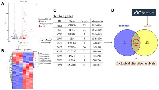
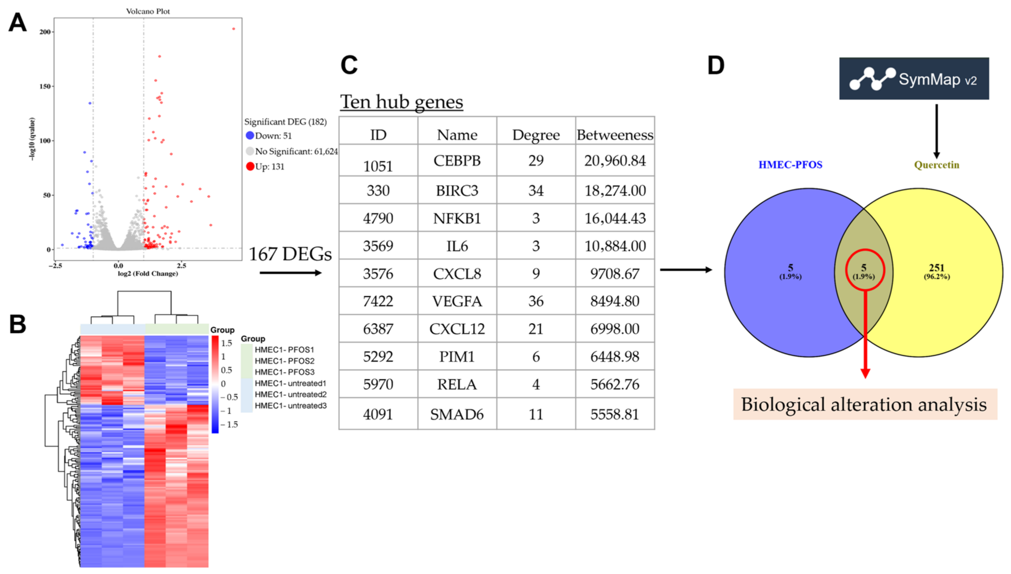
To identify the DEGs of PFOS and quercetin by using RNA-sequencing (RNA-seq) and data mining analysis, we analyzed 182 DEGs composed of 131 up-regulated DEGs and 51 down-regulated DEGs from HMEC-1 treated with PFOS (Figure 1A). In addition, hierarchical clustering showed the gene expression pattern of the DEGs between the PFOS-treated group compared to the vehicle group (Figure 1B). Prior to comparative analysis, these 182 DEGs were mapped to known gene symbols, resulting in 167 annotated genes (Table S1). Topological analysis of the PPI network constructed from these 167 DEGs identified the top 10 hub genes based on betweenness centrality (Figure 1C). A total of five overlapping hub genes among these 10 hub genes and quercetin-associated target genes from the SymMap database were obtained for further analysis (Figure 1D) (Table S1).
Figure 1.
Transcriptomic profiling of PFOS-exposed HMEC-1 cells and identification of quercetin-associated target genes. (A) Volcano plot showing the differentially expressed genes (DEGs) between PFOS-treated and vehicle control groups. Red and blue dots represent significantly up-regulated and down-regulated genes, respectively (|log2FC| ≥ 1, FDR < 0.05). (B) Hierarchical clustering heatmap of the significant DEGs across biological replicates (n = 3). (C) The top ten hub genes are identified and ranked by betweenness centrality from the topological analysis of the 167 DEGs. (D) Venn diagram demonstrating the intersection between the ten hub genes and quercetin-associated target genes retrieved from the SymMap database. This intersection of five overlapping hub genes is used for downstream biological alteration analysis.
3.2. Cellular and Molecular Alterations
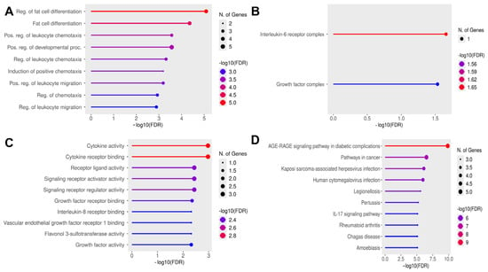
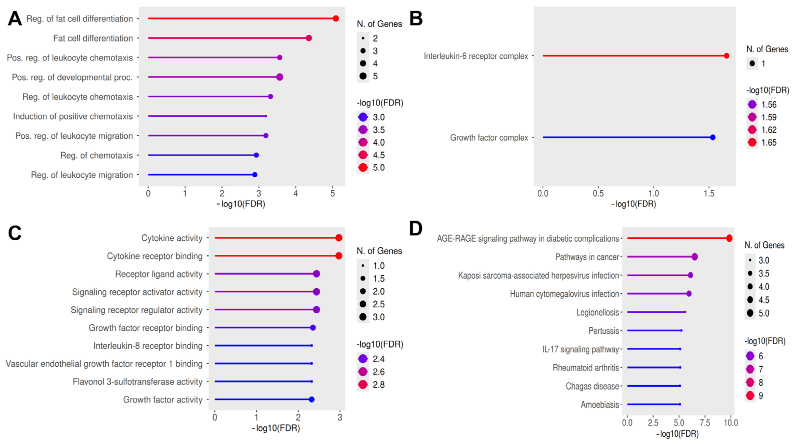
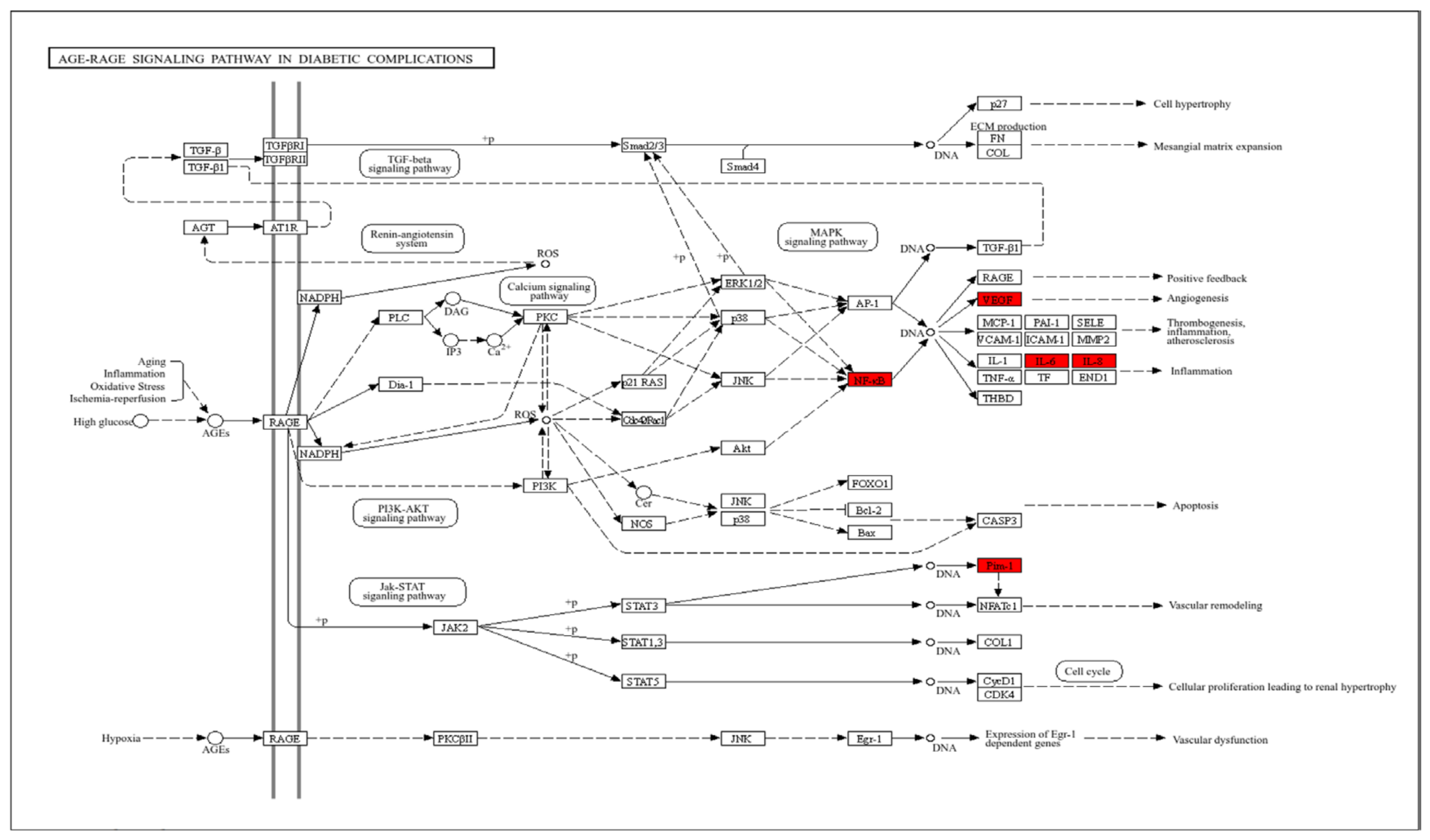
To determine biological alterations using the ShinyGO platform, the five overlapped hub genes were analyzed in terms of gene ontology (GO) and pathway analysis (KEGG). For GO, five overlapped hub genes were categorized into three terms, including biological process (BP), cellular compartment (CC) and molecular function (MF). For BP, regulation of fat cell differentiation, fat cell differentiation and positive regulation of leukocyte chemotaxis were the top three terms (Figure 2A). For CC, interleukin-6 receptor complex and growth factor complex were the top two terms (Figure 2B). MF showed that cytokine activity, cytokine receptor binding and receptor ligand binding activity were the top three terms (Figure 2C). Notably, the AGE–RAGE signaling pathway in diabetic complications was the most enriched pathway, with enriched DEGs including VEGFA, IL-6, IL-8 and PIM1 (Figure 2D and Figure 3 and Table S1).
Figure 2.
Functional enrichment analysis of five overlapping hub genes (IL-6, RELA, VEGFA, CXCL8 and PIM1) between PFOS-induced transcriptomic alterations and quercetin targets. Lollipop charts show significantly enriched terms (FDR < 0.05) across four functional categories: (A) Biological process (BP), (B) Cellular component (CC), (C) Molecular function (MF) and (D) KEGG pathways. The x-axis represents statistical significance expressed as −log10(FDR) The size of the circles corresponds to the number of overlapping genes associated with each specific term, while the color gradient from blue to red indicates increasing statistical significance.
Figure 3.
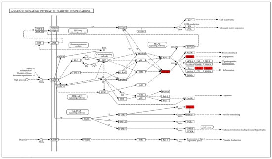
KEGG pathway mapping of the core molecular targets. The AGE–RAGE signaling pathway in diabetic complications (KEGG ID: hsa04933) was identified as the most significantly enriched pathway associated with the five overlapping hub genes. The specific targets overlapping between the PFOS-induced transcriptomic alterations and the quercetin-associated target genes, namely, VEGFA, NF-κB (RELA), IL-6, IL-8 (CXCL8), and PIM1, are highlighted in red. This pathway mapping shows the critical roles of these hub genes as downstream effectors mediating endothelial inflammation, angiogenesis, and vascular remodeling.
3.3. Network and Modular Clustering Analyses
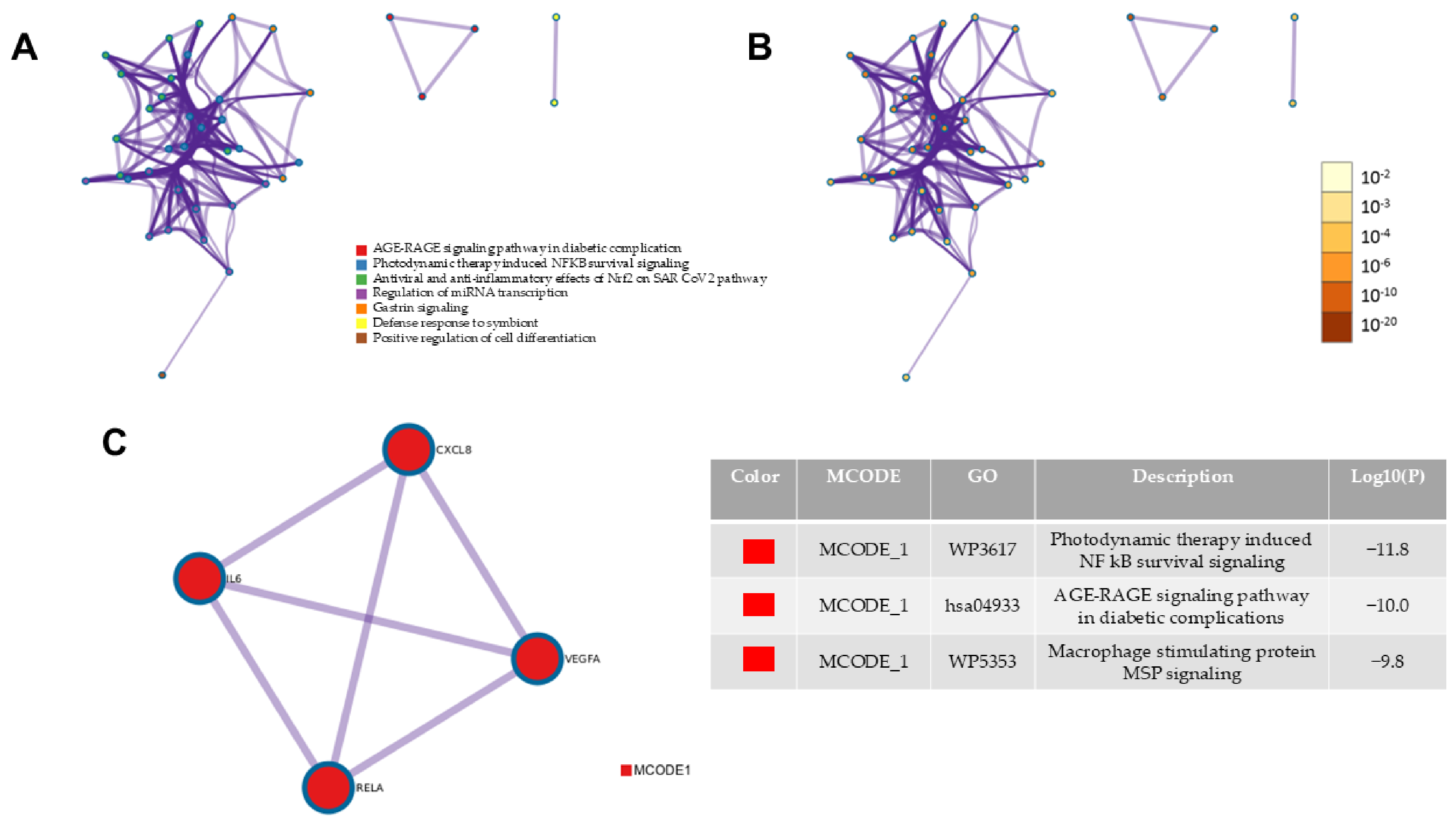
To further substantiate the findings from the KEGG pathway analysis and evaluate the physical connectivity among the five identified therapeutic targets, a Protein–Protein Interaction (PPI) network was constructed, and the Molecular Complex Detection (MCODE) algorithm was applied. The MCODE analysis successfully extracted a densely connected core sub-network (MCODE_1) comprising four of the five overlapping hub genes (IL-6, RELA, VEGFA, and CXCL8) (Figure 4). Functional annotation of this specific core cluster revealed predominant enrichment in the AGE–RAGE signaling pathway in diabetic complications and NF-κB survival signaling. These MCODE clustering results independently confirm our initial KEGG pathway analysis, providing strong evidence that these targets functionally and physically interact as a coordinated core complex to drive endothelial inflammatory responses. Notably, PIM1 was identified as a critical overlapping hub gene; it was not structurally clustered within this specific MCODE1. This topological distinction suggests that although PIM1 is functionally essential to the broader transcriptomic alterations and shares molecular targeting by quercetin, it likely acts as a peripheral modulator or functions through parallel signaling cascades, rather than forms direct, dense physical interactions with the IL-6/RELA/VEGFA/CXCL8 core axis.
Figure 4.
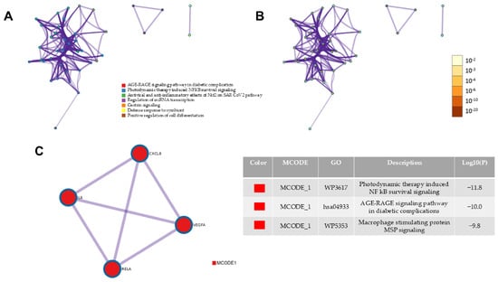
Network analysis and MCODE clustering of the enriched terms associated with the molecular targets. The network was generated using the Metascape platform based on the string database. (A) A network of enriched GO/KEGG terms colored by cluster ID, where terms with high similarity are grouped together as distinct nodes. (B) The identically structured network colored by statistical significance (p-value), with darker nodes representing highly significant terms (lower p-values). (C) The MCODE algorithm was applied to identify the most densely connected protein neighborhood component (MCODE_1) from the protein–protein interaction (PPI) network. This core cluster consists of four highly interacting hub genes (IL-6, RELA, VEGFA and CXCL8) and is predominantly associated with the AGE–RAGE signaling pathway and NF-κB survival signaling.
3.4. Hub Genes Verification
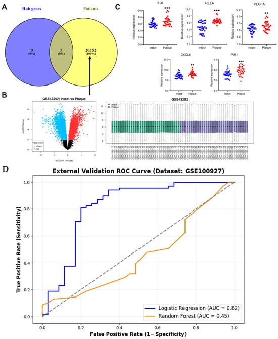
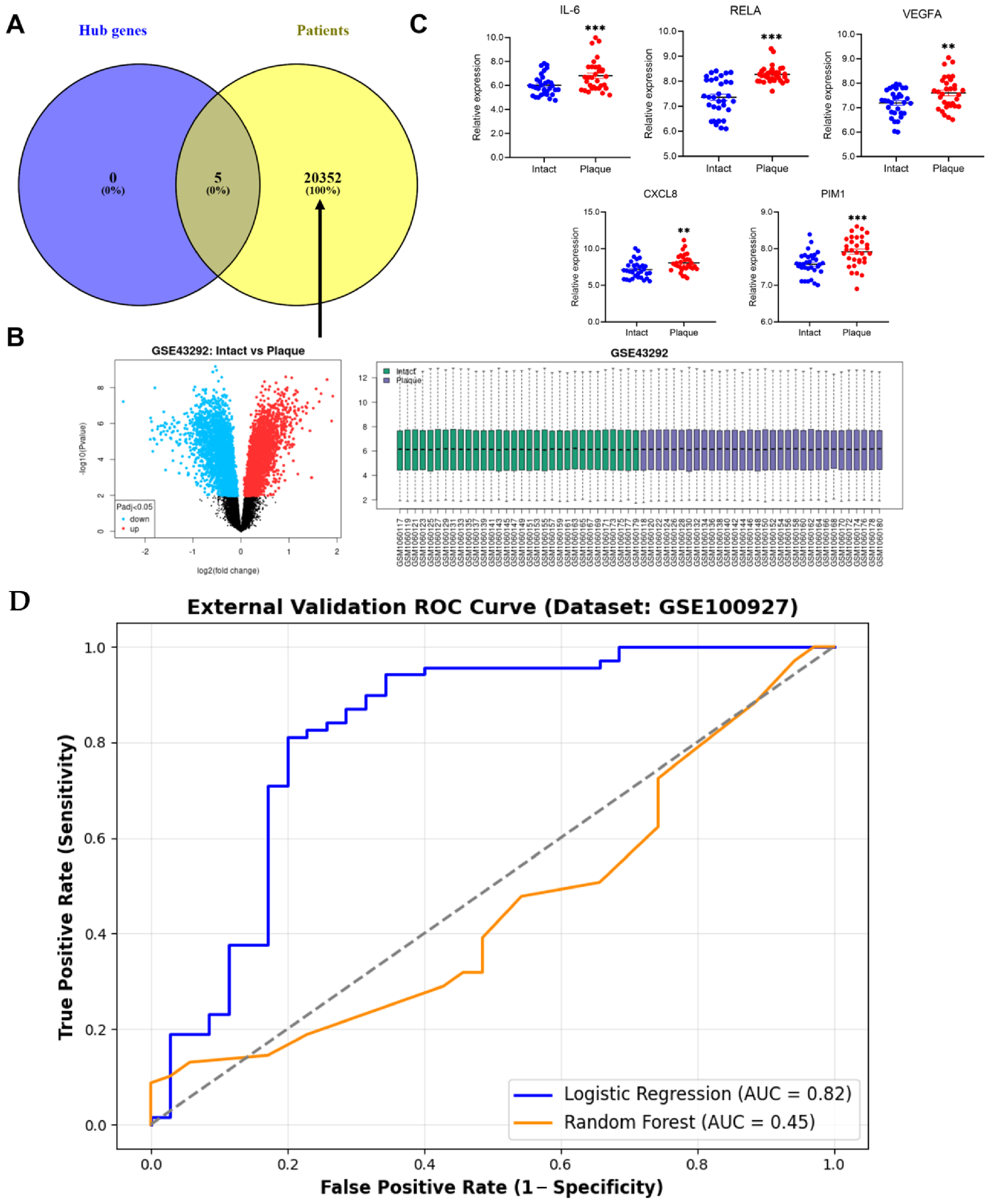
To verify the clinical relevance of the hub genes from the PPI network, we analyzed their expression in the GSE43292 dataset. We found that all five overlapping hub genes (IL-6, RELA, VEGFA, CXCL8 and PIM1) were significantly up-regulated in the atheromatous plaque tissues compared to the distant macroscopically intact arterial tissues, suggesting their deep involvement in localized atherosclerotic plaque pathogenesis (Figure 5A–C). To evaluate the clinical diagnostic relevance and generalizability of the identified five overlapping hub genes, an independent external validation was performed using the microarray dataset GSE100927 (n = 104) as the test cohort, while the GSE43292 dataset served as the training cohort. Two distinct machine learning algorithms, namely, Logistic Regression (LR) and Random Forest (RF), were trained on the GSE43292 dataset and directly evaluated on the unseen GSE100927 dataset. The Logistic Regression model demonstrated exceptional discriminatory power, with an Area Under the Curve (AUC) of 0.82, outperforming the Random Forest classifier (AUC = 0.45) (Figure 5D). This robust external validation performance confirms that the five overlapping hub genes possess high clinical diagnostic value and generalizability for distinguishing atherosclerotic plaques from normal arterial tissues, effectively mitigating overfitting and internal validation bias.
Figure 5.
External validation of the five overlapping hub genes in the clinical GSE43292 dataset and machine learning-based prediction. (A) Venn diagram showing the intersection of five overlapping hub genes with the transcriptomic profile of the clinical dataset GSE43292. (B) Transcriptomic profiling of the GSE43292 dataset. The left panel shows a volcano plot displaying differentially expressed genes (DEGs) between intact vascular tissues and atherosclerotic plaques (Padj < 0.05). The right panel displays a boxplot demonstrating the robust normalization and uniform distribution of microarray data across all clinical samples. (C) Relative expression levels of the five overlapping hub genes (IL-6, RELA, VEGFA, CXCL8 and PIM1) between intact vascular tissues and atherosclerotic plaques. Data are presented as scatter dot plots with the mean. Statistical significance is ** p < 0.01 and *** p < 0.001. (D) Independent external validation ROC curves evaluated on the GSE100927 dataset (n = 104), comparing the predictive performance of the Logistic Regression model (AUC = 0.82, blue line) and the Random Forest model (AUC = 0.45, orange line). Dashed line is random guess line.
3.5. Effects of PFOA on Cell Viability
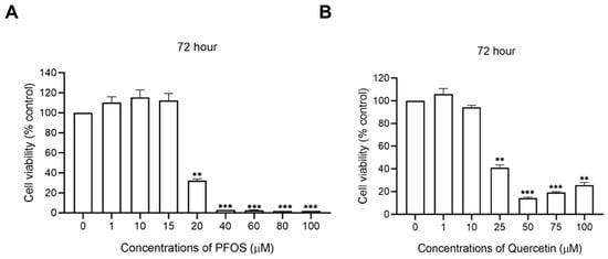
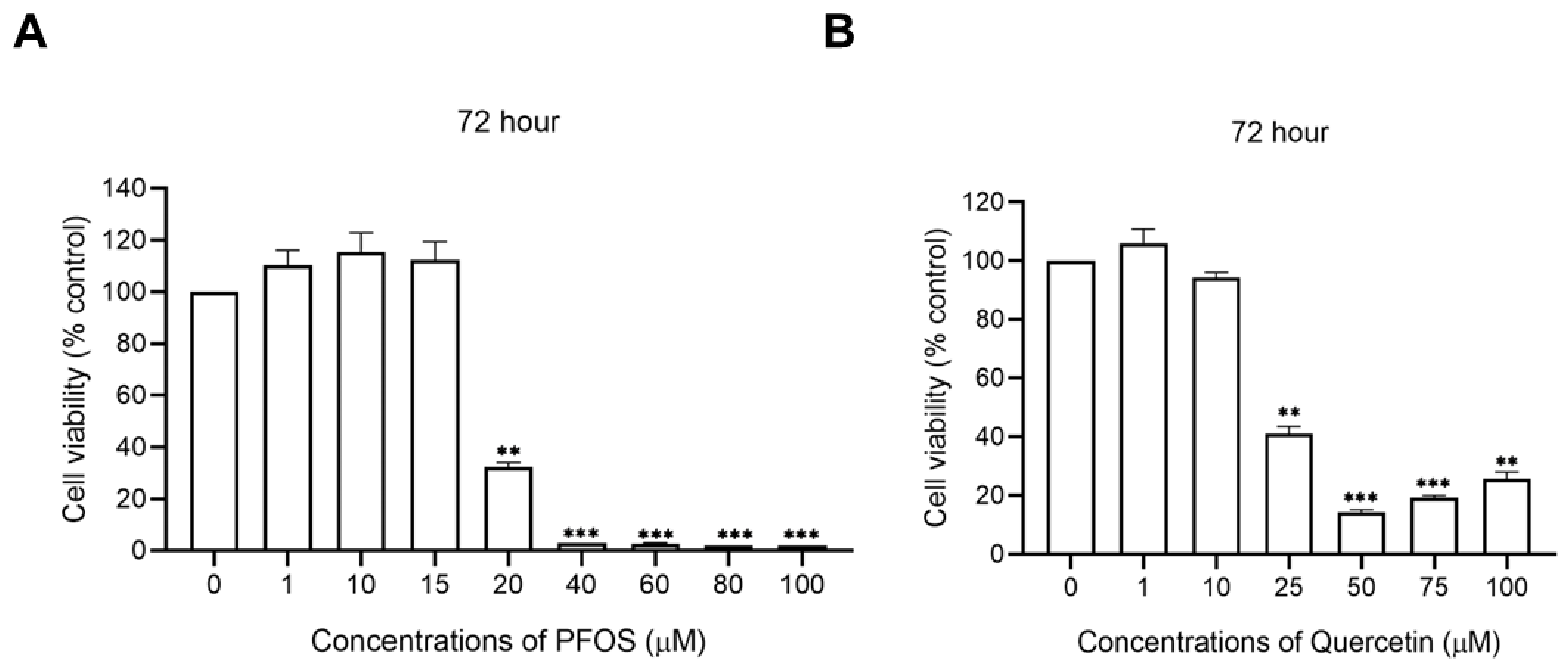
To investigate the appropriate experimental concentrations and ensure that subsequent biological evaluations are not confounded by non-specific cell death, the effects of PFOS and quercetin on HMEC-1 cell viability were assessed over 72 h. Exposure to PFOS at concentrations up to 15 μM did not significantly change viability; however a dramatic and significant reduction in cell survival was observed at 20 μM and higher (p < 0.001) (Figure 6A). Similarly, treatment with quercetin maintained cellular viability near 100% up to a concentration of 10 μM, with significant cytotoxicity initiating at 25 μM (p < 0.001) (Figure 6B). Based on these viability assays, 15 μM of PFOS and 10 μM of quercetin were identified as the highest non-toxic concentrations. Consequently, these specific doses were selected for all downstream transcriptomic profiling and in vitro protein validations. This stringent dose justification ensures that the observed alterations in endothelial inflammatory markers are driven by specific mechanistic signaling pathways rather than being secondary artifacts of generalized cellular toxicity or apoptosis.
Figure 6.
Effects of PFOS and quercetin on cell viability. (A) Cell viability of HMEC-1 cells following exposure to varying concentrations of PFOS (1, 10, 15, 20, 40, 60, 80, and 100 μM) for 72 h compared to vehicle control. (B) Cell viability of HMEC-1 cells following exposure to varying concentrations of quercetin (1, 10, 25, 50, 75, and 100 μM) for 72 h compared to vehicle control. Data are presented as the mean ± standard error of the mean (SEM) of three independent biological replicates. Cell viability is expressed as a percentage relative to the vehicle control, which is normalized to 100%. Statistical significance was determined using a repeated-measures one-way ANOVA, followed by Dunnett’s post hoc test, and is denoted as ** p < 0.01 and *** p < 0.001.
3.6. Quercetin Alleviates PFOS-Induced Endothelial Inflammation
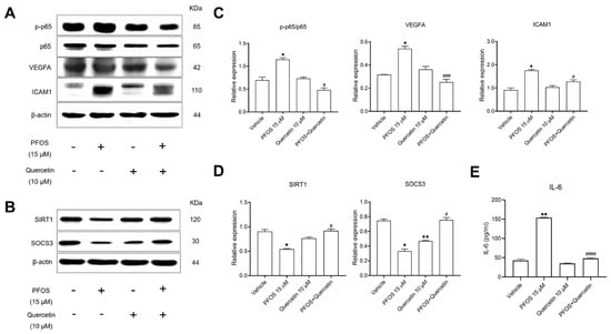
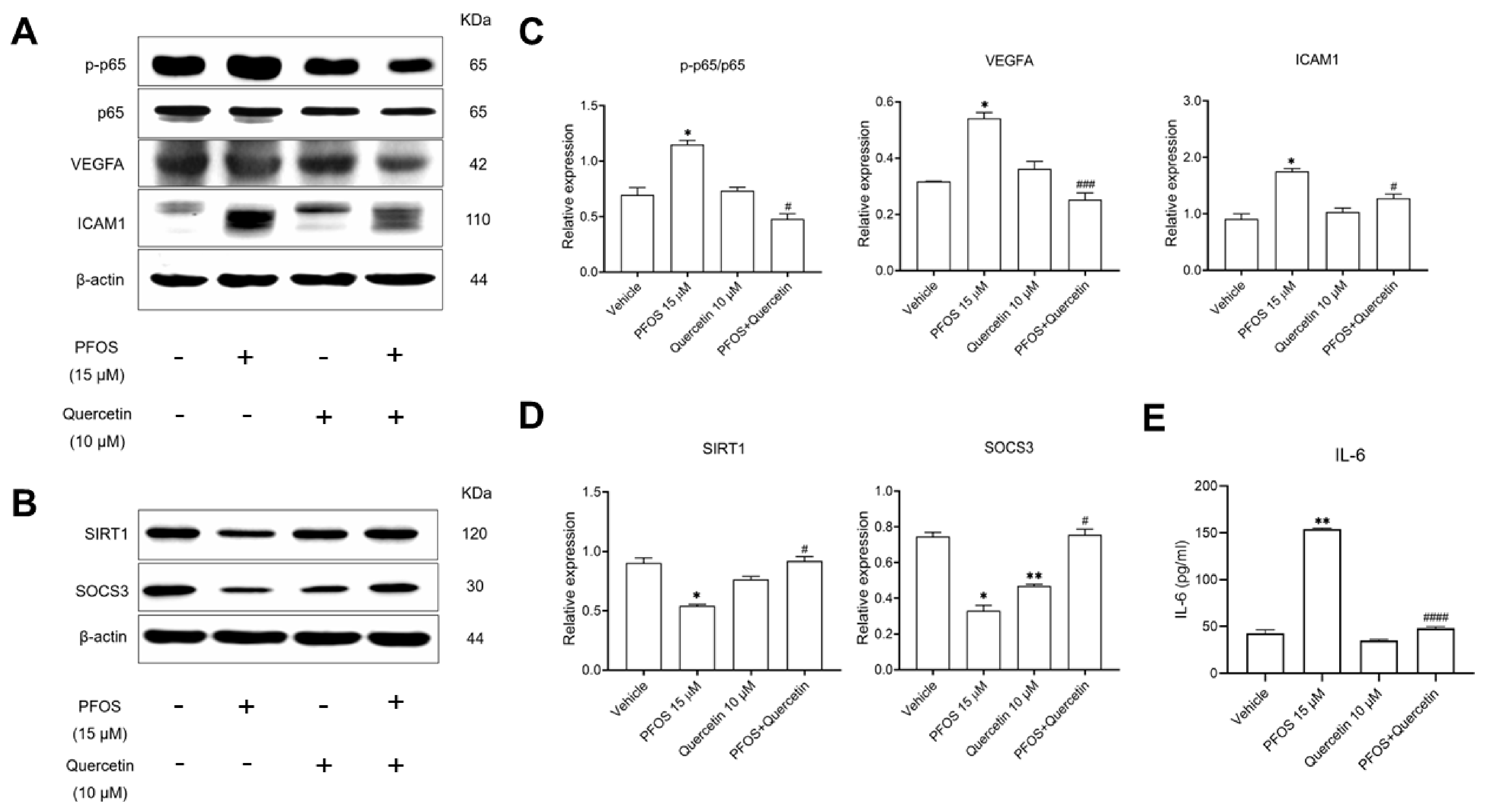
To validate the bioinformatic predictions and clinical dataset analyses, we evaluated the protein expression of the identified core molecular targets. HMEC-1 cells were exposed to sub-lethal concentrations (15 μM of PFOS and 10 μM of quercetin). To comprehensively evaluate the AGE–RAGE/NF-κB functional axis, we prioritized the validation of the core downstream effectors (IL-6, VEGFA and the adhesion marker ICAM1) and the critical upstream negative regulators (SIRT1 and SOCS3) rather than validating all bioinformatic hub genes exhaustively. Consistent with the functional enrichment linking the core hubs to the NF-κB and AGE–RAGE signaling pathways, PFOS exposure significantly induced the phosphorylation of p65 (indicated by an increased p-p65/total p65 ratio, while total p65 remained largely unchanged) and up-regulated the expression of VEGFA and downstream adhesion molecule ICAM1 (Figure 7A,C). Furthermore, PFOS increased the secretion of the pro-inflammatory cytokine IL-6 (Figure 7E). Interestingly, while treatment with quercetin alone reduced SOCS3 expression compared to the vehicle control, co-treatment with 10 μM of quercetin effectively restored the expression of SIRT1 and SOCS3 compared to the PFOS-exposed group (Figure 7B,D). Furthermore, co-treatment effectively attenuated these PFOS-induced molecular alterations, significantly suppressing p65 phosphorylation, VEGFA expression, and IL-6 secretion (p < 0.01 and p < 0.001). Collectively, these validations demonstrated that quercetin alleviates PFOS-induced endothelial inflammatory responses by directly modulating inflammatory hub genes (RELA, VEGFA and IL-6) in our network pipeline.
Figure 7.
In vitro validation of quercetin-mediated suppression of PFOS-induced endothelial inflammation. (A,B) Representative Western blot shows the protein expression levels of p-p65, total p65, VEGFA, ICAM1, SIRT1 and SOCS3 in HMEC-1 cells exposed to the vehicle, 15 μM of PFOS, 10 μM of quercetin or their co-treatment. β-actin served as the internal loading control. (C,D) Quantitative densitometric analysis of Western blot results. Target protein densitometry values (VEGFA, ICAM1, SIRT1, and SOCS3) are directly normalized to the corresponding β-actin internal controls. The relative expression of p-p65 is presented as the ratio of phosphorylated p65 to total p65. The y-axis represents these normalized relative expression values. (E) The concentration of IL-6 secreted into the cell culture supernatant was quantified using ELISA. Data are expressed as the mean ± SEM of independent biological replicates with individual observations. Statistical significance was determined using a repeated-measures one-way ANOVA, followed by Sidak’s multiple comparisons test, and is indicated as follows: * p < 0.05 and ** p < 0.01 compared to the vehicle control, whereas # p < 0.05, ### p < 0.001 and #### p < 0.0001 are compared to the PFOS-treated group.
4. Discussion
PFOS is a fluorinated chemical widely used in many industries and household products [1]. A previous study indicated that PFOS induced endothelial dysfunctions to promote atherosclerosis in ApoE−/− mice [18]. Quercetin is a plant-derived flavonoid that exhibits free radical scavenging and anti-inflammatory properties [13]. A previous study reported that quercetin has the potential to alleviate endothelial dysfunctions via autophagy in hypertensive rats [19]. Unfortunately, the molecular mechanisms of quercetin-alleviated PFOS-induced endothelial dysfunctions in HMEC-1 have to be investigated by the integration of transcriptomic analysis with experimental study.
Previous studies have established the cardiovascular toxicity of PFOS and the broader protective properties of quercetin, such as its modulation of oxidative stress in ApoE−/− mice [20] and its mitigation of structural myocardial injury in rat models [21]. However, the direct transcriptomic alterations in the human endothelium remain largely unexplored. Our study provides a novel contribution by using an unbiased RNA-seq and bioinformatic approach, specifically in human microvascular endothelial cells (HMEC-1). Unlike previous animal models, our functional enrichment analyses uniquely identify the AGE–RAGE signaling pathway as a primary target of the PFOS–quercetin interaction. Furthermore, we demonstrated that quercetin actively restores the anti-inflammatory regulators SIRT1 and SOCS3, which are explicitly down-regulated by PFOS exposure. Crucially, by validating our in vitro hub genes (IL-6, RELA, VEGFA, CXCL8 and PIM1) against the human atherosclerotic microarray dataset GSE43292, our findings indicate distinct clinical translational value by proposing that these hub genes are involved in the atherosclerotic plaque pathogenesis.
For biological alterations, we found that cytokine activity is the most enriched in the molecular function term and advanced glycation end products, and the receptor for the AGEs (AGE–RAGE) signaling pathway is the most enriched pathway. The AGE–RAGE signaling pathway has been recognized as a critical mediator of chronic inflammation, where ligand–receptor interaction activates downstream pathways, including NF-κB, MAPK, and PI3K/Akt, that amplify pro-inflammatory cytokines secretion [22]. A previous study reported that AGE–RAGE signaling pathway activation promoted pro-inflammatory TNFα secretion, oxidative stress and endothelial dysfunctions in Type II diabetes mice [23]. Notably, our pathway diagram showed that NF-κB, VEGF, IL-6, CXCL8 and PIM1 associated with AGE–RAGE signaling pathway activation should be further investigated.
As protein–protein interaction (PPI) network and modular clustering analysis, we also found that the AGE–RAGE signaling pathway in diabetic complication was the most enriched pathway, including IL-6, VEGFA and CXCL8. Moreover, network analysis showed that IL-6, RELA (p65) and CXCL8 were the top three enrichments, with a high betweenness score. Interleukin 6 (IL-6) is a multifunctional cytokine that plays a crucial role in acute-phase responses and immune regulation [24]. Elevated levels of IL-6 and other inflammatory markers, such as high-sensitivity C-reactive protein (hs-CRP), have been associated with endothelial dysfunctions and chronic inflammation [25,26]. Vascular Endothelial Growth Factor A (VEGFA) plays a crucial role in plaque progression and instability during atherosclerosis progression [27]. A previous study reported that overexpression of VEGF has been associated with enhanced angiogenesis and accelerated plaque development [28]. CXCL8 is a chemokine involved in immune cells at the site of tissue injury and inflammation [29]. CXCL8 has been implicated in atherosclerosis progression by modulating lipid metabolism, promoting macrophage recruitment and accelerating foam cell formation [30]. RELA (NF-κB, p65), a key subunit of the NF-κB transcription factor complex, is a crucial regulator of inflammation, immunity, cell survival and proliferation [31]. Activation of the NF-κB signaling pathway has been strongly linked to endothelial dysfunctions associated with atherosclerosis [32]. Therefore, these finding supported that biological alterations of the AGE–RAGE signaling pathway, including IL-6, VEGFA, CXCL8 and RELA, may be therapeutic targets of PFOS-quercetin exposure in HMEC-1 cells that should be further verified.
The identification of IL-6, RELA, VEGFA, CXCL8 and PIM1 as core hub genes provides a robust framework for understanding quercetin’s protective efficacy. The down-regulation of RELA (p65) by quercetin likely serves as a primary regulator, dampening the NF-κB-mediated transcription of subsequent pro-inflammatory and pro-angiogenic targets, specifically, IL-6, CXCL8, and VEGFA, thereby preventing pathogenic endothelial remodeling [33]. However, it is important to acknowledge that changes in transcript and protein expression alone are associative and do not definitively establish these molecules as the exclusive direct mediators of quercetin’s protective effects. Quercetin is a highly pleiotropic molecule, and alternative mechanisms, such as its direct biochemical capacity to scavenge reactive oxygen species (ROS) prior to receptor activation, may contribute significantly to endothelial protection [34]. Furthermore, the absence of gene silencing (e.g., siRNA targeting RELA or SIRT1) or specific pharmacological inhibition in this study limits our ability to confirm direct causality. Future investigations using genetic knockdown models are necessary to definitively validate whether the AGE–RAGE/NF-κB signaling pathway is the obligatory pathway for quercetin-mediated alleviation of PFOS-induced endothelial dysfunctions.
Regarding clinical relevance, our analysis of the GSE43292 dataset demonstrated that the five overlapping hub genes (IL-6, RELA, VEGFA, CXCL8 and PIM1) were significantly up-regulated in localized atheromatous plaque tissues compared to intact arterial tissues. Previous studies have established certain targets like IL-6 as systemic circulating inflammatory markers [35]. The collective up-regulation of these five hub genes in our analysis specifically implicates them as localized tissue effectors in atherosclerotic progression [36,37,38,39]. To establish their diagnostic potential and overcome internal validation bias, we implemented an independent external validation strategy using the GSE100927 clinical dataset (n = 104). By training machine learning models on the GSE43292 cohort and evaluating them on the unseen GSE100927 cohort, the Logistic Regression model demonstrated exceptional discriminatory power, with an AUC of 0.82, outperforming the Random Forest classifier (AUC = 0.45). This robust external validation confirms that these five overlapping hub genes possess significant predictive value and generalizability for distinguishing atherosclerotic lesions. However, it is important to note that tissue expression differences alone do not establish these molecules as early clinical biomarkers. Our validation primarily relies on localized tissue expression, and we have not yet evaluated their diagnostic performance in circulating biofluids such as plasma or serum. Future large-scale clinical studies are required to rigorously evaluate the diagnostic utility of these hub genes.
To investigate the viable cell test, a previous study reported that exposure to 15 µM of PFOS for 72 h did not change the percentage of viable cells compared to the vehicle control in human endothelial HMEC-1 cells [16]. Similarly, we found that exposure to 15 µM of PFOS and 10 µM of quercetin for 72 h did not change the percentage of viable cells compared to the vehicle control in HMEC-1. Therefore, we selected 15 µM of PFOS and 10 µM of quercetin for further studies.
While a direct MTT viability assay was not performed for the co-exposure condition, the preservation of cell viability and uniform cell mass was validated through our downstream assays. Specifically, total protein quantification prior to Western blotting and ELISA demonstrated consistent protein yields across the vehicle, PFOS, quercetin and co-exposure groups. Furthermore, the robust and uniform expression of the internal housekeeping protein (β-actin) in the co-exposure group confirmed that substantial cytotoxicity or cell loss did not occur. Therefore, the observed mechanistic alterations and inflammatory resolution in the co-exposure group were independent of overt cell death.
Moreover, in interpreting these in vitro findings, the clinical relevance of the selected concentrations and timelines must be considered. While the 15 µM PFOS concentration utilized in this study is higher than the typical serum levels observed in the general population, it aligns with serum concentrations reported in occupationally exposed workers or populations in highly contaminated regions. For instance, biomonitoring studies of occupationally exposed workers in fluorochemical manufacturing have reported serum PFOS concentrations reaching up to 12.83 µg/mL (equivalent to 25 µM) [40]. Furthermore, applying this concentration over a 72 h period serves as an established in vitro accelerated model. Because continuous, low-dose chronic exposure over decades in vivo cannot be perfectly replicated in a standard cell culture timeframe, higher sub-lethal concentrations are required in vitro to reliably induce and evaluate the mechanistic, cumulative molecular alterations of endothelial stress [41]. In contrast, the selected dose of 10 µM quercetin is highly translationally relevant. While basal dietary intake yields nanomolar plasma levels, targeted pharmacological supplementation and highly bioavailable formulations can safely elevate human plasma and local tissue concentrations into the low micromolar range (up to 10 µM), making this an appropriate and physiologically achievable in vitro working concentration [42].
Endothelial inflammation is a key pathological response that can lead to tissue damage and promotes pathogenesis of various diseases, including endothelial dysfunctions associated with atherosclerosis [43]. In this study, we investigated the underlying molecular mechanisms of PFOS and quercetin in HMEC-1 by using the immunoblotting assay. We found that PFOS up-regulated phospho-p65, VEGFA, and ICAM1 and increased IL-6 secretion. Conversely, quercetin exposure down-regulated phospho-p65, VEGFA, and ICAM and decreased IL-6 secretion. Similarly, a previous study reported that PFOS induced inflammation by up-regulating p65-caused endothelial dysfunctions in HMEC-1 cells [16]. Meanwhile, quercetin exposure attenuated inflammation by down-regulating p65 in in vivo and in vitro models [44]. In addition, a previous study demonstrated that VEGFA-containing conditioned medium from PFOS-exposed human granulosa cells rather than direct PFOS exposure promoted endothelial angiogenesis in human EA.hy926 cells [45]. Conversely, quercetin exposure inhibited endothelial angiogenesis through the cross-talk between the Akt/NF-κB and VEGFA/VEGFR-2 signaling pathways in a rat model [46]. Intercellular adhesion molecule 1 (ICAM1) is a transmembrane protein that facilitates cell–cell interactions and promotes leukocyte adhesion and transmigration across the vascular wall [47]. A previous study reported that PFOS significantly up-regulated ICAM1 expression in HMEC-1 cells [16]. In contrast, quercetin attenuated TNFα-induced ICAM1 expression through modulation of AP-1 activation and the JNK signaling pathway in human endothelial cells [48]. For cytokine secretion, a previous study suggested that PFOS increased the level of IL-6 secretion in HMEC-1 cells [16], whereas quercetin exposure inhibited the level of IL-6 secretion in LPS-stimulated neutrophils [49].
In terms of anti-inflammation, sirtuin 1 (SIRT1) is a nuclear deacetylase that regulates gene expression, cellular metabolism, and aging and prevents inflammation [50]. A previous study suggested that SIRT1 prevented DNA damage and attenuated atherosclerosis in human vascular smooth muscle cells (VSMCs) [51]. In this study, we found that PFOS down-regulated SIRT1 expression, while quercetin exposure up-regulated SIRT1 expression. Similarly, a previous study reported that PFOS down-regulated SIRT1 expression in human endothelial cells [16], while quercetin up-regulated SIRT1 expression against inflammatory responses via the activation of the SIRT1/Nrf2/HO-1 signaling pathway in rats’ cartilage tissue [52]. Suppressor of cytokine signaling 3 (SOCS3) is a negative regulator of the JAK2/STAT3 pathway that plays an important role in anti-inflammation, cell proliferation and immune responses [53]. In this study, we found that PFOS down-regulated SOCS3 expression, whereas quercetin exposure up-regulated SOCS3 expression. Similarly, a previous study suggested that PFOS down-regulated SOCS3 expression in placental and fetal mouse tissues [54], whereas quercetin exposure up-regulated SOCS3 expression through the activation of the PPARγ signaling pathway in arthritic mice [55]. Therefore, these findings suggested that the NF-κB signaling pathway may be the molecular target of PFOS; meanwhile, it also acts as a therapeutic target of quercetin exposure in HMEC-1 cells.
Despite these mechanistic insights, this study possesses several inherent limitations that must be acknowledged. First, our assessments were restricted to transcriptomic and protein-level inflammatory markers, lacking direct functional evaluations of endothelial dysfunctions such as nitric oxide (NO) bioavailability, endothelial permeability, or in vitro angiogenesis (tube formation) assays. Second, our experiments used an immortalized human microvascular endothelial cell line (HMEC-1). While HMEC-1 serves as a robust and standard baseline model for evaluating generic endothelial activation and transcriptomic toxicity, atherosclerosis is fundamentally a macrovascular disease affecting large- and medium-sized arteries [56]. Consequently, extrapolating microvascular responses directly to arterial atherogenesis requires caution. Additionally, while three independent biological replicates (n = 3) are a standard minimum for in vitro molecular screening, we acknowledge that this small sample size inherently restricts statistical precision and effect size estimation. Finally, as a strictly in vitro observational study, these findings cannot be interpreted as clinical evidence of disease prevention. Future research incorporating primary arterial endothelial cells such as human aortic endothelial cells, functional physiological assays, and in vivo cardiovascular disease models are imperative to validate the therapeutic potential of quercetin against PFOS-induced atherosclerosis.
5. Conclusions
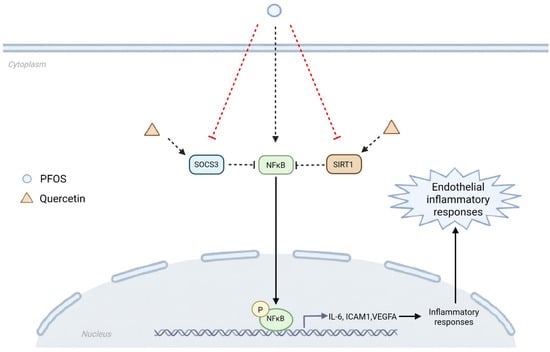
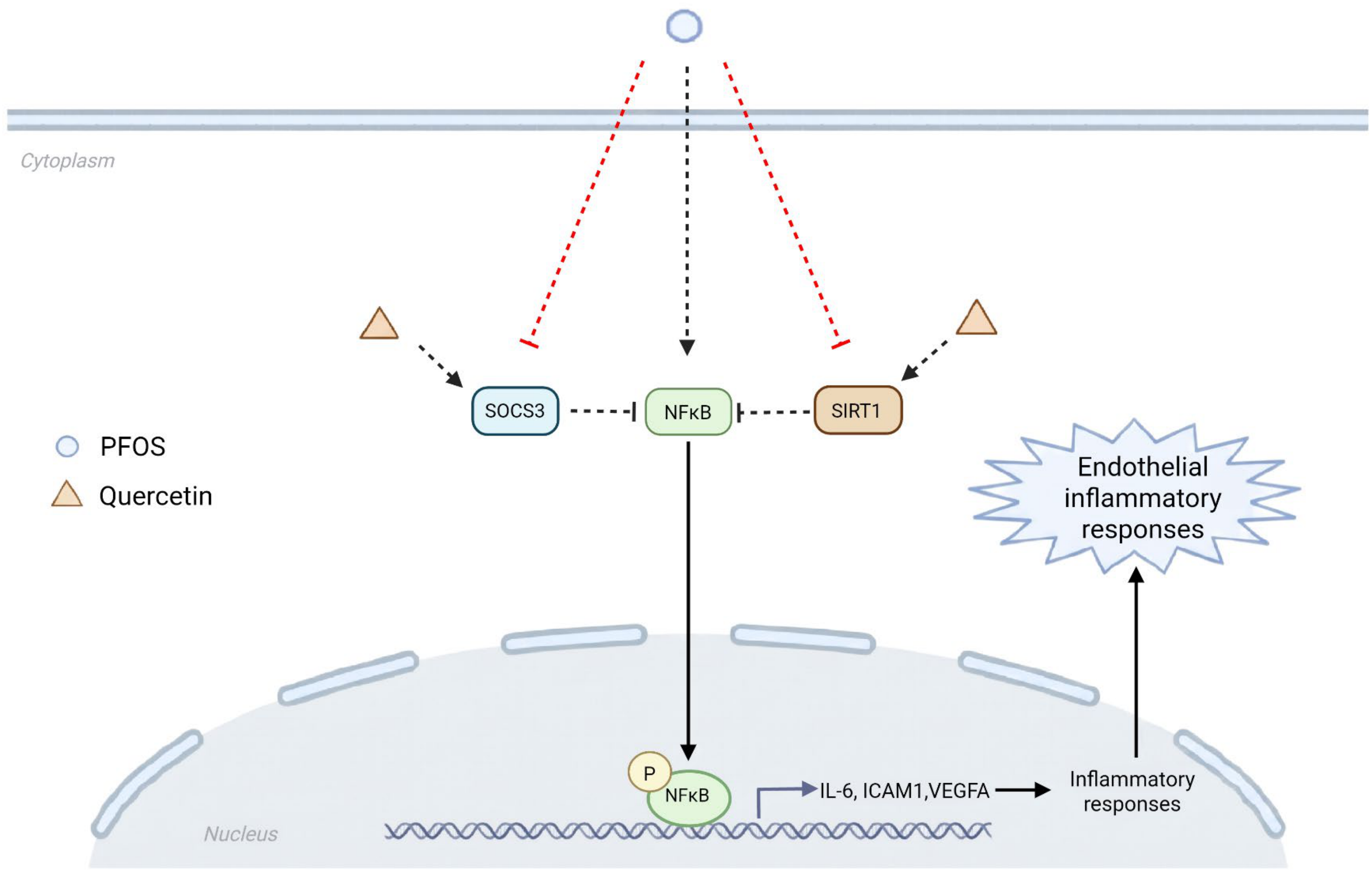
In summary, we found that the AGE–RAGE signaling pathway and cytokine activity were the most enriched terms of pathway analysis and molecular function. Similarly, the PPI network showed that the AGE–RAGE signaling pathway and cytokine activity were the most enriched pathways. Furthermore, we found that five overlapping hub genes, including IL6, RELA, VEGFA, CXCL8, and PIM1, were up-regulated in plaque compared to intact tissues from GSE43292, suggesting their clinical relevance in atherosclerotic plaque pathogenesis. Crucially, our independent external validation using machine learning (Logistic Regression) on the external GSE100927 dataset showed that these five overlapping hub genes possess strong discriminatory predictive capability (AUC = 0.82), strongly supporting their clinical diagnostic relevance and generalizability. At the protein level, PFOS triggered endothelial inflammatory responses by up-regulating p65, VEGFA, and ICAM1 expression; increasing IL-6 levels; and down-regulating SIRT1 and SOCS3. Conversely, quercetin successfully attenuated these inflammatory responses by reversing these protein alterations (Figure 8). Collectively, these findings provide novel insights into the molecular mechanisms by which quercetin alleviates PFOS-induced inflammatory responses in HMEC-1 cells, suggesting its promising potential to modulate early molecular events associated with endothelial activation and atherosclerosis.
Figure 8.
Proposed mechanistic model of quercetin-mediated alleviation of PFOS-induced endothelial inflammatory responses. PFOS exposure induces the activation of NF-κB, leading to the up-regulation of downstream inflammatory markers (IL-6, ICAM1, and VEGFA). Co-treatment with quercetin effectively attenuates PFOS-induced molecular alterations. Solid lines represent the direct experiment confirmed in this study (protein expression and cytokine secretion). Dashed lines represent hypothesized upstream regulatory mechanisms, including SIRT1/SOCS3 to the NF-κB axis, and inferred signaling connections based on bioinformatic enrichment analysis.
Supplementary Materials
The following supporting information can be downloaded at: https://www.mdpi.com/article/10.3390/medsci14070797/s1, supplementary S1: coding S1; supplementary S2: Full Gel; Tables S1–S4.
Author Contributions
N.V.: Writing—original draft, Writing—review and editing, Methodology, Investigation, Data curation and Data analysis. S.C.: Writing—review and editing, Conceptualization, Methodology, Investigation, Data curation, Data analysis and Supervision. J.S.: Writing—review and editing, Conceptualization, Resources, Supervision and Project administration. All authors have read and agreed to the published version of the manuscript.
Funding
This study was supported by Chulabhorn Research Institute (CRI-2569-01/3-05).
Institutional Review Board Statement
Not applicable.
Data Availability Statement
The original contributions presented in this study are included in the article/Supplementary Material. Further inquiries can be directed to the corresponding author.
Acknowledgments
We thank Thomas J. Lawley (Centers for Disease Control and Prevention, Atlanta, GA, USA) for kindly providing the HMEC-1 cell line used in this study.
Conflicts of Interest
The authors declare no conflicts of interest.
Abbreviations
The following abbreviations are used in this manuscript:
| AS | Atherosclerosis |
| HMEC-1 | Human microvascular endothelial cell 1 |
| ICAM1 | Intercellular adhesion molecule 1 |
| IL-6 | Interleukin 6 |
| PFOS | Perfluorooctane sulfonic acid |
| SIRT1 | Sirtuin 1 |
| SOCS3 | Suppressor of cytokine signaling 3 |
| VEGFA | Vascular endothelial growth factor A |
References
- Jarvis, A.L.; Justice, J.R.; Elias, M.C.; Schnitker, B.; Gallagher, K. Perfluorooctane sulfonate in US ambient surface waters: A review of occurrence in aquatic environments and comparison to global concentrations. Environ. Toxicol. Chem. 2021, 40, 2425–2442. [Google Scholar] [CrossRef] [Scilit] [PubMed]
- Leung, S.C.E.; Shukla, P.; Chen, D.; Eftekhari, E.; An, H.; Zare, F.; Ghasemi, N.; Zhang, D.; Nguyen, N.-T.; Li, Q. Emerging technologies for PFOS/PFOA degradation and removal: A review. Sci. Total. Environ. 2022, 827, 153669. [Google Scholar] [CrossRef] [Scilit] [PubMed]
- Olsen, G.W.; Burris, J.M.; Ehresman, D.J.; Froehlich, J.W.; Seacat, A.M.; Butenhoff, J.L.; Zobel, L.R. Half-life of serum elimination of perfluorooctanesulfonate, perfluorohexanesulfonate, and perfluorooctanoate in retired fluorochemical production workers. Environ. Health Perspect. 2007, 115, 1298–1305. [Google Scholar] [CrossRef] [Scilit] [PubMed]
- Fenton, S.E.; Ducatman, A.; Boobis, A.; DeWitt, J.C.; Lau, C.; Ng, C.; Smith, J.S.; Roberts, S.M. Per-and polyfluoroalkyl substance toxicity and human health review: Current state of knowledge and strategies for informing future research. Environ. Toxicol. Chem. 2021, 40, 606–630. [Google Scholar] [CrossRef] [Scilit] [PubMed]
- Mišľanová, C.; Valachovičová, M. Health Impacts of Per-and Polyfluoroalkyl Substances (PFASs): A Comprehensive Review. Life 2025, 15, 573. [Google Scholar] [CrossRef] [Scilit] [PubMed]
- Wee, S.Y.; Zaharin, A.A. Environmental impacts, exposure pathways, and health effects of PFOA and PFOS. Ecotoxicol. Environ. Saf. 2023, 267, 115663. [Google Scholar] [CrossRef] [Scilit] [PubMed]
- Jebari-Benslaiman, S.; Galicia-Garcia, U.; Larrea-Sebal, A.; Olaetxea, J.R.; Alloza, I.; Vandenbroeck, K.; Benito-Vicente, A.; Martin, C. Pathophysiology of Atherosclerosis. Int. J. Mol. Sci. 2022, 23, 3346. [Google Scholar] [CrossRef] [Scilit] [PubMed]
- Nedkoff, L.; Briffa, T.; Zemedikun, D.; Herrington, S.; Wright, F.L. Global trends in atherosclerotic cardiovascular disease. Clin. Ther. 2023, 45, 1087–1091. [Google Scholar] [CrossRef] [Scilit] [PubMed]
- Goh, R.S.J.; Chong, B.; Jayabaskaran, J.; Jauhari, S.M.; Chan, S.P.; Kueh, M.T.W.; Shankar, K.; Li, H.; Chin, Y.H.; Kong, G. The burden of cardiovascular disease in Asia from 2025 to 2050: A forecast analysis for East Asia, South Asia, South-East Asia, Central Asia, and high-income Asia Pacific regions. Lancet. Reg. Health West. Pac. 2024, 49, 101138. [Google Scholar] [CrossRef] [Scilit] [PubMed]
- Gerdanovics, A.; Stănescu, I.C.; Mîrza, C.M.; Dogaru, G.B.; Nicula, C.A.; Boarescu, P.-M.; Gerdanovics, C.-A.; Bulboacă, A.E. Proinflammatory Risk Factors in Patients with Ischemic Stroke: A Systematic Review and Meta-Analysis. Antioxidants 2025, 14, 1229. [Google Scholar] [CrossRef] [Scilit] [PubMed]
- Oh, K.K.; Adnan, M.; Cho, D.H. A network pharmacology analysis on drug-like compounds from Ganoderma lucidum for alleviation of atherosclerosis. J. Food. Biochem. 2021, 45, e13906. [Google Scholar] [CrossRef] [Scilit] [PubMed]
- Latif, R.; Nawaz, T. Medicinal plants and human health: A comprehensive review of bioactive compounds, therapeutic effects, and applications. Phytochem. Rev. 2025, 25, 2299–2342. [Google Scholar] [CrossRef] [Scilit]
- Vollmannová, A.; Bojňanská, T.; Musilová, J.; Lidiková, J.; Cifrová, M. Quercetin as one of the most abundant represented biological valuable plant components with remarkable chemoprotective effects-A review. Heliyon 2024, 10, e33342. [Google Scholar] [CrossRef] [Scilit] [PubMed]
- Kleemann, R.; Verschuren, L.; Morrison, M.; Zadelaar, S.; van Erk, M.J.; Wielinga, P.Y.; Kooistra, T. Anti-inflammatory, anti-proliferative and anti-atherosclerotic effects of quercetin in human in vitro and in vivo models. Atherosclerosis 2011, 218, 44–52. [Google Scholar] [CrossRef] [Scilit] [PubMed]
- Luo, G.; Xiang, L.; Xiao, L. Quercetin alleviates atherosclerosis by suppressing oxidized LDL-induced senescence in plaque macrophage via inhibiting the p38MAPK/p16 pathway. J. Nutr. Biochem. 2023, 116, 109314. [Google Scholar] [CrossRef] [Scilit] [PubMed]
- Vajeethaveesin, N.; Kanitwithayanun, J.; Suriyo, T.; Chujan, S.; Satayavivad, J. Perfluorooctane sulfonic acid: A possible risk factor of endothelial dysfunction based on in silico and in vitro studies. Arch. Toxicol. 2025, 99, 2975–2991. [Google Scholar] [CrossRef] [Scilit] [PubMed]
- Ge, S.X.; Jung, D.; Yao, R. ShinyGO: A graphical gene-set enrichment tool for animals and plants. J. Bioinf. Comput. Biol. 2020, 36, 2628–2629. [Google Scholar] [CrossRef] [Scilit] [PubMed]
- Zhang, B.; Li, Q.; Wang, W.; Tian, M.; Xu, D.; Xie, Y. PFOS and Its Substitute OBS Cause Endothelial Dysfunction to Promote Atherogenesis in ApoE–/–Mice. EHS 2025, 3, 526–538. [Google Scholar] [CrossRef] [Scilit] [PubMed]
- Lin, X.; Han, T.; Fan, Y.; Wu, S.; Wang, F.; Wang, C. Quercetin improves vascular endothelial function through promotion of autophagy in hypertensive rats. Life Sci. 2020, 258, 118106. [Google Scholar] [CrossRef] [Scilit] [PubMed]
- Luo, M.; Tian, R.; Lu, N. Quercetin inhibited endothelial dysfunction and atherosclerosis in apolipoprotein E-deficient mice: Critical roles for NADPH oxidase and heme oxygenase-1. J. Agric. Food Chem. 2020, 68, 10875–10883. [Google Scholar] [CrossRef] [Scilit] [PubMed]
- Mandour, D.A.; Morsy, M.M.; Fawzy, A.; Mohamed, N.M.; Ahmad, M.M. Structural and molecular changes in the rat myocardium following perfluoroctane sulfonate (PFOS) exposure are mitigated by quercetin via modulating HSP 70 and SERCA 2. J. Mol. Histol. 2023, 54, 283–296. [Google Scholar] [CrossRef] [Scilit] [PubMed]
- Ramasamy, R.; Yan, S.F.; Schmidt, A.M. Receptor for AGE (RAGE): Signaling mechanisms in the pathogenesis of diabetes and its complications. Ann. N. Y. Acad. Sci. 2011, 1243, 88–102. [Google Scholar] [CrossRef] [Scilit] [PubMed]
- Gao, X.; Zhang, H.; Schmidt, A.M.; Zhang, C. AGE/RAGE produces endothelial dysfunction in coronary arterioles in type 2 diabetic mice. Am. J. Physiol. Heart Circ. Physiol. 2008, 295, H491–H498. [Google Scholar] [CrossRef] [Scilit] [PubMed]
- Tanaka, T.; Narazaki, M.; Kishimoto, T. IL-6 in inflammation, immunity, and disease. Cold Spring Harb. Perspect. Biol. 2014, 6, a016295. [Google Scholar] [CrossRef] [Scilit] [PubMed]
- Gager, G.M.; Biesinger, B.; Hofer, F.; Winter, M.-P.; Hengstenberg, C.; Jilma, B.; Eyileten, C.; Postula, M.; Lang, I.M.; Siller-Matula, J.M. Interleukin-6 level is a powerful predictor of long-term cardiovascular mortality in patients with acute coronary syndrome. Vasc. Pharmacol. 2020, 135, 106806. [Google Scholar] [CrossRef] [Scilit] [PubMed]
- Kaptoge, S.; Seshasai, S.R.K.; Gao, P.; Freitag, D.F.; Butterworth, A.S.; Borglykke, A.; Di Angelantonio, E.; Gudnason, V.; Rumley, A.; Lowe, G.D. Inflammatory cytokines and risk of coronary heart disease: New prospective study and updated meta-analysis. Eur. Hear. J. 2014, 35, 578–589. [Google Scholar] [CrossRef] [Scilit] [PubMed]
- Dabravolski, S.A.; Khotina, V.A.; Omelchenko, A.V.; Kalmykov, V.A.; Orekhov, A.N. The role of the VEGF family in atherosclerosis development and its potential as treatment targets. Int. J. Mol. Sci. 2022, 23, 931. [Google Scholar] [CrossRef] [Scilit] [PubMed]
- Celletti, F.L.; Waugh, J.M.; Amabile, P.G.; Brendolan, A.; Hilfiker, P.R.; Dake, M.D. Vascular endothelial growth factor enhances atherosclerotic plaque progression. Nat. Med. 2001, 7, 425–429. [Google Scholar] [CrossRef] [Scilit] [PubMed]
- Asokan, S.; Bandapalli, O.R. CXCL8 signaling in the tumor microenvironment. Adv. Exp. Med. Biol. 2021, 1302, 25–39. [Google Scholar] [CrossRef] [Scilit] [PubMed]
- Apostolakis, S.; Vogiatzi, K.; Amanatidou, V.; Spandidos, D.A. Interleukin 8 and cardiovascular disease. Cardiovasc. Res. 2009, 84, 353–360. [Google Scholar] [CrossRef] [Scilit] [PubMed]
- Msaki, A.; Sánchez, A.M.; Koh, L.F.; Barré, B.; Rocha, S.; Perkins, N.D.; Johnson, R.F. The role of RelA (p65) threonine 505 phosphorylation in the regulation of cell growth, survival, and migration. Mol. Cell. 2011, 22, 3032–3040. [Google Scholar] [CrossRef] [Scilit] [PubMed]
- Bourcier, T.; Sukhova, G.; Libby, P. The nuclear factor κ-B signaling pathway participates in dysregulation of vascular smooth muscle cells in vitroand in human atherosclerosis. J. Biol. Chem. 1997, 272, 15817–15824. [Google Scholar] [CrossRef] [Scilit] [PubMed]
- Cho, S.-Y.; Park, S.-J.; Kwon, M.-J.; Jeong, T.-S.; Bok, S.-H.; Choi, W.-Y.; Jeong, W.-I.; Ryu, S.-Y.; Do, S.-H.; Lee, C.-S. Quercetin suppresses proinflammatory cytokines production through MAP kinases and NF-κB pathway in lipopolysaccharide-stimulated macrophage. Mol. Cell. Biochem. 2003, 243, 153–160. [Google Scholar] [CrossRef] [Scilit] [PubMed]
- Carrillo-Martinez, E.J.; Flores-Hernández, F.Y.; Salazar-Montes, A.M.; Nario-Chaidez, H.F.; Hernández-Ortega, L.D. Quercetin, a flavonoid with great pharmacological capacity. Molecules 2024, 29, 1000. [Google Scholar] [CrossRef] [Scilit] [PubMed]
- Held, C.; White, H.D.; Stewart, R.A.; Budaj, A.; Cannon, C.P.; Hochman, J.S.; Koenig, W.; Siegbahn, A.; Steg, P.G.; Soffer, J. Inflammatory biomarkers interleukin-6 and C-reactive protein and outcomes in stable coronary heart disease: Experiences from the STABILITY (stabilization of atherosclerotic plaque by initiation of darapladib therapy) trial. J. Am. Heart Assoc. 2017, 6, e005077. [Google Scholar] [CrossRef] [Scilit] [PubMed]
- Chhotaray, S.; Jal, S. Identifying Biomarkers for Atherosclerosis via Gene Expression and Biological Networking. Curr. Cardiol. Rev. 2025, 21, E1573403X1340118. [Google Scholar] [CrossRef] [Scilit] [PubMed]
- Wang, J.; Hu, X.; Hu, X.; Gao, F.; Li, M.; Cui, Y.; Wei, X.; Qin, Y.; Zhang, C.; Zhao, Y. MicroRNA-520c-3p targeting of RelA/p65 suppresses atherosclerotic plaque formation. Int. J. Biochem. Cell Biol. 2021, 131, 105873. [Google Scholar] [CrossRef] [Scilit] [PubMed]
- Braile, M.; Marcella, S.; Cristinziano, L.; Galdiero, M.R.; Modestino, L.; Ferrara, A.L.; Varricchi, G.; Marone, G.; Loffredo, S. VEGF-A in cardiomyocytes and heart diseases. Int. J. Mol. Sci. 2020, 21, 5294. [Google Scholar] [CrossRef] [Scilit] [PubMed]
- Nock, S.; Karim, E.; Unsworth, A.J. Pim kinases: Important regulators of cardiovascular disease. Int. J. Mol. Sci. 2023, 24, 11582. [Google Scholar] [CrossRef] [Scilit] [PubMed]
- Olsen, G.W.; Logan, P.W.; Hansen, K.J.; Simpson, C.A.; Burris, J.M.; Burlew, M.M.; Vorarath, P.P.; Venkateswarlu, P.; Schumpert, J.C.; Mandel, J.H. An occupational exposure assessment of a perfluorooctanesulfonyl fluoride production site: Biomonitoring. AIHA J. 2003, 64, 651–659. [Google Scholar] [CrossRef]
- Wielsøe, M.; Long, M.; Ghisari, M.; Bonefeld-Jørgensen, E.C. Perfluoroalkylated substances (PFAS) affect oxidative stress biomarkers in vitro. Chemosphere 2015, 129, 239–245. [Google Scholar] [CrossRef] [Scilit] [PubMed]
- Riva, A.; Ronchi, M.; Petrangolini, G.; Bosisio, S.; Allegrini, P. Improved Oral Absorption of Quercetin from Quercetin Phytosome®, a New Delivery System Based on Food Grade Lecithin: A. Riva et al. Eur. J. Drug Metab. Pharmacokinet. 2019, 44, 169–177. [Google Scholar] [CrossRef] [Scilit] [PubMed]
- Gusev, E.; Sarapultsev, A. Atherosclerosis and inflammation: Insights from the theory of general pathological processes. Int. J. Mol. Sci. 2023, 24, 7910. [Google Scholar] [CrossRef] [Scilit] [PubMed]
- Meng, X.; Li, Y.; Wang, F.; Li, T.; Wang, B.; Wang, Q.; Long, J.; Xie, H.; Zhang, Y.; Li, J. Quercetin attenuates inflammation in rosacea by directly targeting p65 and ICAM-1. Life Sci. 2024, 347, 122675. [Google Scholar] [CrossRef] [Scilit] [PubMed]
- Tomanic, T.; Nenadov, D.S.; Stanic, B.; Pogrmic-Majkic, K.; Pletikosic, S.R.; Andric, N. Vascular endothelial growth factor-containing conditioned medium from PFOS-exposed human granulosa cells promotes endothelial angiogenesis in vitro. Toxicology 2025, 519, 154322. [Google Scholar] [CrossRef] [Scilit] [PubMed]
- Li, X.; Chen, Y.; Wang, L.; Shang, G.; Zhang, C.; Zhao, Z.; Zhang, H.; Liu, A. Quercetin alleviates pulmonary angiogenesis in a rat model of hepatopulmonary syndrome. Braz. J. Med. Biol. Res. 2016, 49, e5326. [Google Scholar] [CrossRef] [Scilit] [PubMed]
- Haydinger, C.D.; Ashander, L.M.; Tan, A.C.R.; Smith, J.R. Intercellular adhesion molecule 1: More than a leukocyte adhesion molecule. Biology 2023, 12, 743. [Google Scholar] [CrossRef] [Scilit] [PubMed]
- Kobuchi, H.; Roy, S.; Sen, C.K.; Nguyen, H.G.; Packer, L. Quercetin inhibits inducible ICAM-1 expression in human endothelial cells through the JNK pathway. Am. J. Physiol. Cell. Physiol. 1999, 277, C403–C411. [Google Scholar] [CrossRef] [Scilit] [PubMed]
- Liu, J.; Li, X.; Yue, Y.; Li, J.; He, T.; He, Y. The inhibitory effect of quercetin on IL-6 production by LPS-stimulated neutrophils. Cell. Mol. Immunol. 2005, 2, 455–460. [Google Scholar] [PubMed]
- Rahman, S.; Islam, R. Mammalian Sirt1: Insights on its biological functions. Cell. Commun. Signal. 2011, 9, 11. [Google Scholar] [CrossRef] [Scilit] [PubMed]
- Gorenne, I.; Kumar, S.; Gray, K.; Figg, N.; Yu, H.; Mercer, J.; Bennett, M. Vascular smooth muscle cell sirtuin 1 protects against DNA damage and inhibits atherosclerosis. Circulation 2013, 127, 386–396. [Google Scholar] [CrossRef] [Scilit] [PubMed]
- Ruan, H.; Zhu, T.; Wang, T.; Guo, Y.; Liu, Y.; Zheng, J. Quercetin Modulates Ferroptosis via the SIRT1/Nrf− 2/HO− 1 Pathway and Attenuates Cartilage Destruction in an Osteoarthritis Rat Model. Int. J. Mol. Sci. 2024, 25, 7461. [Google Scholar] [CrossRef] [Scilit] [PubMed]
- Gao, A.; Van Dyke, T.E. Role of suppressors of cytokine signaling 3 in bone inflammatory responses. Front. Immunol. 2014, 4, 506. [Google Scholar] [CrossRef] [Scilit] [PubMed]
- Ho, T.C.; Wan, H.T.; Lee, W.K.; Lam, T.K.Y.; Lin, X.; Chan, T.F.; Lai, K.P.; Wong, C.K.C. Effects of In Utero PFOS Exposure on Epigenetics and Metabolism in Mouse Fetal Livers. Environ. Sci. Technol. 2023, 57, 14892–14903. [Google Scholar] [CrossRef] [Scilit] [PubMed]
- Yang, Y.; Shi, G.N.; Wu, X.; Xu, M.; Chen, C.J.; Zhou, Y.; Wei, Y.Z.; Wu, L.; Cui, F.F.; Sun, L. Quercetin impedes Th17 cell differentiation to mitigate arthritis involving pparγ-driven transactivation of SOCS3 and redistribution corepressor SMRT from PPARγ to STAT3. Mol. Nutr. Food. Res. 2022, 66, 2100826. [Google Scholar] [CrossRef] [Scilit] [PubMed]
- Ades, E.W.; Candal, F.J.; Swerlick, R.A.; George, V.G.; Summers, S.; Bosse, D.C.; Lawley, T.J. HMEC-1: Establishment of an immortalized human microvascular endothelial cell line. J. Investig. Dermatol. 1992, 99, 683–690. [Google Scholar] [CrossRef] [Scilit] [PubMed]
Disclaimer/Publisher’s Note: The statements, opinions and data contained in all publications are solely those of the individual author(s) and contributor(s) and not of MDPI and/or the editor(s). MDPI and/or the editor(s) disclaim responsibility for any injury to people or property resulting from any ideas, methods, instructions or products referred to in the content. |
© 2026 by the authors. Licensee MDPI, Basel, Switzerland. This article is an open access article distributed under the terms and conditions of the Creative Commons Attribution (CC BY) license.