


Abstract
Direct molecular evidence for ancient photosynthetic pigment inputs in early Proterozoic sedimentary rocks remains scarce, partly because of long-standing concerns about contamination in Precambrian biomarker studies. Here, we report maleimide and phthalimide compounds released by chromic-acid oxidation of bitumen-free kerogen from ca. 2.1 Ga black shales of the Franceville Basin, Gabon. The detected assemblage includes 2-methylmaleimide, 2,3-dimethylmaleimide, 2-methyl-3-ethylmaleimide, and phthalimide, whereas higher-alkyl maleimides commonly associated with bacteriochlorophyll c-, d-, and e-derived inputs were not detected. Paired analyses of kerogen isolated from exterior and interior rock portions show near-unity exterior/interior ratios and broadly similar relative compositions, supporting a kerogen-bound origin rather than substantial surficial contamination. In the context of organic-rich marine shales containing evidence for microbial ecosystems, the detected compounds are most plausibly interpreted as reflecting photosynthetic pigment inputs, especially (bacterio)chlorophyll-related tetrapyrrole precursors. The absence of higher-alkyl maleimides may reflect either limited original inputs from anoxygenic phototrophs or the preferential degradation of higher-alkyl maleimides during thermal alteration. Overall, these results show that kerogen-bound maleimide-type compounds provide a promising approach for tracing ancient tetrapyrrole and photosynthetic pigment inputs in Precambrian sedimentary rocks.
1. Introduction
The Great Oxidation Event (GOE), a pivotal transformation in Earth’s biogeochemical cycles and life, occurred about 2.4 to 2.0 billion years ago [1]. This event marked a critical turning point driven by primary productivity and the production of oxygen, which overpowered the reduced species consuming oxygen [2]. Understanding the ecological role of photosynthetic organisms during and after the GOE is therefore central to reconstructing early Proterozoic biogeochemical cycles.
Many models explaining the GOE implicitly or explicitly assume the abundant presence of cyanobacteria prior to the GOE, given that cyanobacteria are the only extant bacteria that can perform oxygenic photosynthesis. Relaxed clock analyses place the origin of cyanobacteria in the Archaean, prior to the GOE, with substantial uncertainty [3]. Additionally, the oldest undisputed cyanobacterial microfossils, Eoentophysalis belcherensis, originate from the Belcher Group, dating to about 2016.5 Ma [4,5]. However, although lineages related to oxygenic phototrophy likely existed by the early Proterozoic, the ecological abundance, productivity, and environmental significance of chlorophyll-based oxygenic phototrophy remain incompletely constrained [3,6].
In this paper, the term “biomarker” refers specifically to organic molecular fossils. Biomarkers in rocks provide insights into the origins and structures of ancient (bacterio)chlorophyll molecules. However, concerns about contamination are heightened in ancient rocks from the Precambrian era. External contamination could be an issue, particularly in biomarker analyses targeting bitumen [7]. Although the oldest cyanobacterial biomarkers from bitumen extracts were once thought to date back to 2.8 Ga [8], current evidence for biomarkers from bitumen extracts dates to 1.1 Ga [9] and potentially 1.73 Ga [10]. To address possible contamination issues in Precambrian rocks, this study focuses on tetrapyrrole-derived molecular products preserved within kerogen from the early Proterozoic era. Unlike bitumen, kerogen is a macromolecule with a lower risk of external contamination; thus, information derived from kerogen is generally believed to be inherent to the rock. For example, the oldest evidence of life’s metabolism, dating back approximately 3.95 billion years, was obtained from carbon isotopic signals of kerogen in the Saglek Block, northern Labrador [11]. When verifying contamination in bitumen, data from kerogen is assumed to be correct for comparison [7]. However, because kerogen is an insoluble, solid macromolecule, investigating molecular fossils within it is challenging. In our study, we employed chromic acid oxidative degradation of kerogen, which fragmented it into shorter molecules to enable its analysis by gas chromatography-mass spectrometry (GC-MS).
In chromic acid oxidation, kerogen-bound tetrapyrrole-derived compounds decompose into pyrrole units and are extracted as maleimides [12,13,14]. The side chains of these maleimides reflect those of (bacterio)chlorophylls, providing information about tetrapyrrole precursor structures and, in some cases, pigment sources. Maleimides with side chains of more than four carbon atoms are derived from the B ring of bacteriochlorophylls c, d, and e, which are uniquely produced by specific bacterial groups, primarily green sulfur bacteria (GSB). Therefore, maleimides with C4 or higher alkyl substitution, such as 2-methyl-3-propyl-maleimide (MnPrMi) and 2-methyl-3-isobutyl-maleimide (MiBuMi), are considered evidence for GSB pigment inputs in ancient ecosystems [15,16]. The ratio of maleimides from bacteriochlorophylls c, d, and e characteristic of GSB to those derived mainly from chlorophylls in oxygenic photosynthetic organisms has been proposed as an indicator for estimating the redox environment of ancient marine settings [13,17,18].
In this study, we analyze maleimides and phthalimide released from bitumen-free kerogen in ca. 2.1 Ga black shales of the Franceville Basin, Gabon. We first evaluate whether the detected compounds are consistent with an indigenous kerogen-bound signal rather than surficial contamination, using paired exterior–interior analyses. We then discuss the possible tetrapyrrole precursor sources and the implications of the detected assemblage for photosynthetic pigment inputs and depositional redox conditions in the early Proterozoic ocean.
2. Materials and Methods
2.1. Geological Setting
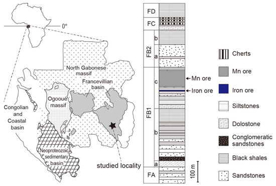
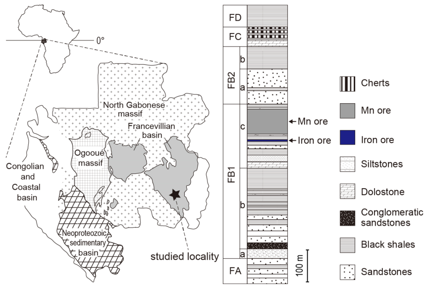
In this study, we focused on the Franceville Basin in Gabon, Africa (Figure 1). The Franceville Group consists of sedimentary layers deposited in a basin formed by rifting in the Early Proterozoic. Stratigraphically, the Franceville Group consists of five formations, FA, FB, FC, FD and FE. The FA Formation, predominantly composed of sandstones, is interpreted as fluvial deposits [19]. Units above the FB Formation are of marine origin [20,21]. These formations comprise sandstones, black shales, limestone, and cherts. The depositional environment of the FB2 Formation, including the FB2a and FB2b subunits, has been interpreted as a turbidite lobe in the prodelta or upper slope, with a depth exceeding 200 m [22,23,24,25]. Lithologically, the FB2a unit (Poubara sandstones) is mainly composed of massive sandstone with thin interlayered black shale, whereas the FB2b unit is composed of black shales interbedded with thin layers of siltstones [26,27]. Geobiologically, the FB2 Member is characterized by the occurrence of centimeter-sized, highly organized structures representing spatially discrete populations of early colonial macroorganisms [22]. Carbon and sulphur isotopic data demonstrate that these biogenic structures were fossilized by pyritization during early diagenesis [22]. Furthermore, iron-speciation analyses suggest that the sediments were deposited under a well-oxygenated water column [22]. The FC Formation comprises layers containing cherts, carbonates (mainly dolostones), and stromatolites, interpreted to have been deposited primarily in a shallow marine environment. The FD Formation was deposited in a foreland basin environment influenced by terrigenous material supply and subjected to tectonic extension and crustal subsidence, resulting in deep burial, reaching up to 1000 m in thickness. The subsidence and sediment thickness suggest that the FD Formation was deposited at significant depths [23,28] in an euxinic environment, as evidenced by high reactive iron ratios and trace metal enrichments [29]. Age constraints have been obtained from intrusive rocks such as the N’Goutou complex and ash layers. U-Pb dating of zircons from intrusive rocks in the FB Formation yields an age of 2191 ± 13 Ma [30], while the youngest U-Pb dates from zircons in the tuff of the FD Formation are 2083 ± 6 Ma [31]. The maximum temperature and pressure that the rocks of the Franceville Group experienced are estimated to be approximately 180–210 °C and ~1 kbar, respectively, based on fluid-inclusion microthermometry of diagenetic quartz overgrowths and carbonate cements [32,33]. The samples analyzed in this study were organic-rich black shales from the FB2b and FD Formation. These samples were collected from quarries at the coordinates 1°37′41″ South, 13°27′56″ East for FB2b unit samples OK 62, OK 412, and OK 429, and 1°38′27″ South, 13°32′33″ East for FD Formation samples OK 77 and OK 78. Photographs of the samples used for analyses are shown in Figure 2. The total organic carbon content of the FB2b unit and FD Formation is generally high, up to ~8% and ~12%, respectively [29,34].
Figure 1.
Simplified geological map of Gabon and schematic stratigraphic column of the Francevillian Basin, based on Parize et al. [23] and Ngombi-Pemba et al. [34], respectively. On the map, different fill patterns denote sedimentary basins of different depositional ages: the Francevillian Basin represents Palaeoproterozoic deposits (ca. 2200–2050 Ma), whereas the other basins shown correspond to younger depositional sequences.
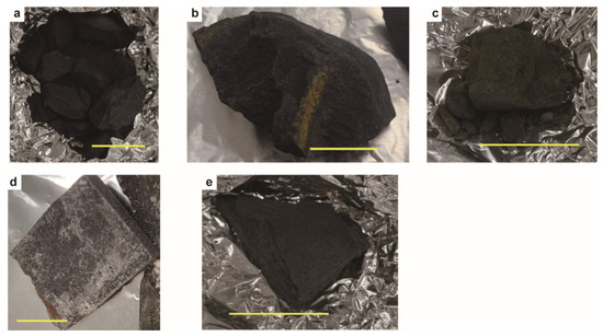
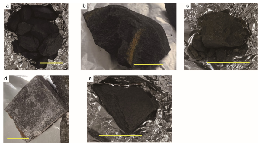
Figure 2.
Rock samples from the Francevillian group in Gabon prior to separation into exterior and interior parts. (a) the black shale (OK77) from the FD Formation. (b) the black shale (OK78) from the FD Formation. (c) the black shale (OK62) from the FB2b unit. (d) the black shale (OK412) from the FB2b unit. (e) the black shale (OK429) from the FB2b unit. Scale bar = 4 cm.
2.2. Methods
All glassware used in the experiments was pre-heated at 450 °C or 550 °C for 10 h to remove organic contaminants. Cylindrical ceramic tiles and silicon carbide were heated at 550 °C for 10 h before use. Teflon containers were cleaned with acetone, methanol, and dichloromethane before use in the experiments.
Our samples were collected from quarries, and if any weathered surface was observed, it was limited to the extreme outer surface layer (Figure 2). After removing any apparent surface contamination, the rock samples were crushed into pieces several centimeters in size using an iron mortar (Nichika Inc., Kyoto, Japan). Subsequently, a micro-ablation technique using a KG-1 Mini-Sonic tumbler (Diamond Pacific Tool Corporation, Barstow, CA, USA) was employed to separate the interior and exterior parts of the rock into powders [35]. The terms “exterior” and “interior” here refer to the outer surface and the interior of rock lumps collected from outcrops. The technique for separating the exterior and interior portions of rocks has been applied not only to drilling cores but also to samples collected from outcrops [36,37]. The rock samples, cylindrical ceramic tiles, and silicon carbide were placed in a stainless-steel hopper mounted on an ultrasonic polisher to rotate and polish the rock samples. Polishing continued until the rock fragments were approximately half their original size, and the slurry obtained during this stage was designated as the exterior part. The slurry from further polishing was designated as the interior part and collected. The collected samples were centrifuged, and the supernatant was discarded. The samples were then frozen, placed in a freeze dryer for 3–4 days, and dried. The dried samples were homogenized using an agate mortar (Nichika Inc., Kyoto, Japan) and pestle and then subjected to solvent extraction for bitumen removal.
The powdered sample was placed in a Teflon container thoroughly cleaned with organic solvents. Ultrasonic extraction was performed to remove bitumen three times using a dichloromethane: methanol mixture (9:1, v/v). The dried extraction residue was treated with hydrofluoric acid, stirred intermittently, and left for 24 h. The hydrofluoric acid used for acid treatment was purified by three liquid–liquid extractions with hexane. After the reaction, centrifugation was performed, and the supernatant was removed. The residue was washed three times with ultrapure water, followed by centrifugation each time to remove the supernatant. The residue was frozen overnight and then freeze-dried for 3–4 days. The freeze-dried sample was ground and homogenized with an agate mortar and pestle, then subjected to the same extraction procedure again to remove any remaining extractable organic matter.
Maleimides were extracted using a modified protocol based on Kawabata et al. [18]. Specifically, minerals were removed prior to chromic-acid oxidation (hydrofluoric acid demineralization; see Section above), and oxidation was applied to the resulting organic-rich residue rather than to whole-rock powder. The dried residue after the extraction (typically 1–2 g) and a stir bar were placed in a 110 mL glass tube. A mixture of 25% (v/v) sulfuric acid (5 mL) and 10% (w/v) chromium trioxide solution (5 mL) was added, and the mixture was stirred for 1 h in an ice bath under a nitrogen atmosphere. During the oxidation, the reaction vessel was kept under low-light conditions by shielding it with aluminum foil and a blackout curtain. After 1 h, the glass tube was removed from the ice bath and stirred for another hour at room temperature. Sodium bisulfite was gradually added to the glass tube to neutralize the chromium, which turned the solution green. The contents of the glass tube were rinsed with ultrapure water and benzene and transferred to a Teflon bottle. The Teflon bottle was capped, stirred for 3 min, centrifuged, and the supernatant was transferred to a 10 mL glass tube. This process was repeated twice more with benzene. The collected benzene was concentrated under a nitrogen atmosphere. For comparison, a procedural blank sample was similarly processed using the same protocol.
The measurements were performed using a Nexis GC-2030 gas chromatograph (Shimadzu Corporation, Kyoto, Japan) connected to a quadrupole mass spectrometer (TQ 8050NX, Shimadzu) and a computer system. The injection port was of the splitless type, and high-purity helium was used as the carrier gas. Sample injection was performed using an auto-sampler system. Mass spectrometry was conducted in full scan mode or multiple reaction monitoring (MRM) mode using electron ionization (EI). In MRM mode, a trace signal denotes a weak response at the expected retention time that does not form a clearly defined chromatographic peak, whereas undetected indicates no discernible response above baseline noise. The concentrated benzene extract was analyzed by GC–MS using a DB-FFAP capillary column (60 m, 0.25 mm i.d., 0.25 μm film thickness). The injection port temperature was 230 °C. The oven temperature was initially held at 50 °C for 2 min, then increased to 150 °C at 10 °C/min, and then to 230 °C at 3 °C/min, and held at 230 °C for 50 min.
Maleimides were identified by comparison of the mass spectra and GC retention times of sample peaks with corresponding data from an acid-treated kerogen sample from the Meishan section and a freeze-dried sediment sample from Lake Kaiike, together with relative GC retention times and published mass spectra [38,39,40]. The Meishan reference sample is a carbonate rock from the Changhsing Formation at the Meishan section, South China, of the latest Permian [41], collected from a stratigraphic level 86.5 cm below the top of Bed 24. In the present study, prior to maleimide analysis, this sample was subjected to sequential acid treatment with hydrochloric acid, hydrofluoric acid, and hydrochloric acid, followed by ultrasonic extraction with dichloromethane:methanol (9:1, v/v) to remove extractable organic matter. Maleimides have previously been reported from this section [17]. This sample was provided by Prof. Kunio Kaiho (Tohoku University, Japan). The Lake Kaiike reference sample is a sediment sample collected from Lake Kaiike, Japan, at a water depth of 9.1 m using an Ekman grab sampler (RIGO Co., Ltd., Tokyo, Japan) deployed from a rowboat, and subsequently freeze-dried prior to analysis. Previous analyses of sediments and lake water from Lake Kaiike identified bacteriochlorophyll e homologues derived from GSB, which are anoxygenic phototrophic organisms [42]. In the present study, the freeze-dried Lake Kaiike sediment was directly subjected to chromic-acid oxidation for maleimide analysis and used as a reference for maleimides associated with anoxygenic phototrophs, particularly GSB, in an immature sedimentary setting, rather than as an analogue of mature kerogen.
Since authentic standards are not available, we do not report absolute concentrations. Prior to GC-MS analysis, the exterior and interior extracts of each sample were adjusted to the same final volume, which was either 50 or 100 μL depending on the sample. Peak areas were corrected for final extract volume and normalized to the dry weight of the organic-rich residue, so that the exterior and interior portions of each sample were compared under equivalent conditions. Exterior/interior ratios were calculated from these normalized values. The relative proportions of maleimides and phthalimide calculated from the normalized peak areas should not be regarded as direct quantitative estimates of the original kerogen-bound tetrapyrrole precursor distribution, because differential recovery, partitioning, or secondary alteration during chromic-acid oxidation may affect the relative abundances of individual compounds.
The reproducibility of the analytical procedure was confirmed by three independent chromic-acid oxidation experiments using the acid-treated, bitumen-free kerogen isolated from the Meishan reference sample, which yielded consistent chromatographic patterns and relative maleimide compositions (Figures S1–S3). A schematic overview of the sample preparation, exterior/interior separation, bitumen removal, demineralization, chromic-acid oxidation, and GC–MS/MS analysis is provided in Figure S4.
3. Results
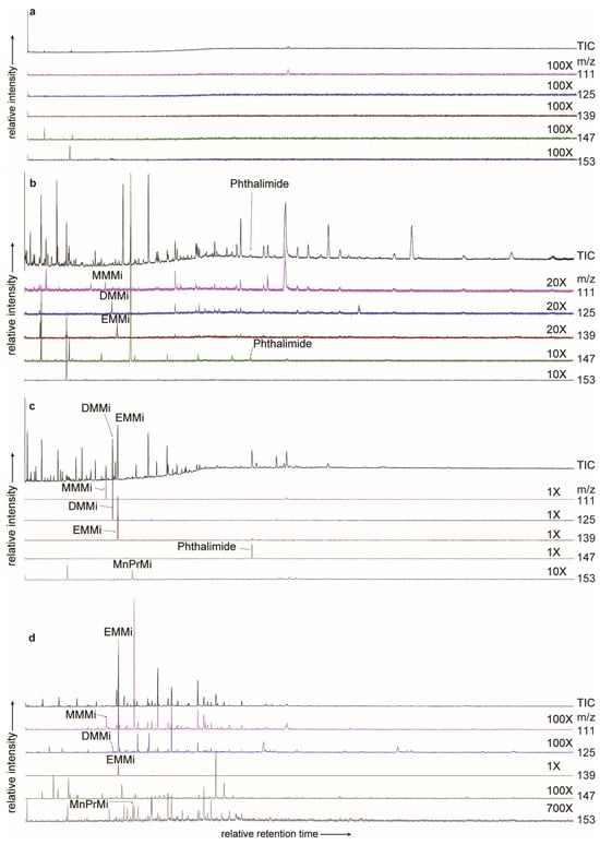
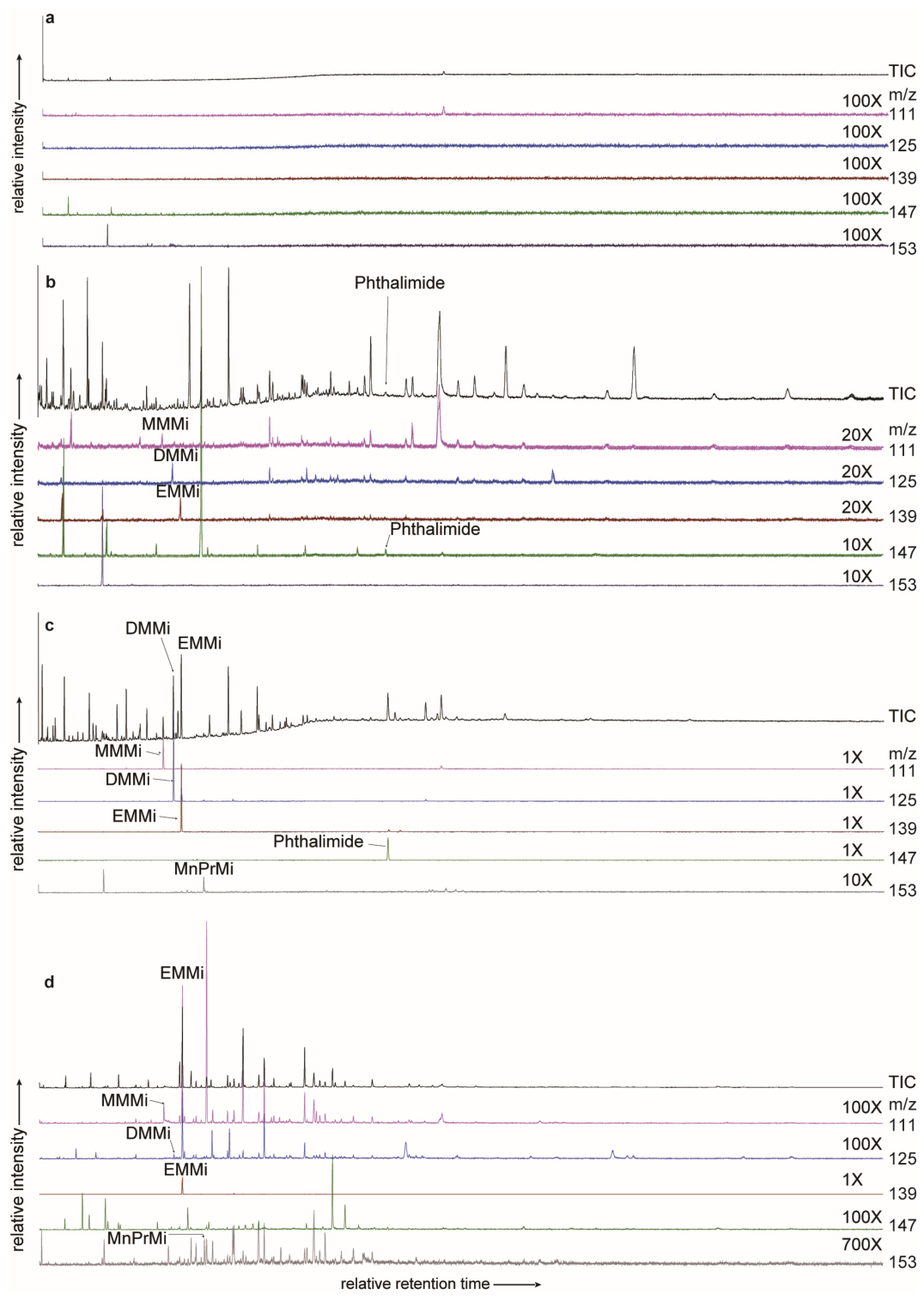
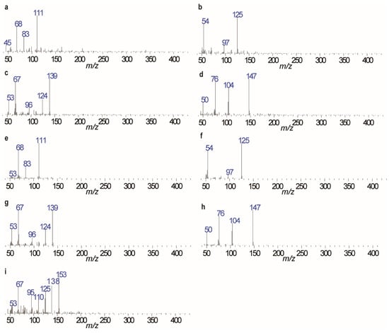
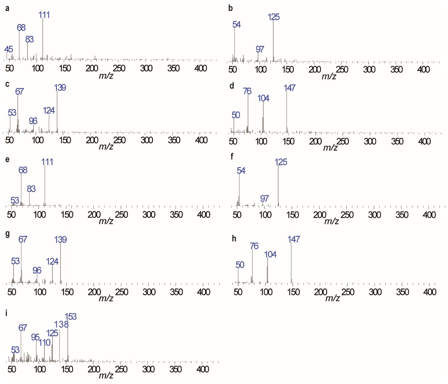
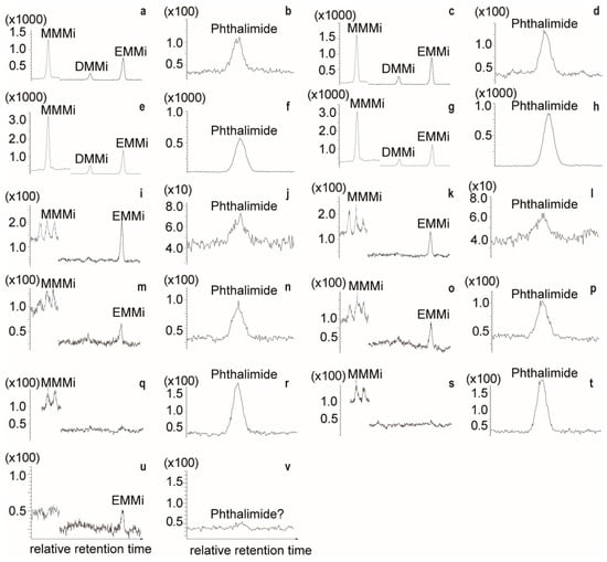
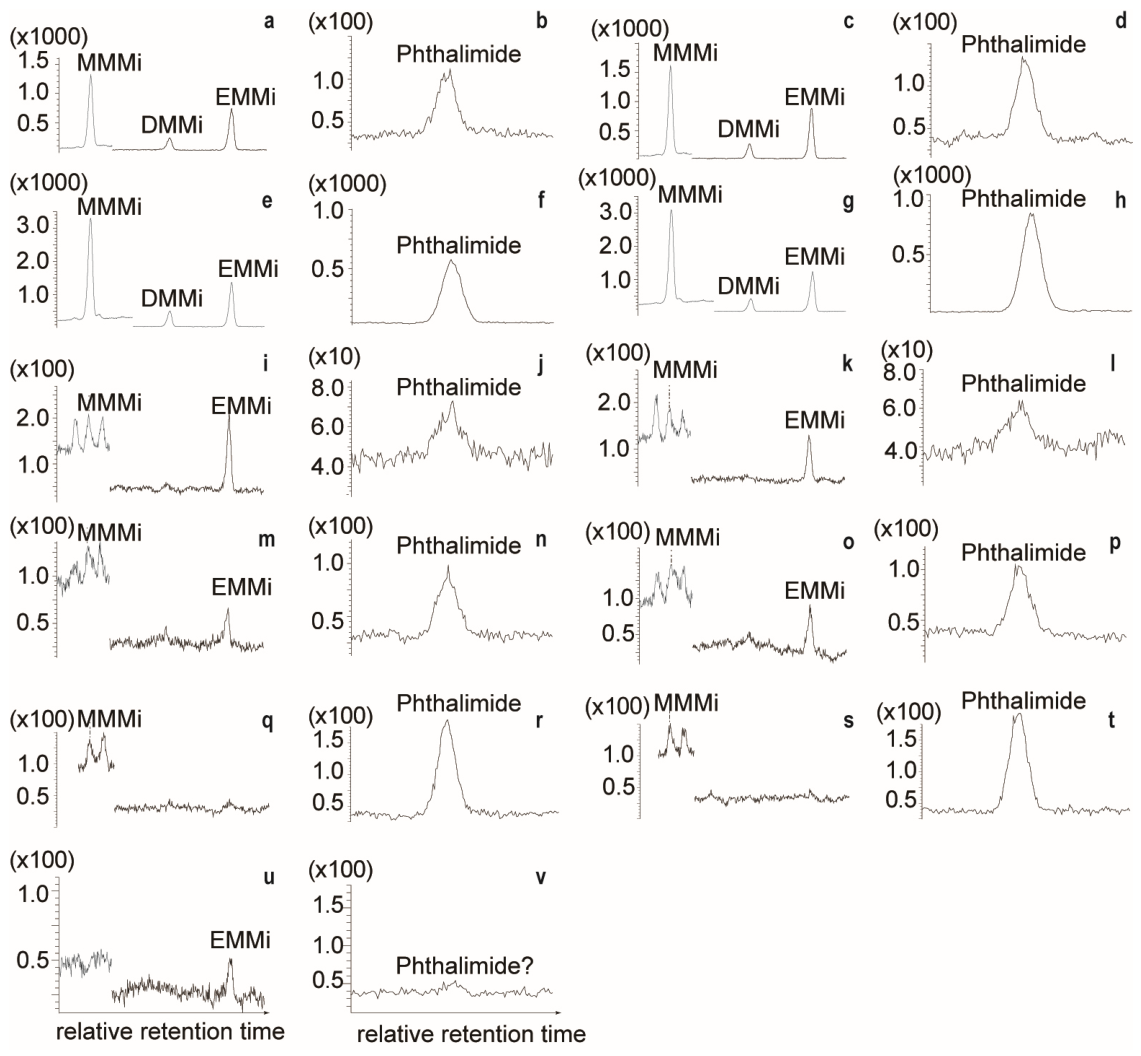
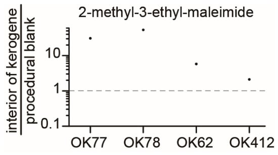
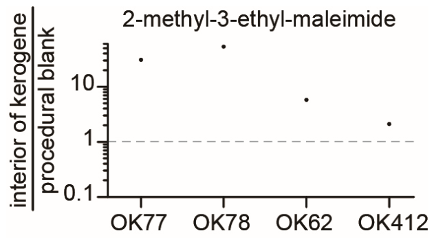
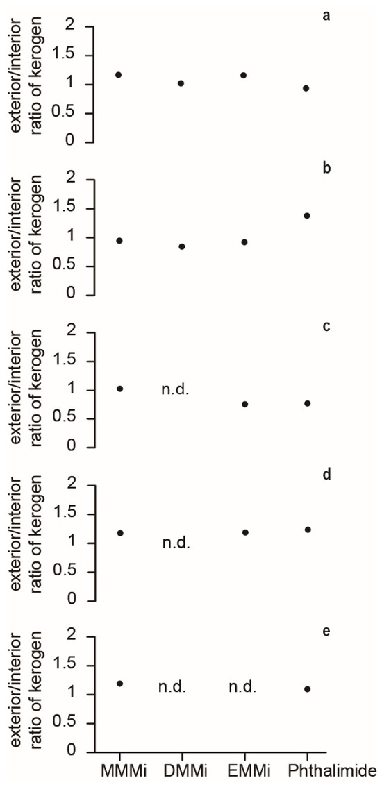
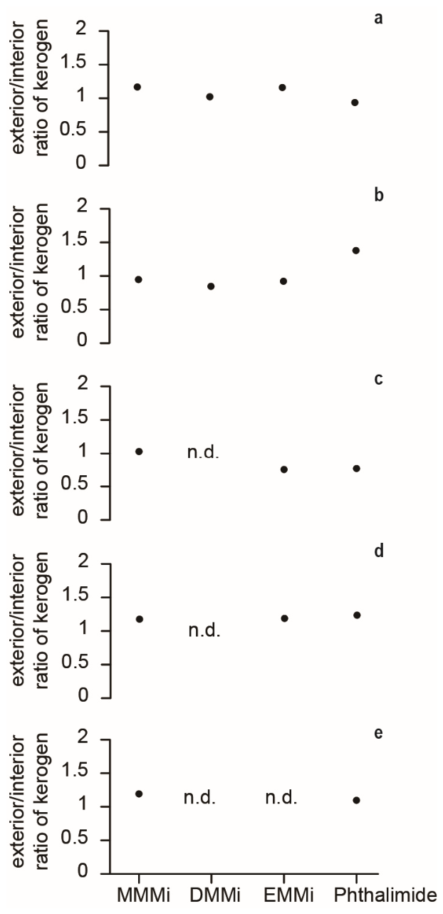
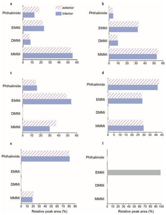
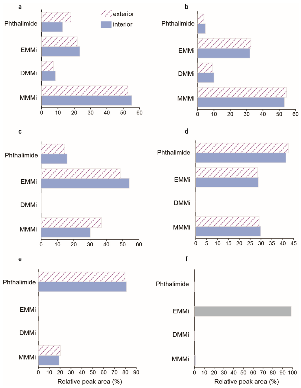
Chromic-acid oxidation of kerogen from black shale samples of the FB2b unit and FD Formation of the Franceville Group yielded MMMi, DMMi, EMMi, and phthalimide (Figure 3, Figure 4, Figure 5, Figure 6, Figure 7 and Figure 8). The mass spectra and chromatographic characteristics of the compounds detected in the Francevillian samples were compared with those of the Meishan and Lake Kaiike reference samples (Figure 3 and Figure 4). The spectral patterns are consistent with those reported in previous studies [38,39]. Additionally, the retention times of the maleimides detected in the Francevillian samples are consistent with those observed in the Meishan section and Lake Kaiike, providing confirmation for the identification of these compounds. Full scan mode measurements of the procedural blank did not detect any maleimides or phthalimides (Figure 3a). However, MRM mode measurements of the procedural blank detected EMMi (Figure 5u,v). In MRM mode analysis, the above-mentioned maleimides and phthalimides were detected in the black shale samples from the FD Formation, while some black shale samples from the FB2b Formation had DMMi and EMMi below the detection limit (Figure 5i–t). Additionally, MnPrMi and MiBuMi were below the detection limit in all black shale samples from the FB2b unit and FD Formation of the Franceville Group. The sample/procedural blank ratio for EMMi was higher in the FD Formation but lower in the FB2b unit (Figure 6). For the detected maleimides and phthalimide, the exterior/interior (E/I) ratios of normalized peak areas in kerogen are close to unity, suggesting no systematic enrichment in the exterior portions (Figure 7). In addition, the relative peak-area proportions of MMMi, DMMi, EMMi, and phthalimide were broadly similar between the exterior and interior portions within each sample, with no systematic compositional difference observed between the two fractions (Figure 8). The Francevillian samples show relatively higher proportions of MMMi and phthalimide than the Lake Kaiike and Meishan reference samples (Figure 8 and Figure S3).
Figure 3.
Total ion chromatograms (TIC) and extracted ion chromatograms from maleimide analyses of (a) the procedural blank, (b) kerogen isolated from the Francevillian Group FD Formation black shale sample OK78i (Gabon), (c) the Meishan section sample from the Changhsing Formation (South China), and (d) Lake Kaiike sediment (Japan).
Figure 4.
Background-subtracted mass spectra of tentatively assigned maleimides and phthalimide from samples shown in Figure 3. (a–d) kerogen isolated from the Francevillian Group FD Formation black shale sample OK78i (Gabon): (a) MMMi; (b) DMMi; (c) EMMi; (d) phthalimide. (e–h) Meishan sample (Changhsing Formation, South China): (e) MMMi; (f) DMMi; (g) EMMi; (h) phthalimide. (i), Lake Kaiike sediment sample (Japan): MnPrMi. Abbreviations: MMMi, 2-methyl-maleimide; DMMi, 2,3-dimethyl-maleimide; EMMi, 2-methyl-3-ethyl-maleimide; MnPrMi, 2-methyl-3-propyl maleimide.
Figure 5.
Multiple reaction monitoring chromatograms of maleimides from kerogens isolated from black shale samples of the Francevillian group in Gabon and from the procedural blank. (a,b) the interior kerogen of the black shale (OK77) from the FD Formation. (c,d) the exterior kerogen of the black shale (OK77) from the FD Formation. (e,f), the interior kerogen of the black shale (OK78) from the FD Formation. (g,h) the exterior kerogen of the black shale (OK78) from the FD Formation. (i,j) the interior kerogen of the black shale (OK62) from the FB2b unit. (k,l) the exterior kerogen of the black shale (OK62) from the FB2b unit. (m,n) the interior kerogen of the black shale (OK412) from the FB2b unit. (o,p) the exterior kerogen of the black shale (OK412) from the FB2b unit. (q,r) the interior kerogen of the black shale (OK429) from the FB2b unit. (s,t) the exterior kerogen of the black shale (OK429) from the FB2b unit. (u,v) procedural blank sample. MMMi, 2-methyl-maleimide. DMMi, 2,3-dimethyl-maleimide. EMMi, 2-methyl-3-ethyl-maleimide. The MRM transitions used were m/z 111 > 83 for MMMi, m/z 125 > 54 for DMMi, m/z 139 > 124 for EMMi, and m/z 147 > 103 for phthalimide. Note: In panels (q,s), acquisition of the m/z 111 > 83 MRM trace began at a slightly later retention time than in the other runs.
Figure 6.
Interior/procedural blank ratios of 2-methyl-3-ethyl-maleimide for the Francevillian black shale samples OK77 and OK78 from the FD Formation and OK62 and OK412 from the FB2b unit.
Figure 7.
Exterior/interior ratios of maleimides and phthalimide from kerogens isolated from black shale samples of Francevillian group in Gabon. (a) black shale (OK77) from the FD Formation. (b) black shale (OK78) from the FD Formation. (c) black shale (OK62) from the FB2b unit. (d) black shale (OK412) from the FB2b unit. (e) black shale (OK429) from the FB2b unit. MMMi, 2-methyl-maleimide. DMMi, 2,3-dimethyl-maleimide. EMMi, 2-methyl-3-ethyl-maleimide. n.d., not detected/below detection limit.
Figure 8.
Relative peak-area proportions of maleimides and phthalimide in the exterior and interior kerogen fractions of the Francevillian samples and in the Lake Kaiike reference sample. (a) OK77, (b) OK78, (c) OK62, (d) OK412, (e) OK429, and (f) Lake Kaiike. For the Francevillian samples, hatched bars indicate exterior fractions and blue bars indicate interior fractions. MMMi, 2-methyl-maleimide; DMMi, 2,3-dimethyl-maleimide; EMMi, 2-methyl-3-ethyl-maleimide.
4. Discussion
4.1. Contamination Check
Potential contamination of ancient rock samples can be assessed by examining the concentration ratio of each organic molecule between the exterior and interior of the rock samples (E/I ratio) [9,10,35,43,44,45]. The consistent E/I ratios for maleimides and phthalimide from the Francevillian kerogen samples suggest no systematic enrichment in exterior fractions and are therefore consistent with limited surficial contamination (Figure 7). These results are consistent with a kerogen-bound origin for the detected compounds and argue against substantial surficial contamination. The EMMi signal in some FB2b samples should be interpreted cautiously because it is close to the procedural background. This does not affect the exterior/interior comparison, but it indicates that low-abundance EMMi in FB2b samples should not be overinterpreted.
4.2. Origin of Maleimides and Phthalimide
Chromic acid oxidation of (bacterio)chlorophylls produces various maleimides. This process notably produces EMMi and 2-methyl-3-vinyl-maleimide (VMMi) as primary products from chlorophyll a [46]. EMMi and VMMi have also been detected as major maleimides in recent sediments [15]. For bacteriochlorophylls c, d, and e from anoxygenic phototrophs, such as GSB, chromic acid oxidation produces higher-alkyl maleimides, including MnPrMi, with an abundance approximately half that of EMMi [47]. These quantitative relationships are confirmed by stratigraphic records of the Triassic–Jurassic extinction event, where GSB thrived in the euxinic photic zone [48].
In the maleimide fraction of examined samples, EMMi was detected, while MnPrMi was below the detection limit. Other maleimides commonly associated with GSB, which are anoxygenic phototrophs (e.g., MnPrMi and MiBuMi) were also below the detection limit. This absence may indicate that the photic zone was relatively oxic and/or that H2S availability was low, limiting the contribution of GSB. However, this absence should be interpreted with caution, as the Francevillian Group rocks experienced burial temperatures of approximately 180–210 °C and pressures of ~1 kbar [32,33], conditions that may have promoted preferential degradation of higher-alkyl maleimides. Previous pyrolysis experiments have demonstrated that these maleimides, particularly MnPrMi, can undergo thermal decomposition to form simpler maleimides such as MMMi, DMMi, and phthalimide [17]. The relative enrichments in MMMi and phthalimide in the Francevillian samples, compared with the Meishan and Kaiike samples (Figure 8 and Figure S3), are consistent with preferential degradation of higher-alkyl maleimides during thermal alteration. Therefore, the absence of higher-alkyl maleimides indicative of GSB may reflect either limited original inputs from anoxygenic phototrophs or the preferential degradation of higher-alkyl maleimides during thermal alteration.
In addition to the secondary thermal degradation of higher-alkyl maleimides discussed above, MMMi, DMMi, and phthalimide detected in the Francevillian samples may also have formed through other diagenetic transformations of tetrapyrrole precursors. Phthalimide can be produced through two pathways: the Diels–Alder reaction involving the 3-methyl-4-vinylpyrrole unit, which also yields MMMi [49], or through aromatization including trans-alkylation and cyclization of 3-ethyl-4-methylpyrrole [50]. MMMi can additionally originate from decarboxylation following ring E degradation of tetrapyrroles under acidic conditions [15].
The co-detection of both MMMi and DMMi in our samples may retain some information on redox conditions, although this signal could have been overprinted by thermal alteration. Pickering and Keely [51] demonstrated that co-oxidation of the vinyl group of tetrapyrroles with hydrogen sulfide produces a C-3 aldehyde intermediate, which is subsequently converted to MMMi under oxic conditions or to DMMi under reducing conditions. The detection of DMMi is consistent with the euxinic depositional environment of the FD Formation inferred from trace metal and iron speciation data [29], while the co-occurrence of MMMi may reflect production in a relatively more oxidative portion of the water column, potentially the photic zone [25,51]. DMMi can also form through cleavage of the C-C bond at the allylic position of an alkyl chain, followed by addition of a hydrogen radical to the resulting allylic carbon radical [17]. Determining these formation pathways is complex; however, it is important to note that all proposed pathways are consistent with formation from tetrapyrrole-derived precursors, including (bacterio)chlorophyll-related compounds.
Additional origins of the detected maleimides and phthalimides in this study may include phycobilin pigments and heme [52,53,54,55]. Phycobilins are light-harvesting pigments in oxygenic photosynthetic organisms like cyanobacteria, red algae, and cryptomonads [56], and their biosynthetic pathway for phycobilins likely existed during the GOE [57]. Similarly, the presence of heme-containing peroxidases in the basal oxyphotobacterial genus Gloeobacter suggests that the heme biosynthetic pathway had already evolved by the time of the GOE [55,58], but heme itself is not source-specific because it serves as a prosthetic group in a wide variety of proteins, including antioxidant enzymes and cytochromes involved in electron-transfer reactions such as anaerobic metabolism [59,60]. Although maleimides are not strictly source-specific, prior studies have commonly interpreted naturally occurring maleimides in sediments and sedimentary rocks as transformation products of (bacterio)chlorophylls, and have used them accordingly in palaeoenvironmental reconstruction [12,13,15,17,48]. The Francevillian succession contains organic-rich black shales, with the FB Formation representing one of the greatest accumulations of organic carbon of its age [61], and it also preserves stromatolites, microfossils, and microbial mat-related structures [62,63]. These observations independently indicate the presence of microbial ecosystems that likely included photosynthetic primary production in the basin by ca. 2.1 Ga [62,63]. In this depositional context, (bacterio)chlorophyll-derived tetrapyrroles were likely the major precursor source of the detected maleimides, whereas any contribution from phycobilin-derived tetrapyrroles would still be consistent with a photosynthetic origin [64]. This view is also consistent with porphyrin geochemistry. Although heme-derived porphyrins can be preserved in sedimentary rocks, chlorophyll is biosynthesized in far greater quantities than heme, making chlorophyll-derived tetrapyrroles the more plausible dominant precursor source of the detected maleimides [65]. Consistent with this view, modern marine environmental samples show that chlorophyll is significantly more abundant than heme, even though few studies have directly compared the quantities of chlorophyll and heme in photosynthetic organisms [66,67]. Thus, although contributions from phycobilins and heme cannot be ruled out, the detected maleimides are most plausibly interpreted as being derived from photosynthetic tetrapyrrole pigments, including chlorophyll- and/or bacteriochlorophyll-related precursors.
4.3. Implication for the Early Proterozoic Ecosystem
Widely accepted indigenous biomarker records from sedimentary rocks older than ca. 2.0 Ga remain rare and controversial; previous research results are believed to be influenced by younger contamination [68]. Thus, direct comparisons with widely accepted biomarker records of similar age remain limited. In contrast, indigenous biomarker results have been reported from sedimentary rocks of the mid-Proterozoic era [9,10,36,44,45,69,70,71]. These rocks are characterized by low diversity of eukaryotic fossils and the absence of steranes, which are molecular fossils of eukaryotic sterols. Instead, biomarkers such as hopanes, trimethyl aryl isoprenoids, C40 carotenoid pigments, porphyrins, and protosteroids have been detected from sedimentary rocks of mid-Proterozoic age [9,10,36,44,45,69,70,71]. In particular, porphyrin data have been interpreted as evidence for cyanobacterial primary production, whereas trimethyl aryl isoprenoids are interpreted in relation to phototrophic sulfur bacteria, with specific assignments depending on methylation patterns and associated carotenoid evidence. These findings suggest the presence of taxonomically diverse phototrophic communities in some mid-Proterozoic ecosystems.
The maleimide assemblage reported here is most conservatively interpreted as evidence for tetrapyrrole-derived organic inputs in ca. 2.1 Ga marine sediments, with photosynthetic pigments representing the most plausible dominant precursor source. Because the detected maleimides are most plausibly interpreted as being derived from (bacterio)chlorophyll-related precursors, with possible additional contributions from phycobilins and heme, a dominant contribution from non-specific heme-derived sources alone is less likely. Considering the depositional depth (>200 m), the environment was euxinic, particularly within the FD Formation as indicated by trace metal and iron speciation data [29], while the photic zone might have been oxidative [25,72]. If the tetrapyrrole precursors were derived mainly from chlorophylls and/or phycobilins, as argued above, the data are consistent with input from photosynthetic biomass produced in the photic zone, potentially under locally or episodically oxygenated conditions.
5. Conclusions
We analyzed kerogen-bound tetrapyrrole-derived compounds in ca. 2.1 Ga marine sedimentary rocks from the Franceville Group, Gabon. Chromic-acid oxidation of bitumen-free kerogen released 2-methylmaleimide, 2,3-dimethylmaleimide, 2-methyl-3-ethylmaleimide, and phthalimide. Exterior/interior comparisons showed near-unity ratios of normalized peak areas and broadly similar relative compositions, supporting a kerogen-bound origin rather than substantial surficial contamination. These observations indicate that the detected compounds are most consistent with an indigenous or near-indigenous tetrapyrrole-derived signal that survived diagenesis and thermal maturation. Although contributions from heme- and phycobilin-derived tetrapyrroles cannot be excluded, the detected assemblage is most plausibly interpreted as reflecting photosynthetic tetrapyrrole inputs, including chlorophyll- and/or bacteriochlorophyll-related precursors. The absence of higher-alkyl maleimides should be interpreted cautiously, because it may reflect thermal alteration rather than the original absence of anoxygenic phototrophs. Overall, our results demonstrate that kerogen-bound maleimide- and phthalimide-type compounds provide a promising, although non-unique, approach for tracing ancient tetrapyrrole and photosynthetic pigment inputs in Precambrian sedimentary rocks.
Supplementary Materials
The following supporting information can be downloaded at: https://www.mdpi.com/article/10.3390/geosciences16070287/s1, Supplementary Note S1: Reproducibility of chromic-acid oxidation experiments using Meishan reference kerogen [73,74,75]; Figure S1: Total ion chromatograms from maleimide analyses of the Meishan section sample from the Changhsing Formation, South China; Figure S2: Extracted ion chromatograms from maleimide analyses of the Meishan section sample from the Changhsing Formation, South China; Figure S3: Relative peak-area proportions of maleimides and phthalimide obtained from three independent chromic acid oxidation replicates of the bitumen-free Meishan kerogen; Figure S4: Schematic workflow of the analytical procedure used in this study; Table S1: Final extract volumes and dry kerogen weights used for normalization of GC–MS peak areas in the exterior and interior fractions of the Francevillian samples; Table S2: Exterior/interior ratios of maleimides and phthalimide in the kerogens from the Francevillian group in Gabon; Table S3: Interior/blank ratios of 2-methyl-3-ethyl-maleimide in the kerogens from the Francevillian group in Gabon.
Author Contributions
Conceptualization, R.S.; methodology, R.S.; formal analysis, R.S. and A.T.; investigation, R.S., A.T., Y.S., M.M. and A.E.-M.; resources, Y.S.; data curation, R.S.; writing—original draft preparation, R.S.; writing—review and editing, R.S., A.T., Y.S., M.M. and A.E.-M.; visualization, R.S.; project administration, R.S.; funding acquisition, R.S. and Y.S. All authors have read and agreed to the published version of the manuscript.
Funding
This research was funded by the Japan Society for the Promotion of Science (JSPS) KAKENHI, grant numbers JP22H01345 and JP24K21570 to R.S. and JP17H04858 to Y.S.; the ExCELLS Project Research “Creation of Neo-Life Forms” under the Advanced Co-Creation Platform of the Exploratory Research Center on Life and Living Systems (ExCELLS), National Institutes of Natural Sciences (NINS), to R.S.; and the Island Campus Project of the Kagoshima Remote Islands Promotion Council, which supported field sampling at Lake Kaiike.
Data Availability Statement
The original contributions presented in this study are included in the Supplementary Tables.
Acknowledgments
We thank Kunio Kaiho for providing the Meishan sample. We also thank Yuka Miyake for assistance in obtaining permission for the field investigation. We are grateful to the Academic Editor and three anonymous reviewers for their constructive comments, which helped improve the manuscript. We thank Masahiro UEDA, Kiyotaka MIYAZAKI, Tatsuji TERADA, and Nozomi ITOU of the Production Division, Advanced Technology Institute, Yamaguchi University, for their technical support in fabricating the custom tumbling containers used for the sequential removal and collection of the exterior and interior fractions of the rock samples.
Conflicts of Interest
The authors declare no conflicts of interest.
Abbreviations
The following abbreviations are used in this manuscript:
| GOE | Great Oxidation Event |
| GC-MS | gas chromatography–mass spectrometry |
| MRM | multiple reaction monitoring |
| E/I ratio | exterior/interior ratio |
| GSB | green sulfur bacteria |
| MMMi | 2-methylmaleimide |
| DMMi | dimethylmaleimide |
| EMMi | 2-methyl-3-ethylmaleimide |
| VMMi | 2-methyl-3-vinyl-maleimide |
| MnPrMi | 2-methyl-3-propyl maleimide |
| MiBuMi | 2-methyl-3-isobutyl maleimide |
References
- Lyons, T.W.; Reinhard, C.T.; Planavsky, N.J. The rise of oxygen in Earth’s early ocean and atmosphere. Nature 2014, 506, 307–315. [Google Scholar] [CrossRef] [Scilit] [PubMed]
- Kasting, J.F. What caused the rise of atmospheric O2? Chem. Geol. 2013, 362, 13–25. [Google Scholar] [CrossRef] [Scilit]
- Schirrmeister, B.E.; Gugger, M.; Donoghue, P.C. Cyanobacteria and the Great Oxidation Event: Evidence from genes and fossils. Palaeontology 2015, 58, 769–785. [Google Scholar] [CrossRef] [Scilit] [PubMed]
- Hofmann, H.J. Precambrian Microflora, Belcher Islands, Canada: Significance and Systematics. J. Paleontol. 1976, 50, 1040–1073. [Google Scholar]
- Hodgskiss, M.S.; Dagnaud, O.M.; Frost, J.L.; Halverson, G.P.; Schmitz, M.D.; Swanson-Hysell, N.L.; Sperling, E.A. New insights on the Orosirian carbon cycle, early Cyanobacteria, and the assembly of Laurentia from the Paleoproterozoic Belcher Group. Earth Planet. Sci. Lett. 2019, 520, 141–152. [Google Scholar] [CrossRef] [Scilit]
- Nishihara, A.; Tsukatani, Y.; Azai, C.; Nobu, M.K. Illuminating the coevolution of photosynthesis and bacteria. Proc. Natl. Acad. Sci. USA 2024, 121, e2322120121. [Google Scholar] [CrossRef] [Scilit] [PubMed]
- Rasmussen, B.; Fletcher, I.R.; Brocks, J.J.; Kilburn, M.R. Reassessing the first appearance of eukaryotes and cyanobacteria. Nature 2008, 455, 1101–1104. [Google Scholar] [CrossRef] [Scilit] [PubMed]
- Brocks, J.J.; Logan, G.A.; Buick, R.; Summons, R.E. Archean Molecular Fossils and the Early Rise of Eukaryotes. Science 1999, 285, 1033–1036. [Google Scholar] [CrossRef] [Scilit] [PubMed]
- Gueneli, N.; McKenna, A.M.; Ohkouchi, N.; Boreham, C.J.; Beghin, J.; Javaux, E.J.; Brocks, J.J. 1.1-billion-year-old porphyrins establish a marine ecosystem dominated by bacterial primary producers. Proc. Natl. Acad. Sci. USA 2018, 115, E6978–E6986. [Google Scholar] [CrossRef] [Scilit] [PubMed]
- Vinnichenko, G.; Jarrett, A.J.M.; Hope, J.M.; Brocks, J.J. Discovery of the oldest known biomarkers provides evidence for phototrophic bacteria in the 1.73 Ga Wollogorang Formation, Australia. Geobiology 2020, 18, 544–559. [Google Scholar] [CrossRef] [Scilit] [PubMed]
- Tashiro, T.; Ishida, A.; Hori, M.; Igisu, M.; Koike, M.; Mejean, P.; Takahata, N.; Sano, Y.; Komiya, T. Early trace of life from 3.95 Ga sedimentary rocks in Labrador, Canada. Nature 2017, 549, 516–518. [Google Scholar] [CrossRef] [Scilit] [PubMed]
- Kozono, M.; Nomoto, S.; Mita, H.; Shimoyama, A. Detection of maleimides and their characteristics in Neogene sediments of the Shinjo basin, Japan. Geochem. J. 2001, 35, 225–236. [Google Scholar] [CrossRef] [Scilit]
- Asahina, K.; Takahashi, S.; Saito, R.; Kaiho, K.; Oba, Y. Maleimide index: A paleo-redox index based on fragmented fossil-chlorophylls obtained by chromic acid oxidation. RSC Adv. 2022, 12, 31061–31067. [Google Scholar] [CrossRef] [Scilit] [PubMed]
- Kawabata, K.; Ikeda, M.; Kano, A.; Fujibayashi, M.; Senda, R.; Saito, R. Increase in marine productivity during the recovery phase of carbon isotope excursion from the Toarcian Oceanic Anoxic Event at the Northwest of the Panthalassa Ocean. Palaeogeogr. Palaeoclimatol. Palaeoecol. 2026, 681, 113392. [Google Scholar] [CrossRef] [Scilit]
- Grice, K.; Gibbison, R.; Atkinson, J.E.; Schwark, L.; Eckardt, C.B.; Maxwell, J.R. Maleimides (1H-pyrrole-2,5-diones) as molecular indicators of anoxygenic photosynthesis in ancient water columns. Geochim. Cosmochim. Acta 1996, 60, 3913–3924. [Google Scholar] [CrossRef] [Scilit]
- Pancost, R.D.; Crawford, N.; Maxwell, J.R. Molecular evidence for basin-scale photic zone euxinia in the Permian Zechstein Sea. Chem. Geol. 2002, 188, 217–227. [Google Scholar] [CrossRef] [Scilit]
- Naeher, S.; Schaeffer, P.; Adam, P.; Schubert, C.J. Maleimides in recent sediments—Using chlorophyll degradation products for palaeoenvironmental reconstructions. Geochim. Cosmochim. Acta 2013, 119, 248–263. [Google Scholar] [CrossRef] [Scilit]
- Smolarek, J.; Marynowski, L.; Trela, W.; Kujawski, P.; Simoneit, B.R.T. Redox conditions and marine microbial community changes during the end-Ordovician mass extinction event. Glob. Planet. Change 2017, 149, 105–122. [Google Scholar] [CrossRef] [Scilit]
- Gauthier-Lafaye, F.; Weber, F. Natural nuclear fission reactors: Time constraints for occurrence, and their relation to uranium and manganese deposits and to the evolution of the atmosphere. Precambrian Res. 2003, 120, 81–100. [Google Scholar] [CrossRef] [Scilit]
- Pambo, F.; Guiraud, M.; Quesne, D.; Gauthier-Lafaye, F.; Azzibrouck, G.; Lang, J. The Proterozoic Franceville Basin (SE Gabon): An example of interaction between marine sedimentation and extensional faulting. Afr. Geosci. Rev. 2006, 13, 77–106. [Google Scholar]
- Ossa Ossa, F.; Eickmann, B.; Hofmann, A.; Planavsky, N.J.; Asael, D.; Pambo, F.; Bekker, A. Two-step deoxygenation at the end of the Paleoproterozoic Lomagundi Event. Earth Planet. Sci. Lett. 2018, 486, 70–83. [Google Scholar] [CrossRef] [Scilit]
- El Albani, A.; Bengtson, S.; Canfield, D.E.; Bekker, A.; Macchiarelli, R.; Mazurier, A.; Hammarlund, E.U.; Boulvais, P.; Dupuy, J.-J.; Fontaine, C.; et al. Large colonial organisms with coordinated growth in oxygenated environments 2.1 Gyr ago. Nature 2010, 466, 100–104. [Google Scholar] [CrossRef] [Scilit] [PubMed]
- Parize, O.; Feybesse, J.-L.; Guillocheau, F.; Mulder, T. Were the 2.1-Gyr fossil colonial organisms discovered in the Francevillian basin (Palaeoproterozoic, Gabon) buried by turbidites? Comptes Rendus Geosci. 2013, 345, 101–110. [Google Scholar] [CrossRef] [Scilit]
- Ndongo, A.; Guiraud, M.; Vennin, E.; Mbina, M.; Buoncristiani, J.-F.; Thomazo, C.; Flotté, N. Control of fluid-pressure on early deformation structures in the Paleoproterozoic extensional Franceville Basin (SE Gabon). Precambrian Res. 2016, 277, 1–25. [Google Scholar] [CrossRef] [Scilit]
- Aubineau, J.; El Albani, A.; Chi Fru, E.; Kipp, M.A.; Ngwal’ghoubou Ikouanga, J.; Bekker, A. Benthic redox conditions and nutrient dynamics in the ca. 2.1 Ga Franceville sub-basin. Precambrian Res. 2021, 360, 106234. [Google Scholar] [CrossRef] [Scilit]
- Weber, F.; Gauthier-Lafaye, F.; Whitechurch, H.; Ulrich, M.; El Albani, A. The 2-Ga Eburnean Orogeny in Gabon and the opening of the Francevillian intracratonic basins: A review. Comptes Rendus Geosci. 2016, 348, 572–586. [Google Scholar] [CrossRef] [Scilit]
- Bankole, O.M.; El Albani, A.; Meunier, A.; Pambo, F.; Paquette, J.L.; Bekker, A. Earth’s oldest preserved K-bentonites in the ca. 2.1 Ga Francevillian Basin, Gabon. Am. J. Sci. 2018, 318, 409–434. [Google Scholar] [CrossRef] [Scilit] [PubMed]
- Feybesse, J.L.; Johan, V.; Triboulet, C.; Guerrot, C.; Mayaga-Mikolo, F.; Bouchot, V.; Eko N’dong, J. The West Central African belt: A model of 2.5–2.0Ga accretion and two-phase orogenic evolution. Precambrian Res. 1998, 87, 161–216. [Google Scholar] [CrossRef] [Scilit]
- Canfield, D.E.; Ngombi-Pemba, L.; Hammarlund, E.U.; Bengtson, S.; Chaussidon, M.; Gauthier-Lafaye, F.; Meunier, A.; Riboulleau, A.; Rollion-Bard, C.; Rouxel, O.; et al. Oxygen dynamics in the aftermath of the Great Oxidation of Earth’s atmosphere. Proc. Natl. Acad. Sci. USA 2013, 110, 16736–16741. [Google Scholar] [CrossRef] [Scilit] [PubMed]
- Sawaki, Y.; Moussavou, M.; Sato, T.; Suzuki, K.; Ligna, C.; Asanuma, H.; Sakata, S.; Obayashi, H.; Hirata, T.; Edou-Minko, A. Chronological constraints on the Paleoproterozoic Francevillian Group in Gabon. Geosci. Front. 2017, 8, 397–407. [Google Scholar] [CrossRef] [Scilit]
- Horie, K.; Hidaka, H.; Gauthier-Lafaye, F. U-Pb geochronology and geochemistry of zircon from the Franceville series at Bidoudouma, Gabon. In Proceedings of the 15th Annual Goldschmidt Conference, Moscow, ID, USA, 20–25 May 2005. [Google Scholar]
- Gauthier-Lafaye, F.; Weber, F. The Francevillian (Lower Proterozoic) uranium ore deposits of Gabon. Econ. Geol. 1989, 84, 2267–2285. [Google Scholar] [CrossRef] [Scilit]
- Mathieu, R.; Cuney, M.; Cathelineau, M. Geochemistry of palaeofluids circulation in the Franceville basin and around Oklo natural nuclear reaction zones (Gabon). J. Geochem. Explor. 2000, 69–70, 245–249. [Google Scholar] [CrossRef] [Scilit]
- Ngombi-Pemba, L.; El Albani, A.; Meunier, A.; Grauby, O.; Gauthier-Lafaye, F. From detrital heritage to diagenetic transformations, the message of clay minerals contained within shales of the Palaeoproterozoic Francevillian basin (Gabon). Precambrian Res. 2014, 255, 63–76. [Google Scholar] [CrossRef] [Scilit]
- Jarrett, A.J.M.; Schinteie, R.; Hope, J.M.; Brocks, J.J. Micro-ablation, a new technique to remove drilling fluids and other contaminants from fragmented and fissile rock material. Org. Geochem. 2013, 61, 57–65. [Google Scholar] [CrossRef] [Scilit]
- Luo, G.; Hallmann, C.; Xie, S.; Ruan, X.; Summons, R.E. Comparative microbial diversity and redox environments of black shale and stromatolite facies in the Mesoproterozoic Xiamaling Formation. Geochim. Cosmochim. Acta 2015, 151, 150–167. [Google Scholar] [CrossRef] [Scilit]
- Brocks, J.J.; Jarrett, A.J.; Sirantoine, E.; Hallmann, C.; Hoshino, Y.; Liyanage, T. The rise of algae in Cryogenian oceans and the emergence of animals. Nature 2017, 548, 578–581. [Google Scholar] [CrossRef] [Scilit] [PubMed]
- Ellsworth, R.K.; Aronoff, S. Investigations on the biogenesis of chlorophyll a: I. purification and mass spectra of maleimides from the oxidation of chlorophyll and related compounds. Arch. Biochem. Biophys. 1968, 124, 358–364. [Google Scholar] [CrossRef] [Scilit] [PubMed]
- Barwise, A.J.G.; Whitehead, E.V. Separation and structure of petroporphyrins. Phys. Chem. Earth 1980, 12, 181–192. [Google Scholar] [CrossRef] [Scilit]
- Shimoyama, A.; Kozono, M.; Mita, H.; Nomoto, S. Maleimides in the cretaceous/tertiary boundary sediments at Kawaruppu, Hokkaido, Japan. Geochem. J. 2001, 35, 365–375. [Google Scholar] [CrossRef] [Scilit]
- Yin, H.; Jiang, H.; Xia, W.; Feng, Q.; Zhang, N.; Shen, J. The end-Permian regression in South China and its implication on mass extinction. Earth-Sci. Rev. 2014, 137, 19–33. [Google Scholar] [CrossRef] [Scilit]
- Ohkouchi, N.; Nakajima, Y.; Okada, H.; Ogawa, N.O.; Suga, H.; Oguri, K.; Kitazato, H. Biogeochemical processes in the saline meromictic Lake Kaiike, Japan: Implications from molecular isotopic evidences of photosynthetic pigments. Environ. Microbiol. 2005, 7, 1009–1016. [Google Scholar] [CrossRef] [Scilit] [PubMed]
- Schinteie, R.; Brocks, J.J. Evidence for ancient halophiles? Testing biomarker syngeneity of evaporites from Neoproterozoic and Cambrian strata. Org. Geochem. 2014, 72, 46–58. [Google Scholar] [CrossRef] [Scilit]
- Nguyen, K.; Love, G.D.; Zumberge, J.A.; Kelly, A.E.; Owens, J.D.; Rohrssen, M.K.; Bates, S.M.; Cai, C.; Lyons, T.W. Absence of biomarker evidence for early eukaryotic life from the Mesoproterozoic Roper Group: Searching across a marine redox gradient in mid-Proterozoic habitability. Geobiology 2019, 17, 247–260. [Google Scholar] [CrossRef] [Scilit] [PubMed]
- Brocks, J.J.; Nettersheim, B.J.; Adam, P.; Schaeffer, P.; Jarrett, A.J.M.; Guneli, N.; Liyanage, T.; van Maldegem, L.M.; Hallmann, C.; Hope, J.M. Lost world of complex life and the late rise of the eukaryotic crown. Nature 2023, 618, 767–773. [Google Scholar] [CrossRef] [Scilit] [PubMed]
- Suzuki, Y.; Tanabe, K.; Shioi, Y. Determination of chemical oxidation products of chlorophyll and porphyrin by high-performance liquid chromatography. J. Chromatogr. A 1999, 839, 85–91. [Google Scholar] [CrossRef] [Scilit]
- Morley, H.V.; Holt, A.S. Studies on Chlorobium Chlorophylls: Ii. The Resolution of Oxidation Products of Chlorobium Pheophorbide (660) by Gas–Liquid Partition Chromatography. Can. J. Chem. 1961, 39, 755–760. [Google Scholar] [CrossRef] [Scilit]
- Naeher, S.; Grice, K. Novel 1 H -Pyrrole-2,5-dione (maleimide) proxies for the assessment of photic zone euxinia. Chem. Geol. 2015, 404, 100–109. [Google Scholar] [CrossRef] [Scilit]
- Kozono, M.; Nomoto, S.; Shimoyama, A. The First Experimental Simulation of Thermal Transformation of Chlorophylls into Benzoporphyrins in Sediments. Chem. Lett. 2002, 31, 470–471. [Google Scholar] [CrossRef] [Scilit]
- Nomoto, S.; Satou, M.; Yoshida, T.; Mita, H.; Kumagai, G.; Nomoto, K.; Kigoshi, H.; Kashiyama, Y. The First Experimental Demonstration of Benzoporphyrin Formation from Sedimentary Porphyrins. Chem. Lett. 2008, 37, 490–491. [Google Scholar] [CrossRef] [Scilit]
- Pickering, M.D.; Keely, B.J. Origins of enigmatic C-3 methyl and C-3 H porphyrins in ancient sediments revealed from formation of pyrophaeophorbide d in simulation experiments. Geochim. Cosmochim. Acta 2013, 104, 111–122. [Google Scholar] [CrossRef] [Scilit]
- Glazer, A.N.; Apell, G.S.; Hixson, C.S.; Bryant, D.A.; Rimon, S.; Brown, D.M. Biliproteins of cyanobacteria and Rhodophyta: Homologous family of photosynthetic accessory pigments. Proc. Natl. Acad. Sci. USA 1976, 73, 428–431. [Google Scholar] [CrossRef] [Scilit] [PubMed]
- Brown, S.B.; Houghton, J.D.; Vernon, D.I. New trends in photobiology biosynthesis of phycobilins. Formation of the chromophore of phytochrome, phycocyanin and phycoerythrin. J. Photochem. Photobiol. B 1990, 5, 3–23. [Google Scholar] [CrossRef] [Scilit] [PubMed]
- Paoli, M.; Marles-Wright, J.; Smith, A. Structure-function relationships in heme-proteins. DNA Cell Biol. 2002, 21, 271–280. [Google Scholar] [CrossRef] [Scilit] [PubMed]
- Fischer, W.W.; Hemp, J.; Johnson, J.E. Evolution of Oxygenic Photosynthesis. Annu. Rev. Earth Planet. Sci. 2016, 44, 647–683. [Google Scholar] [CrossRef] [Scilit]
- Cornejo, J.; Beale, S.I. Phycobilin biosynthetic reactions in extracts of cyanobacteria. Photosynth. Res. 1997, 51, 223–230. [Google Scholar] [CrossRef] [Scilit]
- Tomitani, A.; Okada, K.; Miyashita, H.; Matthijs, H.C.; Ohno, T.; Tanaka, A. Chlorophyll b and phycobilins in the common ancestor of cyanobacteria and chloroplasts. Nature 1999, 400, 159–162. [Google Scholar] [CrossRef] [Scilit] [PubMed]
- Zamocky, M.; Hofbauer, S.; Schaffner, I.; Gasselhuber, B.; Nicolussi, A.; Soudi, M.; Pirker, K.F.; Furtmuller, P.G.; Obinger, C. Independent evolution of four heme peroxidase superfamilies. Arch. Biochem. Biophys. 2015, 574, 108–119. [Google Scholar] [CrossRef] [Scilit] [PubMed]
- Dale, J.R.; Wade, J.R.; DiChristina, T.J. A Conserved Histidine in Cytochrome c Maturation Permease CcmB of Shewanella putrefaciens Is Required for Anaerobic Growth below a Threshold Standard Redox Potential. J. Bacteriol. 2007, 189, 1036–1043. [Google Scholar] [CrossRef] [Scilit] [PubMed]
- Kranz, R.G.; Richard-Fogal, C.; Taylor, J.-S.; Frawley, E.R. Cytochrome c biogenesis: Mechanisms for covalent modifications and trafficking of heme and for heme-iron redox control. Microbiol. Mol. Biol. Rev. 2009, 73, 510–528. [Google Scholar] [CrossRef] [Scilit] [PubMed]
- Mossman, D.J.; Gauthier-Lafaye, F.; Jackson, S.E. Black shales, organic matter, ore genesis and hydrocarbon generation in the Paleoproterozoic Franceville Series, Gabon. Precambrian Res. 2005, 137, 253–272. [Google Scholar] [CrossRef] [Scilit]
- Bertrand-Sarfati, J.; Potin, B. Microfossiliferous cherty stromatolites in the 2000 Ma Franceville Group, Gabon. Precambrian Res. 1994, 65, 341–356. [Google Scholar] [CrossRef] [Scilit]
- Aubineau, J.; El Albani, A.; Chi Fru, E.; Gingras, M.; Batonneau, Y.; Buatois, L.A.; Geffroy, C.; Labanowski, J.; Laforest, C.; Lemée, L.; et al. Unusual microbial mat-related structural diversity 2.1 billion years ago and implications for the Francevillian biota. Geobiology 2018, 16, 476–497. [Google Scholar] [CrossRef] [Scilit] [PubMed]
- Beale, S.I. Biosynthesis of Phycobilins. Chem. Rev. 1993, 93, 785–802. [Google Scholar] [CrossRef] [Scilit]
- Ohkouchi, N.; Kashiyama, Y. Geochemistry of Porphyrins as Molecular Markers of Chlorophylls. Kogosei Kenkyu 2009, 19, 142–154. (In Japanese) [Google Scholar]
- Gledhill, M.; Achterberg, E.P.; Honey, D.J.; Nielsdottir, M.C.; Rijkenberg, M.J.A. Distributions of particulate Heme b in the Atlantic and Southern Oceans—Implications for electron transport in phytoplankton. Glob. Biogeochem. Cycles 2013, 27, 1072–1082. [Google Scholar] [CrossRef] [Scilit]
- Bellworthy, J.; Gledhill, M.; Esposito, M.; Achterberg, E.P. Abundance of the iron containing biomolecule, heme b, during the progression of a spring phytoplankton bloom in a mesocosm experiment. PLoS ONE 2017, 12, e0176268. [Google Scholar] [CrossRef] [Scilit] [PubMed]
- French, K.L.; Hallmann, C.; Hope, J.M.; Schoon, P.L.; Zumberge, J.A.; Hoshino, Y.; Peters, C.A.; George, S.C.; Love, G.D.; Brocks, J.J.; et al. Reappraisal of hydrocarbon biomarkers in Archean rocks. Proc. Natl. Acad. Sci. USA 2015, 112, 5915–5920. [Google Scholar] [CrossRef] [Scilit] [PubMed]
- Brocks, J.J.; Love, G.D.; Summons, R.E.; Knoll, A.H.; Logan, G.A.; Bowden, S.A. Biomarker evidence for green and purple sulphur bacteria in a stratified Palaeoproterozoic sea. Nature 2005, 437, 866–870. [Google Scholar] [CrossRef] [Scilit] [PubMed]
- Blumenberg, M.; Thiel, V.; Riegel, W.; Kah, L.C.; Reitner, J. Biomarkers of black shales formed by microbial mats, Late Mesoproterozoic (1.1Ga) Taoudeni Basin, Mauritania. Precambrian Res. 2012, 196, 113–127. [Google Scholar] [CrossRef] [Scilit]
- Duda, J.-P.; König, H.; Reinhardt, M.; Shuvalova, J.; Parkhaev, P. Molecular fossils within bitumens and kerogens from the ~ 1 Ga Lakhanda Lagerstätte (Siberia, Russia) and their significance for understanding early eukaryote evolution. PalZ 2021, 95, 577–592. [Google Scholar] [CrossRef] [Scilit]
- Ossa Ossa, F.; Spangenberg, J.E.; Bekker, A.; König, S.; Stüeken, E.E.; Hofmann, A.; Poulton, S.W.; Yierpan, A.; Varas-Reus, M.I.; Eickmann, B.; et al. Moderate levels of oxygenation during the late stage of Earth’s Great Oxidation Event. Earth Planet. Sci. Lett. 2022, 594, 117716. [Google Scholar] [CrossRef] [Scilit]
- Baker, E.W.; Palmer, S.E. Geochemistry of porphyrins. In The Porphyrins, Volume I: Structure and Synthesis, Part A; Dolphin, D., Ed.; Academic Press: New York, NY, USA, 1978; pp. 485–551. [Google Scholar]
- Furlong, E.T. Sediment Geochemistry of Photosynthetic Pigments in Oxic and Anoxic Marine and Lacustrine Sediments: Dabob Bay, Saanich Inlet, and Lake Washington. Ph.D. Thesis, University of Washington, Seattle, WA, USA, 1986. [Google Scholar]
- Furlong, E.T.; Carpenter, R. Pigment preservation and remineralization in oxic coastal marine sediments. Geochim. Cosmochim. Acta 1988, 52, 87–99. [Google Scholar] [CrossRef] [Scilit]
Disclaimer/Publisher’s Note: The statements, opinions and data contained in all publications are solely those of the individual author(s) and contributor(s) and not of MDPI and/or the editor(s). MDPI and/or the editor(s) disclaim responsibility for any injury to people or property resulting from any ideas, methods, instructions or products referred to in the content. |
© 2026 by the authors. Licensee MDPI, Basel, Switzerland. This article is an open access article distributed under the terms and conditions of the Creative Commons Attribution (CC BY) license.