


Simple Summary
Viral infections are one of the main causes of diarrhea in calves and can lead to significant economic losses for the cattle industry. Bovine rotavirus (BRV), bovine coronavirus (BCoV), and bovine norovirus (BNoV) are enteric viruses associated with diarrhea in calves. However, their epidemiological distribution and genetic diversity in Yunnan Province, southwestern China, remain insufficiently characterized. In this study, 403 fecal samples from diarrheic calves in 12 administrative regions of Yunnan were screened. Co-detection involving two or all three viruses was also identified. Genetic analysis identified two BRV strains of the G10P[11] genotype, revealed genetic differences among different BCoV strains, and identified a putative recombination signal in one BCoV S1 sequence. The nearly complete genome sequence of BNoV was classified as genotype GIII.2. These findings provide baseline information on the detection of BRV, BCoV, and BNoV and their genetic characteristics in diarrheic calves in Yunnan Province. They also support the need for continued surveillance to monitor the circulation and genetic diversity of these viruses in cattle populations.
Abstract
Neonatal calf diarrhea (NCD) is one of the major causes of economic losses in the cattle industry. BRV, BCoV and BNoV are viral pathogens associated with this disease. However, there is currently a lack of sufficient understanding regarding the epidemiological distribution and genetic characteristics of these three viruses in Yunnan Province, southwestern China. In this study, a total of 403 fecal samples were collected from calves with diarrhea across 12 administrative regions in Yunnan Province and screened for the three viruses. The results showed that the overall detection rates for BRV, BCoV, and BNoV were 24.3% (98/403; 95% CI: 20.4–28.7%), 29.8% (120/403; 95% CI: 25.5–34.4%), and 7.2% (29/403; 95% CI: 5.1–10.1%), respectively. Kunming City had the highest detection rates for BRV (31.7%) and BCoV (39.8%), while Dehong Prefecture had the highest detection rate for BNoV at 16.7%. Analysis of mixed infections revealed that BRV/BCoV was the most common dual-infection combination (n = 17), and three viruses were detected simultaneously in 6 samples. Molecular characterization identified two BRV strains of the G10P[11] genotype, with nucleotide sequence identities ranging from 92.17 to 98.52% to representative P[11] strains and from 83.56 to 97.55% to representative G10 strains. Phylogenetic analysis of the BCoV S1 gene classified the four strains into two distinct evolutionary clades: PZ095466 and PZ095467 shared 98.51–99.65% nucleotide sequence identity with BCoV strains from Chinese cattle, whereas PZ095464 and PZ095465 showed nucleotide sequence identity of 99.57–99.86% with goat-derived strains from different geographical regions. Recombination analyses identified a putative recombination signal in PZ095464, with OR077311.1 and OP037396.1 inferred as the major and minor parental sequences, respectively, and a predicted breakpoint interval at nucleotides 420–578. The near-complete BNoV genome sequence PX919795 was classified as genotype GIII.2, with nucleotide sequence identity ranging from 77.91 to 96.15% compared to other GIII.2 strains. This study reveals the detection rate , co-infection patterns, and genetic characteristics of BRV, BCoV, and BNoV in calves with diarrhea in Yunnan Province, providing a data foundation for further epidemiological surveillance of these entero viruses.
1. Introduction
Neonatal calf diarrhea (NCD) remains one of the most important infectious diseases affecting global cattle production and is a major cause of morbidity and mortality in neonatal calves [1]. In U.S. dairy herds, diarrhea accounts for approximately 57% of pre-weaning mortality [2]. A recent systematic review in China estimated that approximately 27.2% of dairy calves were affected by diarrhea, with associated direct and indirect economic losses exceeding 10 billion Chinese yuan annually [3,4]. Despite substantial advances in nutrition management, husbandry, vaccination, and biosecurity, enteric diseases remain an important constraint on the sustainable development of both dairy and beef cattle production. Neonatal calves are particularly susceptible to enteric infections because of the immaturity of their gastrointestinal barrier and immune systems, together with the progressive decline of maternally derived passive immunity during the early postnatal period. These physiological characteristics facilitate colonization by enteric pathogens and increase the susceptibility of calves to clinical diarrhea [5].
Calf diarrhea is generally considered a multifactorial condition caused by interactions among infectious, nutritional, environmental, and management-related factors rather than by a single pathogen [6]. Mixed infections involving viral, bacterial, and protozoal enteropathogens are frequently reported in diarrheic calves under both dairy and beef production systems worldwide [7]. Importantly, calves infected with multiple enteric pathogens may develop more severe clinical disease than calves infected with a single pathogen. Among the viral pathogens associated with NCD, BRV, BCoV, and BNoV are consistently recognized as important enteric pathogens and can circulate concurrently within the same calf populations, contributing to mixed infections. Therefore, incorporating these three viruses into integrated epidemiological and molecular surveillance can provide a more comprehensive assessment of their prevalence patterns, genetic diversity, and patterns of co-infection in NCD.
BRV, BCoV, and BNoV belong to different viral families and possess distinct genome organizations; they share the characteristic evolutionary features of RNA viruses, including high mutation rates, extensive genetic diversity, and rapid evolution. Consequently, different genomic regions are used as molecular markers to characterize the epidemiology and evolution of each virus. BRV, a member of the family Sedoreoviridae, possesses an 11-segment double-stranded RNA genome. The outer capsid proteins VP7 and VP4 determine the G and P genotypes, respectively, and constitute the basis of the internationally accepted classification system [8]. Accordingly, the VP7 and VP4 genes are widely used for molecular epidemiological surveillance and genotyping of BRV [9]. BCoV belongs to the genus Betacoronavirus within the family Coronaviridae and possesses a positive-sense single-stranded RNA genome of approximately 31 kb [10,11]. Among its structural proteins, the S1 subunit of the spike glycoprotein mediates receptor binding and contains major antigenic determinants. Because S1 exhibits greater sequence variability than most other genomic regions, it has become the preferred molecular marker for investigating the genetic diversity and evolution of BCoV [12]. BNoV, a member of the genus Norovirus within the family Caliciviridae, possesses a positive-sense single-stranded RNA genome of approximately 7.3–7.5 kb [13,14]. Recombination has been documented during the evolution of BNoV, and analyses based on partial genomic regions may not fully resolve their evolutionary relationships. Genome-scale sequence information can therefore improve phylogenetic resolution and facilitate the investigation of potential recombination, although the reliability of such analyses also depends on sequence completeness and the quality and diversity of reference sequences.
Although BRV, BCoV, and BNoV infections have been reported in cattle worldwide, many previos studies have focused on individual viruses or specific genomic regions [15,16]. Consequently, comprehensive information on the molecular epidemiology, co-infection patterns, genetic diversity, and evolutionary characteristics of BRV, BCoV, and BNoV remains limited, particularly in Yunnan Province, a major livestock-producing region in southwestern China where cross-border animal movement may facilitate viral introduction and genetic exchange [17]. Therefore, this study aimed to investigate the detection rates and co-infection patterns of BRV, BCoV, and BNoV in diarrheic calves across 12 administrative regions of Yunnan Province, China, and to characterize the genetic diversity and evolutionary relationships of representative viral strains.
2. Materials and Methods
2.1. Study Areas and Sampling
A total of 403 fecal samples were collected from diarrheic calves aged ≤6 months from cattle farms in 12 administrative regions of Yunnan Province, China, during routine veterinary diagnostic visits between March 2024 and May 2026. Diarrhea was identified based on fecal consistency and appearance. Eligible diarrheic calves available for sampling during these visits were included. Fresh fecal specimens were collected after natural defecation into sterile tubes containing phosphate-buffered saline (PBS, pH 7.4) supplemented with 1% penicillin–streptomycin. Samples were transported to the laboratory on dry ice and processed within 24 h of collection.
2.2. Main Reagents
The main reagents and materials used in this study included a Total RNA Extraction Kit (Accurate Biology Co., Ltd., Changsha, China), 2 × Taq Plus Master Mix II and GV Gel Extraction Kit (Vazyme Biotech Co., Ltd., Nanjing, China), agarose, DL2000 DNA Marker, and GelRed nucleic acid stain (Sangon Biotech Co., Ltd., Shanghai, China), pMD19-T vector, and competent Escherichia coli DH5α cells (Takara Bio Inc., Shiga, Japan).
2.3. RNA Extraction and Viral Detection
Total RNA was extracted from fecal samples using a Total RNA Extraction Kit (Accurate Biology Co., Ltd., Changsha, China). First-strand cDNA was synthesized from total RNA using the Evo M-MLV RT Kit for qPCR(Accurate Biology Co., Ltd., Changsha, China). Virus-specific primers targeting the conserved regions of the VP6 gene of bovine rotavirus (BRV), the N gene of BCoV, and the RdRp gene of BNoV were used for viral detection (Table 1). PCR amplification was performed with 2 × Taq Plus Master Mix II (Vazyme Biotech Co., Ltd., Nanjing, China). The amplification products were analyzed by electrophoresis on 1.5% agarose gels stained with GelRed. Products showing bands of the expected sizes were submitted to Sangon Biotech (Shanghai, China) for Sanger sequencing, and samples were considered positive after sequence confirmation.
Table 1.
Primers used for epidemiological detection of BRV, BCoV, and BNoV.
2.4. Genome Amplification and Sequencing
Positive samples were selected for amplification of the VP4 and VP7 genes of s BRV, the partial S1 gene of BCoV, and the near-complete genome of BNoV using the primer sets listed in Table 2. PCR amplification was performed with 2× Taq PCR Mix (Vazyme Biotech Co., Ltd., Nanjing, China). The PCR products were examined by electrophoresis on 1.5% agarose gels. Target bands were purified using the GV Gel Extraction Kit (Vazyme Biotech Co., Ltd., Nanjing, China), ligated into the pMD19-T vector, and transformed into Escherichia coli DH5α competent cells (Takara Bio Inc., Shiga, Japan). Positive recombinant clones were confirmed by PCR and submitted to Sangon Biotech (Shanghai, China) for Sanger sequencing.
Table 2.
Primers used for amplification of BRV, BCoV, and BNoV.
2.5. Sequence Assembly and Phylog2.5 Genetic Analysis
Detection rates and their corresponding 95% confidence intervals (CIs) were calculated using Microsoft Excel 2021. The sequencing data were assembled and edited using the SeqMan module of DNASTAR Lasergene. Sequence similarity was analyzed using BLASTn against the NCBI nucleotide (nt) database (accessed on July 18 2026) and the Sequence Demarcation Tool (SDT) v1.2. Multiple sequence alignments were performed using the ClustalW algorithm implemented in MEGA 11. The best-fitting nucleotide substitution models for each gene dataset (BRV VP4, BRV VP7, BCoV S1, and BNoV VP1) were selected separately using MEGA 11 based on the Bayesian Information Criterion (BIC). The selected substitution models were subsequently applied to the corresponding maximum-likelihood (ML) phylogenetic analyses, with 1000 bootstrap replicates to assess the reliability of the inferred phylogenetic relationships. The phylogenetic trees and sequence identity plots were further visualized and refined using ChiPlot. Representative reference sequences used for sequence comparison and phylogenetic analysis were retrieved from the GenBank database.
2.6. Recombination Analysis
Potential recombination events were analyzed using RDP5, which incorporates seven recombination detection methods, including RDP, GENECONV, BootScan, MaxChi, Chimaera, SiScan, and 3Seq. Recombination events were considered credible when supported by at least four of the seven methods with a p value of <1 × 10⁻⁶. Putative recombination events identified by RDP5 were further verified using SimPlot v3.5.1. Only recombination signals consistently detected by both RDP5 and SimPlot were considered reliable.
3. Results
3.1. Detection Rates and Co-Infection of BRV, BCoV and BNoV
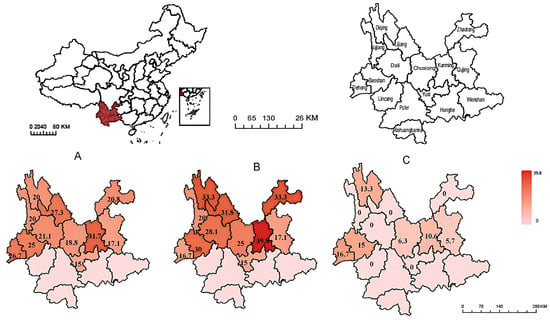
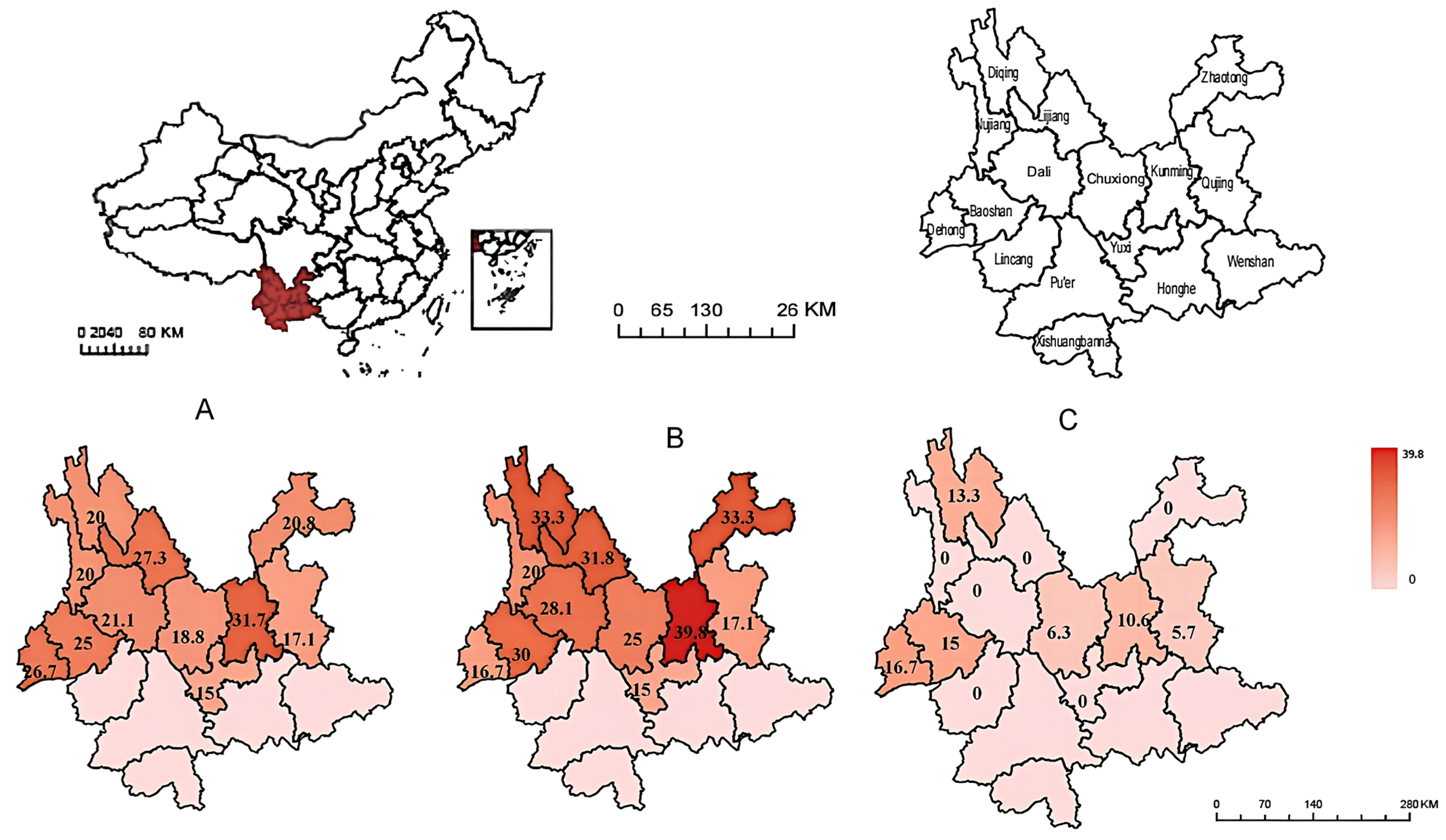
A total of 403 fecal samples were collected from diarrheic calves across 12 regions of Yunnan Province. BRV, BCoV, and BNoV were detected in 24.3% (98/403; 95% CI: 20.4–28.7%), 29.8% (120/403; 95% CI: 25.5–34.4%), and 7.2% (29/403; 95% CI: 5.1–10.1%) of the samples, respectively. Their detection rates varied across regions (Figure 1). BRV and BCoV were most frequently detected in Kunming, with rates of 31.7% and 39.8%, whereas BNoV showed its highest detection rate in Dehong (16.7%) (Figure 1A–C).
Figure 1.
Geographic distribution of BRV, BCoV, and BNoV detection rates in Yunnan Province, China. The maps show the detection rates of (A) BRV, (B) BCoV, and (C) BNoV among fecal samples collected from diarrheic calves in 12 administrative regions of Yunnan Province. The numbers within each region indicate the detection rate (%) of the corresponding virus, calculated as the number of positive samples divided by the total number of samples tested in that region (n/N × 100%). The color gradient represents the detection rate, with darker red indicating a higher rate and lighter pink indicating a lower rate. Regions without sampled specimens are shown without detection-rate data. The maps display regional detection rates rather than absolute numbers of positive samples.
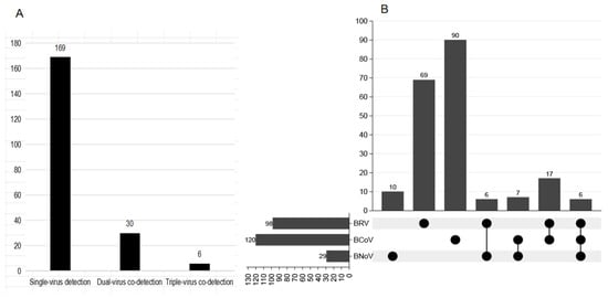
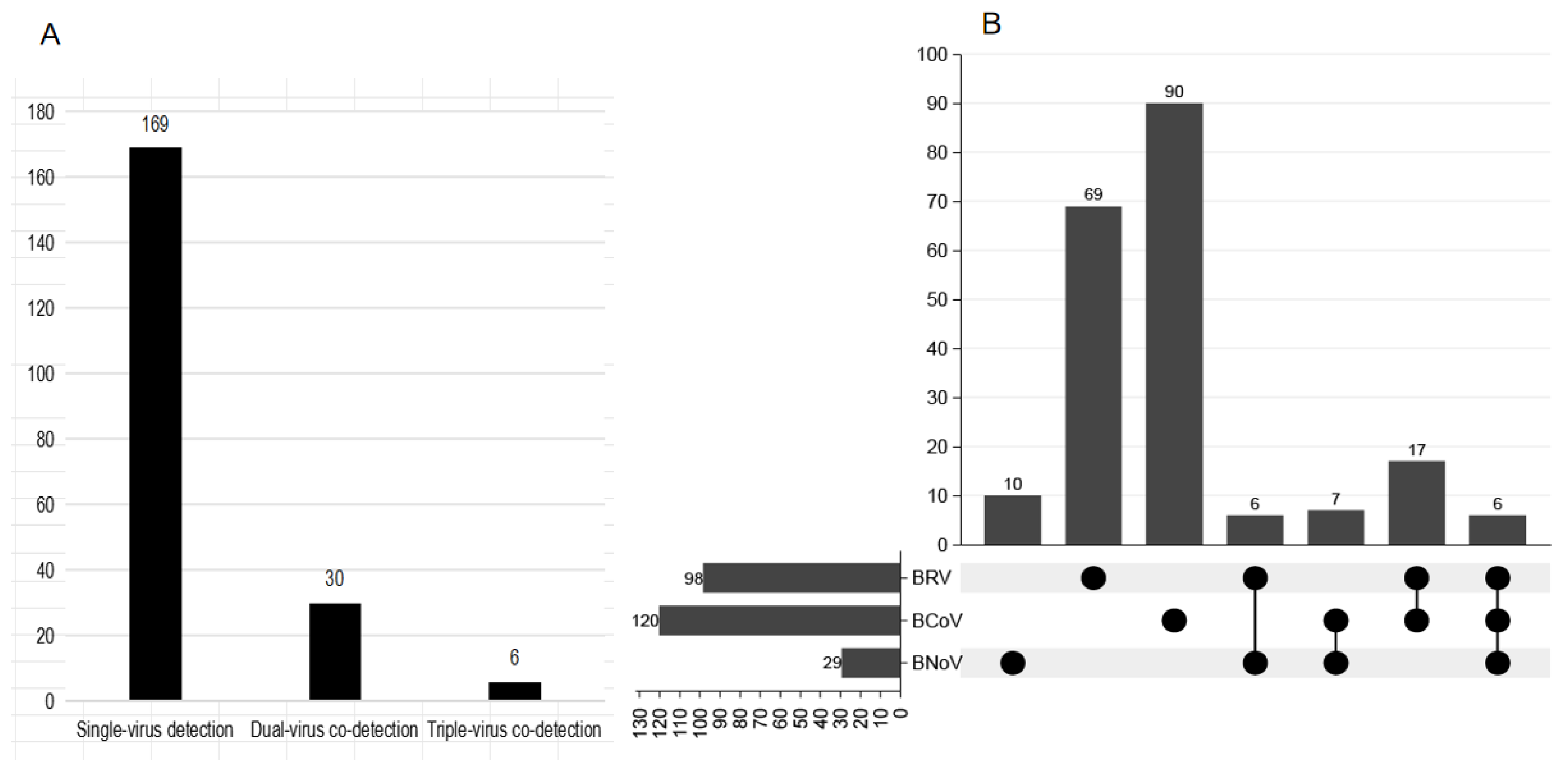
Single-pathogen detection predominated, with BCoV being the most frequently detected single pathogen (n = 90). Co-detection with other viruses was observed in 29.6% (29/98) of BRV-positive samples, 25.0% (30/120) of BCoV-positive samples, and 65.5% (19/29) of BNoV-positive samples. Among the dual-pathogen co-detection combinations, BRV/BCoV was the most common (n = 17), followed by BCoV/BNoV (n = 7) and BRV/BNoV (n = 6). Triple-pathogen co-detection involving BRV, BCoV and BNoV was detected in six samples (Figure 2A,B).
Figure 2.
Distribution of single and mixed detection with BRV, BCoV and BNoV in diarrhoeic calves. (A) Numbers of samples with single-virus detection, dual-virus co-detection, and triple-virus co-detection. (B) Distribution of different combinations of BRV, BCoV, and BNoV detection. The horizontal bars on the left indicate the total number of samples positive for each virus, the vertical bars at the top represent the number of samples in each detection combination, and the dot matrix below indicates the viruses included in each combination. BRV, bovine rotavirus; BCoV, bovine coronavirus; BNoV, bovine norovirus.
3.2. Molecular Characterization of BRV
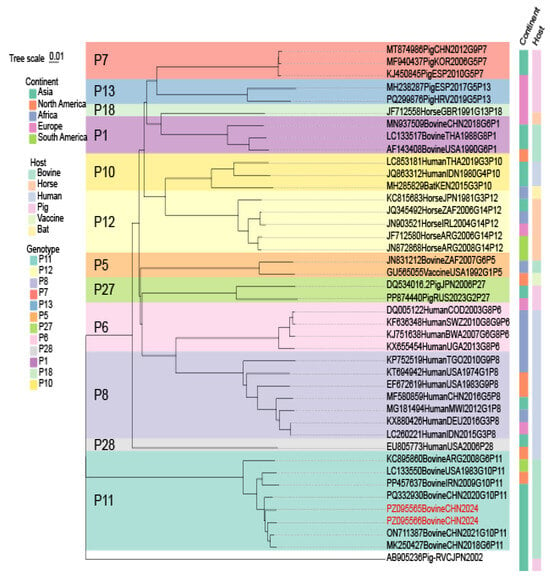
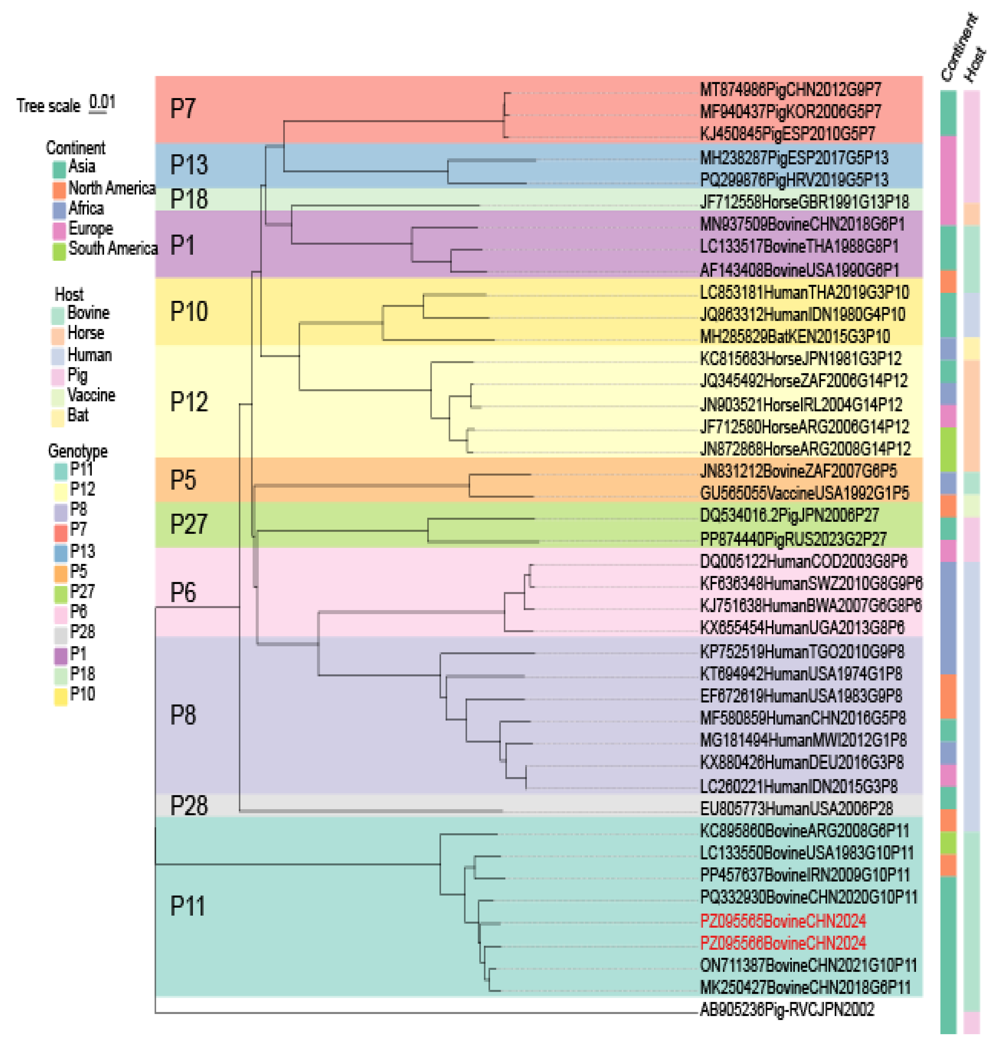
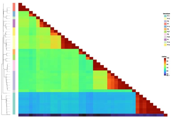
Representative BRV-positive samples were selected for VP4 and VP7 gene amplification, sequencing, and genotype determination. PCR amplification yielded products of approximately 1650 bp and 780 bp, respectively. Phylogenetic analysis of the VP4 gene showed that the two BRV strains identified in this study (PZ095565 and PZ095566) were assigned to the P[11] genotype (Figure 3). Both isolates clustered with bovine P[11] reference strains reported from China, Iran, the United States, and Argentina, indicating a close genetic relationship with contemporary bovine P[11] strains. The nucleotide identity analysis supported the phylogenetic classification (Figure 4). The VP4 sequences shared 92.17–98.52% nucleotide identity with representative P[11] strains, whereas identities with the remaining P genotypes (P[1], P[5], P[6], P[7], P[8], P[10], P[12], P[13], P[18], P[27], and P[28]) ranged from 57.15% to 60.87%. These results confirmed that both isolates belonged to the P[11] genotype and were clearly differentiated from the other VP4 genotypes included in the analysis.
Figure 3.
Phylogenetic analysis of the VP4 gene of BRV.
Figure 4.
Nucleotide sequence identity analysis of the BRV VP4 gene.
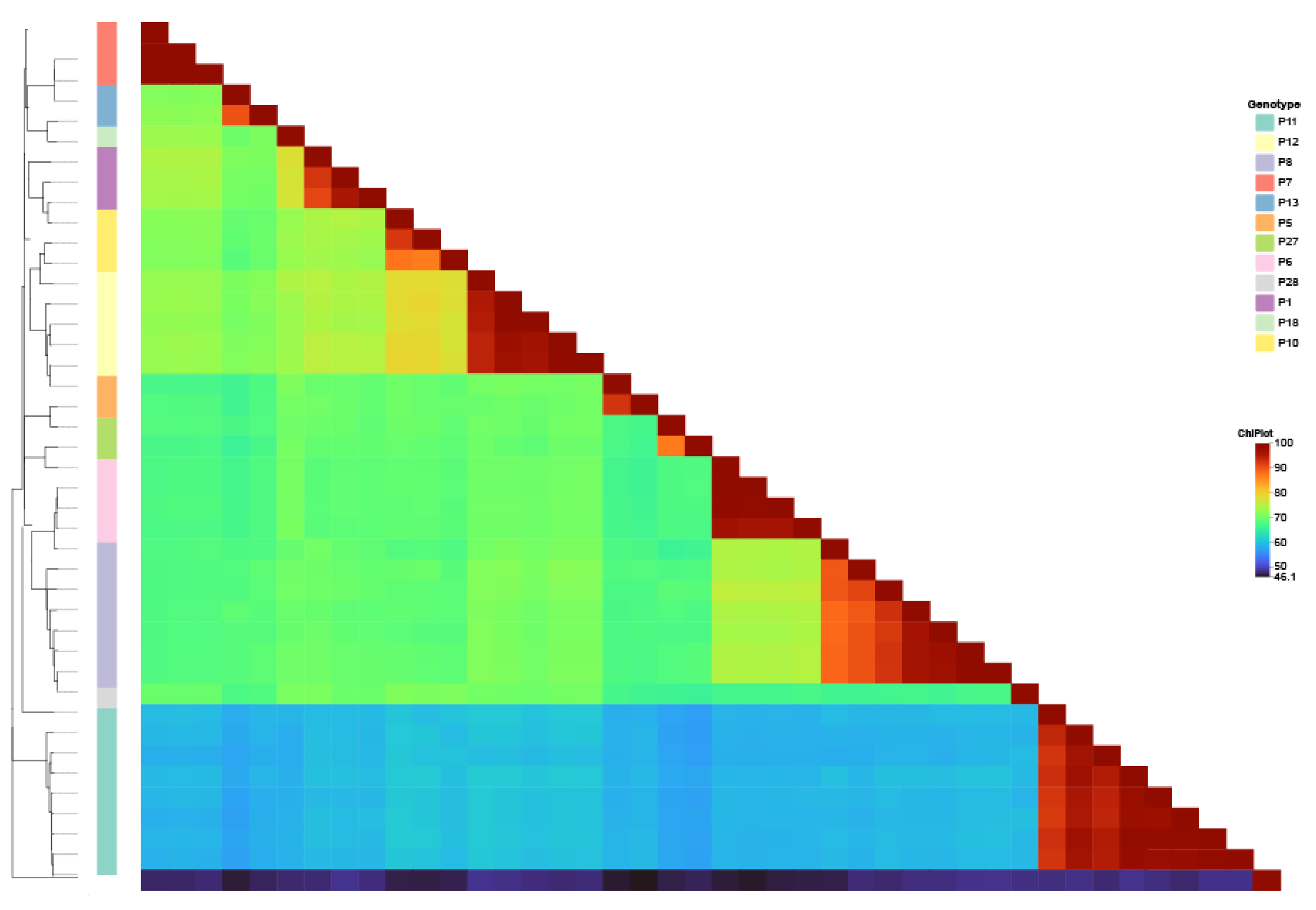
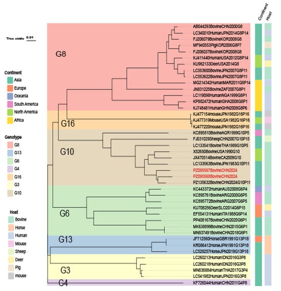
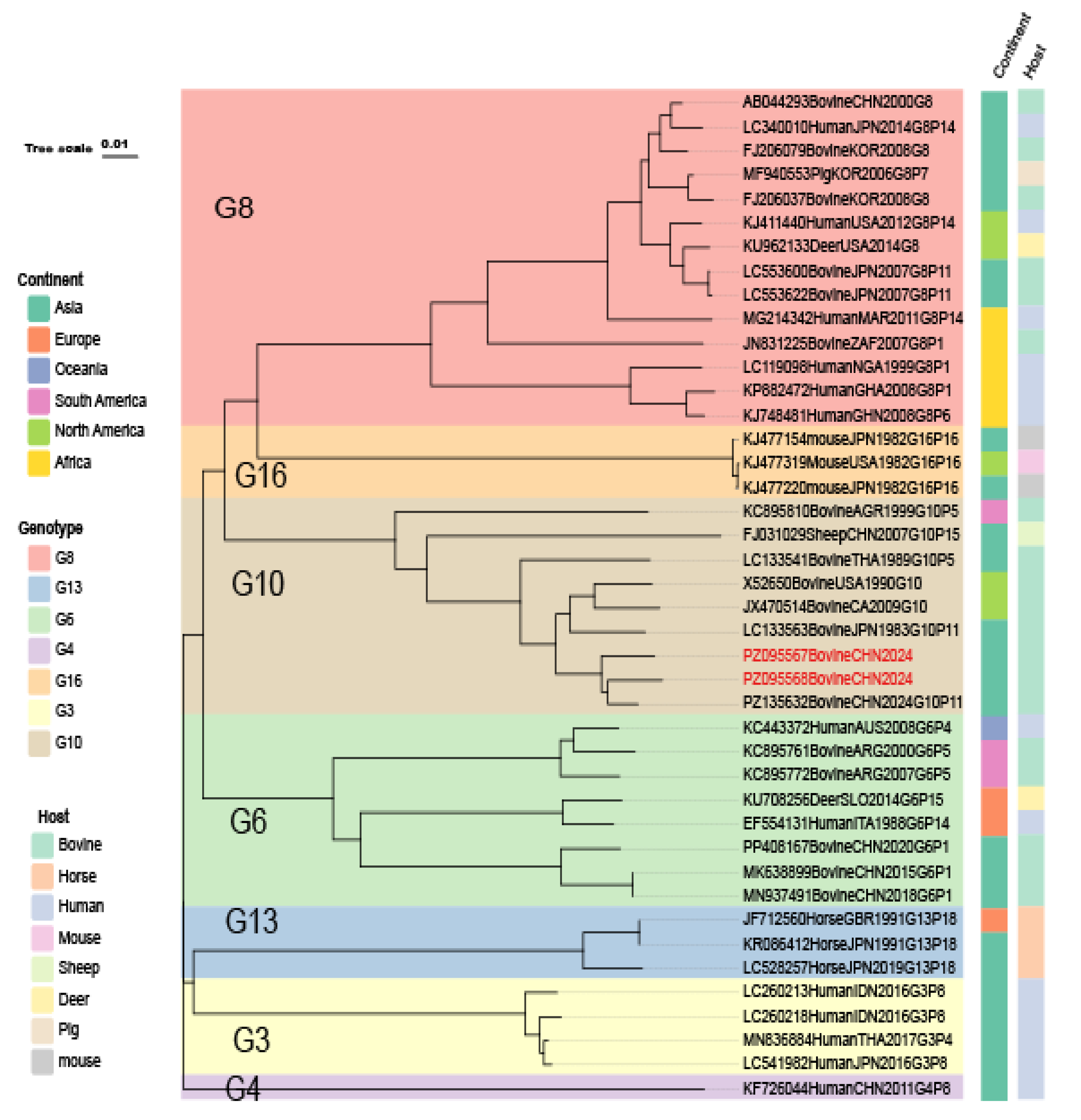
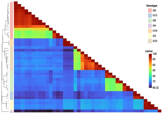
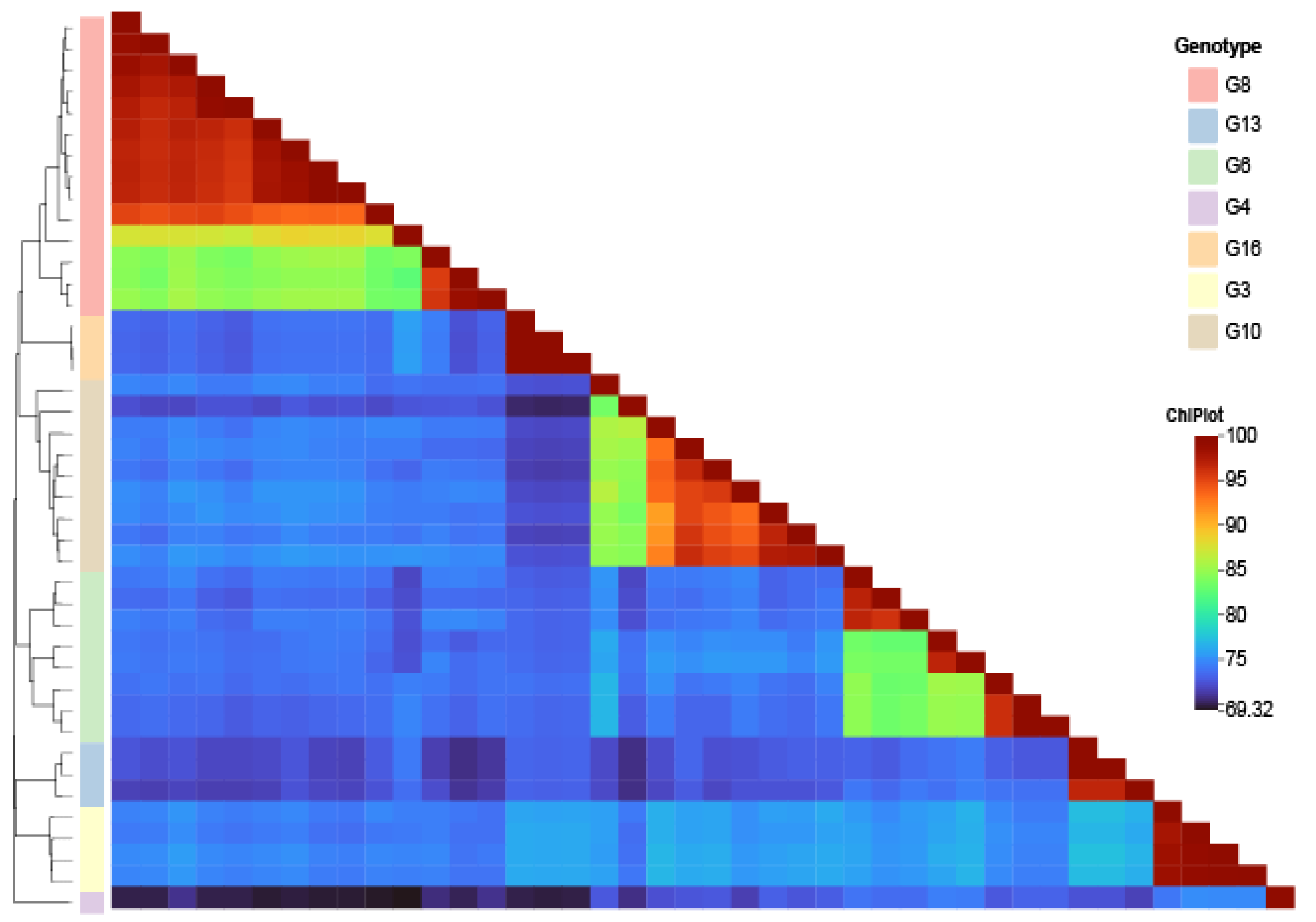
A similar result was obtained for the VP7 gene. Phylogenetic analysis placed strains PZ095567 and PZ095568 within the G10 genotype (Figure 5). The two strains clustered together with the Guangxi isolate (PZ135632), which was also identified as the G10P[11] genotype, suggesting that G10P[11] was the predominant genotype combination among the BRV strains characterized in this study. The G10 cluster also contained bovine and ovine reference strains from China, Thailand, the United States, and Canada, while remaining clearly separated from the G3, G4, G6, G8, G13, and G16 genotypes. The VP7 nucleotide identity analysis showed the same clustering pattern (Figure 6). The study strains shared 83.56–97.55% nucleotide identity with representative G10 strains, whereas identities with the other G genotypes ranged from 71.41% to 75.83%. Taken together, the phylogenetic and sequence identity analyses consistently identified the BRV strains characterized in this study as the G10P[11] genotype.
Figure 5.
Phylogenetic analysis of the VP7 gene of BRV.
Figure 6.
Nucleotide sequence identity analysis of the BRV VP7 gene.
3.3. Molecular Characterization of BCoV
Representative BCoV-positive samples were selected for partial S1 gene amplification, sequencing, and phylogenetic analysis, generating PCR products of approximately 1600 bp. The four BCoV partial S1 gene sequences obtained in this study were deposited in GenBank under accession numbers PZ095464–PZ095467.
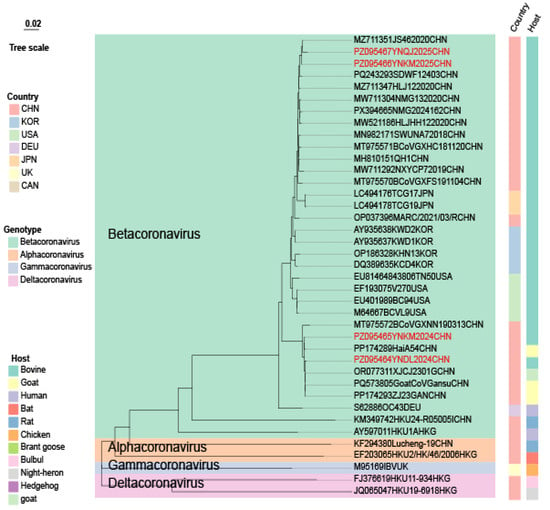
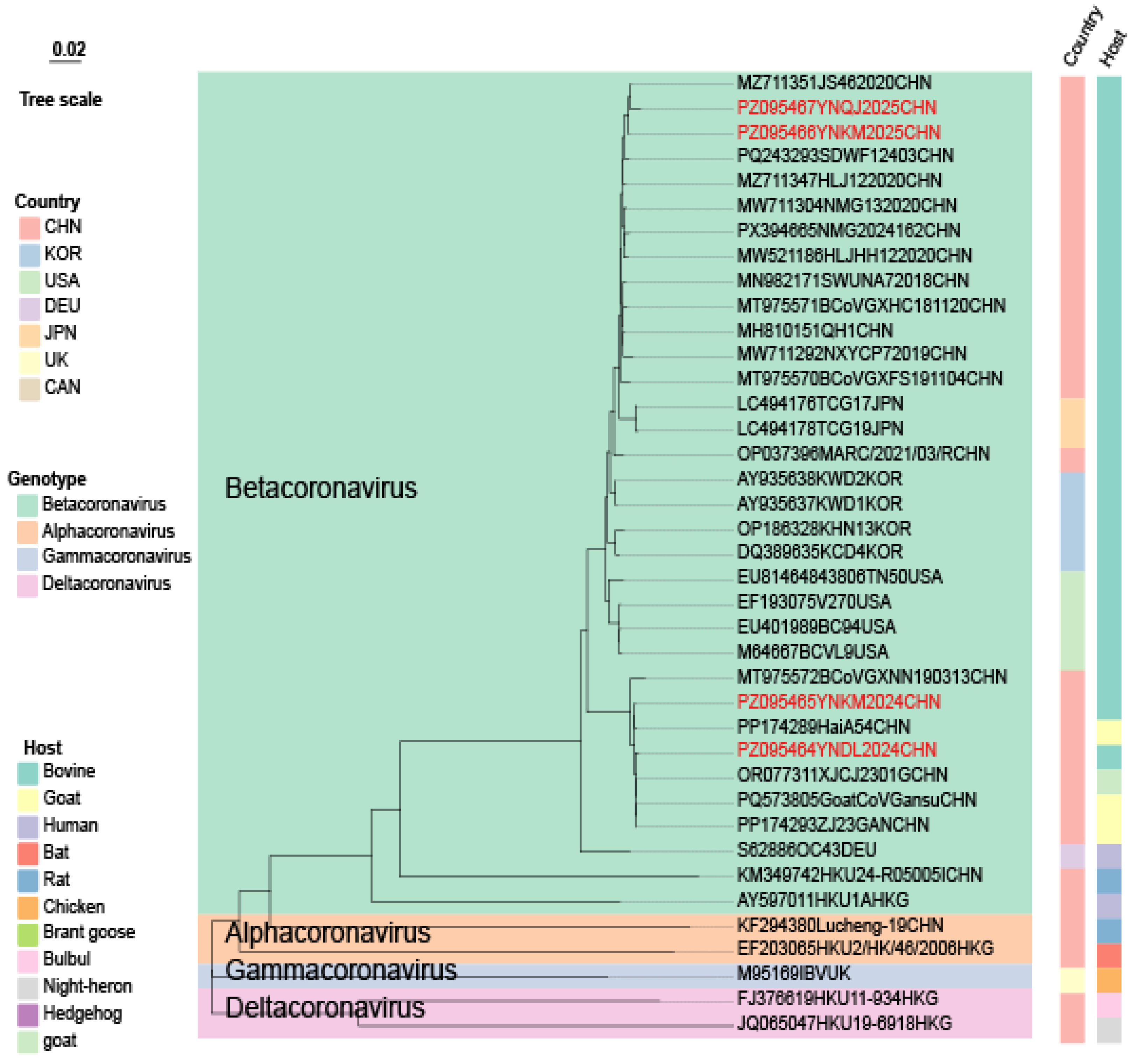
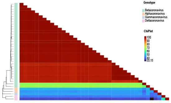
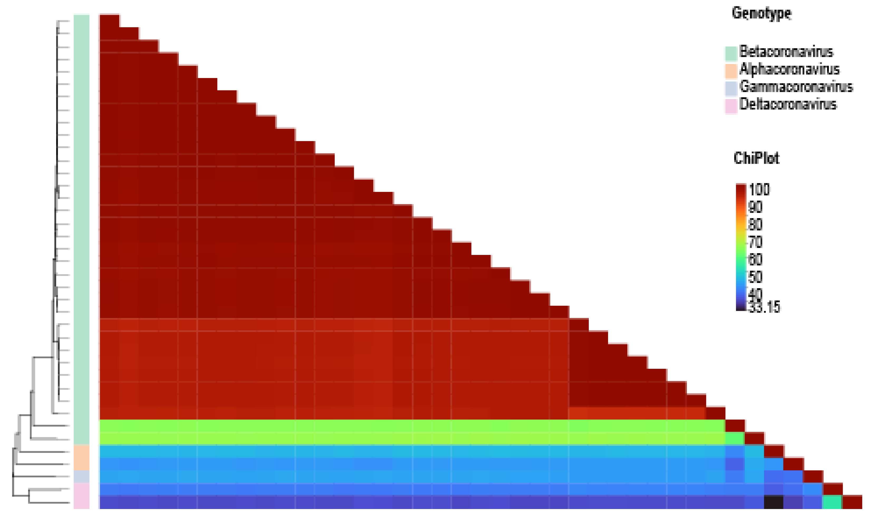
Phylogenetic analysis based on the partial S1 gene showed that all four BCoV strains clustered within the Betacoronavirus genus, clearly separated from the Alphacoronavirus, Gammacoronavirus, and Deltacoronavirus outgroups (Figure 7), and formed two distinct subclusters. Strains PZ095466 and PZ095467 grouped with the recent Chinese strain MZ711351JS462020CHN, sharing 99.65% and 99.22% nucleotide identity, respectively, and also showed high identity with PQ243293SDWF12403CHN from Shandong (99.29% and 98.51%, respectively). Strains PZ095464 and PZ095465 formed the second subcluster together with the goat-derived strain OR077311XJCJ2301GCHN, and shared high nucleotide identity with three independently reported goat-derived BCoV strains, PQ573805GoatCoVGansuCHN (Gansu), PP174293ZJ23GANCHN (Zhejiang), and PP174289HaiA54CHN (Hainan), reaching 99.79%/99.86% and 99.57%/99.79%, respectively. Consistent with the phylogenetic topology, pairwise nucleotide identity among bovine-associated Betacoronavirus strains remained high (92.9–100%), but decreased to 61.99% when more distantly related members of the genus (e.g., human coronaviruses HKU1, HKU24, and OC43) were included, and further declined to 33.2–49.7% when compared with representatives of the other three coronavirus genera (Figure 8). These findings demonstrate that the study strains are highly conserved within the bovine-associated Betacoronavirus lineage while remaining clearly differentiated from other coronavirus genera. The close phylogenetic relationship and high sequence identity between PZ095464/PZ095465 and multiple geographically distinct goat-derived BCoV strains suggested possible interspecies genetic exchange, prompting further investigation for evidence of recombination.
Figure 7.
Phylogenetic analysis of the S1 gene of BCoV.
Figure 8.
Nucleotide sequence identity analysis of the BCoV S1 gene.
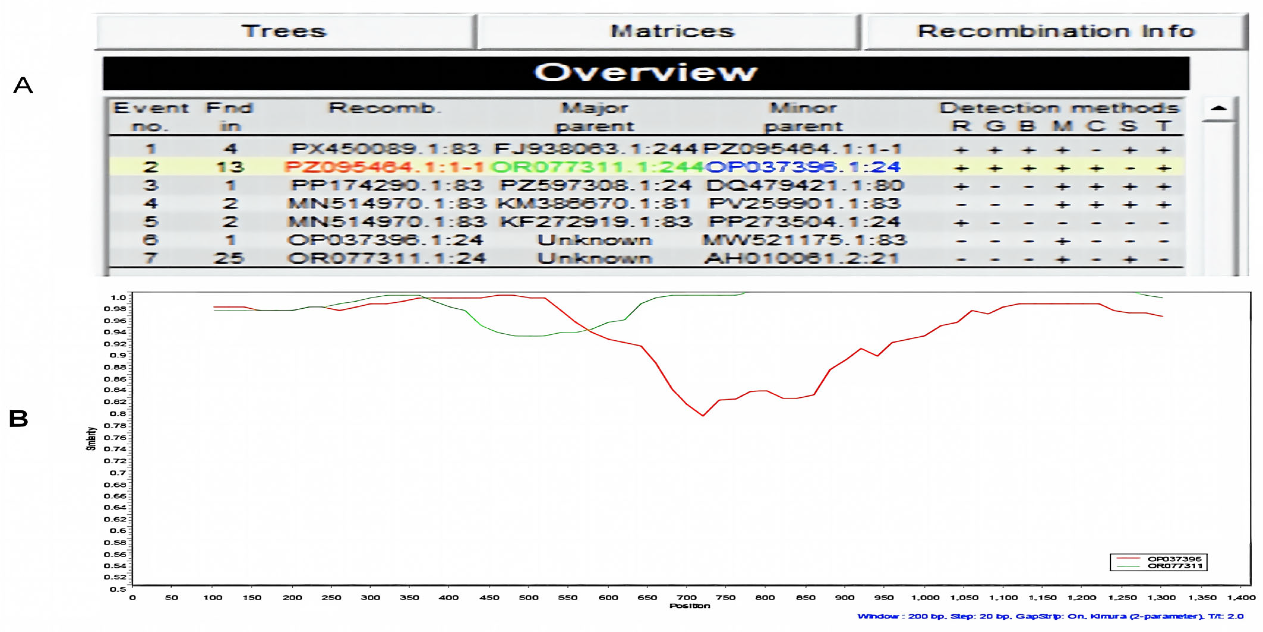
The BCoV S1 gene sequences were analyzed for recombination using RDP5 with seven detection algorithms (RDP, GENECONV, BootScan, MaxChi, Chimaera, SiScan, and 3Seq). Seven candidate recombination events were identified, of which only one involved a strain obtained in this study. A significant recombination event was detected in strain PZ095464 and was supported by six of the seven algorithms implemented in RDP5, whereas SiScan did not detect a significant signal (Figure 9A). OR077311.1 (goat-derived BCoV) and OP037396.1 (bovine BCoV) were inferred as the major and minor parental strains, respectively. The predicted recombination breakpoints were located at nucleotide positions 420–578. Strain PZ095464 shared 99.79% nucleotide identity with the major parent and 94.47% identity with the minor parent. SimPlot analysis provided additional evidence of a potential recombination signal (Figure 9B). Across most of the analyzed S1 gene region, strain PZ095464(red) exhibited greater similarity to the major parent, OR077311.1(green), than to the minor parent, OP037396.1(blue). However, within the putative recombination region (approximately nt 400–540), sequence similarity shifted toward the minor parent. Although this pattern was broadly consistent with the RDP5 results, the regions identified by the two methods partially overlapped but did not fully coincide. Therefore, SimPlot provided additional support for a potential recombination event but did not precisely confirm the breakpoint positions predicted by RDP5.
Figure 9.
Recombination analysis of the BCoV S1 gene. (A) Detection of a putative recombination event in strain PZ095464 using RDP5. (B) SimPlot analysis showing nucleotide sequence similarity between PZ095464 and the putative parental sequences OR077311.1 and OP037396.1.
3.4. Molecular Characterization of BNoV
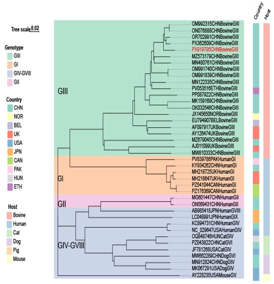
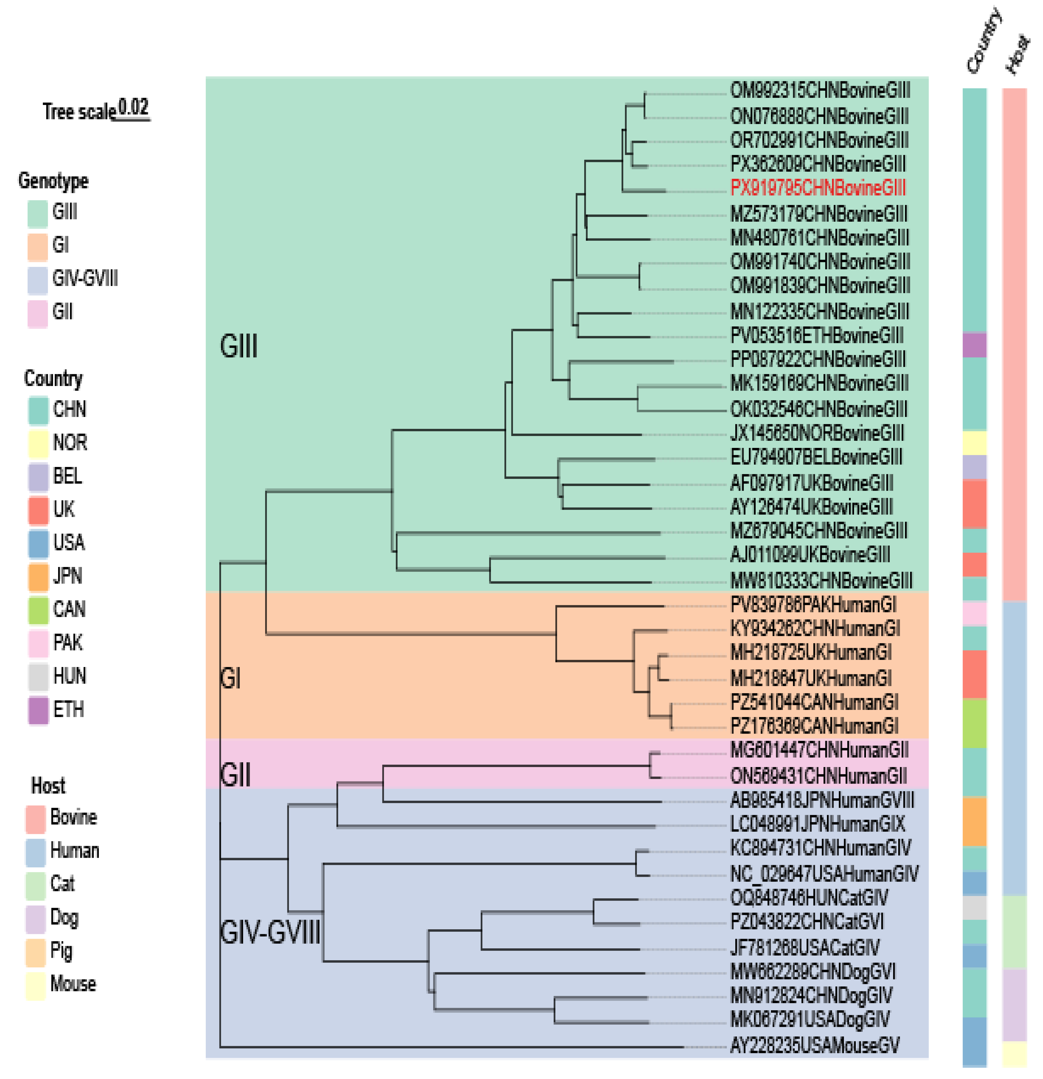
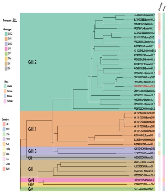
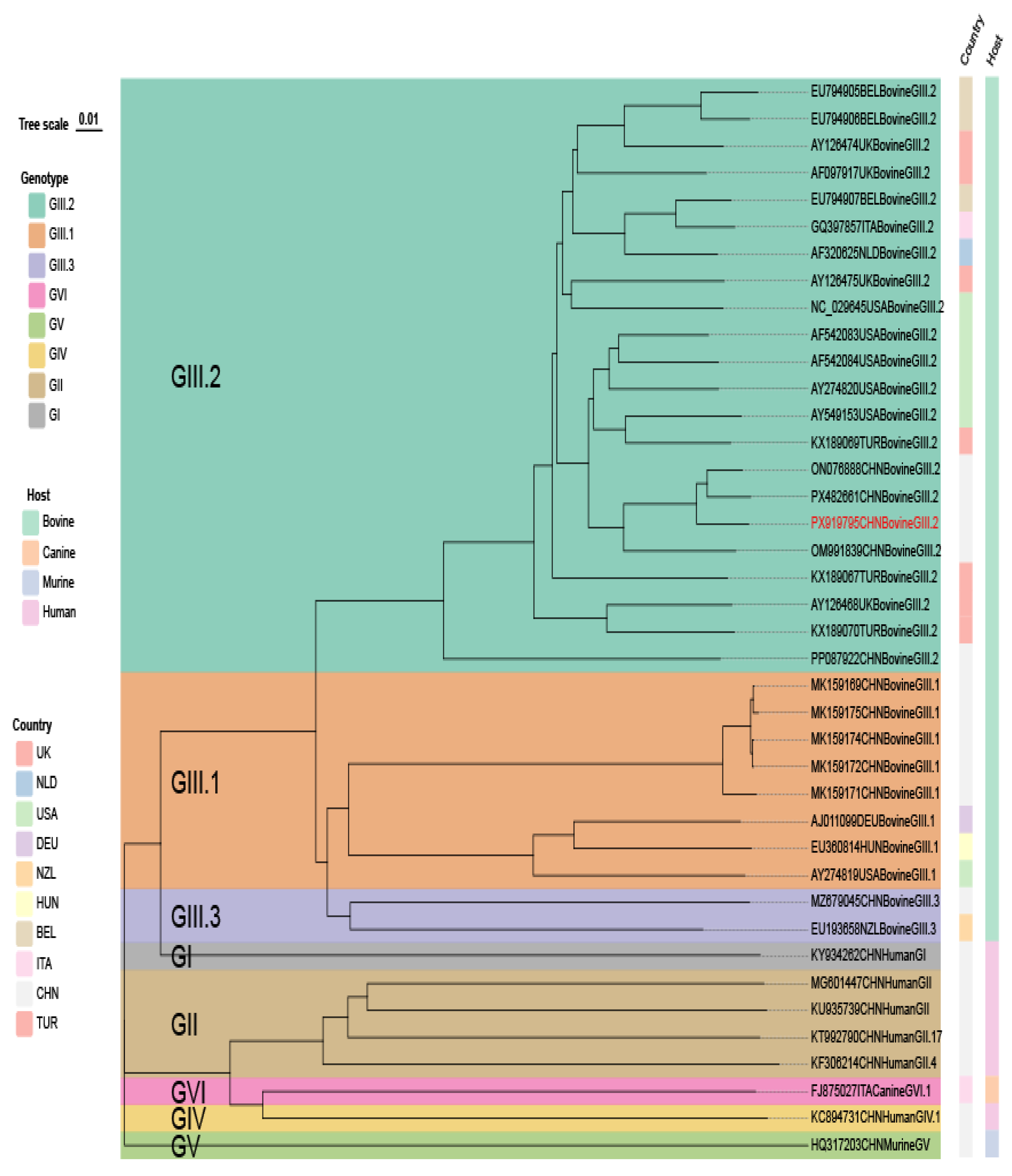
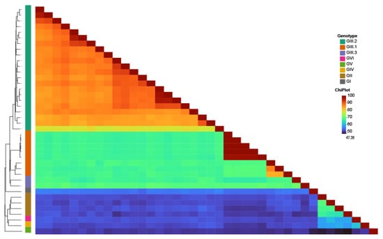
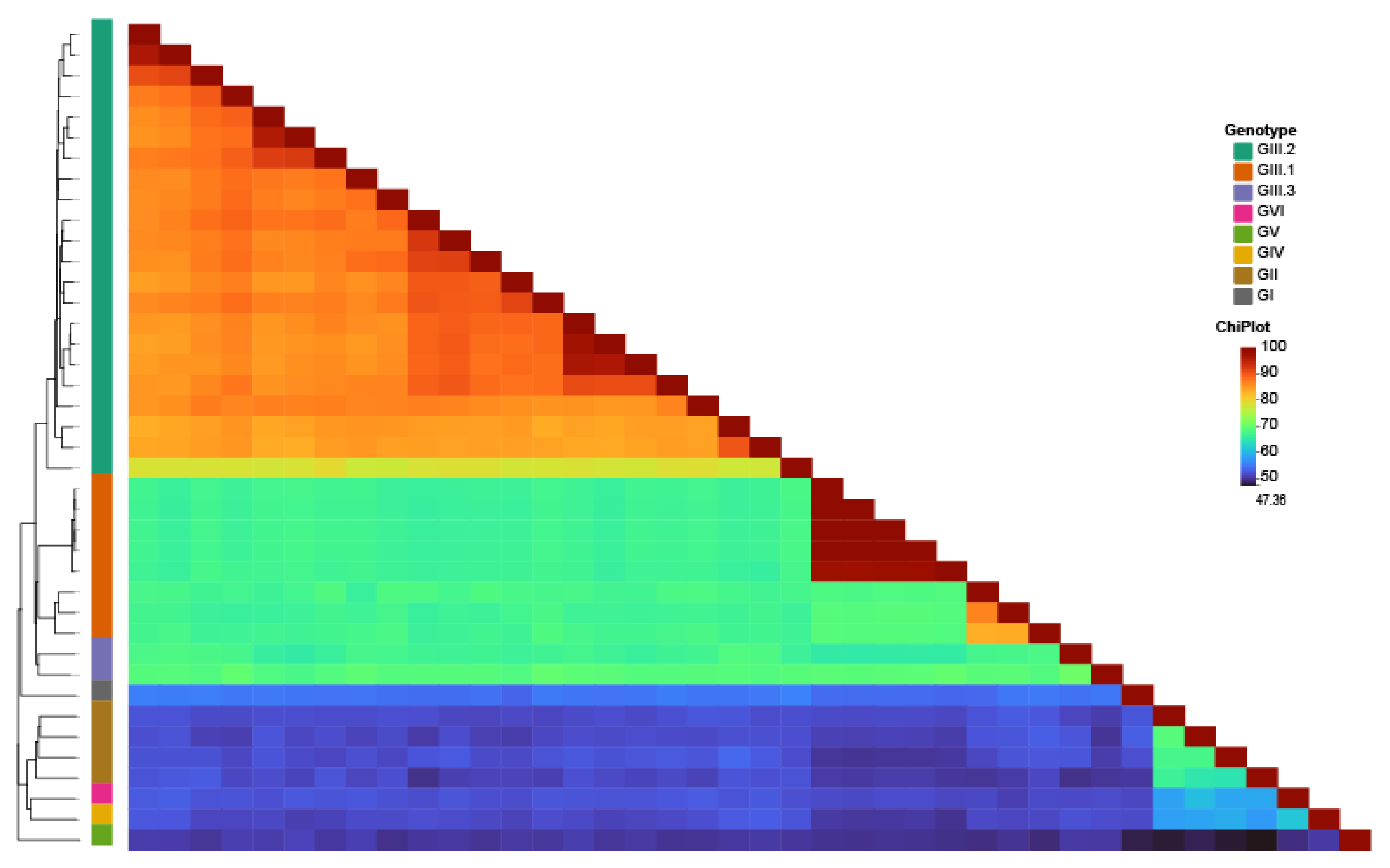
The study used the five pairs of primers listed in Table 2 to perform PCR amplification on different regions of the BNoV genome. The amplified products were cloned and sequenced in both directions. Then, using SeqMan (DNASTAR 12), the overlapping sequences of each fragment were assembled to obtain a nearly complete BNoV genome sequence, which was submitted to GenBank under the accession number PX919795. Phylogenetic analysis based on this sequence showed that PX919795 belongs to the GIII genotype (Figure 10), and further genetic evolution analysis of vp1 determined it as the GIII.2 subtype (Figure 11). Nucleotide sequence identity analysis showed that PX919795 shares 77.91–96.15% nucleotide sequence identity with GIII.2 strains, with the highest similarity to the Chinese strain ON076888. The nucleotide sequence identity with GIII.1 and GIII.3 strains is 66.82–67.34% and 66.43–68.91%, respectively, while nucleotide sequence identity with other genotype strains is lower, at 50.30–54.84% (Figure 12). In addition, neither RDP5 nor SimPlot identified a clear recombination signal in the analyzed BNoV sequence.
Figure 10.
Phylogenetic analysis of BNoV.
Figure 11.
Phylogenetic Analysis of the BNoV VP1 Gene.
Figure 12.
Nucleotide sequence identity analysis of the BNoV VP1 gene.
4. Discussion
Calf diarrhea is one of the most important diseases affecting calf health and the production efficiency of the cattle industry [18]. In 2018, research results published by the United States National Animal Health Monitoring System indicated that 39% of calf deaths within 3 weeks after birth were due to diarrhea [19]. Calf diarrhea is usually associated with infections by multiple pathogens, among which BRV, BCoV, and BNoV are common viral pathogens. In recent years, an increasing number of studies have demonstrated that these three viruses possess considerable genetic variability and recombination potential and are capable of crossing host barriers. As a segmented RNA virus, BRV has been shown to undergo genetic reassortment among different animal hosts [20,21], and cases of zoonotic transmission of bovine rotavirus to humans, including disease in children, have been documented [22]. BCoV is closely related phylogenetically to human coronavirus HCoV-OC43 [23] and has also been detected in dogs, turkeys, and various wild ruminants, indicating a broad host range [24]. BNoV, which belongs to the family Caliciviridae together with human noroviruses, has also attracted attention because of its genetic diversity and potential for cross-species transmission [25], with antibodies against BNoV detected in certain human populations [26]. These genetic recombination and cross-species transmission events may not only influence viral host adaptation and epidemiological characteristics but may also have potential public health implications. RT-PCR was employed in this study in order to test 403 fecal samples from diarrheic calves across 12 regions in Yunnan Province. The results showed that the overall detection rates of BRV, BCoV, and BNoV were 24.3%, 29.8%, and 7.2%, respectively, with BCoV having the highest detection rate. Relevant meta-analyses indicate that the overall prevalence of BCoV in China is 30.8% [4], while a previous survey in Yunnan Province documented a detection rate of 28.73% [10], both of which are relatively close to the results of this study. BRV showed a comparable pattern. The detection rate of 24.3% identified in the present study was comparable to the 21.5% (95% CI: 17.5–25.8%) previously observed in Yunnan Province [27]. However, it was lower than the pooled national prevalence of BRV in China, which was estimated at 35.7%. BNoV had the lowest detection rate at 7.2%. Currently, epidemiological data on BNoV in diarrheic calves in Yunnan Province remain limited, and this study provides basic data on the prevalence of BNoV in this region. Compared with other regions, the BNoV detection rate in this study is lower than that in Xinjiang at 19.82% [28], six provinces in China at 20.4% [29], and Guangdong Province at 15.98% [30]. This regional difference may be related to factors such as geographic environment, breeding methods, sample sources, and testing conditions.
In addition to single-pathogen detection, co-detection of multiple pathogens was also frequently observed in this study. The proportions of mixed detection among BRV-, BCoV-, and BNoV-positive samples were 29.6% (29/98), 25.0% (30/120), and 65.5% (19/29), respectively, with the highest proportion observed among BNoV-positive samples. BRV/BCoV was the most common dual-pathogen co-detection combination (n = 17), and 6 cases of triple-pathogen co-detection were also detected. These findings were broadly consistent with previous studies in China. A study involving 1646 bovine faecal samples from 14 provinces in China reported that BCoV/BRV was the most common co-detection combination, accounting for 46.58% of co-detection cases [31]. Co-detection with other viruses was also detected in 75.9% of BRV-positive samples in a six-province survey [32]. For BNoV, the proportion was even higher, with 88.4% of BNoV-positive samples showing co-detection with BRV, BCoV, and BVDV in another six-province survey [29]. Although co-detection rates varied among studies, these findings suggest that BRV, BCoV, and BNoV are frequently detected alongside other viruses in diarrheic calves, with BNoV-positive samples showing a relatively higher proportion of co-detection. It should be noted that this study focused only on BRV, BCoV, and BNoV, and did not include screening for other pathogens, such as bacteria and parasites, that may also be associated with calf diarrhea. Therefore, the pathogen composition of diarrheic calves could not be fully characterized, and the possible involvement of other pathogens in co-detection could not be assessed. Further studies incorporating bacteriological, parasitological, and broader pathogen screening are needed to provide a more comprehensive understanding of the pathogens associated with calf diarrhea and their potential interactions.
The VP4 and VP7 genes are important molecular markers for BRV genotyping and phylogenetic analysis, as they encode the two major outer capsid proteins. In this study, sequence analysis of representative positive samples identified the BRV strains as belonging to the G10P[11] genotype. The distribution of BRV genotypes varies across different regions of the world. In Chinese cattle herds, combinations such as G6P[1], G6P[5], and G6P[11] are relatively common [21], whereas G10P[11] has been reported more frequently in Japan. In South Korea [33], Bangladesh [34], and Uruguay [35], G6P[5] or G6P[11] are the main genotypes detected, with G10P[11] accounting for only a small proportion. In 2008, the G10P[11] BRV genotype was reported in Inner Mongolia, China, and since then, this genotype has been detected in other regions of the country. Liu et al. isolated the G10P[11] BRV strain SHH2023001 from a calf with severe diarrhea in Shanghai [36], and this study has again detected the same genotype in Yunnan, indicating that G10P[11] has been identified in different regions of China. Notably, the strain from this study clusters closely with the G10P[11] strain PZ135632, isolated from water buffalo in Guangxi and deposited in GenBank, in the VP7 phylogenetic tree and exhibits high nucleotide sequence identity, suggesting a close genetic relationship between the two in the VP7 gene. Taken together, G10P[11] BRV has been successively detected in multiple geographically distant provinces across China, from Inner Mongolia and Shanghai to Guangxi and Yunnan. Given the geographical proximity of Yunnan and Guangxi and the movement of cattle between these regions, regional animal movement may contribute to the spread of this genotype. However, the close genetic relationship between the bovine and water buffalo-derived strains does not establish cross-host transmission. Further molecular epidemiological investigations involving more samples and complete genome sequences are needed to clarify the transmission patterns, host distribution, and geographic spread of G10P[11] BRV in China.
The S1 gene of BCoV encodes the major surface envelope glycoprotein (spike protein) of the virus, which plays a key role in receptor binding, membrane fusion, and the induction of neutralizing antibody responses [37]. Due to its high genetic variability, it is a commonly used target gene in BCoV molecular typing and evolutionary analysis [38]. Analysis of the S1 gene in representative BCoV-positive samples revealed that the studied strains fall into two subclusters, clustering with domestic bovine strains and goat strains from multiple regions, respectively. Phylogenetic analysis based on the S gene typically classifies strains into lineages such as the classical lineage (GIa), the European lineage (GIb), and the Asian-American lineage (GII, including the GIIa and GIIb sublineages) [39]. Among these, the GIIb sublineage is the primary lineage of strains currently circulating in Yunnan and most other regions of China; it exhibits distinct geographic clustering and shows some divergence from strains circulating in countries such as Japan, South Korea, and Vietnam. The BCoV strains detected in this study also belong to the GIIb lineage, which is generally consistent with previous reports on the genetic characteristics of BCoV in Yunnan and other regions of China [10,31]. Notably, this study identified a putative recombination signal in the partial S1 sequence of PZ095464. Although the RDP5 and SimPlot analyses provided supporting evidence for this signal, the use of partial-gene sequences limits the certainty of the inference. Further analysis using longer genomic sequences is required to validate the potential recombination event and clarify the evolutionary relationship between bovine- and goat-associated coronaviruses. This finding is closely related to the broad host range of BCoV itself: previous reports have shown that BCoV can infect a variety of species, including goats, deer, llamas, and even humans [24,39,40,41], and its ability to cross species barriers far exceeds that of typical enteroviruses. Recombination analysis further revealed that the primary parental strain of PZ095464 was a goat-derived strain (OR077311.1, 99.79% nucleotide sequence identity). This strain originated from a recent goat population study, where it was identified as a novel betacoronavirus (provisionally named cpCoV) that is evolutionarily distinct from classical BCoV [42]; its overall nucleotide sequence identity with members of the BCoV subgenus is typically below 95%. Nevertheless, the bovine strain PZ095464 in this study showed high nucleotide sequence identity with this goat-derived strain in a specific gene region (nt 420–578), suggesting that local gene exchange may occur between bovine- and goat-derived coronaviruses. This finding is consistent with observations from previous regional studies—for example, a study in the southwestern region reported that local bovine BCoV strains clustered with goat-derived strains and showed signs of recombination [37], suggesting that in the southwestern mountainous areas, such as Yunnan, where mixed cattle and goat farming is common, localized gene exchange of coronaviruses among different ruminant hosts may be an ongoing phenomenon rather than an isolated occurrence. Noroviruses can be classified into multiple genogroups (GI–GX) based on differences in the amino acid sequences of the major capsid protein VP1 [43], and these genogroups exhibit distinct host preferences. BNoV is currently the only known norovirus associated with the GIII genogroup. Since the discovery of the Newbury2 strain in the United Kingdom and the Jena strain in Germany in the 1970s [44], GIII has been further divided into two major genotypes, GIII.1 and GIII.2. GIII.2 has a broader geographic distribution than GIII.1 and has been detected in more countries worldwide [29]. Based on comparisons with the prototype GIII.1 and GIII.2 strains available in the NCBI database, the strain PX919795 identified in the present study was assigned to genotype GIII.2, further confirming the circulation of this genotype in China. The first GIII.2 BNoV reported in China was CH-HNSC-2018, which was identified in diarrheic calves in Henan Province [45]. The complete genome of this strain was sequenced, and phylogenetic analysis based on the VP1 sequence classified it as GIII.2; several amino acid variations were also identified in VP1 and the P2 subdomain. Subsequently, the complete genome of another BNoV strain, Bo/CH/HB/BD/2019, was reported in Hebei, and this strain also belonged to the GIII.2-related lineage [46]. These findings indicate that GIII.2 BNoV has been detected in cattle from different regions of China and exhibits a degree of genetic diversity. The identification of a GIII.2 BNoV strain in Yunnan Province in the present study further expands the molecular epidemiological information on BNoV in southwestern China.
5. Conclusions
This study investigated the detection rates, co-infection patterns, and genetic characteristics of BRV, BCoV, and BNoV in 403 fecal samples collected from diarrheic calves across 12 administrative regions of Yunnan Province, China. The overall detection rates were 24.3% for BRV, 29.8% for BCoV, and 7.2% for BNoV, with BCoV showing the highest detection rate. Co-detection involving two or three viruses was also identified. Molecular characterization assigned the two BRV strains to genotype G10P[11], revealed two distinct subclusters among the four BCoV S1 sequences, and identified a putative recombination signal in one partial BCoV S1 sequence. The near-complete BNoV genome belonged to genogroup GIII, and VP1 phylogenetic analysis classified it as genotype GIII.2. These findings provide baseline molecular epidemiological information on three enteric viruses in diarrheic calves in Yunnan Province. Further surveillance involving non-diarrheic controls and additional genomic sequences is needed to clarify the epidemiological significance of viral co-detection and to investigate the genetic diversity and evolution of these viruses.
Author Contributions
P.Z. and Y.W. collected the samples and performed the experiments. P.Z. conducted the molecular, phylogenetic, and recombination analyses and drafted the manuscript. B.L., X.Z. and Y.L. contributed to the laboratory experiments and data analysis. C.J., S.F. and W.L. revised and critically reviewed the manuscript. All authors have read and agreed to the published version of the manuscript.
Funding
This study was supported by the Yunnan Joint International R&D Center of Veterinary Public Health (Grant No. 202403AP140033) and the Revitalizing Yunnan Talents Support Program (Grant No. XDYC-GDWZ-2025-006), both funded by the Yunnan Provincial Department of Science and Technology.
Institutional Review Board Statement
This study did not involve any direct intervention in cattle. Bovine fecal samples were collected non-invasively after natural defecation during routine farm management, with prior consent from the farm owners.
Informed Consent Statement
Not applicable.
Data Availability Statement
The datasets analysed during the current study have been uploaded to the NCBI GenBank database under the accession numbers PZ095464, PZ095465, PZ095466, PZ095467, PZ095565, PZ095566, PZ095567, PZ095568, and PX919795. The accession numbers correspond to the following sequences: BCoV S1 gene: PZ095464–PZ095467; BRV VP4 gene: PZ095565 and PZ095566; BRV VP7 gene: PZ095567 and PZ095568; BNoV complete genome: PX919795.
Acknowledgments
We would like to thank all the projects for funding this study and all the authors for their help.
Conflicts of Interest
The authors declare no conflicts of interest.
Abbreviations
| BRV | Bovine rotavirus |
| BCoV | Bovine coronavirus |
| BNoV | Bovine norovirus |
| NCD | Neonatal calf diarrhea |
| RT-PCR | Reverse transcription polymerase chain reaction |
| VP4 | Viral protein 4 |
| VP7 | Viral protein 7 |
| S1 | Spike protein 1 |
References
- Papp, H.; László, B.; Jakab, F.; Ganesh, B.; De Grazia, S.; Matthijnssens, J.; Ciarlet, M.; Martella, V.; Bányai, K. Review of group A rotavirus strains reported in swine and cattle. Vet. Microbiol. 2013, 165, 190–199. [Google Scholar] [CrossRef] [Scilit]
- System, N.A.H.M. Dairy 2007: Heifer Calf Health and Management Practices on US Dairy Operations, 2007; U.S. Department of Agriculture, Animal and Plant Health Inspection Service: Riverdale, MD, USA, 2010. [Google Scholar]
- Qu, J.; Meng, C.; Li, L.; Zhu, H.; Yin, G. Epidemiology of Diarrhea for Chinese Dairy Calves: A Systematic Review and Meta-analysis. Indian J. Anim. Res. 2023, 57, 139–146. [Google Scholar] [CrossRef] [Scilit]
- Chen, J.; Meng, W.; Zeng, H.; Wang, J.; Liu, S.; Jiang, Q.; Chen, Z.; Ma, Z.; Wang, Z.; Li, S.; et al. Epidemiological survey of calf diarrhea related viruses in several areas of Guangdong Province. Front. Microbiol. 2024, 15, 1441419. [Google Scholar] [CrossRef] [Scilit]
- Mee, J. Invited review: Bovine neonatal morbidity and mortality—Causes, risk factors, incidences, sequelae and prevention. Reprod. Domest. Anim. 2023, 58, 15–22. [Google Scholar] [CrossRef] [Scilit]
- Cho, Y.-i.; Yoon, K.-J. An overview of calf diarrhea-infectious etiology, diagnosis, and intervention. J. Vet. Sci. 2014, 15, 1. [Google Scholar] [CrossRef] [Scilit]
- Torreggiani, C.; Pupillo, G.; Garbarino, C.A.; Rugna, G.; Prosperi, A.; Chiapponi, C.; Luppi, A. Major etiological agents isolated from neonatal calf diarrhea outbreaks in Northern Italy. Pathogens 2025, 14, 847. [Google Scholar] [CrossRef] [Scilit]
- Matthijnssens, J.; Attoui, H.; Bányai, K.; Brussaard, C.P.; Danthi, P.; Del Vas, M.; Dermody, T.S.; Duncan, R.; Fāng, Q.; Johne, R.; et al. ICTV virus taxonomy profile: Sedoreoviridae 2022. J. Gen. Virol. 2022, 103, 001782. [Google Scholar] [CrossRef] [Scilit]
- Hasan, M.A.; Suzuki, M.; Sakai, K.; Kabir, M.H.; Miyaoka, Y.; Hakim, H.; Kadota, C.; Shoham, D.; Takehara, K. Complete genome constellations of two bovine rotavirus A strains isolated in Japan reveal a unique T9 NSP3 genotype. J. Vet. Med. Sci. 2023, 85, 998–1003. [Google Scholar] [CrossRef] [Scilit]
- Qi, S.; Yang, Y.; Deng, J.; Wang, X.; Li, Y.; Sun, J.; Li, Q.; Yang, S.; Yin, G.; Li, Y.; et al. Whole-Genome Sequencing Reveals the Evolutionary Characterization of Bovine Coronavirus. Transbound. Emerg. Dis. 2025, 2025, 5152361. [Google Scholar] [CrossRef] [Scilit]
- Colina, S.E.; Serena, M.S.; Echeverría, M.G.; Metz, G.E. Clinical and molecular aspects of veterinary coronaviruses. Virus Res. 2021, 297, 198382. [Google Scholar] [CrossRef] [Scilit]
- Park, G.-N.; Choe, S.; Song, S.; Kim, K.-S.; Shin, J.; An, B.-H.; Moon, S.H.; Hyun, B.-H.; An, D.-J. Characterization and spike gene analysis of a candidate attenuated live bovine coronavirus vaccine. Animals 2024, 14, 389. [Google Scholar] [CrossRef] [Scilit]
- Han, M.G.; Smiley, J.R.; Thomas, C.; Saif, L.J. Genetic recombination between two genotypes of genogroup III bovine noroviruses (BoNVs) and capsid sequence diversity among BoNVs and Nebraska-like bovine enteric caliciviruses. J. Clin. Microbiol. 2004, 42, 5214–5224. [Google Scholar] [CrossRef] [Scilit]
- Oliver, S.; Brown, D.; Green, J.; Bridger, J. A chimeric bovine enteric calicivirus: Evidence for genomic recombination in genogroup III of the Norovirus genus of the Caliciviridae. Virology 2004, 326, 231–239. [Google Scholar] [CrossRef] [Scilit]
- Min, F.; Liang, Y.; Jiang, Q.; Tao, X.; Wan, J.; Xue, T.; Li, N.; Chen, R.; Zhong, Q.; Yao, G.; et al. Isolation, identification and pathogenicity analysis of BCoV epidemic strains in Xinjiang. Front. Microbiol. 2026, 17, 1788466. [Google Scholar] [CrossRef] [Scilit]
- Li, C.; Wang, X.; Zhu, Q.; Sun, D. Isolation and identification of BRV G6P [1] strain in Heilongjiang province, Northeast China. Front. Vet. Sci. 2024, 11, 1416465. [Google Scholar] [CrossRef] [Scilit]
- Dai, M.; Yi, Y.; Tang, H.; Liu, H.; Yang, H.; Li, C.; Shen, C.; Zhang, Y. Analysis of Cross-Border Movement of Cattle in Yunnan Province, China: Insights Into Transboundary Animal Diseases Control. Transbound. Emerg. Dis. 2025, 2025, 5561414. [Google Scholar] [CrossRef] [Scilit]
- Hur, T.-Y.; Jung, Y.-H.; Choe, C.-Y.; Cho, Y.-I.; Kang, S.-J.; Lee, H.-J.; Ki, K.-S.; Baek, K.-S.; Suh, G.-H. The dairy calf mortality: The causes of calf death during ten years at a large dairy farm in Korea. Korean J. Vet. Sci. 2013, 53, 103–108. [Google Scholar] [CrossRef] [Scilit]
- Kim, H.S.; Whon, T.W.; Sung, H.; Jeong, Y.-S.; Jung, E.S.; Shin, N.-R.; Hyun, D.-W.; Kim, P.S.; Lee, J.-Y.; Lee, C.H.; et al. Longitudinal evaluation of fecal microbiota transplantation for ameliorating calf diarrhea and improving growth performance. Nat. Commun. 2021, 12, 161. [Google Scholar] [CrossRef] [Scilit]
- Bwogi, J.; Jere, K.C.; Karamagi, C.; Byarugaba, D.K.; Namuwulya, P.; Baliraine, F.N.; Desselberger, U.; Iturriza-Gomara, M. Whole genome analysis of selected human and animal rotaviruses identified in Uganda from 2012 to 2014 reveals complex genome reassortment events between human, bovine, caprine and porcine strains. PLoS ONE 2017, 12, e0178855. [Google Scholar] [CrossRef] [Scilit]
- Ghonaim, A.H.; Lei, M.; Zeng, Y.; Xu, Q.; Hong, B.; Li, D.; Yang, Z.; Zhou, J.; Liu, C.; He, Q.; et al. Molecular Surveillance and Whole Genomic Characterization of Bovine Rotavirus A G6P[1] Reveals Interspecies Reassortment with Human and Feline Strains in China. Vet. Sci. 2025, 12, 742. [Google Scholar] [CrossRef] [Scilit]
- Doan, Y.H.; Nakagomi, T.; Aboudy, Y.; Silberstein, I.; Behar-Novat, E.; Nakagomi, O.; Shulman, L.M. Identification by full-genome analysis of a bovine rotavirus transmitted directly to and causing diarrhea in a human child. J. Clin. Microbiol. 2013, 51, 182–189. [Google Scholar] [CrossRef] [Scilit]
- Vijgen, L.; Keyaerts, E.; Moës, E.; Thoelen, I.; Wollants, E.; Lemey, P.; Vandamme, A.M.; Van Ranst, M. Complete genomic sequence of human coronavirus OC43: Molecular clock analysis suggests a relatively recent zoonotic coronavirus transmission event. J. Virol. 2005, 79, 1595–1604. [Google Scholar] [CrossRef] [Scilit]
- Amer, H.M. Bovine-like coronaviruses in domestic and wild ruminants. Anim. Health Res. Rev. 2018, 19, 113–124. [Google Scholar] [CrossRef] [Scilit]
- Mattison, K.; Shukla, A.; Cook, A.; Pollari, F.; Friendship, R.; Kelton, D.; Bidawid, S.; Farber, J.M. Human noroviruses in swine and cattle. Emerg. Infect. Dis. 2007, 13, 1184–1188. [Google Scholar] [CrossRef] [Scilit]
- Widdowson, M.A.; Rockx, B.; Schepp, R.; van der Poel, W.H.; Vinje, J.; van Duynhoven, Y.T.; Koopmans, M.P. Detection of serum antibodies to bovine norovirus in veterinarians and the general population in the Netherlands. J. Med. Virol. 2005, 76, 119–128. [Google Scholar] [CrossRef] [Scilit]
- Zhu, P.; Liu, Y.; Khan, M.; Liu, H.; Punyapornwithaya, V.; Zhang, C.; Wu, X.; Yan, H.; Gao, H.; Li, W. Isolation and Identification of G8P[1] Bovine Rotavirus A Among Neonatal Diarrheic Calves in Yunnan, China. Animals 2026, 16, 1274. [Google Scholar] [CrossRef] [Scilit]
- Ma, Z.; Jiang, Q.; Quan, C.; Liu, L.; Zhang, Z.; Xie, J.; Zhao, L.; Zhong, Q.; Yao, G.; Ma, X. The first complete genome sequence and genetic evolution analysis of bovine norovirus in Xinjiang, China. J. Vet. Res. 2024, 68, 1–8. [Google Scholar] [CrossRef] [Scilit]
- Wang, Y.; Yue, H.; Tang, C. Prevalence and complete genome of bovine norovirus with novel VP1 genotype in calves in China. Sci. Rep. 2019, 9, 12023. [Google Scholar] [CrossRef] [Scilit]
- Xie, L.; Zhang, J.; Zhang, W.; Wei, Z.; Ma, J.; Xie, R.; Pan, X.; Chen, S.; Li, J. Prevalence and genetic characterization of diarrhea viruses among cattle in Guangdong, China. BMC Vet. Res. 2025, 21, 637. [Google Scholar] [CrossRef] [Scilit]
- Li, S.; Huang, J.; Cai, X.; Mao, L.; Xie, L.; Wang, F.; Zhou, H.; Yuan, X.; Sun, X.; Fu, X.; et al. Prevalence and Evolutionary Characteristics of Bovine Coronavirus in China. Vet. Sci. 2024, 11, 230. [Google Scholar] [CrossRef] [Scilit]
- Liu, X.; Yan, N.; Yue, H.; Wang, Y.; Zhang, B.; Tang, C. Detection and molecular characteristics of bovine rotavirus A in dairy calves in China. J. Vet. Sci. 2021, 22, e69. [Google Scholar] [CrossRef] [Scilit]
- Cho, H.-C.; Kim, E.-M.; Shin, S.-U.; Park, J.; Choi, K.-S. Molecular surveillance of rotavirus A associated with diarrheic calves from the Republic of Korea and full genomic characterization of bovine-porcine reassortant G5P [7] strain. Infect. Genet. Evol. 2022, 100, 105266. [Google Scholar] [CrossRef] [Scilit]
- Huang, Y.; Li, Z.; Fu, Y.; Wang, S.Q.; Kang, M.; Meng, R. Diagnosis of bovine rotavirus: An overview of currently available methods. Front. Microbiol. 2025, 16, 1550601. [Google Scholar] [CrossRef] [Scilit]
- Castells, M.; Caffarena, R.D.; Casaux, M.L.; Schild, C.; Miño, S.; Castells, F.; Castells, D.; Victoria, M.; Riet-Correa, F.; Giannitti, F.; et al. Phylogenetic Analyses of Rotavirus A from Cattle in Uruguay Reveal the Circulation of Common and Uncommon Genotypes and Suggest Interspecies Transmission. Pathogens 2020, 9, 570. [Google Scholar] [CrossRef] [Scilit]
- Liu, J.; Yang, X.; Gui, Y.; Xia, Q.; Feng, G.; Yang, D.; Xu, P.; Tao, J.; Ma, Y.; Ma, J.; et al. Isolation, Identification, and Comprehensive Genomic Characterization of a Bovine Rotavirus G10P[11] Strain in China. Pol. J. Microbiol. 2025, 74, 318–328. [Google Scholar] [CrossRef] [Scilit]
- Li, Q.; Bai, H.; Pan, Y.; Liao, Y.; Pei, Z.; Wu, C.; Ma, C.; Chen, Z.; Li, C.; Gong, Y.; et al. Genome-Wide Genomic Analysis and Evolutionary Insights into Bovine Coronavirus Strains in Southwest China. Vet. Sci. 2024, 12, 9. [Google Scholar] [CrossRef] [Scilit]
- Zhu, Q.; Li, B.; Sun, D. Advances in Bovine Coronavirus Epidemiology. Viruses 2022, 14, 1109. [Google Scholar] [CrossRef] [Scilit]
- Suzuki, T.; Otake, Y.; Uchimoto, S.; Hasebe, A.; Goto, Y. Genomic Characterization and Phylogenetic Classification of Bovine Coronaviruses Through Whole Genome Sequence Analysis. Viruses 2020, 12, 183. [Google Scholar] [CrossRef] [Scilit]
- Alekseev, K.P.; Vlasova, A.N.; Jung, K.; Hasoksuz, M.; Zhang, X.; Halpin, R.; Wang, S.; Ghedin, E.; Spiro, D.; Saif, L. Bovine-like coronaviruses isolated from four species of captive wild ruminants are homologous to bovine coronaviruses, based on complete genomic sequences. J. Virol. 2008, 82, 12422–12431. [Google Scholar] [CrossRef] [Scilit]
- Glotov, A.G.; Nefedchenko, A.V.; Yuzhakov, A.G.; Koteneva, S.V.; Glotova, T.I.; Komina, A.K.; Krasnikov, N.Y. Genetic diversity of Siberian bovine coronavirus isolates (Coronaviridae: Coronavirinae: Betacoronavirus-1: Bovine-Like coronaviruses). Probl. Virol. 2022, 67, 465–474. [Google Scholar] [CrossRef] [Scilit]
- Mao, L.; Cai, X.; Li, J.; Li, X.; Li, S.; Li, W.; Lu, H.; Dong, Y.; Zhai, J.; Xu, X.; et al. Discovery of a novel Betacoronavirus 1, cpCoV, in goats in China: The new risk of cross-species transmission. PLoS Pathog. 2025, 21, e1012974. [Google Scholar] [CrossRef] [Scilit]
- Zakhour, M.; Ruvoën-Clouet, N.; Charpilienne, A.; Langpap, B.; Poncet, D.; Peters, T.; Bovin, N.; Le Pendu, J. The alphaGal epitope of the histo-blood group antigen family is a ligand for bovine norovirus Newbury2 expected to prevent cross-species transmission. PLoS Pathog. 2009, 5, e1000504. [Google Scholar] [CrossRef] [Scilit]
- Di Felice, E.; Mauroy, A.; Pozzo, F.D.; Thiry, D.; Ceci, C.; Di Martino, B.; Marsilio, F.; Thiry, E. Bovine noroviruses: A missing component of calf diarrhoea diagnosis. Vet. J. 2016, 207, 53–62. [Google Scholar] [CrossRef] [Scilit]
- Shi, Z.; Wang, W.; Xu, Z.; Zhang, X.; Lan, Y. Genetic and phylogenetic analyses of the first GIII.2 bovine norovirus in China. BMC Vet. Res. 2019, 15, 311. [Google Scholar] [CrossRef] [Scilit]
- Wang, M.-J.; Fan, J.; Fan, H.-X.; Liu, Y.; Peng, Z.-H.; Ma, Y.-Z.; Li, Q.-Y.; Zuo, Y.-Z. Complete genome and sequence and phylogenetic analyses of the first bovine norovirus strain identified in northern China. Authorea 2020. [Google Scholar] [CrossRef] [Scilit]

Disclaimer/Publisher’s Note: The statements, opinions and data contained in all publications are solely those of the individual author(s) and contributor(s) and not of MDPI and/or the editor(s). MDPI and/or the editor(s) disclaim responsibility for any injury to people or property resulting from any ideas, methods, instructions or products referred to in the content. |
© 2026 by the authors. Licensee MDPI, Basel, Switzerland. This article is an open access article distributed under the terms and conditions of the Creative Commons Attribution (CC BY) license.