


Abstract
Background: Burning mouth syndrome (BMS) is a chronic orofacial pain disorder characterized by persistent intraoral burning recurring daily for at least 2 h over more than 3 months, without explanatory mucosal or laboratory findings. It affects 1.7–7.7% of the population, predominantly perimenopausal and postmenopausal women, and conventional pharmacotherapy offers only partial relief. Aim: This narrative review examines the associative and mechanistic evidence linking the oral microbiome to BMS and evaluates the rationale for lactic acid bacteria (LAB) as a candidate therapeutic strategy. Methods: PubMed/MEDLINE, Scopus, and Web of Science were searched for English-language literature on BMS, the oral microbiome, and probiotics, supplemented by mechanistic data from related conditions. Results: BMS patients may exhibit a compositionally distinct salivary microbiome, with reduced alpha diversity in psychiatric-comorbid subsets, though findings are heterogeneous. LAB, particularly Lacticaseibacillus paracasei, show antimicrobial and immunomodulatory properties relevant to oral homeostasis, but direct clinical evidence in BMS remains scarce and largely preclinical. Conclusions: Current evidence is predominantly cross-sectional and associative; the oral dysbiosis–BMS link and the therapeutic potential of LAB should be regarded as hypothesis-generating, warranting biomarker-anchored, strain-specific randomized trials.
1. Introduction
Burning mouth syndrome (BMS) is a chronic idiopathic orofacial pain disorder characterized by intraoral burning or dysesthetic discomfort persisting for more than 2 h daily for at least 3 months, in the absence of mucosal lesions or laboratory findings adequate to account for the symptoms [1,2]. Pooled epidemiological estimates indicate a general population prevalence of approximately 1.7%, rising to 7.7% among patients in clinical care settings [3]. The condition imposes a substantial functional and psychosocial burden, with a marked predilection for perimenopausal and postmenopausal women, in whom reported prevalence reaches 10–40% [4,5].
Despite decades of clinical recognition, the pathogenesis of BMS remains incompletely understood and lacks a unified mechanistic explanation. Current leading hypotheses encompass trigeminal small-fiber neuropathy with altered expression of TRPV1, NGF, P2X3, and voltage-gated sodium channels (Nav1.7, Nav1.9); central sensitization with possible nociplastic features; hormonal dysregulation involving gonadal and neuroactive steroids; micronutrient deficiencies (vitamin B12, folate, iron, and zinc); Helicobacter pylori infection; psychiatric comorbidities including anxiety and depression; and—most relevant to the present review—oral microbial dysbiosis [6,7,8,9]. Conventional pharmacological strategies, including topical and systemic clonazepam, alpha-lipoic acid, gabapentinoids, tricyclic antidepressants, and capsaicin, yield only partial and inconsistent symptom relief, and their adverse-effect profiles constrain long-term tolerability [10,11]. The lack of disease-modifying treatments has prompted renewed investigation into biological substrates that are both upstream of symptom generation and amenable to therapeutic intervention—foremost among which is the oral microbiome.
The human oral cavity supports the second largest microbial community in the body, encompassing more than 700 bacterial species organized into spatially structured biofilms across distinct ecological niches, including saliva, tongue dorsum, supra- and subgingival plaque, buccal mucosa, hard and soft palate, and tonsils [12,13]. Under healthy conditions, the oral microbiome is dominated by genera such as Streptococcus, Veillonella, Neisseria, Actinomyces, Rothia, Haemophilus, and Prevotella, with site-specific tropism giving rise to stable yet compositionally distinct microenvironments [13]. Disruption of this homeostatic balance—referred to as oral dysbiosis—has been associated with caries, periodontitis, oral cancer, mucosal disorders, and, increasingly, chronic orofacial pain conditions, including BMS [7,14,15]. Although the oral microbiome literature specific to BMS remains limited, an emerging consensus suggests that BMS patients exhibit reproducible compositional shifts and, in subsets stratified by pain intensity or psychiatric comorbidity, reduced alpha diversity [16,17].
Lactic acid bacteria (LAB)—encompassing the genera Lactobacillus, Bifidobacterium, Streptococcus, and Lactococcus—are Gram-positive, fermentative microorganisms that produce lactic acid as the principal end-product of carbohydrate catabolism [18,19]. Certain species are indigenous to the human oral cavity, whereas others are introduced through dietary sources such as fermented dairy products, vegetables, and soybean-based foods, or administered as exogenous probiotic supplements [14,20]. Classified as Generally Recognized as Safe (GRAS), LAB have an established record of use in both food production and clinical settings [18,21]. Among LAB, Lacticaseibacillus paracasei (formerly Lactobacillus paracasei) [22] has emerged as a particularly promising candidate for oral health applications, with whole-genome sequencing of multiple strains revealing conserved core functions in carbohydrate metabolism, cellular adhesion, and stress tolerance [23,24].
The present review integrates current evidence across three rapidly advancing fields: (i) the clinical features and etiopathogenesis of BMS, with emphasis on neuropathic, hormonal, nutritional, and microbial mechanisms; (ii) the composition, dynamics, and dysbiotic alterations of the oral microbiome in BMS; and (iii) the mechanistic and clinical evidence supporting LAB—particularly L. paracasei—as potential modulators of oral mucosal homeostasis and orofacial pain. Special attention is directed toward translational links among yeast-derived metabolites, short-chain fatty acids, GABAergic signaling, and trigeminal nociceptor function, which together constitute a biologically plausible oral-gut–brain axis through which probiotic interventions may exert antinociceptive effects. We conclude by identifying critical knowledge gaps—including the predominance of cross-sectional associative data and the paucity of BMS-specific probiotic RCTs—and propose a methodological framework and research agenda for biomarker-anchored, strain-specific clinical trials designed to move the field from association toward mechanism.
Search Strategy
This article is a narrative review. To enhance transparency and minimize the risk of selection bias, the literature underpinning this review was identified through a structured, non-systematic search of the PubMed/MEDLINE, Scopus, and Web of Science databases for English-language articles published from database inception to May 2026. The search combined Medical Subject Headings (MeSH) and free-text terms relating to three thematic domains, using Boolean operators: (i) the clinical entity—“burning mouth syndrome”, “stomatodynia”, “glossodynia”, “oral dysesthesia”; (ii) the microbial domain—“oral microbiome”, “oral microbiota”, “dysbiosis”, “salivary microbiome”, “Candida”; and (iii) the therapeutic domain—“lactic acid bacteria”, “probiotics”, “Lactobacillus”, “Lacticaseibacillus paracasei”, “postbiotics”. Reference lists of relevant articles and reviews were screened manually to identify additional sources (snowballing). Priority was given to peer-reviewed original research, systematic reviews, and meta-analyses; given the limited number of studies addressing the oral microbiome and probiotic intervention specifically in BMS, mechanistic and clinical evidence derived from in vitro studies, animal models, and related oral and systemic conditions was also incorporated and is explicitly identified as such throughout the text. Articles were selected on the basis of relevance to the review’s thematic scope, methodological soundness, and contribution to the mechanistic narrative, with preference for the most recent and authoritative sources where overlapping evidence existed. Non-English publications, conference abstracts without full text, and non-peer-reviewed sources were excluded.
2. Burning Mouth Syndrome: Clinical Features and Etiopathogenesis
2.1. Definition, Classification, and Diagnostic Criteria
Under the third edition of the International Classification of Headache Disorders (ICHD-3), BMS is operationally defined as “an intraoral burning or dysaesthetic sensation, recurring daily for more than 2 h per day for more than 3 months, without clinically evident causative lesions” [1]. Complementary nosological frameworks—the International Classification of Orofacial Pain [2] and the ICD-11 criteria endorsed by the International Association for the Study of Pain (IASP)—introduce a clinically important dichotomy between primary BMS, classified as a chronic primary pain disorder, and secondary BMS arising from identifiable local or systemic pathology such as oral candidiasis, lichen planus, hyposalivation, haematinic deficiencies, diabetes mellitus, Sjögren syndrome, or causative medications [7,8]. The IASP scheme also allows stratification based on whether quantitative sensory testing (QST) reveals somatosensory abnormalities, thereby capturing heterogeneity in underlying neuropathic phenotypes [2]. To reduce ongoing diagnostic ambiguity, an international Delphi process has proposed revisions to BMS terminology and ICD-11 diagnostic thresholds [25].
Diagnosis is principally one of exclusion: clinically evident mucosal lesions, systemic conditions, and medications known to cause oral burning must be ruled out through history, intraoral examination, salivary flow assessment, complete blood count, fasting glucose or HbA1c, vitamin B12, folate, ferritin, zinc, autoantibody screening when indicated, and Candida culture or smear [11,26].
2.2. Epidemiology
A large-scale systematic review and meta-analysis of 41 studies comprising over 26,000 participants estimated the global pooled prevalence of BMS at 1.73% in the general population and 7.72% among patients in clinical care settings, with substantial regional heterogeneity: Asia 1.05%, Europe 5.58%, and North America 1.10% [3]. A subsequent meta-analysis conducted under ICHD diagnostic criteria documented a marked female predominance, with female-to-male ratios ranging from approximately 3:1 to 7:1, peak incidence concentrated in the sixth and seventh decades of life, and statistically significant associations with comorbid anxiety, depression, and suboptimal oral hygiene [8]. Emerging evidence suggests that the COVID-19 pandemic may have contributed to an increase in BMS-like presentations: the frequency of COVID-19-associated oral burning has been estimated at approximately 4% in mild-to-moderate cases and 15% in severely hospitalized patients [27], while a survey of individuals with long-COVID found that 78% reported at least one oral manifestation, including burning mouth sensation and dysgeusia [28].
2.3. Clinical Presentation
The hallmark presentation of BMS is a persistent or near-persistent sensation of burning, scalding, or stinging within the oral cavity, with the anterior two-thirds of the tongue—particularly the tongue tip and lateral borders—representing the most frequently affected site, followed in descending order by the hard palate, lips, and buccal mucosa. A characteristic diurnal pattern is observed: symptom intensity tends to be relatively mild upon waking and escalates progressively throughout the day, with transient relief commonly reported during eating, drinking, or chewing—a feature that helps distinguish BMS from other chronic orofacial pain conditions [26]. A substantial proportion of patients—estimated at 70–80%—concurrently report dysgeusia, manifesting as persistent metallic, bitter, or otherwise altered taste perception, and xerostomia, defined as the subjective sensation of oral dryness; notably, objective measurement of salivary output reveals true hyposalivation in only a minority of affected individuals [29,30]. Beyond the primary oral symptoms, sleep disturbance, mood disorders, and measurable reductions in oral health-related quality of life frequently co-occur and contribute substantially to the overall disease burden [8].
2.4. Pathophysiological Mechanisms
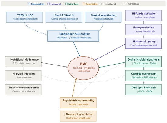
The pathophysiology of BMS is multifactorial and incompletely understood, with several interrelated mechanisms proposed to contribute to symptom generation and maintenance. These encompass peripheral and central neuro-pathic alterations, hormonal and neuroactive steroid dysregulation, nutritional and endocrine deficiencies together with infectious contributors, psychiatric comorbidity, and disturbances of salivary function and composition. Ra-ther than acting in isolation, these mechanisms are increasingly recognized to interact within a convergent network that culminates in the characteristic burning sensation, providing the conceptual framework within which the po-tential role of the oral microbiome—the principal focus of this review—can be situated. An integrated overview of these contributing mechanisms is presented in Figure 1, and each is examined in turn in the subsections below.
Figure 1.
BMS: multifactorial pathophysiology.
2.4.1. Peripheral and Central Neuropathic Mechanisms
The neuropathic basis of BMS was first substantiated histopathologically by Lauria et al. [6], who reported a marked reduction in epithelial and subpapillary nerve fiber densities in tongue biopsies from BMS patients compared with matched controls, providing the first direct evidence of trigeminal small-fiber neuropathy in this disorder. These foundational observations were subsequently corroborated and expanded by a comprehensive systematic review, which cataloged a range of neurobiological abnormalities, including immunohistochemical upregulation of fibers expressing TRPV1, NGF, and P2X3; overexpression of the voltage-gated sodium channel Nav1.7 accompanied by modest downregulation of Nav1.9 mRNA; prolonged blink-reflex latencies; and corneal confocal microscopy findings indicative of peripheral nerve fiber loss [7]. Complementary evidence from quantitative sensory testing (QST) reveals elevated cool-detection thresholds and increased cold-pain thresholds in BMS patients, a somatosensory profile that closely parallels that of established small-fiber neuropathy conditions, including diabetic peripheral neuropathy and fibromyalgia. In parallel with the peripheral neuropathic framework, a nociplastic pain model has been proposed to account for central pain-processing dysregulation observed in a clinically meaningful subset of BMS patients, suggesting that aberrant central sensitization may contribute independently to symptom generation and maintenance [31].
2.4.2. Hormonal and Neuroactive Steroid Dysregulation
The marked female preponderance and the clustering of BMS onset around the peri- and postmenopausal transition implicate gonadal steroid-dependent modulation of oral nociceptive processing as a key pathophysiological contributor. Estrogen receptors are widely expressed across oral mucosal epithelium, salivary gland tissue, and trigeminal sensory neurons, and the precipitous decline in circulating estrogen accompanying menopause is associated with dysregulated synthesis of neuroactive steroids at both mucosal and central nervous system levels, potentially lowering the excitability threshold of trigeminal and glossopharyngeal primary afferents [4,5]. This hormonal vulnerability is further compounded by the effects of chronic psychological stress and anxiety, which dysregulate hypothalamic–pituitary–adrenal (HPA) axis activity and alter adrenal glucocorticoid output, thereby amplifying the underlying neuroendocrine imbalance and potentially perpetuating central sensitization.
2.4.3. Nutritional, Endocrine, and Infectious Contributors
Deficiencies in hematinic micronutrients—specifically vitamin B12, folate, and iron—as well as zinc insufficiency, are disproportionately prevalent in BMS patient cohorts, and symptom improvement following nutritional repletion has been reported in a proportion of affected individuals, lending support to their designation as reversible causes of secondary BMS [9,32]. In subsets of patients presenting with iron deficiency, concomitant hyperhomocysteinemia and serological evidence of gastric parietal cell autoimmunity have also been documented, suggesting an underlying autoimmune or malabsorptive mechanism [32]. An elevated prevalence of Helicobacter pylori infection among BMS patients has led to the hypothesis that this pathogen may indirectly contribute to oral mucosal dysesthesia. The proposed mechanism centers on H. pylori-induced chronic atrophic gastritis, which progressively impairs gastric acid secretion and thereby compromises the luminal absorption of iron and vitamin B12—micronutrients whose deficiency is independently implicated in BMS pathogenesis [32,33]. A secondary pathway involves ectopic oral colonization by H. pylori, which may sustain a chronic, low-grade proinflammatory cytokine environment, potentially sensitizing trigeminal nociceptors at the mucosal level. However, a definitive causal relationship between H. pylori infection and BMS symptomatology has yet to be established, and prospective eradication trials with rigorous symptom endpoints are needed to resolve this question.
2.4.4. Psychiatric Comorbidity
Comorbid anxiety and depression occur at elevated rates among BMS patients, a pattern corroborated by meta-analytic evidence demonstrating statistically robust associations between BMS and both mood disorders [8]. The precise nature of this relationship remains a subject of ongoing debate: psychological distress may precede and contribute to the development of BMS through neuroendocrine and central sensitization mechanisms, may arise as a consequence of chronic pain and functional impairment, or may interact bidirectionally in a self-perpetuating cycle that amplifies both symptom burden and affective dysregulation [8]. Regardless of directionality, the extensively characterized reciprocal relationship between mood state and nociceptive processing—mediated through descending corticolimbic modulatory pathways, hypothalamic–pituitary–adrenal axis dysregulation, and neuroactive metabolites of gut–brain axis origin—provides a compelling mechanistic rationale for treatment approaches that integrate psychological intervention with pharmacological strategies targeting both pain and affective comorbidity [8,31].
2.4.5. Salivary Function and Composition
Despite the absence of clinically overt xerostomia in the majority of BMS patients, converging evidence from multiple independent studies indicates a modest but measurable reduction in unstimulated salivary flow rates in affected cohorts relative to healthy controls (mean 0.11 mL/min versus 0.21 mL/min), with approximately 46% of patients fulfilling established criteria for hyposalivation and a substantial proportion concurrently experiencing subjective oral dryness [29,30,34]. At the biochemical level, salivary concentrations of cortisol and α-amylase are consistently elevated in BMS, a pattern interpreted as a reflection of heightened sympathoadrenal and hypothalamic–pituitary–adrenal axis activation in response to chronic pain and psychological stress; by contrast, salivary cytokine profiles—including IL-2, IL-6, IL-18, and TNF-α—have yielded less reproducible findings across studies, likely attributable to methodological heterogeneity and patient selection differences [35,36]. Emerging evidence from salivary metabolomics has begun to delineate a molecular signature characteristic of BMS, with downregulation of caffeine metabolic pathways and reduced concentrations of tyrosine pathway intermediates—including L-dopa, L-tyrosine, and tyramine—identified as potentially discriminating biochemical features that distinguish BMS patients from unaffected controls [37].
2.5. Current Management
Current BMS management is inherently multimodal and requires individualization according to the patient’s clinical phenotype, comorbidity profile, and predominant pathophysiological mechanism. A comprehensive systematic review of available therapeutic options concluded that topical clonazepam, alpha-lipoic acid at doses of 300–800 mg/day, low-level laser therapy, capsaicin administered topically or as an oral rinse, and cognitive-behavioral therapy (CBT) demonstrate the most consistent evidence for both short- and long-term symptom attenuation, and are generally associated with acceptable tolerability and mild adverse-effect profiles [38]. Combination regimens incorporating clonazepam alongside alpha-lipoic acid have been associated with additive symptomatic benefit, while CBT has demonstrated particularly durable efficacy in patients with prominent comorbid anxiety or depressive disorders, likely through its capacity to modulate descending pain inhibitory pathways and maladaptive pain-related cognitions [26,39]. Nevertheless, no currently available monotherapy or combination regimen reliably achieves complete and sustained remission across the heterogeneous BMS population, underscoring an unmet therapeutic need and providing a strong rationale for investigating biologically targeted interventions—including microbiome-modulating strategies designed to correct underlying oral mucosal and systemic dysbiosis—as adjunctive or alternative treatment approaches. Critically, the limited efficacy of current empirical regimens reflects not only the absence of disease-modifying agents but also a deeper conceptual limitation: the treatment of a mechanistically heterogeneous disorder as though it were a homogeneous syndrome [7,26,38] (Table 1).
Table 1.
Summary of major BMS management modalities, their proposed mechanisms, and current evidence.
2.6. Evolution of BMS Management Paradigms
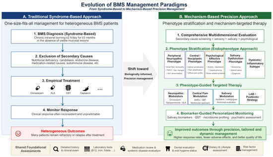
The foregoing limitations of syndrome-based, exclusion-driven management have catalyzed a conceptual shift toward mechanism-based precision medicine in BMS (Figure 2). The traditional paradigm (Figure 2A) applies a uniform diagnostic and therapeutic sequence—secondary-cause exclusion followed by empirical pharmacotherapy with clonazepam, alpha-lipoic acid (ALA), cognitive-behavioral therapy (CBT), or capsaicin—irrespective of the patient’s underlying pathophysiology. This one-size-fits-all approach produces heterogeneous and frequently unsatisfactory outcomes, with a substantial proportion of patients remaining refractory or relapsing after initial treatment [26,38].
Figure 2.
Evolution of BMS management paradigms: from syndrome-based to mechanism-based precision management. (A) The traditional syndrome-based approach applies a uniform diagnostic and empirical treat-ment sequence to all patients, producing heterogeneous outcomes. (B) The emerging mecha-nism-based precision approach stratifies patients into distinct endophenotypes (peripheral neu-ropathic, central/nociplastic, psychological/affective, salivary dysfunction, and dysbi-otic/inflammatory) to guide targeted therapy and biomarker-anchored monitoring.
The emerging mechanism-based precision approach (Figure 2B) proposes replacing uniform empirical treatment with a structured multidimensional evaluation that integrates quantitative sensory testing (QST), salivary function assessment, microbiome profiling, and psychological screening alongside standard secondary-cause exclusion [7,26,38]. On this basis, patients are stratified into distinct endophenotypes: a Peripheral Neuropathic Phenotype characterized by small-fiber neuropathy, TRPV1/Nav channel alterations, and QST abnormalities [6,7]; a Central/Nociplastic Phenotype defined by central sensitization, pain amplification, and diffuse pain profiles [31]; a Psychological/Affective Phenotype with prominent anxiety, depression, catastrophizing, and stress-related symptoms [8,17]; a Salivary Dysfunction Phenotype presenting with xerostomia, altered salivary composition, and dysgeusia [30,34]; and a Dysbiotic/Inflammatory Subtype associated with compositional shifts in the oral microbiome and heightened mucosal inflammatory tone [16,17]. Each endophenotype informs targeted therapeutic selection: neuropathic modulators (clonazepam, gabapentinoids, ALA) for peripheral phenotypes; SNRIs and multimodal CBT for central and psychological phenotypes; sialogogues and hormonal evaluation for salivary phenotypes; and lactic acid bacteria (LAB)/postbiotic strategies for dysbiotic subtypes [38,44,46]. This phenotype-guided approach is further complemented by biomarker-anchored monitoring—incorporating salivary cortisol, inflammatory cytokines, QST, microbiome profiling, and psychiatric assessment—enabling dynamic and responsive management throughout the clinical course [35,36,47]. Shared foundational assessments, including detailed clinical history, targeted laboratory screening (vitamin B12, iron, folate), medication review, dental evaluation, and dietary assessment, remain integral to both paradigms but serve in the precision model as the springboard for individualized stratification rather than as therapeutic endpoints in themselves [9,32]. This evolution from phenotype-agnostic exclusion to mechanism-anchored endophenotyping represents the direction toward which future BMS clinical trial design, biomarker development, and therapeutic innovation—including the oral microbiome-targeting strategies reviewed in subsequent sections—should be systematically oriented [7,25,38].
3. The Oral Microbiome in Health and BMS
3.1. Composition and Site-Specific Architecture of the Healthy Oral Microbiome
Cataloged within the Human Oral Microbiome Database, the healthy oral microbiome encompasses more than 700 phylotypes distributed across five major phyla: Firmicutes (Streptococcus, Veillonella, Granulicatella), Proteobacteria (Neisseria, Haemophilus), Actinobacteria (Rothia, Corynebacterium, Actinomyces), Bacteroidetes (Prevotella, Porphyromonas, Capnocytophaga), and Fusobacteria (Fusobacterium) [12]. Each oral anatomical niche—saliva, tongue dorsum, buccal mucosa, hard palate, and supra- and subgingival plaque—sustains a reproducibly distinct microbial community shaped by local physicochemical gradients and nutrient availability. The tongue dorsum warrants particular emphasis in the BMS context: high-resolution spatial imaging has demonstrated that its microbial architecture consists of structured multispecies consortia anchored to epithelial cell surfaces, with tongue-tropic Streptococcus species (S. salivarius, S. parasanguinis, S. infantis, S. australis) occupying central positions within these biogeographic arrays [13]. Because saliva continuously samples shed cells and planktonic bacteria from all niche communities, it serves as a practical and representative specimen for clinical microbiome investigations [14].
Defining a universally shared “core” oral microbiome has proven challenging because of inter-individual variability, but a panel of genera—Streptococcus, Veillonella, Granulicatella, Neisseria, Haemophilus, Rothia, Fusobacterium, Prevotella, and Porphyromonas—is consistently identified across healthy adults, with aggregate Streptococcus abundances of 12–23% across saliva, tongue, plaque, and oral rinse [14,48].
3.2. Microbiome Dysbiosis in Burning Mouth Syndrome
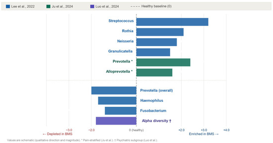
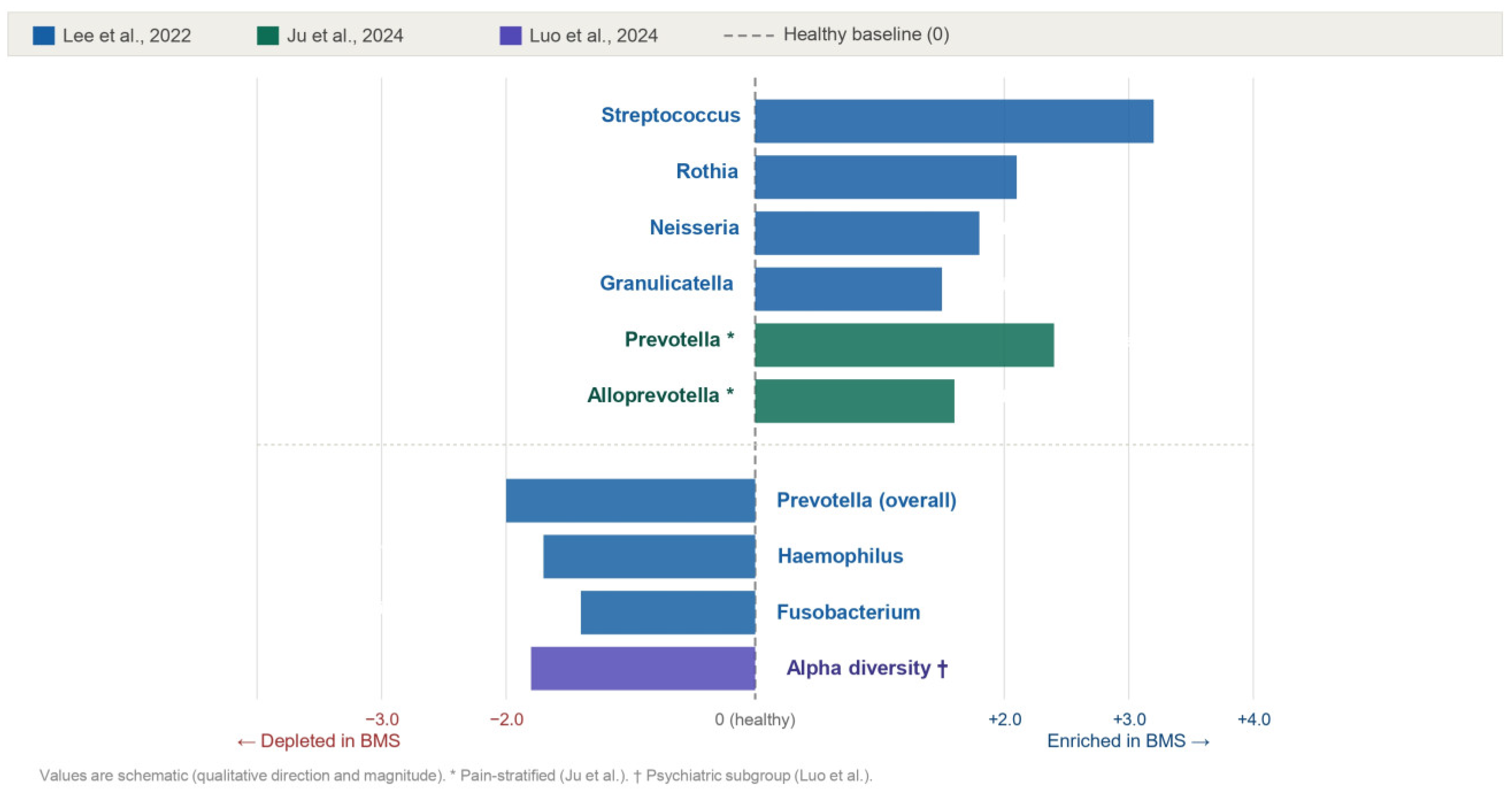
The intersection of BMS and oral microbiome research constitutes a rapidly developing area of inquiry, and accumulating evidence from independent 16S rRNA amplicon sequencing and shotgun metagenomic studies has begun to delineate a recognizable pattern of salivary dysbiosis in affected individuals. In a case–control investigation employing V3–V4 region amplicon sequencing of unstimulated whole saliva obtained from 19 primary BMS patients and 22 healthy volunteers, Lee et al. [16] documented relative enrichment of Streptococcus, Rothia, Neisseria, and Granulicatella in the BMS salivary microbiome, with concurrent depletion of Prevotella, Haemophilus, and Fusobacterium. In a study specifically examining the psychiatric-comorbid BMS subpopulation, Luo et al. [17] identified significantly reduced alpha diversity, as quantified by Shannon and Chao1 indices, accompanied by salivary metabolomic profiles indicative of perturbation in amino acid and nucleotide biosynthetic pathways. Further stratification by pain severity revealed that individuals classified as high-pain BMS harbored higher relative abundances of Prevotella and Alloprevotella than both low-pain BMS patients and healthy controls, suggesting that salivary microbial community composition may track clinical symptom intensity [47]. Importantly, reductions in alpha diversity are not a universal finding across BMS populations; rather, they appear most pronounced in subgroups characterized by concurrent psychiatric comorbidity or elevated pain burden, whereas aggregate case–control analyses yield heterogeneous and sometimes conflicting results [7]. The individual oral microbiome studies in BMS discussed above are summarized in Table 2, and the taxonomic and alpha-diversity shifts identified across these studies are synthesized and compared by genus and study source in Figure 3, which highlights both cross-study recurring patterns and subgroup-specific variations in microbial composition.
Table 2.
Summary of oral microbiome studies in burning mouth syndrome.
Figure 3.
Comparison of oral microbiome composition in healthy individuals and BMS patients [16,17,47].
While a consensus dysbiotic signature remains to be established, the cumulative evidence consistently supports three overarching themes: (i) relative enrichment of Streptococcus is the most reproducible compositional feature of the BMS salivary microbiome across independent cohorts; (ii) microbial compositional shifts exhibit meaningful correlations with both pain intensity and psychiatric comorbidity burden; and (iii) accompanying functional metabolomic disturbances raise the possibility that oral dysbiosis may be associated with altered local concentrations of metabolites capable of modulating trigeminal nociceptor activity [17,37].
It must be emphasized, however, that essentially all available microbiome data in BMS derive from cross-sectional, case–control studies with modest sample sizes. Such designs can establish association but cannot determine the direction of causality, and they do not permit the conclusion that dysbiosis is a cause—rather than a consequence or epiphenomenon—of BMS. Observed microbial differences may equally reflect secondary effects of reduced salivary flow, altered oral pH, medication use, dietary modification, or psychiatric comorbidity that frequently accompany the condition. Accordingly, the associations described above should be interpreted as suspected relationships requiring confirmation through prospective, longitudinal, and interventional studies rather than as evidence of a definitive causal role.
3.3. Candida Species, Oral Candidiasis, and BMS
Candida albicans is the most prevalent fungal commensal in the human oral cavity, with colonization rates ranging from 20% to 60% among immunocompetent adults. Symptomatic oral candidiasis represents a well-established cause of secondary BMS and is of particular diagnostic significance because it can produce a burning sensation clinically indistinguishable from that of primary BMS in the absence of visible mucosal lesions or classical pseudomembranous plaques, thereby necessitating microbiological confirmation via Candida culture or cytological smear as a mandatory component of the routine BMS diagnostic workup [49].
The growing prevalence of multidrug-resistant Candida auris, including its documented capacity for oral mucosal colonization, has substantially intensified interest in non-antifungal therapeutic strategies—among which LAB-based probiotic interventions have emerged as particularly promising candidates—for both the prophylaxis and adjunctive clinical management of oral candidiasis [50,51].
3.4. Salivary Biomarkers, Metabolomics, and Functional Outputs
Extending beyond taxonomic characterization, functional interrogation of the oral microenvironment in BMS yields complementary and mechanistically informative insights. Salivary concentrations of cortisol and α-amylase are reproducibly elevated in BMS patients relative to healthy controls, a biochemical pattern consistent with chronic upregulation of hypothalamic–pituitary–adrenal (HPA) axis activity and heightened sympathoadrenal tone; by contrast, salivary cytokine profiles—encompassing IL-2, IL-6, IL-18, and TNF-α—exhibit considerable inter-study variability, likely reflecting heterogeneity in patient phenotype, sampling protocols, and analytical methodology [35,36]. Untargeted salivary metabolomics has further revealed suppression of caffeine catabolic pathways and dysregulation of tyrosine pathway intermediates in BMS patients compared with controls, pointing toward broader disruptions in aromatic amino acid metabolism that may have downstream consequences for neurotransmitter biosynthesis and mucosal nociceptive signaling [37]. Collectively, these functional biochemical readouts delineate a salivary phenotypic substrate that is both biologically plausible as a target for microbiome-modulating interventions and sufficiently well-characterized to serve as a reference framework against which the effects of LAB-based probiotic administration can be systematically benchmarked in future mechanistic and clinical trials.
4. Lactic Acid Bacteria as Therapeutic Modulators of the Oral Cavity
4.1. General Properties and Taxonomy of LAB Relevant to Oral Health
The term “lactic acid bacteria” refers to a phenotypically heterogeneous and phylogenetically broad grouping of Gram-positive, non-spore-forming, facultatively anaerobic microorganisms that share the defining metabolic characteristic of producing lactic acid as the principal end-product of carbohydrate fermentation. Within the context of oral health, the genera of greatest clinical relevance include the extensively reclassified Lactobacillus sensu lato—a taxon that has undergone major taxonomic reorganization and now encompasses approximately 25 distinct genera, among them Lacticaseibacillus and Limosilactobacillus—alongside Bifidobacterium, Streptococcus (notably S. salivarius, which occupies a prominent ecological role on the tongue dorsum), Lactococcus, and Weissella. Within the oral ecosystem, certain LAB taxa function as autochthonous residents, whereas others are introduced transiently through consumption of fermented dietary products—including yogurt, kefir, kimchi, and doenjang—or via deliberately formulated exogenous probiotic preparations. The vast majority of LAB species used in clinical and experimental research have been granted Generally Recognized as Safe (GRAS) status by the United States Food and Drug Administration, a designation that reflects their extensive history of safe human consumption across diverse cultural and dietary contexts [18,19].
4.2. Antimicrobial Mechanisms
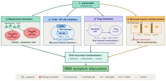
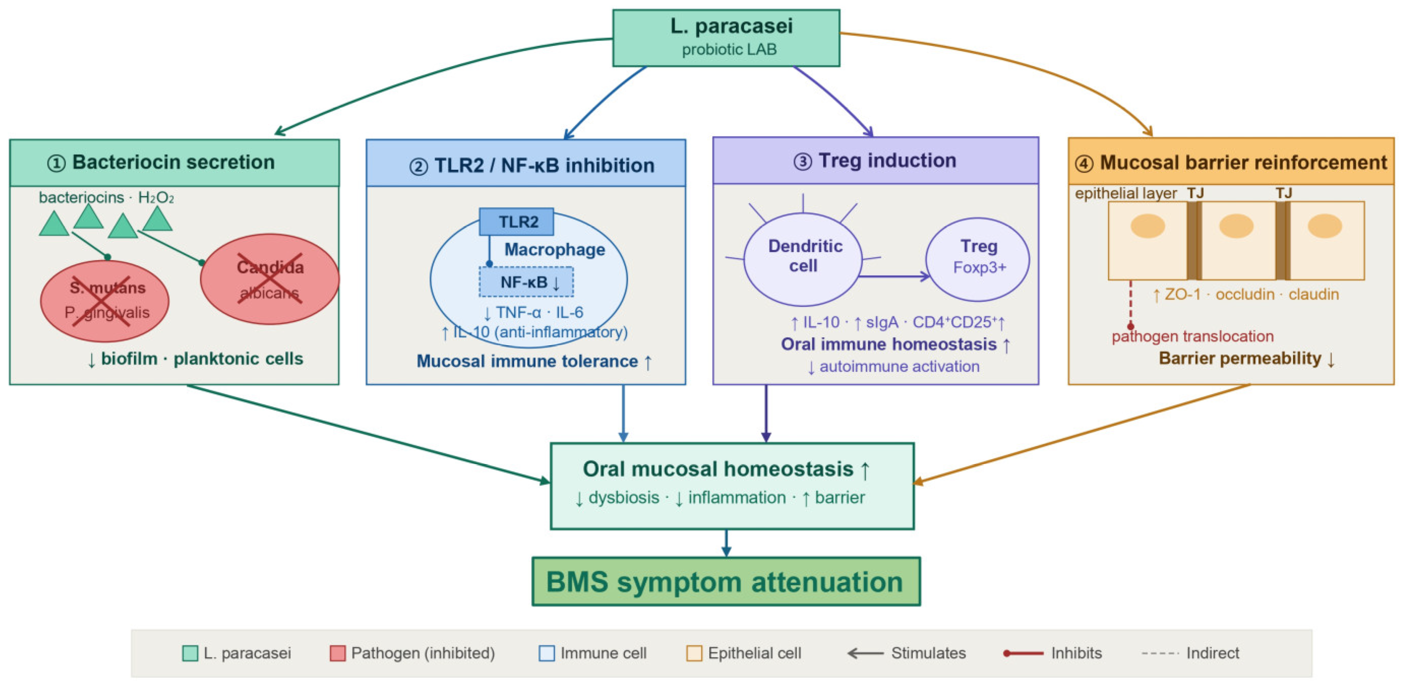
LAB exert antagonistic activity against oral pathogens through several mechanistically distinct and complementary pathways: (i) elaboration of short-chain organic acids—including lactic, acetic, and propionic acid—which reduce local environmental pH and selectively inhibit acid-sensitive pathogenic species; (ii) generation of reactive oxygen species, principally hydrogen peroxide, with broad-spectrum antimicrobial activity; (iii) biosynthesis and secretion of proteinaceous bacteriocins and bacteriocin-like inhibitory substances; (iv) competitive exclusion of pathogens from mucosal and dental surfaces through occupation of adhesion receptors and interference with biofilm establishment; and (v) physical co-aggregation with target pathogens, facilitating their mechanical clearance from the oral cavity [52,53]. Among LAB, Lacticaseibacillus paracasei strains have been shown to elaborate bacteriocins spanning a molecular weight range of 3–56 kDa, with documented inhibitory activity against Porphyromonas gingivalis, Streptococcus mutans, and additional clinically relevant oral pathogens [50]. Notably, in vitro studies have shown that postbiotic preparations derived from L. paracasei 28.4 have demonstrated the capacity to inhibit Candida albicans in both planktonic and sessile biofilm states, including recalcitrant persister cell populations, and additionally exhibit antifungal activity against the emergent multidrug-resistant pathogen Candida auris [51]. Cell-free culture supernatants of Ligilactobacillus salivarius have been shown to attenuate S. mutans biofilm formation by downregulating phosphoenolpyruvate-dependent phosphotransferase systems, ATP-binding cassette (ABC) transporters, two-component regulatory systems, and exopolysaccharide biosynthetic gene clusters; bioactive compounds responsible for these effects include phenyllactic acid, sorbitol, and honokiol [54]. Limosilactobacillus fermentum strains of human oral origin have similarly been reported to suppress S. mutans proliferation and virulence through the combined action of reuterin and organic acid production, further expanding the breadth of the antimicrobial repertoire available within the LAB clade [55]. The multifaceted therapeutic potential of L. paracasei at the oral mucosal interface—encompassing direct pathogen suppression, antifungal activity, and downstream modulation of host mucosal immune responses—is synthesized and schematically illustrated in Figure 4.
Figure 4.
Antimicrobial and immunomodulatory mechanisms of Lacticaseibacillus paracasei.
4.3. Immunomodulatory Effects
LAB modulate both innate and adaptive immune circuitry through engagement of pattern-recognition receptors—including Toll-like receptor 2 (TLR2), TLR9, and nucleotide-binding oligomerization domain (NOD) receptors—thereby driving context-dependent cytokine reprogramming, facilitating the expansion of immunoregulatory T-cell subsets, and enhancing secretory immunoglobulin A (sIgA) production at mucosal surfaces. At the strain-specific level, selected L. paracasei strains have been shown to attenuate lipopolysaccharide-induced secretion of TNF-α and IL-6 from human peripheral blood mononuclear cells through upregulation of endogenous negative regulators of the NF-κB signaling cascade via a TLR2–IRAK4-dependent mechanism, thereby dampening proinflammatory cytokine output without global immunosuppression [56]. In alignment with this anti-inflammatory phenotype, L. paracasei MSMC39-1 has been reported to reduce TNF-α production in intestinal epithelial cell models, while L. paracasei WIS43 significantly attenuated systemic and colonic concentrations of TNF-α, IL-6, and IL-1β in a dextran sulfate sodium-induced murine colitis model [57,58]. L. paracasei strains, including KBL382, have additionally been shown to promote IL-10 secretion and drive expansion of the CD4+CD25+Foxp3+ regulatory T-cell compartment within mesenteric lymph nodes, with postbiotic cell-free fractions of the same strain further augmenting mucosal sIgA output—an immunological effector critical for first-line defense against opportunistic oral pathogens [59,60]. Considered in aggregate, the immunomodulatory properties documented across L. paracasei strains exhibit a high degree of mechanistic complementarity with the immune–neuroendocrine dysregulation characterizing BMS, which encompasses chronically elevated salivary cortisol and α-amylase, aberrant proinflammatory cytokine profiles, and a permissive mucosal microenvironment that facilitates pathobiont expansion and sustained dysbiosis.
4.4. Lacticaseibacillus paracasei: Whole-Genome Insights and Probiotic Profile
The comparative genomics of L. paracasei has advanced considerably over the past decade. The foundational pan-genome analysis conducted by Smokvina et al. [23] delineated approximately 4200 orthologous gene clusters across the species, with each genome encoding a conserved core of roughly 1800 genes within a total of 2800–3100 protein-coding sequences. Subsequent whole-genome characterization of strains recovered from ecologically diverse sources—including dairy products, fermented vegetables, palm sap, kefir, and human breast milk—has consistently identified a repertoire of genomic determinants underlying key probiotic-relevant phenotypes: gastrointestinal stress tolerance, mediated by bile salt hydrolase and acid-responsive chaperone systems; mucosal adhesion capacity, conferred by sortase-dependent pilus biosynthesis loci, mucus-binding surface proteins, and exopolysaccharide biosynthetic gene clusters; antimicrobial activity, encoded by sactipeptide and class II bacteriocin operons with specificity against P. gingivalis and S. mutans; and broad-spectrum carbohydrate catabolic versatility, which collectively reflect the remarkable ecological plasticity of the species across nutritionally heterogeneous environmental niches [23,24,61]. Systematic genomic safety assessment has reproducibly confirmed the absence of horizontally transferable antibiotic resistance determinants and recognized virulence factor genes across examined strains, providing robust genomic corroboration for the GRAS designation assigned to orally administered L. paracasei formulations and substantiating their suitability for human clinical application [24]. The probiotic credentials of L. paracasei relevant to oral health applications are summarized in Table 3.
Table 3.
Summary of the probiotic credentials of Lacticaseibacillus paracasei relevant to oral health applications.
4.5. Clinical Evidence in BMS and Related Oral Conditions
In interpreting the therapeutic potential of LAB, it is important to distinguish the level of evidence underlying each claim, as the supporting data span a hierarchy of translational maturity. The antimicrobial and immunomodulatory mechanisms described in Section 4.2 and Section 4.3 are derived predominantly from in vitro experiments (e.g., bacteriocin assays, biofilm and postbiotic studies, and cell-culture immunoassays) and, to a lesser extent, from animal models. Evidence of clinical efficacy in humans is currently confined largely to oral conditions adjacent to BMS—such as periodontitis, oral candidiasis, and halitosis—whereas direct interventional evidence in BMS populations themselves remains scarce and preliminary. The distinction between these tiers of evidence—in vitro, animal, related clinical conditions, and BMS-specific clinical data—is made explicit below and should be borne in mind when appraising the translational readiness of LAB-based strategies for BMS. The clinical evidence for LAB-based interventions in BMS and related oral conditions discussed in this section in summarized in Table 4.
Table 4.
Summary of clinical evidence for LAB-based interventions in BMS and related oral conditions.
Direct evidence from randomized clinical trials evaluating LAB-based interventions specifically in BMS remains limited, though available data are mechanistically plausible and clinically encouraging. In the most methodologically rigorous trial to date, Loncar-Brzak et al. [44] allocated 80 BMS patients across four parallel arms—B-vitamin supplementation, oral probiotic lozenges containing Limosilactobacillus reuteri Prodentis, low-level laser therapy, or provision of verbal and written information alone—and documented statistically significant improvements in Oral Health Impact Profile-14 (OHIP-14) quality-of-life scores in both the probiotic and laser therapy arms at one month of follow-up, with no treatment-related adverse events recorded in either group. Complementary and mechanistically supportive evidence is available from a growing body of clinical research examining LAB interventions in conditions of adjacent oral pathology: L. reuteri Prodentis lozenges have demonstrated efficacy in reducing clinical indices of gingival inflammation and periodontal pocket depth in patients with chronic periodontitis [45,63,64]; L. rhamnosus, L. acidophilus, and Streptococcus salivarius K12 have each been shown to attenuate oral Candida colonization burden in both clinical cohort and experimental model settings [65,66,67]; and multi-strain formulations combining L. gasseri with L. paracasei have significantly reduced salivary hydrogen sulfide concentrations and total volatile sulfur compound levels in patients with halitosis [68]. Considered collectively, these findings establish a coherent clinical-translational framework that provides both biological rationale and preliminary evidence of efficacy for the design and conduct of BMS-specific, biomarker-anchored randomized trials targeting L. paracasei strains as candidate therapeutic agents.
5. Mechanistic Links: From Oral Dysbiosis to Neuropathic Pain
5.1. Microbe–Immune–Neural Crosstalk in the Oral Mucosa
Communication between the oral microbial community and the trigeminal somatosensory system operates through multiple receptor- and metabolite-dependent pathways. Lipopolysaccharides shed by Gram-negative periodontal pathogens, such as Porphyromonas gingivalis, bind TLR4 on trigeminal nociceptors, stimulating calcitonin gene-related peptide (CGRP) release and NF-κB nuclear translocation; these events heighten neuronal excitability and, over time, may potentially contribute to a chronic pain state [7,69]. Additional pathogenic microbial-derived signals—including N-formyl peptides and pore-forming cytolysins—sensitize TRPV1 and allied ion channels, potentiating nociceptor firing. In contrast, fermentative metabolites, including short-chain fatty acids (SCFAs), indole derivatives, and gamma-aminobutyric acid (GABA), exert opposing anti-nociceptive and anti-inflammatory effects [70].
5.2. Short-Chain Fatty Acids, GABA, and the Gut–Brain–Oral Axis
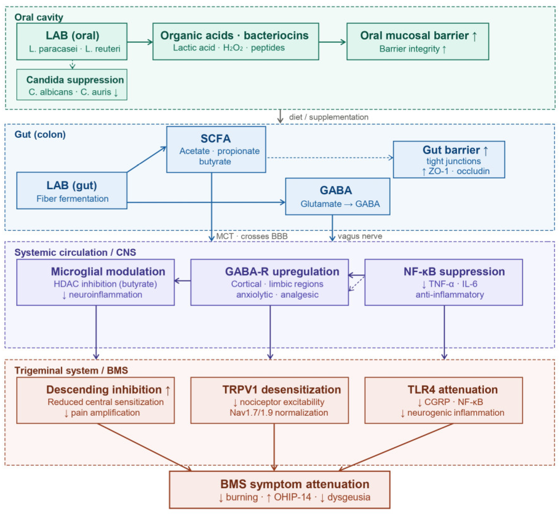
The three principal SCFAs—acetate, propionate, and butyrate—are generated by LAB and allied fermentative bacteria through two anatomically distinct processes: salivary carbohydrate catabolism within the oral biofilm, and dietary fiber fermentation in the colonic lumen. These bioactive molecules transduce their effects through the G protein-coupled free fatty acid receptors FFAR2 (GPR43) and FFAR3 (GPR41), which are expressed on enteroendocrine cells, circulating immune cells, and peripheral sensory neurons. Among the three, acetate and butyrate are capable of traversing the blood–brain barrier via monocarboxylate transporter-mediated uptake, whereupon they modulate central neuroimmune tone by inhibiting histone deacetylase (HDAC) activity and thereby reshaping microglial activation phenotype [70,71]. At the epithelial mucosal surface, SCFAs transcriptionally upregulate structural tight junction proteins—including claudins and occludin—reinforcing paracellular barrier integrity in both the intestinal epithelium and, by anatomical extension, the oral mucosa [62]. Through a mechanistically independent pathway, select Lactobacillus strains enzymatically decarboxylate glutamate to produce GABA via glutamate decarboxylase; prolonged colonization with such strains has been shown to remodel GABA receptor subunit expression in cortical and limbic brain regions, yielding quantifiable anxiolytic and antinociceptive behavioral outcomes in rodent models [72,73].
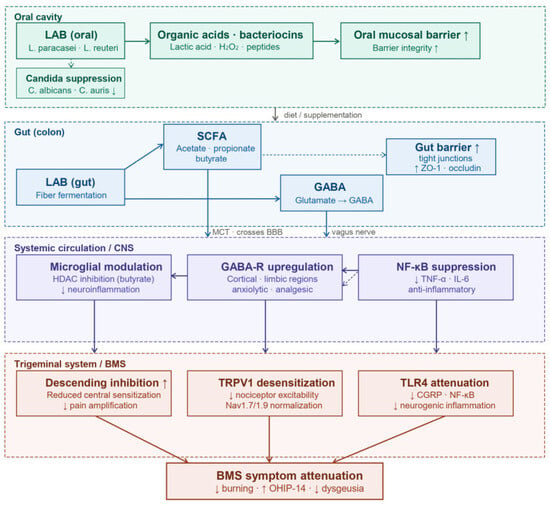
Within the pathophysiological context of BMS, these mechanistic threads converge to support a biologically coherent oral–gut–brain axis model: dysbiotic compositional shifts in the BMS salivary microbiome may collectively diminish local SCFA and GABA biosynthetic output, compromise trigeminal mucosal barrier integrity, amplify TLR-mediated peripheral nociceptor sensitization, and ultimately may be associated with the establishment and maintenance of central sensitization—each of these steps representing a discrete and potentially tractable target for LAB-based therapeutic intervention (as synthesized in Figure 5). Extending this framework to central nervous system endpoints, Siviero et al. [74] have additionally implicated olfactory bulb neuroplasticity in BMS pathophysiology, identifying a CNS-level target that may itself be amenable to modulation through systemic and mucosal microbial inputs.
Figure 5.
The Oral–Gut–Brain Axis in BMS: A systemic framework for microbial-mediated pain modulation.
5.3. LAB-Derived Metabolites as Anti-Nociceptive Agents
Beyond SCFAs and GABA, a broader repertoire of LAB-derived bioactive compounds—encompassing conjugated linoleic acid (CLA), strain-specific exopolysaccharides, indole-3-aldehyde, phenyllactic acid, and ribosomally synthesized bacteriocins—has been shown to possess antinociceptive, anti-inflammatory, and immune-tolerogenic properties through mechanistically distinct pathways [75,76,77]. Among LAB species, L. paracasei strains are particularly notable for their capacity to produce CLA and structurally complex exopolysaccharides at comparatively high yields, both of which have independently demonstrated anti-inflammatory activity in experimental models at the mucosal and systemic levels [54]. The aggregate of these microbially elaborated metabolic outputs constitutes a pharmacologically tractable postbiotic armamentarium—one that, if appropriately formulated, could be deployed therapeutically in BMS with a potentially favorable safety profile, circumventing the theoretical engraftment risks and regulatory complexities inherent to live-organism probiotic administration.
6. Future Directions and Knowledge Gaps
6.1. Limitations of the Current Evidence
Before outlining future directions, it is essential to critically appraise the limitations of the evidence synthesized in this review, as these constraints temper the strength of the conclusions that can presently be drawn. First, there is substantial heterogeneity in the definition and diagnostic criteria of BMS across studies. Although the ICHD-3 and ICOP frameworks have improved nosological consistency, many of the microbiome and interventional studies cited here predate or did not uniformly apply these criteria, and they vary in their distinction between primary and secondary BMS. This diagnostic heterogeneity limits the comparability of findings across cohorts and may partly account for the inconsistent microbial signatures reported. Second, the oral microbiome studies in BMS are few in number and constrained by small sample sizes, with most enrolling fewer than 50 participants per group; such studies are vulnerable to type II error, inflated effect-size estimates, and limited generalizability. Methodological variability among these studies—including differences in sampling site (saliva, tongue dorsum, mucosal swab), DNA extraction protocols, sequencing platforms, hypervariable region targeted, and bioinformatic pipelines—further complicates cross-study comparison and meta-analytic synthesis. Third, the mechanistic evidence supporting LAB, and L. paracasei in particular, is derived predominantly from in vitro experiments and animal models, or extrapolated from other oral and systemic conditions such as periodontitis, oral candidiasis, and inflammatory bowel disease; direct clinical evidence in BMS populations remains scarce. The few interventional studies conducted in BMS are limited methodologically—most notably, the randomized trial by Loncar-Brzak et al. [44] combined probiotics with multiple co-interventions (B vitamins, low-level laser therapy, and verbal/written information) and lacked a true placebo-controlled probiotic arm, precluding attribution of any benefit specifically to the probiotic component. Fourth, the cross-sectional and observational design of nearly all available microbiome studies precludes determination of causal direction; it remains unresolved whether dysbiosis contributes to BMS pathogenesis, arises secondary to altered salivary composition, medication use, or dietary changes in affected patients, or reflects uncontrolled confounding by factors such as age, sex, menopausal status, smoking, and psychiatric comorbidity. Collectively, these limitations indicate that the proposed associations between oral dysbiosis and BMS, and the therapeutic rationale for LAB, should be regarded as hypothesis-generating rather than established, and interpreted with appropriate caution.
6.2. Priority Research Directions
Despite encouraging mechanistic and clinical signals, the BMS–microbiome–LAB triangle remains incompletely explored. We identify five priority research directions:
First, adequately powered, randomized, double-blind, placebo-controlled trials of strain-specific LAB interventions—particularly L. paracasei strains validated by whole-genome sequencing—are needed in well-characterized BMS cohorts stratified by IASP somatosensory phenotype, psychiatric comorbidity, and microbiome baseline. Endpoint panels should include pain intensity (VAS, BPI), oral health-related quality of life (OHIP-14), salivary flow, biomarker panels (cortisol, α-amylase, cytokines), and 16S/shotgun microbiome characterization at baseline and follow-up.
Second, the current evidence base is predominantly cross-sectional and associative; prospective longitudinal studies and interventional designs are needed to establish the directionality and causal relevance of oral dysbiosis in BMS pathophysiology. Mechanistic studies should clarify the bidirectional relationships between oral dysbiosis and trigeminal small-fiber neuropathy in BMS, leveraging tongue biopsy with intra-epidermal nerve fiber density quantification, corneal confocal microscopy, and matched salivary metabolomics.
Third, the role of Candida species—particularly emerging multidrug-resistant species such as C. auris—in BMS phenotypes warrants systematic re-investigation using species- and strain-level molecular identification, with parallel evaluation of L. paracasei postbiotic preparations as antifungal-sparing interventions.
Fourth, the oral–gut–brain axis remains a hypothesis-rich but evidence-limited domain in BMS. Integrated multi-omics studies (oral and stool microbiome, salivary and serum metabolomics, neuroimaging) are required to move from association to mechanism.
Fifth, postbiotic and engineered-LAB approaches—exploiting defined bacteriocins, exopolysaccharides, and metabolite preparations—may offer pharmacologically tractable alternatives to live-organism administration, with fewer safety concerns and greater regulatory clarity.
7. Conclusions
Burning mouth syndrome remains a clinically challenging, mechanistically heterogeneous chronic orofacial pain disorder for which conventional pharmacotherapy delivers only partial relief. Converging associative evidence from salivary microbiome surveys and metabolomic profiling, contextualized within established frameworks of neuropathic pain neurobiology and mucosal immunology, raises the hypothesis that oral microbial dysbiosis constitutes a biologically plausible and potentially modifiable contributor to BMS pathophysiology. It bears emphasis that the existing microbiome data are predominantly cross-sectional and associative in nature; direct evidence for a causal role of oral dysbiosis in BMS etiology remains to be established through longitudinal and interventional study designs. Within this evidence landscape, targeted modulation of the oral microbiome with lactic acid bacteria—particularly L. paracasei—may offer a mechanistically coherent and clinically testable therapeutic avenue. Whole-genome sequencing of L. paracasei strains, combined with a robust evidence base of antimicrobial, immunomodulatory, and barrier-protective properties, positions L. paracasei as a compelling candidate for next-generation BMS clinical trials. Realization of this therapeutic potential will require strain-specific, biomarker-anchored, adequately powered randomized controlled trials conducted within an integrative oral–gut–brain framework—investigations that this review is intended to motivate and inform.
Author Contributions
Conceptualization, Y.-J.R.; methodology, Y.-J.R. and J.-A.Y.; investigation, Y.-J.R., J.-A.Y. and Y.J.J.; writing—original draft preparation, Y.-J.R.; writing—review and editing, Y.-J.R., J.-A.Y. and Y.J.J.; visualization, Y.-J.R.; supervision, Y.-J.R. All authors have read and agreed to the published version of the manuscript.
Funding
This research received no external funding.
Institutional Review Board Statement
Not applicable. This review article is based entirely on previously published literature and did not involve human subjects or animal experiments.
Informed Consent Statement
Not applicable.
Data Availability Statement
No new data were created or analyzed in this study. Data sharing is not applicable to this article.
Acknowledgments
The authors thank the faculty of the Department of Otorhinolaryngology–Head and Neck Surgery and Kangwon National University Hospital for their collegial support during the preparation of this manuscript.
Conflicts of Interest
The authors declare no conflicts of interest.
References
- Headache Classification Committee of the International Headache Society (IHS). The International Classification of Headache Disorders, 3rd edition. Cephalalgia 2018, 38, 1–211. [Google Scholar] [CrossRef] [Scilit] [PubMed]
- International Classification of Orofacial Pain, 1st edition (ICOP). Cephalalgia 2020, 40, 129–221. [CrossRef] [Scilit] [PubMed]
- Wu, S.; Zhang, W.; Yan, J.; Noma, N.; Young, A.; Yan, Z. Worldwide prevalence estimates of burning mouth syndrome: A systematic review and meta-analysis. Oral Dis. 2022, 28, 1431–1440. [Google Scholar] [PubMed]
- Dahiya, P.; Kamal, R.; Kumar, M.; Gupta, R. Burning mouth syndrome and menopause. Int. J. Prev. Med. 2013, 4, 15–20. [Google Scholar] [PubMed]
- Woda, A.; Dao, T.; Gremeau-Richard, C. Steroid dysregulation and stomatodynia (burning mouth syndrome). J. Orofac. Pain 2009, 23, 202–210. [Google Scholar] [PubMed]
- Lauria, G.; Majorana, A.; Borgna, M.; Lombardi, R.; Penza, P.; Padovani, A. Trigeminal small-fiber sensory neuropathy causes burning mouth syndrome. Pain 2005, 115, 332–337. [Google Scholar] [CrossRef] [Scilit] [PubMed]
- Kouri, M.; Adamo, D.; Vardas, E.; Georgaki, M.; Canfora, F.; Mignogna, M.D.; Nikitakis, N. Small Fiber Neuropathy in Burning Mouth Syndrome: A Systematic Review. Int. J. Mol. Sci. 2024, 25, 11442. [Google Scholar] [CrossRef] [Scilit] [PubMed]
- de Lima-Souza, R.A.; Perez-de-Oliveira, M.E.; Normando, A.G.C.; Louredo, B.V.R.; Mariano, F.V.; Farag, A.M.; Santos-Silva, A.R. Clinical and epidemiological profile of burning mouth syndrome patients following the International Headache Society classification: A systematic review and meta-analysis. Oral Surg. Oral Med. Oral Pathol. Oral Radiol. 2024, 137, 119–135. [Google Scholar] [CrossRef] [Scilit] [PubMed]
- Gholizadeh, N.; Sheykhbahaei, N. Micronutrients status as a contributing factor in secondary burning mouth syndrome: A review of the literature. Health Sci. Rep. 2024, 7, e1906. [Google Scholar] [CrossRef] [Scilit] [PubMed]
- Tan, H.L.; Smith, J.G.; Hoffmann, J.; Renton, T. A systematic review of treatment for patients with burning mouth syndrome. Cephalalgia 2022, 42, 128–161. [Google Scholar] [PubMed]
- Sangalli, L.; Mirfarsi, S.; Kramer, J.M.; Eisa, E.; Miller, C.S. Managing Burning Mouth Syndrome: Current and Future Directions. Drugs 2025, 85, 1109–1131. [Google Scholar] [CrossRef] [Scilit] [PubMed]
- Dewhirst, F.E.; Chen, T.; Izard, J.; Paster, B.J.; Tanner, A.C.R.; Yu, W.H. The human oral microbiome. J. Bacteriol. 2010, 192, 5002–5017. [Google Scholar] [CrossRef] [Scilit] [PubMed]
- Wilbert, S.A.; Mark Welch, J.L.; Borisy, G.G. Spatial ecology of the human tongue dorsum microbiome. Cell Rep. 2020, 30, 4003–4015.e4003. [Google Scholar] [CrossRef] [Scilit] [PubMed]
- Sedghi, L.; DiMassa, V.; Harrington, A.; Lynch, S.V.; Kapila, Y.L. The oral microbiome: Role of key organisms and complex networks in oral health and disease. Periodontol. 2000 2021, 87, 107–131. [Google Scholar]
- La Rosa, G.R.M.; Gattuso, G.; Pedulla, E.; Rapisarda, E.; Nicolosi, D.; Salmeri, M. Association of oral dysbiosis with oral cancer development. Oncol. Lett. 2020, 19, 3045–3058. [Google Scholar] [CrossRef] [Scilit] [PubMed]
- Lee, B.M.; Park, J.W.; Jo, J.H.; Oh, B.; Chung, G. Comparative analysis of the oral microbiome of burning mouth syndrome patients. J. Oral Microbiol. 2022, 14, 2052632. [Google Scholar] [CrossRef] [Scilit] [PubMed]
- Luo, S.; Lou, F.; Yan, L.; Dong, Y.; Zhang, Y.; Liu, Y.; Ji, P.; Jin, X. Comprehensive analysis of the oral microbiota and metabolome change in patients of burning mouth syndrome with psychiatric symptoms. J. Oral. Microbiol. 2024, 16, 2362313. [Google Scholar] [CrossRef] [Scilit] [PubMed]
- Sirichoat, A.; Flórez, A.B.; Vázquez, L.; Buppasiri, P.; Panya, M.; Lulitanond, V.; Mayo, B. Antibiotic susceptibility profiles of lactic acid bacteria from the human vagina and genetic basis of acquired resistances. Int. J. Mol. Sci. 2020, 21, 2594. [Google Scholar] [CrossRef] [Scilit] [PubMed]
- Tham, C.S.C.; Peh, K.K.; Bhat, R.; Liong, M.T. Probiotic properties of bifidobacteria and lactobacilli isolated from local dairy products. Ann. Microbiol. 2012, 62, 1079–1087. [Google Scholar] [CrossRef] [Scilit]
- Homayouni Rad, A.; Pourjafar, H.; Mirzakhani, E. A comprehensive review of the application of probiotics and postbiotics in oral health. Front Cell Infect. Microbiol. 2023, 13, 1120995. [Google Scholar] [CrossRef] [Scilit] [PubMed]
- Anumudu, C.K.; Miri, T.; Onyeaka, H. Multifunctional applications of lactic acid bacteria: Enhancing safety, quality, and nutritional value in foods and fermented beverages. Foods 2024, 13, 3714. [Google Scholar] [CrossRef] [Scilit] [PubMed]
- Zheng, J.; Wittouck, S.; Salvetti, E.; Franz, C.; Harris, H.M.B.; Mattarelli, P. A taxonomic note on the genus Lactobacillus: Description of 23 novel genera, emended description of the genus Lactobacillus Beijerinck 1901, and union of Lactobacillaceae and Leuconostocaceae. Int. J. Syst. Evol. Microbiol. 2020, 70, 2782–2858. [Google Scholar] [CrossRef] [Scilit] [PubMed]
- Smokvina, T.; Wels, M.; Polka, J.; Chervaux, C.; Brisse, S.; Boekhorst, J. Lactobacillus paracasei comparative genomics: Towards species pan-genome definition and exploitation of diversity. PLoS ONE 2013, 8, e68731. [Google Scholar] [CrossRef] [Scilit] [PubMed]
- Lee, H.B.; Bang, W.Y.; Shin, G.R.; Jeon, H.J.; Jung, Y.H.; Yang, J. Isolation, Characterization, and Safety Evaluation of the Novel Probiotic Strain Lacticaseibacillus paracasei IDCC 3401 via Genomic and Phenotypic Approaches. Microorganisms 2023, 12, 85. [Google Scholar] [CrossRef] [Scilit] [PubMed]
- Chmieliauskaite, M.; Stelson, E.A.; Epstein, J.B.; Klasser, G.D.; Farag, A.; Carey, B.; Albuquerque, R.; Mejia, L.; Ariyawardana, A.; Nasri-Heir, C.; et al. Consensus agreement to rename burning mouth syndrome and improve ICD-11 disease criteria: An international Delphi study. Pain 2021, 162, 2548–2557. [Google Scholar] [CrossRef] [Scilit] [PubMed]
- Tan, H.L.; Renton, T. Burning mouth syndrome: An update. Cephalalgia Rep. 2020, 3, 2515816320970143. [Google Scholar] [CrossRef] [Scilit]
- Williams, L.D.; Zis, P. COVID-19-Related Burning Eye Syndrome and Burning Mouth Syndrome: A Systematic Review and Meta-analysis. Pain Ther. 2023, 12, 621–630. [Google Scholar] [CrossRef] [Scilit] [PubMed]
- Patel, D.; Louca, C.; Machuca Vargas, C. Oral manifestations of long COVID and the views of healthcare professionals. Br. Dent. J. 2024, 236, 111–116. [Google Scholar] [CrossRef] [Scilit] [PubMed]
- López-Jornet, P.; Camacho-Alonso, F.; Andujar-Mateos, P. A prospective, randomized study on the efficacy of tongue protector in patients with burning mouth syndrome. Oral Dis. 2014, 20, 692–699. [Google Scholar]
- Poon, R.; Su, N.; Ching, V.; Darling, M.; Grushka, M. Reduction in unstimulated salivary flow rate in burning mouth syndrome. Br. Dent. J. 2014, 217, E14. [Google Scholar] [CrossRef] [Scilit] [PubMed]
- Pinto-Pardo, N. Redefining burning mouth syndrome: A nociplastic pain disorder? J. Dent. Sci. 2025, 20, 1382–1383. [Google Scholar] [CrossRef] [Scilit] [PubMed]
- Lin, H.P.; Wang, Y.P.; Chen, H.M.; Kuo, Y.S.; Lang, M.J.; Sun, A. Significant association of hematinic deficiencies and high blood homocysteine levels with burning mouth syndrome. J. Formos. Med. Assoc. 2013, 112, 319–325. [Google Scholar] [CrossRef] [Scilit] [PubMed]
- Hershko, C.; Skikne, B. Pathogenesis and management of iron deficiency anemia: Emerging role of celiac disease, helicobacter pylori, and autoimmune gastritis. Semin Hematol. 2009, 46, 339–350. [Google Scholar] [CrossRef] [Scilit] [PubMed]
- Canfora, F.; Calabria, E.; Spagnuolo, G.; Coppola, N.; Armogida, N.G.; Mazzaccara, C.; Solari, D.; D’Aniello, L.; Aria, M.; Pecoraro, G.; et al. Salivary Complaints in Burning Mouth Syndrome: A Cross Sectional Study on 500 Patients. J. Clin. Med. 2023, 12, 5561. [Google Scholar] [CrossRef] [Scilit] [PubMed]
- Barry, A.; O’Halloran, K.D.; McKenna, J.P.; McCreary, C.; Downer, E.J. Plasma IL-8 signature correlates with pain and depressive symptomatology in patients with burning mouth syndrome: Results from a pilot study. J. Oral. Pathol. Med. 2018, 47, 158–165. [Google Scholar] [CrossRef] [Scilit] [PubMed]
- Jang, S.; Kim, J.E.; Lee, Y.H.; Jung, W. Changes in salivary biomarkers of burning mouth syndrome patients after clonazepam treatment. J. Oral Facial Pain Headache 2024, 38, 111–118. [Google Scholar] [CrossRef] [Scilit] [PubMed]
- Ye, L.; Dai, Q.; Hou, F.; Wu, C.; Qiu, X.; Yuan, P.; Chen, F.; Meng, Y.; Feng, X.; Jiang, L. Salivary metabolomics of burning mouth syndrome: A cross-sectional study. Arch. Oral Biol. 2022, 144, 105550. [Google Scholar] [CrossRef] [Scilit]
- Rossetti, A.; Teixeira, A.; Milhazes, N. Efficacy of different therapeutic options for pain relief and treatment of burning mouth syndrome: A systematic review. Clin. Oral Investig. 2025, 29, 551. [Google Scholar] [CrossRef] [Scilit] [PubMed]
- Shim, Y. Treatment for burning mouth syndrome: A clinical review. J. Oral Med. Pain 2023, 48, 11–22. [Google Scholar] [CrossRef] [Scilit]
- Heckmann, S.M.; Kirchner, E.; Grushka, M.; Wichmann, M.G.; Hummel, T. A double-blind study on clonazepam in patients with burning mouth syndrome. Laryngoscope 2012, 122, 813–816. [Google Scholar] [CrossRef] [Scilit] [PubMed]
- Femiano, F.; Scully, C. Burning mouth syndrome: Double blind controlled study of alpha-lipoic acid (thioctic acid) therapy. J. Oral Pathol. Med. 2002, 31, 267–269. [Google Scholar] [CrossRef] [Scilit] [PubMed]
- Banik, S.; Ghosh, A.; Sato, H.; Onoue, S. The efficacy of alpha-lipoic acid in the management of burning mouth syndrome: An updated systematic review of randomized controlled clinical trials. Health Sci. Rep. 2023, 6, e1186. [Google Scholar] [CrossRef] [Scilit] [PubMed]
- Silvestre, F.J.; Silvestre-Rangil, J.; Tamarit-Santafé, C.; Bautista, D. Application of a capsaicin rinse in the treatment of burning mouth syndrome. Med. Oral Patol. Oral Cir. Bucal 2012, 17, e1–e4. [Google Scholar] [CrossRef] [Scilit] [PubMed]
- Loncar-Brzak, B.; Skrinjar, I.; Brailo, V.; Vidovic-Juras, D.; Sumilin, L.; Andabak-Rogulj, A. Burning Mouth Syndrome (BMS)-Treatment with Verbal and Written Information, B Vitamins, Probiotics, and Low-Level Laser Therapy: A Randomized Clinical Trial. Dent. J. 2022, 10, 44. [Google Scholar] [CrossRef] [Scilit] [PubMed]
- Schlagenhauf, U.; Rehder, J.; Gelbrich, G.; Jockel-Schneider, Y. Consumption of Lactobacillus reuteri-containing lozenges improves periodontal health in navy sailors at sea: A randomized controlled trial. J. Periodontol. 2020, 91, 1328–1338. [Google Scholar] [CrossRef] [Scilit] [PubMed]
- Tanzer, J.M.; Thompson, A.; Lang, C.; Cooper, B.; Hareng, L.; Gamer, A. Caries inhibition by and safety of Lactobacillus paracasei DSMZ16671. J. Dent. Res. 2010, 89, 921–926. [Google Scholar] [CrossRef] [Scilit] [PubMed]
- Ju, H.M.; Ahn, Y.W.; Ok, S.M.; Jeong, S.H.; Na, H.S.; Chung, J. Distinctive salivary oral microbiome in patients with burning mouth syndrome depending on pain intensity compared to healthy subjects. J. Dent. Sci. 2024, 19, 2174–2182. [Google Scholar]
- Zaura, E.; Keijser, B.J.F.; Huse, S.M.; Crielaard, W. Defining the healthy “core microbiome” of oral microbial communities. BMC Microbiol. 2009, 9, 259. [Google Scholar] [CrossRef] [Scilit] [PubMed]
- Terai, H.; Shimahara, M. Tongue pain: Burning mouth syndrome vs Candidaassociated lesion. Oral Dis. 2007, 13, 440–442. [Google Scholar] [CrossRef] [Scilit] [PubMed]
- Rossoni, R.D.; de Barros, P.P.; de Alvarenga, J.A.; Ribeiro, F.D.C.; Velloso, M.D.S.; Fuchs, B.B. Antifungal activity of clinical Lactobacillus strains against Candida albicans biofilms: Identification of potential probiotic candidates to prevent oral candidiasis. Biofouling 2018, 34, 212–225. [Google Scholar] [CrossRef] [Scilit] [PubMed]
- Rossoni, R.D.; de Barros, P.P.; Mendonca, I.D.C.; Medina, R.P.; Silva, D.H.S.; Fuchs, B.B.; Junqueira, J.C.; Mylonakis, E. The Postbiotic Activity of Lactobacillus paracasei 28.4 Against Candida auris. Front Cell Infect. Microbiol. 2020, 10, 397. [Google Scholar] [CrossRef] [Scilit] [PubMed]
- Pangsomboon, K.; Bansal, S.; Martin, G.P.; Suntinanalert, P.; Kaewnopparat, S.; Srichana, T. Further characterization of a bacteriocin produced by Lactobacillus paracasei HL32. J. Appl. Microbiol. 2009, 106, 1928–1940. [Google Scholar] [CrossRef] [Scilit] [PubMed]
- Lozo, J.; Jovcic, B.; Kojic, M.; Dalgalarrondo, M.; Chobert, J.M.; Haertle, T.; Topisirovic, L. Molecular characterization of a novel bacteriocin and an unusually large aggregation factor of Lactobacillus paracasei subsp. paracasei BGSJ2-8, a natural isolate from homemade cheese. Curr. Microbiol. 2007, 55, 266–271. [Google Scholar] [CrossRef] [Scilit] [PubMed]
- Ma, N.; Yang, W.; Chen, B.; Bao, M.; Li, Y.; Wang, M.; Yang, X.; Liu, J.; Wang, C.; Qiu, L. Exploration of the primary antibiofilm substance and mechanism employed by Lactobacillus salivarius ATCC 11741 to inhibit biofilm of Streptococcus mutans. Front Cell Infect. Microbiol. 2025, 15, 1535539. [Google Scholar] [CrossRef] [Scilit] [PubMed]
- Park, D.Y.; Hwang, J.; Kim, Y.; Lee, D.; Kim, Y.Y.; Kim, H.S.; Hwang, I. Antimicrobial activity of Limosilactobacillus fermentum strains isolated from the human oral cavity against Streptococcus mutans. Sci. Rep. 2023, 13, 7969. [Google Scholar] [CrossRef] [Scilit] [PubMed]
- Sun, K.Y.; Xu, D.H.; Xie, C.; Plummer, S.; Tang, J.; Yang, X.F.; Ji, X.H. Lactobacillus paracasei modulates LPS-induced inflammatory cytokine release by monocyte-macrophages via the up-regulation of negative regulators of NF-kappaB signaling in a TLR2-dependent manner. Cytokine 2017, 92, 1–11. [Google Scholar] [CrossRef] [Scilit] [PubMed]
- Ladda, B.; Jantararussamee, C.; Pradidarcheep, W.; Kasorn, A.; Matsathit, U.; Taweechotipatr, M. Anti-Inflammatory and Gut Microbiota Modulating Effects of Probiotic Lactobacillus paracasei MSMC39-1 on Dextran Sulfate Sodium-Induced Colitis in Rats. Nutrients 2023, 15, 1388. [Google Scholar] [CrossRef] [Scilit] [PubMed]
- Fu, Y.; Chen, S.; Cui, L.; Cao, Y.; Dong, Y.; Duan, Y.; Wu, C. Lactobacillus paracasei WIS43 alleviates DSS-induced colitis by modulating gut microbiota and suppressing inflammation. Front Microbiol. 2025, 16, 1721585. [Google Scholar] [CrossRef] [Scilit] [PubMed]
- Lavasani, S.; Dzhambazov, B.; Nouri, M.; Fak, F.; Buske, S.; Molin, G.; Thorlacius, H.; Alenfall, J.; Jeppsson, B.; Westrom, B. A novel probiotic mixture exerts a therapeutic effect on experimental autoimmune encephalomyelitis mediated by IL-10 producing regulatory T cells. PLoS ONE 2010, 5, e9009. [Google Scholar] [CrossRef] [Scilit] [PubMed]
- Kim, W.K.; Jang, Y.J.; Han, D.H.; Jeon, K.; Lee, C.; Han, H.S.; Ko, G. Lactobacillus paracasei KBL382 administration attenuates atopic dermatitis by modulating immune response and gut microbiota. Gut Microbes 2020, 12, 1–14. [Google Scholar] [CrossRef] [Scilit] [PubMed]
- Tarrah, A.; Pakroo, S.; Corich, V.; Giacomini, A. Whole-genome sequence and comparative genome analysis of Lactobacillus paracasei DTA93, a promising probiotic lactic acid bacterium. Arch. Microbiol. 2020, 202, 1997–2003. [Google Scholar] [CrossRef] [Scilit] [PubMed]
- Suzuki, T. Regulation of the intestinal barrier by nutrients: The role of tight junctions. Anim. Sci. J. 2021, 91, e13357. [Google Scholar]
- Twetman, S.; Derawi, B.; Keller, M.; Ekstrand, K.; Yucel-Lindberg, T.; Stecksen-Blicks, C. Short-term effect of chewing gums containing probiotic Lactobacillus reuteri on the levels of inflammatory mediators in gingival crevicular fluid. Acta Odontol. Scand. 2009, 67, 19–24. [Google Scholar] [PubMed]
- Ince, G.; Gürsoy, H.; İpçi, S.D.; Cakar, G.; Emekli-Alturfan, E.; Yılmaz, S. Clinical and biochemical evaluation of lozenges containing Lactobacillus reuteri as an adjunct to non-surgical periodontal therapy in chronic periodontitis. J. Periodontol. 2015, 86, 746–754. [Google Scholar] [CrossRef] [Scilit] [PubMed]
- Mendonça, F.H.; Santos, S.S.; Faria, I.S.; Gonçalves e Silva, C.R.; Jorge, A.O.; Leão, M.V. Effects of probiotic bacteria on Candida presence and IgA anti-Candida in the oral cavity of elderly. Braz. Dent. J. 2012, 23, 534–538. [Google Scholar] [PubMed]
- Hu, L.; Zhou, M.; Young, A.; Zhao, W.; Yan, Z. In vivo effectiveness and safety of probiotics on prophylaxis and treatment of oral candidiasis: A systematic review and meta-analysis. BMC Oral Health 2019, 19, 140. [Google Scholar] [CrossRef] [Scilit] [PubMed]
- de Carvalho, F.M.; Rocha, L.M.; dos Santos, J.R.L. Efficacy of probiotics for oral candidiasis management: A systematic review. BMC Oral Health 2025, 25, 411. [Google Scholar] [CrossRef] [Scilit]
- Choi, J.H.; Song, S.; Jang, M.J.; Haque, M.A.; Lee, H.E.; Kim, D.H.; Kim, Y.J.; Cho, J.W.; Moon, J.S.; Heo, K.; et al. A randomized, double-blind, placebo-controlled study on probiotic treatment for halitosis: Novel insights into glucose and phosphorus metabolism. Probiotics Antimicrob. Proteins 2025, 18, 1722–1735. [Google Scholar] [PubMed]
- Chiu, I.M.; Heesters, B.A.; Ghasemlou, N.; Von Hehn, C.A.; Zhao, F.; Tran, J. Bacteria activate sensory neurons that modulate pain and inflammation. Nature 2013, 501, 52–57. [Google Scholar] [CrossRef] [Scilit] [PubMed]
- Silva, Y.P.; Bernardi, A.; Frozza, R.L. The role of short-chain fatty acids from gut microbiota in gut-brain communication. Front Endocrinol. 2020, 11, 25. [Google Scholar] [CrossRef] [Scilit]
- Dalile, B.; Van Oudenhove, L.; Vervliet, B.; Verbeke, K. The role of short-chain fatty acids in microbiota-gut-brain communication. Nat. Rev. Gastroenterol. Hepatol. 2019, 16, 461–478. [Google Scholar] [CrossRef] [Scilit] [PubMed]
- Bravo, J.A.; Forsythe, P.; Chew, M.V.; Escaravage, E.; Savignac, H.M.; Dinan, T.G. Ingestion of Lactobacillus strain regulates emotional behavior and central GABA receptor expression in a mouse via the vagus nerve. Proc. Natl. Acad. Sci. USA 2011, 108, 16050–16055. [Google Scholar] [CrossRef] [Scilit] [PubMed]
- Cryan, J.F.; Dinan, T.G. Mind-altering microorganisms: The impact of the gut microbiota on brain and behaviour. Nat. Rev. Neurosci. 2012, 13, 701–712. [Google Scholar] [CrossRef] [Scilit] [PubMed]
- Siviero, M.; Teixeira, M.J.; Siqueira, J.T.; Siqueira, S.R. Central mechanisms in burning mouth syndrome involving the olfactory nerve: A preliminary study. Clinics (Sao Paulo) 2011, 66, 509–512. [Google Scholar] [CrossRef] [Scilit] [PubMed]
- Mathur, H.; Beresford, T.P.; Cotter, P.D. Health Benefits of Lactic Acid Bacteria (LAB) Fermentates. Nutrients 2020, 12, 1679. [Google Scholar] [CrossRef] [Scilit] [PubMed]
- Zelante, T.; Iannitti, R.G.; Cunha, C.; De Luca, A.; Giovannini, G.; Pieraccini, G.; Zecchi, R.; D’Angelo, C.; Massi-Benedetti, C.; Fallarino, F.; et al. Tryptophan catabolites from microbiota engage aryl hydrocarbon receptor and balance mucosal reactivity via interleukin-22. Immunity 2013, 39, 372–385. [Google Scholar] [CrossRef] [Scilit] [PubMed]
- Cotter, P.D.; Ross, R.P.; Hill, C. Bacteriocins—A viable alternative to antibiotics? Nat. Rev. Microbiol. 2013, 11, 95–105. [Google Scholar] [CrossRef] [Scilit] [PubMed]

Disclaimer/Publisher’s Note: The statements, opinions and data contained in all publications are solely those of the individual author(s) and contributor(s) and not of MDPI and/or the editor(s). MDPI and/or the editor(s) disclaim responsibility for any injury to people or property resulting from any ideas, methods, instructions or products referred to in the content. |
© 2026 by the authors. Licensee MDPI, Basel, Switzerland. This article is an open access article distributed under the terms and conditions of the Creative Commons Attribution (CC BY) license.