


Abstract
The cell envelope of Gram-positive bacteria is a primary target of host immune defenses and antibiotics, and its stability is influenced by environmental factors, including the availability of the divalent cations Mg2+ and Ca2+. Alanine also plays a critical role in cell envelope integrity, contributing to peptidoglycan cross-linking, D-alanine modification of teichoic acids, and protein synthesis. However, how these factors functionally interact to maintain envelope stability in S. aureus remains unclear. Here, we demonstrate that growth of S. aureus under Mg2+-limited and Ca2+-limited conditions requires increased alanine uptake mediated by the transporter AapA. Loss of AapA results in increased cell lysis and impaired growth under cation-limited conditions, and removing alanine from the growth medium phenocopies these aapA mutant defects. Alanine limitation increases susceptibility to the detergent Triton X-100 and the membrane-targeting antibiotic daptomycin, consistent with defects in envelope stability. Furthermore, aapA function contributes to bacterial fitness in insect and murine infection models. Together, these findings indicate that Mg2+, Ca2+, and alanine play overlapping roles in stabilizing the S. aureus cell envelope, pointing to AapA as a target that may leveraged to enhance antimicrobial efficacy.
1. Introduction
Staphylococcus aureus is a Gram-positive pathogen frequently implicated in invasive and chronic human infections. Clinical isolates often exhibit intrinsic antibiotic tolerance and a high incidence of resistance [1,2], posing a persistent challenge to treatment. Antibiotics of last resort against resistant S. aureus, such as vancomycin and daptomycin, target the bacterial cell envelope; in response to envelope stress caused by antibiotics or host-derived antimicrobials, S. aureus remodels its cell wall to enhance survival [3,4,5,6,7].
In addition to the role of L-alanine in proteinogenesis, L- and D-alanine are essential for peptidoglycan synthesis and modification of teichoic acids in the cell wall [reviewed in [8,9]]. Cellular alanine requirements can be met through de novo biosynthesis or uptake from the environment. The AapA transporter (SAUSA300_1642), also known as CycA, mediates efficient uptake of both D- and L-alanine, and deletion of aapA increases sensitivity to D-cycloserine and β-lactam antibiotics [10,11]. Although inactivation of aapA substantially slows alanine depletion from the culture medium [10], residual uptake indicates that AapA is not the sole alanine transporter. However, genome-wide screens suggest that aapA contributes to fitness under stress conditions, including abscess formation, salt stress, and daptomycin exposure [11,12,13,14]. De novo alanine synthesis requires D-alanine aminotransferase (Dat) and alanine racemase (Alr) [15], and previous studies suggest that de novo synthesis is insufficient to meet demand under certain stress conditions [11,12,15], suggesting a critical role for environmental alanine acquisition.
Envelope stabilization by the divalent cations Mg2+ and Ca2+ has been reported in some Gram-positive bacteria; these cations protect cells from lysis and antimicrobial targeting of the membrane [16,17,18,19]. However, it is unclear if this stabilizing effect applies to S. aureus, as excessive concentrations of these divalent cations can be lethal to S. aureus specifically [20]. Thus, the role of Mg2+ and Ca2+ associated with the cell envelope at physiological concentrations remains poorly defined in S. aureus, and the functional interplay between alanine uptake and envelope-associating divalent cations is not yet well understood.
During infection, host nutritional immunity limits the availability of essential bacterial nutrients, including the divalent cations Mg2+ and Ca2+ [21,22]. In this study, we investigated how alanine uptake contributes to bacterial survival under cation-limited conditions. We show that Mg2+ and Ca2+ limitation increases the cellular requirement for alanine and that this demand cannot be met by biosynthesis alone. Instead, AapA-mediated uptake is required to maintain growth and prevent lysis in some strains of S. aureus. Furthermore, in vivo infection models confirm the importance of AapA for fitness in a host environment.
2. Materials and Methods
2.1. Bacterial Growth Conditions
Staphylococcus aureus strains were routinely cultured on tryptic soy agar (TSA) medium at 37 °C. All experiments measuring growth began with overnight culture in tryptic soy broth (TSB). A 1:50 dilution of the overnight culture was then grown to mid-exponential phase, at which point cells were pelleted and resuspended in phosphate-buffered saline (PBS; 8 g/L NaCl, 0.2 g/L KCl, 1.44 Na2HPO4, 0.24 KH2PO4). These cells were then inoculated into experimental media to a final optical density (OD600) of 0.01 in a 96-well plate. OD600 was measured hourly using a BMG SpectroStar Nano plate reader with shaking at 37 °C (BMG LABTECH GmbH, Ortenberg, Germany). All experimental growth tests were carried out in a chemically defined medium adapted from Hussain et al. [23] and Sebulsky et al. [24] and detailed in the Supplemental Materials. The basal chemically defined medium is prepared with low Mg2+/Ca2+, defined as 5 mg/L MgSO4 (41 µM Mg2+) and 5 mg/L CaCl2-2H2O (34 µM Ca2+). All amino acids (Sigma-Aldrich, St. Louis, MO, USA) excluding alanine were added to this basal medium. Where appropriate, alanine was added to a specified concentration, and an additional 2 mM Mg2+ and/or 2 mM Ca2+ was added to create high Mg2+/Ca2+ conditions.
2.2. S. aureus Strain Preparation
All strains used in this study are detailed in Table 1. Transposon mutants (aapA::tn and alr::tn) were provided by the Nebraska Transposon Mutant Library [25]. Plasmids were maintained (as in the aapA complemented strain and as appropriate during allelic exchange) using 10 µg/mL chloramphenicol in overnight cultures. For allelic exchange to create a dat deletion mutant, the flanking regions of dat were amplified by PCR (5′-CATAGAGGTACCCAACCTCGCCTGTTTCTATG, 5′-TTTATTCATTATGCGTGGAGGAATAATATAATTCTTTCATCATATTTTTAGATTAAATT for amplification of downstream flanking region and 5′-ATATTATTCCTCCACGCATAATGAATAAA, CATAGAGGTACCGGATCCATCTATCGGTGTGAATG for upstream flanking region). PCR products were then stitched together using overlap extension PCR. The PCR product and pIMAY-Z plasmid [26] were digested using KpnI, and the plasmid was then phosphatase treated, and digested products were then ligated using T4 DNA ligase and transformed into chemically competent E. coli strain IM08B [26]. The plasmid was isolated from IM08B and transformed by electroporation into S. aureus strain JE2. The process of allelic exchange was then performed as detailed by Monk et al. [26]. For complementation of aapA, the aapA gene and promoter were amplified by PCR (5′-ACAGGAGCTCGATAAATTTCCCCTTTACTGTTTCTTTATG and 5′-ATAGGGTACCAGCTCAACGAGCTGTACATTAT). The PCR product and pRMC2 plasmid [27] were digested using the SacI and KpnI enzymes, and ligated together. The ligation product was transformed into IM08B, PCR verified, then isolated and transformed by electroporation into the aapA mutant.
Table 1.
Strains used in this study.
Diverse isolates tested for growth in low Mg2+ and Ca2+ with no alanine included clinical human-derived strains isolated by others (see Table S1), as well as bovine mastitis strains (UH5 and IJ4) isolated from infected dairy cow milk as part of this study. DNA isolated from UH5 was Illumina- and Nanopore-sequenced by SeqCenter (Pittsburgh, PA, USA) services. This resulted in contiguous chromosome and plasmid sequences (NCBI accession ID: SAMN57330621). DNA isolated from IJ4 was Illumina-sequenced, also by SeqCenter (NCBI accession ID: SAMN57331713).
2.3. Supernatant eDNA Quantification Using Propidium Iodide
As an indirect measure of cell lysis, overnight cultures were first sub-cultured in TSB, washed, and then inoculated into defined medium at an OD600 of 0.1 in a volume of 2 mL and incubated for 4 h with shaking at 37 °C. Cells were then pelleted by centrifugation, and the supernatant collected. Propidium iodide was added to the supernatant at a final concentration of 0.5 µg/mL. This was then vortexed thoroughly and incubated at room temperature in the dark for 10 min, followed by measurement of propidium iodide fluorescence in a 96-well plate at 490/640 nm excitation/emission wavelengths.
2.4. Triton X-100 Sensitivity Assay
As a test of cell envelope integrity, strains were grown overnight in TSB, then sub-cultured in TSB for 2 h. These cells were then washed in PBS and resuspended in fresh defined medium with the specified alanine/cation additives, including 0.05% Triton X-100. These cells were then transferred to a 96-well plate and incubated with shaking at 37 °C. OD600 was measured hourly using a BMG SpectroStar Nano plate reader and reported as a percentage of the OD600 measurement at the first hour after inoculation (BMG LABTECH GmbH, Ortenberg, Germany).
2.5. Daptomycin Susceptibility Testing
Susceptibility to daptomycin (MedChemExpress, Monmouth Junction, NJ, USA) was tested according to our standard growth assay described above. The specified concentration of daptomycin was added to cells at an OD600 of 0.01 and growth was measured hourly in defined medium containing 2 mM Ca2+ and 0.04 mM Mg2+ with either 0.1 or 1 mM L-alanine.
2.6. Infection of Galleria mellonella
G. mellonella were infected by injection of 105 colony forming units (CFUs) of either wild-type JE2 or the aapA mutant prepared by subculturing overnight cultures in TSB, resuspending and diluting in PBS, and then injecting 10 µL at the third left proleg (n = 10 per group in 3 separate experiments). The infected larvae were then incubated at 37 °C and death of larvae was assessed at 24-h intervals. Control larvae were injected with PBS alone.
2.7. Infection of RAW 264.7 Cells
The mouse macrophage-derived RAW 264.7 cells were grown in 50/50 DMEM/RPMI with 10% FBS and antibiotics. The 6-well plates were seeded and allowed to incubate for 24 h, and 70–80% confluency was confirmed by visual inspection. Fresh medium without antibiotic was added to the RAW 264.7 cells, and they were then infected with S. aureus at an MOI of ~30, with equal portions of wild-type JE2 and the aapA mutant (confirmed by CFU counts). Synchronized phagocytosis was then accomplished by centrifugation (5 min at 300 RPM) and the cells were allowed to incubate for 30 min. Gentamicin (100 µg/mL) was then added to kill extracellular bacteria. The gentamicin was removed after 30 min and fresh medium added. As a control, treatment of the inoculum suspended in DMEM (without exposure to RAW cells) using this gentamicin treatment method was confirmed to eliminate detectable CFUs. The bacteria from two wells were immediately collected using 0.01% Triton X-100 and plated to determine the intracellular CFUs at this point. Cells from the remaining four wells were collected in a similar manner 5 h after gentamicin treatment. Plating was done on TSA and TSA + 5 µg/mL erythromycin to differentiate recovered wild-type cells versus aapA::tn cells (which are erythromycin resistant). The competition index (CI) was calculated for the aapA mutant relative to the wild-type strain at 0 h (immediately after gentamicin treatment) and 5 h (5 h post-gentamicin treatment). The CI of aapA::tn = (CFUs of aapA::tn in experimental condition/CFUs of aapA::tn in inoculum)/(CFUs of JE2 Wt in experimental condition/CFUs of JE2 Wt in inoculum). This was performed 4 times in independent experiments.
2.8. Subcutaneous Infection of Mice
BALB/cJ mice were infected subcutaneously with an average of 5.5 × 10⁷ CFUs/mL with 1:1 JE2:aapA::tn strains in two separate experiments. In total, 4 male and 4 female mice were infected (n = 4 mice per experiment). Infected mice were euthanized after 48 h and infected tissue and the spleen harvested. Harvested samples were homogenized in 1 mL PBS and plated for CFUs. Plating was done on TSA and TSA + 5 µg/mL erythromycin to differentiate recovered wild type versus aapA::tn, as erythromycin resistance is encoded within the transposon inserted into aapA. The same JE2:aapA::tn mix used for injections was also grown for 24 h in TSB and plated as a control.
2.9. Statistical Analysis
Statistical analyses were performed using GraphPad Prism 10 software (GraphPad Software, San Diego, CA, USA). Statistically significant differences in endpoint growth assays were assessed using a two-way ANOVA, with the exception of multi-strain comparisons, which were analyzed using a one-way ANOVA. In vivo competition assays were analyzed using a one-sample t-test with a theoretical mean of 1. Other infections were analyzed using a two-sample t-test. Growth curve experiments are reported as the mean of three separate experiments, with error bars showing the Standard Error of the Mean (SEM) between experiments.
3. Results
3.1. Alanine Starvation of Staphylococcus aureus Results in Defective Growth and Increased Lysis When Mg2+ and Ca2+ Are Limited
In S. aureus, growing evidence indicates that alanine uptake by the alanine uptake transporter AapA (SAUSA300_1642) is necessary to survive envelope-targeting antibiotics, suggesting a role in envelope stress tolerance [10,11]. Mg2+ and Ca2+ are also likely to influence cell envelope stability, yet their contributions to cell envelope physiology remain poorly understood in S. aureus. To investigate the relationship between divalent cation availability and alanine transport, experiments were conducted using the USA300-LAC derivative strain JE2 in a chemically defined medium. In this medium, 0.04 mM and 2 mM Mg2+/Ca2+ are defined as low and high concentrations, respectively.
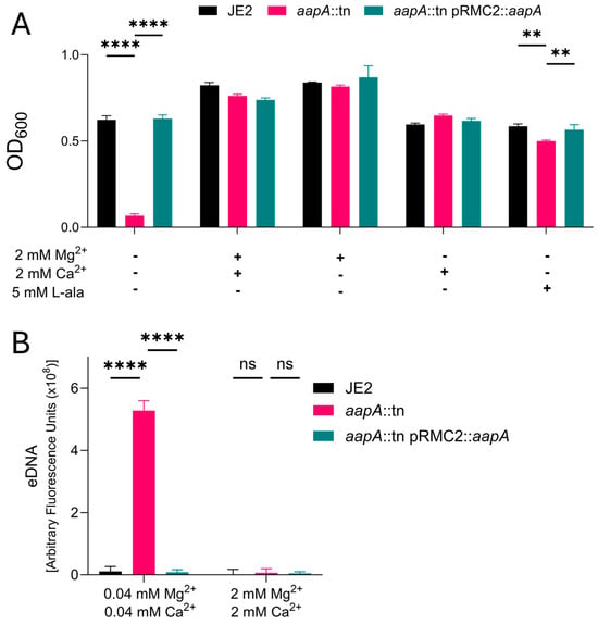
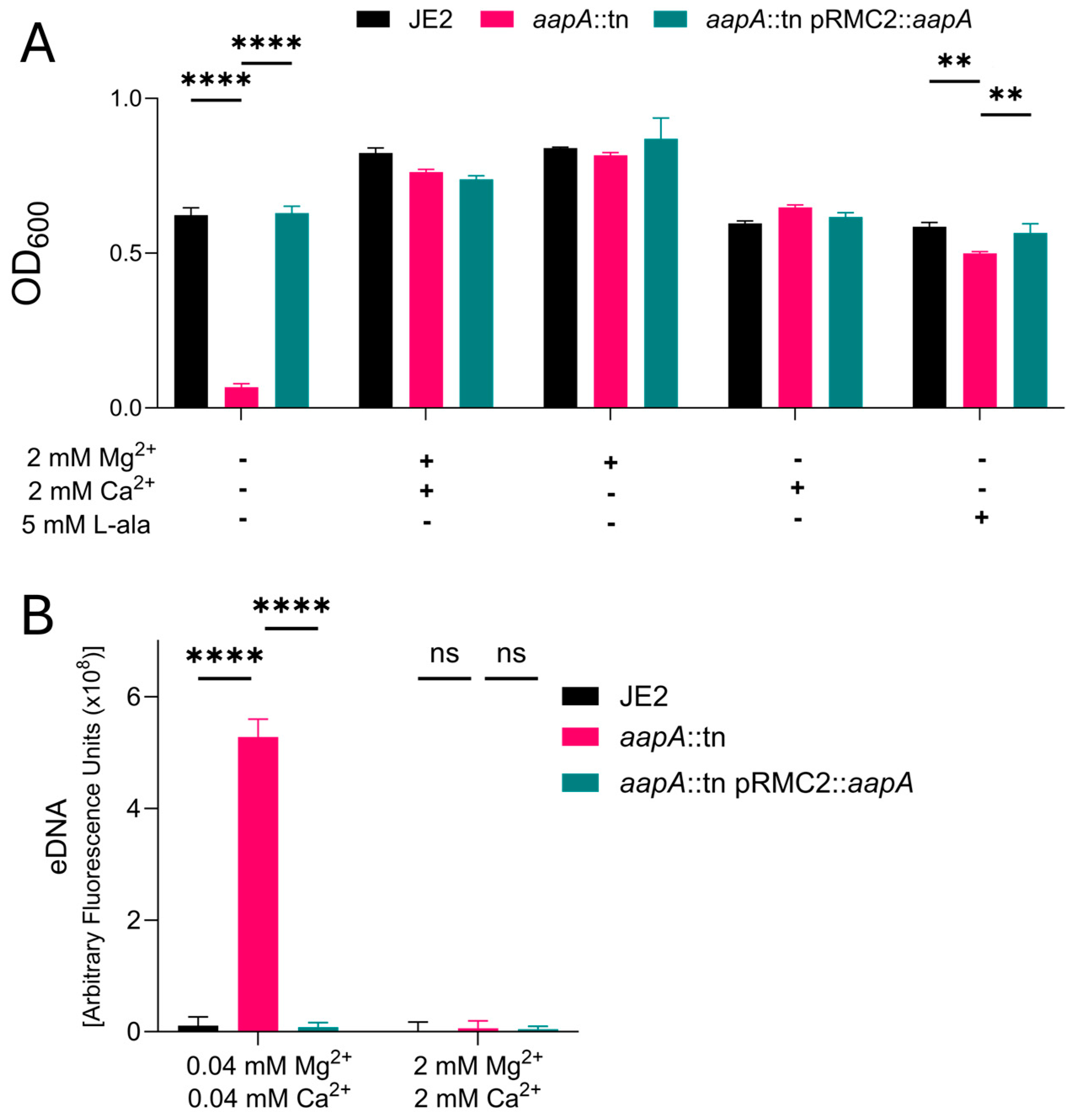
Growth of S. aureus JE2 and an aapA transposon mutant in the same strain (aapA::tn) was measured in a chemically defined medium containing 1 mM L-alanine and low Mg2+/Ca2+. In these conditions, aapA::tn exhibited a significant growth defect compared to the wild type. However, the aapA mutant grew similar to the wild-type strain when the medium was supplemented with an additional 5 mM of L-alanine, high Mg2+, or high Ca2+. Complementation of the mutant with a functional copy of aapA also restored normal growth (Figure 1A). Given the role of the divalent cations Mg2+ and Ca2+ in cell envelope stability in other species [16,17,18,19], we hypothesized that the growth defect observed in the aapA mutant when cultured in low Mg2+/Ca2+ conditions was due to increased cell lysis. As a proxy for cell lysis, extracellular DNA (eDNA) was measured in culture supernatants. Supernatant eDNA from aapA::tn was significantly increased relative to that of the wild-type strain following growth in low Mg2+/Ca2+ conditions. In contrast, after growth in medium containing high Mg2+/Ca2+, eDNA levels in the aapA mutant and wild-type JE2 supernatants were equally low (Figure 1B). We interpret this to mean that alanine uptake prevents cell lysis in low Mg2+/Ca2+ conditions, suggesting a role for alanine uptake or Mg2+ and Ca2+ in cell envelope maintenance.
Figure 1.
Alanine uptake by the AapA transporter is necessary for S. aureus (strain JE2) growth and protection from cell lysis when cultured in minimal concentrations of Mg2+ and Ca2+. (A) Optical density following 10-h incubation in chemically defined media containing 1 mM L-alanine and 0.04 mM each of Mg2+ and Ca2+. These basal concentrations are designated with (−), and the medium was also supplemented with additional alanine, Mg2+, or Ca2+, as indicated in the figure with (+). (B) Relative levels of S. aureus eDNA in culture supernatants following incubation in defined medium with low Mg2+ and Ca2+ or supplemented with 2 mM Mg2+ and Ca2+. Data are displayed as the mean +/− SEM of 3 biological replicates containing 3 technical replicates per experiment. Statistical significance was calculated using a two-way ANOVA (** = p-value < 0.005; **** = p-value < 0.0005).
3.2. Mg2+/Ca2+ Depletion Increases Alanine Demand in S. aureus
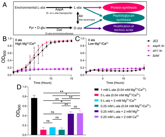
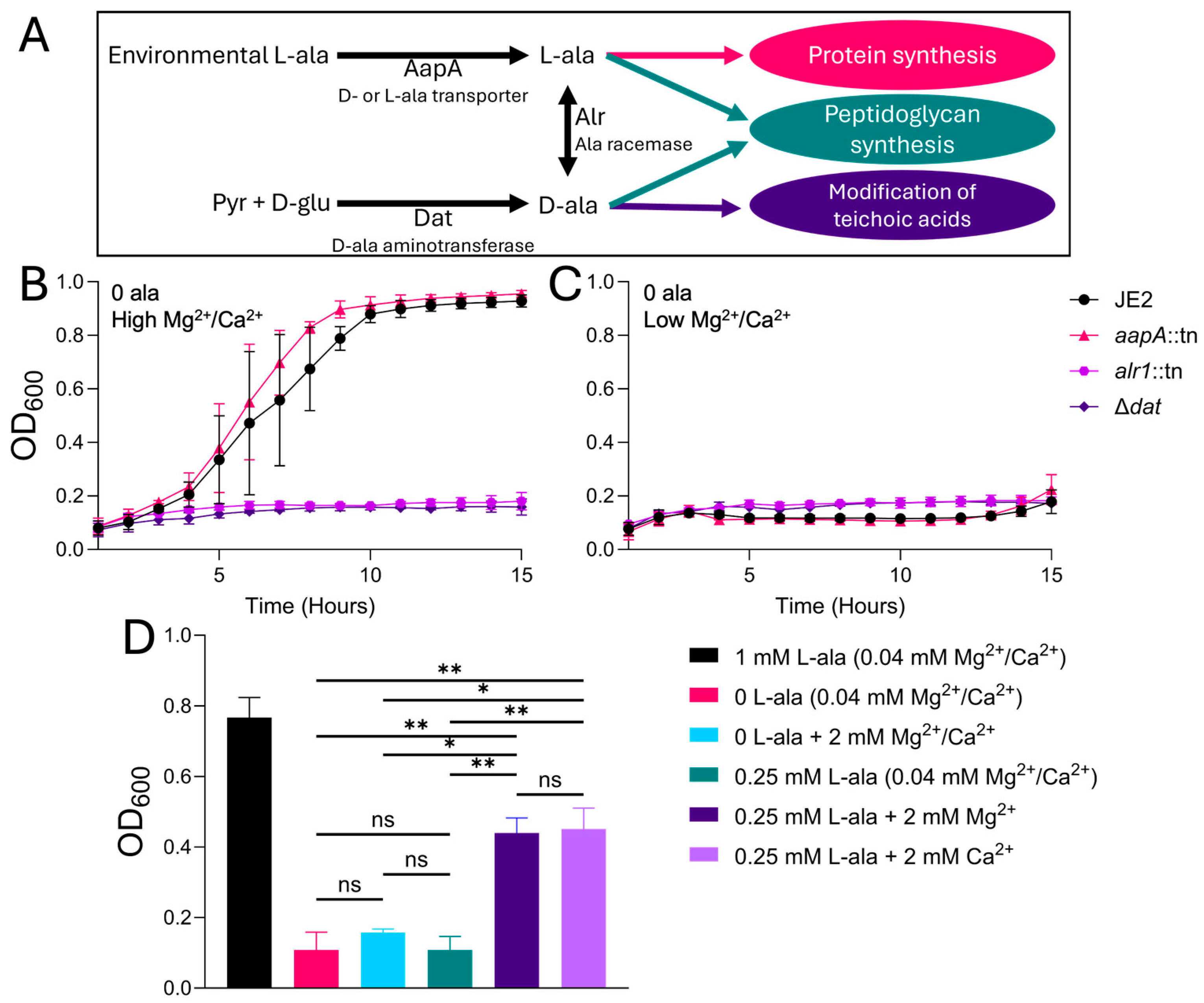
The results above suggest overlapping roles for Mg2+/Ca2+ and alanine in maintaining cell envelope stability and confirm that alanine uptake via AapA somehow compensates for environmental depletion of these divalent cations. However, S. aureus can supply its own alanine through de novo synthesis. Dat synthesizes D-alanine from glutamate, and Alr catalyzes interconversion between D- and L-alanine (Figure 2A) [15]. This raises the question as to why Dat/Alr-dependent de novo synthesis is unable to support growth of the aapA mutant in low Mg2+/Ca2+. We investigated the possibility that alanine demand increases as Mg2+/Ca2+ concentrations decrease, and that this increased demand cannot be met by Dat/Alr biosynthesis. To address this possibility, we obtained dat and alr mutants (Δdat and alr::tn, respectively) and confirmed their inability to grow in the absence of exogenous alanine (Figure 2B). Under the same conditions, JE2 and aapA::tn exhibit robust growth, as would be expected. However, when alanine is absent and Mg2+/Ca2+ are low (Figure 2C), all four strains fail to grow. This shows that Dat and Alr can supply adequate D- and L-alanine for growth only when Mg2+/Ca2+ concentrations are sufficiently high. This finding rules out the notion that AapA-dependent uptake is somehow inhibited by low Mg2+/Ca2+ concentrations. If this were the case, growth of the wild type in the absence of exogenous alanine (where AapA is superfluous) would not be inhibited in low Mg2+/Ca2+. We went on to assess how much exogenous L-alanine is required to support growth of the Δdat strain in low Mg2+/Ca2+ (Figure 2D), finding that 1 mM L-alanine is sufficient to restore robust growth, while 0.25 mM L-alanine is not sufficient. However, when Mg2+ or Ca2+ levels are elevated to 2 mM, 0.25 mM L-alanine partially restores growth to the Δdat strain. This finding allows us to reject the notion that Dat biosynthetic activity is somehow inhibited by low Mg2+/Ca2+; if this were the case, the amount of exogenous alanine required for growth of a dat mutant would not vary as a function of Mg2+/Ca2+ concentration. Instead, our results point to low Mg2+/Ca2+ imposing a higher cellular demand for alanine, independent of its source.
Figure 2.
Growth of S. aureus lacking either exogenous alanine or endogenous alanine synthesis demonstrates an increased alanine requirement under Mg2+ and Ca2+ limitation. (A) A depiction of alanine metabolism in Staphylococcus aureus. (B) Growth of wild-type JE2, the alanine transporter gene aapA transposon mutant (aapA::tn), the alanine racemase gene alr transposon mutant (alr::tn), or a strain with a clean deletion of the alanine synthesis gene dat (∆dat) in defined medium containing no alanine and 2 mM Mg2+/Ca2+ or (C) no alanine and low (0.04 mM each) Mg2+/Ca2+. (D) Growth at 10 h of the alanine-synthesis defective dat deletion mutant strain with no exogenous alanine (0 L-ala) or 0.25 mM L-ala in defined medium containing low Mg2+ and Ca2+ (0.04 mM each) or 2 mM Mg2+ and Ca2+ supplemented. Data are displayed as the mean +/− SEM of 3 biological replicates containing 3 technical replicates per experiment. Statistical significance was calculated using a two-way ANOVA. (* = p-value < 0.05; ** = p-value < 0.005).
3.3. Exogenous Alanine Starvation Results in Increased Sensitivity to Membrane Destabilization
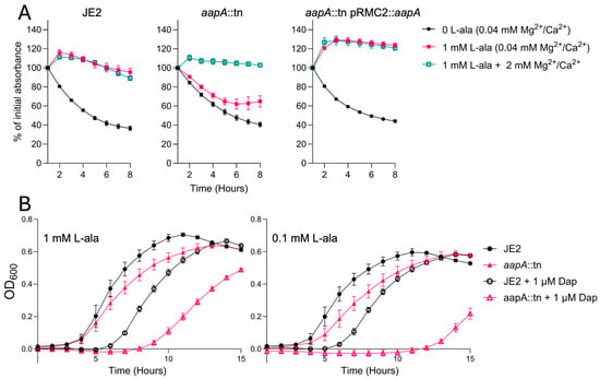
We hypothesized that the increase in alanine demand in low Mg2+/Ca2+ is attributable to reinforcing cell wall components such as peptidoglycan or teichoic acids (both of which require alanine). It has been shown previously that S. aureus cell wall defects potentiate cell lysis by the membrane-destabilizing detergent Triton X-100 [31]. Thus, we expected alanine-starved cells to be Triton sensitive. To test Triton sensitivity, bacteria were collected during exponential growth in TSB, washed, and then suspended in media containing 0.05% Triton X-100 and specified amounts of Mg2+/Ca2+ and alanine (see Figure 3A). Optical density was then used to measure lysis over 8 h. Lysis of wild-type bacteria occurs in low Mg2+/Ca2+ when alanine is absent from the medium, but is prevented by supplementation of 1 mM L-alanine. For the aapA mutant, however, lysis occurs in either condition. We conclude from these results that S. aureus requires exogenous alanine to reinforce the cell wall in low Mg2+/Ca2+ conditions, subsequently preventing detergent-induced lysis.
Figure 3.
Tolerance of S. aureus to membrane perturbation by Triton X-100 in low Mg2+ and Ca2+ or daptomycin treatment requires efficient alanine uptake or 2 mM Mg2+/Ca2+. (A) Lysis in the presence of 0.05% Triton X-100 of the wild-type JE2, the alanine transporter mutant (aapA::tn), or the complemented aapA mutant (aapA::tn pRMC2::aapA) in defined medium containing 0 alanine and low (0.04 mM each) Mg2+ and Ca2+ or with the addition of 1 mM L-alanine in low Mg2+ and Ca2+, or 1 mM L-alanine and 2 mM Mg2+ and Ca2+. (B) Growth of wild-type JE2 or alanine transporter mutant aapA::tn in defined medium containing 2 mM Ca2+ and 0.1 or 1 mM L-alanine with or without 1 µM Daptomycin. Data are displayed as the mean +/− SEM of 3 biological replicates containing 3 technical replicates per experiment.
The antibiotic daptomycin targets the membrane of Gram-positive bacteria through a Ca2+-dependent mechanism, causing membrane depolarization and lysis [32,33]. In response to sublethal daptomycin exposure, S. aureus stabilizes its cell wall through increased peptidoglycan synthesis and D-alanine modification of teichoic acids [6,34,35]. Given our evidence that these processes may be compromised by exogenous alanine starvation, we expected exogenous alanine uptake to influence daptomycin sensitivity. Daptomycin sensitivity was measured by comparing the growth of wild-type and aapA mutant bacteria upon exposure to 1 µM daptomycin in media containing 1.0 or 0.1 mM L-alanine in low Mg2+. Daptomycin treatment moderately extended the lag phase for the wild-type strain, with equal growth delays in 1.0 and 0.1 mM L-alanine conditions. For the aapA mutant, the lag phase exceeded that of the wild type by approximately 3 h in 1 mM L-alanine; in 0.1 mM L-alanine, the lag phase exceeded that of the wild type by approximately 7 h (Figure 3B). Importantly, these experiments were performed in the presence of high Ca2+ (required for daptomycin activity), indicating that alanine uptake is critical to tolerate daptomycin-induced membrane stress even when divalent cations are available.
3.4. The Requirement for Exogenous Alanine Varies Among S. aureus Strains
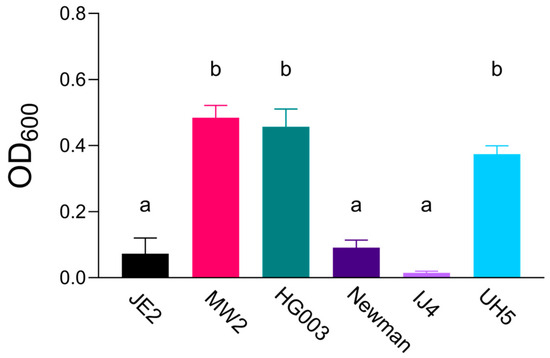
We next sought to determine if the increased demand for alanine uptake in low Mg2+/Ca2+ is shared across S. aureus strains of diverse sequence types (ST) and methicillin resistance traits (MSSA/MRSA). Growth of tested strains in low Mg2+/Ca2+ containing no alanine varied substantially, with JE2 (ST8/MRSA), Newman (ST254/MSSA), and IJ4 (ST151/MSSA) strains exhibiting negligible growth, whereas MW2 (ST1/MRSA), HG003 (ST8/MSSA), and UH5 (ST2187/MSSA) strains were capable of growth (Figure 4). These results indicate that individual strains differ significantly in their need for exogenous alanine uptake when cultured in low Mg2+/Ca2+ conditions.
Figure 4.
Responses of S. aureus to alanine, Mg2+, and Ca2+ depletion varies between strains. S. aureus strains were grown in defined medium containing low (0.04 mM each) Mg2+/Ca2+ and no alanine and growth measured at 15 h. Strains include the USA300 lineage strain JE2 (ST8), human isolate MW2 (ST1), lab strain HG003 (ST8), human isolate Newman (ST8), bovine mastitis isolate IJ4 (ST151) and bovine mastitis isolate UH5 (ST2187). Letters denote statistically significant differences (p-value < 0.05 in a one-way ANOVA) between strains designated “a” and those designated “b”. Data are displayed as the mean +/− SEM of 2 biological replicates containing 3 technical replicates per experiment.
3.5. AapA Contributes to Bacterial Survival During Infection
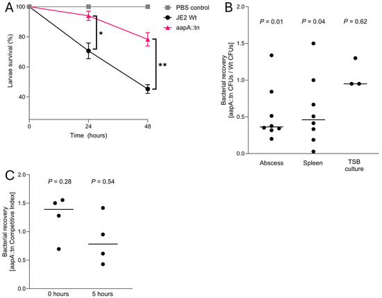
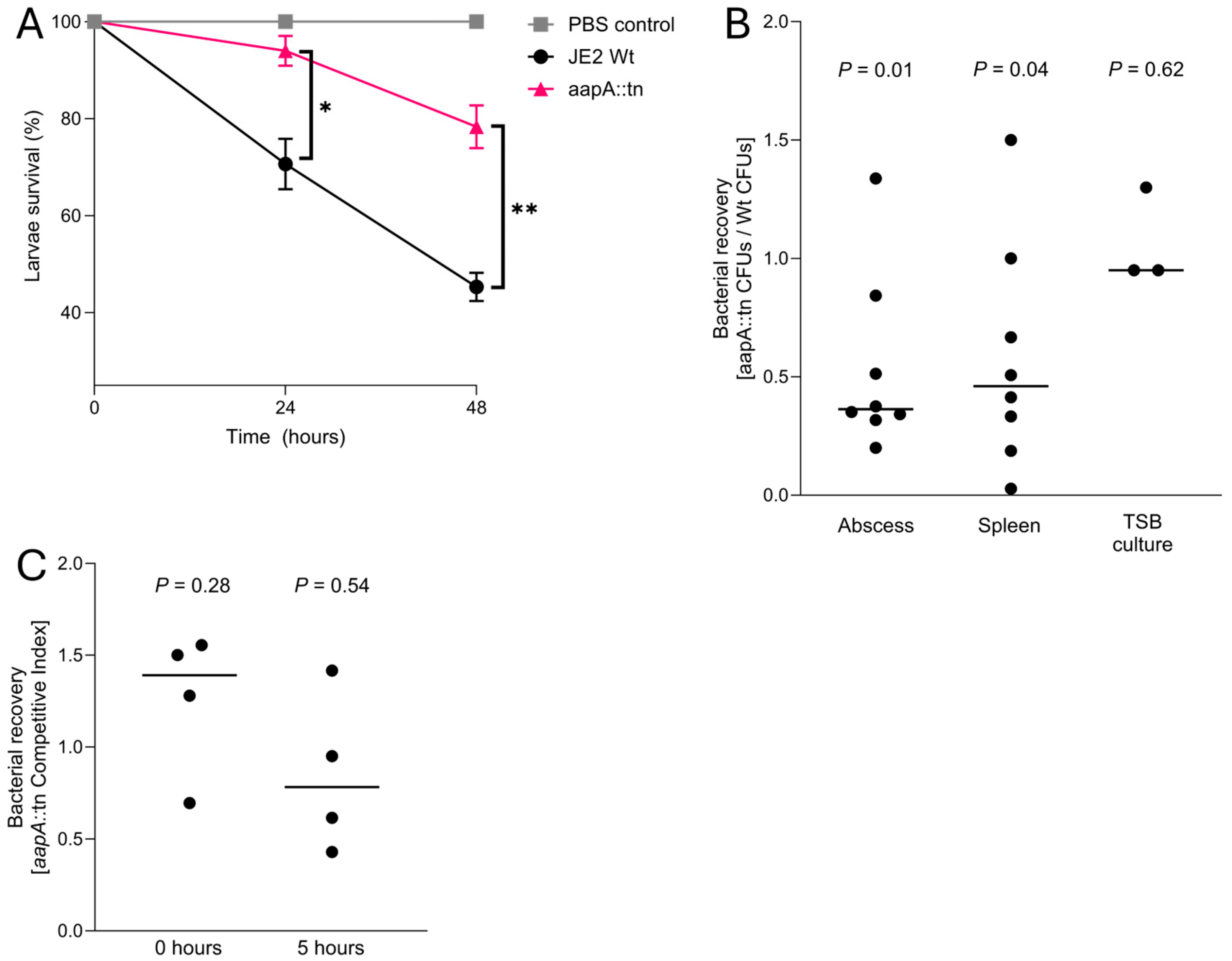
Galleria mellonella insect larvae have been used to measure intrinsic virulence properties of S. aureus [36]. The insect immune system controls bacterial infection through a combination of innate immune factors, including phagocytic cells and antimicrobial peptides. Upon G. mellonella infection, we observed that aapA::tn exhibited significantly reduced virulence compared to wild-type bacteria, as measured by survival of larvae (Figure 5A). Next, we measured bacterial survival in a murine subcutaneous infection model. In competition assays between wild-type and aapA::tn strains, significantly fewer aapA::tn CFUs were recovered from abscesses relative to the wild-type strain. Spleens harvested from the co-infected mice also contained fewer aapA::tn than wild-type bacteria, although the ratio of recovered CFUs was more variable than in abscesses (Figure 5B).
Figure 5.
Mutation of aapA attenuates the infective capacity of S. aureus. (A) G. mellonella larvae survival after inoculation with 105 CFUs of wild-type JE2 or aapA::tn and incubation at 37 °C for 48 h, with survival assessed every 24 h. Injections were performed in groups of at least 10 larvae in 3 separate experiments. Significance was calculated using a two-way ANOVA. (B) Ratio of aapA::tn to wild type recovered after 48 h from a murine co-infection with equal quantities of both strains introduced by subcutaneous injection. Two experiments were performed (n = 4 per experiment). Significance for mouse infection was calculated using a theoretical mean of 1 in a one-sample t-test. (C) Survival of S. aureus after synchronized phagocytosis by RAW cells assessed immediately after gentamicin treatment of RAW cells (0 h) and 5 h after gentamicin treatment (5 h). RAW cell testing was performed in quadruplicate in four separate experiments. The competitive index was calculated using the CFUs either immediately after gentamicin treatment (T0) or 5 h after gentamicin treatment (T5) relative to the inoculum. Immediately after gentamicin treatment, a non-significant increase in phagocytosed aapA::tn cells relative to wild-type cells was observed, as well as a non-significant decrease in viability after 5 h. Significance was calculated using a theoretical mean of 1 in a one-sample t-test. (* = p-value < 0.05; ** = p-value < 0.005).
Assays were next designed to specifically assess intracellular survival of wild-type and aapA::tn bacteria following phagocytosis by murine macrophages. Gentamicin protection assays were performed by adding equal numbers of mutant and wild-type bacteria to the mouse macrophage cell line RAW 264.7 [37]. Equivalent numbers of wild-type and aapA::tn bacteria were recovered immediately following phagocytosis and gentamicin treatment, indicating similar invasion/phagocytosis efficiencies of both strains. Following five hours of incubation within these phagocytic cells, plating and enumeration of surviving bacteria showed a non-significant trend toward increased survival of wild-type bacteria (Figure 5C).
4. Discussion
Maintaining cell envelope stability is essential for bacterial growth and survival, with peptidoglycan providing the primary structural support in Gram-positive bacteria. Alanine is required for pentapeptide stem formation and cross-linking, while the divalent cations Mg2+ and Ca2+ associate with the envelope through electrostatic interactions with various envelope constituents [16]. Although these roles are well established, their functional interplay in supporting cell envelope stability has not been well defined. Here, we demonstrate that under cation-limited conditions, some strains of Staphylococcus aureus exhibit an increased dependence on AapA-mediated alanine uptake to maintain cell envelope integrity. Disruption of alanine acquisition in these conditions results in increased cell lysis in vitro and reduces fitness in infection models, linking nutrient availability to cell envelope stability.
Our findings support a model in which depletion of Mg2+ and Ca2+ shifts the burden of envelope stabilization toward alanine-dependent processes, such as peptidoglycan and teichoic acid modification. D-alanine modification of the negatively charged groups of teichoic acid neutralizes a major source of negative charge in the cell wall, and may decrease the need for ionic bridging accomplished by envelope-associated Mg2+ and Ca2+. Peptidoglycan thickening also requires alanine, and has been shown to improve tolerance of S. aureus to envelope stress [1,5]. Under conditions that may require these processes, such as exposure to envelope-targeting antibiotics or detergents, de novo alanine synthesis appears insufficient to meet alanine demand, making alanine uptake via AapA critical for growth and fitness. A previous study of S. aureus strain MW2 reported deficient D-alanine modification of teichoic acids in aapA mutant cells [11], an essential process for tolerating envelope stress [4,38]. Another study in strain JE2 found modest changes in peptidoglycan cross-link composition between the wild type and the aapA mutant [10]. Based on our findings, we would expect the differences in cell envelope composition between the two strains to be amplified upon experiencing a secondary stress, such as divalent cation depletion.
The variability in growth observed across S. aureus strains cultured under conditions of alanine and cation limitation suggests the presence of compensatory mechanisms for cell wall stabilization. These differences may reflect variation in alanine biosynthetic capacity, cation-responsive regulatory networks [39,40], or altered peptidoglycan cross-linking or teichoic acid modification [5]. We consider it most likely that all strains experience an increased demand for alanine upon divalent cation depletion, but some are better equipped to upregulate de novo biosynthesis.
Host nutritional immunity is typically associated with sequestration of trace metals, such as Mn2+ and Fe2+ [41,42,43]. However, additional mechanisms of nutritional immunity may target more abundant divalent cations, such as Mg2+ and Ca2+. For example, the host divalent metal transporter NRAMP1 depletes Mg2+ from the phagosome [22], and Ca2+ efflux from the neutrophil phagosome has also been documented [21]. Thus, we predicted that the aapA mutant may be more susceptible to the harsh environment inside phagocytic cells due to Mg2+/Ca2+ depletion and other cell envelope stressors. Consistent with this, we observe that the aapA mutant is less virulent than the wild type in an insect infection model and less capable of survival in a murine infection model. However, our results did not indicate a statistically significant defect in proliferation/survival after five hours of the aapA mutant in a macrophage cell line. This may be because intracellular S. aureus manipulates Ca2+ inside of host cells—a phenomenon that has been observed previously [44]. Alternatively, it could be that peptides available in host cells provide a sufficient supply of alanine.
Previous studies suggest AapA contributes to bacterial fitness in some host environments but not others. For example, genome-wide screens have indicated that AapA plays a role in S. aureus strain HG003 for survival in abscesses, and in strain TM283 (nearly isogenic to JE2 used in this study) under salt stress [12,13]. Conversely, an aapA mutant in strain JE2 showed minimal or no fitness defects in a bloodstream infection model [10]. These differences may reflect variation in nutrient and cation availability across host niches.
Disruption of AapA increases the sensitivity of S. aureus to the membrane targeting antibiotic daptomycin, as shown in this work with strain JE2, and by others with strain MW2 [11]; this is also evidenced by transposon insertion screens performed by others in various strains, including TM283, MW2, HG003 and others [14]. The aapA mutant also has increased sensitivity to other cell wall-targeting antibiotics, including β-lactams [10,11]. It seems likely that alanine starvation and cell wall-targeting agents act additively, with each compromising cell wall cross-linking by distinct mechanisms. Under these conditions of compromised cell wall synthesis, we have shown that S. aureus becomes increasingly dependent on envelope stabilization by Mg2+ and Ca2+. Links between cation chelation and envelope-targeting antibiotic susceptibility have been shown [45], and are further supported by the findings of this study.
In summary, this work identifies alanine uptake as an important determinant of cell envelope stability in S. aureus under conditions of divalent cation limitation. Within the context of host nutritional immunity, where Mg2+ and Ca2+ availability is restricted, alanine acquisition becomes increasingly important for maintaining cell wall integrity and preventing lysis. This conditional dependence is supported by in vivo observations, in which disruption of AapA-mediated alanine acquisition reduces bacterial fitness in multiple infection models. Together, these findings link nutrient limitation to cell wall homeostasis and support growing evidence that alanine uptake represents a vulnerability that could be leveraged to enhance antimicrobial strategies.
Supplementary Materials
The following supporting information can be downloaded at https://www.mdpi.com/article/10.3390/microorganisms14061332/s1, Table S1: Strains used in this study.
Author Contributions
Experiments were performed by T.G.B. and S.K. Writing was by T.G.B. and E.W. Editing was done by T.G.B., D.L.E. and V.T. All authors have read and agreed to the published version of the manuscript.
Funding
This research was funded by NIH grant RO1AI149491.
Institutional Review Board Statement
The animal study protocol was approved by the Institutional Animal Care and Use Committee at Brigham Young University (Protocol no. 25-0507, approved on 13 May 2025).
Informed Consent Statement
Not applicable.
Data Availability Statement
The original contributions presented in this study are included in the article/Supplementary Materials. Further inquiries can be directed to the corresponding author.
Conflicts of Interest
The authors declare no conflicts of interest.
References
- Ledger, E.V.K.; Mesnage, S.; Edwards, A.M. Human serum triggers antibiotic tolerance in Staphylococcus aureus. Nat. Commun. 2022, 13, 2041. [Google Scholar] [CrossRef] [Scilit] [PubMed]
- Carrel, M.; Smith, M.; Shi, Q.; Hasegawa, S.; Clore, G.S.; Perencevich, E.N.; Goto, M. Antimicrobial Resistance Patterns of Outpatient Staphylococcus aureus Isolates. JAMA Netw. Open 2024, 7, e2417199. [Google Scholar] [CrossRef] [Scilit] [PubMed]
- Mishra, N.N.; Yang, S.-J.; Sawa, A.; Rubio, A.; Nast, C.C.; Yeaman, M.R.; Bayer, A.S. Analysis of Cell Membrane Characteristics of In Vitro-Selected Daptomycin-Resistant Strains of Methicillin-Resistant Staphylococcus aureus. Antimicrob. Agents Chemother. 2009, 53, 2312–2318. [Google Scholar] [CrossRef] [Scilit]
- Mishra, N.N.; Bayer, A.S.; Weidenmaier, C.; Grau, T.; Wanner, S.; Stefani, S.; Cafiso, V.; Bertuccio, T.; Yeaman, M.R.; Nast, C.C.; et al. Phenotypic and Genotypic Characterization of Daptomycin-Resistant Methicillin-Resistant Staphylococcus aureus Strains: Relative Roles of mprF and dlt Operons. PLoS ONE 2014, 9, e107426. [Google Scholar] [CrossRef] [Scilit]
- Cui, L.; Ma, X.; Sato, K.; Okuma, K.; Tenover, F.C.; Mamizuka, E.M.; Gemmell, C.G.; Kim, M.-N.; Ploy, M.-C.; El Solh, N.; et al. Cell Wall Thickening Is a Common Feature of Vancomycin Resistance in Staphylococcus aureus. J. Clin. Microbiol. 2003, 41, 5–14. [Google Scholar] [CrossRef] [Scilit] [PubMed]
- Dengler, V.; Meier, P.S.; Heusser, R.; Berger-Bächi, B.; McCallum, N. Induction kinetics of the Staphylococcus aureus cell wall stress stimulon in response to different cell wall active antibiotics. BMC Microbiol. 2011, 11, 16. [Google Scholar] [CrossRef] [Scilit]
- Ledger, E.V.K.; Edwards, A.M. Host-induced cell wall remodeling impairs opsonophagocytosis of Staphylococcus aureus by neutrophils. mBio 2024, 15, e0164324. [Google Scholar] [CrossRef] [Scilit]
- Vollmer, W.; Blanot, D.; De Pedro, M.A. Peptidoglycan structure and architecture. FEMS Microbiol. Rev. 2008, 32, 149–167. [Google Scholar] [CrossRef] [Scilit]
- Reichmann, N.T.; Cassona, C.P.; Gründling, A. Revised mechanism of D-alanine incorporation into cell wall polymers in Gram-positive bacteria. Microbiology 2013, 159, 1868–1877. [Google Scholar] [CrossRef] [Scilit]
- Gallagher, L.A.; Shears, R.K.; Fingleton, C.; Alvarez, L.; Waters, E.M.; Clarke, J.; Bricio-Moreno, L.; Campbell, C.; Yadav, A.K.; Razvi, F.; et al. Impaired Alanine Transport or Exposure to d-Cycloserine Increases the Susceptibility of MRSA to β-lactam Antibiotics. J. Infect. Dis. 2019, 221, 1000–1016. [Google Scholar] [CrossRef] [Scilit]
- Suzuki, Y.; Kawada-Matsuo, M.; Thuan, V.T.T.; Le, M.N.-T.; Sakaguchi, T.; Komatsuzawa, H. D-alanine synthesis and exogenous alanine affect the antimicrobial susceptibility of Staphylococcus aureus. Antimicrob. Agents Chemother. 2025, 69, e0193624. [Google Scholar] [CrossRef] [Scilit] [PubMed]
- Schuster, C.F.; Wiedemann, D.M.; Kirsebom, F.C.M.; Santiago, M.; Walker, S.; Gründling, A. High-throughput transposon sequencing highlights the cell wall as an important barrier for osmotic stress in methicillin resistant Staphylococcus aureus and underlines a tailored response to different osmotic stressors. Mol. Microbiol. 2019, 113, 699–717. [Google Scholar] [CrossRef] [Scilit]
- Valentino, M.D.; Foulston, L.; Sadaka, A.; Kos, V.N.; Villet, R.A.; Maria, J.S.; Lazinski, D.W.; Camilli, A.; Walker, S.; Hooper, D.C.; et al. Genes Contributing to Staphylococcus aureus Fitness in Abscess- and Infection-Related Ecologies. mBio 2014, 5, e01729-14. [Google Scholar] [CrossRef] [Scilit]
- Coe, K.A.; Lee, W.; Stone, M.C.; Komazin-Meredith, G.; Meredith, T.C.; Grad, Y.H.; Walker, S. Multi-strain Tn-Seq reveals common daptomycin resistance determinants in Staphylococcus aureus. PLoS Pathog. 2019, 15, e1007862. [Google Scholar] [CrossRef] [Scilit]
- Panda, S.; Jayasinghe, Y.P.; Shinde, D.D.; Bueno, E.; Stastny, A.; Bertrand, B.P.; Chaudhari, S.S.; Kielian, T.; Cava, F.; Ronning, D.R.; et al. Staphylococcus aureus counters organic acid anion-mediated inhibition of peptidoglycan cross-linking through robust alanine racemase activity. eLife 2024, 13, RP95389. [Google Scholar] [CrossRef] [Scilit]
- Thomas, K.J.; Rice, C.V. Revised model of calcium and magnesium binding to the bacterial cell wall. Biometals 2014, 27, 1361–1370. [Google Scholar] [CrossRef] [Scilit] [PubMed]
- Abee, T.; Rombouts, F.M.; Hugenholtz, J.; Guihard, G.; Letellier, L. Mode of Action of Nisin Z against Listeria monocytogenes Scott A Grown at High and Low Temperatures. Appl. Environ. Microbiol. 1994, 60, 1962–1968. [Google Scholar] [CrossRef] [Scilit]
- Kumar, M.; Srivastava, S. Effect of calcium and magnesium on the antimicrobial action of enterocin LR/6 produced by Enterococcus faecium LR/6. Int. J. Antimicrob. Agents 2011, 37, 572–575. [Google Scholar] [CrossRef] [Scilit]
- Tesson, B.; Dajkovic, A.; Keary, R.; Marlière, C.; Dupont-Gillain, C.C.; Carballido-López, R. Magnesium rescues the morphology of Bacillus subtilis mreB mutants through its inhibitory effect on peptidoglycan hydrolases. Sci. Rep. 2022, 12, 1137. [Google Scholar] [CrossRef] [Scilit]
- Xie, Y.; Yang, L. Calcium and Magnesium Ions Are Membrane-Active against Stationary-Phase Staphylococcus aureus with High Specificity. Sci. Rep. 2016, 6, 20628. [Google Scholar] [CrossRef] [Scilit]
- Dewitt, S.; Green, J.; Laffafian, I.; Lewis, K.J.; Hallett, M.B. Intraphagosomal Free Ca2+ Changes during Phagocytosis. Int. J. Mol. Sci. 2024, 25, 4254. [Google Scholar] [CrossRef] [Scilit]
- Cunrath, O.; Bumann, D. Host resistance factor SLC11A1 restricts Salmonella growth through magnesium deprivation. Science 2019, 366, 995–999. [Google Scholar] [CrossRef] [Scilit] [PubMed]
- Hussain, M.; Hastings, J.G.M.; White, P.J. A chemically defined medium for slime production by coagulase-negative staphylococci. J. Med. Microbiol. 1991, 34, 143–147. [Google Scholar] [CrossRef] [Scilit]
- Sebulsky, M.T.; Speziali, C.D.; Shilton, B.H.; Edgell, D.R.; Heinrichs, D.E. FhuD1, a ferric hydroxamate-binding lipoprotein in Staphylococcus aureus: A case of gene duplication and lateral transfer. J. Biol. Chem. 2004, 279, 53152–53159. [Google Scholar] [CrossRef] [Scilit]
- Fey, P.D.; Endres, J.L.; Yajjala, V.K.; Widhelm, T.J.; Boissy, R.J.; Bose, J.L.; Bayles, K.W. A Genetic Resource for Rapid and Comprehensive Phenotype Screening of Nonessential Staphylococcus aureus Genes. mBio 2013, 4, e00537-12. [Google Scholar] [CrossRef] [Scilit] [PubMed]
- Monk, I.R.; Stinear, T.P. From cloning to mutant in 5 days: Rapid allelic exchange in Staphylococcus aureus. Access Microbiol. 2020, 3, 000193. [Google Scholar] [CrossRef] [Scilit] [PubMed]
- Corrigan, R.M.; Foster, T.J. An improved tetracycline-inducible expression vector for Staphylococcus aureus. Plasmid 2009, 61, 126–129. [Google Scholar] [CrossRef] [Scilit]
- Baba, T.; Takeuchi, F.; Kuroda, M.; Yuzawa, H.; Aoki, K.-I.; Oguchi, A.; Nagai, Y.; Iwama, N.; Asano, K.; Naimi, T.; et al. Genome and virulence determinants of high virulence community-acquired MRSA. Lancet 2002, 359, 1819–1827. [Google Scholar] [CrossRef] [Scilit]
- Sassi, M.; Felden, B.; Augagneur, Y. Draft Genome Sequence of Staphylococcus aureus subsp. aureus Strain HG003, an NCTC8325 Derivative. Genome Announc. 2014, 2, e00855-14. [Google Scholar] [CrossRef] [Scilit]
- Duthie, E.S.; Lorenz, L.L. Staphylococcal Coagulase: Mode of Action and Antigenicity. Microbiology 1952, 6, 95–107. [Google Scholar] [CrossRef] [Scilit]
- Raychaudhuri, D.; Chatterjee, A.N. Use of resistant mutants to study the interaction of triton X-100 with Staphylococcus aureus. J. Bacteriol. 1985, 164, 1337–1349. [Google Scholar] [CrossRef] [Scilit]
- Buttress, J.A.; Schäfer, A.-B.; Koh, A.; Wheatley, J.; Mickiewicz, K.; Wenzel, M.; Strahl, H. The last resort antibiotic daptomycin exhibits two independent antibacterial mechanisms of action. Nat. Commun. 2025, 16, 10320. [Google Scholar] [CrossRef] [Scilit]
- Taylor, S.D.; Palmer, M. The action mechanism of daptomycin. Bioorg. Med. Chem. 2016, 24, 6253–6268. [Google Scholar] [CrossRef] [Scilit]
- Ledger, E.V.K.; Edwards, A.M. Growth Arrest of Staphylococcus aureus Induces Daptomycin Tolerance via Cell Wall Remodelling. mBio 2023, 14, e0355822. [Google Scholar] [CrossRef] [Scilit]
- Muthaiyan, A.; Silverman, J.A.; Jayaswal, R.K.; Wilkinson, B.J. Transcriptional Profiling Reveals that Daptomycin Induces the Staphylococcus aureus Cell Wall Stress Stimulon and Genes Responsive to Membrane Depolarization. Antimicrob. Agents Chemother. 2008, 52, 980–990. [Google Scholar] [CrossRef] [Scilit] [PubMed]
- Alnezary, F.S.; Almutairi, M.S.; Alhifany, A.A.; Almangour, T.A. Assessing Galleria mellonella as a preliminary model for systemic Staphylococcus aureus infection: Evaluating the efficacy and impact of vancomycin and Nigella sativa oil on gut microbiota. Saudi Pharm. J. 2023, 31, 101824. [Google Scholar] [CrossRef] [Scilit]
- Taciak, B.; Białasek, M.; Braniewska, A.; Sas, Z.; Sawicka, P.; Kiraga, Ł.; Rygiel, T.; Król, M. Evaluation of phenotypic and functional stability of RAW 264.7 cell line through serial passages. PLoS ONE 2018, 13, e0198943. [Google Scholar] [CrossRef] [Scilit]
- Collins, L.V.; Kristian, S.A.; Weidenmaier, C.; Faigle, M.; van Kessel, K.P.M.; van Strijp, J.A.G.; Götz, F.; Neumeister, B.; Peschel, A. Staphylococcus aureus strains lacking D-alanine modifications of teichoic acids are highly susceptible to human neutrophil killing and are virulence attenuated in mice. J. Infect. Dis. 2002, 186, 214–219. [Google Scholar] [CrossRef] [Scilit] [PubMed]
- Kuijk, M.M.; Tusveld, E.; Lehmann, E.; van Dalen, R.; Lasa, I.; Ingmer, H.; Pannekoek, Y.; van Sorge, N.M. The two-component system ArlRS is essential for wall teichoic acid glycoswitching in Staphylococcus aureus. mBio 2025, 16, e0266824. [Google Scholar] [CrossRef] [Scilit]
- Memmi, G.; Nair, D.R.; Cheung, A. Role of ArlRS in Autolysis in Methicillin-Sensitive and Methicillin-Resistant Staphylococcus aureus Strains. J. Bacteriol. 2012, 194, 759–767. [Google Scholar] [CrossRef] [Scilit] [PubMed]
- Ruiz, V.M.R.; Freiberg, J.A.; Weiss, A.; Green, E.R.; Jobson, M.-E.; Felton, E.; Shaw, L.N.; Chazin, W.J.; Skaar, E.P. Coordinated adaptation of Staphylococcus aureus to calprotectin-dependent metal sequestration. mBio 2024, 15, e0138924. [Google Scholar] [CrossRef] [Scilit]
- Kehl-Fie, T.E.; Zhang, Y.; Moore, J.L.; Farrand, A.J.; Hood, M.I.; Rathi, S.; Chazin, W.J.; Caprioli, R.M.; Skaar, E.P. MntABC and MntH Contribute to Systemic Staphylococcus aureus Infection by Competing with Calprotectin for Nutrient Manganese. Infect. Immun. 2013, 81, 3395–3405. [Google Scholar] [CrossRef] [Scilit]
- Antelo, G.T.; Vila, A.J.; Giedroc, D.P.; Capdevila, D.A. Molecular Evolution of Transition Metal Bioavailability at the Host–Pathogen Interface. Trends Microbiol. 2020, 29, 441–457. [Google Scholar] [CrossRef] [Scilit]
- Stelzner, K.; Winkler, A.-C.; Liang, C.; Boyny, A.; Ade, C.P.; Dandekar, T.; Fraunholz, M.J.; Rudel, T. Intracellular Staphylococcus aureus Perturbs the Host Cell Ca2+ Homeostasis to Promote Cell Death. mBio 2020, 11, 10-1128. [Google Scholar] [CrossRef] [Scilit]
- Olson, J.; Khetarpal, A.K.; Jankeel, A.; Lorenzana, A.; Nizet, V.; Sakoulas, G.; Ulloa, E.R. Ethylenediaminetetraacetic Acid Enhances Vancomycin and Reactive Oxygen Species–Mediated Killing of Vancomycin-Intermediate Staphylococcus aureus. Open Forum Infect. Dis. 2025, 12, ofaf291. [Google Scholar] [CrossRef] [Scilit]

Disclaimer/Publisher’s Note: The statements, opinions and data contained in all publications are solely those of the individual author(s) and contributor(s) and not of MDPI and/or the editor(s). MDPI and/or the editor(s) disclaim responsibility for any injury to people or property resulting from any ideas, methods, instructions or products referred to in the content. |
© 2026 by the authors. Licensee MDPI, Basel, Switzerland. This article is an open access article distributed under the terms and conditions of the Creative Commons Attribution (CC BY) license.