


Abstract
Bacterial membrane vesicles (BMVs) are non-replicative, bilayered nanostructures secreted by both Gram-negative and Gram-positive bacteria. Rather than being passive byproducts of cell envelope turnover, BMVs are increasingly recognized as regulated particles that selectively package proteins, lipids, nucleic acids, and other bioactive molecules. Through these cargos, BMVs mediate a wide range of biological processes, including bacterial stress adaption, intercellular communication, virulence delivery, and host immune modulation. In this review, we integrate recent advancements in understanding the molecular mechanisms underlying BMV biogenesis and composition and discuss how their heterogeneity contributes to their functional diversity. Beyond their biological roles, we critically examine the translational potential of BMVs in vaccine development, targeted drug delivery, cancer therapy, diagnostic tools, and biotechnological applications. However, significant challenges related to their safety, efficacy, and large-scale production must be addressed to realize their full clinical potential. We review recent progress and ongoing obstacles in the use of BMVs across various biomedical applications and propose strategies for their clinical translation.
1. Introduction
Bacteria release nanoscale, spherical structures known as bacterial membrane vesicles (BMVs) into their extracellular environment. Initially considered incidental byproducts of cell lysis or envelope turnover, BMVs are now recognized as an evolutionarily conserved and actively regulated secretion system found across nearly all prokaryotes, including both Gram-negative and Gram-positive bacteria [1]. In Gram-negative bacteria, these vesicles are often referred to as outer membrane vesicles (OMVs), which form from localized bulging of the outer membrane, encapsulating periplasmic components within a lipid bilayer. For many years, Gram-positive bacteria were thought to be incapable of vesicle production due to their thick peptidoglycan cell wall. However, accumulating evidence now demonstrates that Gram-positive species actively generate cytoplasmic membrane vesicles (CMVs) through coordinated cell wall remodeling and turgor-driven membrane extrusion processes [2,3]. Additionally, vesicle production has been documented in various archaeal species, suggesting that membrane vesiculation is a universal biological process that likely predates the evolutionary divergence of prokaryotic lineages [4]. More recently, the identification of outer-inner membrane vesicles (OIMVs) in Gram-negative bacteria, which encapsulate components from both the outer and inner membranes, has further expanded our understanding of BMV structural heterogeneity and underscored the mechanistic diversity of vesicle biogenesis pathways [5,6].
BMVs serve a wide array of functions that span microbial physiology, ecology, and pathogenesis. As specialized delivery vehicles, BMVs selectively package and transport proteins, lipids, nucleic acids, metabolites, and signaling molecules, thereby facilitating stress responses, horizontal gene transfer, quorum sensing, and biofilm formation [7,8,9,10,11]. In pathogenic bacteria, BMVs contribute to immune evasion, toxin delivery, modulation of host signaling pathways, and the spread of antimicrobial resistance determinants [12,13,14,15]. In contrast, BMVs from commensal and probiotic bacteria can exert beneficial effects on the host, promoting immune tolerance, reinforcing epithelial barrier integrity, and supporting mutualistic host–microbe interactions [16,17]. Beyond their endogenous biological roles, BMVs are increasingly viewed as modular and engineerable nanosystems with substantial translational potential. Their intrinsic stability, nanoscale dimensions, and ability to integrate immunostimulatory components with functional cargos make BMVs particularly promising for applications in vaccinology, antimicrobial therapy, targeted drug delivery, diagnostics, and cancer immunotherapy.
In this review, we provide a comprehensive and updated overview of BMV biology. We first summarize current models of BMV biogenesis, followed by an in-depth examination of vesicle composition. We then discuss BMV functions in bacterial homeostasis, stress adaptation, interspecies interactions, and host modulation. Finally, we highlight the emerging translational potential of BMVs in vaccines, drug delivery, diagnostics, and cancer immunotherapy, while critically addressing the key technical and regulatory challenges that must be overcome for clinical and industrial implementation. By integrating these perspectives, this review aims to provide an updated conceptual framework for BMV biology and illustrate how insights into bacterial vesiculation may inform future therapeutic and synthetic biology strategies.
2. Biogenesis of BMVs
The biogenesis of BMVs is a tightly regulated and evolutionarily conserved process observed across diverse bacterial and archaeal lineages. Although the precise mechanisms of vesicle formation vary among taxa, all bacteria capable of vesiculation share the ability to remodel their cell envelope and to selectively package molecular cargo into nanoscale membranous structures (Figure 1).
Figure 1.
Biogenesis pathways of BMVs. In Gram-negative bacteria, BMVs are generated through outer and/or inner membrane blebbing and explosive cell lysis. (a) Outer membrane blebbing results in the formation of outer membrane vesicles (OMVs), typically driven by disturbances in cell envelope integrity, such as the insertion of hydrophobic molecules into the outer membrane or defects in peptidoglycan biosynthesis. Since the inner membrane remains intact during this process, OMVs generally lack cytoplasmic components. (b,c) Weakening of the peptidoglycan layer by endolysins can induce protrusion of the inner membrane into the periplasmic space, followed by pinching-off of the outer membrane, resulting in the formation of outer-inner membrane vesicles (OIMVs). (d) In contrast, explosive cell lysis is triggered by endolysin-mediated degradation of the peptidoglycan layer. Upon loss of cell wall integrity, bacterial cells round up and undergo lysis, and the resulting membrane fragments spontaneously self-assemble into explosive outer-inner membrane vesicles (EOIMVs) and explosive outer-membrane vesicles (EOMVs). Unlike OMVs and OIMVs formed by membrane blebbing, EOMVs and EOIMVs encapsulate cytoplasmic components in largely non-selective manner. (e,f) In Gram-positive bacteria, BMV biogenesis occurs through cytoplasmic membrane blebbing and/or a process termed bubbling cell death. Endolysin-mediated degradation of the thick peptidoglycan layer facilitates cytoplasmic membrane protrusion and vesicle release, leading to the formation of cytoplasmic membrane vesicles (CMVs). CMVs can encapsulate both membrane-associated and cytoplasmic components. Created with BioRender.com.
2.1. BMV Biogenesis in Gram-Negative Bacteria
Virtually all Gram-negative bacteria constitutively release OMVs, which typically range from 20 to 400 nm in diameter [1]. OMVs are generated through localized outward blebbing and subsequent pinching-off of the outer membrane without compromising overall membrane integrity. Several non-mutually exclusive mechanistic models have been proposed to explain how outer membrane remodeling drives vesicle formation.
The earliest and most widely discussed model attributes OMV biogenesis to the weakening or loss of covalent linkages between the outer membrane and the underlying peptidoglycan layer [18,19]. Disruption of these cross-links, often mediated by alterations in lipoproteins such as Lpp, creates localized envelope instability. When the expansion of the outer membrane exceeds that of the peptidoglycan layer, membrane tension is relieved through outward protrusion, ultimately resulting in vesicle release.
A second model focuses on periplasmic crowding. The accumulation of peptidoglycan fragments, misfolded proteins, or aggregated periplasmic material increases localized osmotic or turgor pressure against the inner face of the outer membrane [15,19]. This pressure induces membrane curvature and bulging, leading to the release of OMVs enriched in periplasmic components. Environmental conditions that disrupt protein folding or peptidoglycan turnover, such as heat shock, oxidative stress, or antibiotic exposure, often trigger OMV formation via this pathway [11,20,21].
A third mechanism involves the localized enrichment of curvature-inducing molecules within the outer membrane. In Pseudomonas aeruginosa (P. aeruginosa), the quorum-sensing molecule Pseudomonas quinolone signal (PQS) intercalates into the outer leaflet of the outer membrane, sequestering divalent cations that stabilize negatively charged B-band lipopolysaccharide (LPS), thereby enhancing electrostatic repulsion and inducing outer membrane blebbing and OMV release [8,22]. While well characterized, this mechanism appears largely species-specific, as PQS production is predominantly restricted to P. aeruginosa.
More recently, disruption of outer membrane lipid asymmetry has emerged as a potentially conserved driver of OMV biogenesis. Impairment or downregulation of the VacJ/Yrb ATP-binding cassette (ABC) transporter system, particularly under iron-limiting conditions regulated by Fur, leads to the accumulation of phospholipids in the outer leaflet of the outer membrane [23]. This lipid enrichment increases lateral membrane pressure, promotes membrane curvature, and stimulates OMV formation. Lipid asymmetry-driven vesiculation has been demonstrated in Haemophilus influenzae (H. influenzae), Vibrio cholerae (V. cholerae), and Escherichia coli (E. coli), suggesting that regulated OMV release may trigger a general physiological response to nutrient limitation and envelope stress.
Beyond regulated outer membrane blebbing, Gram-negative bacteria can also generate BMVs through two alternative pathways: explosive cell lysis and inner membrane blebbing [6,24]. Explosive cell lysis is triggered under severe stress conditions, such as DNA damage, bacteriophage infection, antibiotic exposure, or host-derived lytic enzyme activity, which leads to peptidoglycan degradation and eventual rupture of the cell envelope [1]. Following lysis, membrane fragments self-assemble into vesicles that are either derived exclusively from the outer membrane, referred to as explosive OMVs (EOMVs), or from both outer and inner membranes, termed explosive outer-inner membrane vesicles (EOIMVs) [24]. Inner membrane blebbing, distinct from explosive lysis, represents a regulated form of vesiculation. For instance, we recently demonstrated that X-ray irradiation induces inner membrane blebbing in P. aeruginosa, followed by outer membrane pinching-off, leading to the formation of OIMVs [6]. A notable example of lysis-driven vesiculation occurs in Shewanella vesiculosa M7 (S. vesiculosa M7), where prophage-inducible explosive lysis generates complex, multilayered EOIMVs [25]. These structures often contain multiple internal vesicles and are morphologically distinct from OIMVs produced via membrane blebbing.
Collectively, these diverse biogenetic routes give rise to structurally and compositionally distinct vesicle subtypes with specialized biological functions. Canonical OMVs generated by outer membrane blebbing are typically enriched in LPS and periplasmic proteins, while largely lacking cytoplasmic material. In contrast, vesicles produced through explosive lysis or OIMV pathways encapsulate inner membrane lipids, cytoplasmic proteins, and nucleic acids, reflecting their origin in more extensive and disruptive envelope remodeling events.
2.2. BMV Biogenesis in Gram-Positive Bacteria
Despite lacking an outer membrane and possessing a thick peptidoglycan layer, Gram-positive bacteria are also capable of producing BMVs, commonly referred to as cytoplasmic membrane vesicles (CMVs) [1]. CMV biogenesis involves cytoplasmic membrane protrusion followed by passage through the rigid cell wall, a process enabled by localized wall remodeling or stress-induced weakening. This remodeling is largely mediated by autolysins and cell wall hydrolases, which transiently loosen peptidoglycan cross-links to facilitate vesicle extrusion. In Staphylococcus aureus (S. aureus), for example, the autolysin Sle1 has been shown to facilitate CMV release by locally weakening the cell wall structure [3]. Additional factors, such as teichoic acid composition, membrane fluidity, and lipid microdomain organization, further modulate vesiculation by influencing envelope rigidity and membrane curvature [26].
Studies in Bacillus subtilis (B. subtilis) has revealed that a prophage-encoded endolysin can induce CMV formation through a process known as “bubbling cell death” [2]. Unlike the complete explosive lysis observed in certain Gram-negative bacteria, B. subtilis cells undergoing this process often retain partial structural integrity, forming so-called “ghost cells” that contain intracellular vesicles. In S. aureus, amphipathic phenol-soluble modulins (PSMs) insert into the cytoplasmic membrane, perturb lipid bilayer organization, and promote curvature conducive to vesicle budding, while subsequent autolysin activity enables vesicle passage through the cell wall. Electron microscopy analyses consistently reveal continuous morphological connections between cytoplasmic membrane protrusions and released extracellular vesicles, supporting that CMVs are actively generated rather than being passive byproducts of cell death [3].
3. Composition of BMVs
BMVs display a complex, heterogeneous, and highly dynamic molecular composition, shaped by their biogenesis pathways, the physiological state of the producing bacteria, and the surrounding environmental conditions (Figure 2). These nanoscale vesicles encapsulate a broad spectrum of biomolecules, including proteins, lipids, polysaccharides, and nucleic acids [27]. In Gram-negative bacteria, OMVs are typically enriched in outer membrane-derived components such as LPS, outer membrane proteins (OMPs), phospholipids, hydrophobic molecules, and virulence-associated factors [28]. In contrast, CMVs produced by Gram-positive bacteria predominantly contain cytoplasmic membrane proteins, cytosolic contents, membrane-associated enzymes, and secreted toxins [2,3]. Vesicle subtypes arising from alternative biogenetic routes, including OIMVs and CMVs, frequently encapsulate DNA, RNA, and small regulatory RNAs, contributing to bacterial communication, horizontal gene transfer, stress adaptation, and host immune modulation [1,6]. Accumulating proteomic and lipidomic evidence demonstrates that BMV cargo loading is highly selective rather than stochastic, highlighting their role as specialized and regulated vehicles for intra- and interspecies interactions [29,30,31,32,33]. Table 1 summarizes the distinct cargo profiles found in BMVs.
Figure 2.
Types of BMVs with distinct structures and compositions. In Gram-negative bacteria, BMVs are generated via membrane blebbing or explosive cell lysis. OMVs are formed through blebbing of the outer membrane; their cargo lacks cytoplasmic components but is enriched in periplasmic and outer membrane proteins. OIMVs arise from inner membrane blebbing and, in addition to outer membrane and periplasmic proteins, contain inner membrane proteins, cytoplasmic proteins, and nucleic acids. EOMVs and EOIMVs are produced through explosive cell lysis, during which shattered membrane fragments self-assemble. Unlike OMVs, EOMVs and EOIMVs carry cytosolic contents, including genomic DNA. In Gram-positive bacteria, CMVs can be formed through membrane blebbing, where phenol-soluble modulins disrupt the cytoplasmic membrane, followed by CMV release through the cell wall after autolysin-mediated weakening of peptidoglycan cross-linking, and through bubbling cell death triggered by endolysin. CMVs contain cytoplasmic membrane proteins, cytosolic contents, and often toxins. ECMVs result from bubbling cell death induced by endolysins, stress-triggered autolysis, or exogenous peptidoglycan hydrolysis. ECMVs may carry endolysins, phages, or other lytic factors. Created with BioRender.com.
Table 1.
The distinct cargo profiles found in BMVs.
3.1. Proteins
Mass spectrometry-based proteomic analyses have revealed a remarkably diverse repertoire of proteins packaged within BMVs, offering critical insights into their biological functions and contributions to bacterial physiology and virulence [34]. BMV-associated proteins participate in essential processes such as environmental sensing, host cell adhesion, nutrient acquisition, and immune evasion [35,36,37,38]. Many BMVs contain abundant periplasmic and cytoplasmic enzymes, such as proteases, peptidases, nucleases, and β-lactamases, which enhance bacterial survival under stress conditions and facilitate adaptation to antimicrobial pressure [39,40,41,42]. Notably, the proteomic composition of BMVs varies substantially depending on bacterial species, growth phase, nutrient availability, and environmental cues. For example, during biofilm formation or nutrient limitation, P. aeruginosa OMVs are enriched in enzymes involved in extracellular matrix remodeling and stress tolerance, whereas Salmonella enterica serovar Typhimurium (S. enterica serovar Typhimurium) OMVs often contain immunomodulatory proteins that promote host colonization and immune manipulation [43,44,45].
Although canonical OMVs originate primarily from the outer membrane, increasing evidence supports the presence of inner membrane and cytoplasmic proteins within specific vesicle subpopulations. In P. aeruginosa and S. vesiculosa, prophage-encoded endolysins degrade the peptidoglycan layer, triggering explosive vesiculation and generating EOIMVs that encapsulate cytoplasmic proteins [24,25]. Similar phenomena have been observed in Stenotrophomonas maltophilia (S. maltophilia) and E. coli under genotoxic or antibiotic stress [46,47,48]. These alternative vesiculation pathways, which involve membrane rupture or dual-membrane extrusion, account for the presence of cytoplasmic proteins, metabolic proteins, and nucleic acid-binding factors, thereby markedly expanding the proteomic diversity and functional versatility of BMVs.
3.2. LPS
LPS is a major amphiphilic constituent of the outer membrane in Gram-negative bacteria and plays a central role in defining the biophysical and immunological properties of BMVs [22]. Composed of lipid A, a core oligosaccharide, and an O-antigen polysaccharide chain, LPS influence vesicle curvature, surface charge, and immunogenicity [49]. In classical OMVs, LPS is predominantly derived from the outer leaflet of the bacterial outer membrane and generally reflects the lipid A structure and O-antigen profile of the parental cell [50]. However, growing evidence indicates that LPS incorporated into OMVs can differ from that of the parental outer membrane in acylation state, phosphorylation pattern, and O-antigen chain length, pointing to selective enrichment during vesicle formation [51,52]. Such selective sorting may favor LPS chemotypes that promote membrane curvature, vesicle stability, or immune evasion. Notably, shorter or “rough” LPS variants are frequently associated with enhanced vesiculation and altered host immune recognition [53]. In contrast, OIMVs exhibit a hybrid lipid composition derived from both outer and inner membranes, incorporating LPS alongside phospholipids [54]. Although direct experimental evidence remains limited, this dual-membrane architecture, enriched in inner membrane proteins and cytoplasmic components, may enhance membrane fusion capacity or modulate interactions with host cells. Even greater LPS heterogeneity is observed in EOMVs, which arise from explosive cell lysis and therefore contain mixtures of intact and truncated LPS species [24]. Collectively, these structural variations shape vesicle morphology, immunogenic potential, and functional specialization.
3.3. Phospholipids
Phospholipids form the structural backbone of bacterial membranes and play a pivotal role in determining the physicochemical properties, curvature dynamics, and functional capacities of BMVs [55]. In Gram-negative bacteria, the outer membrane displays pronounced lipid asymmetry, with LPS dominating the outer leaflet and phospholipids, primarily phosphatidylethanolamine (PE), phosphatidylglycerol (PG), and cardiolipin, enriching the inner leaflet [56,57]. Although this asymmetry is partially retained during OMV formation, accumulating evidence indicates selective enrichment of specific phospholipid species within vesicles, highlighting the active and regulated nature of vesiculation [22]. Classical OMVs often exhibit increased levels of PE and PG, which facilitate membrane fluidity and curvature, while cardiolipin preferentially accumulates in regions of high curvature and promotes vesicle scission [40]. Environmental stressors, including nutrient limitation, antibiotic exposure, and osmotic imbalance, further remodel membrane lipid composition, generating OMVs with distinct phospholipid signatures [58,59,60]. In P. aeruginosa and E. coli, stress-induced increases in cardiolipin or unsaturated fatty acids correlate with enhanced vesicle production and improved envelope adaptation [61,62]. In contrast to classical OMVs, OIMVs incorporate substantial amounts of inner membrane material alongside outer membrane components, resulting in a dual-lipid architecture that modulates membrane fluidity, fusion potential, and the efficiency of effector molecule delivery [1]. EOMVs display the greatest lipid heterogeneity and are frequently enriched in inner membrane phospholipids and oxidized lipid species, reflecting their non-selective, fragmentation-driven origin [24].
Phospholipid diversity within BMVs carries important biological implications. Membrane charge, curvature stress, and fluidity collectively govern vesicle stability, interactions with host cells, and selective cargo packaging [63]. Moreover, specific phospholipids, including cardiolipin and lysophospholipids, can function as innate immune stimuli, directly shaping host inflammatory responses [64]. Overall, BMV phospholipid composition reflects a dynamic interplay among membrane architecture, environmental signals, and bacterial physiology.
3.4. Nucleic Acids
BMVs are increasingly recognized as active carriers of diverse nucleic acids, including chromosomal DNA, plasmid DNA, mRNA, tRNA, and small regulatory RNAs (sRNAs) [1,9,22,27]. Depending on vesicle subtype and biogenetic route, these nucleic acids may originate from cytoplasmic, periplasmic, or membrane-associated compartments [2,5,6,48]. Although early studies attributed vesicle-associated nucleic acids to contamination or cell lysis artifacts, it is now evident that many are selectively packaged and functionally relevant [6]. The nucleic acid cargo of BMVs plays crucial roles in bacterial communication, biofilm development, horizontal gene transfer, and host–pathogen interactions [9,24,65,66]. Bacteria-derived extracellular DNA, released during growth or biofilm formation, can associate with the surface of BMVs after vesicle release. In addition, DNA originating from cytoplasmic or periplasmic compartments may be incorporated into vesicles during biogenesis and thus protected within the vesicle lumen. In classical OMVs, extracellular DNA contributes to biofilm formation, antimicrobial tolerance, and immune activation [67,68]. OIMVs commonly harbor cytoplasmic or plasmid DNA, facilitating horizontal gene transfer and dissemination of antimicrobial resistance determinants [1,5]. Explosive OMVs and EOIMVs contain highly heterogeneous DNA fragments consistent with their lytic and non-selective origin [24,25]. Beyond DNA, BMVs also transport functionally active RNA species. sRNAs delivered by OMVs from pathogens such as P. aeruginosa and Helicobacter pylori (H. pylori) have been shown to modulate host immune responses, cellular metabolism, and inflammatory signaling pathways [69,70]. Vesicle-associated mRNAs may further support metabolic coordination within microbial communities or promote stress adaptation. Selective RNA loading is thought to involve RNA-binding proteins, lipid microdomains, and stress-responsive membrane remodeling, underscoring the intentional and regulated nature of nucleic acid incorporation into BMVs [6,71].
4. Functions of BMVs
BMVs are now recognized as multifunctional nanoscale structures that play central roles in bacterial physiology, intercellular communication, and host–pathogen interactions [27]. Once regarded as inert byproducts of cell growth or envelope turnover, BMVs are increasingly understood as actively produced, tightly regulated vehicles that selectively deliver biomolecules, sensing environmental cues, and modulate both microbial and host responses [72]. Their functional repertoire spans bacterial homeostasis and stress adaptation, virulence delivery, immunomodulation, microbiome dynamics, and host adaptation (Figure 3) [35,37,73].
Figure 3.
Biological functions of BMVs. BMVs play diverse roles in bacterial physiology and pathogenesis. These functions include horizontal gene transfer, biofilm formation and stabilization, intercellular communication and signaling, nutrient acquisition, toxin transport, and delivery of virulence factors. BMVs also mediate competitive interactions by inhibiting or killing competing microbes, facilitating host cell adhesion, infection, and invasion; modulating immune responses; and sequestering antibiotics. Created with BioRender.com.
4.1. Physiological Roles in Bacterial Homeostasis and Stress Adaptation
BMV production is closely linked to bacterial stress responses and the maintenance of cellular homeostasis. Beyond serving as a mechanism for eliminating toxic metabolites or misfolded proteins, BMVs selectively package specific cargos in response to environmental stressors. For example, under antibiotic pressure, ciprofloxacin-resistant E. coli produces OMVs enriched in proteins involved in energy metabolism and amino acid biosynthesis [74]. These vesicles can alter membrane potential and intracellular homeostasis in neighboring cells, thereby inhibiting or killing antibiotic-sensitive strains and conferring a competitive advantage to resistant populations.
BMVs also function as extracellular reservoirs of enzymes that mediate nutrient acquisition, redox regulation, and macromolecule degradation. Vesicle-associated hydrolases, such as chitinase and proteases, enable bacteria to exploit complex environmental substrates and support metabolic cooperation within microbial communities [22,27,35]. Exposure to antibiotics, oxidative stress, nutrient limitation, or membrane perturbation frequently triggers a marked increase in BMV release, supporting the view that vesiculation constitutes a rapid and adaptive response to envelope stress.
In addition, BMVs play a critical role in genetic exchange. By encapsulating chromosomal DNA, plasmids, and mobile genetic elements, BMVs facilitate horizontal gene transfer, thereby enhancing genetic diversity, population resilience, and adaptive potential [9,27,75]. Vesicle-mediated dissemination of genetic material has been documented in pathogens such as P. aeruginosa and Acinetobacter baumannii (A. baumannii), highlighting BMVs as important drivers of genomic plasticity and the spread of antimicrobial resistance [76,77,78]. Moreover, BMVs represent an integral component of the biofilm matrix. BMVs produced by P. aeruginosa are a significant and active source of extracellular DNA, which is crucial for biofilm stability, cell–cell cohesion, and the structural integrity of the community [42,72]. Studies have shown that biofilm-derived OMVs contain more DNA compared to their planktonic counterparts and can enhance DNA levels within the biofilm [79].
4.2. Pathogenic and Virulence-Associated Functions
In pathogenic bacteria, BMVs serve as efficient and highly targeted delivery vehicles for virulence factors, enabling manipulation of host cells without direct physical contact [36,37]. BMVs encapsulate diverse virulence determinants, including toxins, proteases, adhesins, and LPS, and gain access to host cells via endocytic uptake or membrane fusion. This vesicle-mediated delivery allows precise spatial and temporal modulation of host signaling pathways [22,27,50]. BMVs produced by pathogens such as P. aeruginosa, H. pylori, and Porphyromonas gingivalis (P. gingivalis) have been shown to disrupt epithelial barrier integrity, induce apoptosis, suppress innate immune responses, and create permissive niches for infection [50,80,81,82]. Importantly, BMVs can co-package multiple virulence factors, enabling synergistic effects upon host cell entry [83,84]. For instance, pore-forming toxins may facilitate cytosolic access of co-delivered proteases, thereby amplifying pathogenic outcomes [13]. In addition, BMVs can interfere directly with immune recognition and effector mechanisms by displaying host-mimicking surface structures or carrying enzymes that degrade immune components, such as complement factors, further enhancing immune evasion and pathogenic fitness [22,51,81].
4.3. Immunomodulatory Activities
BMVs are enriched in pathogen-associated molecular patterns (PAMPs) that are readily sensed by pattern-recognition receptors (PRRs) expressed on immune cells, leading to inflammasome activation and the production of proinflammatory cytokines [19,28,85]. For example, BMVs derived from Listeria monocytogenes (L. monocytogenes) strongly activate macrophages, inducing robust secretion of TNF-α, IL-6, and IL-1β [86]. Similarly, LPS-rich OMVs potentiate the stimulation of Toll-like receptors (TLRs), driving innate immune activation, cytokine release, and leukocyte recruitment [22,87].
Conversely, BMVs from certain bacteria exert immunoregulatory or immunosuppressive effects. BMVs released by Enterococcus faecalis (E. faecalis) MNC-168 activate the host STING pathway and promote type I interferon (IFN-β) production, suggesting potential applications in cancer immunotherapy [88]. More broadly, BMVs produced by commensal and probiotic bacteria contribute to the maintenance of immune homeostasis [89,90]. These effects include the induction of regulatory T cell (Treg) differentiation and the promotion of anti-inflammatory cytokines, such as IL-10 [91]. Recent studies have shown that polysaccharide A (PSA)-containing BMVs from Bacteroides fragilis (B. fragilis) induce a significant expansion of Treg populations, highlighting their potent immunoregulatory capacity. These OMVs also promote the production of anti-inflammatory cytokines, such as IL-10, which further contribute to the maintenance of immune homeostasis and mucosal immune tolerance [92]. Beyond proteins and lipids, BMVs also transport small RNAs and other regulatory molecules that modulate host transcriptional programs, further highlighting their context-dependent immunological roles at the host–microbiome interface [93].
4.4. Interbacterial and Interkingdom Communication
BMVs function as sophisticated molecular messengers that mediate communication both within bacterial communities and across biological kingdoms. They participate in quorum sensing by packaging and transporting signaling molecules such as PQS, and they distribute metabolites, enzymes, and genetic material to coordinate collective behaviors, including biofilm formation and virulence activation [27,67,72]. BMVs can also deliver antagonistic factors, such as bacteriocins, autolysins, and antimicrobial enzymes, to suppress competing species, as demonstrated in A. baumannii and Myxococcus xanthus (M. xanthus) [94,95].
Beyond bacterial interactions, BMVs exert broad effects on fungi, plant, and host cells. For instance, BMVs from pathogens such as H. pylori influence fungal morphology in co-culture models, and vesicles from marine bacteria can alter protist grazing behavior, highlighting the ecological breadth of BMV-mediated interkingdom signaling [75,96,97]. In plant-microbe interactions, BMVs are crucial for symbiotic outcomes. Rhizobia-derived BMVs can carry nodulation factors essential for initiating nitrogen-fixing symbiosis with legume roots [98]. In interactions with host cells, BMVs play multifaceted and pivotal roles in both health and disease. For example, BMVs from Bacteroides thetaiotaomicron (B. thetaiotaomicron) can exert beneficial effects by modulating host immune homeostasis and reinforcing intestinal barrier function [99]. Conversely, pathogenic BMVs are critical virulence vehicles that facilitate infection and disease progression. BMVs enable the targeted delivery of toxins and effector molecules directly into host cells. For example, H. pylori BMVs carry virulence factors such as adhesins (BabA, SabA) and the vacuolating cytotoxin VacA, which facilitate adhesion to gastric epithelial cells and subsequent induction of inflammation and apoptosis [100]. Together, these findings underscore the ecological, immunological, and therapeutic significance of BMVs in shaping microbial communities, host–microbe interactions, and disease outcomes.
5. Applications of BMVs
As nanoscale lipid bilayer structures, BMVs, with their unique structure and biological properties, including intrinsic biocompatibility, structural stability, and the capacity to encapsulate diverse biomolecular cargos, have positioned themselves as versatile and highly adaptable platforms across biomedical and biotechnological fields. Importantly, BMVs naturally incorporate PAMPs, such as LPS, lipoproteins, proteins, and nucleic acids, enabling direct engagement with host immune systems and supporting applications in vaccinology, drug delivery, synthetic biology, diagnostics, and cancer immunotherapy (Figure 4) [101]. Table 2 summarizes clinical and preclinical studies of BMVs in vaccine and cancer immunotherapy applications from 2020 to 2026.
Figure 4.
Biomedical applications of BMVs. BMVs hold significant potential in various biomedical applications, including as vaccine platforms, drug delivery vehicles, cancer immunotherapy agents, diagnostics biomarkers, nanobioreactors, and environmental biosensors. Created with BioRender.com.
5.1. BMVs as Vaccine Platforms
BMVs possess intrinsic immunogenicity due to their enrichment in PAMPs, including LPS, lipoproteins, and peptidoglycan fragments. These components activate pattern recognition receptors (PRRs), such as TLR4 and TLR2, inducing robust innate immune responses that bridge to adaptive immunity [53]. The successful licensure of Bexsero®, a vaccine for protection against Neisseria meningitidis serogroup B (N. meningitidis) exemplifies this concept. This vaccine is based on OMVs bioengineered to display multiple protective antigens, including factor H-binding protein (fHbp), Neisserial heparin binding antigen (NHBA), and Neisseria adhesin A (NadA), demonstrating the capacity of BMVs to present a multivalent antigenic repertoire and induce broad protective immunity [102,103,104].
Advances in genetic and synthetic biology have greatly expanded both the safety and versatility of BMV-based vaccines [105]. A key innovation is the precise control of antigen localization, which critically shapes the immune response. Surface display of antigens using fusion scaffolds like ClyA, Lpp-OmpA, or autotransporter systems (e.g., Hbp and AIDA-I) preferentially promotes humoral immunity by enhancing B-cell accessibility [106,107,108,109]. In contrast, targeting antigens to the OMV lumen through signal peptides (e.g., PelB, OmpA, Lpp) favors intracellular antigen processing and MHC-I presentation, eliciting robust CD8+ T-cell responses that are essential for protection against intracellular pathogens [110].
Further engineering strategies aim to minimize reactogenicity while preserving immunogenicity. Producer strains such as E. coli BL21(DE3), which generates rough LPS lacking O-antigen, or ClearColi™, which synthesizes detoxified lipid A, are widely used to generate safer OMVs. Hypervesiculating strains created by disrupting envelope maintenance genes (e.g., ΔmlaA, ΔnlpI, ΔtolR) significantly increase vesicle yield, a critical parameter for scalable manufacturing. Furthermore, BMV-based vaccines can be administered via mucosal routes, such as intranasal or oral delivery, eliciting both systemic immunity and local secretory IgA responses—an advantage for vaccines targeting respiratory or enteric pathogens [111,112].
5.2. BMVs as Drug Delivery Vehicles
BMVs function as natural nanocarriers capable of stabilizing and delivering a wide range of therapeutic cargos, including antibiotics, chemotherapeutic agents, siRNA, and CRISPR–Cas components [113,114,115]. Their lipid bilayer structure protects encapsulated molecules from enzymatic degradation and enhances bioavailability. For instance, doxorubicin-loaded OMVs from Klebsiella pneumoniae (K. pneumoniae) preferentially accumulate in tumor tissue via the enhanced permeability and retention effect, resulting in targeted cytotoxicity in lung cancer models [116]. Similarly, E. coli-derived OMVs have been used to deliver siRNA for oncogene silencing or antimicrobial peptides to site of infection [117,118].
Through genetic and biochemical engineering, BMVs can be actively loaded with chemotherapeutics or genome-editing machinery, further broadening their utility in oncology and gene therapy [119,120]. BMV surfaces can be bioengineered via genetic modification or chemical conjugation to display targeting ligands or receptor-binding motifs, enhancing tissue specificity while reducing off-target toxicity [121,122]. Ligands such as affibodies, aptamers, or peptides can be conjugated to BMVs to enhance specificity for cancer or immune cells [123]. Notably, BMVs exhibit the capacity to traverse biological barriers, including the blood–brain barrier, enabling therapeutic delivery to the central nervous system [124,125]. Compared with synthetic nanocarriers such as liposomes or polymeric nanoparticles, BMVs offer superior biocompatibility and intrinsic biological functionality, supporting their use in precision and personalized medicine [73].
5.3. BMVs as Cancer Immunotherapy Agents
BMVs have emerged as powerful platforms for cancer immunotherapy due to their intrinsic immunostimulatory properties, nanoscale architecture, and high degree of engineerability [126,127]. A defining advantage of BMVs is their ability to potentiate innate immune pathways while simultaneously promoting adaptive antitumor immunity through efficient antigen delivery and cross-presentation [128,129]. PAMPs embedded in BMVs drive dendritic cell maturation and enhance antigen cross-presentation, ultimately inducing cytotoxic T-cell responses. Preclinical studies have shown that BMVs loaded with immunomodulatory cargos, such as STING agonists, effectively suppress tumor growth and metastasis in murine models [130]. Additionally, engineered OMVs displaying immune checkpoint inhibitors, such as PD-1 antagonists, can overcome therapeutic resistance by reprogramming the tumor microenvironment [131]. BMVs also synergize with established therapeutic modalities. For example, in photodynamic therapy, BMVs loaded with photosensitizers generate reactive oxygen species under light activation, inducing immunogenic cell death and amplifying antitumor immune responses [115]. Combination strategies integrating BMVs with chemotherapy or radiotherapy further enhance systemic immunity and abscopal effects by promoting tumor antigen release and immune priming [132].
Leveraging their natural tropism and immunogenicity, BMVs have also been successfully repurposed as in situ cancer immunotherapeutic agents. Upon systemic administration, OMVs derived from engineered bacteria preferentially accumulate in tumor tissues through the enhanced permeability and retention effect [133]. Within the tumor microenvironment, these BMVs are efficiently internalized by antigen-presenting cells (APCs), where their PAMP cargos trigger robust activation. This process promotes the production of key antitumor cytokines and chemokines, such as interferon-gamma (IFN-γ) and CXCL10, which are critical for recruiting and activating cytotoxic T lymphocytes and natural killer cells [121]. In murine models, this BMV-mediated remodeling of the tumor immune landscape has generated potent antitumor responses, capable of inhibiting tumor growth and, in some cases, achieving complete tumor regression [134]. Beyond serving as standalone immunostimulants, BMVs provide a versatile platform for engineered cancer vaccines. Through surface display systems, such as antigen fusion to CytolysinA (ClyA) or hemoglobin protease (Hbp), or luminal encapsulation strategies, OMVs can be functionalized to present tumor-specific or tumor-associated antigens, directing precise and potent adaptive immune responses against cancer cells [128,135]. Collectively, these advances position engineered BMVs as a multifaceted and highly promising modality in next-generation cancer immunotherapies, combining targeted delivery, potent immune activation, and extensive capacity for functionalization.
Table 2.
Preclinical and clinical studies of BMVs in bacterial vaccines and cancer immunotherapy agents from 2020 to 2026.
5.4. BMVs as Diagnostics Biomarkers
BMVs represent valuable sources for diagnostic and biomarker discovery because they encapsulate and protect biomolecules that reflect the physiological state of their parental bacteria. BMVs isolated from clinical specimens, including blood, urine, sputum, and gastric fluid, carry pathogen-specific proteins, lipids, and nucleic acids that enable early infection detection, pathogen identification, and therapeutic monitoring [147,148]. For example, OMVs derived from H. pylori or Mycobacterium tuberculosis (M. tuberculosis) can be detected in serum using proteomic or nucleic acid-based assays, providing non-invasive biomarkers for disease diagnosing and progression assessment [149,150]. In parallel, engineered BMVs are being developed as biosensing platforms capable of signal amplification and molecular recognition [151].
5.5. BMVs in Biotechnology and Synthetic Engineering
Beyond therapeutic applications, BMVs serve as versatile tools in biotechnology and synthetic biology. Engineered BMVs can act as nanobioreactors by encapsulating enzymes, thereby enhancing catalytic efficiency, stability, and reaction specificity [151]. Hypervesiculating bacterial strains enable large-scale BMV production, supporting industrial biomanufacturing workflows [105]. In environmental biotechnology, functionalized BMVs are being developed as biosensors capable of detecting pollutants, toxins, or heavy metals [152]. For example, E. coli OMVs engineered to display metal-binding proteins have been applied to quality monitoring and environmental surveillance [153].
Despite these promising applications, several challenges remain before BMV-based technologies can be widely translated into clinical or industrial practice. One major limitation lies in the scalable and reproducible production of BMVs. Vesicle yield and composition are highly dependent on bacterial strain, culture conditions, and genetic modifications, which can introduce substantial batch-to-batch variability and complicate process standardization. In addition, BMVs are inherently heterogeneous in their composition and cargo content, carrying a complex and dynamic repertoire of proteins, lipids, and nucleic acids derived from the parental bacteria. Another important challenge involves the efficient and controllable loading of exogenous cargos, particularly large biomolecules such as nucleic acids, for which current strategies remain relatively limited. Furthermore, while BMVs possess natural tropism toward certain tissues or immune cells, achieving precise and predictable targeting in vivo remains an area requiring further engineering optimization [154,155]. Finally, issues related to biosafety, endotoxin-associated reactogenicity, and product standardization represent important considerations for clinical translation.
6. Safety and Quality Control in BMV Applications
The successful translation of BMVs into vaccines, therapeutics, and diagnostics requires rigorous control over safety, purity, and stability. As biologically derived nanoparticles that retain components of their parental bacteria, BMVs must meet regulatory standards comparable to those applied to advanced medicinal products. Key challenges include achieving high-purity isolation, attenuating the reactogenicity of immunostimulatory components such as LPS, and ensuring long-term stability during manufacturing, storage and distribution.
High-purity BMV production requires the effective removal of residual bacterial cells, membrane debris, genomic DNA, free cytosolic proteins, and surface appendages such as flagella and fimbriae. While ultracentrifugation remains widely used, it is limited by low throughput and the potential co-isolation of contaminants [156]. Ultrafiltration offers improved scalability but may retain high-molecular-weight aggregates [157]. High-resolution techniques, including density gradient centrifugation (DGC) and size-exclusion chromatography (SEC), yield superior purity [158]. For example, combining ultrafiltration with SEC increases purity by approximately 7-fold compared to ultracentrifugation alone, while DGC achieves the highest purity, with a 34-fold improvement [159]. Consequently, integrated multi-step purification workflows, typically combining ultrafiltration with chromatographic polishing, are increasingly adopted to meet regulatory requirements for clinical-grade BMVs [160].
A central safety concern is the intrinsic endotoxicity of LPS, particularly its lipid A moiety. Although LPS underpins the potent adjuvant properties of BMVs, excessive TLR4 activation can lead to pronounced reactogenicity or systemic toxicity [161]. Precise molecular engineering of lipid A biosynthesis offers a validated strategy to decouple endotoxicity from adjuvanticity. Genetic modification of late acyltransferase genes, such as lpxL1 or pagL, enables the production of BMVs with attenuated endotoxicity while preserving immunogenicity. Specifically, N. meningitidis lpxL1 mutant strains exhibit a roughly 100-fold reduction in endotoxin activity. However, lpxL1 mutant LPS retains robust adjuvant activity, restoring immunogenicity of major outer membrane proteins to wild-type levels and eliciting bactericidal antibody titers 100-fold higher than those achieved with lpxL2 mutant LPS [162]. Similarly, deletion of msbB (encoding a myristoyl acyltransferase) represents another promising clinical strategy. Salmonella and E. coli ΔmsbB mutants produce penta-acylated lipid A with markedly reduced TLR4 activation and inflammatory potential yet preserve strong immunostimulatory capacity for vaccine applications [163,164]. Alternative strategies include the use of rough LPS mutants or engineered strains, such as E. coli BL21(DE3) Δ60, which generate simplified LPS structures and reduced proteome complexity, thereby improving safety profiles [165].
Long-term stability remains an essential consideration for BMV-based products. BMVs are susceptible to structural degradation, aggregation, and cargo loss under suboptimal storage conditions [166]. While frozen (−80 °C) or refrigerated (2–8 °C) storage generally preserves BMV integrity, repeated freeze–thaw cycles or exposure to elevated temperatures can compromise membrane structure and antigenicity [53]. However, lyophilized vesicles supplemented with trehalose have been shown to remain stable for over 12 months at 4 °C, providing a promising approach to enhance the long-term stability of BMVs [167]. Addressing these challenges will require systematic optimization of formulation buffers, cryoprotectants, lyophilization protocols, and cold-chain logistics to ensure consistent product quality throughout manufacturing and distribution.
7. Conclusions and Future Perspectives
BMVs have emerged as fundamental and versatile mediators of microbial physiology, ecological dynamics, and host–microbe communication. Once regarded as passive byproducts of cell growth or envelope turnover, BMVs are now recognized as actively regulated, evolutionarily conserved nanostructures produced by both Gram-negative and Gram-positive bacteria. Accumulating evidence demonstrates that vesiculation is not a uniform or incidental process but rather a tightly controlled phenomenon driven by multiple biogenetic routes, including localized membrane blebbing, perturbation of lipid asymmetry, autolysin- or phage-mediated cell wall remodeling, and stress-induced explosive cell lysis. Each pathway generates vesicle subpopulations with distinct molecular architectures, cargo profiles, and biological functions, collectively underscoring the remarkable structural and functional heterogeneity of BMVs.
Advances in high-resolution imaging, multi-omics profiling, and genetic perturbation strategies have profoundly reshaped our understanding of BMV composition and function. It is now evident that BMV cargo loading is highly selective rather than stochastic. Proteins, lipids, polysaccharides, and nucleic acids are differentially enriched according to vesiculation mechanisms, bacterial physiological states, and environmental pressures. This selective packaging underpins the diverse biological activities of BMVs, which extend from envelope remodeling and stress adaptation to interbacterial competition, biofilm stabilization, immune modulation, and targeted delivery of virulence determinants.
From a translational perspective, BMVs represent a highly promising yet still underexploited class of biologically derived nanomaterials. Naturally immunogenic OMVs have already achieved clinical success as licensed vaccines, and engineered BMVs offer a modular platform for antigen presentation, nucleic acid delivery, immune pathway modulation, and precision drug targeting. In biotechnology and synthetic biology, BMVs function as secretion vehicles, nanobioreactors, and biosensors, highlighting their adaptability across diverse application domains. However, realizing their full translational promise requires careful consideration of safety, endotoxin attenuation, large-scale purification, and long-term stability. Engineered BMVs are emerging as next-generation vaccine platforms and precision delivery vehicles, yet their clinical translation faces challenges including scalability, reproducibility, and biosafety. Standardization of isolation, characterization, and quantification methods is urgently needed to ensure consistency across studies and facilitate regulatory approval. In addition, minimizing the intrinsic toxicity of bacterial components, particularly LPS, remains a key step toward safe therapeutic application.
Looking forward, several emerging directions are poised to redefine the BMV field. The integration of BMV biology computational and synthetic engineering will enable the rational design of vesicles with tailored cargo composition, targeting specificity, and immunological profiles. This will be accelerated by cutting-edge computational and imaging technologies. For instance, AlphaFold3-guided design of autotransporter (T5SS) and other outer membrane protein scaffolds offers an exciting approach for optimizing surface display of antigens, improving the precision and efficacy of BMV-based vaccines. Additionally, single-vesicle cryo-electron tomography will play a crucial role in resolving vesicle heterogeneity and uncovering cargo-sorting mechanisms, which remain poorly understood. These imaging advances will provide deep insights into structure–function relationships and cargo-specific interactions, ultimately advancing the rational design of BMVs for therapeutic applications. Cryo-electron microscopy and single-vesicle analytical technologies will be instrumental in resolving vesicle heterogeneity, and uncovering cargo-sorting rules and structure–function relationships that remain poorly understood. As fundamental principles governing bacterial vesiculation continue to unfold, BMVs are positioned at the intersection of microbiology, immunology, nanotechnology, and translational medicine. Continued interdisciplinary efforts will not only deepen our understanding of microbial life and host–microbe interactions but also unlock innovative strategies for vaccinology, infectious disease control, cancer immunotherapy, and precision medicine.
Author Contributions
Writing—original draft preparation, L.Z.; writing—review and editing, L.Z. and Y.H.; visualization, G.W. and J.S.; supervision, Y.C.; project administration, Z.W.; funding acquisition, L.Z. All authors have read and agreed to the published version of the manuscript.
Funding
This research was funded by Postdoctoral Research Fund of West China Hospital, Sichuan University (2024HXBH169) and the Science and Technology Department of Sichuan Province (2025ZNSFSC1739).
Institutional Review Board Statement
Not applicable.
Informed Consent Statement
Not applicable.
Data Availability Statement
No new data were created or analyzed in this study. Data sharing is not applicable to this article.
Conflicts of Interest
The authors declare no conflicts of interest.
Abbreviations
The following abbreviations are used in this manuscript:
| BMVs | Bacterial membrane vesicles |
| OMVs | Outer membrane vesicles |
| CMVs | Cytoplasmic membrane vesicles |
| OIMVs | Outer-inner membrane vesicles |
| EOIMVs | Explosive outer-inner membrane vesicles |
| EOMVs | Explosive outer membrane vesicles |
| PQS | Pseudomonas quinolone signal |
| LPS | Lipopolysaccharide |
| DNA | DeoxyriboNucleic Acid |
| ABC | ATP-binding cassette |
| PSMs | Phenol-soluble modulins |
| OMPs | Outer membrane proteins |
| RNA | Ribonucleic Acid |
| PE | Phosphatidylethanolamine |
| PG | Phosphatidylglycerol |
| mRNA | Messenger RNA |
| tRNA | Transfer RNA |
| sRNAs | Small regulatory RNAs |
| PAMPs | Pathogen-associated molecular patterns |
| PRRs | Pattern-recognition receptors |
| TNF-α | Tumor necrosis factor-alpha |
| IL | Interleukin |
| TLRs | Toll-like receptors |
| STING | Stimulator of Interferon Genes |
| IFN-β | Interferon-beta |
| Treg | Regulatory T cell |
| IFN-γ | Interferon-gamma |
| CXCL10 | CXC chemokine ligand 10 |
| fHbp | Factor H-binding protein |
| NHBA | Neisserial heparin binding antigen |
| NadA | Neisseria adhesin A |
| ClyA | Cytolysin A |
| Lpp-OmpA | Lipoprotein-OmpA fusion protein |
| AIDA-I | Adhesin involved in diffuse adherence I |
| PelB | Pectate lyase B |
| MHC-I | Major Histocompatibility Complex Class I |
| IgA | Immunoglobulin A |
| siRNA | Small interfering RNA |
| CRISPR–Cas | Clustered Regularly Interspaced Short Palindromic Repeats/CRISPR-associated proteins |
| PD-1 | Programmed Cell Death Protein 1 |
| DGC | Density gradient centrifugation |
| SEC | Size-exclusion chromatography |
References
- Toyofuku, M.; Nomura, N.; Eberl, L. Types and origins of bacterial membrane vesicles. Nat. Rev. Microbiol. 2019, 17, 13–24. [Google Scholar] [CrossRef] [Scilit] [PubMed]
- Toyofuku, M.; Carcamo-Oyarce, G.; Yamamoto, T.; Eisenstein, F.; Hsiao, C.C.; Kurosawa, M.; Gademann, K.; Pilhofer, M.; Nomura, N.; Eberl, L. Prophage-triggered membrane vesicle formation through peptidoglycan damage in Bacillus subtilis. Nat. Commun. 2017, 8, 481. [Google Scholar] [CrossRef] [Scilit]
- Wang, X.; Thompson, C.D.; Weidenmaier, C.; Lee, J.C. Release of Staphylococcus aureus extracellular vesicles and their application as a vaccine platform. Nat. Commun. 2018, 9, 1379. [Google Scholar] [CrossRef] [Scilit] [PubMed]
- Liu, J.; Cvirkaite-Krupovic, V.; Commere, P.-H.; Yang, Y.; Zhou, F.; Forterre, P.; Shen, Y.; Krupovic, M. Archaeal Extracellular Vesicles Are Produced in an ESCRT-Dependent Manner and Promote Gene Transfer and Nutrient Cycling in Extreme Environments. ISME J. 2021, 15, 2892–2905. [Google Scholar] [CrossRef] [Scilit] [PubMed]
- Pérez-Cruz, C.; Delgado, L.; López-Iglesias, C.; Mercade, E. Outer-inner membrane vesicles naturally secreted by gram-negative pathogenic bacteria. PLoS ONE 2015, 10, e0116896. [Google Scholar] [CrossRef] [Scilit]
- Zhang, L.; Shen, Z.; Chen, Y.; Ma, C.; Huang, M.; He, Y.; Wang, G.; Huang, D.; Su, B.; Jiang, B.; et al. X-Ray Irradiation of Pseudomonas aeruginosa Induces Biogenesis of Outer-Inner Membrane Vesicles with Potential as a Vaccine Against Acute Pneumonia. J. Extracell. Vesicles 2025, 14, e70151. [Google Scholar] [CrossRef] [Scilit]
- Toyofuku, M.; Morinaga, K.; Hashimoto, Y.; Uhl, J.; Shimamura, H.; Inaba, H.; Philippe, S.K.; Eberl, L.; Nomura, N. Membrane vesicle-mediated bacterial communication. ISME J. 2017, 11, 1504–1509. [Google Scholar] [CrossRef] [Scilit]
- Florez, C.; Raab, J.E.; Cooke, A.C.; Schertzer, J.W. Membrane Distribution of the Pseudomonas Quinolone Signal Modulates Outer Membrane Vesicle Production in Pseudomonas aeruginosa. mBio 2017, 8, e01034-17. [Google Scholar] [CrossRef] [Scilit]
- Wen, A.X.; Herman, C. Horizontal gene transfer and beyond: The delivery of biological matter by bacterial membrane vesicles to host and bacterial cells. Curr. Opin. Microbiol. 2024, 81, 102525. [Google Scholar] [CrossRef] [Scilit]
- Dell’Annunziata, F.; Dell’Aversana, C.; Doti, N.; Donadio, G.; Dal Piaz, F.; Izzo, V.; De Filippis, A.; Galdiero, M.; Altucci, L.; Boccia, G.; et al. Outer Membrane Vesicles Derived from Klebsiella pneumoniae Are a Driving Force for Horizontal Gene Transfer. Int. J. Mol. Sci. 2021, 22, 8732. [Google Scholar] [CrossRef] [Scilit]
- Mozaheb, N.; Mingeot-Leclercq, M.P. Membrane Vesicle Production as a Bacterial Defense Against Stress. Front. Microbiol. 2020, 11, 600221. [Google Scholar] [CrossRef] [Scilit]
- Miao, B.; Han, J.; Jiang, Y.; Yu, L.; Liu, Y.; Du, F.; Zhu, X.; Gong, X.; Li, Z.; Chen, Z.; et al. Bacterial outer membrane vesicles: A novel target in mediating bacterial infection and host immune responses. World J. Microbiol. Biotechnol. 2025, 41, 363. [Google Scholar] [CrossRef] [Scilit] [PubMed]
- Macion, A.; Wyszyńska, A.; Godlewska, R. Delivery of Toxins and Effectors by Bacterial Membrane Vesicles. Toxins 2021, 13, 845. [Google Scholar] [CrossRef] [Scilit]
- Skerniškytė, J.; Karazijaitė, E.; Lučiūnaitė, A.; Sužiedėlienė, E. OmpA Protein-Deficient Acinetobacter baumannii Outer Membrane Vesicles Trigger Reduced Inflammatory Response. Pathogens 2021, 10, 407. [Google Scholar] [CrossRef] [Scilit]
- Liu, X.F.; Xiao, J.Y.; Wang, S.M.; Zhou, J.X.; Qin, J.L.; Jia, Z.B.; Wang, Y.F.; Wang, Z.G.; Zhang, Y.M.; Hao, H.F. Research Progress on Bacterial Membrane Vesicles and Antibiotic Resistance. Int. J. Mol. Sci. 2022, 23, 11553. [Google Scholar] [CrossRef] [Scilit]
- Fonseca, S.; Carvalho, A.L.; Miquel-Clopés, A.; Jones, E.J.; Juodeikis, R.; Stentz, R.; Carding, S.R. Extracellular vesicles produced by the human gut commensal bacterium Bacteroides thetaiotaomicron elicit anti-inflammatory responses from innate immune cells. Front. Microbiol. 2022, 13, 1050271, Erratum in Front. Microbiol. 2024, 14, 1353539. https://doi.org/10.3389/fmicb.2022.1050271. [Google Scholar] [CrossRef] [Scilit] [PubMed]
- Kaur, H.; Ali, S.A.; Yan, F. Interactions between the gut microbiota-derived functional factors and intestinal epithelial cells—Implication in the microbiota-host mutualism. Front. Immunol. 2022, 13, 1006081. [Google Scholar] [CrossRef] [Scilit]
- McMillan, H.M.; Kuehn, M.J. The extracellular vesicle generation paradox: A bacterial point of view. EMBO J. 2021, 40, e108174. [Google Scholar] [CrossRef] [Scilit]
- Sartorio, M.G.; Pardue, E.J.; Feldman, M.F.; Haurat, M.F. Bacterial Outer Membrane Vesicles: From Discovery to Applications. Annu. Rev. Microbiol. 2021, 75, 609–630. [Google Scholar] [CrossRef] [Scilit]
- Schwechheimer, C.; Kuehn, M.J. Synthetic effect between envelope stress and lack of outer membrane vesicle production in Escherichia coli. J. Bacteriol. 2013, 195, 4161–4173. [Google Scholar] [CrossRef] [Scilit]
- Brand, C.; Newton-Foot, M.; Grobbelaar, M.; Whitelaw, A. Antibiotic-induced stress responses in Gram-negative bacteria and their role in antibiotic resistance. J. Antimicrob. Chemother. 2025, 80, 1165–1184. [Google Scholar] [CrossRef] [Scilit] [PubMed]
- Yu, Y.J.; Wang, X.H.; Fan, G.C. Versatile effects of bacterium-released membrane vesicles on mammalian cells and infectious/inflammatory diseases. Acta Pharmacol. Sin. 2018, 39, 514–533. [Google Scholar] [CrossRef] [Scilit]
- Roier, S.; Zingl, F.G.; Cakar, F.; Durakovic, S.; Kohl, P.; Eichmann, T.O.; Klug, L.; Gadermaier, B.; Weinzerl, K.; Prassl, R.; et al. A novel mechanism for the biogenesis of outer membrane vesicles in Gram-negative bacteria. Nat. Commun. 2016, 7, 10515. [Google Scholar] [CrossRef] [Scilit]
- Turnbull, L.; Toyofuku, M.; Hynen, A.L.; Kurosawa, M.; Pessi, G.; Petty, N.K.; Osvath, S.R.; Carcamo-Oyarce, G.; Gloag, E.S.; Shimoni, R.; et al. Explosive cell lysis as a mechanism for the biogenesis of bacterial membrane vesicles and biofilms. Nat. Commun. 2016, 7, 11220. [Google Scholar] [CrossRef] [Scilit] [PubMed]
- Baeza, N.; Delgado, L.; Comas, J.; Mercade, E. Phage-Mediated Explosive Cell Lysis Induces the Formation of a Different Type of O-IMV in Shewanella vesiculosa M7T. Front. Microbiol. 2021, 12, 713669. [Google Scholar] [CrossRef] [Scilit]
- Stachowiak, J.C.; Hayden, C.C.; Sasaki, D.Y. Steric confinement of proteins on lipid membranes can drive curvature and tubulation. Proc. Natl. Acad. Sci. USA 2010, 107, 7781–7786. [Google Scholar] [CrossRef] [Scilit]
- Toyofuku, M.; Schild, S.; Kaparakis-Liaskos, M.; Eberl, L. Composition and functions of bacterial membrane vesicles. Nat. Rev. Microbiol. 2023, 21, 415–430. [Google Scholar] [CrossRef] [Scilit]
- Abolhasani, F.S.; Vaghefinanekaran, N.; Yarahmadi, A.; Akrami, S.; Mirmahdavi, S.; Yousefi, M.H.; Afkhami, H.; Shafiei, M. Outer membrane vesicles in gram-negative bacteria and its correlation with pathogenesis. Front. Immunol. 2025, 16, 1541636. [Google Scholar] [CrossRef] [Scilit]
- Yu, M.S.C.; Chiang, D.M.; Reithmair, M.; Meidert, A.; Brandes, F.; Schelling, G.; Ludwig, C.; Meng, C.; Kirchner, B.; Zenner, C.; et al. The proteome of bacterial membrane vesicles in Escherichia coli—A time course comparison study in two different media. Front. Microbiol. 2024, 15, 1361270. [Google Scholar] [CrossRef] [Scilit]
- Weinberger, V.; Darnhofer, B.; Thapa, H.B.; Mertelj, P.; Stentz, R.; Jones, E.; Grabmann, G.; Mohammadzadeh, R.; Shinde, T.; Karner, C.; et al. Proteomic and metabolomic profiling of extracellular vesicles produced by human gut archaea. Nat. Commun. 2025, 16, 5094. [Google Scholar] [CrossRef] [Scilit]
- Morales Aparicio, J.; Barrán Berdón, A.; Green, K.; Wen, Z.; Brady, L.J. Lipidomic and proteomic evaluation of extracellular membrane vesicles from Streptococcus mutans wild-type, ΔsrtA and Δsfp strains. FASEB J. 2019, 33, 796.9. [Google Scholar] [CrossRef] [Scilit]
- McMillan Hannah, M.; Kuehn Meta, J. Proteomic Profiling Reveals Distinct Bacterial Extracellular Vesicle Subpopulations with Possibly Unique Functionality. Appl. Environ. Microbiol. 2022, 89, e01686-22. [Google Scholar] [CrossRef] [Scilit]
- Shang, K.; Gao, Y.; Du, J.; Liu, C.; Dai, J.; Zhang, J.; Jia, Y.; Yu, Z.; Chen, S.; Liu, Z. Proteomic analysis of outer membrane vesicles derived from the type A5 Strain of Mannheimia haemolytica. Front. Cell. Infect. Microbiol. 2025, 15, 1578027. [Google Scholar] [CrossRef] [Scilit]
- Huang, Y.; Zhu, F.; Koh, J.; Stanton, D.; Chen, S.; Wang, N. Proteomic and bioinformatic analyses of proteins in the outer membrane and extracellular compartments and outer membrane vesicles of Candidatus Liberibacter species. Front. Microbiol. 2022, 13, 977710. [Google Scholar] [CrossRef] [Scilit] [PubMed]
- Gan, Y.; Zhao, G.; Wang, Z.; Zhang, X.; Wu, M.X.; Lu, M. Bacterial Membrane Vesicles: Physiological Roles, Infection Immunology, and Applications. Adv. Sci. 2023, 10, e2301357. [Google Scholar] [CrossRef] [Scilit]
- Moghaddam, Z.S.; Dehghan, A.; Halimi, S.; Najafi, F.; Nokhostin, A.; Naeini, A.E.; Akbarzadeh, I.; Ren, Q. Bacterial Extracellular Vesicles: Bridging Pathogen Biology and Therapeutic Innovation. Acta Biomater. 2025, 200, 1–20. [Google Scholar] [CrossRef] [Scilit] [PubMed]
- Magaña, G.; Harvey, C.; Taggart, C.C.; Rodgers, A.M. Bacterial Outer Membrane Vesicles: Role in Pathogenesis and Host-Cell Interactions. Antibiotics 2024, 13, 32. [Google Scholar] [CrossRef] [Scilit]
- Berleman, J.; Auer, M. The role of bacterial outer membrane vesicles for intra- and interspecies delivery. Environ. Microbiol. 2013, 15, 347–354. [Google Scholar] [CrossRef] [Scilit]
- Lu, Y.; Wen, Z.; Liu, X.; Zhang, T.; Liu, M.; Zhang, L.; Qiu, J.; Wang, M. Research progress on bacterial outer membrane vesicles in antibiotic resistance and clinical anti-infective therapy. Front. Microbiol. 2025, 16, 1670307. [Google Scholar] [CrossRef] [Scilit]
- Federica, D.A.; Cosimato, I.; Salzano, F.; Mensitieri, F.; Andretta, V.; Santoro, E.; Boccia, G.; Folliero, V.; Franci, G. Adaptations of Bacterial Extracellular Vesicles in Response to Antibiotic Pressure. Int. J. Mol. Sci. 2025, 26, 5025. [Google Scholar] [CrossRef] [Scilit]
- Woo, J.M.; Kim, M.Y.; Song, J.W.; Baeg, Y.; Jo, H.J.; Cha, S.S.; Park, J.B. Engineering of a bacterial outer membrane vesicle to a nano-scale reactor for the biodegradation of β-lactam antibiotics. J. Biotechnol. 2022, 356, 1–7. [Google Scholar] [CrossRef] [Scilit]
- Zhu, X.X.; Zou, A.L.; Xianyu, Y.L. Bacterial membrane vesicles: From biogenesis to antibiotic resistance. Biomater. Sci. 2025, 13, 6545–6561. [Google Scholar] [CrossRef] [Scilit]
- Cooke, A.C.; Florez, C.; Dunshee, E.B.; Lieber, A.D.; Terry, M.L.; Light, C.J.; Schertzer, J.W. Pseudomonas quinolone signal-induced outer membrane vesicles enhance biofilm dispersion in Pseudomonas aeruginosa. mSphere 2020, 5, e01109-20. [Google Scholar] [CrossRef] [Scilit]
- De Plano, L.M.; Caratozzolo, M.; Conoci, S.; Guglielmino, S.P.P.; Franco, D. Impact of Nutrient Starvation on Biofilm Formation in Pseudomonas aeruginosa: An Analysis of Growth, Adhesion, and Spatial Distribution. Antibiotics 2024, 13, 987. [Google Scholar] [CrossRef] [Scilit] [PubMed]
- Shang, K.; Choi, Y.R.; Son, J.E.; Kim, G.J.; Zhang, J.F.; Kim, K.W.; Jang, H.K.; Wei, B.; Kang, M. Cross-Protective Efficacy of Outer Membrane Vesicles (OMVs) Derived from Salmonella enterica Serovar Typhimurium Against Salmonella enterica Serovars Colonization in SPF Chicken. Biology 2025, 15, 11. [Google Scholar] [CrossRef] [Scilit]
- Chandramowli, D.; Vitse, J.; Stremersch, S.; Raemdonck, K.; Braeckmans, K.; Devreese, B. DNA damage-associated vesicle production in Stenotrophomonas maltophilia is mediated by a cryptic tailocin endolysin. bioRxiv 2025. [Google Scholar] [CrossRef] [Scilit]
- Mandal, P.K.; Ballerin, G.; Nolan, L.M.; Petty, N.K.; Whitchurch, C.B. Bacteriophage infection of Escherichia coli leads to the formation of membrane vesicles via both explosive cell lysis and membrane blebbing. Microbiology 2021, 167, 001021. [Google Scholar] [CrossRef] [Scilit] [PubMed]
- Ojima, Y.; Toda, K.; Sawabe, T.; Kumazoe, Y.; Tahara, Y.O.; Miyata, M.; Azuma, M. Budding and explosive membrane vesicle production by hypervesiculating Escherichia coli strain ΔrodZ. Front. Microbiol. 2024, 15, 1400434. [Google Scholar] [CrossRef] [Scilit] [PubMed]
- Bertani, B.; Ruiz, N. Function and Biogenesis of Lipopolysaccharides. EcoSal Plus 2018, 8, 10. [Google Scholar] [CrossRef] [Scilit]
- Zhou, J.; Zou, S.; Dai, D.; He, L.; Mou, X.; Zhao, N.; Li, H.; Bao, R. Bacterial Outer Membrane Vesicles: From Physics to Clinical. MedComm Biomater. Appl. 2025, 4, e70013. [Google Scholar] [CrossRef] [Scilit]
- Elhenawy, W.; Bording-Jorgensen, M.; Valguarnera, E.; Haurat, M.; Wine, E.; Feldman, M. LPS Remodeling Triggers Formation of Outer Membrane Vesicles in Salmonella. mBio 2016, 7, e00940-16. [Google Scholar] [CrossRef] [Scilit]
- May, K.L.; Akiyama, T.; Parker, B.G.; Kim, M.; Grabowicz, M. LPS O-antigen polysaccharide length impacts outer membrane permeability of enteric gram-negative bacteria. mBio 2025, 16, e02518-25. [Google Scholar] [CrossRef] [Scilit]
- Garling, A.; Auvray, F.; Epardaud, M.; Oswald, É.; Branchu, P. Outer Membrane Vesicles as a Versatile Platform for Vaccine Development: Engineering Strategies, Applications and Challenges. J. Extracell. Vesicles 2025, 14, e70150. [Google Scholar] [CrossRef] [Scilit]
- Tashiro, Y. Bacterial membrane vesicles with multiple lipid bilayers: Vesicles harboring organelle-like structures. Biosci. Biotechnol. Biochem. 2022, 86, 967–973. [Google Scholar] [CrossRef] [Scilit]
- Strahl, H.; Errington, J. Bacterial Membranes: Structure, Domains, and Function. Annu. Rev. Microbiol. 2017, 71, 519–538. [Google Scholar] [CrossRef] [Scilit]
- Yeow, J.; Luo, M.; Chng, S.S. Molecular mechanism of phospholipid transport at the bacterial outer membrane interface. Nat. Commun. 2023, 14, 8285. [Google Scholar] [CrossRef] [Scilit] [PubMed]
- Giordano, N.P.; Cian, M.B.; Dalebroux, Z.D. Outer Membrane Lipid Secretion and the Innate Immune Response to Gram-Negative Bacteria. Infect. Immun. 2020, 88, e00920-19. [Google Scholar] [CrossRef] [Scilit]
- Walczak-Skierska, J.; Ludwiczak, A.; Sibińska, E.; Pomastowski, P. Environmental Influence on Bacterial Lipid Composition: Insights from Pathogenic and Probiotic Strains. ACS Omega 2024, 9, 37789–37801. [Google Scholar] [CrossRef] [Scilit] [PubMed]
- Kaur, M.; Mingeot-Leclercq, M.P. Maintenance of bacterial outer membrane lipid asymmetry: Insight into MlaA. BMC Microbiol. 2024, 24, 186. [Google Scholar] [CrossRef] [Scilit]
- Jasim, R.; Han, M.-L.; Zhu, Y.; Hu, X.; Hussein, M.H.; Lin, Y.-W.; Zhou, Q.; Dong, C.Y.D.; Li, J.; Velkov, T. Lipidomic analysis of the outer membrane vesicles from paired polymyxin-susceptible and-resistant Klebsiella pneumoniae clinical isolates. Int. J. Mol. Sci. 2018, 19, 2356. [Google Scholar] [CrossRef] [Scilit] [PubMed]
- Macdonald, I.A.; Kuehn, M.J. Stress-induced outer membrane vesicle production by Pseudomonas aeruginosa. J. Bacteriol. 2013, 195, 2971–2981. [Google Scholar] [CrossRef] [Scilit]
- Nepper, J.F.; Lin, Y.C.; Weibel, D.B. Rcs Phosphorelay Activation in Cardiolipin-Deficient Escherichia coli Reduces Biofilm Formation. J. Bacteriol. 2019, 201, e00804-18. [Google Scholar] [CrossRef] [Scilit] [PubMed]
- Hu, W.; Song, M.; Zhu, K. Phospholipid Diversity and Biosynthesis Pathway in Bacteria: Potential Antibacterial Targets. Biomed. J. 2026, 100946. [Google Scholar] [CrossRef] [Scilit]
- Chai, Z.; Zhou, Y.; Hossain, S.; Huynh, K.; Hajimirzaei, S.; Zhang, Y.; Qi, J.; Zhang, G.; Li, Z.; Wei, Y. Cardiolipin’s multifaceted role in immune response: A focus on interacting proteins. Front. Immunol. 2025, 16, 1680326. [Google Scholar] [CrossRef] [Scilit] [PubMed]
- Li, J.; Li, C.; Han, Y.; Yang, J.; Hu, Y.; Xu, H.; Zhou, Y.; Zuo, J.; Tang, Y.; Lei, C.; et al. Bacterial membrane vesicles from swine farm microbial communities harboring and safeguarding diverse functional genes promoting horizontal gene transfer. Sci. Total Environ. 2024, 951, 175639. [Google Scholar] [CrossRef] [Scilit]
- Rodriguez, B.V.; Kuehn, M.J. Staphylococcus aureus secretes immunomodulatory RNA and DNA via membrane vesicles. Sci. Rep. 2020, 10, 18293. [Google Scholar] [CrossRef] [Scilit]
- Sakalauskienė, G.V.; Radzevičienė, A. Biofilm and Outer Membrane Vesicle Formation in ESKAPE Gram-Negative Bacteria: A Comprehensive Review. Int. J. Mol. Sci. 2025, 26, 9857. [Google Scholar] [CrossRef] [Scilit]
- Wang, H.; Sun, J.; Ma, G.; You, F.; Heng, B.C.; Bai, Y.; Deng, X. The role and mechanism of bacterial outer membrane vesicles in the development of periodontitis. Front. Microbiol. 2025, 16, 1654137. [Google Scholar] [CrossRef] [Scilit]
- Koeppen, K.; Hampton, T.H.; Jarek, M.; Scharfe, M.; Gerber, S.A.; Mielcarz, D.W.; Demers, E.G.; Dolben, E.L.; Hammond, J.H.; Hogan, D.A.; et al. A Novel Mechanism of Host-Pathogen Interaction through sRNA in Bacterial Outer Membrane Vesicles. PLoS Pathog. 2016, 12, e1005672. [Google Scholar] [CrossRef] [Scilit] [PubMed]
- Zhang, H.; Zhang, Y.; Song, Z.; Li, R.; Ruan, H.; Liu, Q.; Huang, X. sncRNAs packaged by Helicobacter pylori outer membrane vesicles attenuate IL-8 secretion in human cells. Int. J. Med. Microbiol. 2020, 310, 151356. [Google Scholar] [CrossRef] [Scilit]
- Corrado, C.; Barreca, M.M.; Zichittella, C.; Alessandro, R.; Conigliaro, A. Molecular Mediators of RNA Loading into Extracellular Vesicles. Cells 2021, 10, 3355. [Google Scholar] [CrossRef] [Scilit]
- Xiu, L.; Wu, Y.; Lin, G.; Zhang, Y.; Huang, L. Bacterial membrane vesicles: Orchestrators of interkingdom interactions in microbial communities for environmental adaptation and pathogenic dynamics. Front. Immunol. 2024, 15, 1371317. [Google Scholar] [CrossRef] [Scilit]
- Collins, S.M.; Brown, A.C. Bacterial Outer Membrane Vesicles as Antibiotic Delivery Vehicles. Front. Immunol. 2021, 12, 733064. [Google Scholar] [CrossRef] [Scilit] [PubMed]
- Bauwens, A.; Kunsmann, L.; Karch, H.; Mellmann, A.; Bielaszewska, M. Antibiotic-Mediated Modulations of Outer Membrane Vesicles in Enterohemorrhagic Escherichia coli O104:H4 and O157:H7. Antimicrob. Agents Chemother. 2017, 61, e00937-17. [Google Scholar] [CrossRef] [Scilit]
- Zlatkov, N.; Nadeem, A.; Uhlin, B.E.; Wai, S.N. Eco-evolutionary feedbacks mediated by bacterial membrane vesicles. FEMS Microbiol. Rev. 2021, 45, fuaa047. [Google Scholar] [CrossRef] [Scilit] [PubMed]
- Geisinger, E.; Isberg, R.R. Antibiotic modulation of capsular exopolysaccharide and virulence in Acinetobacter baumannii. PLoS Pathog. 2015, 11, e1004691. [Google Scholar] [CrossRef] [Scilit] [PubMed]
- Uddin, M.J.; Dawan, J.; Jeon, G.; Yu, T.; He, X.; Ahn, J. The Role of Bacterial Membrane Vesicles in the Dissemination of Antibiotic Resistance and as Promising Carriers for Therapeutic Agent Delivery. Microorganisms 2020, 8, 670. [Google Scholar] [CrossRef] [Scilit]
- Fulsundar, S.; Domingues, S.; Nielsen, K. Vesicle-Mediated Gene Transfer in Acinetobacter baumannii. Methods Mol. Biol. 2019, 1946, 87–94. [Google Scholar] [CrossRef] [Scilit] [PubMed]
- Johnston, E.; Zavan, L.; Bitto, N.; Petrovski, S.; Hill, A.; Kaparakis-Liaskos, M. Planktonic and Biofilm-Derived Pseudomonas aeruginosa Outer Membrane Vesicles Facilitate Horizontal Gene Transfer of Plasmid DNA. Microbiol. Spectr. 2023, 11, e0517922. [Google Scholar] [CrossRef] [Scilit]
- Winter, J.; Letley, D.; Rhead, J.; Atherton, J.; Robinson, K. Helicobacter pylori membrane vesicles stimulate innate pro- and anti-inflammatory responses and induce apoptosis in Jurkat T cells. Infect. Immun. 2014, 82, 1372–1381. [Google Scholar] [CrossRef] [Scilit]
- Cecil, J.D.; O’Brien-Simpson, N.M.; Lenzo, J.C.; Holden, J.A.; Singleton, W.; Perez-Gonzalez, A.; Mansell, A.; Reynolds, E.C. Outer membrane vesicles prime and activate macrophage inflammasomes and cytokine secretion in vitro and in vivo. Front. Immunol. 2017, 8, 1017. [Google Scholar] [CrossRef] [Scilit]
- Davis, J.M.; Carvalho, H.M.; Rasmussen, S.B.; O’Brien, A.D. Cytotoxic necrotizing factor type 1 delivered by outer membrane vesicles of uropathogenic Escherichia coli attenuates polymorphonuclear leukocyte antimicrobial activity and chemotaxis. Infect. Immun. 2006, 74, 4401–4408. [Google Scholar] [CrossRef] [Scilit]
- Schwechheimer, C.; Kuehn, M.J. Outer-membrane vesicles from Gram-negative bacteria: Biogenesis and functions. Nat. Rev. Microbiol. 2015, 13, 605–619. [Google Scholar] [CrossRef] [Scilit]
- Ellis, T.N.; Kuehn, M.J. Virulence and immunomodulatory roles of bacterial outer membrane vesicles. Microbiol. Mol. Biol. Rev. 2010, 74, 81–94. [Google Scholar] [CrossRef] [Scilit]
- Sun, J.; Huang, Y.; Li, X.; Xu, X.; Cui, X.; Hao, F.; Ji, Q.; Chen, C.; Bao, G.; Liu, Y. Characterization and immunological effect of outer membrane vesicles from Pasteurella multocida on macrophages. Appl. Microbiol. Biotechnol. 2024, 108, 238. [Google Scholar] [CrossRef] [Scilit] [PubMed]
- Tang, M.; Tian, S.; Chen, K.; Zhang, Q.; Lei, Y.; Tang, T.; Zeng, J.; Wang, C. Membrane vesicles derived from Listeria monocytogenes might be a potential antigen delivery vector. Int. J. Pharm. 2023, 644, 123275. [Google Scholar] [CrossRef] [Scilit] [PubMed]
- Mazgaeen, L.; Gurung, P. Recent Advances in Lipopolysaccharide Recognition Systems. Int. J. Mol. Sci. 2020, 21, 379. [Google Scholar] [CrossRef] [Scilit]
- Xian, Y.; Chen, Z.; Lan, Z.; Zhang, C.; Sun, H.; Liu, Z.; Kong, P.; Liang, Y.; Zhao, Y.; Liu, S.M.; et al. Live biotherapeutic enterococcus lactis MNC-168 promotes the efficacy of immune checkpoint blockade in cancer therapy by activating STING pathway via bacterial membrane vesicles. Gut Microbes 2025, 17, 2557978. [Google Scholar] [CrossRef] [Scilit]
- Cañas, M.A.; Fábrega, M.J.; Giménez, R.; Badia, J.; Baldomà, L. Outer Membrane Vesicles From Probiotic and Commensal Escherichia coli Activate NOD1-Mediated Immune Responses in Intestinal Epithelial Cells. Front. Microbiol. 2018, 9, 498. [Google Scholar] [CrossRef] [Scilit]
- Wang, X.; Lin, S.; Wang, L.; Cao, Z.; Zhang, M.; Zhang, Y.; Liu, R.; Liu, J. Versatility of bacterial outer membrane vesicles in regulating intestinal homeostasis. Sci. Adv. 2023, 9, eade5079. [Google Scholar] [CrossRef] [Scilit] [PubMed]
- Träger, C.; Kaiser, M.; Freudenstein, D.; Heckscher, S.; Dettmer, K.; Oefner, P.J.; Liebisch, G.; Hiergeist, A.; Gessner, A.; Lee, D.H.; et al. A probiotic approach identifies a Treg-centred immunoregulation via modulation of gut microbiota metabolites in people with multiple sclerosis and healthy individuals. eBioMedicine 2025, 116, 105743. [Google Scholar] [CrossRef] [Scilit] [PubMed]
- Shagaleeva, O.Y.; Kashatnikova, D.A.; Kardonsky, D.A.; Efimov, B.A.; Ivanov, V.A.; Smirnova, S.V.; Evsiev, S.S.; Zubkov, E.A.; Abramova, O.V.; Zorkina, Y.A.; et al. Bacteroides vesicles promote functional alterations in the gut microbiota composition. Microbiol. Spectr. 2024, 12, e00636-24. [Google Scholar] [CrossRef] [Scilit]
- Sahr, T.; Escoll, P.; Rusniok, C.; Bui, S.; Pehau-Arnaudet, G.; Lavieu, G.; Buchrieser, C. Translocated Legionella pneumophila small RNAs mimic eukaryotic microRNAs targeting the host immune response. Nat. Commun. 2022, 13, 762. [Google Scholar] [CrossRef] [Scilit] [PubMed]
- Jun, S.H.; Lee, J.H.; Kim, B.R.; Kim, S.I.; Park, T.I.; Lee, J.C.; Lee, Y.C. Acinetobacter baumannii Outer Membrane Vesicles Elicit a Potent Innate Immune Response via Membrane Proteins. PLoS ONE 2013, 8, e71751. [Google Scholar] [CrossRef] [Scilit]
- Berleman, J.E.; Allen, S.; Danielewicz, M.A.; Remis, J.P.; Gorur, A.; Cunha, J.; Hadi, M.Z.; Zusman, D.R.; Northen, T.R.; Witkowska, H.E.; et al. The lethal cargo of Myxococcus xanthus outer membrane vesicles. Front. Microbiol. 2014, 5, 474. [Google Scholar] [CrossRef] [Scilit]
- Chen, X.; Lin, Z.; Wang, N.; Zhou, Y.; Cheng, L.; Ren, B. Helicobacter pylori-derived outer membrane vesicles: Pathogenic roles, microbiota interactions, and biomedical applications. J. Adv. Res. 2025, in press. [Google Scholar] [CrossRef] [Scilit]
- Guillonneau, R.; Baraquet, C.; Molmeret, M. Marine Bacteria Display Different Escape Mechanisms When Facing Their Protozoan Predators. Microorganisms 2020, 8, 1982. [Google Scholar] [CrossRef] [Scilit]
- Ayala-García, P.; Moreno-de Castro, N.; Jiménez-Guerrero, I.; Müsken, M.; Arce-Rodríguez, A.; Pérez-Montaño, F.; Borrero-de Acuña, J.M. Isolation, Quantification, and Visualization of Extracellular Membrane Vesicles in Rhizobia Under Free-Living Conditions. Methods Mol. Biol. 2024, 2751, 219–228. [Google Scholar] [CrossRef] [Scilit]
- Durant, L.; Stentz, R.; Noble, A.; Brooks, J.; Gicheva, N.; Reddi, D.; O’Connor, M.J.; Hoyles, L.; McCartney, A.L.; Man, R.; et al. Bacteroides thetaiotaomicron-derived outer membrane vesicles promote regulatory dendritic cell responses in health but not in inflammatory bowel disease. Microbiome 2020, 8, 88. [Google Scholar] [CrossRef] [Scilit]
- Parker, H.; Chitcholtan, K.; Hampton, M.B.; Keenan, J.I. Uptake of Helicobacter pylori outer membrane vesicles by gastric epithelial cells. Infect. Immun. 2010, 78, 5054–5061. [Google Scholar] [CrossRef] [Scilit]
- Zahid, A.; Ismail, H.; Wilson, J.C.; Grice, I.D. Bioengineering Outer-Membrane Vesicles for Vaccine Development: Strategies, Advances, and Perspectives. Vaccines 2025, 13, 767. [Google Scholar] [CrossRef] [Scilit] [PubMed]
- Petousis-Harris, H. Impact of meningococcal group B OMV vaccines, beyond their brief. Hum. Vaccines Immunother. 2018, 14, 1058–1063. [Google Scholar] [CrossRef] [Scilit]
- Petousis-Harris, H.; Radcliff, F.J. Exploitation of Neisseria meningitidis Group B OMV Vaccines Against N. gonorrhoeae to Inform the Development and Deployment of Effective Gonorrhea Vaccines. Front. Immunol. 2019, 10, 683. [Google Scholar] [CrossRef] [Scilit] [PubMed]
- Deghmane, A.E.; Taha, M.K. Product review on the IMD serogroup B vaccine Bexsero®. Hum. Vaccines Immunother. 2022, 18, 2020043. [Google Scholar] [CrossRef] [Scilit]
- Qiao, L.; Rao, Y.; Zhu, K.; Rao, X.; Zhou, R. Engineered Remolding and Application of Bacterial Membrane Vesicles. Front. Microbiol. 2021, 12, 729369. [Google Scholar] [CrossRef] [Scilit]
- Huang, W.; Wang, S.; Yao, Y.; Xia, Y.; Yang, X.; Li, K.; Sun, P.; Liu, C.; Sun, W.; Bai, H.; et al. Employing Escherichia coli-derived outer membrane vesicles as an antigen delivery platform elicits protective immunity against Acinetobacter baumannii infection. Sci. Rep. 2016, 6, 37242. [Google Scholar] [CrossRef] [Scilit]
- Lattemann, C.T.; Maurer, J.; Gerland, E.; Meyer, T.F. Autodisplay: Functional Display of Active β-Lactamase on the Surface of Escherichia coli by the AIDA-I Autotransporter. J. Bacteriol. 2000, 182, 3726–3733. [Google Scholar] [CrossRef] [Scilit]
- Schetters, S.T.T.; Jong, W.S.P.; Horrevorts, S.K.; Kruijssen, L.J.W.; Engels, S.; Stolk, D.; Daleke-Schermerhorn, M.H.; Garcia-Vallejo, J.; Houben, D.; Unger, W.W.J.; et al. Outer membrane vesicles engineered to express membrane-bound antigen program dendritic cells for cross-presentation to CD8+ T cells. Acta Biomater. 2019, 91, 248–257. [Google Scholar] [CrossRef] [Scilit]
- Chen, D.J.; Osterrieder, N.; Metzger, S.M.; Buckles, E.; Doody, A.M.; DeLisa, M.P.; Putnam, D. Delivery of foreign antigens by engineered outer membrane vesicle vaccines. Proc. Natl. Acad. Sci. USA 2010, 107, 3099–3104. [Google Scholar] [CrossRef] [Scilit] [PubMed]
- Garling, A.; Goursat, C.; Seguy, C.; Martin, P.; Goman, A.; Nougayrède, J.P.; Oswald, É.; Auvray, F.; Branchu, P. Development of intimin-enriched outer membrane vesicles (OMVs) as a vaccine to control intestinal carriage of Enterohemorrhagic Escherichia coli. Vaccine 2025, 52, 126899. [Google Scholar] [CrossRef] [Scilit]
- Kim, S.H.; Kim, K.S.; Lee, S.R.; Kim, E.; Kim, M.S.; Lee, E.Y.; Gho, Y.S.; Kim, J.W.; Bishop, R.E.; Chang, K.T. Structural modifications of outer membrane vesicles to refine them as vaccine delivery vehicles. Biochim. Biophys. Acta 2009, 1788, 2150–2159. [Google Scholar] [CrossRef] [Scilit] [PubMed]
- Leitner, D.R.; Feichter, S.; Schild-Prüfert, K.; Rechberger, G.N.; Reidl, J.; Schild, S. Lipopolysaccharide modifications of a cholera vaccine candidate based on outer membrane vesicles reduce endotoxicity and reveal the major protective antigen. Infect. Immun. 2013, 81, 2379–2393. [Google Scholar] [CrossRef] [Scilit]
- Aytar Çelik, P.; Erdogan-Gover, K.; Barut, D.; Enuh, B.M.; Amasya, G.; Sengel-Türk, C.T.; Derkus, B.; Çabuk, A. Bacterial Membrane Vesicles as Smart Drug Delivery and Carrier Systems: A New Nanosystems Tool for Current Anticancer and Antimicrobial Therapy. Pharmaceutics 2023, 15, 1052. [Google Scholar] [CrossRef] [Scilit]
- Jain, S.; Pillai, J. Bacterial membrane vesicles as novel nanosystems for drug delivery. Int. J. Nanomed. 2017, 12, 6329–6341. [Google Scholar] [CrossRef] [Scilit]
- Jiang, Y.; Zhou, Z.; Liu, C.; Wang, L.; Li, C. Bacterial outer membrane vesicles as drug delivery carrier for photodynamic anticancer therapy. Front. Chem. 2023, 11, 1284292. [Google Scholar] [CrossRef] [Scilit]
- Kuerban, K.; Gao, X.; Zhang, H.; Liu, J.; Dong, M.; Wu, L.; Ye, R.; Feng, M.; Ye, L. Doxorubicin-loaded bacterial outer-membrane vesicles exert enhanced anti-tumor efficacy in non-small-cell lung cancer. Acta Pharm. Sin. B 2020, 10, 1534–1548. [Google Scholar] [CrossRef] [Scilit] [PubMed]
- Gujrati, V.; Kim, S.; Kim, S.H.; Min, J.J.; Choy, H.E.; Kim, S.C.; Jon, S. Bioengineered bacterial outer membrane vesicles as cell-specific drug-delivery vehicles for cancer therapy. ACS Nano 2014, 8, 1525–1537. [Google Scholar] [CrossRef] [Scilit] [PubMed]
- Gonçalves, M.O.; Pincela Lins, P.M.; Kassab, G.; Bagnato, V.; Zucolotto, V. Outer membrane vesicles (OMVs) and their therapeutic potential as anti-infectious agents. Nano Trends 2025, 11, 100129. [Google Scholar] [CrossRef] [Scilit]
- Zhao, S.; Xu, W.; Zeng, W.; He, S.; Luo, X.; Ge, D.; Duan, Y.; Han, R.; Chen, C.-Y.; Yang, Y.; et al. Engineered bacterial outer membrane vesicles co-delivering Angio-3 and doxorubicin to enhance tumor therapy. Colloids Surf. B Biointerfaces 2025, 253, 114707. [Google Scholar] [CrossRef] [Scilit]
- Li, G.; Pu, S.; You, L.; Gao, Y.; Zhong, Y.; Zhao, H.; Fan, D.; Lu, X. Innovative Strategies in Oncology: Bacterial Membrane Vesicle-Based Drug Delivery Systems for Cancer Diagnosis and Therapy. Pharmaceutics 2025, 17, 58. [Google Scholar] [CrossRef] [Scilit]
- Yang, Y.; Wu, Y. Potential of bacterial outer membrane vesicles in tumor vaccine: Characteristics, advancements, and future directions. Essays Biochem. 2025, 69, 181–193. [Google Scholar] [CrossRef] [Scilit]
- Laotee, S.; Arunmanee, W. Genetically surface-modified Escherichia coli outer membrane vesicles targeting MUC1 antigen in cancer cells. Biotechnol. Rep. 2024, 44, e00854. [Google Scholar] [CrossRef] [Scilit]
- Marquez, C.A.; Oh, C.-I.; Ahn, G.; Shin, W.-R.; Kim, Y.-H.; Ahn, J.-Y. Synergistic vesicle-vector systems for targeted delivery. J. Nanobiotechnol. 2024, 22, 6. [Google Scholar] [CrossRef] [Scilit]
- Shi, R.; Lv, R.; Dong, Z.; Cao, Q.; Wu, R.; Liu, S.; Ren, Y.; Liu, Z.; van der Mei, H.C.; Liu, J.; et al. Magnetically-targetable outer-membrane vesicles for sonodynamic eradication of antibiotic-tolerant bacteria in bacterial meningitis. Biomaterials 2023, 302, 122320. [Google Scholar] [CrossRef] [Scilit] [PubMed]
- Zeng, B.; Li, Y.; Xia, J.; Xiao, Y.; Khan, N.; Jiang, B.; Liang, Y.; Duan, L. Micro Trojan horses: Engineering extracellular vesicles crossing biological barriers for drug delivery. Bioeng. Transl. Med. 2024, 9, e10623. [Google Scholar] [CrossRef] [Scilit] [PubMed]
- Luo, Z.; Cheng, X.; Feng, B.; Fan, D.; Liu, X.; Xie, R.; Luo, T.; Wegner, S.V.; Ma, D.; Chen, F.; et al. Engineering Versatile Bacteria-Derived Outer Membrane Vesicles: An Adaptable Platform for Advancing Cancer Immunotherapy. Adv. Sci. 2024, 11, e2400049. [Google Scholar] [CrossRef] [Scilit]
- Zheng, K.; Feng, Y.; Li, L.; Kong, F.; Gao, J.; Kong, X. Engineered bacterial outer membrane vesicles: A versatile bacteria-based weapon against gastrointestinal tumors. Theranostics 2024, 14, 761–787. [Google Scholar] [CrossRef] [Scilit]
- Cheng, K.; Zhao, R.; Li, Y.; Qi, Y.; Wang, Y.; Zhang, Y.; Qin, H.; Qin, Y.; Chen, L.; Li, C.; et al. Bioengineered bacteria-derived outer membrane vesicles as a versatile antigen display platform for tumor vaccination via Plug-and-Display technology. Nat. Commun. 2021, 12, 2041. [Google Scholar] [CrossRef] [Scilit]
- Liang, J.; Cheng, K.; Li, Y.; Xu, J.; Chen, Y.; Ma, N.; Feng, Q.; Zhu, F.; Ma, X.; Zhang, T.; et al. Personalized cancer vaccines from bacteria-derived outer membrane vesicles with antibody-mediated persistent uptake by dendritic cells. Fundam. Res. 2022, 2, 23–36. [Google Scholar] [CrossRef] [Scilit]
- Lötvall, J.; Ordouzadeh, N.; Crescitelli, R.; Deshmukh, M.; Jin, T.; Park, K.S. STING Agonist Drug Delivery by Bacterial Extracellular Vesicles Induces Synergistic Immuno-Oncology Responses and Efficient Inhibition of Tumour Growth. J. Extracell. Vesicles 2025, 14, e70117. [Google Scholar] [CrossRef] [Scilit] [PubMed]
- Li, Y.; Zhao, R.; Cheng, K.; Zhang, K.; Wang, Y.; Zhang, Y.; Li, Y.; Liu, G.; Xu, J.; Xu, J.; et al. Bacterial Outer Membrane Vesicles Presenting Programmed Death 1 for Improved Cancer Immunotherapy via Immune Activation and Checkpoint Inhibition. ACS Nano 2020, 14, 16698–16711. [Google Scholar] [CrossRef] [Scilit]
- Chen, M.H.; Liu, T.Y.; Chen, Y.C.; Chen, M.H. Combining Augmented Radiotherapy and Immunotherapy through a Nano-Gold and Bacterial Outer-Membrane Vesicle Complex for the Treatment of Glioblastoma. Nanomaterials 2021, 11, 1661. [Google Scholar] [CrossRef] [Scilit] [PubMed]
- Thomas, S.C.; Madaan, T.; Kamble, N.S.; Siddiqui, N.A.; Pauletti, G.M.; Kotagiri, N. Engineered Bacteria Enhance Immunotherapy and Targeted Therapy through Stromal Remodeling of Tumors. Adv. Healthc. Mater. 2022, 11, e2101487. [Google Scholar] [CrossRef] [Scilit]
- Kim, O.Y.; Park, H.T.; Dinh, N.T.H.; Choi, S.J.; Lee, J.; Kim, J.H.; Lee, S.W.; Gho, Y.S. Bacterial outer membrane vesicles suppress tumor by interferon-γ-mediated antitumor response. Nat. Commun. 2017, 8, 626. [Google Scholar] [CrossRef] [Scilit]
- Suri, K.; D’Souza, A.; Huang, D.; Bhavsar, A.; Amiji, M. Bacterial extracellular vesicle applications in cancer immunotherapy. Bioact. Mater. 2022, 22, 551–566. [Google Scholar] [CrossRef] [Scilit]
- Li, X.J.; Li, X.H.; Shi, J.Q.; Li, Y.J.; Zhang, H.Y.; Chen, T.J.; Pang, H.; Zhang, S.Y.; Luo, S.N.; Liu, F.Y.; et al. Engineered outer membrane vesicles enhance solid tumour CAR-T cell therapy. Nat. Biomed. Eng. 2026. Epub ahead of print. [Google Scholar] [CrossRef] [Scilit]
- Won, S.; Lee, C.; Bae, S.; Lee, J.; Choi, D.; Kim, M.G.; Song, S.; Lee, J.; Kim, E.; Shin, H.; et al. Mass-produced gram-negative bacterial outer membrane vesicles activate cancer antigen-specific stem-like CD8+ T cells which enables an effective combination immunotherapy with anti-PD-1. J. Extracell. Vesicles 2023, 12, e12357. [Google Scholar] [CrossRef] [Scilit] [PubMed]
- Li, S.P.; Li, X.; Meng, J.Q.; Bao, W.E.; Wang, S.; Ye, P.; Gao, X.; Wei, W. Dynamic shielding of bacterial outer membrane vesicles for safe and efficient chemo-immunotherapy against tumors. Nano Res. 2024, 17, 836–847. [Google Scholar] [CrossRef] [Scilit]
- Lin, Y.; Chen, C.; Chen, M.; Xu, L.; Tian, X.; Cheung, S.; Wu, Y.; Siriwon, N.; Wu, S.; Mou, K. The Bacterial Outer Membrane Vesicle-Cloaked Immunostimulatory Nanoplatform Reinvigorates T Cell Function and Reprograms Tumor Immunity. ACS Nano 2025, 19, 19866–19889. [Google Scholar] [CrossRef] [Scilit]
- Rezaei Adriani, R.; Mousavi Gargari, S.; Bakherad, H.; Amani, J. Anti-EGFR bioengineered bacterial outer membrane vesicles as targeted immunotherapy candidate in triple-negative breast tumor murine model. Sci. Rep. 2023, 13, 16403. [Google Scholar] [CrossRef] [Scilit]
- Su, L.; Tian, Y.; Zheng, Q.; Cao, Y.; Yao, M.; Wang, S.; Xu, W.; Xi, C.; Clocchiatti, A.; Nie, G.; et al. Anti-tumor immunotherapy using engineered bacterial outer membrane vesicles fused to lysosome-targeting chimeras mediated by transferrin receptor. Cell Chem. Biol. 2024, 31, 1219–1230.e5. [Google Scholar] [CrossRef] [Scilit] [PubMed]
- Huang, W.; Shu, C.; Hua, L.; Zhao, Y.; Xie, H.; Qi, J.; Gao, F.; Gao, R.; Chen, Y.; Zhang, Q.; et al. Modified bacterial outer membrane vesicles induce autoantibodies for tumor therapy. Acta Biomater. 2020, 108, 300–312. [Google Scholar] [CrossRef] [Scilit]
- Li, M.; Li, S.; Zhou, H.; Tang, X.; Wu, Y.; Jiang, W.; Tian, Z.; Zhou, X.; Yang, X.; Wang, Y. Chemotaxis-driven delivery of nano-pathogenoids for complete eradication of tumors post-phototherapy. Nat. Commun. 2020, 11, 1126. [Google Scholar] [CrossRef] [Scilit]
- Li, M.; Zhou, H.; Jiang, W.; Yang, C.; Miao, H.; Wang, Y. Nanovaccines integrating endogenous antigens and pathogenic adjuvants elicit potent antitumor immunity. Nano Today 2020, 35, 101007. [Google Scholar] [CrossRef] [Scilit]
- Chen, Q.; Bai, H.; Wu, W.; Huang, G.; Li, Y.; Wu, M.; Tang, G.; Ping, Y. Bioengineering Bacterial Vesicle-Coated Polymeric Nanomedicine for Enhanced Cancer Immunotherapy and Metastasis Prevention. Nano Lett. 2020, 20, 11–21. [Google Scholar] [CrossRef] [Scilit] [PubMed]
- Zhuang, Q.; Xu, J.; Deng, D.; Chao, T.; Li, J.; Zhang, R.; Peng, R.; Liu, Z. Bacteria-derived membrane vesicles to advance targeted photothermal tumor ablation. Biomaterials 2021, 268, 120550. [Google Scholar] [CrossRef] [Scilit]
- Lyu, L.; Jia, H.; Liu, Q.; Ma, W.; Li, Z.; Pan, L.; Zhang, X. Individualized lipid profile in urine-derived extracellular vesicles from clinical patients with Mycobacterium tuberculosis infections. Front. Microbiol. 2024, 15, 1409552. [Google Scholar] [CrossRef] [Scilit]
- Liu, C.; Yazdani, N.; Moran, C.S.; Salomon, C.; Seneviratne, C.J.; Ivanovski, S.; Han, P. Unveiling clinical applications of bacterial extracellular vesicles as natural nanomaterials in disease diagnosis and therapeutics. Acta Biomater. 2024, 180, 18–45. [Google Scholar] [CrossRef] [Scilit]
- Subsomwong, P.; Asano, K.; Akada, J.; Matsumoto, T.; Nakane, A.; Yamaoka, Y. Proteomic Profiling of Extracellular Vesicles Reveals Potential Biomarkers for Helicobacter pylori Infection and Gastric Cancer. Helicobacter 2025, 30, e70022. [Google Scholar] [CrossRef] [Scilit] [PubMed]
- Arya, R.; Jit, B.P.; Kumar, V.; Kim, J.J. Exploring the Potential of Exosomes as Biomarkers in Tuberculosis and Other Diseases. Int. J. Mol. Sci. 2024, 25, 2885. [Google Scholar] [CrossRef] [Scilit]
- Li, R.; Liu, Q. Engineered Bacterial Outer Membrane Vesicles as Multifunctional Delivery Platforms. Front. Mater. 2020, 7, 202. [Google Scholar] [CrossRef] [Scilit]
- Fu, Y.; Shi, Y.; Zhou, Z.; Huang, Y.; Song, Y.; Li, C. Bacterial Extracellular Vesicles: An Emerging Concern in the Field of Environmental Contamination—Characterization, Impacts, and Response Strategies. Microorganisms 2025, 13, 2413. [Google Scholar] [CrossRef] [Scilit]
- Jia, X.; Li, Y.; Xu, T.; Wu, K. Display of lead-binding proteins on Escherichia coli surface for lead bioremediation. Biotechnol. Bioeng. 2020, 117, 3820–3834. [Google Scholar] [CrossRef] [Scilit]
- Gao, J.; Wang, S.; Dong, X.; Wang, Z. RGD-expressed bacterial membrane-derived nanovesicles enhance cancer therapy via multiple tumorous targeting. Theranostics 2021, 11, 3301–3316. [Google Scholar] [CrossRef] [Scilit] [PubMed]
- Zhao, M.M.; Cheng, X.H.; Shao, P.W.; Dong, Y.; Wu, Y.J.; Xiao, L.; Cui, Z.Y.; Sun, X.D.; Gao, C.C.; Chen, J.N.; et al. Bacterial protoplast-derived nanovesicles carrying CRISPR-Cas9 tools re-educate tumor-associated macrophages for enhanced cancer immunotherapy. Nat. Commun. 2024, 15, 950. [Google Scholar] [CrossRef] [Scilit]
- Castillo-Romero, K.F.; Santacruz, A.; González-Valdez, J. Production and purification of bacterial membrane vesicles for biotechnology applications: Challenges and opportunities. Electrophoresis 2023, 44, 107–124. [Google Scholar] [CrossRef] [Scilit] [PubMed]
- Nordin, J.Z.; Lee, Y.; Vader, P.; Mäger, I.; Johansson, H.J.; Heusermann, W.; Wiklander, O.P.; Hällbrink, M.; Seow, Y.; Bultema, J.J. Ultrafiltration with size-exclusion liquid chromatography for high yield isolation of extracellular vesicles preserving intact biophysical and functional properties. Nanomed. Nanotechnol. Biol. Med. 2015, 11, 879–883. [Google Scholar] [CrossRef] [Scilit]
- Dauros Singorenko, P.; Chang, V.; Whitcombe, A.; Simonov, D.; Hong, J.; Phillips, A.; Swift, S.; Blenkiron, C. Isolation of membrane vesicles from prokaryotes: A technical and biological comparison reveals heterogeneity. J. Extracell. Vesicles 2017, 6, 1324731. [Google Scholar] [CrossRef] [Scilit]
- Cano-Castaño, B.; Corral-Lugo, A.; Gato, E.; Terrón, M.; Martín-Galiano, A.; Sotillo, J.; Pérez, A.; McConnell, M.J. Loss of Lipooligosaccharide Synthesis in Acinetobacter baumannii Produces Changes in Outer Membrane Vesicle Protein Content. Int. J. Mol. Sci. 2024, 25, 9272. [Google Scholar] [CrossRef] [Scilit] [PubMed]
- Preto, R.M.; dos Santos, V.C.T.; Lordelo, M.V.S.; Pereira, G.H.F.; Leite, L.C.d.C.; Gonçalves, V.M.; Barazzone, G.C. Optimization of methods for isolation and purification of outer membrane vesicles (OMVs) from Neisseria lactamica. Appl. Microbiol. Biotechnol. 2025, 109, 82. [Google Scholar] [CrossRef] [Scilit]
- Wang, S.; Zhang, K.; Song, X.; Huang, Q.; Lin, S.; Deng, S.; Qi, M.; Yang, Y.; Lu, Q.; Zhao, D.; et al. TLR4 Overexpression Aggravates Bacterial Lipopolysaccharide-Induced Apoptosis via Excessive Autophagy and NF-κB/MAPK Signaling in Transgenic Mammal Models. Cells 2023, 12, 1769. [Google Scholar] [CrossRef] [Scilit]
- Zariri, A.; Beskers, J.; van de Waterbeemd, B.; Hamstra, H.J.; Bindels, T.H.; van Riet, E.; van Putten, J.P.; van der Ley, P. Meningococcal Outer Membrane Vesicle Composition-Dependent Activation of the Innate Immune Response. Infect. Immun. 2016, 84, 3024–3033. [Google Scholar] [CrossRef] [Scilit]
- Zanella, I.; König, E.; Tomasi, M.; Gagliardi, A.; Frattini, L.; Fantappiè, L.; Irene, C.; Zerbini, F.; Caproni, E.; Isaac, S.J.; et al. Proteome-minimized outer membrane vesicles from Escherichia coli as a generalized vaccine platform. J. Extracell. Vesicles 2021, 10, e12066. [Google Scholar] [CrossRef] [Scilit] [PubMed]
- Yang, L.; Chai, Z.; Qi, J.; Zhang, Y.; Zhou, Y.; Li, Z.; Wei, Y. Deficiency in the msbB Gene Reduced the Salmonella Typhimurium Virulence Through Mechanisms Beyond LPS Modification. Microorganisms 2025, 13, 2510. [Google Scholar] [CrossRef] [Scilit] [PubMed]
- Liu, Q.; Li, Y.; Zhao, X.; Yang, X.; Liu, Q.; Kong, Q. Construction of Escherichia coli Mutant with Decreased Endotoxic Activity by Modifying Lipid A Structure. Mar. Drugs 2015, 13, 3388–3406. [Google Scholar] [CrossRef] [Scilit] [PubMed]
- Xie, J.; Li, Q.; Haesebrouck, F.; Van Hoecke, L.; Vandenbroucke, R.E. The tremendous biomedical potential of bacterial extracellular vesicles. Trends Biotechnol. 2022, 40, 1173–1194. [Google Scholar] [CrossRef] [Scilit]
- Ahmadian, S.; Jafari, N.; Tamadon, A.; Ghaffarzadeh, A.; Rahbarghazi, R.; Mahdipour, M. Different storage and freezing protocols for extracellular vesicles: A systematic review. Stem Cell Res. Ther. 2024, 15, 453. [Google Scholar] [CrossRef] [Scilit] [PubMed]
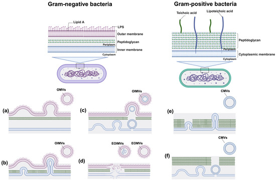
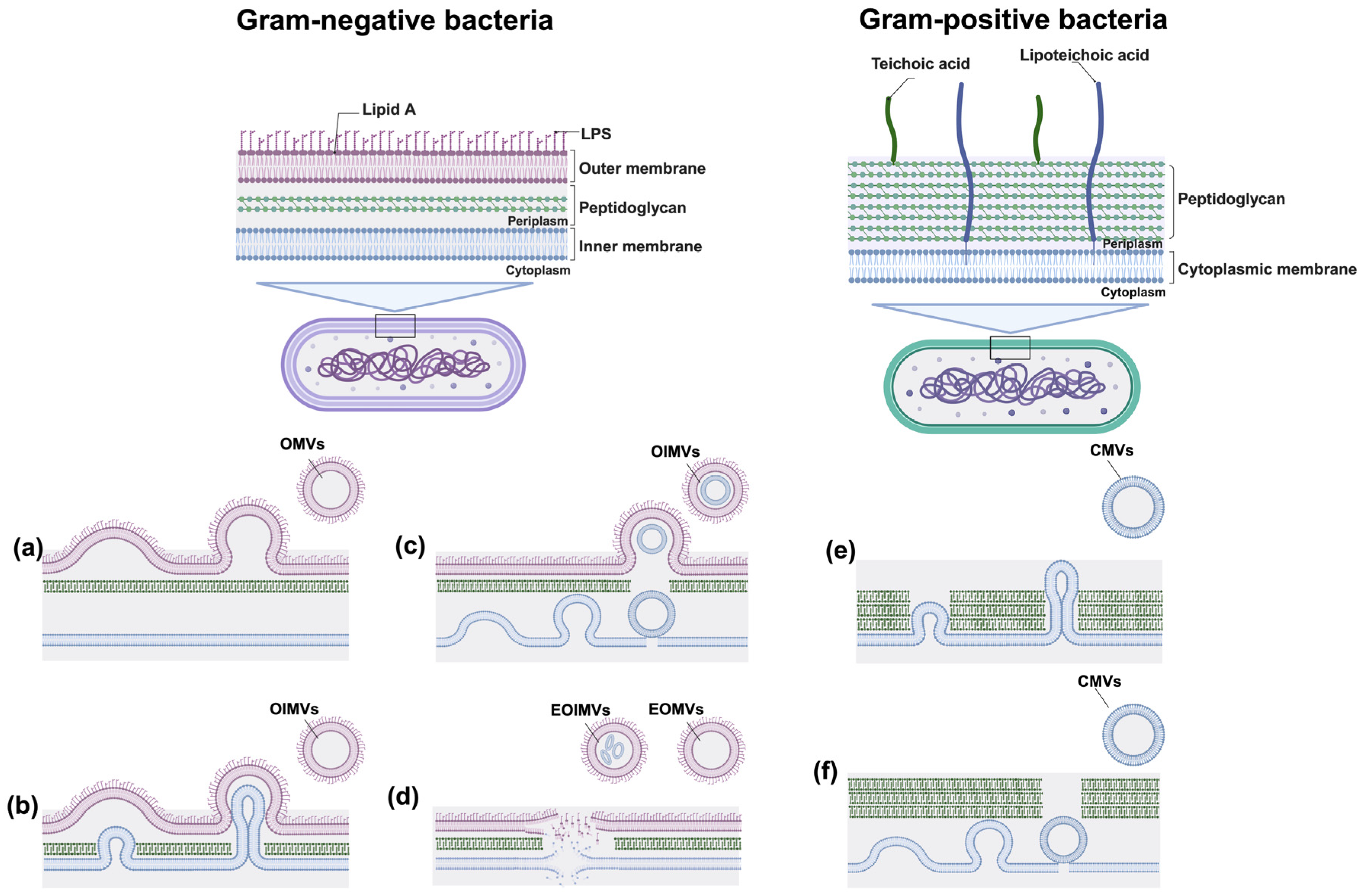
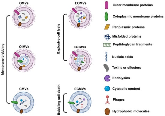
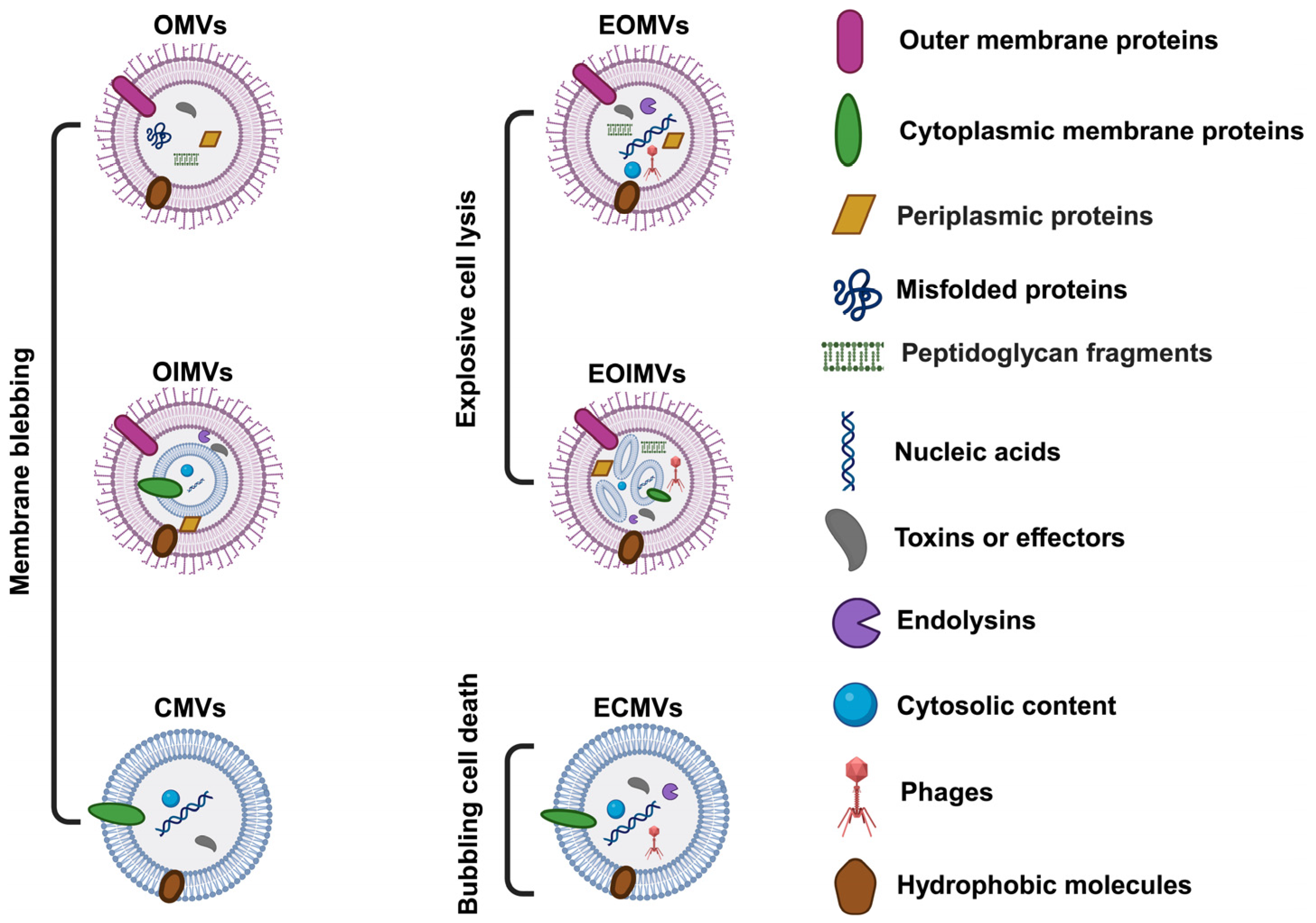
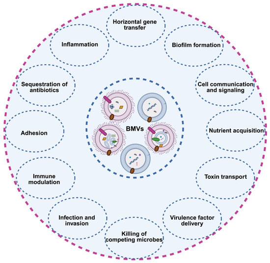
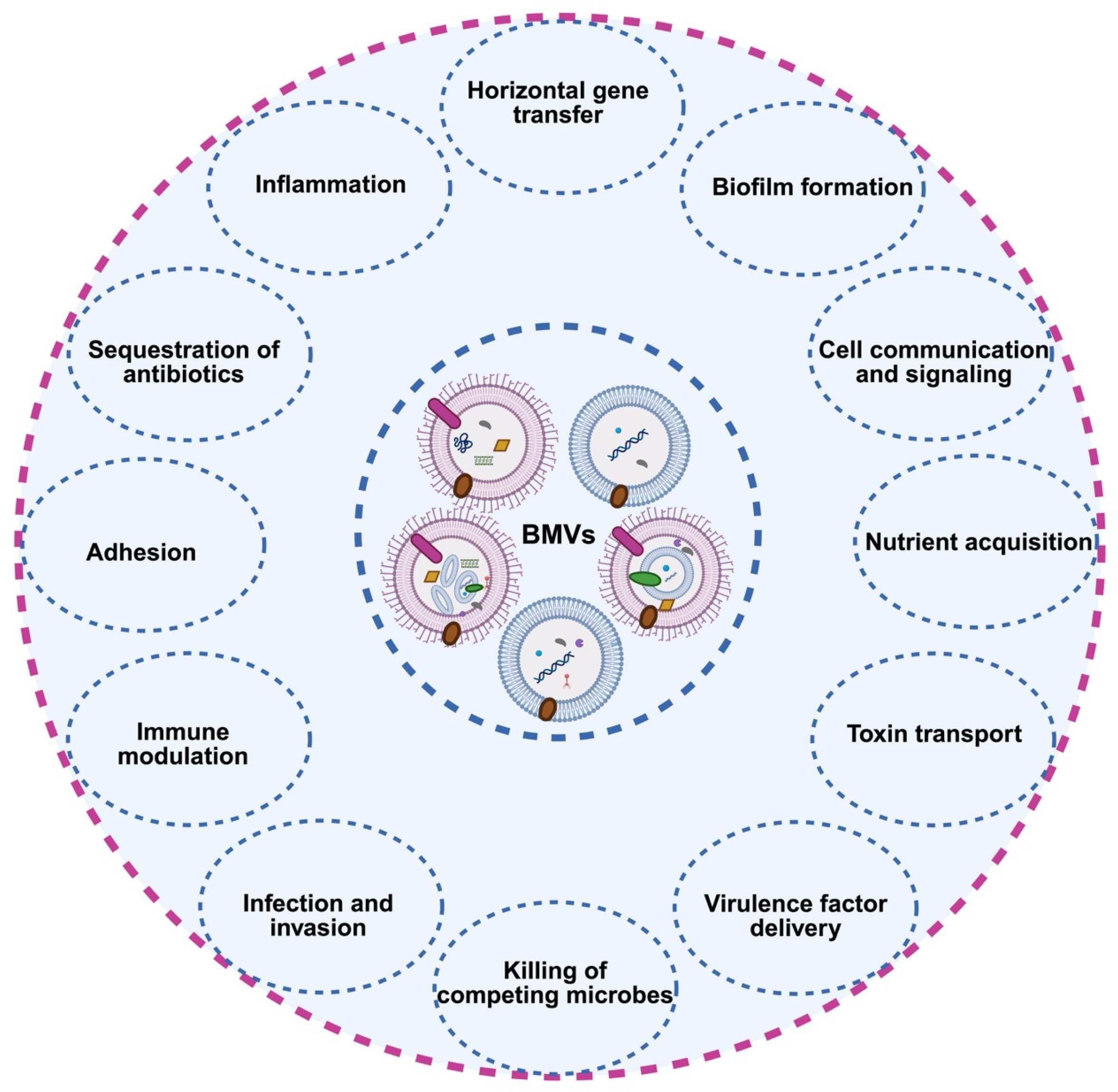
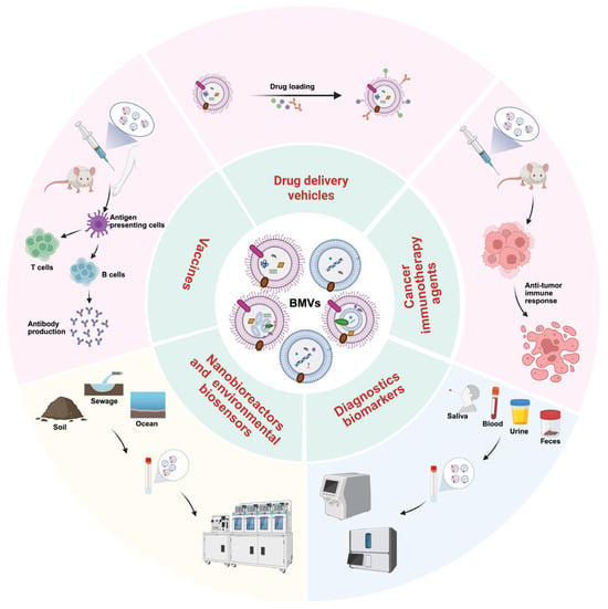
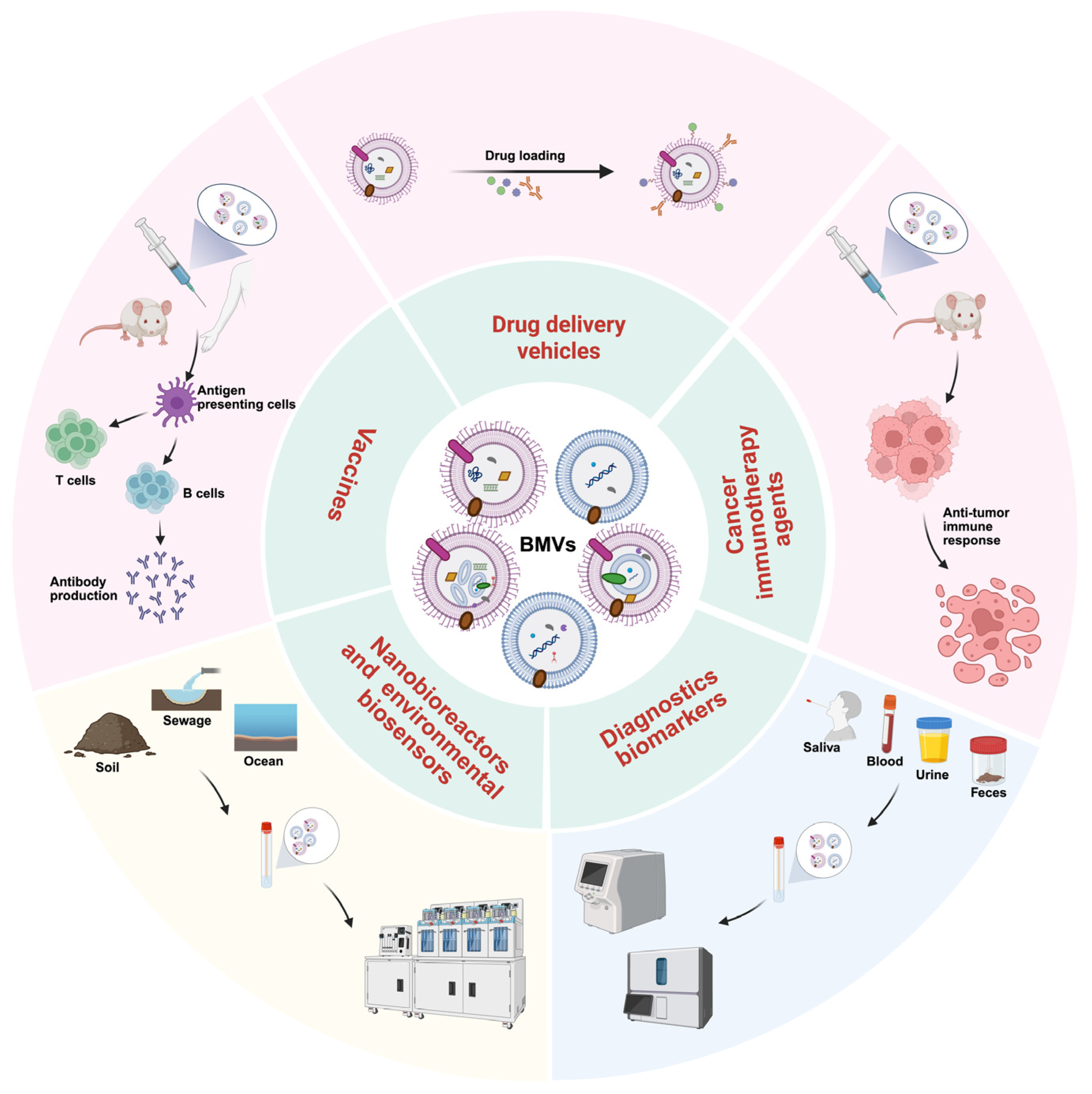

Disclaimer/Publisher’s Note: The statements, opinions and data contained in all publications are solely those of the individual author(s) and contributor(s) and not of MDPI and/or the editor(s). MDPI and/or the editor(s) disclaim responsibility for any injury to people or property resulting from any ideas, methods, instructions or products referred to in the content. |
© 2026 by the authors. Licensee MDPI, Basel, Switzerland. This article is an open access article distributed under the terms and conditions of the Creative Commons Attribution (CC BY) license.