


Abstract
Respiratory viruses with broad host ranges are relevant to veterinary epidemiology, biological safety, and One Health surveillance. This retrospective study integrated companion-animal and wildlife records to examine two complementary surveillance questions: SARS-CoV-2 detection in dogs and cats with close human contact, and influenza A virus detection across a broader multispecies panel that included wild birds. We analyzed 256 archived animal samples collected in Kazakhstan during 2022–2023, comprising 158 dogs, 76 cats, and 22 animals of other domestic and wild species. A previously developed in-house multiplex RT-qPCR assay was used to detect SARS-CoV-2 and influenza A virus RNA, and a previously developed in-house SM-antigen-based ELISA was used to assess SARS-CoV-2-associated serum reactivity. SARS-CoV-2 RNA was detected in 104/256 samples (40.6%; 95% CI, 34.8–46.7), including 68 dogs and 36 cats. Influenza A virus RNA was detected in four wild birds (1.6%): two partridges and two mallards. According to the assay-specific classification recorded in the source dataset, 34 sera were reactive, 14 were doubtful, and 208 were non-reactive. All SARS-CoV-2 RT-qPCR-positive samples were ELISA-non-reactive; all reactive or doubtful ELISA results occurred among RT-qPCR-negative animals. Dogs and cats did not differ significantly in SARS-CoV-2 RT-qPCR positivity, ELISA reactivity, or Ct distributions. Because the ELISA had not been formally validated for the animal species studied, reactivity was not interpreted as confirmed SARS-CoV-2 seropositivity. The archived 2022–2023 dataset therefore provides a historical baseline for multispecies respiratory-virus surveillance in Kazakhstan and supports cautious integration of molecular and serological data.
1. Introduction
Respiratory viruses capable of crossing species barriers are an important focus of veterinary microbiology and One Health surveillance. SARS-CoV-2 is a betacoronavirus with a broad mammalian host range under natural and experimental conditions, and reverse zoonotic transmission from infected people to animals has been documented. Influenza A virus (IAV) likewise infects multiple avian and mammalian hosts and can undergo reassortment and host adaptation. These characteristics make integrated animal surveillance relevant for distinguishing current viral RNA detection from serological evidence of prior or possible exposure [1,2,3,4].
Companion animals are of particular interest because of their close and frequent contact with people. Reported SARS-CoV-2 molecular and serological detection frequencies in dogs and cats vary markedly among studies according to sampling period, exposure setting, clinical status, and study design; non-detection has been reported in some surveys, whereas higher frequencies have been observed in animals from households with confirmed human COVID-19 [2,5,6]. These patterns support the interpretation that observed frequencies are highly context dependent and that most documented infections in pets are associated with human-to-animal transmission rather than sustained independent circulation in dogs or cats.
Serological surveillance in animals requires assays that are validated for the species and antigenic target under investigation. Multi-species ELISAs based on SARS-CoV-2 antigens have demonstrated high diagnostic performance in experimentally or naturally infected animals [7]. Comparative evaluations in cats and dogs indicate that spike-protein-based ELISAs can provide strong diagnostic discrimination when assessed against well-characterized reference sera, while emphasizing the need to control for cross-reactivity and to confirm cut-off values using appropriate positive and negative panels [8]. These considerations are particularly important when an in-house assay is used for field surveillance across more than one animal species.
Influenza A virus (IAV) has an exceptionally broad host range and undergoes continuous genetic and antigenic evolution. Dogs and cats can be infected by influenza viruses of avian, equine, swine, or human origin, and canine-adapted H3N8 and H3N2 lineages illustrate the capacity of IAV to establish transmission in a new mammalian host [9,10,11]. Reassortment and host adaptation in dogs are of One Health concern because companion animals occupy a close ecological interface with humans. A recent meta-analysis confirmed that influenza infection and serological evidence of exposure are reported in dogs worldwide, although estimates vary substantially by geography, study design, and diagnostic method [12].
Kazakhstan spans multiple ecological zones and includes interfaces among people, companion animals, livestock, wildlife, animal shelters, veterinary clinics, and zoological settings. Recent surveillance in Almaty detected influenza A virus RNA in dogs and demonstrated serological evidence of influenza exposure, supporting the relevance of veterinary respiratory-virus monitoring in Kazakhstan [13]. The present study was therefore designed as an integrated epizootic-monitoring analysis using archived animal samples collected during veterinary investigations in 2022–2023. Although the specimens are historical, the dataset remains relevant as a documented baseline for interpreting host-specific detection patterns and for designing prospectively standardized multispecies surveillance in Kazakhstan. Previously developed and analytically characterized in-house multiplex RT-qPCR and SARS-CoV-2 ELISA systems were used as diagnostic tools rather than as objects of primary assay development. The principal objectives were to characterize the distribution of SARS-CoV-2 and influenza A molecular detections across animal species, describe SARS-CoV-2 serological classifications, compare molecular and serological patterns, and evaluate species-specific differences within the monitored dataset. Because the sample set was clinically enriched and not obtained by probability sampling, the reported proportions characterize the monitored animals and are not interpreted as population prevalence estimates. A previous Kazakhstan conference paper reported SARS-CoV-2 detection in domestic animals from the same broader monitoring program [14]. The present article substantially extends that earlier report by integrating influenza A virus molecular surveillance, SARS-CoV-2-associated serological assessment, geographic and temporal analyses, comparative statistics, and a combined interpretation of molecular and serological findings.
2. Materials and Methods
2.1. Study Design and Diagnostic Systems
This retrospective epizootic-monitoring study analyzed 256 archived animal records obtained during veterinary investigations in Kazakhstan in 2022–2023. The two diagnostic systems used in the present analysis were non-commercial, in-house systems developed previously by the authors’ institutions: a multiplex RT-qPCR assay for detection of influenza A virus and SARS-CoV-2 RNA [15] and the SARS-CoV-2-IgG-RIBSP-ELISA for serological testing [16]. Both systems were actually used to generate the molecular and serological results analyzed in this study; they were not additional post hoc tests. Their analytical development and prior characterization are reported in the cited publications, while the present work uses them as tools for multispecies veterinary monitoring. The study focused on host distribution, target-specific molecular detection, serological classification, Ct distributions, and relationships between molecular and serological findings.
2.2. Composition of the In-House ELISA
The SARS-CoV-2-IgG-RIBSP-ELISA was an in-house, non-commercial assay developed previously by the authors’ institutions; it therefore has no commercial catalogue number. The test system is registered as No. RK-VP-2-5999-26. The kit included microplates sensitized with a recombinant chimeric SARS-CoV-2 SM protein, positive and negative control sera, conjugate solution, 20-fold concentrated phosphate-buffered saline with Tween 20 (PBS-T), TMB substrate, and stop reagent. The assay design, recombinant antigen production, and analytical characterization are described in detail in the original method publication [16].
Briefly, the recombinant SM antigen contains a spike-protein fragment spanning amino acids 554–593 and a membrane-protein fragment spanning amino acids 1–19 joined by the flexible glycine/serine-rich SSGGGGSS linker [16]. In the previous human-serum characterization, the reported OD450 cut-off was 0.213, with a sensitivity of 90.48% (95% CI: 80.74–95.56%), a specificity of 93.65% (95% CI: 84.78–97.50%), an AUC of 0.9623, and a positive likelihood ratio of 14.25. These values describe prior human-serum assay characterization and were not used as species-specific sensitivity or specificity estimates for the present animal surveillance dataset.
2.3. Composition of the Multiplex RT-qPCR Kit
The multiplex RT-qPCR reagent set was an in-house system designed for 55 reactions and contained 5× PCR buffer, a primer-and-probe mixture, positive and negative control materials, and nuclease-free deionized water. The positive control was a constructed plasmid DNA preparation (0.01 ng) containing inserts corresponding to a fragment of the influenza A virus M gene and a fragment of the SARS-CoV-2 RdRp gene. The analytical performance of this assay was established previously [15], including testing against a respiratory-virus panel with no observed cross-amplification and an analytical detection limit of 6 target copies per reaction for both viral targets.
2.4. Animal Sampling and Specimen Collection
The archived source register contained 256 animal records collected previously during veterinary monitoring activities in the Republic of Kazakhstan. The panel comprised 158 dogs, 76 cats, and 22 animals of other species, including partridges (n = 4), mallards (n = 3), muskrats (n = 3), chickens (n = 2), hares (n = 2), crows (n = 2), roe deer (n = 2), beavers (n = 2), one lioness, and one fox. Dogs and cats were included because of their close contact with people and because this clinically enriched subset had signs considered compatible with coronavirus infection. The additional species originated from broader veterinary and field-monitoring activities and were retained to preserve the multispecies surveillance scope; importantly, the only influenza A virus detections occurred in wild birds. Results for these small wildlife groups are therefore presented descriptively only and are not used for species-level prevalence inference. Sampling sites included veterinary clinics, temporary holding facilities, zoological or captive settings, and field-monitoring locations in several regions of Kazakhstan. Because the material was assembled previously and subsequently re-used for integrated surveillance analysis, the present dataset should not be interpreted as a population-based prevalence sample. For geographic visualization, the six named geographic subsets were plotted at representative administrative centers solely for geographic orientation; exact specimen-level geographic coordinates were not available in the archived register, and map markers therefore do not denote exact sampling sites.
Nasal and rectal swabs had been collected during the original broader veterinary surveillance program using sterile synthetic swabs and placed in sterile 1.5–2.0 mL tubes containing phosphate-buffered transport medium (pH 7.2–7.4). Nasal swabs are the more appropriate respiratory specimens for influenza A virus surveillance; rectal swabs were included in the original sampling scheme primarily because the program also targeted coronaviruses and other veterinary surveillance objectives. The archived final-status dataset does not contain a complete specimen-type field for every individual record, so the present retrospective analysis cannot reliably stratify influenza A detections by specimen type. Whole blood was collected into sterile vacuum tubes containing a clot activator or separation gel. Blood was allowed to clot at room temperature for 30–45 min and was then centrifuged at 3000 rpm for 10–15 min. The separated serum was transferred into sterile microtubes.
2.5. Transport and Storage
Swab specimens and serum samples were stored at −70 °C until viral RNA extraction or serological testing in order to minimize RNA degradation and preserve antibody reactivity.
2.6. Ethics and Regulatory Compliance
Animal sampling was conducted within veterinary surveillance activities in accordance with applicable regulatory legal acts of the Republic of Kazakhstan. The protocol application for the project under which the animal sampling was performed was reviewed and approved by the Local Commission on Biological Ethics of the Scientific Research Institute for Biological Safety Problems (Protocol No. 5, 29 November 2021). The previously published sampling study reports compliance with national veterinary sampling and biosafety requirements, including Order No. 7-1/393 of the Minister of Agriculture of the Republic of Kazakhstan dated 30 April 2015, which regulates sampling of transported objects and biological material [14].
2.7. Viral RNA Extraction
Viral RNA was extracted under biosafety level 2 (BSL-2) laboratory conditions using the innuPREP Virus DNA/RNA Kit (Innuscreen GmbH, Berlin, Germany) according to the manufacturer’s instructions.
2.8. Multiplex Real-Time RT-PCR
The reaction mixture was prepared in a sterile PCR workstation on a cooling block maintained at approximately 4 °C. Each 25.0 µL reaction contained 6.0 µL of 5× PCR buffer, 7.5 µL of the oligonucleotide primer/probe mixture, 5.5 µL of sterile nuclease-free deionized water, and 6.0 µL of extracted RNA or control material. The 19.0 µL master mix was dispensed into 0.2 mL reaction tubes, followed by addition of the corresponding template or control. Tubes were mixed by gentle pipetting and briefly centrifuged before amplification.
Amplification was performed on a Rotor-Gene Q instrument (otor-Gene Q instrument (QIAGEN GmbH, Hilden, Germany), with fluorescence data analyzed using Rotor-Gene Q Software version 1.8.187.5. The cycling protocol consisted of reverse transcription at 55 °C for 25 min, initial denaturation at 94 °C for 2 min, and 45 cycles of 94 °C for 15 s, 55 °C for 30 s, and 72 °C for 20 s. Fluorescence was recorded in the Green channel for influenza A virus and in the Orange channel for SARS-CoV-2. In the source protocol, a sample was considered positive when an exponential amplification curve was observed with a Ct value ≤38 [15]. In the present dataset, SARS-CoV-2-positive Ct values ranged from 25.87 to 34.99 and influenza A-positive Ct values from 33.12 to 35.25. No sequence-confirmation or independent confirmatory-test results were available in the archived dataset analyzed here; this is therefore acknowledged as a limitation rather than inferred retrospectively.
2.9. SARS-CoV-2 IgG ELISA Procedure
Polysorp microplates (Nunc, Denmark) were coated with recombinant chimeric SM antigen in 0.01 M carbonate-bicarbonate buffer (pH 9.6). After antigen coating, unoccupied binding sites were blocked with 1% bovine serum albumin in PBS-T (137 mM NaCl, 2.7 mM KCl, 10 mM Na2HPO4, 2 mM KH2PO4, 0.1% Tween 20; pH 7.5). Serum samples were diluted 1:100 in PBS-T and incubated for 1 h at 37 °C, after which the wells were washed with PBS-T.
Bound antibodies were detected using Pierce Recombinant Protein G, Peroxidase Conjugated (catalog No. 31499; Thermo Fisher Scientific, Waltham, MA, USA) diluted 1:15,000 and incubated for 30 min at 37 °C. After washing, 100 µL of TMB substrate was added per well and the plates were incubated for 15 min at room temperature in the dark. The reaction was stopped by adding 100 µL of 1 M H2SO4 per well. Optical density (OD) was measured at 450 nm using an ImmunoChem-2100 microplate reader. For the present retrospective analysis, reactive, doubtful, and non-reactive animal-serum results were taken from the final laboratory classification recorded in the archived dataset. The assay had previously been characterized using human sera, and no species-specific diagnostic cut-off, diagnostic sensitivity, or diagnostic specificity had been established for the animal species included here. Accordingly, an ELISA-reactive result was interpreted as reactivity in this assay rather than as independently confirmed SARS-CoV-2 seropositivity. This distinction is consistent with WOAH guidance that diagnostic assays should be validated for the species, specimen matrix, and intended purpose in which they are used [17].
2.10. Statistical Analysis
Descriptive statistics were used to summarize species composition, target-specific RT-qPCR results, Ct values, and final ELISA reactivity classifications. Proportions are reported with 95% Wilson confidence intervals where appropriate. Dogs and cats were compared for SARS-CoV-2 RT-qPCR positivity and ELISA reactivity (reactive versus all other assay classifications) using Fisher’s exact test. Ct distributions among SARS-CoV-2 RT-qPCR-positive dogs and cats were compared using the two-sided Mann–Whitney U test because Ct values were not assumed to be normally distributed. A two-sided p value < 0.05 was considered statistically significant. Because the monitored sample set was clinically enriched and was not assembled by probability sampling, proportions describe findings within the investigated animals and are not interpreted as population prevalence estimates. Molecular and serological results were not used to calculate ELISA sensitivity, specificity, predictive values, or diagnostic agreement because RT-qPCR and serology represent different biological measurements and no independent species-specific serological reference standard was available.
3. Results
3.1. Study Population and Sample Distribution
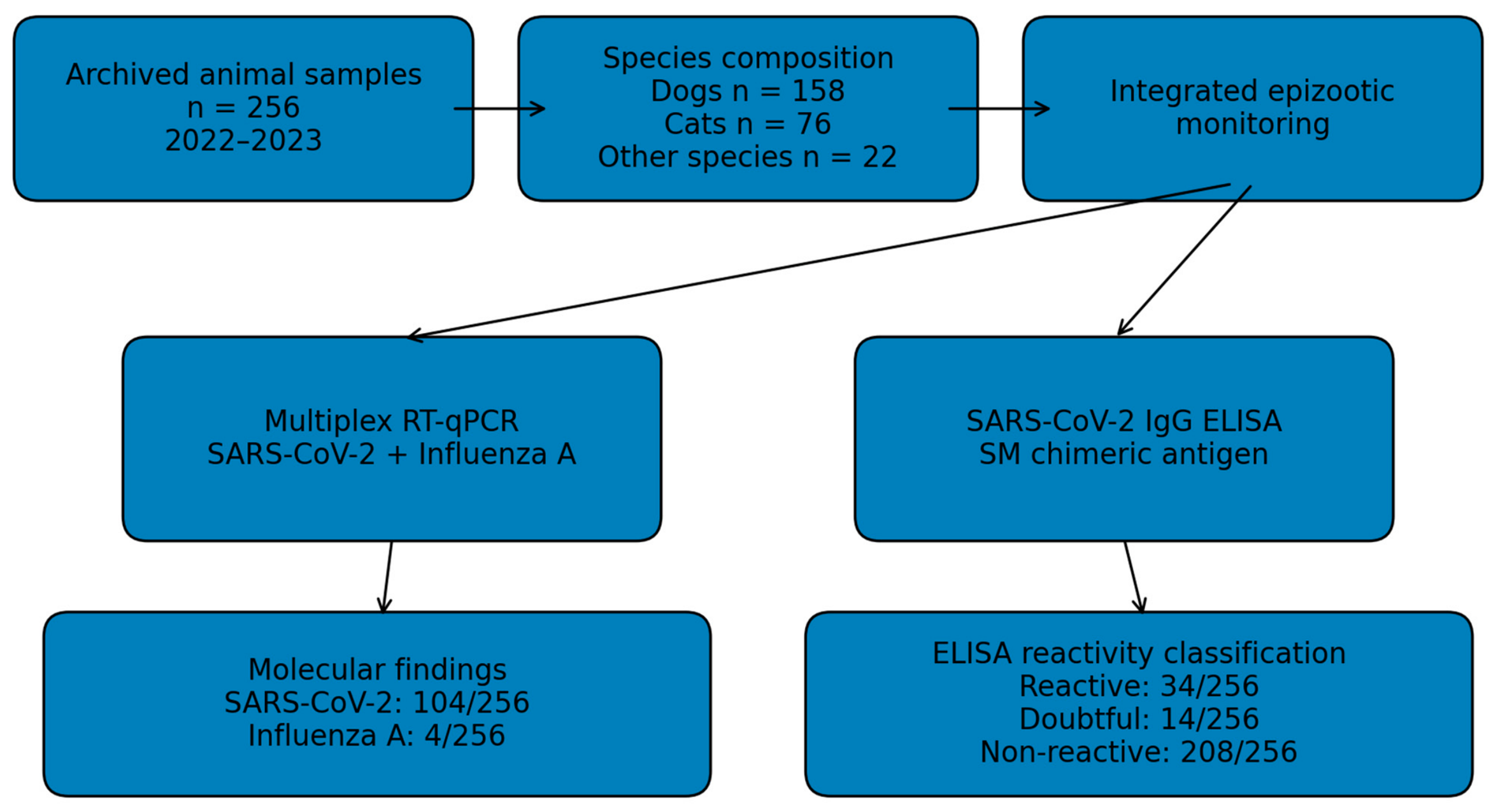
The monitoring dataset contained 256 animal records collected between July 2022 and April 2023. Dogs represented 158/256 (61.7%) records and cats 76/256 (29.7%). The remaining 22/256 (8.6%) records comprised partridges (n = 4), mallards (n = 3), muskrats (n = 3), chickens (n = 2), hares (n = 2), crows (n = 2), roe deer (n = 2), beavers (n = 2), one lioness, and one fox. Dogs and cats together accounted for 234/256 (91.4%) of the monitored animals (Table 1; Figure 1).
Table 1.
Species composition and target-specific findings in the epizootic-monitoring dataset.
Figure 1.
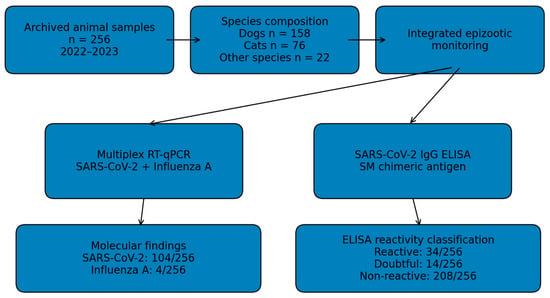
Study design for integrated molecular and serological epizootic monitoring of SARS-CoV-2 and influenza A virus in the 256-animal dataset.
3.2. Geographic and Temporal Distribution
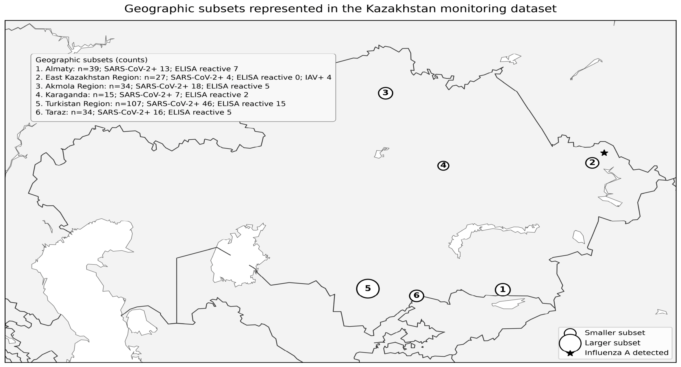
Sampling records covered six geographic subsets in Kazakhstan and four main collection periods between July 2022 and April 2023 (Figure 2, Table 2).The largest subset originated from Turkistan Region (107/256), followed by Almaty (39/256), Akmola Region (34/256), Taraz (34/256), East Kazakhstan Region (27/256), and Karaganda (15/256). SARS-CoV-2 RT-qPCR-positive samples were identified in all six geographic subsets. The highest descriptive proportion of SARS-CoV-2 RT-qPCR positivity was observed in the Akmola subset (18/34, 52.9%), followed by Taraz (16/34, 47.1%), Karaganda (7/15, 46.7%), Turkistan Region (46/107, 43.0%), Almaty (13/39, 33.3%), and East Kazakhstan Region (4/27, 14.8%). These proportions are descriptive and should not be interpreted as regional prevalence estimates because sampling was targeted, the number and composition of animals differed among locations, and the study was not based on probability sampling.
Sampling intensity was markedly uneven across the six geographic subsets. Turkistan Region contributed 107/256 records (41.8%), followed by Almaty (15.2%), Akmola Region and Taraz (13.3% each), East Kazakhstan Region (10.5%), and Karaganda (5.9%). The collection schedule was also clustered: five subsets were sampled between July and December 2022, whereas the Turkistan subset additionally included sampling in April 2023. Consequently, geographic subset, collection period, host composition, and sampling intensity are partly confounded and should not be analyzed as independent regional risk factors.
Table 2.
Geographic and temporal distribution of molecular and serological findings.
Figure 2.
Geographic distribution of the six sampling subsets represented in the Kazakhstan monitoring dataset. Marker area is proportional to the number of records in each subset (n); labels show the numbers of SARS-CoV-2 RT-qPCR-positive and ELISA-reactive samples, and influenza A virus (IAV) detections are indicated where present. Markers are positioned at representative administrative centers for geographic orientation and do not denote exact specimen-level collection coordinates. Regional results are descriptive and do not represent prevalence estimates.
3.3. Molecular Detection of SARS-CoV-2 and Influenza A Virus
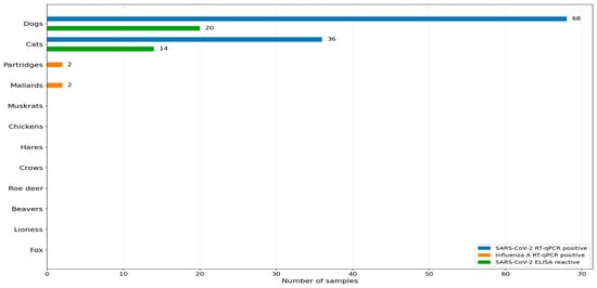
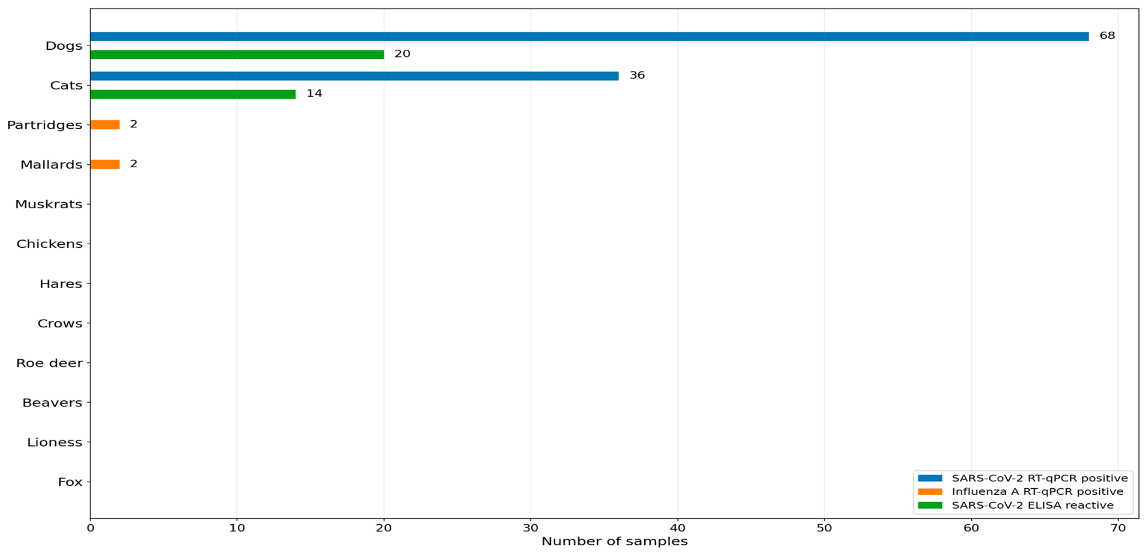
Target-specific RT-qPCR results were available for all 256 records. SARS-CoV-2 RNA was detected in 104/256 samples (40.6%; 95% CI, 34.8–46.7), including 68/158 dogs (43.0%; 95% CI, 35.6–50.8) and 36/76 cats (47.4%; 95% CI, 36.5–58.4). No SARS-CoV-2-positive samples were recorded among the other monitored species. The overall SARS-CoV-2 Ct range was 25.87–34.99, with a mean ± SD of 32.48 ± 1.61 and a median of 32.44 (IQR, 31.22–33.76). Influenza A virus RNA was detected in 4/256 samples (1.6%; 95% CI, 0.6–3.9): two of four partridges and two of three mallards. Influenza A Ct values ranged from 33.12 to 35.25. No dual SARS-CoV-2/influenza A RT-qPCR-positive sample was identified (Table 3; Figure 3).
Figure 3.
Target-specific molecular detections and SARS-CoV-2 ELISA reactivity by animal species. Bars show the number of SARS-CoV-2 RT-qPCR-positive, influenza A RT-qPCR-positive, and SARS-CoV-2 ELISA-reactive samples; species with zero detections or reactivity are retained to show the complete monitored host panel.
Table 3.
Descriptive Ct statistics for target-specific RT-qPCR-positive samples.
3.4. ELISA Reactivity Classification
Field sera were tested in triplicate using the SM-based SARS-CoV-2 ELISA. According to the final assay-specific classification in the source dataset, 34/256 samples (13.3%; 95% CI, 9.7–18.0) were reactive, 208/256 (81.3%) were non-reactive, and 14/256 (5.5%) were doubtful. Among dogs, 20/158 (12.7%; 95% CI, 8.3–18.7) were reactive, 127/158 (80.4%) non-reactive, and 11/158 (7.0%) doubtful. Among cats, 14/76 (18.4%; 95% CI, 11.3–28.6) were reactive, 59/76 (77.6%) non-reactive, and 3/76 (3.9%) doubtful. All 22 samples from other species were classified as ELISA-non-reactive. The previously reported analytical characteristics of the assay were established using human sera [16]; therefore, the animal-serum classifications reported here indicate assay reactivity only and are not interpreted as confirmed SARS-CoV-2 antibody status.
3.5. Molecular–Serological Relationship
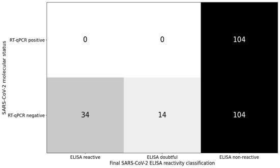
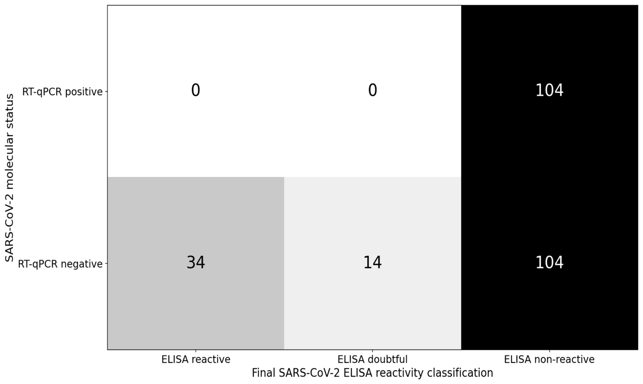
All 104 SARS-CoV-2 RT-qPCR-positive samples were classified as ELISA-non-reactive. Among the 152 SARS-CoV-2 RT-qPCR-negative samples, 34 were ELISA-reactive, 14 were doubtful, and 104 were ELISA-non-reactive (Table 4; Figure 4). Thus, no RT-qPCR-positive sample was simultaneously classified as ELISA-reactive or doubtful. This pattern is reported descriptively and is not treated as a diagnostic-agreement analysis. RT-qPCR detects viral RNA, whereas the ELISA measures antibody-associated binding reactivity; moreover, the ELISA had not been validated for the animal species represented in this dataset.
Table 4.
Relationship between SARS-CoV-2 RT-qPCR status and final ELISA reactivity classification.
Figure 4.
Cross-classification of SARS-CoV-2 RT-qPCR status and final ELISA reactivity classification. Cell values are sample counts. The matrix is descriptive and is not interpreted as diagnostic agreement or confirmed SARS-CoV-2 antibody status because the ELISA had not been species-specifically validated for the animals studied.
3.6. Comparative Analysis of Dogs and Cats
SARS-CoV-2 RT-qPCR positivity did not differ significantly between dogs (68/158, 43.0%) and cats (36/76, 47.4%; Fisher’s exact p = 0.575). ELISA reactivity was also not significantly different between dogs (20/158, 12.7%) and cats (14/76, 18.4%; Fisher’s exact p = 0.242). Among RT-qPCR-positive animals, dogs had a median SARS-CoV-2 Ct of 32.34 (IQR, 31.22–33.70; range, 25.87–34.99), whereas cats had a median Ct of 32.55 (IQR, 31.28–33.76; range, 30.12–34.98). The difference in Ct distributions was not statistically significant (Mann–Whitney U = 1178, p = 0.756). These results do not indicate a detectable species-level difference in molecular findings or assay-defined ELISA reactivity between dogs and cats within the monitored dataset.
4. Discussion
This study provides an integrated molecular and serological description of SARS-CoV-2 and influenza A virus findings in a multispecies epizootic-monitoring dataset from Kazakhstan. Its value lies primarily in documenting host-specific patterns within a clinically enriched historical dataset rather than estimating population prevalence. Three observations are particularly relevant. First, SARS-CoV-2 RNA was detected exclusively in dogs and cats, with comparable proportions and Ct distributions in the two companion-animal groups. Second, influenza A virus RNA was detected only in wild birds, specifically partridges and mallards. Third, molecular and ELISA reactivity profiles did not overlap: RT-qPCR-positive animals were ELISA-non-reactive, whereas ELISA-reactive and doubtful classifications occurred among RT-qPCR-negative animals. Because the ELISA was not species-specifically validated, these serum reactions should be regarded as assay reactivity rather than confirmed evidence of SARS-CoV-2 antibodies. The molecular and serological findings therefore provide distinct, cautiously interpretable layers of surveillance information.
The proportion of SARS-CoV-2 RT-qPCR-positive samples in dogs and cats was higher than that reported in many population-based pet surveys. This difference is expected because the present dataset was clinically enriched rather than randomly sampled, and observed frequencies are strongly influenced by exposure setting, time period, clinical selection, and sampling design. Studies of animals from households with confirmed human COVID-19 have consistently shown higher molecular or serological detection than broad screening populations [2,5,6]. Recent active surveillance in Texas similarly demonstrated that exposure setting strongly shapes observed frequencies: 4.8% of dogs and 13.4% of cats from COVID-19-affected households were RT-qPCR-positive, while neutralizing antibodies were detected in 25% and 36%, respectively [18]. In contrast, Ferrara et al. found evidence of SARS-CoV-2 exposure in hunting and stray dogs in southern Italy using a different sampling context and diagnostic approach [19], while Castillo et al. reported SARS-CoV-2 detection in dogs and cats owned by COVID-19-positive people in southeastern Brazil [20]. These studies reinforce that direct numerical comparisons across surveys require caution. National surveillance data from the United States likewise showed that clinical and epidemiological features of SARS-CoV-2 in dogs and cats varied with surveillance setting and exposure context [21]. Within the broader AR13067641 surveillance project, a dog-derived SARS-CoV-2 isolate, SARS-CoV-2/Canis lupus familiaris/KAZ/CCoV_Almaty_KZ_2022 (GenBank accession PP898122), was previously obtained and characterized [22]. However, the archived dataset analyzed in the present retrospective study does not permit record-level linkage of that isolate to a specific one of the 104 RT-qPCR-positive records; accordingly, it is not treated here as confirmatory testing for the present dataset. The high positivity observed in the present clinically selected dataset should therefore be interpreted together with the previously reported analytical specificity of the multiplex assay [15] and should not be extrapolated as infection prevalence in the general animal population.
Dogs and cats showed similar SARS-CoV-2 RT-qPCR positivity in the present dataset (43.0% versus 47.4%), and the difference was not statistically significant. Likewise, Ct distributions did not differ between the two species. These data do not support a species-level difference in the intensity of detectable viral RNA within this monitored population. However, other studies have reported species-related variation in susceptibility, serological response, and infection probability depending on exposure intensity and study design [5,18]. The absence of a significant difference here should therefore be interpreted within the limits of the available sample size, clinical enrichment, and lack of standardized exposure histories.
The complete separation between SARS-CoV-2 RT-qPCR positivity and ELISA reactivity requires cautious interpretation. It should not be interpreted as proof of sequential infection stages or as evidence that one method invalidates the other. RT-qPCR detects viral RNA, whereas the ELISA measures antibody-associated binding reactivity, and these signals may occur on different biological timescales. One possible explanation is that some animals sampled during detectable RNA shedding had not yet developed measurable antibody responses, whereas antibody-associated reactivity could persist after RNA was no longer detectable. However, timing alone cannot explain the pattern with certainty because the archived records lack standardized dates of exposure, onset of clinical signs, and longitudinal follow-up. In addition, the ELISA had been characterized using human sera and was not formally validated for the animal species studied. Reactive signals may therefore represent SARS-CoV-2-specific antibodies, cross-reactive antibodies, or nonspecific binding, and these possibilities cannot be distinguished from the archived data. Consequently, the 34 reactive and 14 doubtful results among RT-qPCR-negative animals are reported as assay classifications rather than confirmed prior infection. Species-appropriate confirmation, ideally using an independent reference method such as virus neutralization, would be required to establish true SARS-CoV-2 seropositivity and diagnostic agreement [7,8,17].
The influenza A findings extend the monitoring value of the dataset beyond companion animals. The four RT-qPCR-positive influenza A detections occurred in two partridges and two mallards, whereas all mammals were negative for this target. Wild aquatic birds, particularly members of the Anatidae family, are central hosts in the ecology of avian influenza viruses. A 2026 systematic review and meta-analysis of active surveillance in wild birds found substantial taxonomic and ecological variation in avian influenza virus detection and reported particularly important contributions from water-associated and migratory species [23,24]. Although the numbers in the present study are too small for population inference, detection in mallards and partridges supports continued integration of wildlife sampling into national influenza surveillance.
The geographic distribution of positive findings showed descriptive heterogeneity, but the sampling design does not support causal or prevalence-based regional comparisons. Turkistan Region contributed 41.8% of all records, whereas Karaganda contributed only 5.9%, and collection periods differed among subsets. In addition, host composition was not uniform: the East Kazakhstan subset contained the wild-bird samples and was the only geographic subset in which influenza A virus RNA was detected, whereas the other subsets were dominated by companion animals. Geography is therefore partly confounded by host composition, sampling intensity, and collection period. The map should be interpreted as a visualization of dataset coverage rather than a spatial-risk surface.
This study has several limitations. The monitored dataset was not generated by probability sampling and was dominated by dogs and cats; therefore, molecular detection proportions cannot be generalized to national animal populations. Several wildlife species were represented by only one to four animals and are interpreted descriptively. Detailed exposure histories, owner COVID-19 status, timing of clinical signs, and serial follow-up samples were not available for all records. The ELISA had not been formally validated for the animal species studied, and no independent species-appropriate confirmatory serological method was available; accordingly, the observed ELISA classifications indicate assay reactivity rather than confirmed SARS-CoV-2 seropositivity. Likewise, record-level sequence-confirmation or independent confirmatory-test results for the RT-qPCR-positive records were not available in the archived dataset. Although a dog-derived SARS-CoV-2 isolate (PP898122) was obtained previously within the broader surveillance project [22], the archived dataset used here does not establish a one-to-one link between that isolate and a specific RT-qPCR-positive record in the present analysis. Future surveillance would be strengthened by prospective sampling with standardized epidemiological metadata, paired molecular and serological specimens collected at defined intervals, species-specific ELISA validation using appropriate reference panels, confirmatory neutralization testing, virus sequencing or isolation where feasible, and broader spatial and seasonal coverage. The final status workbook does not provide a complete sample-type field for each individual record, preventing reliable stratification of positivity by specimen type from this dataset alone.
From a One Health perspective, the observed host-specific molecular patterns underline the value of multispecies surveillance at interfaces involving people, companion animals, and wildlife. The previously developed multiplex RT-qPCR system enables simultaneous screening for two respiratory-virus targets, while the ELISA provides an additional layer of antibody-associated reactivity. For animal surveillance, however, such serological signals should be interpreted conservatively until species-specific validation and independent confirmation are available. Used within those constraints, combined molecular and serological approaches can broaden epizootic situational awareness and help guide future, prospectively validated veterinary monitoring in Kazakhstan.
5. Conclusions
This study presents integrated molecular and serological epizootic monitoring of SARS-CoV-2 and influenza A virus in a clinically enriched multispecies animal dataset from Kazakhstan. SARS-CoV-2 RNA was detected in 104/256 samples (40.6%), exclusively in dogs and cats, with no significant difference between the two species; this proportion describes the investigated sample set and is not a population prevalence estimate. Influenza A virus RNA was detected in four wild-bird samples (1.6%), comprising two partridges and two mallards. The ELISA classified 34 samples as reactive, 14 as doubtful, and 208 as non-reactive according to the assay-specific criteria recorded in the source dataset. Because the ELISA had not been formally validated for the animal species studied, these results should not be interpreted as confirmed SARS-CoV-2 seropositivity or evidence of prior infection. The non-overlapping molecular and ELISA reactivity patterns therefore warrant cautious interpretation rather than diagnostic equivalence. The archived 2022–2023 findings provide a historical surveillance baseline and support continued integrated monitoring of companion animals and wildlife, with species-specific serological validation and confirmatory testing incorporated into future One Health surveillance.
Author Contributions
Conceptualization, G.D.N., K.D.Z. and S.K.K.; methodology, G.D.N., O.V.C., K.A.S. and A.K.N.; investigation, G.D.N., A.K.N., K.A.S., A.S.N., N.S.K. and A.M.M.; data curation, G.D.N., A.K.N., K.A.S.; writing—original draft preparation, G.D.N.; writing—review and editing, S.K.K., O.V.C., K.D.Z. and A.T.Z.; supervision, K.D.Z. and S.K.K.; project administration, K.D.Z. and O.V.C.; funding acquisition, K.D.Z. All authors have read and agreed to the published version of the manuscript.
Funding
Development and evaluation of the diagnostic systems were conducted within the scientific and technical program ‘Development of New Diagnostic Test Systems for Particularly Dangerous Viral Infections’ (IRN BR24992948), 2024–2026, with support from the Science Committee of the Ministry of Science and Higher Education of the Republic of Kazakhstan.
Institutional Review Board Statement
The protocol application for the project under which the animal sampling was performed was reviewed and approved by the Local Commission on Biological Ethics of the Scientific Research Institute for Biological Safety Problems (Protocol No. 5, 29 November 2021). No additional experimental procedures on animals were performed specifically for this retrospective analysis.
Informed Consent Statement
Not applicable.
Data Availability Statement
The datasets generated and/or analyzed during the current study are available from the corresponding author on reasonable request, subject to institutional and ethical restrictions.
Acknowledgments
The authors thank the staff of the Scientific Research Institute for Biological Safety Problems who participated directly in the experimental stages of the study. During the preparation of this manuscript, the authors used ChatGPT (OpenAI, GPT-4o, web application; accessed in 2026) to assist with language editing, structural refinement, and formatting. All scientific content, data interpretation, conclusions, and references were reviewed and verified by the authors, who take full responsibility for the content of the publication.
Conflicts of Interest
Authors Gulnur D. Nakhanova, Kamshat A. Shorayeva, Aziz K. Nakhanov, Olga V. Chervyakova, Nurlan S. Kozhabergenov, Aibarys M. Melisbek, Sabit K. Kokanov, and Kuandyk D. Zhugunissov were employed by the company Research Institute for Biological Safety Problems LLP, National Holding QazBioPharm JSC. The remaining authors declare that the research was conducted in the absence of any commercial or financial relationships that could be construed as a potential conflict of interest.
References
- World Organisation for Animal Health (WOAH). SARS-CoV-2. Available online: https://www.woah.org/en/disease/sars-cov-2/ (accessed on 22 September 2026).
- Guo, R.; Wolff, C.; Prada, J.M.; Mughini-Gras, L. When COVID-19 Sits on People’s Laps: A Systematic Review of SARS-CoV-2 Infection Prevalence in Household Dogs and Cats. One Health 2023, 16, 100497. [Google Scholar] [CrossRef] [Scilit] [PubMed]
- Silva, M.J.A.; Santana, D.S.; Lima, M.B.M.; Silva, C.S.; de Oliveira, L.G.; Monteiro, E.O.L.; dos Santos Dias, R.; de Kássia Barbosa Pereira, B.; da Silva Nery, P.A.; Ferreira, M.A.S.; et al. Assessment of the Risk Impact of SARS-CoV-2 Infection Prevalence between Cats and Dogs in America and Europe: A Systematic Review and Meta-Analysis. Pathogens 2024, 13, 314. [Google Scholar] [CrossRef] [Scilit] [PubMed]
- Kamel, M.S.; El-Sayed, A.A.; Munds, R.A.; Verma, M.S. Interactions between Humans and Dogs during the COVID-19 Pandemic: Recent Updates and Future Perspectives. Animals 2023, 13, 524. [Google Scholar] [CrossRef] [Scilit] [PubMed]
- Michelitsch, A.; Allendorf, V.; Conraths, F.J.; Gethmann, J.; Schulz, J.; Wernike, K.; Denzin, N. SARS-CoV-2 Infection and Clinical Signs in Cats and Dogs from Confirmed Positive Households in Germany. Viruses 2023, 15, 837. [Google Scholar] [CrossRef] [Scilit] [PubMed]
- Udom, K.; Jairak, W.; Chamsai, E.; Charoenkul, K.; Boonyapisitsopa, S.; Bunpapong, N.; Techakriengkrai, N.; Amonsin, A. Serological Survey of Antibodies against SARS-CoV-2 in Dogs and Cats, Thailand. Transbound. Emerg. Dis. 2022, 69, 2140–2147. [Google Scholar] [CrossRef] [Scilit] [PubMed]
- Wernike, K.; Aebischer, A.; Michelitsch, A.; Hoffmann, D.; Freuling, C.; Balkema-Buschmann, A.; Graaf, A.; Müller, T.; Osterrieder, N.; Rissmann, M.; et al. Multi-Species ELISA for the Detection of Antibodies against SARS-CoV-2 in Animals. Transbound. Emerg. Dis. 2021, 68, 1779–1785. [Google Scholar] [CrossRef] [Scilit] [PubMed]
- Diezma-Díaz, C.; Álvarez-García, G.; Regidor-Cerrillo, J.; Miró, G.; Villanueva-Saz, S.; Pérez, M.D.; Verde, M.T.; Galán-Malo, P.; Brun, A.; Moreno, S.; et al. A Comparative Study of Eight Serological Methods Shows That Spike Protein-Based ELISAs Are the Most Accurate Tests for Serodiagnosing SARS-CoV-2 Infections in Cats and Dogs. Front. Vet. Sci. 2023, 10, 1121935. [Google Scholar] [CrossRef] [Scilit] [PubMed]
- Borland, S.; Gracieux, P.; Jones, M.; Mallet, F.; Yugueros-Marcos, J. Influenza A Virus Infection in Cats and Dogs: A Literature Review in the Light of the One Health Concept. Front. Public Health 2020, 8, 83. [Google Scholar] [CrossRef] [Scilit] [PubMed]
- Klivleyeva, N.G.; Glebova, T.I.; Shamenova, M.G.; Saktaganov, N.T. Influenza A Viruses Circulating in Dogs: A Review of the Scientific Literature. Open Vet. J. 2022, 12, 676–687. [Google Scholar] [CrossRef] [Scilit] [PubMed]
- Parrish, C.R.; Voorhees, I.E.H. H3N8 and H3N2 Canine Influenza Viruses: Understanding These New Viruses in Dogs. Vet. Clin. N. Am. Small Anim. Pract. 2019, 49, 643–649. [Google Scholar] [CrossRef] [Scilit] [PubMed]
- Leung, C.; King, A.N.; Barker, P.R.A.; Alshallal, A.D.; Lee, J.Y.; Su, L. Global Seroprevalence and Prevalence of Infection of Influenza in Dogs (Canis familiaris): A Systematic Review and Meta-Analysis. Rev. Med. Virol. 2024, 34, e2542. [Google Scholar] [CrossRef] [Scilit] [PubMed]
- Glebova, T.I.; Klivleyeva, N.G.; Saktaganov, N.T.; Shamenova, M.G.; Lukmanova, G.V.; Baimukhametova, A.M.; Baiseiit, S.B.; Ongarbayeva, N.S.; Orynkhanov, K.A.; Ametova, A.V.; et al. Circulation of Influenza Viruses in the Dog Population in Kazakhstan (2023–2024). Open Vet. J. 2024, 14, 1896–1904. [Google Scholar] [CrossRef] [Scilit] [PubMed]
- Sadikaliyeva, S.; Shorayeva, K.; Abay, Z.; Jekebekov, K.; Shayakhmetov, Y.; Kalimolda, E.; Omurtay, A.; Kopeyev, S.; Nakhanov, A.; Yespembetov, B.; et al. Detection of Coronavirus among Domestic Animals. Res. Rural. Dev. 2024, 39, 62–66. [Google Scholar] [CrossRef] [Scilit]
- Almezhanova, M.D.; Kozhabergenov, N.S.; Usserbayev, B.S.; Melisbek, A.M.; Shirinbekov, M.Z.h.; Chervyakova, O.V.; Junushov, A.T.; Sultankulova, K.T. Determination of Specificity and Sensitivity of a Test-System for Detecting RNA of Influenza A and SARS-CoV-2 Viruses by Real-Time Multiplex RT-PCR. Microbiol. Virol. 2026, 2, 332–357. [Google Scholar] [CrossRef] [Scilit]
- Nakhanova, G.; Chervyakova, O.; Shorayeva, K.; Issabek, A.; Moldagulova, S.; Zhunushov, A.; Ulankyzy, A.; Zhakypbek, A.; Omurtay, A.; Nakhanov, A.; et al. Development of an ELISA Using Recombinant Chimeric SM Protein for Serological Detection of SARS-CoV-2 Antibodies. Methods Protoc. 2026, 9, 4. [Google Scholar] [CrossRef] [Scilit] [PubMed]
- World Organisation for Animal Health (WOAH). Validation of Diagnostic Assays for Infectious Diseases of Terrestrial Animals. In Terrestrial Manual; WOAH: Paris, France, 2023; Chapter 1.1.6; Available online: https://www.woah.org/fileadmin/Home/eng/Health_standards/tahm/1.01.06_VALIDATION.pdf (accessed on 2 October 2026).
- Pauvolid-Corrêa, A.; Davila, E.; Auckland, L.D.; Zecca, I.B.; Busselman, R.E.; Tang, W.; Roundy, C.M.; Killian, M.L.; Torchetti, M.K.; Jenkins-Moore, M.; et al. Active surveillance reveals common SARS-CoV-2 infections among cats and dogs from households with human COVID-19 cases in Texas, USA. One Health 2026, 22, 101377. [Google Scholar] [CrossRef] [Scilit] [PubMed]
- Ferrara, G.; Pagnini, U.; Montagnaro, S. SARS-CoV-2 Exposure in Hunting and Stray Dogs of Southern Italy. Vet. Res. Commun. 2024, 48, 4037–4042. [Google Scholar] [CrossRef] [Scilit] [PubMed]
- Pérez Castillo, A.; Oliveira-Silva, A.; Colácio, N.; Pereira, M.R.; Fonseca, P.L.C.; Miranda, J.V.O.; Cerqueira, B.L.; Amaral, V.H.B.; Moreira, R.G.; Spanhol, V.C.; et al. Detection of SARS-CoV-2 and Coinfections in Companion Animals from COVID-19-Positive Owners in Southeast, Brazil. Comp. Immunol. Microbiol. Infect. Dis. 2026, 128, 102497. [Google Scholar] [CrossRef] [Scilit] [PubMed]
- Liew, A.Y.; Carpenter, A.; Moore, T.A.; Wallace, R.M.; Hamer, S.A.; Hamer, G.L.; Fischer, R.S.B.; Zecca, I.B.; Davila, E.; Auckland, L.D.; et al. Clinical and epidemiologic features of SARS-CoV-2 in dogs and cats compiled through national surveillance in the United States. J. Am. Vet. Med. Assoc. 2023, 261, 480–489. [Google Scholar] [CrossRef] [Scilit] [PubMed]
- Sadikaliyeva, S.; Kalimolda, E.; Abay, Z.; Shayakhmetov, Y.; Moldagulova, S.; Shorayeva, K.; Jekebekov, K.; Omurtay, A.; Zhunushov, A.; Kozhabergenov, N.; et al. Influence of Physicochemical Factors on the SARS-CoV-2 Strain Isolated from a Dog. Eurasian J. Appl. Biotechnol. 2024, 3, 197–202. [Google Scholar] [CrossRef] [Scilit]
- Dupas, M.-C.; Falcucci, A.; Pittiglio, C.; Roche, X.; Cinardi, G.; Delgado, A.; Seck, I.; Dhingra, M.; Gilbert, M. Patterns of Avian Influenza Virus Detection from Active Surveillance in Wild Birds: A Systematic Review and Meta-Analysis. One Health 2026, 22, 101338. [Google Scholar] [CrossRef] [Scilit] [PubMed]
- Ramasamy, S.; Bustamante, F.; LaBella, L.C.; Cole, S.D.; Armstrong, J.; Thompson, D.L.; Hardham, J.M.; Kuchipudi, S.V.; Tewari, D. Spillover of SARS-CoV-2 to Domestic Dogs in COVID-19-Positive Households: A One Health Surveillance Study. Virus Res. 2025, 360, 199629. [Google Scholar] [CrossRef] [Scilit] [PubMed]

Disclaimer/Publisher’s Note: The statements, opinions and data contained in all publications are solely those of the individual author(s) and contributor(s) and not of MDPI and/or the editor(s). MDPI and/or the editor(s) disclaim responsibility for any injury to people or property resulting from any ideas, methods, instructions or products referred to in the content. |
© 2026 by the authors. Licensee MDPI, Basel, Switzerland. This article is an open access article distributed under the terms and conditions of the Creative Commons Attribution (CC BY) license.