


Abstract
Oropouche virus (OROV) has re-emerged as a significant public health threat during the 2023–2025 outbreak, characterized by an unprecedented geographic expansion beyond endemic regions. The virus has been detected in various regions, including the Caribbean, as well as in Europe and the United States, among travelers from Central and South America. This epidemic was associated with the expansion of a novel reassortant strain, which may exhibit enhanced viral fitness and immune evasion properties. Although orthobunyaviruses are known to counteract interferon responses, the specific strategy adopted by OROV remains incompletely understood. In this study, we compared the in-vitro behavior of the prototype strain BeAn19991 and a 2024 isolate representing the reassortant virus responsible for the 2023–2025 epidemic (ISCDC-2024), focusing on innate immune response. We examined the sensitivity of the two strains to IFN-β antiviral activity and their ability to induce type I IFN in fibroblast. Furthermore, we characterized the innate immune response triggered by in-vitro infection of PBMCs from healthy donors. Although the two viral strains exhibited a similar plateau in viral RNA accumulation and comparable sensitivity to exogenous IFN-β, the IFN-β induction in fibroblasts was delayed, with higher levels induced by ISCDC-2024 than by BeAn19991. Moreover, the two strains displayed distinct innate immune profiles in PBMCs: BeAn19991 predominantly induced a robust antiviral IFN-driven response, whereas ISCDC-2024 infection was associated with an IL-6-dominated inflammatory pattern. Overall, these findings highlight distinct phenotypic features of the contemporary reassortant isolate and suggest strain-specific differences in the modulation of innate immunity, potentially reflecting distinct immune evasion strategies. If confirmed, this hypothesis could have significant implications for infection transmission, viral persistence and disease severity, highlighting the need for continuous surveillance and functional characterization of emerging OROV strains to drive public health interventions and antiviral countermeasures.
1. Introduction
Oropouche virus (OROV) is a re-emerging arbovirus transmitted primarily through the bite of the midge Culicoides paraensis. During 2023–2025, it was responsible for a significant outbreak that, for the first time, affected geographic areas beyond its endemic region, the Amazon Basin. Since August 2025, the Pan American Health Organization (PAHO) has reported cases in all Brazilian federal units, and in Barbados, Bolivia, Colombia, Cuba, Ecuador, Guyana, Panama, Peru, and Venezuela, and travel-associated cases have been observed in Italy, Spain, Germany, France, the Netherlands, the United States, Chile, Uruguay, and Canada [1]. In the past, OROV outbreaks with even higher numbers of cases have occurred, but the game changer in the last outbreak was the spread outside endemic areas, even in areas with low circulation of the vector [2,3]. In light of the ongoing geographic spread and the start of the arbovirus season in the affected regions, the PAHO/WHO urges at-risk countries to strengthen surveillance systems and implement enhanced preventive measures [4,5]. Particular attention should be given to potential alternative routes of transmission, including the possibility of vertical transmission, which could have implications for vulnerable populations such as pregnant women and infants [6,7]. OROV infection typically presents as an acute febrile illness characterized by headache, myalgia, arthralgia, chills, and gastrointestinal symptoms. Most patients recover within approximately one week, although convalescence may extend for several weeks. A biphasic clinical course has been reported in approximately 60–70% of symptomatic cases [8,9,10]. Severe disease is uncommon but may include aseptic meningitis and adverse pregnancy outcomes [8,11,12,13]. The recent emergence of OROV in previously unaffected regions has prompted increasing investigation into its clinical, epidemiological, pathological, and molecular characteristics, providing new insights into the biology and clinical relevance of this emerging human pathogen. Nevertheless, the factors underlying its recent geographic expansion beyond historically endemic areas and the mechanisms contributing to its increased circulation remain poorly understood. Elucidation of these mechanisms is imperative to understand viral transmission dynamics and to improve targeted strategies for surveillance, prevention, and control.
In June 2024, we described the first cases of Oropouche infection, outside the endemic areas, in two unrelated travelers from Cuba who referred for diagnosis to our laboratory at IRCCS Sacro Cuore Don Calabria Hospital, in Veneto region [14,15]. The virus was isolated and the full genome characterization was performed [16]. As previously outlined, the isolated OROV-IRCCS-SCDC-1/2024 strain (ISCDC_2024) belongs to a novel reassortant lineage among Oropouche viruses that was identified during the 2023–2024 OROV epidemic [5,16]. The recent geographic expansion of OROV has been associated with the emergence and expansion of this reassortant lineage, although the factors underlying its increased circulation and geographic spread remain to be fully elucidated.
In this regard, Scachetti and colleagues [17], provided a comprehensive assessment of Oropouche fever in Brazil during the period 2023–2024, suggesting a potential correlation between the increased incidence of the disease and the enhanced replication efficiency of this novel reassortant virus, as well as a reduced neutralizing capacity against the new OROV strain in individuals with acquired immunity. Moreover, Ribeiro Amorim and colleagues previously reported the ability of OROV to infect and to persist at low titers in human Peripheral Blood Mononuclear Cells (PBMCs), albeit inducing an adequate IFN-response [18]. Consistent with these observations, OROV RNA has been detected in whole blood during the acute phase of infection and has been reported to persist for up to 135 days after symptom onset (DSO) [19,20,21]. These findings highlight the importance of understanding how different OROV strains interact with the host innate immune system. PBMCs provide a relevant ex vivo model to investigate these interactions and to explore whether strain-specific differences in innate immune responses may contribute to distinct patterns of viral persistence or dissemination to specific microenvironments, including the central nervous system [22,23] or testes [15].
Another important factor to consider in this context is the role of viral immune-evasion proteins. In several Orthobunyavirus species, the non-structural protein NSs, encoded by the S segment, has been described as an antagonist of the host IFN response, interfering with innate immune sensing and downstream antiviral signaling [22,24,25,26,27]. Similar immune-evasion mechanisms have been reported for other Bunyavirales; however, whether OROV NSs exerts comparable effects remains to be fully elucidated [28,29,30].
To better characterize the biological features of the contemporary ISCDC-2024 reassortant, we compared its sensitivity to the antiviral activity of IFN-β and its ability to induce type I IFN in vitro with those of the prototype strain BeAn19991. We also assessed viral RNA accumulation kinetics as a complementary measure of the interaction between the two strains and the host IFN response. In parallel, we investigated whether ISCDC-2024 and BeAn19991 elicited distinct patterns of innate immune activation by analyzing IFN and pro-inflammatory cytokine responses in PBMCs from healthy donors. Overall, these experiments were designed to identify phenotypic differences between the contemporary reassortant and prototype OROV strains, with particular emphasis on the timing, magnitude, and profile of the innate immune responses associated with their infection.
2. Materials and Methods
2.1. Cells and Viruses
Vero E6 cells (CRL-1586, American Type Culture Collection (ATCC), Manassas, VA, USA) were maintained in Modified Eagle Medium (MEM, Life Technologies Europe, Bleiswijk, The Netherlands) supplemented with 10% Fetal Bovine Serum (FBS; Gibco™, Thermo Fisher Scientific, Waltham, MA, USA), 1% glutamine (Gibco), and 1% penicillin-streptomycin solution (Gibco) at 37 °C, 5% CO2 atmosphere. The Vero E6 cell line was selected since it sustained a genetic deletion that ablated the type I IFN locus [31,32], making them unable to mount an efficient type I IFN specific innate immune response to viral infections.
MRC-5 cells (CCL-171, ATCC) were maintained in Dulbecco’s Modified Eagle’s Medium (D-MEM, Life Technologies Europe, Gibco) with high glucose concentration (4500 mg/L), supplemented with 1% glutamine, 1% penicillin-streptomycin solution and 10% FBS and incubated at 37 °C, 5% CO2 atmosphere.
The ISCDC-2024 clinical isolate [16] (GenBank Acc. N° PQ363070.1), and the historical OROV prototype strain BeAn19991 (GenBank Acc. N° MG747539.1), kindly provided by Professor Jan Felix Drexler (Institute for Virology, Charité—Universitätsmedizin, Berlin), were used to perform the in-vitro experiments. Virus stocks were prepared under BSL-3 conditions by propagation on 80% confluent Vero E6 cell monolayers, with ISCDC-2024 propagated for two passages (P2) and BeAn19991 for one passage (P1) in our laboratory. Given the low passage numbers used to generate the working stocks, no additional viral sequencing was performed after propagation. After 1 h of adsorption, MEM supplemented with 2% FBS, 1% glutamine (EuroClone, Milan, Italy), 1% P/S (Euroclone) was added to the cells. Clear Cytopathic Effect (CPE) was observed starting from 3 to 5 days after infection by light microscopy. Progeny virus strains were harvested by freezing/thawing the cultures three times. The freeze-thawed cultures were clarified by centrifugation (1125 rcf for 10 min at 4 °C) to separate intact viral particles from cellular debris, host-derived proteins, and residual culture medium components. The clarified supernatant was subsequently subjected to membrane filtration using a microfiltration system (Minisart™ NML, 0.2 μm pore size. 10076891 Sartorius, Thermo Fisher Scientific), to remove remaining particulate contaminants while allowing virions to pass through. The resulting viral suspensions were then aliquoted and stored at −80 °C.
Virus titration was performed on Vero E6 cells by limiting dilution assay (each dilution run in triplicate), determining tissue culture infectious dose (TCID50) with the method of Reed & Muench [33]. As controls, the Encephalomyocarditis virus (EMCV; VR-129B, ATCC) was used at MOI 1 as a positive viral control to induce a robust innate immune response, verify the responsiveness of the PBMC system and the performance of the cytokine assays, and determine the maximum CPE activity, while untreated cells served as the cell control. Virus stock titers were: 106.7 TCID50/mL for OROV ISCDC-2024; 106.6 TCID50/mL for BeAn19991 strain and 107.6 TCID50/mL for EMCV.
Fresh peripheral whole blood (10 mL) was collected from healthy donors into EDTA-containing tubes and processed within 4 h of collection. PBMCs were isolated by density-gradient centrifugation using Ficoll-Paque Plus (GE Healthcare, Uppsala, Sweden). Following isolation, PBMCs were washed with 1× phosphate-buffered saline (PBS; Gibco) and resuspended in complete RPMI-1640 medium (Gibco) supplemented with 10% FBS for subsequent analyses.
2.2. OROV Sensitivity to IFN-β
Virus sensitivity to IFN-β was assessed by virus yield inhibition assay. Specifically, semiconfluent 24-h-old Vero E6 cells were treated for 24 h with increasing concentrations of recombinant IFN-β (0.3, 1, 3, 101, 101.5 102, 102.5, 103, 103.5 and 104 IU/mL) (Betaferon, 250 µg/mL, Bayer, Leverkusen, Germany, 1IU corresponding to approximately 30 pg).
After pretreatment, the IFN-β-containing medium was removed, and the cell monolayers were infected with either OROV strain at a multiplicity of infection (MOI) of 0.01. After 1 h of adsorption, the inoculum was removed, the cells were washed twice, and MEM containing 2% FBS was added.
After 48 h, supernatants were collected and back-titrated to assess virus yield. The experiments were performed six times. For each condition, the virus yield was calculated by back-titration on Vero E6 cells as described above. The percentage reduction in viral yield following IFN-β treatment was calculated as: % reduction = [1 − (viral yield in IFN-β-treated cells/viral yield in untreated control cells)] × 100.
2.3. OROV IFN-β Induction on MRC-5 Cells
The MRC-5 cells were seeded at a concentration of 50,000 cells/well in a 24-well plate and after 72 h were infected with both OROV strains at MOI 1 and 0.1. After 1 h of adsorption, the inoculum was removed, monolayers were washed twice and then D-MEM 2%FBS was added. After 1, 12, 24, 48, and 72 h, the supernatants were collected, treated with 0.01% Triton-x (Millipore, Burlington, MA, USA) to inactivate the virus, and frozen at −80 °C. Cell monolayers were covered with phosphate-buffered saline solution (1 mL, PBS) and frozen. The experiments were performed three times for each condition and time point. The IFN-β levels in the cell supernatants were measured using the VeriKine-HS™ Human Interferon Beta TCM ELISA (41415-1, PBL Assay Science, Piscataway, NJ, USA). The optical density was measured at 450 nm using the Varioskan microplate spectrophotometer (Thermo Fisher Scientific), and the data were then fitted to a 4-parameter standard curve in order to determine the IFN-β concentration in pg/mL. The assay has a lower limit of quantification (LLOQ) of 1.2 pg/mL. Measurements below the LLOQ were considered below the quantifiable range and were assigned a surrogate value of 0.90 pg/mL for statistical analysis.
2.4. RNA Extraction and One-Step Real-Time RT-PCR
The viral RNA was obtained from the MRC-5 monolayers using the QIAamp Viral RNA Mini kit (Qiagen, Milan, Italy) following the manufacturer’s instructions. Total OROV-specific RNA was amplified using Naveca and colleagues’ protocol [34], using the FlexStar® RT-PCR Kit Amplification mix 1.5 and the LDT RT-PCR Detection Mix (Altona Diagnostics, Hamburg, Germany). As negative control, the RNA extracted from uninfected MRC-5 cells, was used. All one-step real-time RT-PCRs were performed on the CFX96 Touch Real-Time PCR instrument (Biorad, Milano, Italy). In order to normalize the viral genome amount the human Ribonuclease P protein subunit p30 (RNAseP) (BioRad, Hercules, CA, USA) was used as human housekeeping gene [35]. The OROV RNA Cycle threshold (Ct) was normalized with the RNAseP Ct to obtain the ΔCt value (Ct OROV− Ct RNAseP).
2.5. Type I and II IFNs, and Other Cytokine Induction in Human PBMCs
The kinetics of the innate immune response were evaluated in human PBMCs exposed in-vitro to either OROV strain at MOI 0.1 and 1, and measuring type I and II interferons and other inflammatory cytokines released in the supernatants. PBMCs were freshly isolated from three healthy donors and seeded in 96-well round-bottom plates at a concentration of 2 × 10⁶ cells/mL (200,000 cells per well). Cells were then infected with either the two OROV strains at MOI 0.1 and 1, or EMCV at MOI 1 as control, followed a 1.5-h adsorption period at 37 °C in a 5% CO2 atmosphere. At 24, 48, and 72 h post-infection, plates were centrifuged at 300× g for 10 min, and supernatants were collected, treated with Triton X-100 (final concentration 0.01%; Millipore) to inactivate OROV, and stored at −80 °C. The PBMCs were washed with PBS and pellets were subsequently frozen. The concentration of cytokines (CXCL10, IFN-γ, IL-6, and TNFα) was determined on inactivated supernatant with the Human Magnetic Luminex Assay (LXSAHM-11; R&D Systems, Minneapolis, MN, USA). Samples were diluted 1:2 for all analytes, and measurements were performed under blinded conditions. Data were acquired on a Luminex® 200 analyser and analysed using the Luminex xPONENT® software v4.3 (R&D Systems). All assays were performed according to the manufacturer’s instructions. The IFN-α levels were measured using the VeriKine-HS™ Human Interferon alfa TCM ELISA (41135; PBL Assay Science; the assay has a reported overall LLOQ of 1.95 pg/mL, although the quantification limits vary among individual IFN-α subtypes). The optical density was measured at 450 nm using a Varioskan microplate spectrophotometer (Thermo Fisher Scientific), and the data were then fitted to a 4-parameter standard curve in order to determine the IFN concentration in pg/mL.
2.6. Ethical and Sample Collection
This study was conducted in accordance with the ethical principles of the Declaration of Helsinki. Ethical clearance was obtained from the local Ethics Committee (Comitato Etico per la Sperimentazione Clinica dell’Area Sud Ovest Veneto), Prot. n. 68600 dated 10 December 2024, and subsequent amendment prot.n. 36834 dated 1 June 2026, all subjects involved signed a written informed consent form.
2.7. Statistical Analysis
Continuous variables were expressed as median and interquartile ranges or mean and standard deviation, depending on data distribution. Non-parametric tests were applied according to the data distribution. Differences between groups were evaluated using the Wilcoxon signed-rank test with continuity correction.
For IFN-β release in cell culture experiments, we performed a two-way mixed-effects ANOVA, considering strain as the between-subjects factor and time as the within-subjects factor, including the strain × time interaction. Absolute values were log-transformed [log(x + 1)] before analysis. Post-hoc comparisons between strains at individual time points were performed with Bonferroni correction.
Due to the small sample size, cytokine levels were analyzed by pooling measurements across the three time points (24, 48, and 72 h). Thus, the resulting n = 9 represents repeated measurements from three donors across three time points and does not correspond to nine independent biological replicates. Differences between the BeAn19991 and ISCDC-2024 groups were assessed using the Wilcoxon signed-rank test. This analysis was considered exploratory. Furthermore, mean cytokine values were log10-transformed and standardized by z-score normalization and visualized as a heatmap with hierarchical clustering (Ward linkage). Statistical analysis using Kruskal–Wallis followed by Dunn’s multiple comparisons test confirmed significant differences between clusters (p < 0.001), supporting the biological relevance of the observed grouping.
Finally, the principal component analysis (PCA) was used to reduce dimensionality and identify major sources of variance across experimental conditions. The significance of group separation in the reduced PCA space was assessed visually based on clustering patterns along principal components explaining the highest proportion of variance. Variance explained by each component was reported as a percentage of the total variance. No additional inferential tests were applied to PCA outputs, as this analysis was used for exploratory data interpretation. Statistical significance was set at p < 0.05.
Statistical analyses were performed using R software v4.2.1 (RCore Team, Vienna, Austria), and all graphs were performed using GraphPad Prism (GraphPad Software v10, San Diego, CA, USA).
3. Results
3.1. OROV Strains Showed Different Abilities to Induce IFN-β in Human Fibroblast MRC-5 Cells
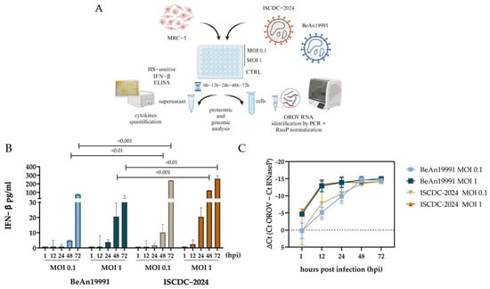
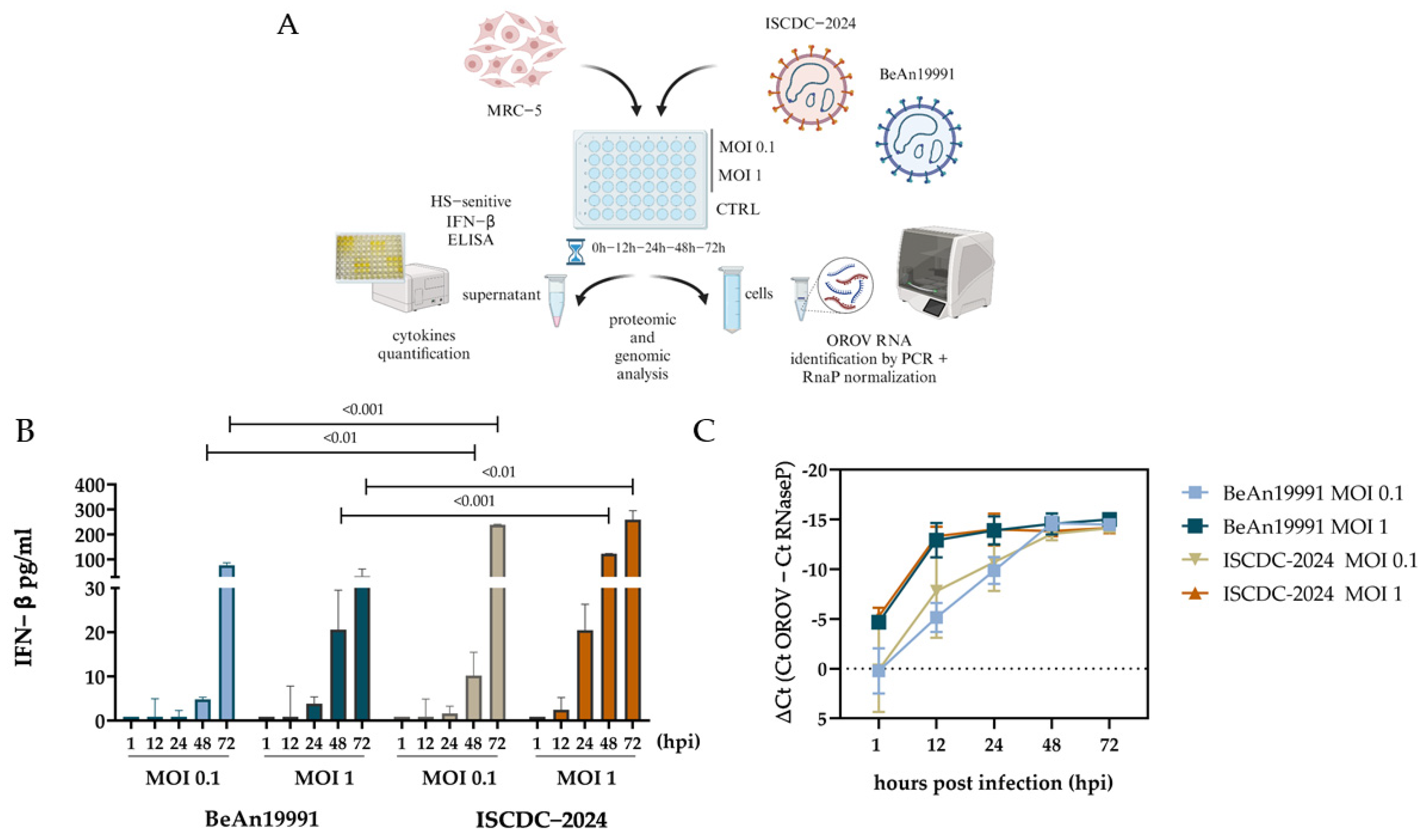
We assessed the IFN-β release over time (1, 12, 24, 48, 72 h) in MRC-5 cells after infection with either OROV strain (BeAn19991 and ISCDC-2024), at two MOIs (0.1 and 1), together with the viral RNA accumulation, as summarized in Figure 1A.
Figure 1.
In-vitro induction of IFN-β and viral RNA accumulation in MRC-5 cells. (A) Schematic representation of experimental design (Created in BioRender. Don calabria, O. S. C. (2026) https://BioRender.com/0ilbep8). (B) Kinetics of IFN-β release following infection with OROV BeAn19991 or ISCDC-2024 at MOIs of 0.1 or 1. IFN-β concentrations are expressed in pg/mL. Bars represent the median, with interquartile range (IQR), from three independent experiments (n = 3). Two-way mixed-effects ANOVA was used, considering strain as the between-subjects factor and time as the within-subjects factor, including the strain × time interaction. Absolute values were log-transformed [log(x + 1)] before analysis. Post-hoc comparisons between strains at individual time points were performed with Bonferroni correction. p-values are in Supplementary Table S1; (C) Time course of viral RNA accumulation in MRC-5 cells infected with OROV BeAn19991 or ISCDC-2024 at MOIs of 0.1 or 1. Viral RNA levels were measured by RT-qPCR at 1, 12, 24, 48, and 72 hpi and expressed as ΔCt values relative to the housekeeping gene RNase P (Ct_OROV − Ct_RNase P). More negative ΔCt values indicate higher viral RNA abundance. Data are presented as mean ± SD from three independent experiments (n = 3).
Both OROV strains induced a dose-dependent increase in IFN-β release, with the highest levels generally observed at MOI 1. IFN-β levels remained close to baseline up to 12 h post-infection (hpi), regardless of viral strain and MOI, suggesting a relatively delayed activation of the IFN-β response. Thereafter, IFN-β levels progressively increased over time, consistent with cumulative cytokine release into the culture supernatant, as shown in Figure 1B. At MOI 1, the contemporary ISCDC-2024 strain induced approximately 20.46 pg/mL of IFN-β at 24 hpi, increasing to more than 123 pg/mL at 48 hpi. In contrast, the prototype BeAn19991 strain showed slower kinetics of IFN-β induction, reaching only approximately 20.56 pg/mL at 48 hpi. Thus, ISCDC-2024 displayed a greater capacity to induce IFN-β during this time interval (Supplementary Table S1).
By 72 hpi, IFN-β levels had further increased for both strains. ISCDC-2024 induced markedly higher concentrations at both MOI 0.1 and MOI 1 (median 238.49 and 258.82 pg/mL, respectively), whereas BeAn19991 reached its highest level at MOI 0.1 (76.87 pg/mL; p < 0.01). Overall, these findings indicate a delayed but progressively increasing IFN-β response, with the contemporary ISCDC-2024 strain inducing a stronger response than BeAn19991, particularly after 48 hpi.
In parallel with IFN-β induction, viral RNA accumulation was assessed in MRC-5 cell lysates by RT-qPCR (Figure 1C and Supplementary Table S2). Viral RNA levels were measured at 1, 12, 24, 48, and 72 hpi and expressed as ΔCt values relative to the housekeeping gene RNase P (Ct_OROV − Ct_RNase P), with more negative ΔCt values indicating higher viral RNA abundance. Both OROV strains showed a progressive increase in viral RNA accumulation over time, reaching comparable levels by 48–72 hpi at both MOIs (ΔCt values ranging from approximately −13 to −14). Overall, the kinetics of viral RNA accumulation in MRC-5 cells were similar for the two OROV strains.
3.2. OROV Strain Sensitivity to IFN-β in Vero E6
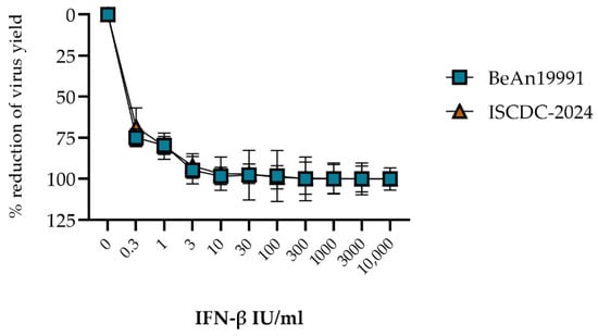
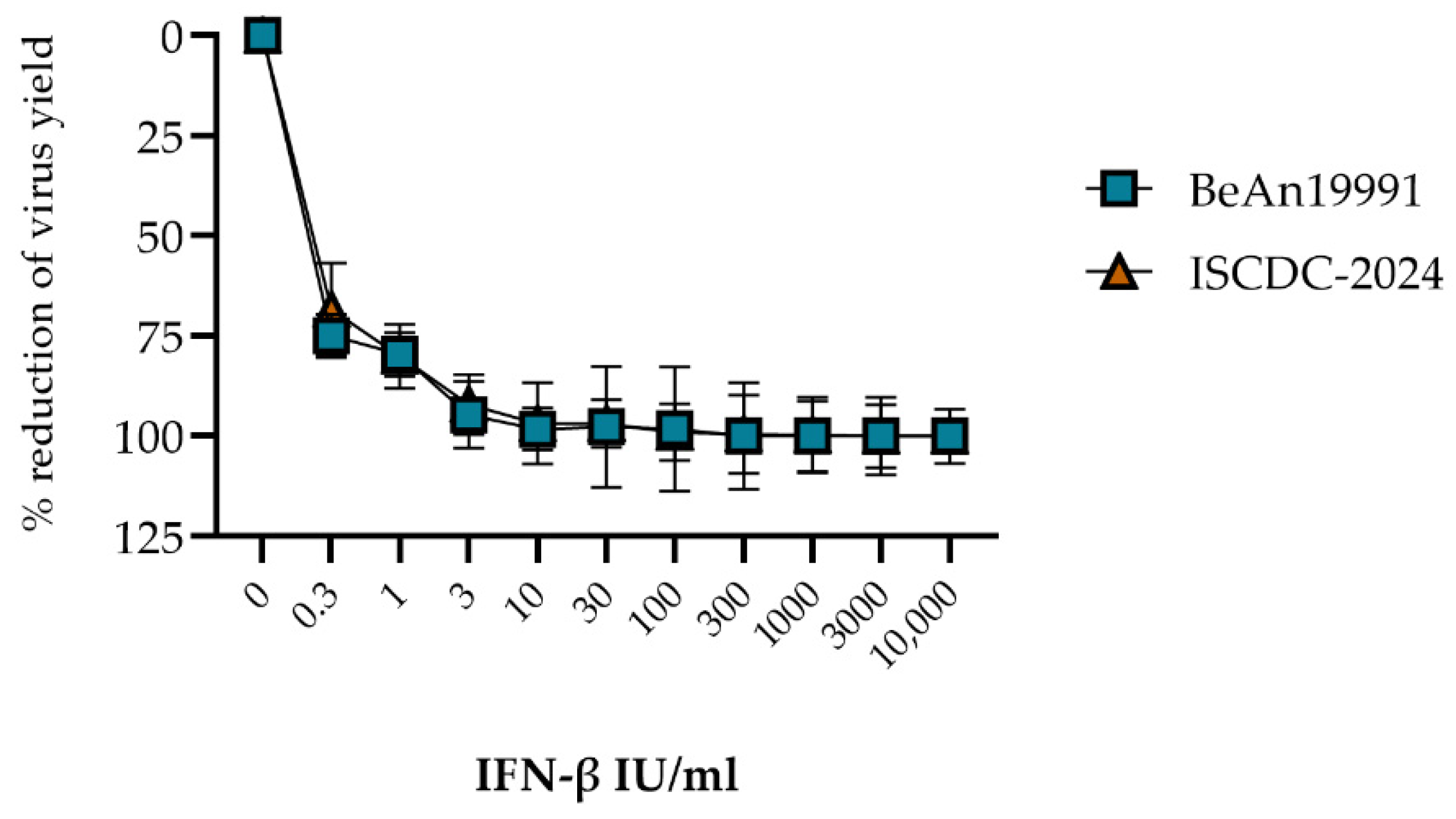
To evaluate the sensitivity of OROV to IFN-β, Vero E6 cells were pretreated with increasing concentrations of recombinant IFN-β. Both OROV strains exhibited a marked dose-dependent reduction in virus yield in response to IFN-β treatment (Figure 2 and Supplementary Table S3). Notably, pretreatment with IFN-β even at the concentration of 0.3 IU/mL reduced viral production by approximately 70–75%, and concentrations ≥3 IU/mL resulted in >90% inhibition. At higher IFN-β concentrations, viral replication was almost completely suppressed. No substantial differences in response to IFN-β treatment were observed between the historical BeAn19991 strain and the contemporary ISCDC-2024 isolate, indicating that both viruses retain a high sensitivity to type I interferon-mediated antiviral activity.
Figure 2.
In-vitro inhibition of virus replication by pretreatment of Vero E6 cells with recombinant IFN-β. Assessment of IFN-β antiviral activity against the replication of the two OROV strains: BeAn19991 (blue square) and ISCDC-2024 (orange triangle). Data are presented as mean percentage reduction in viral replication relative to untreated controls. The percentage reduction in virus yield was assessed by OROV back-titration and is reported as the mean ± SD over six independent experiments.
3.3. Different Levels of Innate Immune Response in PBMC Cultures Exposed to Either OROV Strain
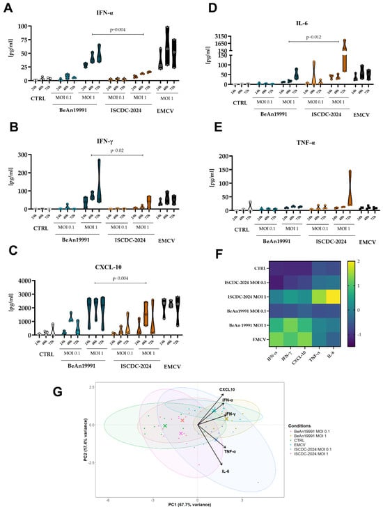
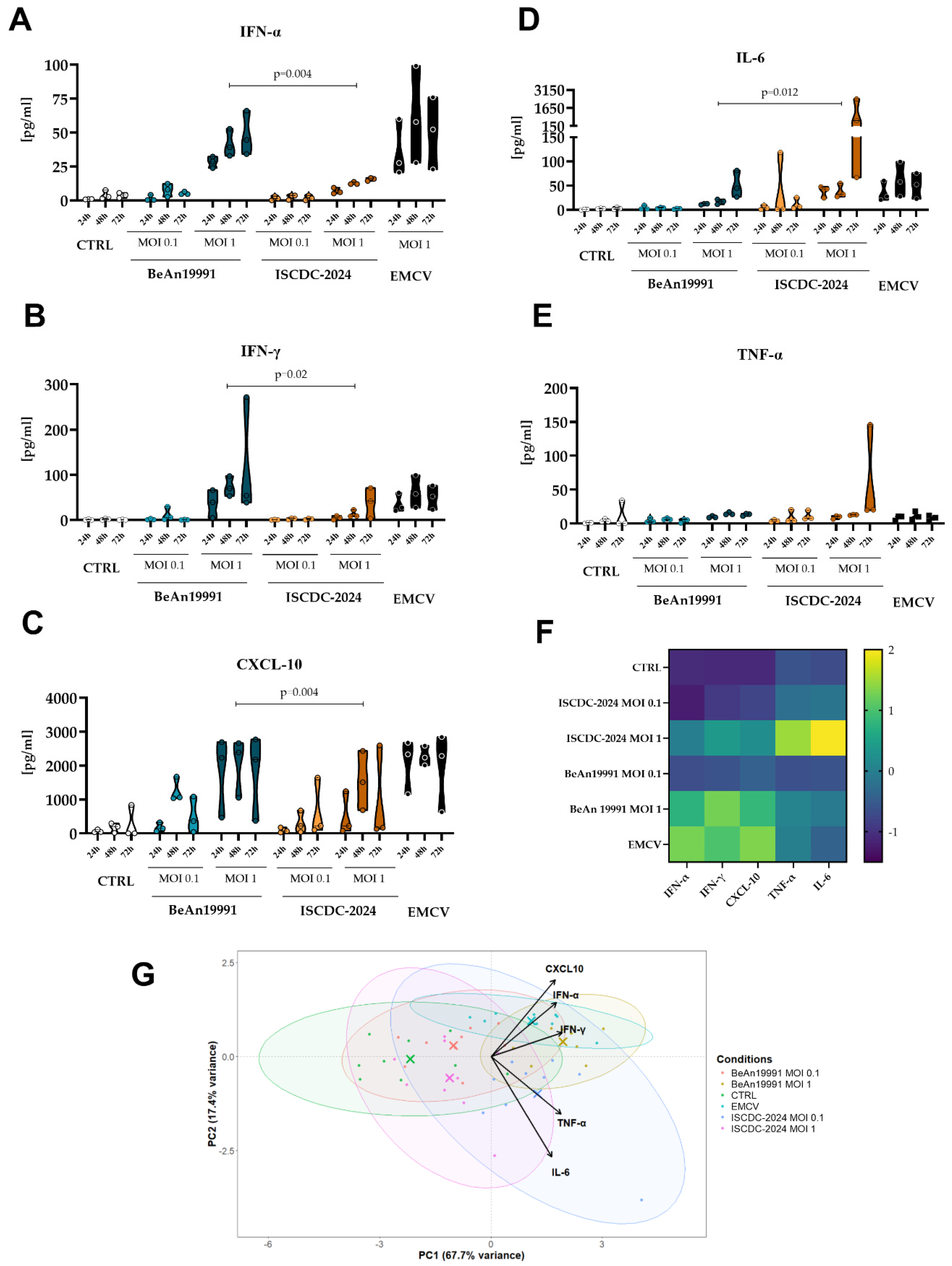
We explored the kinetics of the innate immune response in PBMCs exposed to OROV strain preparations (BeAn19991 and ISCDC-2024) at different MOIs (0.1 and 1) by measuring the cytokine production profile. The results indicate a significantly higher production of IFN-α in PBMCs exposed to the BeAn19991 strain (mean 44.98± 10.6 pg/mL), particularly at MOI 1, compared to the ISCDC-2024 strain (mean 15.63 ± 0.6 pg/mL), after 72 hpi (Figure 3A, Supplementary Table S4A,B).
Figure 3.
Time course of in-vitro induced cytokine release after challenge with different OROV strains on PBMCs. (A–E) Truncated Violin plots represent the distribution of cytokine concentrations (pg/mL) measured in PBMC supernatants at 24, 48, and 72 h after exposure to the OROV prototype strain BeAn19991 or the contemporary isolate ISCDC-2024 at MOIs of 0.1 and 1. Individual values are shown as dots, with the median indicated by the horizontal line within each violin. Uninfected PBMCs (CTRL) and EMCV-infected PBMCs (MOI 1) are shown as controls. For statistical analysis, cytokine levels measured at the different time points were pooled, as reported in Supplementary Table S4. p-values were calculated using the exact Wilcoxon signed-rank test to compare BeAn19991 and ISCDC-2024. (F) Heat map of cytokine expression profiles (IFN-α, IFN-γ, IL-6, TNF-α, and CXCL10) across different viral conditions (CTRL, BeAn19991 MOI 0.1, BeAn19991 MOI 1, ISCDC-2024 MOI 1, EMCV). Data from all biological replicates (D1–D3) and time points (24, 48, 72 h) were pooled (n = 9 per condition), log10(x + 1)-transformed, and standardized by z-score normalization to evaluate overall virus-specific immune signatures. The heatmap was generated using hierarchical clustering with Ward linkage. Color intensity indicates normalized log-scaled cytokine expression levels. (G) Principal component analysis (PCA) of cytokine response. PCA was performed on log10-transformed cytokine concentrations (IFN-α, IFN-γ, IL-6, TNF-α, CXCL-10). Each point represents a PBMC result for each virus concentration at each time point. Group centroids (represented with X) were calculated for visualization purposes. PC1 reflects the overall strength of the immune response, separating control samples from infected and stimulated conditions in a dose-dependent manner. PC2 captures differences in type of response. PCA based on cytokine expression data showed that PC1 and PC2 explained 85.1% of the total variance (PC1 = 67.7%, PC2 = 17.4%).
A similar trend was observed for IFN-γ (Figure 3B) and its induced protein CXCL-10 (Interferon-Induced Protein 10, IP-10) (Figure 3C), with higher levels induced by BeAn19991, especially at MOI 1. In detail, at 72 hpi, the IFN-γ level in the supernatant of PBMCs exposed to BeAn19991 was 2.8 times higher than that measured in PBMCs exposed to ISCDC-2024 (111.6 vs. 38.8 pg/mL, respectively).
CXCL10 levels followed a clear time-dependent, bell-shaped profile. Overall, the prototype strain BeAn19991 elicited a stronger response than ISCDC-2024. At 24 hpi, CXCL10 levels induced by BeAn19991 ranged from 179.8 pg/mL at MOI 0.1 to 1796 pg/mL at MOI 1, whereas ISCDC-2024 induced lower levels, ranging from 89.67 pg/mL at MOI 0.1 to 514.0 pg/mL at MOI 1. BeAn19991 reached peak CXCL10 concentrations at 48 hpi, with approximately 1258 pg/mL at MOI 0.1 and 2034 pg/mL at MOI 1, followed by a decline at 72 hpi (498 pg/mL and approximately 1773 pg/mL respectively). ISCDC-2024 showed a similar bell-shaped temporal profile, but with consistently lower CXCL10 levels across all time points.
In contrast, IL-6 levels were markedly higher following exposure to ISCDC-2024 than to BeAn19991 (mean 831.1 vs. 44.25 pg/mL, respectively), particularly at an MOI of 1 after 72 hpi (Figure 3D). Similarly, TNF-α levels tended to be higher following exposure to ISCDC-2024, especially at an MOI of 1 and at later time points (58.69 vs. 13.2 pg/mL, respectively); however, this difference did not reach statistical significance and should therefore be interpreted cautiously (Figure 3E).
Given the variability observed in cytokine production across donors and time points, cytokine levels measured at the different time points were pooled to provide an overall representation of the cytokine profiles elicited by the two OROV strains and visualized as a heatmap (Figure 3F). The heatmap revealed distinct cytokine-specific patterns, with strong IFN- and CXCL10-driven responses following exposure to BeAn19991 at MOI 1 and to EMCV, whereas ISCDC-2024 at MOI 1 showed a more inflammatory profile, characterized mainly by IL-6 and TNF-α. The cytokine profile induced by EMCV was similar to that observed following exposure to BeAn19991 at MOI 1.
To further assess these differences using a multivariate approach, an unsupervised principal component analysis (PCA) was performed on the complete cytokine dataset, without prior assignment of samples according to strain or experimental condition. As shown in Figure 3G, PCA revealed a clear separation between the two OROV strains, supporting the distinct cytokine profiles associated with PBMC exposure to BeAn19991 and ISCDC-2024 described above. PC1 represented the main source of variability and was strongly associated with the overall magnitude of the immune response, reflecting a global gradient in cytokine production. Along this axis, control samples clustered on the negative side, whereas infected and stimulated conditions showed a progressive shift toward higher PC1 values, with either BeAn19991 and ISCDC-2024 at MOI 1 and EMCV showing the strongest displacement, consistent with the highest immune activation. PC2 captured secondary variability related to the qualitative composition of the cytokine response rather than its overall intensity. This axis was mainly influenced by IFN-expression. Overall, PC2 appeared to distinguish inflammatory and IFN-associated expression profiles across conditions. Two major clusters emerged: a low-response group (CTRL, BeAn19991 and ISCDC-2024 at MOI 0.1) and a high-response group (BeAn19991, ISCDC-2024 and EMCV at MOI 1). At MOI 1 BeAn19991 and EMCV showed strong induction of interferon-related cytokines (IFN-α, IFN-γ, CXCL-10), while ISCDC-2024 infection was characterized by a distinct IL-6–driven inflammatory profile. Overall, BeAn19991 and EMCV share a strong antiviral signature (PC1+), whereas ISCDC-2024 displays a strong IL-6-driven signature (PC2+). Results obtained with both OROV strains at MOI 0.1 and the CTRL condition cluster together, representing a baseline-like state. The variance analysis revealed that the first two components accounted for 85.1% of the total variance in the dataset (PC1 = 67.7%, PC2 = 17.4%).
4. Discussion
Oropouche virus has resurfaced as a major public health concern in the Americas during 2023–2025, with over 23,000 confirmed cases reported to date [1,11]. A key difference between this outbreak and previous OROV outbreaks was the unprecedented geographic expansion of the virus beyond its endemic areas [2,36].
The 2023–2025 OROV epidemic appears to be driven by a novel viral lineage resulting from a reassortment among Oropouche viruses [5,14]. Scachetti et al., reported that an isolate from the 2023–2025 OROV epidemic in Brazil, isolate AM0088, showed increased replication efficiency and seemed to be able to evade the immune response in individuals who previously had acquired immunity, indicating possible immune escape mechanisms [17]. During the 2023–2025 OROV epidemic, we obtained the viral isolate (ISCDC-2024) from a patient returning from Cuba. Although the ISCDC-2024 strain represents only one of the contemporary reassortant strains circulating during the 2023–2025 epidemic and should not be considered representative of all reassortant OROV strains, based on its genomic and phylogenetic characterization [16,36], and in agreement with WHO reports [4], ISCDC-2024 is representative of the novel reassortant OROV lineage associated with the ongoing epidemic.
In our preceding manuscript, we described the ability of the ISCDC-2024 isolate to persist in replication-competent form in whole blood and semen for an extended period, up to 135 and 86 days post-symptom onset, respectively, suggesting that OROV can persist in specific biological compartments longer than previously expected [19,37].
This finding, together with the widespread circulation of OROV during the recent epidemic, underscores the need to further investigate the interaction between contemporary OROV strains and the host immune system. Future studies should help elucidate the biological significance of the phenotypic differences observed between ISCDC-2024 and prototype strains and determine whether these findings are representative of contemporary OROV lineages more broadly.
Considering the central role of the IFN response in antiviral immunity and the ability of Orthobunyaviruses to modulate different components of the innate immune response, including both IFN induction and IFN-mediated signaling [18], characterization of the IFN response and, more broadly, of the innate immune profile elicited by OROV infection is important for further defining the interaction between contemporary OROV strains and the host immune system.
To address this knowledge gap, we characterized the biological phenotype of the contemporary OROV strain ISCDC-2024 in comparison with the prototype strain BeAn19991. We focused on IFN-β induction, viral RNA accumulation kinetics, and sensitivity to exogenous IFN-β in adherent cell culture models, as well as on the cytokine profile elicited in human PBMC cultures.
First, we demonstrated that both ISCDC-2024 and BeAn19991 elicited a time- and dose-dependent IFN-β response in human fibroblast cultures. Overall, IFN-β production remained relatively modest compared with the robust interferon responses typically observed with many RNA viruses, consistent with the reported ability of bunyaviruses to suppress type I IFN induction [27,38,39]. Notably, in the fibroblast model used here, IFN-β became detectable only from 48 hpi onward, suggesting a delayed onset of the response. At later time points, IFN-β levels were higher following infection with ISCDC-2024 than with BeAn19991 (Figure 1A), suggesting that the contemporary strain elicits a quantitatively distinct IFN-β response under the experimental conditions tested.
Previous studies have shown that OROV NSs protein impairs IFN signaling by promoting RNA polymerase II (RPB1) degradation and reducing IFN-β transcription [22,26,27]. These findings support a role for NSs as a viral antagonist of the type I interferon response and as a potential immune evasion factor [40,41,42,43].
Although this study was not designed to directly investigate the mechanistic contribution of NSs to the strain-associated differences in IFN induction observed here, recent evidence supports a role for OROV NSs in modulating the type I IFN response. In a collaborative study involving the newly characterized ISCDC-2024 strain [44], OROV NSs was shown to modulate type I IFN responses in fibroblasts, supporting its role as a potential regulator of innate immune responses. However, these findings do not establish a direct contribution of NSs to the strain-specific phenotypic differences observed in the present study. The experiments were conducted independently, using distinct datasets and experimental conditions, including different MOIs and sampling time points. Although the results are not directly comparable quantitatively, the findings are broadly consistent.
Considering viral RNA accumulation in fibroblasts, we observed similar kinetics between the two strains, with a marked increase during the early phase of infection and comparable plateau levels at 48–72 hpi. Notably, this relatively similar early viral RNA accumulation was accompanied by a delayed IFN-β response, which became evident only at later time points. Thus, the delayed induction of IFN-β observed with ISCDC-2024 does not appear to simply reflect a delayed accumulation of viral RNA during the early phase of infection, suggesting a differential timing of the host IFN response despite comparable early viral RNA kinetics.
Moreover, our data on Vero E6 cells demonstrated that both strains are highly sensitive to the antiviral activity induced by exogenous IFN-β (Figure 2, Supplementary Table S3). The marked antiviral effect observed at sub-IU concentrations (0.3 IU/mL) of IFN-β suggests that even the relatively low amounts of IFN-β induced early in infected fibroblast cultures may substantially contribute to limiting OROV infectivity. This finding is in agreement with a recent report by Wang and colleagues, who observed that the contemporary strain ISCDC-2024 is highly sensitive to type I and III IFNs in a human intestinal organoid study model [45].
Interestingly, analysis of the innate immune responses elicited in human PBMCs by the two OROV strains revealed distinct cytokine profiles (Figure 3). Specifically the prototype strain BeAn19991 primarily induced a typical antiviral response, characterized by higher production of type I and II IFN and CXCL-10 [46,47,48], whereas the contemporary strain ISCDC-2024 appeared to promote a shift from a predominantly IFN-associated response toward a more IL-6-dominated inflammatory profile, compare with BeAn19991.
These findings may reflect differential modulation of innate immune pathways and are consistent with previous observations showing that orthobunyaviruses can shape host innate immune responses according to the cellular and tissue context, potentially influencing viral replication and disease outcome [49,50,51]. Moreover, the observed differences in cytokine profiles are consistent with previous studies reporting the induction of interferon and inflammatory responses following OROV exposure of human PBMCs and in patients with Oropouche fever [21,52].
The described imbalance between antiviral and inflammatory programs observed between the two strains warrant further experimental investigation, as they may provide insights into mechanisms potentially relevant to OROV pathogenesis [8,53,54,55,56,57,58].
A strength of the present study is the direct phenotypic comparison of a contemporary OROV reassortant strain, ISCDC-2024, with the historical prototype BeAn19991, integrating complementary experimental approaches to characterize viral RNA accumulation, IFN-β induction and sensitivity to exogenous IFN-β, as well as the innate immune response elicited in human PBMCs.
These findings should nevertheless be interpreted in light of some limitations. First, productive virus replication was not assessed in the PBMC model. Previous studies have shown that virus-induced activation of interferon and other cytokines in PBMCs may occur in the absence of productive infection and can be triggered by interactions between viral components and immune cells [59,60,61,62,63,64]. At the same time, BeAn19991 has been shown to infect human PBMCs and maintain low viral titers in these cells, suggesting that productive infection may also occur in this cellular context and potentially contribute to viral persistence in specific microenvironments [18]. Although a contribution of cellular components present in the virus preparations or differences in cell viability cannot be completely excluded, the same cell substrate was used to generate both virus stocks, and the distinct cytokine profiles observed following exposure to the two strains argue against a major non-specific effect.
Second, viral RNA levels in PBMCs remained relatively stable across the time points analyzed, suggesting that no substantial further accumulation occurred within the experimental time frame and limiting the additional informative value of later measurements. Finally, the limited number of independent PBMC donors restricts the statistical power and generalizability of these findings; therefore, the PBMC data should be considered preliminary and exploratory and require confirmation in larger cohorts and, ideally, in ex vivo or clinical samples.
Future investigation including clinical samples from OROV-infected patients will be important to determine whether the cytokine and IFN response profiles observed in our experimental models are recapitulated in-vivo and to further explore their relationship with viral replication and host immune responses. In addition, a more comprehensive analysis of viral kinetics in PBMCs, including earlier time points, together with characterization of the cellular transcriptional response, may help clarify the temporal relationship between viral RNA accumulation and cytokine induction. Further genomic and antigenomic analyses of the contemporary and prototype strains may also help identify viral genetic features potentially associated with the distinct phenotypes observed in-vitro. These approaches will also be important to assess the translational relevance of the present findings and their potential association with viral persistence and clinical outcomes.
5. Conclusions
Overall, our findings can be summarized as follows: despite comparable kinetics of viral RNA accumulation and similar sensitivity to exogenous IFN-β, the contemporary OROV strain ISCDC-2024 and the prototype strain BeAn19991 differed in their ability to induce IFN-β in fibroblasts and in the cytokine profiles elicited in human PBMC cultures. BeAn19991 was associated with a predominantly IFN-associated antiviral response, whereas ISCDC-2024 seems to promote a more inflammatory cytokine profile.
These findings highlight distinct phenotypic profiles associated with the two OROV strains, without establishing a causal relationship between the reassortment event and the observed differences. These profiles may reflect differences in the interaction of the two strains with host innate immune pathways, although the underlying mechanisms remain to be defined. Whether the phenotypes observed in-vitro are recapitulated during human infection or are associated with viral persistence or clinical manifestations remains to be determined.
Future studies integrating analyses of clinical samples with viral and host response measurements will be important to assess the broader relevance of these findings and to determine how they relate to factors influencing OROV infection.
Supplementary Materials
The following supporting information can be downloaded at: https://www.mdpi.com/article/10.3390/microorganisms14112496/s1, Table S1: The time course of in-vitro IFN-β release by human fibroblast MRC-5 cells after infection with the two OROV strains at MOI 1 and 0.1. Table S2: The time course of viral RNA accumulation in MRC-5 fibroblasts infected with the two OROV strains at MOI 1 and 0.1. Table S3: Dose-dependent reduction in virus infectivity yield (Log TCID50/mL) and % reduction in virus yield, assessed by scalar dose of exogenous IFN-β on OROV BeAn19991 and ISCDC-2024 isolate at MOI 0.01. Table S4: The time course of in-vitro cytokine release by PBMCs after infection with the two OROV strains at MOI 1 and 0.1. Table S5: Cytokine signature heat-map of viral infection condition. Table S6: Principal component analysis (PCA) of cytokine responses across viral conditions.
Author Contributions
Conceptualization, S.C., M.R.C. and C.C.; methodology, S.C., N.G., L.M.C., E.V., R.P.M., G.G.S. and S.A.; validation, S.C., S.A. and C.C.; formal analysis, S.C. and S.A.; investigation, S.C., N.G., L.M.C., E.V., R.P.M. and S.A.; resources, C.C.; data curation, S.C. and S.A.; data analysis L.S.; writing—original draft preparation, S.C.; writing—review and editing, S.C., S.A., R.P.M., L.S., G.G.S., M.G.C., M.R.C. and C.C.; visualization, S.C.; supervision, C.C., M.G.C. and M.R.C.; project administration, C.C.; funding acquisition, C.C. All authors have read and agreed to the published version of the manuscript.
Funding
This research was funded by the Italian Ministry of Health “Fondi Ricerca Corrente” to IRCCS Sacro Cuore Don Calabria Hospital—L1P3; by “Fondi 5 per mille 2021” (project no. 5M-2021-23684029) and by EU funding within the MUR PNRR Extended Partnership Initiative on Emerging Infectious Diseases (Project no.PE00000007, INF-ACT).
Institutional Review Board Statement
This study was conducted in accordance with the ethical principles of the Declaration of Helsinki. Ethical clearance was obtained from the local Ethics Committee (Comitato Etico per la Sperimentazione Clinica dell’Area Sud Ovest Veneto), Prot. n. 68600 dated 10 December 2024, and subsequent amendment prot.n. 36834 dated 1 June 2026, all subjects involved signed a written informed consent.
Informed Consent Statement
Not applicable.
Data Availability Statement
All data are available in the text and Supplementary Materials section. The dataset containing the experimental data is also available in the Zenodo repository: DOI 10.5281/zenodo.20609134 upon request.
Acknowledgments
The authors thank Jan Felix Drexler (Institute for Virology, Charité—Universitätsmedizin Berlin) for providing us the Prototype OROV strain and the fruitful collaboration.
Conflicts of Interest
The authors declare no conflicts of interest.
Abbreviations
The following abbreviations are used in this manuscript:
| OROV | Oropouche virus |
| PAHO | Pan American Health Organization |
| WHO | World Health Organization |
| ISCDC-2024 | OROV-IRCCS-SCDC-1/2024 strain |
| DSO | days after symptoms onset |
| PBMCs | peripheral blood mononuclear cells |
| NSs | non-structural proteins |
| RIG-I | retinoic acid-inducible gene-I |
| RVFV | Rift Valley fever virus |
| EMCV | encephalomyocarditis virus |
| ATCC | American Type Culture Collection |
| FBS | fetal bovine serum |
| MOI | multiplicities of infection |
| RNAseP | ribonuclease P protein subunit p30 |
| hpi | hours post-infection |
| IFN-β | type I interferon-beta |
| IFN-α | type I interferon- alfa |
| PBS | phosphate-buffered saline |
| IL | interleukin |
| CPE | cytopathic effects |
| TCID50 | tissue culture infectious dose |
| Tregs | regulatory T-cells |
References
- Pan American Health Organization. Epidemiological Update Oropouche in the Region of the Americas—13 August 2025. Available online: https://www.paho.org/en/documents/epidemiological-update-oropouche-region-americas-13-august-2025 (accessed on 25 May 2026).
- European Centre for Disease Prevention and Control. Oropouche Virus Disease Cases Imported into the European Union—9 August 2024; ECDC: Stockholm, Sweden, 2024; Available online: https://www.ecdc.europa.eu/en/publications-data/threat-assessment-brief-oropouche-virus-disease-cases-imported-european-union (accessed on 15 May 2026).
- Pan American Health Organization; World Health Organization. Public Health Risk Assessment Related to Oropouche Virus (OROV) in the Region of the Americas—3 August 2024. Available online: https://www.paho.org/en/documents/public-health-risk-assessment-related-oropouche-virus-orov-region-americas-3-august-2024 (accessed on 15 May 2026).
- World Health Organization (WHO). Risk Evaluation of Oropouche Virus and Its Reassortants—10 June 2025, Technical Document. Available online: https://www.who.int/publications/m/item/risk-evaluation-of-oropouche-virus-and-its-reassortants (accessed on 15 May 2026).
- Pan American Health Organization. Epidemiological Alert Chikungunya and Oropouche in the Americas Region—28 August 2025. Available online: https://www.paho.org/en/documents/epidemiological-alert-chikungunya-and-oropouche-americas-region-28-august-2025 (accessed on 15 May 2026).
- World Health Organization (WHO). Global Arbovirus Initiative. Available online: https://www.who.int/initiatives/global-arbovirus-initiative (accessed on 15 May 2026).
- Charlier, C.; Beaudoin, M.-C.; Couderc, T.; Lortholary, O.; Lecuit, M. Arboviruses and Pregnancy: Maternal, Fetal, and Neonatal Effects. Lancet Child Adolesc. Health 2017, 1, 134–146. [Google Scholar] [CrossRef] [Scilit]
- Travassos Da Rosa, J.F.; De Souza, W.M.; Pinheiro, F.D.P.; Figueiredo, M.L.; Cardoso, J.F.; Acrani, G.O.; Nunes, M.R.T. Oropouche Virus: Clinical, Epidemiological, and Molecular Aspects of a Neglected Orthobunyavirus. Am. Soc. Trop. Med. Hyg. 2017, 96, 1019–1030. [Google Scholar] [CrossRef] [Scilit]
- Pinheiro, F.P.; Travassos da Rosa, A.P.; Travassos da Rosa, J.F.; Ishak, R.; Freitas, R.B.; Gomes, M.L.; LeDuc, J.W.; Oliva, O.F. Oropouche Virus. I. A Review of Clinical, Epidemiological, and Ecological Findings. AM J. Trop. Med. Hyg. 1981, 30, 149–160. [Google Scholar]
- Guagliardo, S.A.J.; Martin, S.; Gould, C.V.; Sutter, R.; Jacobs, D.; O’Laughlin, K.; Huits, R.; Castilletti, C.; Staples, J.E.; GeoSentinel Network Collaborators. Estimation of Incubation Period for Oropouche Virus Disease among Travel-Associated Cases, 2024–2025. Emerg. Infect. Dis. 2025, 31, 250468. [Google Scholar] [CrossRef] [Scilit]
- Salvato, R.S. Re-Emergence of Oropouche Virus as a Novel Global Threat. Curr. Res. Microb. Sci. 2025, 8, 100406. [Google Scholar] [CrossRef] [Scilit]
- Bandeira, A.C.; Pereira, F.M.; Leal, A.; Santos, S.P.O.; Barbosa, A.C.; Souza, M.S.P.L.; de Souza, D.R.; Guimaraes, N.; Fonseca, V.; Giovanetti, M.; et al. Fatal Oropouche Virus Infections in Nonendemic Region, Brazil, 2024. Emerg. Infect. Dis. 2024, 30, 2370–2374. [Google Scholar] [CrossRef] [Scilit]
- Das Neves Martins, F.E.; Chiang, J.O.; Nunes, B.T.D.; Ribeiro, B.D.F.R.; Martins, L.C.; Casseb, L.M.N.; Henriques, D.F.; De Oliveira, C.S.; Maciel, E.L.N.; Azevedo, R.D.S.; et al. Newborns with Microcephaly in Brazil and Potential Vertical Transmission of Oropouche Virus: A Case Series. Lancet Infect. Dis. 2025, 25, 155–165. [Google Scholar] [CrossRef] [Scilit]
- Castilletti, C.; Mori, A.; Matucci, A.; Ronzoni, N.; Van Duffel, L.; Rossini, G.; Sponga, P.; D’Errico, M.L.; Rodari, P.; Cristini, F.; et al. Oropouche Fever Cases Diagnosed in Italy in Two Epidemiologically Non-Related Travellers from Cuba, Late May to Early June 2024. Eurosurveillance 2024, 29, 2400362. [Google Scholar] [CrossRef] [Scilit]
- Castilletti, C.; Huits, R.; Mantovani, R.P.; Accordini, S.; Alladio, F.; Gobbi, F. Replication-Competent Oropouche Virus in Semen of Traveler Returning to Italy from Cuba, 2024. Emerg. Infect. Dis. 2024, 30, 241470. [Google Scholar] [CrossRef] [Scilit]
- Deiana, M.; Malagò, S.; Mori, A.; Accordini, S.; Matucci, A.; Passarelli Mantovani, R.; Gianesini, N.; Huits, R.; Piubelli, C.; Gobbi, F.G.; et al. Full Genome Characterization of the First Oropouche Virus Isolate Imported in Europe from Cuba. Viruses 2024, 16, 1586. [Google Scholar] [CrossRef] [Scilit]
- Scachetti, G.C.; Forato, J.; Claro, I.M.; Hua, X.; Salgado, B.B.; Vieira, A.; Simeoni, C.L.; Barbosa, A.R.C.; Rosa, I.L.; De Souza, G.F.; et al. Re-Emergence of Oropouche Virus between 2023 and 2024 in Brazil: An Observational Epidemiological Study. Lancet Infect. Dis. 2025, 25, 166–175. [Google Scholar] [CrossRef] [Scilit]
- Ribeiro Amorim, M.; Cornejo Pontelli, M.; Fabiano De Souza, G.; Primon Muraro, S.; Toledo-Teixeira, D.A.; Forato, J.; Bispo-dos-Santos, K.; Barbosa, N.S.; Cavalheiro Martini, M.; Lorencini Parise, P.; et al. Oropouche Virus Infects, Persists and Induces IFN Response in Human Peripheral Blood Mononuclear Cells as Identified by RNA PrimeFlowTM and qRT-PCR Assays. Viruses 2020, 12, 785. [Google Scholar] [CrossRef] [Scilit]
- Matucci, A.; Pomari, E.; Mori, A.; Accordini, S.; Gianesini, N.; Passarelli Mantovani, R.; Gobbi, F.G.; Castilletti, C.; Capobianchi, M.R. Persistence and Active Replication Status of Oropouche Virus in Different Body Sites: Longitudinal Analysis of a Traveler Infected with a Strain Spreading in Latin America. Viruses 2025, 17, 852. [Google Scholar] [CrossRef] [Scilit]
- Colavita, F.; Carletti, F.; D’Abramo, A.; Nicastri, E.; Maggi, F. Prolonged Detection of Oropouche Virus RNA in Whole Blood Samples. Lancet Infect. Dis. 2025, 25, e11–e12, Erratum in Lancet Infect Dis. 2025, 25, e137. [Google Scholar] [CrossRef] [Scilit]
- De Souza Luna, L.K.; Rodrigues, A.H.; Santos, R.I.M.; Sesti-Costa, R.; Criado, M.F.; Martins, R.B.; Silva, M.L.; Delcaro, L.S.; Proença-Modena, J.L.; Figueiredo, L.T.M.; et al. Oropouche Virus Is Detected in Peripheral Blood Leukocytes from Patients. J. Med. Virol. 2017, 89, 1108–1111. [Google Scholar] [CrossRef] [Scilit]
- Bridgen, A.; Weber, F.; Fazakerley, J.K.; Elliott, R.M. Bunyamwera Bunyavirus Nonstructural Protein NSs Is a Nonessential Gene Product That Contributes to Viral Pathogenesis. Proc. Natl. Acad. Sci. USA 2001, 98, 664–669. [Google Scholar] [CrossRef] [Scilit]
- Bastos, M.D.S.; Figueiredo, L.T.M.; Naveca, F.G.; Monte, R.L.; Lessa, N.; Pinto De Figueiredo, R.M.; Gimaque, J.B.D.L.; Pivoto João, G.; Ramasawmy, R.; Mourão, M.P.G. Identification of Oropouche Orthobunyavirus in the Cerebrospinal Fluid of Three Patients in the Amazonas, Brazil. Am. Soc. Trop. Med. Hyg. 2012, 86, 732–735. [Google Scholar] [CrossRef] [Scilit]
- Ikegami, T.; Won, S.; Peters, C.J.; Makino, S. Rift Valley Fever Virus NSs mRNA Is Transcribed from an Incoming Anti-Viral-Sense S RNA Segment. J. Virol. 2005, 79, 12106–12111. [Google Scholar] [CrossRef] [Scilit]
- Bouloy, M.; Janzen, C.; Vialat, P.; Khun, H.; Pavlovic, J.; Huerre, M.; Haller, O. Genetic Evidence for an Interferon-Antagonistic Function of Rift Valley Fever Virus Nonstructural Protein NSs. J. Virol. 2001, 75, 1371–1377. [Google Scholar] [CrossRef] [Scilit]
- Weber, F.; Bridgen, A.; Fazakerley, J.K.; Streitenfeld, H.; Kessler, N.; Randall, R.E.; Elliott, R.M. Bunyamwera Bunyavirus Nonstructural Protein NSs Counteracts the Induction of Alpha/Beta Interferon. J. Virol. 2002, 76, 7949–7955. [Google Scholar] [CrossRef] [Scilit]
- Elliott, R.M.; Weber, F. Bunyaviruses and the Type I Interferon System. Viruses 2009, 1, 1003–1021. [Google Scholar] [CrossRef] [Scilit]
- Maes, P.; Alkhovsky, S.V.; Bào, Y.; Beer, M.; Birkhead, M.; Briese, T.; Buchmeier, M.J.; Calisher, C.H.; Charrel, R.N.; Choi, I.R.; et al. Taxonomy of the Family Arenaviridae and the Order Bunyavirales: Update 2018. Arch. Virol. 2018, 163, 2295–2310. [Google Scholar] [CrossRef] [Scilit]
- Tilston-Lunel, N.L.; Acrani, G.O.; Randall, R.E.; Elliott, R.M. Generation of Recombinant Oropouche Viruses Lacking the Nonstructural Protein NSm or NSs. J. Virol. 2016, 90, 2616–2627. [Google Scholar] [CrossRef] [Scilit]
- Ly, H.J.; Ikegami, T. Rift Valley Fever Virus NSs Protein Functions and the Similarity to Other Bunyavirus NSs Proteins. Virol. J. 2016, 13, 118. [Google Scholar] [CrossRef] [Scilit]
- Prescott, J.; Hall, P.; Acuna-Retamar, M.; Ye, C.; Wathelet, M.G.; Ebihara, H.; Feldmann, H.; Hjelle, B. New World Hantaviruses Activate IFNλ Production in Type I IFN-Deficient Vero E6 Cells. PLoS ONE 2010, 5, e11159. [Google Scholar] [CrossRef] [Scilit]
- Emeny, J.M.; Morgan, M.J. Regulation of the Interferon System: Evidence That Vero Cells Have a Genetic Defect in Interferon Production. J. Gen. Virol. 1979, 43, 247–252. [Google Scholar] [CrossRef] [Scilit]
- Reed, L.J.; Muench, H. A Simple Method of Estimating Fifty per Cent Endpoints12. Am. J. Epidemiol. 1938, 27, 493–497. [Google Scholar] [CrossRef] [Scilit]
- Naveca, F.G.; Nascimento, V.A.D.; Souza, V.C.D.; Nunes, B.T.D.; Rodrigues, D.S.G.; Vasconcelos, P.F.D.C. Multiplexed Reverse Transcription Real-Time Polymerase Chain Reaction for Simultaneous Detection of Mayaro, Oropouche, and Oropouche-like Viruses. Mem. Inst. Oswaldo Cruz 2017, 112, 510–513. [Google Scholar] [CrossRef] [Scilit]
- Vogels, C.B.F.; Brito, A.F.; Wyllie, A.L.; Fauver, J.R.; Ott, I.M.; Kalinich, C.C.; Petrone, M.E.; Casanovas-Massana, A.; Catherine Muenker, M.; Moore, A.J.; et al. Analytical Sensitivity and Efficiency Comparisons of SARS-CoV-2 RT–qPCR Primer–Probe Sets. Nat. Microbiol. 2020, 5, 1299–1305. [Google Scholar] [CrossRef] [Scilit]
- Gräf, T.; Delatorre, E.; Do Nascimento Ferreira, C.; Rossi, A.; Santos, H.G.G.; Pizzato, B.R.; Nascimento, V.; Souza, V.; De Lima, G.B.; Dezordi, F.Z.; et al. Expansion of Oropouche Virus in Non-Endemic Brazilian Regions: Analysis of Genomic Characterisation and Ecological Drivers. Lancet Infect. Dis. 2025, 25, 379–389. [Google Scholar] [CrossRef] [Scilit]
- Gourjault, C.; Pezzi, L.; Doudier, B.; Minodier, P.; Klitting, R.; Cano, P.; Ayhan, N.; Touret, F.; Grard, G.; Durand, G.A.; et al. Persistence of Oropouche Virus in Body Fluids among Imported Cases in France, 2024. Lancet Infect. Dis. 2025, 25, e64–e65. [Google Scholar] [CrossRef] [Scilit]
- Randall, R.E.; Goodbourn, S. Interferons and Viruses: An Interplay between Induction, Signalling, Antiviral Responses and Virus Countermeasures. J. Gen. Virol. 2008, 89, 1–47. [Google Scholar] [CrossRef] [Scilit]
- Haller, O.; Kochs, G.; Weber, F. The Interferon Response Circuit: Induction and Suppression by Pathogenic Viruses. Virology 2006, 344, 119–130. [Google Scholar] [CrossRef] [Scilit]
- Yoneyama, M.; Kikuchi, M.; Natsukawa, T.; Shinobu, N.; Imaizumi, T.; Miyagishi, M.; Taira, K.; Akira, S.; Fujita, T. The RNA Helicase RIG-I Has an Essential Function in Double-Stranded RNA-Induced Innate Antiviral Responses. Nat. Immunol. 2004, 5, 730–737. [Google Scholar] [CrossRef] [Scilit]
- Ikegami, T.; Narayanan, K.; Won, S.; Kamitani, W.; Peters, C.J.; Makino, S. Rift Valley Fever Virus NSs Protein Promotes Post-Transcriptional Downregulation of Protein Kinase PKR and Inhibits eIF2α Phosphorylation. PLoS Pathog. 2009, 5, e1000287. [Google Scholar] [CrossRef] [Scilit]
- Habjan, M.; Andersson, I.; Klingström, J.; Schümann, M.; Martin, A.; Zimmermann, P.; Wagner, V.; Pichlmair, A.; Schneider, U.; Mühlberger, E.; et al. Processing of Genome 5′ Termini as a Strategy of Negative-Strand RNA Viruses to Avoid RIG-I-Dependent Interferon Induction. PLoS ONE 2008, 3, e2032. [Google Scholar] [CrossRef] [Scilit]
- Yoshikawa, R.; Ishii, Y.; Sano, N.; Yasuda, J. Nucleolar Targeting by Oropouche Virus Nonstructural Protein Mediates RNA Polymerase II Degradation and Interferon Antagonism. J. Infect. Dis. 2026, 234, 198–210. [Google Scholar] [CrossRef] [Scilit]
- Rizzo, L.; Alessandri, G.; Gori Savellini, G.; Caldrer, S.; Castilletti, C.; Cusi, M.G. Oropouche Virus: Comparative Study of Two Strains Reveals Distinct Innate Immunity Modulation. Viruses 2026, 18, 828. [Google Scholar] [CrossRef] [Scilit]
- Wang, X.; Li, J.; Biharie, A.; De Vries, A.C.; Bijvelds, M.J.C.; Janssen, H.L.A.; Wang, W.; Souza, W.M.D.; Pan, Q. Oropouche Virus Infects Primary Human Intestinal Organoids and Is Inhibited by Type I and III Interferon Treatment. mBio 2026, 17, e03003-25. [Google Scholar] [CrossRef] [Scilit]
- Elemam, N.; Talaat, I.; Maghazachi, A. CXCL10 Chemokine: A Critical Player in RNA and DNA Viral Infections. Viruses 2022, 14, 2445. [Google Scholar] [CrossRef] [Scilit]
- Buttmann, M.; Merzyn, C.; Rieckmann, P. Interferon-β Induces Transient Systemic IP-10/CXCL10 Chemokine Release in Patients with Multiple Sclerosis. J. Neuroimmunol. 2004, 156, 195–203. [Google Scholar] [CrossRef] [Scilit]
- Liu, M.; Guo, S.; Hibbert, J.M.; Jain, V.; Singh, N.; Wilson, N.O.; Stiles, J.K. CXCL10/IP-10 in Infectious Diseases Pathogenesis and Potential Therapeutic Implications. Cytokine Growth Factor Rev. 2011, 22, 121–130. [Google Scholar] [CrossRef] [Scilit]
- Elliott, R.M. Orthobunyaviruses: Recent Genetic and Structural Insights. Nat. Rev. Microbiol. 2014, 12, 673–685. [Google Scholar] [CrossRef] [Scilit]
- Proenca-Modena, J.L.; Hyde, J.L.; Sesti-Costa, R.; Lucas, T.; Pinto, A.K.; Richner, J.M.; Gorman, M.J.; Lazear, H.M.; Diamond, M.S. Interferon-Regulatory Factor 5-Dependent Signaling Restricts Orthobunyavirus Dissemination to the Central Nervous System. J. Virol. 2016, 90, 189–205. [Google Scholar] [CrossRef] [Scilit]
- Eifan, S.; Schnettler, E.; Dietrich, I.; Kohl, A.; Blomström, A.-L. Non-Structural Proteins of Arthropod-Borne Bunyaviruses: Roles and Functions. Viruses 2013, 5, 2447–2468. [Google Scholar] [CrossRef] [Scilit]
- De Oliveira, E.; Azevedo, R.D.S.S.; Coelho-dos-Reis, J.G.; Antonelli, L.R.D.V.; Ferreira, M.S.; Campi-Azevedo, A.C.; Costa-Silva, M.F.; Martins, L.C.; Chiang, J.O.; Teixeira-Carvalho, A.; et al. IFN-α as a Time-Sensitive Biomarker during Oropouche Virus Infection in Early and Late Seroconverters. Sci. Rep. 2019, 9, 17924. [Google Scholar] [CrossRef] [Scilit]
- Santos, R.I.; Almeida, M.F.P.; Paula, F.E.; Rodrigues, A.H.; Saranzo, A.M.; Paula, A.E.; Silva, M.L.; Correa, V.M.A.; Acrani, G.O.; Neder, L.; et al. Experimental Infection of Suckling Mice by Subcutaneous Inoculation with Oropouche Virus. Virus Res. 2012, 170, 25–33. [Google Scholar] [CrossRef] [Scilit]
- Yarilina, A.; Park-Min, K.-H.; Antoniv, T.; Hu, X.; Ivashkiv, L.B. TNF Activates an IRF1-Dependent Autocrine Loop Leading to Sustained Expression of Chemokines and STAT1-Dependent Type I Interferon–Response Genes. Nat. Immunol. 2008, 9, 378–387. [Google Scholar] [CrossRef] [Scilit]
- Tanaka, T.; Narazaki, M.; Kishimoto, T. IL-6 in Inflammation, Immunity, and Disease. Cold Spring Harb. Perspect. Biol. 2014, 6, a016295. [Google Scholar] [CrossRef] [Scilit]
- McNab, F.; Mayer-Barber, K.; Sher, A.; Wack, A.; O’Garra, A. Type I Interferons in Infectious Disease. Nat. Rev. Immunol. 2015, 15, 87–103. [Google Scholar] [CrossRef] [Scilit]
- Ivashkiv, L.B.; Donlin, L.T. Regulation of Type I Interferon Responses. Nat. Rev. Immunol. 2014, 14, 36–49. [Google Scholar] [CrossRef] [Scilit]
- Nan, Y.; Nan, G.; Zhang, Y.-J. Interferon Induction by RNA Viruses and Antagonism by Viral Pathogens. Viruses 2014, 6, 4999–5027. [Google Scholar] [CrossRef] [Scilit]
- Capobianchi, M.R.; Facchini, J.; Marco, P.D.; Antonelli, G.; Dianzani, F. Induction of Alpha Interferon by Membrane Interaction between Viral Surface and Peripheral Blood Mononuclear Cells. Exp. Biol. Med. 1985, 178, 551–556. [Google Scholar] [CrossRef] [Scilit]
- Ankel, H.; Westra, D.F.; Welling-Wester, S.; Lebon, P. Induction of Interferon-α by Glycoprotein D of Herpes Simplex Virus: A Possible Role of Chemokine Receptors. Virology 1998, 251, 317–326. [Google Scholar] [CrossRef] [Scilit]
- Castilletti, C.; Bordi, L.; Lalle, E.; Rozera, G.; Poccia, F.; Agrati, C.; Abbate, I.; Capobianchi, M.R. Coordinate Induction of IFN-α and -γ by SARS-CoV Also in the Absence of Virus Replication. Virology 2005, 341, 163–169. [Google Scholar] [CrossRef] [Scilit]
- Ameglio, F.; Capobianchi, M.R.; Castilletti, C.; Cordiali Fei, P.; Fais, S.; Trento, E.; Dianzani, F. Recombinant Gp120 Induces IL-10 in Resting Peripheral Blood Mononuclear Cells; Correlation with the Induction of Other Cytokines. Clin. Exp. Immunol. 2008, 95, 455–458. [Google Scholar] [CrossRef] [Scilit]
- Capobianchi, M.R.; Ankel, H.; Ameglio, F.; Paganelli, R.; Pizzoli, P.M.; Dianzani, F. Recombinant Glycoprotein 120 of Human Immunodeficiency Virus Is a Potent Interferon Inducer. AIDS Res. Hum. Retrovir. 1992, 8, 575–579. [Google Scholar] [CrossRef] [Scilit]
- Ankel, H.; Capobianchi, M.R.; Frezza, F.; Castilletti, C.; Dianzani, F. Interferon Induction by HIV-1-Infected Cells: A Possible Role of Sulfatides or Related Glycolipids. Virology 1996, 221, 113–119. [Google Scholar] [CrossRef] [Scilit]
Disclaimer/Publisher’s Note: The statements, opinions and data contained in all publications are solely those of the individual author(s) and contributor(s) and not of MDPI and/or the editor(s). MDPI and/or the editor(s) disclaim responsibility for any injury to people or property resulting from any ideas, methods, instructions or products referred to in the content. |
© 2026 by the authors. Licensee MDPI, Basel, Switzerland. This article is an open access article distributed under the terms and conditions of the Creative Commons Attribution (CC BY) license.