


Abstract
The oral cavity may serve as a reservoir for Gram-negative bacteria in critically ill patients. This study compared short-term oral hygiene protocols and explored factors associated with subsequent Gram-negative oral colonization. In this prospective, single-center, non-randomized comparative interventional study, 90 ICU patients were enrolled, of whom 86 received mechanical oral cleaning alone, mechanical cleaning with 0.2% chlorhexidine, or mechanical cleaning with hexetidine according to a predefined cyclic allocation sequence. Saliva samples were collected at ICU admission and on day 3. Multivariable logistic regression analyses were exploratory. Oral hygiene protocol was not significantly associated with day-3 g-negative bacterial growth after adjustment (overall p = 0.479). Baseline Gram-negative colonization (adjusted OR = 2256.88, 95% CI: 21.80–233,657.01) and visible dental plaque (adjusted OR = 41.18, 95% CI: 2.42–701.66) were associated with day-3 growth, but estimates were imprecise and potentially affected by sparse-data bias and overfitting. No statistically significant association between oral hygiene protocol and short-term Gram-negative colonization was detected. Equivalence cannot be inferred. Associations with baseline colonization and visible plaque require confirmation in larger studies.
1. Introduction
Oral biofilm is increasingly recognized as a clinically relevant reservoir of potentially pathogenic microorganisms in critically ill patients. In healthy individuals, the oral microbiota is dominated by complex commensal communities that contribute to mucosal homeostasis and host defense [1,2,3,4]. During critical illness, however, this ecological balance may be rapidly disrupted by impaired consciousness, reduced salivary flow, systemic inflammation, antibiotic exposure, endotracheal intubation and dependence on nursing staff for oral care [5,6,7,8]. These conditions facilitate oral dysbiosis and promote colonization by Gram-negative bacteria with increased virulence potential, including Pseudomonas aeruginosa, Acinetobacter baumannii and Klebsiella pneumoniae. The aforementioned colonization usually happens within 48 h after the ICU administration [6,9,10,11].
The clinical relevance of this shift is particularly important in the intensive care unit (ICU), where dental plaque, tongue coating and oropharyngeal secretions may serve as reservoirs for respiratory pathogens [8,9,12,13,14]. Microaspiration of colonized oropharyngeal secretions is considered one of the key mechanisms contributing to hospital-acquired and ventilator-associated pneumonia (VAP) [15,16,17,18]. VAP remains one of the most frequent nosocomial infections among mechanically ventilated ICU patients and has been associated with prolonged mechanical ventilation, increased mortality and higher treatment costs [19,20,21,22,23,24,25,26]. Several studies have demonstrated microbiological overlap between oral and tracheal isolates in ventilated patients, supporting the role of the oral cavity as a potential source of lower respiratory tract colonization [27,28,29,30].
Oral hygiene is therefore a routine component of infection prevention strategies in ICU patients. Mechanical removal of dental plaque remains biologically essential because mature biofilm may limit the effect of antiseptic agents and because sponge-based oral care does not adequately remove adherent plaque [13,31,32,33,34,35,36,37,38]. Chlorhexidine has been widely used as an adjunctive antiseptic in ICU oral care and has shown benefit in some studies and meta-analyses, particularly in reducing VAP incidence [19,34,35,39,40,41,42]. However, the evidence is inconsistent regarding its effect on mortality, duration of mechanical ventilation and ICU length of stay, and concerns have been raised about potential adverse effects and reduced susceptibility among Gram-negative organisms [39,43,44,45,46,47,48,49]. These uncertainties have renewed interest in alternative or complementary antiseptic strategies.
Hexetidine is less frequently studied in ICU oral care than chlorhexidine, but its antimicrobial and anti-biofilm properties make it a potentially relevant adjunct. Hexetidine has activity against several bacterial and fungal species, including Candida spp., and experimental data suggest that it may reduce microbial adhesion and biofilm formation on oral or airway-related surfaces [50,51,52,53,54,55,56]. Nevertheless, clinical evidence comparing hexetidine with chlorhexidine or mechanical cleaning alone in ICU patients remains limited. This is particularly relevant because ICU oral care protocols vary considerably, and there is still no universally accepted optimal approach for preventing pathogenic oral colonization in critically ill patients [32,37,39,57,58,59,60,61].
Despite the widespread use of oral hygiene protocols in ICU settings, it remains unclear whether short-term antiseptic-based oral care meaningfully modifies Gram-negative oral colonization during the early ICU stay. In addition, the predictive value of baseline oral status has not been sufficiently emphasized in risk stratification. Identifying patients at higher risk for persistent or newly acquired Gram-negative oral colonization could help refine oral care protocols and support targeted preventive strategies. Therefore, the aim of this study was to compare the short-term effects of three oral hygiene protocols—mechanical cleaning alone, mechanical cleaning with hexetidine and mechanical cleaning with 0.2% chlorhexidine—on Gram-negative oral microbiota in ICU patients. A secondary aim was to determine factors which might contribute to Gram-negative colonization in the oral cavity of ICU patients.
2. Materials and Methods
2.1. Study Design and Setting
This prospective, single-center, non-randomized comparative interventional study was conducted in the Department of Intensive Medicine, University Hospital Centre “Sestre milosrdnice”, Zagreb, Croatia. Microbiological analyses were performed at the Clinical Department of Microbiology and Parasitology of the same institution. The study was approved by the Ethics Committee of University Hospital Centre “Sestre milosrdnice” and by the Ethics Committee of the School of Dental Medicine, University of Zagreb (class: [EP-5992/17-3] and approval no. [05-PA-15-5/2017], respectively). Written informed consent was obtained from all participants capable of providing consent. For patients unable to provide consent because of their clinical condition, written informed consent was obtained from a family member or legal guardian before inclusion in the study. Patient recruitment took place from December 2019 to January 2021. The study was retrospectively registered at ClinicalTrials.gov (identifier: NCT07868549; registration date: 8 October 2026) under the title “Effects of Three Oral Hygiene Protocols on Oral Bacteria in Intensive Care Patients” (University of Zagreb protocol record: 05-PA-15-5/2017).
The study was designed to evaluate short-term changes in oral microbiota during the early ICU stay and to compare three oral hygiene protocols: mechanical oral cleaning alone, mechanical oral cleaning with hexetidine, and mechanical oral cleaning with 0.2% chlorhexidine.
2.2. Participants
Patients admitted to the ICU were screened for eligibility. The study population included adult (>18 years) ICU patients of both sexes, irrespective of age, who were hospitalized because of acute medical conditions requiring intensive care. Patients were eligible regardless of respiratory support modality, including spontaneous breathing, oxygen therapy, non-invasive ventilation, and invasive mechanical ventilation. Oxygen therapy, when required, was initiated after ICU admission and delivered using a binasal cannula, oronasal mask, Venturi mask, non-invasive ventilation via oronasal mask, or invasive mechanical ventilation through an orally placed endotracheal tube.
Patients were categorized according to antibiotic exposure at ICU admission. One clinical subgroup consisted of patients with pneumonia or acute exacerbation of chronic obstructive pulmonary disease who received antibiotic therapy according to standard clinical practice. The second subgroup consisted of patients with pulmonary embolism or cardiac decompensation who did not require antibiotic therapy at admission.
Inclusion criteria were: diagnosis requiring ICU admission within the predefined clinical categories, expected survival longer than 72 h, clinically healthy oral mucosa without pathological lesions, and signed informed consent by the patient or, when applicable, by a family member or legal guardian. Exclusion criteria were known allergy to chlorhexidine or hexetidine, current immunosuppressive or corticosteroid therapy, previous radiotherapy to the head and neck region, and the presence of oral mucosal lesions. Participants were withdrawn from the study if they or their legal representative requested withdrawal or if death occurred during the observation period. Four participants died during the observation period and were therefore excluded from the final analysis.
2.3. Baseline Clinical and Oral Assessment
After informed consent, all potentially eligible patients underwent a clinical oral examination to confirm eligibility and assess oral status. The examination included assessment of the teeth, oral mucosa, presence of removable dentures, and visible dental plaque. Visible dental plaque was assessed clinically by the author (VV) and recorded as a binary variable: visible plaque present or absent. Baseline data included age, sex, admission diagnosis, hospitalization history, antibiotic use during the preceding three months, presence of dental prostheses, and previous toothbrushing habits.
For the purposes of the present analysis, visible dental plaque at the ICU admission was considered the main oral-status predictor. Baseline microbiological status of saliva was considered the main microbiological predictor.
2.4. Allocation and Oral Hygiene Protocols
After baseline assessment and collection of the first saliva sample, participants were allocated to the three oral hygiene protocols using a non-random cyclic allocation procedure (mechanical cleaning alone → mechanical cleaning with chlorhexidine → mechanical cleaning with hexetidine → repeat). The sequence was applied across the entire cohort, rather than separately within antibiotic-exposure subgroups. No random sequence was generated, and allocation was not concealed. Antibiotic exposure at ICU admission was used to characterize the clinical subgroups; it did not constitute randomized stratification.
The three oral hygiene protocols were:
- Mechanical cleaning alone: tooth and oral cleaning using a toothbrush or sponge with toothpaste.
- Mechanical cleaning with chlorhexidine: tooth and oral cleaning using a toothbrush or sponge with toothpaste, followed by application of 0.2% chlorhexidine solution.
- Mechanical cleaning with hexetidine: tooth and oral cleaning using a toothbrush or sponge with toothpaste, followed by application of 1 mg/mL hexetidine solution.
A sodium-lauryl-sulfate-free toothpaste was used to avoid potential inactivation of chlorhexidine. Mechanical cleaning was performed using toothbrushes with a suction channel in dentate patients and toothbrushes or sponges with a suction channel in patients with few teeth, in order to reduce the risk of aspiration. Suction was performed by connecting the device to a vacuum pump. Oral hygiene was performed twice daily for three minutes.
In the antiseptic groups, the antiseptic solution was applied after mechanical cleaning using a sterile spatula wrapped with sterile gauze. The solution was applied directly to the tooth surfaces, gingiva and buccal mucosa. The oral cavity was not rinsed after antiseptic application. Patients who were clinically able to perform toothbrushing did so under the supervision of medical staff; in patients unable to do so, oral care was provided by trained staff. Nutritional intake during the study period depended on clinical status, with some patients eating and drinking orally and others receiving nutrition through a nasogastric tube.
2.5. Blinding
The study was conducted with blinded microbiological assessment. The author (VV) did not have access to microbiological results until completion of the study, and the microbiologist performing the analyses was unaware of the oral hygiene protocol assigned to each patient.
2.6. Saliva Sampling
Unstimulated saliva samples were collected at two time points. The first saliva sample was collected within 24 h of ICU admission, before the first session of the assigned oral hygiene protocol. The second sample was collected on the third ICU day, after at least 48 h of protocol exposure and 12 h after the preceding oral-care session. This interval was selected to assess short-term changes during the early ICU stay.
For each sampling, 1 mL of unstimulated saliva accumulated in the oral cavity was aspirated using sterile plastic collectors. Samples were transported immediately to the microbiology laboratory and inoculated onto bacteriological culture media. Each participant included in the final paired analysis contributed two saliva samples, giving 172 samples in total. Saliva provided a common sampling matrix across participants with natural teeth and removable dentures and across different respiratory-support modalities. Dental plaque was assessed clinically but was not sampled microbiologically. Salivary culture therefore cannot establish the composition or bacterial burden of adherent dental biofilm.
2.7. Microbiological Analysis
Microbiological analysis was performed at the Clinical Department of Microbiology and Parasitology, University Hospital Centre “Sestre milosrdnice”. A calibrated sterile disposable loop was used to inoculate 0.01 mL of saliva onto MacConkey agar for Gram-negative bacteria and Sabouraud agar for fungi. Plates were incubated aerobically at 37 °C for 48 h. Colonies were counted separately for each identified species, and counts were multiplied by 100 to obtain colony-forming units per milliliter (CFU/mL). This quantified cultivable growth under the specified conditions rather than the total oral microbiome. The primary statistical endpoint was Gram-negative bacterial growth, not a quantitative comparison of microbial-load changes.
Gram-negative bacteria were identified by their biochemical profiles using conventional microbiological methods; no automated identification system or commercial biochemical panel was used. In mixed cultures, morphologically distinct colonies were assessed and identified separately. For statistical categorization, samples containing Gram-negative bacteria were assigned to the Gram-negative bacterial growth category, irrespective of concomitant Candida spp. growth. Samples containing Candida spp. without Gram-negative bacteria were assigned to the Candida spp. growth category. Samples in which neither was detected were classified as physiological oral flora under the specified culture conditions.
Candida albicans was differentiated from other Candida species using the germ-tube test. No additional media were used to characterize physiological oral flora. No saliva samples were rejected or repeated because of suspected contamination. Growth in saliva was interpreted as an operational measure of oral colonization, not as evidence of clinical infection. For statistical analysis, findings were categorized as physiological oral flora, Candida spp. growth, or Gram-negative bacterial growth.
Antimicrobial susceptibility testing was performed using the disk diffusion method on Mueller–Hinton agar. Bacterial suspensions were adjusted to the 0.5 McFarland standard, and inoculated plates were incubated at 35 ± 1 °C for 18 ± 2 h. The antimicrobial panel comprised imipenem (10 µg), meropenem (10 µg), ciprofloxacin (5 µg), gentamicin (10 µg), ampicillin (10 µg), amoxicillin/clavulanic acid (20/10 µg), cefuroxime (30 µg), ceftriaxone (30 µg), ertapenem (10 µg), ceftazidime (10 µg), and trimethoprim/sulfamethoxazole (1.25/23.75 µg), according to the bacterial species and the applicable interpretive criteria. Antimicrobial susceptibility was interpreted in accordance with the European Committee on Antimicrobial Susceptibility Testing (EUCAST) clinical breakpoint tables, version 10.0 (2020) [62]. Results were classified as susceptible (S), susceptible at increased exposure (I), or resistant (R). Quality control was performed using Escherichia coli ATCC 25922 and Pseudomonas aeruginosa ATCC 27853. All Gram-negative isolates recovered at the two sampling time points underwent susceptibility testing; no technical replicates were performed.
2.8. Outcomes
The primary outcome of the study was the presence of Gram-negative bacterial colonization in the second saliva sample, obtained on the third ICU day after implementation of the assigned oral hygiene protocol.
Secondary outcomes were:
- Change in oral microbiota category between the first and second saliva samples;
- De novo Gram-negative bacterial colonization after ICU admission;
- Persistence of or reduction in Gram-negative bacterial growth according to oral hygiene protocol;
- Identification of factors affecting Gram-negative bacterial growth in the second saliva sample;
- Antimicrobial susceptibility profile of isolated Gram-negative bacteria.
For the purpose of this manuscript, de novo colonization was defined as the isolation of Gram-negative bacteria in the second saliva sample in a patient in whom Gram-negative bacteria were not isolated from the first saliva sample.
2.9. Sample Size
An a priori sample-size calculation was performed using G*Power version 3.1.3, based on the primary endpoint of Gram-negative bacterial colonization on day 3. An omnibus chi-square test for a 2 × 3 contingency table (df = 2) was specified to compare the three oral-hygiene protocols. Assuming clinically relevant differences in colonization rates of 10%, 30%, and 50% across approximately equally sized groups, corresponding to Cohen’s w = 0.356, a two-sided significance level of 0.05 and 80% statistical power, the minimum required total sample size was 76 patients. The anticipated differences were informed by previously reported variation in microbiological responses to oral antiseptic interventions [55]. To accommodate potential attrition in this critically ill population, the planned recruitment target was 90 participants, with 30 allocated to each oral hygiene protocol. Four participants died during the observation period and were excluded from the final analysis, leaving 86 participants.
2.10. Statistical Analysis
Statistical analysis was performed using IBM SPSS Statistics version 29.0 (IBM Corp., Armonk, NY, USA). Categorical variables were presented as frequencies and percentages. Continuous variables were presented as medians with interquartile ranges or as means with standard deviations, depending on distribution. The distribution of continuous variables was assessed using the Shapiro–Wilk test before selecting parametric or non-parametric tests. Differences in categorical variables were analyzed using the chi-square test or Fisher’s exact test, as appropriate. Differences in continuous variables between groups were analyzed using the Mann–Whitney U test. Changes in the three-category microbiological outcome between the first and second saliva samples were assessed using the Stuart–Maxwell test of marginal homogeneity, which accounts for the paired nature of the observations. All statistical tests were two-sided, and p < 0.05 was considered statistically significant.
For statistical analysis, culture results were categorized as physiological flora, Candida spp., or Gram-negative bacteria. Multivariable multinomial logistic regression was used to evaluate factors associated with baseline microbiological status. The dependent variable was the first-sample culture category, with physiological flora as the reference category. Independent variables included age, sex, admission diagnosis, previous oral hygiene habits, presence of dental prostheses, antibiotic therapy status, and visible dental plaque.
Multivariable binary logistic regression was used to evaluate factors associated with Gram-negative bacterial growth in the second saliva sample. The dependent variable was the presence of Gram-negative bacteria in the second saliva sample. Independent variables included Gram-negative bacterial status in the first saliva sample, visible dental plaque, sex, age, Simplified Acute Physiology Score (SAPS) II score, antibiotic therapy at ICU admission, and oral hygiene protocol. Associations were reported as adjusted odds ratios with 95% confidence intervals and p-values. Because the number of Gram-negative outcomes in the second saliva sample was limited relative to the number of parameters included in the multivariable model, effect estimates were interpreted cautiously, with particular attention to the width of the 95% confidence intervals and the potential influence of sparse-data bias and model overfitting.
3. Results
3.1. Study Population and Baseline Characteristics
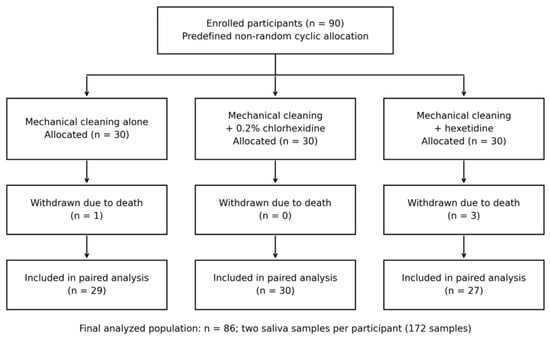
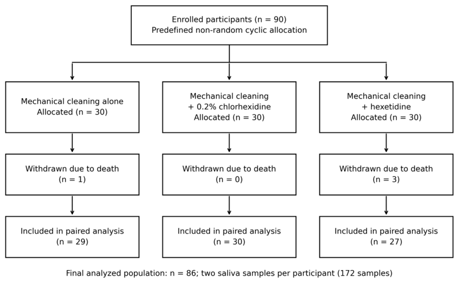
A total of 90 participants were enrolled and allocated to the three oral hygiene protocols, with 30 participants per group. Four participants died during the observation period and were excluded from the final analysis, leaving 86 participants (Figure 1). Of these, 43 patients were receiving antibiotic therapy at ICU admission and 43 were not.
Figure 1.
Participant flow diagram showing allocation to the three oral hygiene protocols, withdrawals due to death during the observation period, and the final population included in the paired analysis.
The two groups did not differ significantly in disease severity, age, sex distribution, previous antibiotic exposure or dentition status. Median SAPS II score was 37 in patients without antibiotic therapy and 38 in those receiving antibiotics. Median age was 73 years in the non-antibiotic group and 70 years in the antibiotic group. Visible dental plaque was present in 44/86 patients (51.2%) and was significantly more frequent among patients receiving antibiotic therapy than among those not receiving antibiotics (60.5% vs. 41.8%; p = 0.048). Oral hygiene habits also differed between the groups, with less frequent daily toothbrushing among patients receiving antibiotic therapy (p = 0.032). Respiratory support differed significantly between the two groups: patients not receiving antibiotics more frequently breathed spontaneously or received oxygen via nasal cannula, whereas patients receiving antibiotics more frequently required non-invasive ventilation or invasive mechanical ventilation (p < 0.001) (Table 1). The final analysis included 29 participants receiving mechanical cleaning alone, 30 receiving mechanical cleaning with chlorhexidine, and 27 receiving mechanical cleaning with hexetidine. The unequal final group sizes resulted from withdrawals due to death after allocation. Both saliva samples were available for all 86 participants included in the final analysis.
Table 1.
Baseline demographic, clinical and oral-status characteristics of ICU patients according to antibiotic therapy status.
3.2. Baseline Oral Microbiota at ICU Admission
At ICU admission, Gram-negative bacteria were isolated from the first saliva sample in 30/86 patients (34.9%). Gram-negative colonization at admission was significantly more frequent in men than in women (p = 0.018). A multinomial logistic regression model evaluating predictors of the first saliva culture was statistically significant and showed good explanatory capacity. In this model, dentition status, visible plaque and previous antibiotic therapy were the most relevant predictors of baseline microbiological status, whereas other variables showed weaker or non-significant associations (Table 2 and Table 3). However, several estimates had very wide confidence intervals. Model significance and pseudo-R2 do not establish reliable prediction, and these findings require cautious interpretation because of model complexity and sparse categories.
Table 2.
Predictors of baseline saliva microbiological status at ICU admission.
Table 3.
Significant predictors from the baseline multinomial logistic regression model.
3.3. Overall Changes in Oral Microbiota After Oral Care
No statistically significant overall change in the recorded microbiological categories was observed between ICU admission and day 3 when the paired nature of the observations was taken into account.
Among all 86 patients, the second saliva sample showed physiological oral flora in 27 patients, Candida spp. growth in 30 patients and Gram-negative bacterial growth in 29 patients. Overall, no statistically significant shift was detected in microbiota category during the observation period (Stuart–Maxwell χ2 = 0.385, df = 2, p = 0.825) (Table 4).
Table 4.
Distribution of saliva microbiota at ICU admission and after oral care.
3.4. Effect of Oral Hygiene Protocol on Gram-Negative Oral Colonization
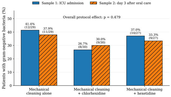
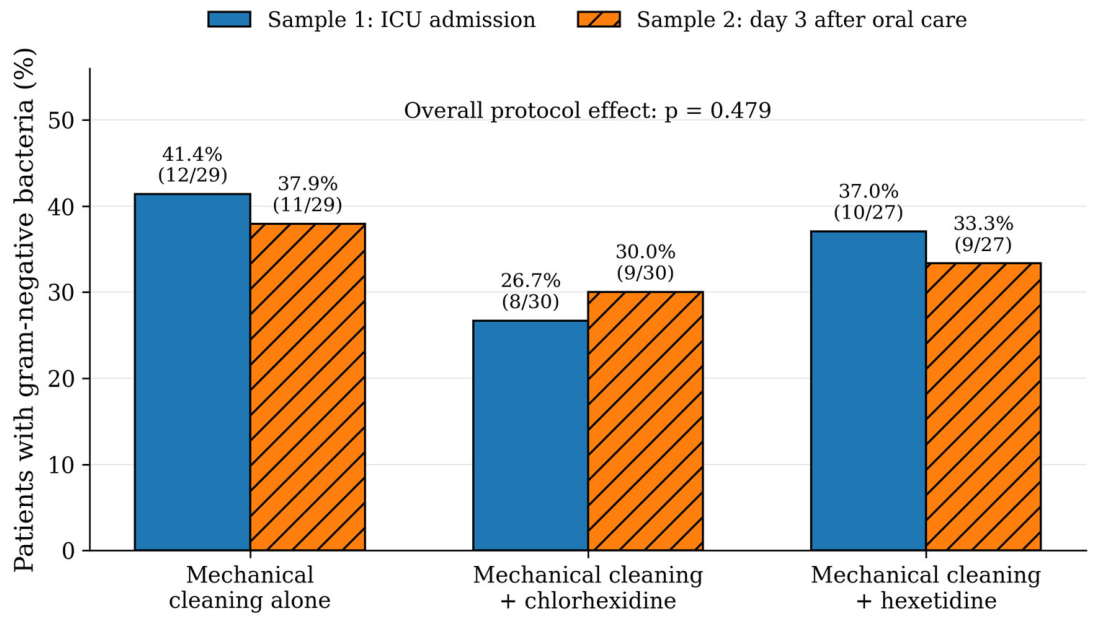
When microbiological outcomes were analyzed according to the assigned oral hygiene protocol, no protocol demonstrated statistically significant superiority in reducing Gram-negative oral colonization. The proportion of patients with Gram-negative bacterial growth remained broadly similar between the first and second saliva samples across all three protocols. Mechanical cleaning alone showed a small reduction from 12/29 (41.4%) to 11/29 (37.9%), chlorhexidine showed a small increase from 8/30 (26.7%) to 9/30 (30.0%), and hexetidine showed a small reduction from 10/27 (37.0%) to 9/27 (33.3%). In the multivariable model, oral hygiene protocol was not independently associated with Gram-negative bacterial growth in the second saliva sample (overall p = 0.479) (Figure 2).
Figure 2.
Gram-negative bacterial colonization before and after oral care according to oral hygiene protocol. The proportion of patients with Gram-negative bacterial growth remained broadly similar between saliva samples across all three protocols. Mechanical cleaning alone showed a change from 12/29 (41.4%) to 11/29 (37.9%), chlorhexidine from 8/30 (26.7%) to 9/30 (30.0%), and hexetidine from 10/27 (37.0%) to 9/27 (33.3%). Oral hygiene protocol was not independently associated with Gram-negative bacterial growth in the second saliva sample in the multivariable model.
3.5. Newly Detected and Persistent Gram-Negative Colonization
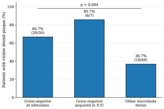
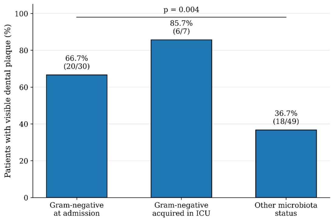
Newly detected Gram-negative colonization was observed in 3/86 patients (3.5%), defined as Gram-negative bacterial growth in the day-3 saliva sample that was not detected in the baseline sample. This represents new detection, not proof of hospital acquisition or clinical infection. The presence of Gram-negative bacteria in the first saliva sample was strongly associated with Gram-negative bacterial growth in the second sample (p < 0.001), indicating a strong relationship between baseline colonization and subsequent microbiological status. Visible dental plaque at ICU admission was also associated with Gram-negative bacterial growth in the second saliva sample (p = 0.023). The distribution of visible dental plaque according to Gram-negative microbiota status is shown in Figure 3. In the multivariable logistic regression model, the presence of Gram-negative bacteria in the first saliva sample showed the largest adjusted association with Gram-negative bacterial growth in the second saliva sample (adjusted OR = 2256.88, 95% CI: 21.80–233,657.01; p = 0.001). Visible dental plaque at ICU admission was also associated with the outcome after multivariable adjustment (adjusted OR = 41.18, 95% CI: 2.42–701.66; p = 0.010). Male sex and higher SAPS II score were additionally associated with increased odds of Gram-negative bacterial growth, whereas antibiotic therapy at ICU admission was associated with lower odds. The assigned oral hygiene protocol was not independently associated with Gram-negative bacterial growth in the second saliva sample (overall p = 0.479) (Table 5).
Figure 3.
Visible dental plaque according to Gram-negative microbiota status. Visible dental plaque was more frequent in patients with Gram-negative bacteria at ICU admission and in those who acquired Gram-negative bacteria during ICU stay than in patients with other microbiota status. Values are shown as percentages with absolute counts. p = 0.004.
Table 5.
Exploratory multivariable logistic regression model for Gram-negative bacterial growth in the second saliva sample.
The multivariable model included 8 predictor degrees of freedom and was based on 29 patients with Gram-negative bacterial growth in the second saliva sample. The limited number of outcome events relative to model complexity, together with the very large odds-ratio estimates and wide confidence intervals for several variables, indicates limited precision and potential sparse-data effects. Accordingly, the multivariable findings should be regarded as exploratory, and the magnitude of individual odds-ratio estimates should be interpreted cautiously.
3.6. Antimicrobial Susceptibility of Gram-Negative Isolates
Gram-negative bacteria were detected in 30 oral samples obtained at ICU admission and 29 samples collected on day 3. Eleven Gram-negative bacterial taxa were included in the antimicrobial susceptibility analysis, comprising 59 g-negative bacterial isolates recovered across both sampling time points (Table 6). The antimicrobial susceptibility profiles varied among the identified bacterial taxa. The Acinetobacter baumannii complex exhibited resistance to imipenem and meropenem, whereas Acinetobacter lwoffii was susceptible to both agents. Enterobacter spp., Citrobacter koseri, Escherichia coli, Klebsiella oxytoca, Klebsiella pneumoniae, Proteus mirabilis, and Serratia marcescens were classified as susceptible to meropenem, ciprofloxacin, gentamicin, ceftriaxone, ertapenem, and ceftazidime. Resistance to ampicillin was present in the susceptibility profiles of Enterobacter spp., Citrobacter koseri, Klebsiella oxytoca, Klebsiella pneumoniae, Proteus mirabilis, and Serratia marcescens. Resistance to amoxicillin/clavulanic acid was identified in Enterobacter spp., Klebsiella pneumoniae, and Serratia marcescens. Escherichia coli was susceptible to all antimicrobial agents included in its susceptibility profile. Pseudomonas putida was classified as susceptible at increased exposure to imipenem, meropenem, ciprofloxacin, and gentamicin. Stenotrophomonas maltophilia was classified as susceptible at increased exposure to trimethoprim/sulfamethoxazole.
Table 6.
Antimicrobial susceptibility profiles of isolated Gram-negative bacteria.
4. Discussion
No statistically significant association between the assigned oral hygiene protocol and day-3 g-negative bacterial growth was detected after adjustment for baseline microbiological status, visible plaque, sex, age, disease severity and antibiotic therapy. Baseline colonization and visible plaque were associated with the outcome in exploratory adjusted analyses, but large estimates and wide confidence intervals indicate substantial uncertainty. These findings generate hypotheses concerning baseline oral status; they do not establish comparative equivalence, treatment efficacy or validated clinical prediction.
Visible plaque remained associated with Gram-negative bacterial growth in the second saliva sample after adjustment. Because plaque is readily assessed at the bedside, this association warrants further investigation. The present study does not establish its predictive performance or demonstrate a benefit from intensified oral care.
The presence of Gram-negative bacteria in the first saliva sample showed the largest adjusted association with Gram-negative bacterial growth in the second sample. This finding suggests that baseline microbiological status is closely related to short-term subsequent colonization. However, the very large adjusted odds ratio should not be interpreted as a precise estimate of effect size. The wide confidence interval, together with the limited number of outcome events relative to model complexity, indicates substantial uncertainty and possible sparse-data inflation of the maximum-likelihood estimate. Therefore, interpretation should focus on the presence and direction of the association rather than on the exact magnitude of the odds ratio. From a clinical perspective, this suggests that preventive oral care should begin as early as possible, ideally at ICU admission, before mature biofilm and pathogenic colonization become established.
The association between visible dental plaque and second-sample Gram-negative growth is one of the clinically relevant findings of this study. Dental plaque is a complex biofilm that may reduce the penetration and effectiveness of antiseptic agents, particularly when mechanical disruption is insufficient [12,31,33,34,35,36,37,38,63]. Kiyoshi Teo et al. [32] have emphasized that sponge-based oral care is more comfortable but less effective than toothbrushing for removing adherent plaque. Our findings are consistent with this concept and suggest that the presence of visible plaque may be a clinically useful marker of biofilm burden and pathogenic colonization risk. Therefore, ICU oral care protocols should not rely solely on antiseptic application but should include systematic assessment and mechanical disruption of oral biofilm.
Chlorhexidine was not associated with a reduction in Gram-negative bacterial colonization in the present analysis and was not statistically superior to mechanical cleaning alone or hexetidine. This finding is consistent with the broader uncertainty in the literature regarding chlorhexidine-based ICU oral care. Several studies and meta-analyses have reported that chlorhexidine may reduce VAP incidence, particularly in selected populations, but evidence regarding its effect on mortality, duration of mechanical ventilation and ICU length of stay remains inconsistent [3,31,49,63]. In addition, concerns have been raised regarding mucosal toxicity, possible adverse outcomes and reduced susceptibility among some Gram-negative organisms [12,13,14,63,64,65,66,67]. In this context, our findings support a cautious interpretation: chlorhexidine may be useful as an adjunct in selected patients, but the present data do not support its universal superiority over mechanical biofilm control during the early ICU stay.
Hexetidine did not show superiority over mechanical cleaning or chlorhexidine in reducing Gram-negative oral colonization. Nevertheless, its inclusion in the present study is clinically relevant because hexetidine has been less extensively investigated in ICU oral care despite antimicrobial and anti-biofilm properties [50,51,52,53,54,55,56,68]. Experimental and dental literature suggests potential activity against bacterial and fungal species, including Candida spp., but clinical evidence in critically ill patients remains limited [52,54,55,56,68,69]. In the present study, no statistically significant association between hexetidine allocation and day-3 g-negative bacterial growth was detected; the confidence interval was wide. Further studies with larger samples and longer follow-up are needed to determine whether hexetidine has a role as an alternative antiseptic in patients in whom chlorhexidine is contraindicated or poorly tolerated.
An additional finding was that male sex and higher SAPS II score were associated with higher adjusted odds of Gram-negative bacterial growth in the second saliva sample. The association with SAPS II is biologically plausible because greater disease severity may reflect impaired host defense, reduced ability to maintain oral clearance and greater dependence on assisted ventilation and nursing care. The association with male sex should be interpreted cautiously, but it is consistent with the baseline observation that Gram-negative bacteria were more frequently detected in men at ICU admission. Whether this reflects differences in oral health status, comorbidity burden, smoking history, dentition, hygiene habits or other unmeasured factors cannot be determined from the present data.
Antibiotic therapy at ICU admission was associated with lower odds of Gram-negative bacterial growth in the second saliva sample after adjustment. This finding should be interpreted with caution because antibiotic exposure was not randomized and was closely linked to admission diagnosis and clinical status. A possible explanation is that systemic antibiotic therapy temporarily suppressed susceptible Gram-negative bacteria in saliva. However, antibiotics may also promote dysbiosis, fungal overgrowth and selection of resistant organisms, depending on agent, spectrum and duration of exposure [70,71,72,73,74]. Therefore, this result should not be interpreted as evidence that antibiotic therapy is protective for oral microbiota, but rather as an adjusted association observed in a clinically heterogeneous ICU cohort.
The antimicrobial susceptibility findings provide additional context. The recorded profiles included clinically relevant Enterobacterales and non-fermenting Gram-negative bacilli such as Acinetobacter baumannii complex, Klebsiella spp., Pseudomonas spp., Proteus mirabilis and Serratia marcescens. These organisms are important in ICU settings because of their association with nosocomial infections and their ability to acquire resistance mechanisms [70,71,72,73,75,76,77,78,79,80,81,82,83,84,85,86]. Although this study did not assess clinical infection outcomes or changes in antimicrobial resistance over time, the detection of these organisms in saliva supports the role of the oral cavity as a potential reservoir of clinically relevant pathogens. These findings do not, however, establish infection, transmission or hospital acquisition. The susceptibility results should be interpreted descriptively, as the absence of isolate-level S/I/R counts for each antimicrobial agent precludes estimation of susceptibility proportions. Translating these findings into routine care also depends on staff knowledge, training and consistent implementation of oral hygiene procedures. Our earlier conducted survey at two university hospital centers demonstrated substantial variability in ICU personnel’s knowledge, perceived training, frequency of oral care and selection of mechanical and chemical biofilm-control methods, highlighting the need for standardized protocols and targeted education alongside evidence-based selection of oral care interventions [87].
This study has several limitations. Its single-center design and relatively small sample size limit generalizability and the precision of multivariable estimates. Allocation followed a predefined cyclic sequence rather than randomization and was not concealed, creating the potential for selection bias and baseline imbalance. Antibiotic therapy, respiratory support and admission diagnosis were clinically interrelated, leaving the possibility of residual confounding despite adjustment. Respiratory support was not included in the day-3 model, and adjustment for SAPS II cannot fully account for differences in support modality.
Both the binary and multinomial regression analyses were exploratory and potentially affected by sparse-data bias and overfitting. In particular, the day-3 model included eight predictor degrees of freedom but only 29 outcome-positive observations, limiting the precision and stability of adjusted odds ratios. Neither model underwent internal or external validation. Although microbiological assessment was blinded, healthcare personnel and patients could not be fully blinded to the assigned oral care protocol.
Microbiological assessment was limited to saliva collected at ICU admission and on day 3. Salivary findings do not directly characterize adherent dental biofilm, and selective aerobic culture cannot capture the full oral microbiome. Visible dental plaque was recorded as a binary clinical variable, which is practical at the bedside but provides less detail than validated plaque indices. The short follow-up precludes conclusions about longer-term persistence, clearance or acquisition of Gram-negative colonization. Furthermore, clinical infection outcomes were not assessed, so no conclusions can be drawn regarding VAP incidence, mortality, duration of mechanical ventilation or ICU length of stay. Also, the study was retrospectively registered, which limits the ability to independently verify prespecified outcomes and analyses. Finally, exclusion of four participants who died during follow-up may have introduced attrition bias, and the findings apply to participants who completed both sampling assessments. Future studies should combine saliva and dental plaque sampling with broader microbiological methods, including anaerobic culture and culture-independent approaches, and longer follow-up to better characterize oral microbial communities and their changes over time.
Strengths include prospective assessment, comparison of three oral care protocols, paired saliva sampling at two defined time points, and inclusion of clinical oral-status variables. The observed association with visible plaque is hypothesis-generating; its predictive performance and the effect of intensified plaque removal require independent evaluation.
5. Conclusions
In conclusion, no statistically significant difference in short-term Gram-negative oral colonization was observed among the three oral hygiene protocols, and the present data provided no evidence that chlorhexidine or hexetidine was superior to mechanical cleaning alone. However, the precision of the treatment-effect estimates was limited, and smaller between-group differences cannot be excluded. Visible dental plaque and baseline Gram-negative colonization showed the strongest adjusted associations with Gram-negative bacterial growth after oral care. Given the exploratory nature and limited stability of the multivariable model, the magnitude of these associations should be interpreted cautiously. These findings support early oral assessment and systematic mechanical biofilm control in ICU patients. Adjunctive antiseptics may have a role, but their relative benefit requires confirmation in larger studies with greater statistical precision.
Author Contributions
Conceptualization, V.V., B.Š., A.T.A. and V.B.; methodology, V.V., B.Š., A.T.A. and V.B.; investigation, V.V.; data curation, V.V., B.Š. and L.Š.; formal analysis, L.Š.; visualization, L.Š.; writing—original draft preparation, V.V., B.Š. and L.Š.; writing—review and editing, V.V., B.Š., L.Š., D.N.V., G.M., D.V.J., A.T.A. and V.B.; supervision, A.T.A. and V.B.; project administration, V.V. and B.Š. All authors have read and agreed to the published version of the manuscript.
Funding
This research was funded by the European Union—NextGenerationEU, through the National Recovery and Resilience Plan within the project “Microbiome in oral health and disease _MOZIB” (grant number: SFZG-10-2025).
Institutional Review Board Statement
The study was conducted in accordance with the Declaration of Helsinki and approved by the Ethics Committees of the “Sestre Milosrdnice” University Hospital Centre and the University of Zagreb School of Dental Medicine, Zagreb, Croatia (class: [EP-5992/17-3] and approval no. [05-PA-15-5/2017], respectively).
Informed Consent Statement
Informed consent was obtained from all subjects involved in the study.
Data Availability Statement
Data are available upon request to the corresponding authors.
Conflicts of Interest
The authors declare no conflicts of interest.
Abbreviations
The following abbreviations are used in this manuscript:
| Abbreviation | Definition |
| CI | Confidence interval |
| ICU | Intensive care unit |
| OR | Odds ratio |
| SAPS II | Simplified Acute Physiology Score II |
References
- Kilian, M.; Chapple, I.L.C.; Hannig, M.; Marsh, P.D.; Meuric, V.; Pedersen, A.M.L.; Tonetti, M.S.; Wade, W.G.; Zaura, E. The oral microbiome—An update for oral healthcare professionals. Br. Dent. J. 2016, 221, 657–666. [Google Scholar] [CrossRef] [Scilit] [PubMed]
- Avila, M.; Ojcius, D.M.; Yilmaz, Ö. The oral microbiota: Living with a permanent guest. DNA Cell Biol. 2009, 28, 405–411. [Google Scholar] [CrossRef] [Scilit] [PubMed]
- Jia, G.; Zhi, A.; Lai, P.F.H.; Wang, G.; Xia, Y.; Xiong, Z.; Zhang, H.; Che, N.; Ai, L. The oral microbiota—A mechanistic role for systemic diseases. Br. Dent. J. 2018, 224, 447–455. [Google Scholar] [CrossRef] [Scilit] [PubMed]
- Rajasekaran, J.J.; Krishnamurthy, H.K.; Bosco, J.; Jayaraman, V.; Krishna, K.; Wang, T.; Bei, K. Oral Microbiome: A Review of Its Impact on Oral and Systemic Health. Microorganisms 2024, 12, 1797. [Google Scholar] [CrossRef] [Scilit] [PubMed]
- Estaji, Z.; Alinejad, M.; Rakhshani, M.H.; Rad, M. The comparison of chlorhexidine solution and swab with toothbrush and toothpaste effect on preventing oral lesions in hospitalized patients in intensive care unit. Glob. J. Health Sci. 2015, 8, 211–216. [Google Scholar] [CrossRef] [Scilit] [PubMed]
- Grap, M.J.; Munro, C.L.; Ashtiani, B.; Bryant, S. Oral care interventions in critical care: Frequency and documentation. Am. J. Crit. Care 2003, 12, 113–118. [Google Scholar] [CrossRef] [Scilit]
- Robriquet, L.; Fourrier, F. Oral hygiene and ventilator-associated pneumonia. Curr. Respir. Med. Rev. 2010, 6, 65–71. [Google Scholar] [CrossRef] [Scilit]
- Kanamori, D.; Fujii, T.; Yoshida, M.; Ito, R.; Sakai, A.; Takahashi, H.; Kuramitsu, K.; Funasaka, K.; Ohno, E.; Hirooka, Y.; et al. Oral Care for Intubated Patients in the Intensive Care Unit: Examination of Bacterial Count and Microbiota. Crit. Care 2025, 29, 320. [Google Scholar] [CrossRef] [Scilit] [PubMed]
- Jones, D.J.; Munro, C.L.; Grap, M.J. Natural history of dental plaque accumulation in mechanically ventilated adults: A descriptive correlational study. Intensive Crit. Care Nurs. 2011, 27, 299–304. [Google Scholar] [CrossRef] [Scilit] [PubMed]
- Munro, C.L.; Grap, M.J.; Elswick, R.K., Jr.; McKinney, J.; Sessler, C.N.; Hummel, R.S., III. Oral health status and development of ventilator-associated pneumonia: A descriptive study. Am. J. Crit. Care 2006, 15, 453–460. [Google Scholar] [CrossRef] [Scilit]
- Rathbun, K.P.; Bourgault, A.M.; Sole, M.L. Oral Microbes in Hospital-Acquired Pneumonia: Practice and Research Implications. Crit. Care Nurse 2022, 42, 47–54. [Google Scholar] [CrossRef] [Scilit] [PubMed]
- De Melo Neto, J.P.; Melo, M.S.A.E.; Dos Santos-Pereira, S.A.; Martinez, E.F.; Okajima, L.S.; Saba-Chujfi, E. Periodontal infections and community-acquired pneumonia: A case-control study. Eur. J. Clin. Microbiol. Infect. Dis. 2013, 32, 27–32. [Google Scholar] [CrossRef] [Scilit] [PubMed]
- Marino, P.J.; Hannigan, A.; Haywood, S.; Cole, J.M.; Palmer, N.; Emanuel, C.; Kinsella, T.; A O Lewis, M.; Wise, M.P.; Williams, D.W. Comparison of foam swabs and toothbrushes as oral hygiene interventions in mechanically ventilated patients: A randomised split mouth study. BMJ Open Respir. Res. 2016, 3, e000150. [Google Scholar] [CrossRef] [Scilit] [PubMed]
- Winning, L.; Lundy, F.T.; Blackwood, B.; McAuley, D.F.; El Karim, I. Oral Health Care for the Critically Ill: A Narrative Review. Crit. Care 2021, 25, 353. [Google Scholar] [CrossRef] [Scilit] [PubMed]
- Sands, K.M.; Wilson, M.J.; Lewis, M.A.O.; Wise, M.P.; Palmer, N.; Hayes, A.J.; Barnes, R.A.; Williams, D.W. Respiratory pathogen colonization of dental plaque, the lower airways, and endotracheal tube biofilms during mechanical ventilation. J. Crit. Care 2017, 37, 30–37. [Google Scholar] [CrossRef] [Scilit] [PubMed]
- American Thoracic Society; Infectious Diseases Society of America. Guidelines for the management of adults with hospital-acquired, ventilator-associated, and healthcare-associated pneumonia. Am. J. Respir. Crit. Care Med. 2005, 171, 388–416. [Google Scholar] [CrossRef] [Scilit] [PubMed]
- Safdar, N.; Crnich, C.J.; Maki, D.G. The pathogenesis of ventilator-associated pneumonia: Its relevance to developing effective strategies for prevention. Respir. Care 2005, 50, 725–739. [Google Scholar] [CrossRef] [Scilit]
- Howroyd, F.; Gill, R.; Thompson, J.; Smith, F.G.; Nasa, P.; Gopal, S.; Duggal, N.A.; Ahmed, Z.; Veenith, T. Ventilator-Associated Pneumonia: Mechanisms, an Appraisal of Current Therapies and the Role for Inhaled Antibiotics in Prevention and Treatment. Respir. Med. 2025, 247, 108275. [Google Scholar] [CrossRef] [Scilit] [PubMed]
- Labeau, S.O.; Van de Vyver, K.; Brusselaers, N.; Vogelaers, D.; Blot, S.I. Prevention of ventilator-associated pneumonia with oral antiseptics: A systematic review and meta-analysis. Lancet Infect. Dis. 2011, 11, 845–854. [Google Scholar] [CrossRef] [Scilit] [PubMed]
- Mogyoródi, B.; Dunai, E.; Gál, J.; Iványi, Z. Ventilator-associated pneumonia and the importance of education of ICU nurses on prevention—Preliminary results. Interv. Med. Appl. Sci. 2016, 8, 147–151. [Google Scholar] [CrossRef] [Scilit] [PubMed]
- Klompas, M. What can we learn from international ventilator-associated pneumonia rates? Crit. Care Med. 2012, 40, 3303–3304. [Google Scholar] [CrossRef] [Scilit] [PubMed]
- Kalanuria, A.A.; Zai, W.; Mirski, M. Ventilator-associated pneumonia in the ICU. Crit. Care 2014, 18, 208. [Google Scholar] [CrossRef] [Scilit] [PubMed]
- Vilela, M.C.N.; Ferreira, G.Z.; Santos, P.S.S.; Rezende, N.P.M. Oral care and nosocomial pneumonia: A systematic review. Einstein 2015, 13, 290–296. [Google Scholar] [CrossRef] [Scilit] [PubMed]
- Safdar, N.; Dezfulian, C.; Collard, H.R.; Saint, S. Clinical and economic consequences of ventilator-associated pneumonia: A systematic review. Crit. Care Med. 2005, 33, 2184–2193. [Google Scholar] [CrossRef] [Scilit] [PubMed]
- Rello, J.; Lode, H.; Cornaglia, G.; Masterton, R. A European care bundle for prevention of ventilator-associated pneumonia. Intensive Care Med. 2010, 36, 773–780. [Google Scholar] [CrossRef] [Scilit] [PubMed]
- Papazian, L.; Klompas, M.; Luyt, C.E. Ventilator-Associated Pneumonia in Adults: A Narrative Review. Intensive Care Med. 2020, 46, 888–906. [Google Scholar] [CrossRef] [Scilit] [PubMed]
- Sands, K.M.; Twigg, J.A.; Wise, M.P. Oral hygiene with chlorhexidine in critically ill patients. JAMA Intern. Med. 2015, 175, 316. [Google Scholar] [CrossRef] [Scilit] [PubMed]
- Bellissimo-Rodrigues, W.T.; Menegueti, M.G.; Gaspar, G.G.; Nicolini, E.A.; Auxiliadora-Martins, M.; Basile-Filho, A.; Martinez, R.; Bellissimo-Rodrigues, F. Effectiveness of a dental care intervention in the prevention of lower respiratory tract nosocomial infections among intensive care patients: A randomized clinical trial. Infect. Control Hosp. Epidemiol. 2014, 35, 1342–1348. [Google Scholar] [CrossRef] [Scilit] [PubMed]
- Müller, F. Oral hygiene reduces the mortality from aspiration pneumonia in frail elders. J. Dent. Res. 2015, 94, 14S–16S. [Google Scholar] [CrossRef] [Scilit] [PubMed]
- Kitsios, G.D.; Nguyen, V.D.; Sayed, K.; Al-Yousif, N.; Schaefer, C.; Shah, F.A.; Bain, W.; Yang, H.; Fitch, A.; Li, K.; et al. The Upper and Lower Respiratory Tract Microbiome in Severe Aspiration Pneumonia. iScience 2023, 26, 106832. [Google Scholar] [CrossRef] [Scilit] [PubMed]
- Oliveira, M.S.; Borges, A.H.; Mattos, F.Z.; Semenoff, T.A.D.V.; Semenoff-Segundo, A.; Tonetto, M.R.; Bandeca, M.C.; Porto, A.N. Evaluation of different methods for removing oral biofilm in patients admitted to the intensive care unit. J. Int. Oral Health 2014, 6, 61–64. [Google Scholar] [PubMed]
- Kiyoshi-Teo, H.; Blegen, M. Influence of institutional guidelines on oral hygiene practices in intensive care units. Am. J. Crit. Care 2015, 24, 309–318. [Google Scholar] [CrossRef] [Scilit] [PubMed]
- Labeau, S.O.; Conoscenti, E.; Blot, S.I. Less daily oral hygiene is more in the ICU: Not sure. Intensive Care Med. 2021, 47, 334–336. [Google Scholar] [CrossRef] [Scilit] [PubMed]
- Grap, M.J.; Munro, C.L.; Hamilton, V.A.; Elswick, R.K.; Sessler, C.N.; Ward, K.R. Early, single chlorhexidine application reduces ventilator-associated pneumonia in trauma patients. Heart Lung 2011, 40, e115–e122. [Google Scholar] [CrossRef] [Scilit] [PubMed]
- Munro, C.L.; Grap, M.J.; Jones, D.J.; McClish, D.K.; Sessler, C.N. Chlorhexidine, toothbrushing, and preventing ventilator-associated pneumonia in critically ill adults. Am. J. Crit. Care 2009, 18, 428–437. [Google Scholar] [CrossRef] [Scilit] [PubMed]
- Patil, S.P.; Patil, P.B.; Kashetty, M.V. Effectiveness of different tooth brushing techniques on the removal of dental plaque in 6–8 year old children of Gulbarga. J. Int. Soc. Prev. Community Dent. 2014, 4, 113–116. [Google Scholar] [CrossRef] [Scilit] [PubMed]
- Nagarakanti, S.; Avuluri, J.; Chava, V.K. Evaluation of nurses’ attitude toward the provision of oral hygiene care to hospitalized patients at two private hospitals in South India. Iran. J. Nurs. Midwifery Res. 2019, 24, 472–474. [Google Scholar] [CrossRef] [Scilit] [PubMed]
- Martelli, M.; Rosa, A.; Miranda, M.; Simone, R.; Scarpati Cioffari di Castiglione, M.; De Falco, F.; Gargari, M.; Bollero, P.; Gianfreda, F. Oral Hygiene Management in Critically Ill Patients: Prevention of Ventilator-Associated Pneumonia. Front. Dent. Med. 2026, 7, 1748329. [Google Scholar] [CrossRef] [Scilit] [PubMed]
- Zhao, T.; Wu, X.; Zhang, Q.; Li, C.; Worthington, H.V.; Hua, F. Oral Hygiene Care for Critically Ill Patients to Prevent Ventilator-Associated Pneumonia. Cochrane Database Syst. Rev. 2020, 12, CD008367. [Google Scholar] [CrossRef] [Scilit] [PubMed]
- De Lacerda Vidal, C.F.; Vidal, A.K.L.; Monteiro, J.G.M.; Cavalcanti, A.; Henriques, A.P.T.; Oliveira, M.; Godoy, M.; Coutinho, M.; Sobral, P.D.; Vilela, C.Â.; et al. Impact of oral hygiene involving toothbrushing versus chlorhexidine in the prevention of ventilator-associated pneumonia: A randomized study. BMC Infect. Dis. 2017, 17, 112, Erratum in BMC Infect. Dis. 2017, 17, 173. [Google Scholar] [CrossRef] [Scilit] [PubMed]
- Da Silva Pinto, A.C.; Da Silva, B.M.; Santiago-Junior, J.F.; De Carvalho Sales-Peres, S.H. Efficiency of different protocols for oral hygiene combined with the use of chlorhexidine in the prevention of ventilator-associated pneumonia. J. Bras. Pneumol. 2021, 47, e20190286. [Google Scholar] [CrossRef] [Scilit] [PubMed]
- Huang, H.; Yu, X.; Huang, C.; Zeng, J.; Li, Y. Oral Care Medications for the Prevention and Treatment of Ventilator-Associated Pneumonia in Intensive Care Unit. Front. Oral Health 2025, 6, 1566355. [Google Scholar] [CrossRef] [Scilit] [PubMed]
- Klompas, M.; Speck, K.; Howell, M.D.; Greene, L.R.; Berenholtz, S.M. Reappraisal of routine oral care with chlorhexidine gluconate for patients receiving mechanical ventilation: Systematic review and meta-analysis. JAMA Intern. Med. 2014, 174, 751–761. [Google Scholar] [PubMed]
- Wittekamp, B.H.; Plantinga, N.L. Less daily oral hygiene is more in the ICU: No. Intensive Care Med. 2021, 47, 331–333. [Google Scholar] [CrossRef] [Scilit] [PubMed]
- Plantinga, N.L.; Wittekamp, B.H.J.; Leleu, K.; Depuydt, P.; Van den Abeele, A.M.; Brun-Buisson, C.; Bonten, M.J.M. Oral mucosal adverse events with chlorhexidine 2% mouthwash in ICU. Intensive Care Med. 2016, 42, 620–621. [Google Scholar] [CrossRef] [Scilit] [PubMed]
- Scannapieco, F.A.; Yu, J.; Raghavendran, K.; Vacanti, A.; Owens, S.I.; Wood, K.; Mylotte, J.M. A randomized trial of chlorhexidine gluconate on oral bacterial pathogens in mechanically ventilated patients. Crit. Care 2009, 13, R117. [Google Scholar] [CrossRef] [Scilit] [PubMed]
- Stickler, D.J. Susceptibility of antibiotic-resistant gram-negative bacteria to biocides: A perspective from the study of catheter biofilms. Symp. Ser. Soc. Appl. Microbiol. 2002, 31, 163S–170S. [Google Scholar] [CrossRef] [Scilit]
- Kampf, G. Acquired resistance to chlorhexidine—Is it time to establish an “antiseptic stewardship” initiative? J. Hosp. Infect. 2016, 94, 213–227. [Google Scholar] [CrossRef] [Scilit] [PubMed]
- De Cassai, A.; Pettenuzzo, T.; Busetto, V.; Legnaro, C.; Pretto, C.; Rotondi, A.; Boscolo, A.; Sella, N.; Munari, M.; Navalesi, P. Chlorhexidine Is Not Effective at Any Concentration in Preventing Ventilator-Associated Pneumonia: A Systematic Review and Network Meta-Analysis. J. Anesth. Analg. Crit. Care 2024, 4, 30. [Google Scholar] [CrossRef] [Scilit] [PubMed]
- PubChem. Hexetidine. Available online: https://pubchem.ncbi.nlm.nih.gov/compound/Hexetidine (accessed on 1 May 2026).
- HALMED. Hexoral 1 mg/ml Otopina za Grgljanje/Ispiranje Usta: Sažetak Opisa Svojstava Lijeka. Available online: https://halmed.hr/upl/lijekovi/SPC/Hexoral-SPC_1.pdf (accessed on 17 October 2026).
- Ernst, C.P.; Canbek, K.; Dillenburger, A.; Willershausen, B. Clinical Study on the Effectiveness and Side Effects of Hexetidine and Chlorhexidine Mouthrinses versus a Negative Control. Quintessence Int. 2005, 36, 641–652. [Google Scholar] [PubMed]
- Jones, D.S.; McGovern, J.G.; Woolfson, A.D.; Gorman, S.P. The effects of hexetidine (Oraldene) on the adherence of Candida albicans to human buccal epithelial cells in vitro and ex vivo and on in vitro morphogenesis. Pharm. Res. 1997, 14, 1765–1771. [Google Scholar] [CrossRef] [Scilit] [PubMed]
- Aoun, G.; Nasseh, I.; Saadeh, M.; Cassia, A.; Berberi, A. Effectiveness of hexetidine 0.1% in eliminating Candida albicans colonizing dentures: A randomized clinical in vivo study. J. Int. Oral Health 2015, 7, 1–4. [Google Scholar] [PubMed]
- Aoun, G.; Saadeh, M.; Berberi, A. Effectiveness of Hexetidine 0.1% Compared to Chlorhexidine Digluconate 0.12% in Eliminating Candida albicans Colonizing Dentures: A Randomized Clinical In Vivo Study. J. Int. Oral Health 2015, 7, 5–8. [Google Scholar] [PubMed]
- Gorman, S.P.; McGovern, J.G.; Woolfson, A.D.; Adair, C.G.; Jones, D.S. The concomitant development of poly(vinyl chloride)-related biofilm and antimicrobial resistance in relation to ventilator-associated pneumonia. Biomaterials 2001, 22, 2741–2747. [Google Scholar] [CrossRef] [Scilit] [PubMed]
- Hillier, B.; Wilson, C.; Chamberlain, D.; King, L. Preventing ventilator-associated pneumonia through oral care, product selection, and application method: A literature review. AACN Adv. Crit. Care 2013, 24, 38–58. [Google Scholar] [CrossRef]
- Alhazzani, W.; Smith, O.; Muscedere, J.; Medd, J.; Cook, D. Toothbrushing for critically ill mechanically ventilated patients: A systematic review and meta-analysis of randomized trials evaluating ventilator-associated pneumonia. Crit. Care Med. 2013, 41, 646–655. [Google Scholar] [PubMed]
- Rello, J.; Koulenti, D.; Blot, S.; Sierra, R.; Diaz, E.; De Waele, J.J.; Macor, A.; Agbaht, K.; Rodriguez, A. Oral care practices in intensive care units: A survey of 59 European ICUs. Intensive Care Med. 2007, 33, 1066–1070. [Google Scholar] [CrossRef] [Scilit] [PubMed]
- Feider, L.L.; Mitchell, P.; Bridges, E. Oral care practices for orally intubated critically ill adults. Am. J. Crit. Care 2010, 19, 175–183. [Google Scholar] [CrossRef] [Scilit] [PubMed]
- Ehrenzeller, S.; Klompas, M. Association Between Daily Toothbrushing and Hospital-Acquired Pneumonia: A Systematic Review and Meta-Analysis. JAMA Intern. Med. 2024, 184, 131–142. [Google Scholar] [PubMed]
- The European Committee on Antimicrobial Susceptibility Testing. Breakpoint Tables for Interpretation of MICs and Zone Diameters. Version 10.0, 2020. Available online: https://www.eucast.org/fileadmin/src/media/PDFs/EUCAST_files/Breakpoint_tables/v_10.0_Breakpoint_Tables.pdf (accessed on 11 October 2026).
- Aguilar, C.; Eichwald, C.; Eberl, L. Multicellularity in bacteria: From division of labor to biofilm formation. In Evolutionary Transitions to Multicellular Life; Ruiz-Trillo, I., Nedelcu, A., Eds.; Springer: Dordrecht, The Netherlands, 2015; pp. 79–95. [Google Scholar]
- Vandecandelaere, I.; Coenye, T. Microbial Composition and Antibiotic Resistance of Biofilms Recovered from Endotracheal Tubes of Mechanically Ventilated Patients. Adv. Exp. Med. Biol. 2015, 830, 137–155. [Google Scholar] [CrossRef] [Scilit] [PubMed]
- Scannapieco, F.A. Pneumonia in Nonambulatory Patients: The Role of Oral Bacteria and Oral Hygiene. J. Am. Dent. Assoc. 2006, 137, S21–S25. [Google Scholar]
- Munro, C.L.; Grap, M.J. Oral Health and Care in the Intensive Care Unit: State of the Science. Am. J. Crit. Care 2004, 13, 25–34. [Google Scholar] [CrossRef] [Scilit]
- Vieira, P.C.; de Oliveira, R.B.; da Silva Mendonça, T.M. Should Oral Chlorhexidine Remain in Ventilator-Associated Pneumonia Prevention Bundles? Med. Intensiv. (Engl. Ed.) 2022, 46, 259–268. [Google Scholar] [CrossRef] [PubMed]
- Overview of Antibacterial Susceptibility Testing. Available online: https://www.uptodate.com/contents/overview-of-antibacterial-susceptibility-testing (accessed on 17 October 2026).
- Maziere, M.; Rompante, P.; Andrade, J.C.; Rodrigues, C.F. Are Mouthwashes Really Effective against Candida spp.? J. Fungi 2024, 10, 528. [Google Scholar] [CrossRef] [Scilit] [PubMed]
- Livermore, D.M. Bacterial resistance: Origins, epidemiology, and impact. Clin. Infect. Dis. 2003, 36, S11–S23. [Google Scholar] [CrossRef] [Scilit] [PubMed]
- Procop, G.W.; Church, D.L.; Hall, G.S.; Janda, W.M.; Koneman, E.W.; Schreckenberger, P.C.; Woods, G.L. Antimicrobial susceptibility testing. In Koneman’s Color Atlas and Textbook of Diagnostic Microbiology, 7th ed.; Wolters Kluwer: Philadelphia, PA, USA, 2017. [Google Scholar]
- Holmes, N.E.; Johnson, P.D.R.; Howden, B.P. Relationship between vancomycin-resistant Staphylococcus aureus, vancomycin-intermediate S. aureus, high vancomycin MIC, and outcome in serious S. aureus infections. J. Clin. Microbiol. 2012, 50, 2548–2552. [Google Scholar] [CrossRef] [Scilit] [PubMed]
- Chastre, J.; Fagon, J.Y. Ventilator-associated pneumonia. Am. J. Respir. Crit. Care Med. 2002, 165, 867–903. [Google Scholar] [PubMed]
- Patangia, D.V.; Anthony Ryan, C.; Dempsey, E.; Paul Ross, R.; Stanton, C. Impact of Antibiotics on the Human Microbiome and Consequences for Host Health. MicrobiologyOpen 2022, 11, e1260. [Google Scholar] [CrossRef] [Scilit] [PubMed]
- Souto, R.; Silva-Boghossian, C.M.; Colombo, A.P.V. Prevalence of Pseudomonas aeruginosa and Acinetobacter spp. in subgingival biofilm and saliva of subjects with chronic periodontal infection. Braz. J. Microbiol. 2014, 45, 495–501. [Google Scholar] [CrossRef] [Scilit] [PubMed]
- Wagner, V.E.; Iglewski, B.H. P. aeruginosa biofilms in CF infection. Clin. Rev. Allergy Immunol. 2008, 35, 124–134. [Google Scholar] [CrossRef] [Scilit] [PubMed]
- Pihl, M.; Chávez de Paz, L.E.; Schmidtchen, A.; Svensäter, G.; Davies, J.R. Effects of clinical isolates of Pseudomonas aeruginosa on Staphylococcus epidermidis biofilm formation. FEMS Immunol. Med. Microbiol. 2010, 59, 504–512. [Google Scholar] [PubMed]
- Karlowsky, J.A.; Draghi, D.C.; Jones, M.E.; Thornsberry, C.; Friedland, I.R.; Sahm, D.F. Surveillance for antimicrobial susceptibility among clinical isolates of Pseudomonas aeruginosa and Acinetobacter baumannii from hospitalized patients in the United States, 1998 to 2001. Antimicrob. Agents Chemother. 2003, 47, 1681–1688. [Google Scholar] [CrossRef] [Scilit] [PubMed]
- Luna, C.M.; Aruj, P.K. Nosocomial Acinetobacter pneumonia. Respirology 2007, 12, 787–791. [Google Scholar] [CrossRef] [Scilit] [PubMed]
- Agodi, A.; Barchitta, M.; Cipresso, R.; Giaquinta, L.; Romeo, M.A.; Denaro, C. Pseudomonas aeruginosa carriage, colonization, and infection in ICU patients. Intensive Care Med. 2007, 33, 1155–1161. [Google Scholar] [CrossRef] [Scilit] [PubMed]
- Colombo, A.P.V.; Teles, R.P.; Torres, M.C.; Souto, R.; Rosalém, W.; Mendes, M.C.S.; Uzeda, M. Subgingival microbiota of Brazilian subjects with untreated chronic periodontitis. J. Periodontol. 2002, 73, 360–369. [Google Scholar] [CrossRef] [Scilit] [PubMed]
- Persson, G.R.; Hitti, J.; Paul, K.; Hirschi, R.; Weibel, M.; Rothen, M.; Persson, R.E. Tannerella forsythia and Pseudomonas aeruginosa in subgingival bacterial samples from parous women. J. Periodontol. 2008, 79, 508–516. [Google Scholar] [CrossRef] [Scilit] [PubMed]
- Souto, R.; De Andrade, A.F.B.; Uzeda, M.; Colombo, A.P.V. Prevalence of “non-oral” pathogenic bacteria in subgingival biofilm of subjects with chronic periodontitis. Braz. J. Microbiol. 2006, 37, 208–215. [Google Scholar] [CrossRef] [Scilit]
- Bassin, A.S.; Niederman, M.S. New approaches to prevention and treatment of nosocomial pneumonia. Semin. Thorac. Cardiovasc. Surg. 1995, 7, 70–77. [Google Scholar] [PubMed]
- George, D.L. Epidemiology of nosocomial pneumonia in intensive care unit patients. Clin. Chest Med. 1995, 16, 29–44. [Google Scholar] [CrossRef] [Scilit]
- Christou, A.; Ntagios, M.; Hart, A.; Dahiya, R. GlasVent—The rapidly deployable emergency ventilator. Glob. Chall. 2020, 4, 2000046. [Google Scholar] [CrossRef] [Scilit] [PubMed]
- Vucelić, V.; Bratić, V.; Negovetić Vranić, D.; Tambić Andrašević, A.; Degoricija, V.; Mihaljević, Z.; Ramić, S.; Košutić, I.P.; Šimunović, L.; Špiljak, B.; et al. Understanding and practices of oral hygiene in the intensive care units: Perspectives of medical staff at two university hospital centers. Acta Stomatol. Croat. 2024, 58, 85–93. [Google Scholar] [CrossRef] [Scilit]

Disclaimer/Publisher’s Note: The statements, opinions and data contained in all publications are solely those of the individual author(s) and contributor(s) and not of MDPI and/or the editor(s). MDPI and/or the editor(s) disclaim responsibility for any injury to people or property resulting from any ideas, methods, instructions or products referred to in the content. |
© 2026 by the authors. Licensee MDPI, Basel, Switzerland. This article is an open access article distributed under the terms and conditions of the Creative Commons Attribution (CC BY) license.