


Abstract
Dark septate endophyte (DSE), a group of widely distributed root-symbiotic fungi has been reported to enhance plant growth, nutrient acquisition, and secondary metabolism, yet its mechanisms in Dioscorea opposita (yam) remain poorly understood. Here, we utilized yam-DSE sterile co-culture system, combined with physiological index detection and non-targeted metabolomic analysis of root secretions, to investigate two DSE strains, Poaceascoma sp. and Podospora sp., isolated from the yam roots. The results showed that DSE inoculation promoted yam growth and increased the content of nutritional active components such as flavonoid compounds, polysaccharides, and amino acids in the tubers. DSE inoculation changed the metabolite profiles of the secretions of yam roots. Inoculation of Podospora sp. was associated with changes in tricarboxylic acid (TCA) metabolism, while inoculation of Poaceascoma sp. was associated with changes in galactose metabolism, pentose phosphate metabolism, and ketone body pathways. Our study deepens the understanding of the symbiotic mechanism between plants and DSE, and provides a theoretical framework for further revealing the effects of DSE on active components and root secretions of medicinal plants, and also provides a reference basis for further evaluating DSE strains to improve the growth and quality of medicinal plants.
1. Introduction
As the plant’s second genome, the root-associated microbiome provides the host with multiple functions, such as promoting growth, improving nutrient absorption, and inducing biological and abiotic resistance [1,2,3,4]. Plant roots secrete a large number of potentially valuable compounds into the rhizosphere. These compounds include ions, free oxygen, water, enzymes, mucilage, as well as primary metabolites such as sugars, amino acids, and organic acids, and secondary metabolites like terpenoids, flavonoids, phenolic acids, siderophores, peptides, and enzymes [5]. Root exudates act as mediators of plant-microbe interactions, sustaining the vitality, function, and stability of the rhizosphere ecosystem, and regulating the size and composition of the root microbial community. Conversely, microorganisms also influence plant nutrient uptake and stress responses by modulating root secretion activities [6,7]. Wang et al. [8] found that Trichoderma koningii could enhance resistance of Pinus massoniana to Rhizoctonia solani by altering root exudates such as organic acids. Fungi that colonize plant roots, including arbuscular mycorrhizal fungi (AMF) and dark septate endophytes (DSE), have a more prominent regulatory effect on the metabolism of the host roots, and can significantly alter the composition and content of plant root exudates [9,10]. Li et al. [11] found that AMF colonizing Glycine max altered the structure of root cells and membrane permeability, resulting in a significant increase in the content of specific flavonoids (such as daidzein and genistein) and phenolic acids (such as benzoic acid and p-hydroxybenzoic acid) in the root exudates of soybeans.
DSE are a group of ascomycetous fungi widely inhabiting plant roots, characterized by their melanized microsporocyte and dark, septate hyphae colonizing the root tissues [12,13]. DSE can promote the absorption of nutrients by plants, and produce secondary metabolites that stimulate plant growth [14,15]. DSE induced the expression of plasma membrane H+-ATPase and enhanced the activity of this enzyme as well as H+-fructose phosphatease, thereby improving the nitrogen absorption capacity of rice [16]. In seedlings inoculated with Phialocephala sphaeroides, the lignin biosynthesis pathway was activated during fungal infection [17]. Inoculation with strain T010 increased the content of plant hormones and flavonoids in blueberry and promoted plant growth by activating metabolic pathways and signaling substance synthesis pathways [18]. Furthermore, DSE may act as siderophore producers, increasing plant iron uptake and chlorophyll synthesis, thereby enhancing the plant’s capacity for nutrient synthesis and accumulation [16,19]. In the cultivation system of medicinal plants, DSE can influence the accumulation of active ingredients in medicinal plants and promote their growth. Under salt stress, inoculation with Curvularia lunata D43 enhanced the activity of key enzymes in the glycyrrhizic acid synthesis pathway, induced the expression of related genes, significantly increased the accumulation of glycyrrhizic acid in licorice, and thereby enhanced the medicinal value of licorice [20]. The development of specific DSE agents for medicinal plants and the exploration of their mechanisms of inducing the formation of medicinal plant quality have become the current hot research directions in the field of DSE.
Dioscorea opposita (yam) is a traditional crop and medicinal plant in China, rich in protein, amino acids, various trace elements, dietary fiber, as well as medicinal components such as yam polysaccharides, saponins, and allantoin [21,22]. Studies have shown that the medicinal components in yam play significant roles in immune regulation, antioxidation, protection of cardiac cells, kidney function, and anti-tumor activities. With the continuous research on the nutritional functions of yam, the bioactive substances in yam have received increasing attention [23]. The previous research by our group has confirmed that a variety of DSE strains, including Edenia gomezpompae, Acrocalymma sp., Ochroconis sp., Zopfiella sp. colonize the roots of yam. However, the effects of different DSE strains on the growth and quality formation of yams are still unclear. In this study, two DSE (Poaceascoma sp. and Podospora sp.) isolated from yam roots were selected to establish a sterile cultivation system for yam-DSE, investigating the biological responses of yam to different DSE strains. And the chemical composition of the root exudates of yam was collected and determined. The effects of DSE on the root exudates and secondary metabolic pathways of yam were studied, deeply analyzing the differential regulatory mechanisms of different DSE strains on the phenotype and the accumulation of active components of yam. The following hypotheses are proposed: (1) DSE inoculation can significantly promote the performance and active component content in yam; (2) Different DSE inoculations can promote the formation of yam quality. The answers to these questions contribute to a deeper understanding of the symbiotic mechanism between plants and DSE, and provide a theoretical basis for further revealing the impact of DSE on the active ingredients and root exudates of medicinal plants.
2. Material and Methods
2.1. Fungal Materials
Two DSE strains, Poaceascoma sp. (Poac) (NCBI number OQ430732) and Podospora sp. (Podo) (NCBI number OQ430728), were isolated from yam roots and used as fungal materials for inoculation experiments. Before use, the two DSE strains were activated in potato glucose agar (PDA) at 28 °C for 14 d until dark hyphae or spores were produced.
2.2. Cultivation of Sterile Yam Seedling
The tissue culture seedlings of yam were cultivated with stem segments. The stem segments without leaves were washed with sterile water for 30 min, disinfected with 70% alcohol for 30 s, then washed with distilled water 3 times, disinfected with 0.1% hypochlorous acid for 4 min, and finally washed with sterile water for 5 times. Cut a 1 cm segment from the bottom of the sterile stem section and inoculate in Murashige and Skoog (MS) medium (containing 0.5 mg/L 6-benzylaminopurine, 0.1 mg/L 1-naphthylacetic acid (NAA), 30 g/L sucrose and 6 g/L agar). Each sterile bottle was inoculated with about 6 stem segments, which were cultured in an incubator with light intensity of 2000 xL and temperature of 25 °C for 14 h. After the buds grew to 3 or 4 cm, the stem segments were transferred to rooting medium (MS + 0.1 mg/LNAA) for continuous culture to obtain the aseptic yam seedlings.
2.3. DSE Inoculation Experiments
When the sterile seedlings grew to 5–6 cm, they were transplanted to growth medium (MS + 25.8 mg/L NH4NO3 + 50 mg/L KH2PO4 + 2 mg/L MgSO4 + 4.77 × 10⁻⁴ mg/L FeCl3) for 10 d. Then two 5 mm DSE cakes were symmetrically inoculated at a distance of 3 cm outside the seedlings. The experiment was set up with three treatment groups: control group (C, receiving an equivalent amount of sterile culture-medium plugs without DSE), Poac treatment group (inoculation of Poac) and Podo treatment group (inoculation of Podo), with six independent biological replicates for each treatment. After the inoculation, further cultivation was carried out for 30 d.
2.4. Collection of Plant Samples and Determination of Active Components
After 30 d of seedling cultivation, the leaf number of each plant was recorded. The plant height was measured from the surface of the culture medium to the tip of the tallest leaf. The culture medium attached to the roots was carefully washed off, and surface moisture was thoroughly removed using filter paper before weighing the plant fresh weight. The relative chlorophyll content of plant leaves was determined using a chlorophyll meter (SPAD-502, KONICA MINOLTA, Tokyo, Japan). Leaves at different developmental stages (mature, old, and young leaves) from each plant were selected for measurement. Each leaf type was measured once, and the average value from each leaf position was used to represent the chlorophyll content for that leaf type. Parts of the yam tubers were dried at 50 °C, ground into powder, and then processed for the following active ingredient analysis.
Soluble sugar content was determined using the anthrone-sulfuric acid method, with absorbance recorded at 630 nm for calculation [23]. Total polysaccharide content was determined using a phenol-sulfuric acid assay kit (Beijing Solarbio Science & Technology Co., Ltd., Beijing, China). Total amino acid content was measured using a Solarbio Life Sciences 50T assay kit (Beijing Solarbio Science & Technology Co., Ltd., Beijing, China). Soluble protein content was determined via the Bradford method [24], with absorbance of the extract measured at 595 nm. Total flavonoid content was assessed using the aluminum nitrate colorimetric method [25]. A standard curve was established using rutin as a reference, and absorbance was measured at 510 nm.
2.5. Observation of DSE Colonization Structure and Determination of Colonization Rate
DSE colonization in roots was assessed according to the method of Phillips and Hayman [26] with slight modifications. The fresh roots were placed in a water bath in 10% KOH solution for 20 min, then stained with 0.5% (w/v) acid fuchsin for 1 min. After removing the acidic fuchsin solution, glycerol lactate was added for soaking and decolorization. Once the decolorization was complete, temporary slides were made. Under a microscope, the DSE colonization structure was observed, and the colonization rate was calculated using Equation (1):
Colonization rate (%) = (Number of root segments colonized by DSE/Total number of root segments examined) × 100%
2.6. Collection of Root Exudates and LC-MS Analysis
Based on the method proposed by Dang et al. [27], we appropriately adapted the root secretion collection process. The plant was removed from the culture medium, the roots were cleaned with sterile ultrapure water, and the plant was quickly placed in sterile bottles with 100 mL of sterile ultrapure water and transferred to the incubator where it was initially cultured. After 24 h, root exudates from each biological replicate were processed separately, filtered with medium-speed qualitative filter paper, and then stored in a −80 °C refrigerator for subsequent analysis. Six independent biological samples per treatment were analyzed separately; pooling was used only to prepare the QC sample.
Root secretion components were analyzed using a liquid chromatography–mass spectrometry system (LC-MS). Liquid root-exudate aliquots (100 µL) were vortexed with prechilled 80% methanol, incubated on ice for 5 min, and centrifuged at 15,000× g for 20 min at 4 °C. Supernatants were diluted to 53% methanol with LC-MS-grade water, transferred to fresh tubes, and centrifuged again under the same conditions. The resulting supernatants were analyzed at Novogene Co., Ltd. (Beijing, China) using a Vanquish UHPLC system coupled to a Q Exactive HF Orbitrap mass spectrometer (Thermo Fisher Scientific, Waltham, MA, USA.). Separation was performed using a Hypersil GOLD column (100 × 2.1 mm, 1.9 µm, Thermo Fisher Scientific, Waltham, MA, USA.) at 0.2 mL/min. Mobile phase A was 0.1% formic acid in water for positive-ion mode and 5 mM ammonium acetate (pH 9.0) for negative-ion mode; mobile phase B was methanol. The reported gradient time points were 2% B at 1.5 min, 85% B at 3 min, 100% B at 10 min, and 2% B at 10.1 and 12 min. Mass-spectrometer settings were: spray voltage, 3.5 kV; capillary temperature, 320 °C; sheath gas, 35 psi; auxiliary gas, 10 L/min; S-lens RF level, 60; and auxiliary-gas heater temperature, 350 °C. Compound Discoverer 3.1 was used for peak alignment, detection, and relative quantification with retention-time tolerance 0.2 min, mass tolerance 5 ppm, signal-intensity tolerance 30%, and signal-to-noise ratio 3. Peak intensities were normalized to total spectral intensity; features with QC coefficients of variation > 30% were excluded. Molecular formulae were predicted using precursor/adduct and fragment ions. Features were matched against mzCloud, mzVault, and MassList, and annotated using KEGG, HMDB, and LIPID MAPS. Compound assignments were treated as putative database annotations. The raw metabolome data were uploaded to the MetaboLights database (MTBLS9059).
2.7. Statistical Analysis
Prior to statistical analysis, the normality and homogeneity of variance for each experimental dataset were examined. Any data that did not meet these assumptions were subjected to logarithmic transformation. Differences in plant metabolites under different treatments, as well as differences in nutritional components and growth indicators between treatment groups and the control group, were analyzed using one-way analysis of variance (ANOVA), followed by Tukey HSD tests to identify significant differences at the 5% significance level, using GraphPad Prism 9.0. The metabolic profiles of yam main root exudates following inoculation with different DSE were analyzed using methods such as partial least squares discrimination analysis (PLS-DA). Volcano plots were generated using the ggrepel package in R 4.5.3 to visualize the up- and down-regulation of metabolites after inoculation with the two DSE strains. The heatmaps were created using TB tools 2020 software. For metabolomics, metaX was used for multivariate analysis. Candidate differential features were screened using VIP > 1, a nominal p < 0.05 from pairwise t-tests, and a fold change ≥2 or ≤0.5.
3. Results
3.1. DSE Colonization Status
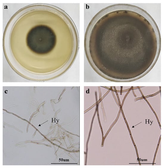
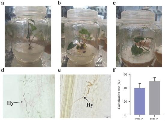
All inoculated yam roots exhibited typical DSE colonization structures (Figure 1), in contrast, no DSE colonization was observed in the control group. Hyphal colonization rate was 40.0% and 50.1% for Poac and Podo treatments, respectively (Figure 2).
Figure 1.
Colony and hyphal morphology of dark septate endophytes (DSE). (a) and (c) indicated the colony morphology and hyphae microscopic structure of Podospora sp. (Podo), respectively. (b) and (d) indicated the colony morphology and hyphae structure of Poaceascoma sp. (Poac), respectively. Hy indicated DSE hyphae.
Figure 2.
Dioscorea opposita (yam) seedling culture and DSE colonization. (a), (b), (c) indicated the growth form of Dioscorea opposita (yam) in control group, Podo treatment group and Poac treatment group, respectively. (d) and (e) indicated colonization status of Podo and Poac in the roots of yam, respectively. (f) indicated the colonization rate of two DSE strains in the root of yam. Poac_P and Podo_P indicated the Poac treatment and Podo treatment, respectively. The average value was derived from 6 biological replicates. Hy indicated DSE hyphae.
3.2. Effect of DSE on the Growth Traits of Yam
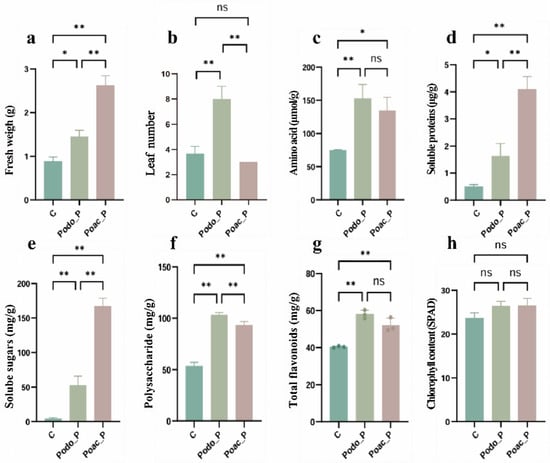
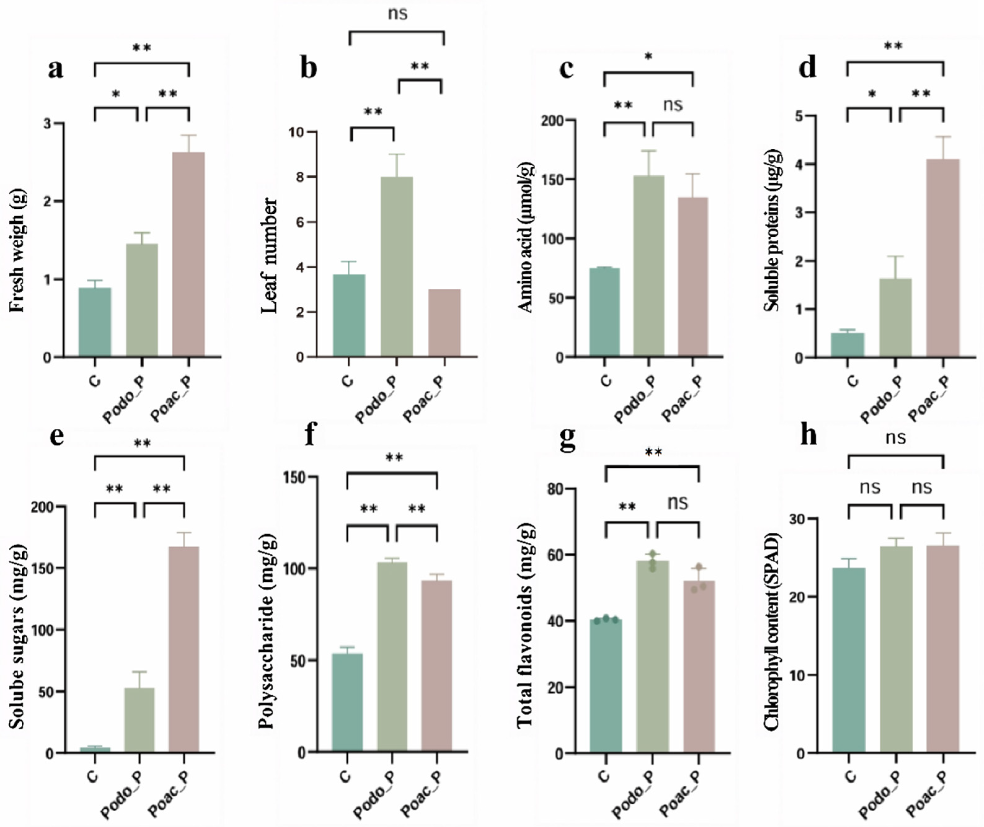
Inoculation with DSE promoted the fresh weight and leaf number of yam. The fresh weights of the two inoculated treatments were both higher than that of the control group, and the fresh weight of Poac treatment was higher than that of Podo treatment (Figure 3a). The leaf number of the Podo treatment was higher than that of the control and Poac treatments, and there was no difference in leaf number between the Poac treatment and the control group (Figure 3b).
Figure 3.
Effect of DSE inoculation treatments on growth parameters and nutrient composition of yam. (a) Fresh weigh. (b) Leaf number. (c) Amino acid. (d) Soluble proteins. (e) Solube sugars. (f) Polysaccharide. (g) Total flavonoids. (h) ChlorophyII content. The C indicated the control group. The “ns” indicated there is no significant difference among different treatments. * indicated significant differences between different treatments. * = p < 0.05; ** = p < 0.01. The average value was derived from six biological replicates.
3.3. The Effect of DSE on the Active Ingredients of Yam
Inoculation with DSE increased the contents of total flavonoids, amino acids, soluble sugars, soluble proteins, and polysaccharides in yams tubers, but had no effect on chlorophyll content in yam leaves. Compared with the control group, the amino acid content in the Poac and Podo treatments increased by 80.2% and 105.1%, respectively. Although there was no difference between the two treatment groups, the amino acid contents in the Podo group were slightly higher than that in the Poac group (Figure 3c). The soluble protein contents in the Poac and Podo treatments were 734.7% and 230.6% higher than that in the control group, respectively, while the Poac group showed a 240.6% increase compared with the Podo group (Figure 3d). For soluble sugar content, the Poac and Podo groups were 11 times and 35 times higher than those in the control group, respectively, with the Poac group being 231.3% higher than the Podo group (Figure 3e). Total flavonoids exhibited a similar trend (Figure 3g), with the Poac and Podo treatment groups showing increases of 29.6% and 44.7%, respectively, compared with the control. Both DSE treatments significantly enhanced polysaccharide content, with the polysaccharide contents in the Poac and Podo groups increasing by 73.8% and 92.7%, respectively, relative to the control (Figure 3f).
3.4. Effect of DSE on the Secretion Pattern of Yam Root Exudates
A total of 1297 metabolites were identified in the root exudates across the three treatments, with 952 substances being annotated. Among these, lipids and lipid-like molecules (22.8%) as well as phenylpropanoids and polyketides (18.3%) constituted the major components of the root exudates (Table 1). In the Poac group, phenylpropanoids and polyketides were the most abundant category with 82 compounds, followed by 69 compounds of lipids and lipid-like molecules, 59 compounds of organic acids and derivatives, and 57 organoheterocyclic compounds. The Podo group was predominantly composed of 97 compounds of lipids and lipid-like molecules, followed by 70 compounds of organic acids and derivatives and 51 organoheterocyclic compounds (Table 1).
Table 1.
The types and quantities of differential root exudates after inoculation with different DSE strains.
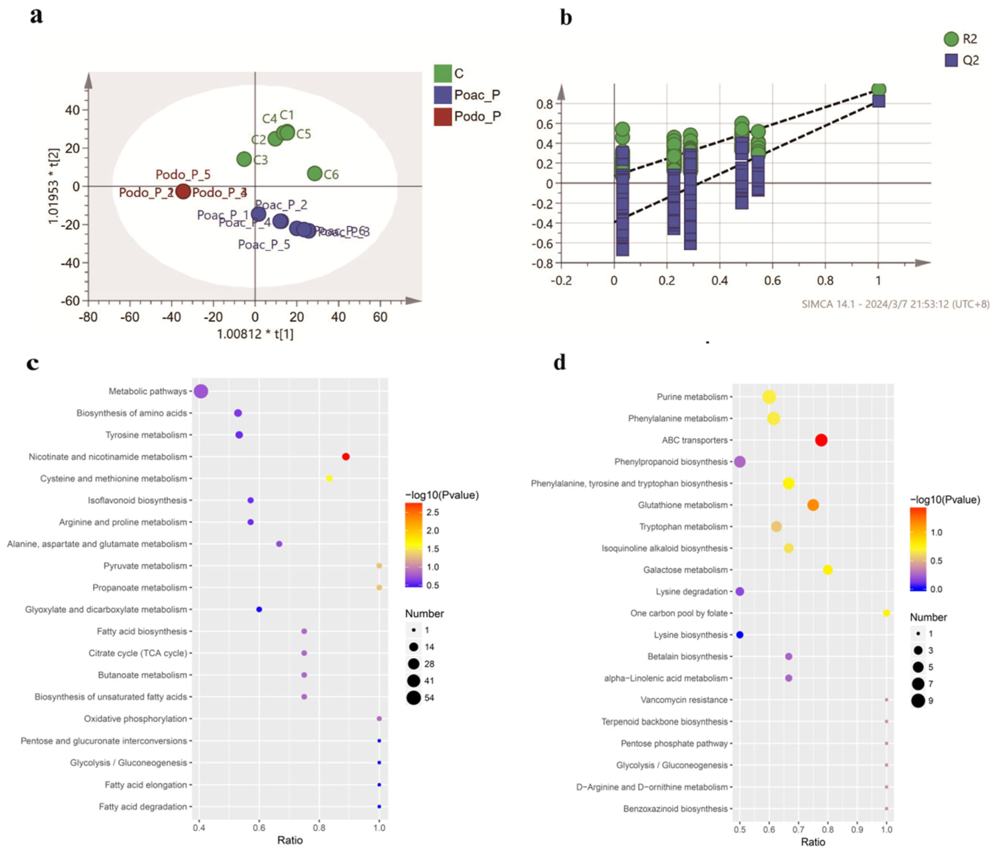
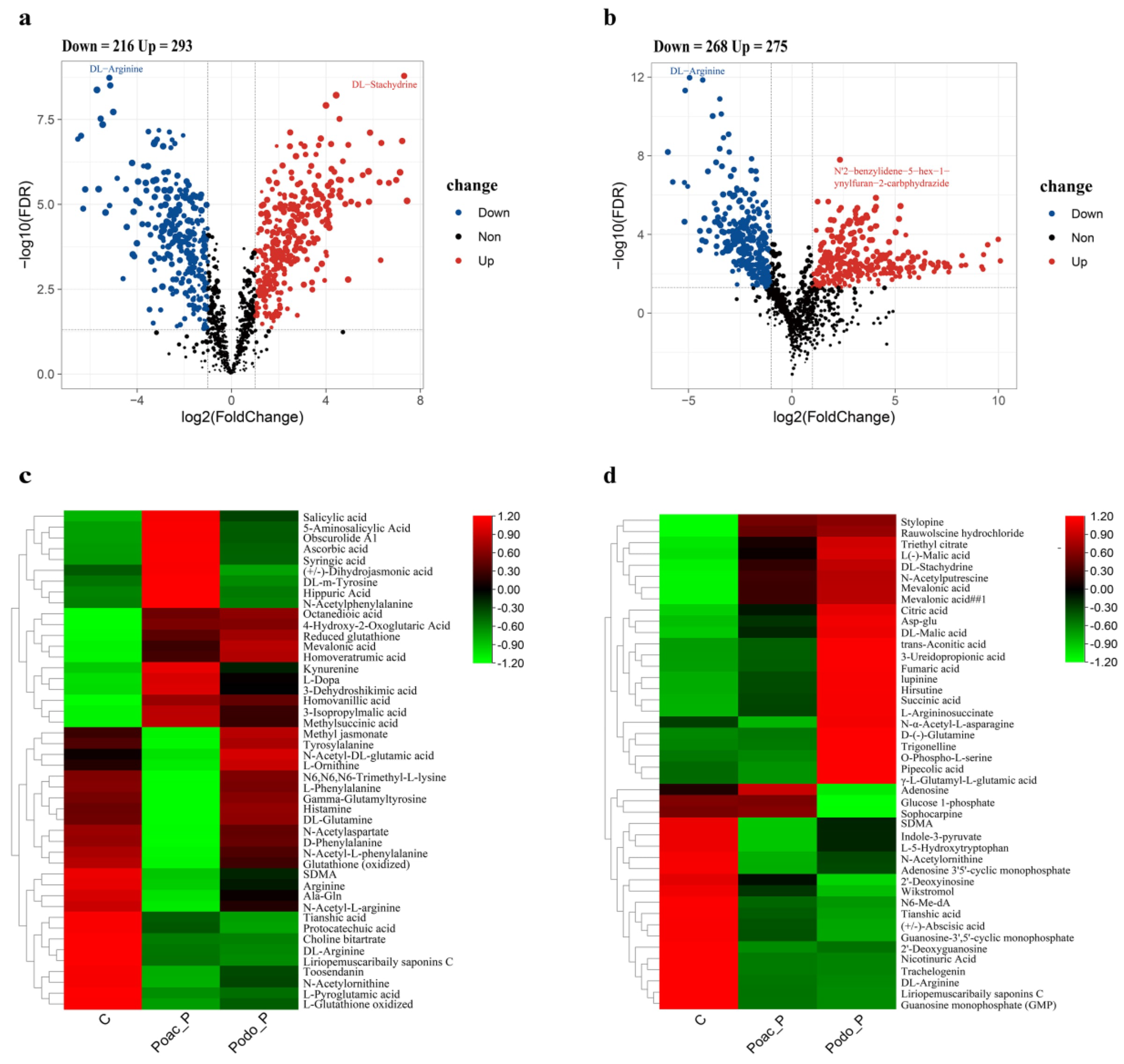
The results of PLS-DA revealed significant differences in the composition of yam root exudates under different treatments (Figure 4a,b). Compared with the control group, 39.2% (509) of metabolites in the Podo treatment group were up-regulated or down-regulated, while 41.9% (543) of metabolites showed changes in the Poac treatment group (Figure 5a,b). There were also significant differences in metabolites between the two DSE treatments (Supplementary Table S1). Volcano plot analysis further elucidated the dynamic changes in differential metabolites: after inoculation with Podo, 293 substances were up-regulated, among which DL-Stachydrine (log2FC = 7.311904043, p-value = 2.53 × 10⁻¹²) showed the most prominent up-regulation, while 216 substances were significantly down-regulated, with DL-Arginine (log2FC = −5.17474, p-value = 1.43 × 10⁻¹²) and methyl 4-methyl-2-oxo-2H-pyran-6-carboxylate (log2FC = −5.69946, p-value = 1.31 × 10⁻¹¹) exhibiting the most notable down-regulation (Figure 5a). For the Poac-inoculation group, 275 compounds were up-regulated and 268 compounds were down-regulated. Among them, N′2-benzylidene-5-hex-1-ynylfuran-2-carbohydrazide (log2FC = 2.32654489, p-value = 1.59 × 10⁻⁸) displayed the most marked up-regulation, while DL-Arginine (log2FC = −4.949652393, p-value = 1.07 × 10⁻¹²) was also significantly down-regulated (Figure 5b).
Figure 4.
The partial least squares discrimination analysis (PLS-DA) of the metabolic differences between Podo treatment and Poac treatment compared to the control group, and the enriched pathways of differential metabolites in Podo treatment and Poac treatment (show the top 20 results). (a) and (b) indicated the graph of OPLS-DA and substitution test graph (validate model: R2 = (0.0, 0.0715) Q2 = (0.0, −0.392)), respectively. In panel a, the “*” symbols in the horizontal and vertical axis labels indicate that the sample scores on the first and second PLS-DA components are multiplied by the respective axis scaling factors of 1.00812 and 1.01953. (c) and (d) indicated Podo and Poac, respectively. The hypergeometric test method was used to obtain the p-value of pathway enrichment. p-value < 0.05 was used as the threshold, and the bubble plot of the KEGG pathway.
The results of hierarchical clustering analysis of metabolites indicated that for the Poac treatment, the two signaling molecules, salicylic acid and jasmonic acid, were significantly up-regulated. Phenolic acid substances related to the phenylpropanoid pathway, including syringic acid, m-tyrosine, and 3-dehydroshikimic acid, were also significantly up-regulated. Notably, the 3-dehydroshikimic acid is a key compound linking primary and secondary metabolism, ultimately leading to the accumulation of products including phenolic acids and coumarins. Furthermore, amino acid derivatives and organic acids were significantly up-regulated too (Figure 5c). For the Podo treatment, intermediate metabolites in the tricarboxylic acid (TCA) cycle, including citric acid, L-malic acid, succinic acid, and argininosuccinic acid, were significantly up-regulated. The stylopine and hirsutine in alkaloids were also significantly up-regulated. Substances involved in nitrogen metabolism and amino acid derivatives, including N-acetylasparagine, D-glutamine, and O-phosphoserine, showed significant up-regulation. Additionally, pipecolic acid was significantly up-regulated (Figure 5d).
3.5. Effect of DSE on Primary Metabolism of Yam
Differential metabolites in the Podo group were enriched in 34 metabolic pathways, among which the Citrate cycle pathway was significantly enriched. This finding aligns with the hierarchical clustering analysis of metabolites mentioned above, which showed that substances including citric acid, L-malic acid, succinic acid, and argininosuccinic acid were significantly up-regulated in the Podo group. This suggested that metabolic pathways centered on the TCA cycle were affected by Podo inoculation. Furthermore, pathways including pyruvate metabolism, glycolysis/gluconeogenesis, fatty acid biosynthesis, and amino acid biosynthesis were also significantly enriched (Figure 4c). Upstream sugars including glucose, sucrose, and glucose-1-phosphate decreased significantly, while downstream products of glycolysis, including D-(−)-ribose and L-arabitol, increased markedly (Figure 6a). Key intermediate products of the TCA cycle, namely citric acid, L-malic acid, succinic acid, trans-aconitic acid, and fumaric acid, all accumulated significantly (Figure 6d). The heatmap of fatty acids (Figure 6b) revealed a significant increase in medium- and long-chain saturated fatty acids such as myristic acid, stearic acid, and valeric acid, accompanied by a synchronous rise in mevalonic acid and 3-hydroxy-3-methylglutaric acid. Aspartate and glutamate are crucial for amino acid biosynthesis. Based on aspartate and glutamate, and complemented by glutamine, a variety of other amino acids can be synthesized. Compared with the control group, the accumulation of aspartate, glutamate, and its derivative glutamine in the rhizosphere of the Podo treatment group was significantly higher (Figure 6e).
Figure 5.
The up- and down-regulation of metabolites secreted after inoculation with different DSE strains. (a) and (b) indicated the volcano plot of Podo treatment and Poac treatment, respectively. (c) and (d) indicated the differential secretions heatmap of treatment and Poac treatment, respectively.
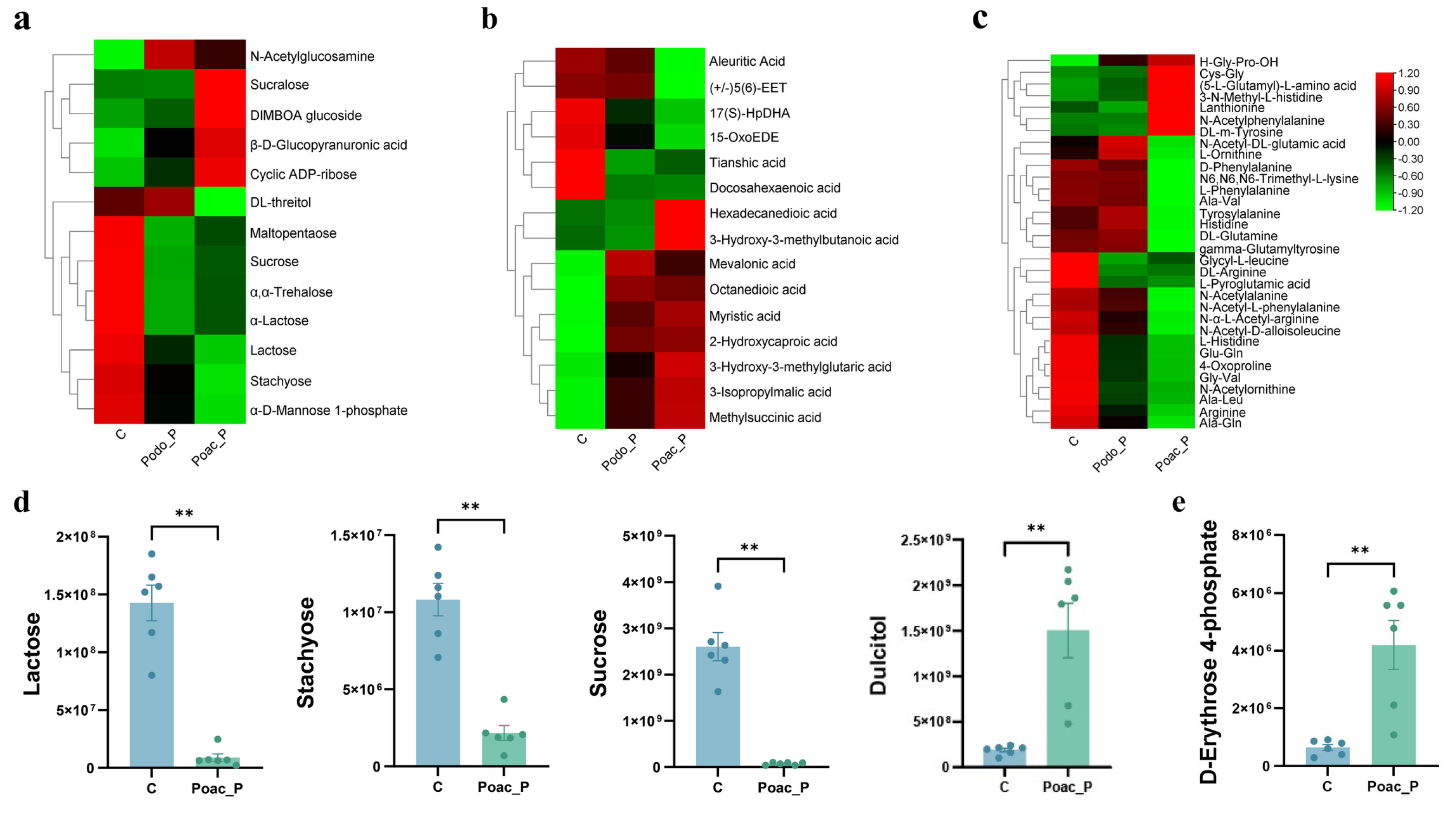
The Poac treatment led to enrichment in the galactose metabolism pathway and the pentose phosphate pathway, along with enrichment in phenylpropanoid biosynthesis and terpenoid backbone biosynthesis pathways (Figure 4d). Upstream sugars in galactose metabolism, including lactose, stachyose, sucrose, and trehalose, decreased significantly, while the downstream product galactitol increased markedly compared to the control group (Figure 7a,d). Additionally, the significant increase in 3-dehydroshikimic acid observed in the hierarchical clustering analysis of metabolites collectively suggests altered activity of the galactose metabolism pathway. The end product of galactose metabolism is D-fructose-6-phosphate, which subsequently enters the pentose phosphate pathway. Concurrently, substances from the pentose phosphate pathway, such as D-erythrose 4-phosphate, were also enriched in the Poac treatment (Figure 7e). This suggests that Poac may regulate plant carbohydrate metabolism by metabolizing sugars like sucrose, stachyose, and lactose via the galactose pathway to generate D-fructose-6-phosphate. This intermediate is then utilized through the pentose phosphate pathway to consume sugars for energy production and the synthesis of other compounds (lipids and nucleic acids). The preferential utilization of sugars via the pentose phosphate pathway may reduce the flux of sugars through glycolysis and the TCA cycle. Consequently, the synthesis of key amino acid precursors, α-ketoglutarate and oxaloacetate, may be diminished, potentially limiting amino acid biosynthesis (Figure 7c). Furthermore, NADPH and glyceraldehyde 3-phosphate generated by the pentose phosphate pathway serve as substrates for the synthesis of fatty acids and steroids in vivo, leading to the substantial production of lipid-like substances in plants (Figure 7b).
Figure 6.
The influence of Podo inoculation on the contents of sugars, fatty acids and amino acids in the root exudates of yam. (a), (b), and (c) indicated the heat maps of the differential metabolites of sugar, fatty acids and amino acids after Podo inoculation, respectively. (d) represented different intermediate products of sugar metabolism. (e) indicated the key amino acids synthesized by inoculation with Podo. * indicated significant differences between different treatments. ** = p < 0.01.
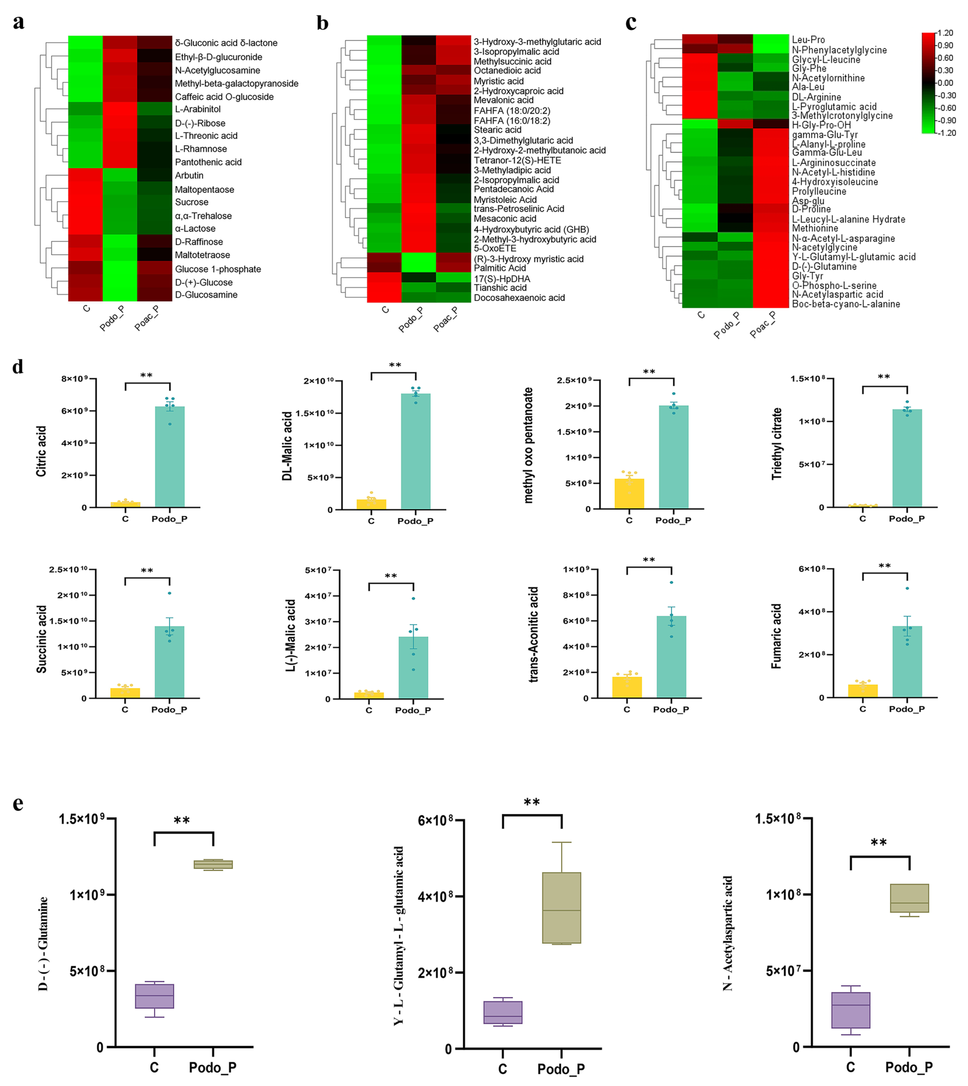
Figure 7.
The influence of Poac inoculation on the contents of sugars, fatty acids and amino acids in the root exudates of yam. (a), (b), and (c) indicated the heat maps of the differential metabolites of sugar, fatty acids and amino acids after Poac inoculation, respectively. (d) represented different intermediate products of galactose metabolism. (e) indicated the products of the pentose phosphate pathway. * indicated significant differences between different treatments. ** = p < 0.01.
3.6. Effect of DSE on Plant Secondary Metabolism
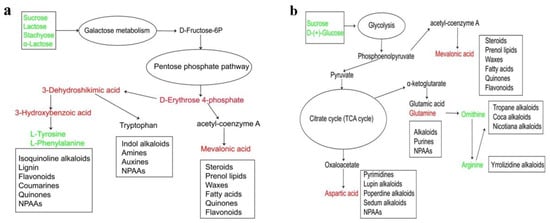
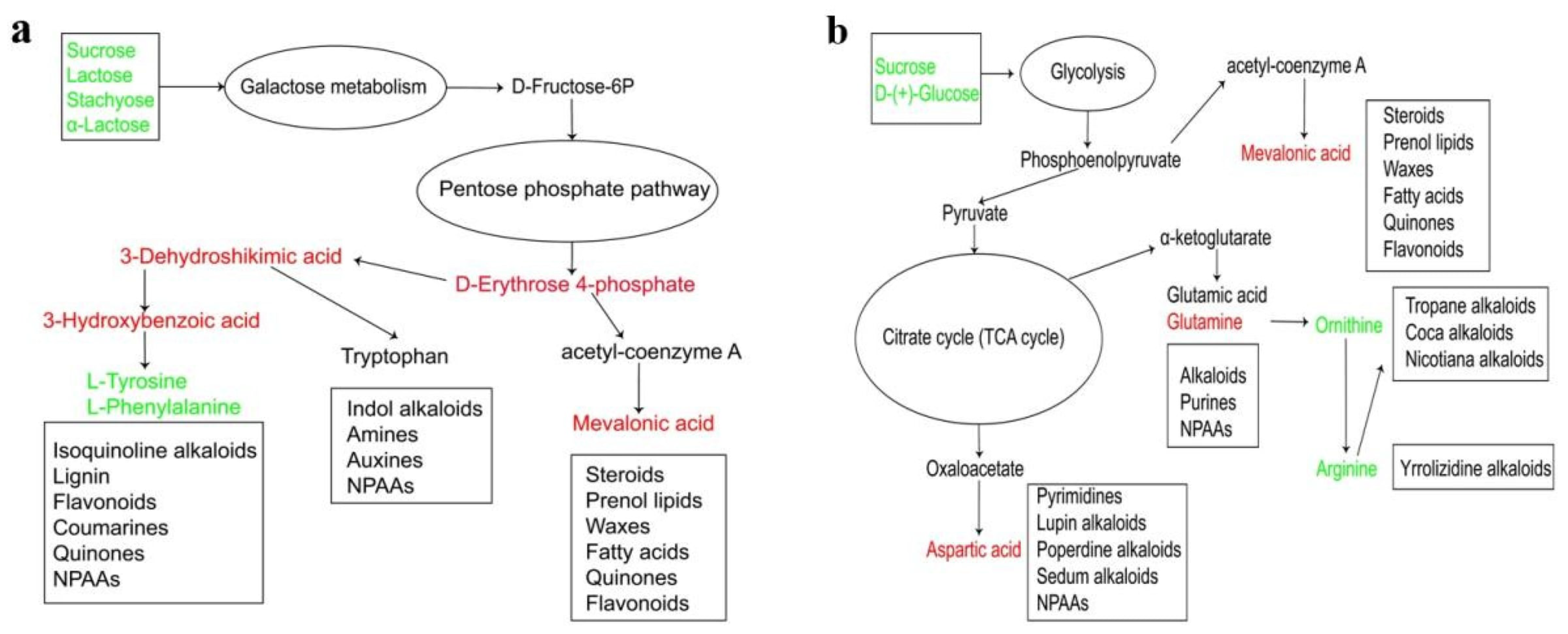
Due to the distinct influences of different DSE strains on plant primary carbohydrate metabolic pathways, the resulting primary metabolites differ, leading to variations in the secondary metabolites synthesized from these primary metabolites and their corresponding biosynthetic pathways. This study revealed that inoculation with Poac was associated with changes in the galactose metabolism and pentose phosphate pathways in yam, alongside increased abundance of erythrose-4-phosphate. This product serves as a key substrate for the shikimate pathway; consequently, yam primarily synthesizes secondary metabolites via this route. By analyzing the exudates, intermediate products of the metabolic pathways were identified, enabling the construction of a metabolic pathway map. The secondary metabolic pathway map (Figure 8a) suggested that Poac may redirect carbon flux from galactose metabolism into the pentose phosphate pathway, generating D-erythrose 4-phosphate. This compound is a crucial precursor for secondary metabolism. It participates in the synthesis of 3-dehydroshikimic acid and 3-hydroxybenzoic acid, which are further utilized to produce L-tyrosine and L-phenylalanine. However, in this experiment, the direct substrates for secondary metabolite synthesis, L-tyrosine and L-phenylalanine, were significantly decreased. This reduction was likely attributable to their substantial consumption for the biosynthesis of secondary metabolites such as flavonoids and coumarins. Furthermore, D-erythrose 4-phosphate is involved in tryptophan synthesis, which subsequently leads to the production of alkaloids and phytohormones. It also contributes to the synthesis of acetyl-CoA, ultimately participating in fatty acid biosynthesis. Key intermediates of the α-ketoglutarate pathway, including DL-arginine, N-acetylornithine, DL-glutamine, arginine, and L-ornithine, were significantly down-regulated (Supplementary Figure S2). This pattern suggests changes in the α-ketoglutarate and shikimate pathways; reduced intermediates may reflect downstream consumption rather than selective inhibition.
Figure 8.
Proposed secondary metabolism pathway of yam inoculated with Poac (a) and Podo (b). Green indicated that the concentration of enriched substances is significantly lower than that of the control group. Red indicated that it is significantly higher than that of the control group.
During the symbiotic association with Podo, yam primarily synthesizes secondary metabolites through the α-ketoglutarate branch and the oxaloacetate branch of the TCA cycle, as well as the mevalonate pathway. Through screening and analysis of the enriched metabolites, the glycolysis-TCA cycle was enhanced by inoculation with Podo, leading to the accumulation of products including acetyl-CoA, α-ketoglutarate, phosphoenolpyruvate, and oxaloacetate (Figure 8b). The oxaloacetate branch mainly activates the pyrimidine synthesis pathway. In this experiment, significant accumulation of N-acetylaspartate, an upstream metabolite in the pyrimidine synthesis pathway, was detected, along with a notable increase in the downstream metabolite aspartate. This promoted the synthesis of pyrimidine-derived metabolites and alkaloids. In the α-ketoglutarate branch, downstream products such as glutamate and glutamine accumulated significantly, driving the biosynthesis of alkaloids, proteins, and other compounds. Phosphoenolpyruvate generated from glycolysis is converted to acetyl-CoA, which subsequently leads to the production of mevalonic acid. This activates the mevalonate pathway, facilitating the synthesis of secondary metabolites such as flavonoids and terpenoids.
In the Poac treatment group, 14 significantly enriched coumarins and their derivatives were detected, of which 12 showed significant up-regulation. In contrast, only 5 significantly enriched coumarins and their derivatives were identified in the Podo treatment group, with merely 4 being significantly up-regulated. The Poac treatment group exhibited significant enrichment of 17 flavonoids that were markedly up-regulated, whereas only 4 were detected in the Podo group. For purine/pyrimidine metabolites, 5 were significantly up-regulated in the Poac group, compared to 10 showing significant accumulation in the Podo group. Regarding alkaloid metabolites, 4 were significantly up-regulated in the Poac group, while 7 displayed significant accumulation in the Podo group (Supplementary Table S2). These findings align with the secondary metabolic pathway maps described above, supporting the interpretation of the proposed primary and secondary metabolic pathway models.
4. Discussion
In this study, inoculation with two DSE strains promoted the growth and quality of yam by enhancing the levels of nutritionally active components, such as flavonoids, polysaccharides, and amino acids, in the roots. This finding was consistent with the general consensus that DSE, as beneficial plant symbionts, can broadly promote host growth and enhance nutrient uptake and metabolic activity [28]. Chu et al. [29] reported that inoculation with Paraphoma chrysanthemicola increased the biomass of Pinus tabulaeformis seedlings, while Li and Wang [30] observed that DSE inoculation significantly increased the biomass, root surface area, and chlorophyll content in Isatis indigotica. Moreover, soluble sugars have been reported to promote plant growth [31], which aligns with the increases in soluble sugar content and plant fresh weight observed in our experiment. Collectively, these results indicated that both Poac and Podo possess potential for further evaluation as growth-promoting inoculants for yam.
Although both strains exhibited growth-promoting effects, there were significant differences in the extent to which they enhanced yam phenotype and various active components. This finding was consistent with the research by Guo et al. [32], suggesting that the variation in DSE-plant interactions might be related to the differential regulation of root exudates by different strains, thereby modulating distinct metabolic pathways. To further elucidate the differential metabolic remodeling mechanisms underlying the strain-specific phenotypic improvements and active component accumulation, this study conducted a root exudate metabolome analysis. The results revealed that inoculation with Podo and Poac was associated with different metabolic responses. These results suggest that yam inoculated with Podo exhibited a bias towards fundamental metabolism via the TCA cycle, whereas those inoculated with Poac favored the pentose phosphate pathway. The TCA cycle provides carbon skeletons, ATP, and other precursors for the biosynthesis of amino acids, fatty acids, nucleotides, and secondary metabolites, thereby supplying more energy for plant growth and development. Conversely, the pentose phosphate pathway supplies carbon skeletons and NADPH for biosynthesis, contributing to the plant’s antioxidant defense system [33].
Furthermore, in this study, inoculation with both DSE strains may influence plant resistance, but their metabolic regulatory mechanisms also differed. Podo inoculation may enhance yam resistance by upregulating pipecolic acid. Pipecolic acid is a non-proteinogenic amino acid that can synergize with signaling molecules such as salicylic acid to form a complex defense signaling network. In contrast, inoculation with the Poac strain may induce systemic resistance in plants by upregulating salicylic acid and jasmonic acid. Research indicated that the accumulation of salicylic acid, jasmonic acid, and pipecolic acid can “prime” plant defense responses, thereby inducing systemic acquired resistance [7,34].
Regarding the nutritional component content in yam roots, in this experiment, inoculation with either Poac or Podo significantly increased the total flavonoid content. This result supported the report by Zhang et al. [35] that DSE inoculation in Sorghum promoted flavonoid synthesis. For plants inoculated with Poac, this effect might be related to the strong activation of the shikimate pathway by Poac. The shikimate pathway is a central route for synthesizing aromatic amino acids (phenylalanine, tyrosine, tryptophan) in plants and microorganisms. These amino acids serve as precursors for various secondary metabolites, with phenylalanine being the core precursor for phenylpropanoid secondary metabolites such as flavonoids, lignin, and coumarins [36]. In the metabolic pathways of Poac-inoculated plants in this experiment, the intermediate metabolite 3-dehydroshikimic acid was significantly upregulated, while the direct substrates L-tyrosine and L-phenylalanine were significantly downregulated. This pattern suggested that the precursors were heavily consumed for downstream synthesis, ultimately leading to substantial flavonoid accumulation. For plants inoculated with Podo, the increase in flavonoid content might be indirectly promoted by Podo through enhancing overall plant metabolic levels, supplying partial precursors (such as acetyl-CoA), or inducing plant defense responses [37]. In the Podo treatment group, enhanced glycolysis could potentially produce more acetyl-CoA, supporting the TCA cycle, fatty acid synthesis, and flavonoid biosynthesis.
Regarding the total amino acid and soluble protein content in plants, inoculation with Poac may have altered the pentose phosphate and shikimate pathways, together with the TCA cycle and the α-ketoglutarate pathway. This suggests that the conventional amino acid synthesis pathways (relying on TCA cycle intermediates) were somewhat constrained. However, the total amino acid content in Poac-inoculated plants was still higher than in the control group. A plausible explanation is that the NADPH generated by the pentose phosphate pathway serves as crucial reducing power for nitrate reduction and amino acid synthesis (e.g., via the glutamate synthase pathway). The increased supply of NADPH may have significantly enhanced the efficiency of nitrogen assimilation and amino acid synthesis [38], ultimately leading to increased soluble protein and total amino acid content. Furthermore, studies have shown that endophytes can enhance plant affinity for nitrogen (N) uptake [15,16,19], which may enable plants to synthesize proteins abundantly, thereby increasing protein content. In contrast, Podo inoculation likely promoted glycolysis and the TCA cycle, generating substantial ATP and key amino acid precursors: α-ketoglutarate (for the glutamate family) and oxaloacetate (for the aspartate family). The enrichment of these key amino donors laid the foundation for the synthesis of other amino acids, ultimately resulting in a significant increase in total amino acid content. Concurrently, the improved TCA cycle may have provided ample ATP, supplying sufficient energy for protein synthesis—including energy-intensive processes such as amino acid activation and peptide chain elongation [39]—which consequently led to a significant increase in soluble protein.
Regarding the soluble sugar content in plants, the levels of glucose, sucrose, and other sugars in the root exudates of Podo-inoculated plants decreased significantly. However, the intermediates of downstream glycolysis and the TCA cycle increased markedly. This suggests that Podo may have stimulated the uptake and metabolic utilization of sugars by the plant; sugars may have been rapidly absorbed into cells for glycolysis and the TCA cycle, providing energy and carbon skeletons for growth. In plants inoculated with Poac, upstream metabolites in galactose metabolism were downregulated, while the downstream product dulcitol was upregulated. Additionally, the key pentose phosphate pathway intermediate D-erythrose 4-phosphate was upregulated, which similarly reflects the metabolism of sugars through specific pathways. Nevertheless, the total soluble sugar content in the plant increased. A possible reason is that DSE inoculation enhanced photosynthesis in the plant. Studies have shown that endophytic fungi can help plants increase their photosynthetic rate, and observed high sugar content indicates that the fungus-plant interaction improves photosynthetic efficiency [40]. In this experiment, the DSE symbiosis may have indirectly stimulated photosynthetic carbon assimilation in the leaves by improving the plant’s nutritional status or through hormonal regulation, thereby generating more sugar sources. Furthermore, the increased number of leaves in Podo-inoculated plants suggests an expansion of the photosynthetic area.
Regarding the polysaccharide content in plants, both DSE treatments in this experiment significantly promoted the growth of the host plant. This growth is manifested in cell division, cell expansion, and the formation of new organs, which require the synthesis of substantial amounts of structural polysaccharides (e.g., cellulose, hemicellulose) and storage polysaccharides (e.g., starch in yam tubers) [41]. For plants inoculated with Poac, the intermediates of the activated pentose phosphate pathway can interconvert with intermediates from other pathways, entering glycolysis or gluconeogenesis. This provides precursors (such as glucose, UDP-glucose) for the synthesis of both structural and storage polysaccharides. For plants inoculated with Podo, the potential promotion of glycolysis leads to an increase in glycolytic intermediates (e.g., glucose-6-phosphate, fructose-6-phosphate). These intermediates serve as key precursors for the synthesis of nucleotides and sugar nucleotides, which are subsequently utilized in the biosynthesis of various polysaccharides [42]. However, these findings were obtained in a 30-day axenic seedling system and require validation in soil-grown plants before conclusions about mature tuber quality or field yield can be drawn. In addition, exudate profiles may include fungal metabolites and transformed plant compounds; they do not directly establish intracellular fluxes or systemic resistance. MS medium and exogenous sucrose may alter exudation. Future studies should combine longer-term soil trials, dry-mass measurements, non-invasive physiological monitoring, and molecular or isotope-tracing validation.
5. Conclusions
This study confirmed through aseptic cultivation experiments that inoculation with two DSE strains promoted the performance and quality of yam by enhancing the levels of nutritionally active components, such as flavonoids, polysaccharides, and amino acids, in the tubers. Further, the differential metabolic responses of different DSE strain inoculations on the phenotype and active ingredient accumulation of yam were analyzed by LC-MS. Podo inoculation may affect the basal metabolism of the tricarboxylic acid (TCA) cycle, while Poac inoculation may affect the galactose metabolism, pentose phosphate metabolism and shikimic acid metabolism pathways. Our research has deepened the understanding of the symbiotic mechanism between plants and DSE, providing a theoretical framework for further exploring the impact of DSE on active ingredients and root secretions. It also offers an important theoretical basis for evaluating the application of specific DSE strains to improve the accumulation of target active components in medicinal plants. Future research will explore the application effects of DSE biotechnology in medicinal plant cultivation and sustainable agriculture.
Supplementary Materials
The following supporting information can be downloaded at: https://www.mdpi.com/article/10.3390/microorganisms14112415/s1, Figure S1:Inoculation of two dark septate endophytes (DSE) significantly alteredmetabolite Venn diagram; Figure S2: Effect of Poac inoculation on key intermediates of the α-ketoglutarate metabolic pathway in plants. Use t-test and Mann Whitney test statistical analysis methods. ** = p < 0.01; *** = p < 0.0005; **** = p < 0.0001. Table S1: Differences in metabolites between the two dark septate endophytes (DSE) treatments. Table S2: Total secondary metabolites across treatments and differential regulatory relationships between treatment groups..
Author Contributions
Conceptualization, W.L., C.H. and X.L.; methodology, W.L., Y.C. and F.Z.; software, Z.M.; validation, J.L. and C.Y.; data curation, W.L. and Y.C.; writing—original draft preparation, W.L. and F.Z.; writing—review and editing, W.L., C.H. and X.L.; supervision, X.H.; project administration, C.H. and X.L.; funding acquisition, C.H. and X.L.; All authors have read and agreed to the published version of the manuscript.
Funding
This work was supported by the National Key Research and Development Program of China [Young Scientist Program, grant numbers: 2025YFC3512100].
Institutional Review Board Statement
Not applicable.
Informed Consent Statement
Not applicable.
Data Availability Statement
The original contributions presented in this study are included in the article/Supplementary Material. Further inquiries can be directed to the corresponding authors.
Conflicts of Interest
The authors declare no conflicts of interest.
References
- Francioli, D.; Kampouris, I.D.; Kuhl-Nagel, T.; Babin, D.; Sommermann, L.; Behr, J.H.; Chowdhury, S.P.; Zrenner, R.; Moradtalab, N.; Schloter, M.; et al. Microbial inoculants modulate the rhizosphere microbiome, alleviate plant stress responses, and enhance maize growth at field scale. Genome Biol. 2025, 26, 148. [Google Scholar] [CrossRef] [Scilit] [PubMed]
- Lugtenberg, B.; Kamilova, F. Plant-growth-promoting rhizobacteria. Annu. Rev. Microbiol. 2009, 63, 541–556. [Google Scholar] [CrossRef] [Scilit] [PubMed]
- Shi, J.; Wang, X.; Wang, E. Mycorrhizal symbiosis in plant growth and stress adaptation: From genes to ecosystems. Annu. Rev. Plant Biol. 2023, 74, 569–607. [Google Scholar] [CrossRef] [Scilit] [PubMed]
- Zhang, H.; Sun, Y.; Xie, X.; Kim, M.S.; Dowd, S.E.; Paré, P.W. A soil bacterium regulates plant acquisition of iron via deficiency-inducible mechanisms. Plant J. 2009, 58, 568–577. [Google Scholar] [CrossRef] [Scilit] [PubMed]
- Mendes, R.; Garbeva, P.; Raaijmakers, J.M. The rhizosphere microbiome: Significance of plant beneficial, plant pathogenic, and human pathogenic microorganisms. Fems. Microbiol. Rev. 2013, 37, 634–663. [Google Scholar] [CrossRef] [Scilit] [PubMed]
- Kawasaki, A.; Dennis, P.G.; Forstner, C.; Raghavendra, A.K.H.; Mathesius, U.; Richardson, A.E.; Delhaize, E.; Gilliham, M.; Watt, M.; Ryan, P.R. Manipulating exudate composition from root apices shapes the microbiome throughout the root system. Plant Physiol. 2021, 187, 2279–2295. [Google Scholar] [CrossRef] [Scilit] [PubMed]
- Venturi, V.; Keel, C. Signaling in the Rhizosphere. Trends Plant Sci. 2016, 21, 187–198. [Google Scholar] [CrossRef] [Scilit] [PubMed]
- Wang, X.; Wei, H.; Lei, J.; Rui, Z.; Yu, C. Trichoderma koningiopsis induced changes in root exudates of masson pine seedlings alter rhizosphere microbiome to enhance damping-off disease resistance. Plant Cell Environ. 2025, 49, 64–79. [Google Scholar] [CrossRef] [Scilit] [PubMed]
- Fracchia, F.; Guinet, F.; Engle, N.L.; Tschaplinski, T.J.; Veneault-Fourrey, C.; Deveau, A. Microbial colonisation rewires the composition and content of poplar root exudates, root and shoot metabolomes. Microbiome 2024, 12, 173. [Google Scholar] [CrossRef] [Scilit] [PubMed]
- Deng, X.; Zhao, D.; Li, Y.; Li, K.; Xia, J.; Yu, S.; Wang, J.; Lin, J. Arbuscular mycorrhizal fungi confer aluminum toxicity tolerance in Ricinus communis via modulating root metabolic mechanisms and the composition and quantity of root exudates. Plant Physiol. Bioch. 2025, 227, 110149. [Google Scholar] [CrossRef] [Scilit] [PubMed]
- Li, Y.; Lu, L.; Wang, Q.; Liu, X.; Tian, J.; Zhang, R.; Liao, H.; Lambers, H.; Wang, X. Arbuscular mycorrhizal fungi promote nodulation and N2 fixation in soybean by specific root exudates. Plant Cell Environ. 2025, 48, 5514–5528. [Google Scholar] [CrossRef] [Scilit] [PubMed]
- Bock, B.M.; Hoeksema, J.D.; Johnson, N.C.; Gehring, C.A. Evidence for common fungal networks among plants formed by a dark septate endophyte in Sorghum bicolor. Commun. Biol. 2025, 8, 996. [Google Scholar] [CrossRef] [Scilit] [PubMed]
- Ruotsalainen, A.L.; Kauppinen, M.; Wäli, P.R.; Saikkonen, K.; Helander, M.; Tuomi, J. Dark septate endophytes: Mutualism from by-products? Trends Plant Sci. 2022, 27, 247–254. [Google Scholar] [CrossRef] [Scilit] [PubMed]
- Wang, S.; Bi, Y.; Quan, W.; Christie, P. Growth and metabolism of dark septate endophytes and their stimulatory effects on plant growth. Fungal Biol. 2022, 126, 674–686. [Google Scholar] [CrossRef] [Scilit] [PubMed]
- Vergara, C.; Araujo, K.E.C.; Alves, L.S.; Souza, S.R.; Santos, L.A.; Santa-Catarina, C.; Silva, K.D.; Pereira, G.M.D.; Xavier, G.R.; Zilli, J.É. Contribution of dark septate fungi to the nutrient uptake and growth of rice plants. Braz. J. Microbiol. 2018, 49, 67–78. [Google Scholar] [CrossRef] [Scilit] [PubMed]
- Vergara, C.; Araujo, K.E.C.; Sperandio, M.V.L.; Santos, L.A.; Urquiaga, S.; Zilli, J.É. Dark septate endophytic fungi increase the activity of proton pumps, efficiency of 15N recovery from ammonium sulphate, N content, and micronutrient levels in rice plants. Braz. J. Microbiol. 2019, 50, 825–838. [Google Scholar] [CrossRef] [Scilit] [PubMed]
- Wen, Z.; Terhonen, E.; Asiegbu, F.O. The dark septate endophyte Phialocephala sphaeroides confers growth fitness benefits and mitigates pathogenic effects of Heterobasidion on Norway spruce. Tree Physiol. 2022, 42, 891–906. [Google Scholar] [CrossRef] [Scilit] [PubMed]
- Wu, F.L.; Li, Y.; Tian, W.; Sun, Y.; Chen, F.; Zhang, Y.; Zhai, Y.; Zhang, J.; Su, H.; Wang, L. A novel dark septate fungal endophyte positively affected blueberry growth and changed the expression of plant genes involved in phytohormone and flavonoid biosynthesis. Tree Physiol. 2020, 40, 1080–1094. [Google Scholar] [CrossRef] [Scilit] [PubMed]
- Vergara, C.; Araujo, K.E.C.; Urquiaga, S.; Santa-Catarina, C.; Schultz, N.; da Silva Araújo, E.; de Carvalho Balieiro, F.; Xavier, G.R.; Zilli, J.É. Dark septate endophytic fungi increase green manure-15n recovery efficiency, N contents, and micronutrients in rice grains. Front. Plant Sci. 2018, 9, 613. [Google Scholar] [CrossRef] [Scilit] [PubMed]
- Tan, J.Y.; Yue, Z.C.; Li, S.T.; Pan, Y.Y.; Chu, Z.Y.; Ban, Y.H.; Xu, Z.Y. Alleviation of salt stress and changes in glycyrrhizic acid accumulation by dark septate endophytes in Glycyrrhiza glabra grown under salt stress. J. Agric. Food Chem. 2024, 72, 14557–14569. [Google Scholar] [CrossRef] [Scilit] [PubMed]
- Guo, C.; Ding, G.; Huang, W.; Wang, Z.; Meng, Z.; Xiao, W. Total saponin of Dioscoreae hypoglaucae rhizoma ameliorates streptozotocin-induced diabetic nephropathy. Drug Des. Devel Ther. 2016, 10, 799–810. [Google Scholar] [CrossRef] [Scilit] [PubMed]
- Obidiegwu, J.E.; Lyons, J.B.; Chilaka, C.A. The dioscorea genus (yam)-an appraisal of nutritional and therapeutic potentials. Foods 2020, 9, 1304. [Google Scholar] [CrossRef] [Scilit] [PubMed]
- Li, X.; Liu, Y.X.; Ye, Q.N.; Xu, M.H.; He, X.L. Application of desert DSEs to nonhost plants: Potential to promote growth and alleviate drought stress of wheat seedlings. Agriculture 2022, 12, 1539. [Google Scholar] [CrossRef] [Scilit]
- Li, L.; Jiao, X.Z. Measurement of protein content by the agency of protein stain coomassie brilliant blue G250. Plant Physiol. Commun. 1980, 6, 52–55. [Google Scholar]
- Kim, D.O.; Jeong, S.W.; Lee, C.Y. Antioxidant capacity of phenolic phytochemicals from various cutivars of plums. Food Chem. 2003, 81, 321–326. [Google Scholar] [CrossRef] [Scilit]
- Phillips, J.M.; Hayman, D.S. Improved procedures for clearing roots and staining parasitic and vesicular–arbuscular mycorrhizal fungi for rapid assessment of infection. Trans. Br. Mycol. Soc. 1970, 55, 158–163. [Google Scholar] [CrossRef] [Scilit]
- Dang, K.; Hou, J.; Liu, H.; Peng, J.; Sun, Y.; Li, J.; Dong, Y. Root exudates of ginger induced by Ralstonia solanacearum infection could inhibit bacterial wilt. J. Agric. Food Chem. 2023, 71, 1957–1969. [Google Scholar] [CrossRef] [Scilit] [PubMed]
- Chu, H.L.; Wang, C.Y.; Li, Z.M.; Wang, H.H.; Xiao, Y.G.; Chen, J. The dark septate endophytes and ectomycorrhizal fungi effect on Pinus tabulaeformis Carr. seedling growth and their potential effects to pine wilt disease resistance. Forests 2019, 10, 140. [Google Scholar] [CrossRef] [Scilit]
- Santos, M.; Cesanelli, I.; Diánez, F.; Sánchez-Montesinos, B.; Moreno-Gavíra, A. Advances in the role of dark septate endophytes in the plant resistance to abiotic and biotic stresses. J. Fungi 2021, 7, 939. [Google Scholar] [CrossRef] [Scilit] [PubMed]
- Li, C.; Wang, M. Application of hairy root culture for bioactive compounds production in medicinal plants. Curr. Pharm. Biotechno. 2021, 22, 592–608. [Google Scholar] [CrossRef] [Scilit] [PubMed]
- Liu, H.; Cheng, S.; Liu, J.; Du, Y.; Bai, Z.; Du, Y. Synthesis of pentasaccharide and heptasaccharide derivatives and their effects on plant growth. J. Agric. Food Chem. 2008, 56, 5634–5638. [Google Scholar] [CrossRef] [Scilit] [PubMed]
- Guo, X.; Zhang, C.; Zhang, Y.; Wang, S.; Wang, R.S.; Xiao, J.C.; Wang, Y.F.; Peng, Z.; Wang, Y.H.; Yan, B.B.; et al. Multi-omics analysis reveals the differential mechanism of host growth and metabolism promotion by endophytic fungus and bacteria in Atractylodes lancea. Med. Plant Biol. 2025, 4, e037. [Google Scholar] [CrossRef] [Scilit]
- Meléndez-Hevia, E.; Waddell, T.G.; Cascante, M. The puzzle of the Krebs citric acid cycle: Assembling the pieces of chemically feasible reactions, and opportunism in the design of metabolic pathways during evolution. J. Mol. Evol. 1996, 43, 293–303. [Google Scholar] [CrossRef]
- Van der Ent, S.; Van Wees, S.C.; Pieterse, C.M. Jasmonate signaling in plant interactions with resistance-inducing beneficial microbes. Phytochemistry 2009, 70, 1581–1588. [Google Scholar] [CrossRef] [Scilit] [PubMed]
- Zhang, Q.M.; Gong, M.G.; Yuan, J.F.; Hou, Y.; Zhang, H.M.; Wang, Y.; Hou, X. Dark septate endophyte improves drought tolerance in sorghum. Int. J. Agric. Biol. 2017, 19, 53–60. [Google Scholar] [CrossRef] [Scilit]
- Li, Q.; Liu, Z.; Jiang, Z.; Jia, M.; Hou, Z.; Dou, D.; Yu, J. Phenylalanine metabolism-dependent lignification confers rhizobacterium-induced plant resistance. Plant Physiol. 2025, 197, kiaf016. [Google Scholar] [CrossRef] [Scilit] [PubMed]
- Yu, P.; He, X.; Baer, M.; Beirinckx, S.; Tian, T.; Moya, Y.A.T.; Zhang, X.; Deichmann, M.; Frey, F.P.; Bresgen, V.; et al. Plant flavones enrich rhizosphere Oxalobacteraceae to improve maize performance under nitrogen deprivation. Nat. Plants 2021, 7, 481–499. [Google Scholar] [CrossRef] [Scilit] [PubMed]
- Tcherkez, G.; Bligny, R.; Gout, E.; Mahé, A.; Hodges, M.; Cornic, G. Respiratory metabolism of illuminated leaves depends on CO2 and O2 conditions. Proc. Natl. Acad. Sci. USA 2008, 105, 797–802. [Google Scholar] [CrossRef] [Scilit] [PubMed]
- Arnold, P.K.; Jackson, B.T.; Paras, K.I.; Brunner, J.S.; Hart, M.L.; Newsom, O.J.; Alibeckoff, S.P.; Endress, J.; Drill, E.; Sullivan, L.B.; et al. A non-canonical tricarboxylic acid cycle underlies cellular identity. Nature 2022, 603, 477–481. [Google Scholar] [CrossRef] [Scilit] [PubMed]
- Wu, L.Q.; Lv, Y.L.; Meng, Z.X.; Chen, J.; Guo, S.X. The promoting role of an isolate of dark-septate fungus on its host plant Saussurea involucrata Kar. et Kir. Mycorrhiza 2010, 20, 127–135. [Google Scholar] [CrossRef] [Scilit]
- Song, C. Research progress on the gene regulatory networks for biosynthesis of plant polysaccharides. Bot. Res. 2025, 3, 151–159. [Google Scholar] [CrossRef]
- Bar-Peled, M.; O’Neill, M.A. Plant nucleotide sugar formation, interconversion, and salvage by sugar recycling. Annu. Rev. Plant Biol. 2011, 62, 127–155. [Google Scholar] [CrossRef] [Scilit] [PubMed]

Disclaimer/Publisher’s Note: The statements, opinions and data contained in all publications are solely those of the individual author(s) and contributor(s) and not of MDPI and/or the editor(s). MDPI and/or the editor(s) disclaim responsibility for any injury to people or property resulting from any ideas, methods, instructions or products referred to in the content. |
© 2026 by the authors. Licensee MDPI, Basel, Switzerland. This article is an open access article distributed under the terms and conditions of the Creative Commons Attribution (CC BY) license.