


Abstract
This study aimed to use quantitative phosphoproteomics to explore phosphorylation characteristics in Klebsiella pneumoniae (Kpn)-induced liver abscess (KPLA) formation and liver injury. The Kpn strain LA-Kpn006 was phenotypically and genotypically characterized. A murine model of KPLA was established via intragastric inoculation. Phosphoproteomics and bioinformatic analyses were conducted to identify and quantify phosphosites and phosphoproteins. LA-Kpn006 displayed a hypermucoviscous phenotype, serotype K1, ST23 genotype, and harbored six major virulence genes. Inoculation induced liver colonization and typical histopathological abscess lesions. We quantified 3017 phosphoproteins covering 12,798 phosphosites (dominated by serine phosphorylation); 1723 proteins were upregulated and 425 downregulated. Bioinformatic analyses revealed remodeling in metabolism, stress response, signal transduction, and cytoskeleton organization. Upregulated proteins converged on fatty acid elongation, inositol phosphate metabolism, and the tricarboxylic acid cycle; downregulated proteins were enriched in PI3K–Akt, IL-17 signaling, and T-cell differentiation. Protein–protein interaction network analysis identified 10 key proteins (Src, Rac1, Actb, Hsp90aa1, Hsp90ab1, Egfr, Rps6, Pik3CA, Itgb1, and Fyn) that mediate inflammatory signaling, cytoskeleton remodeling, and immune infiltration. Dysregulated phosphorylation networks drive pathological metabolic adaptation, suppressed immune homeostasis, and cytoskeletal disorganization, collectively facilitating KPLA progression. The identified hub proteins and pathways represent high-value mechanistic targets and candidate therapeutic vulnerabilities for KPLA.
1. Introduction
Klebsiella pneumoniae (Kpn) is a major Gram-negative pathogen responsible for community-acquired and nosocomial infections [1]. Pyogenic liver abscess caused by Kpn (KPLA) is a potentially life-threatening abdominal infectious disease with significant mortality (approximately 5%), which was first described in Taiwan [2]. Since then, KPLA has been reported in parts of East Asia, including China, the Republic of Korea, and Singapore, accounting for 80% of all cases [3,4,5]. Subsequently, there have also been numerous related reports in the USA, Europe, and Australia [6,7]. Up to now, KPLA has surpassed liver abscess caused by Escherichia coli to become the predominant phenotype in the clinic.
Compared with classical Kpn, which is significantly correlated with pneumonia, studies have shown that KPLA is primarily caused by hypervirulent Kpn (hvKp) [8,9]. Notably, hvKp strains belonging to capsular serotype K1 and sequence type ST23 are epidemiologically predominant worldwide, and often cause infection in healthy individuals [10,11,12]. In addition, hvKp can carry various virulence genes related to the biosynthesis of capsular polysaccharides and lipopolysaccharide, the production of siderophores, as well as fimbriae for promoting bacterial replication, overcoming innate host immunity and maintaining infection, which contribute to playing important roles in the development and progression of liver abscesses [13,14]. However, the underlying precise molecular mechanism(s) detailing how Kpn causes liver abscess formation remain poorly understood.
Increasing evidence indicates that intestinal colonization and subsequent translocation represent a critical route for hvKp targeting the liver [15]. However, the host signaling cascades and post-translational regulatory networks that mediate hepatocellular injury, inflammatory dysregulation, and abscess formation remain poorly defined. Protein phosphorylation is a reversible and tightly controlled post-translational modification that dynamically modulates protein function, subcellular localization, complex assembly, and signal regulation [16,17]. In infectious diseases, pathogen-induced phosphorylation rewires host immunity, metabolism, and cell fate to favor colonization, inflammation, and tissue damage [18,19]. Numerous studies indicate that protein phosphorylation exerts a critical regulatory role in the pathogenesis and progression of liver injury [20,21,22]. Despite its centrality to host–pathogen interplay, a comprehensive phosphoproteomic landscape of KPLA-associated liver injury has not been established.
In this study, we established a murine KPLA model via intragastric hvKp administration and performed high-throughput quantitative phosphoproteomics. We mapped dysregulated phosphoproteins, enriched functional modules, and core signaling pathways, aiming to reveal the phosphorylation-dependent mechanisms driving hvKp-induced liver pathogenesis and identify potential intervention targets.
2. Materials and Methods
2.1. Bacterial Strain and Culture
The hypervirulent Klebsiella pneumoniae strain LA-Kpn006 was isolated from the aspirate of a patient with pyogenic liver abscess. Species identification was confirmed using an automated microbial identification system (MALDI-TOF MS, bioMérieux, Marcy-l’Étoile, France), and confirmed using 16S rRNA sequencing. Bacteria were cultured in a yeast extract (Oxoid, Basingstoke, UK), peptone (Oxoid, Basingstoke, UK) and dextrose (Solarbio, Beijing, China) medium at 37 °C with shaking at 180 rpm. Then, 2 mL of the quantified bacterial suspension was centrifuged at 12,000 rpm for 5 min (Thermo Fisher Scientific, Waltham, MA, USA), and the supernatant was discarded. The bacterial pellet was washed three times with sterile PBS (HyClone, Logan, UT, USA) and resuspended in 1 mL of sterile PBS with thorough mixing by vortexing (Thermo Fisher Scientific, Waltham, MA, USA). The bacterial suspension was diluted to a final concentration of 2 × 10⁹ CFU/mL based on the counting results. To ensure bacterial viability and improve the success rate of model establishment, all bacterial suspensions were freshly prepared immediately before intragastric administration.
2.2. Strain Characterization
The string test was used to assess hypermucoviscosity. Genomic DNA was extracted using a commercial DNeasy kit (Qiagen, Hilden, Germany) following the manufacturer’s instructions, and PCR was performed to determine capsular serotype (K1/K2/K5/K20/K54/K57) and virulence gene carriage (rmpA, rmpA2, allS, iroN, iutA, mrkD) [10,22,23]. MLST analysis was performed as described on the Pasteur Institute MLST website (https://bigsdb.pasteur.fr/klebsiella/, accessed on 13 May 2026).
2.3. Murine KPLA Model
Specific pathogen-free C57BL/6 mice (6–8 weeks, 20–22 g) were purchased from Charles River Laboratories (Beijing, China). All experiments were approved by the Ethics Committee of Capital Institute of Pediatrics (No. DWLL2025010) and conducted in compliance with the 3R principles and institutional animal welfare guidelines. Mice were randomized into control and infected groups (n = 3 per group). After an 8 h fast, mice received 200 μL saturated sodium bicarbonate (Macklin, Shanghai, China) to neutralize gastric acid, followed by intragastric inoculation of 4 × 10⁸ CFU mid-log-phase LA-Kpn006 [24]. The mice were fasted for an additional 2 h and then given free access to food and water. Then they were euthanized at 3 days post-infection for tissue and serum collection. Three biological replicates per group are widely adopted for exploratory quantitative phosphoproteomics in pathogen-infected mouse models. All samples were processed in parallel with unified extraction, enrichment and labeling workflows to minimize technical variation.
2.4. Liver Function and Histopathology
Serum alanine transaminase (ALT) and aspartate transaminase (AST) were measured using commercial kits (Nanjing Jiancheng Bioengineering Institute, Nanjing, China). Liver tissues were fixed in 10% neutral formalin (Solarbio, Beijing, China), paraffin-embedded, sectioned, and stained with hematoxylin and eosin (H&E) for histopathological examination.
2.5. Phosphoproteomic Analysis
Liver tissues were lysed in ice-cold RIPA buffer (Solarbio, Beijing, China), proteins were extracted, digested with trypsin (Promega, Madison, WI, USA), and phosphopeptides were enriched using immobilized metal affinity chromatography. All samples were processed individually without pre-pooling. All six samples were analyzed in one batch to minimize batch effects. Peptides were analyzed by LC–MS/MS in data-independent acquisition (DIA) mode on an Orbitrap Exploris 480 system. Spectronaut (v18) was used for database searching and label-free quantification; false discovery rate was set to 1% for proteins, peptides, and PSMs. A phosphosite localization probability threshold of ≥0.75 was applied for high-confidence site filtering. Standard search parameters were applied: trypsin/P digestion with up to two missed cleavages, variable modifications of phosphorylation and oxidation, 10 ppm precursor tolerance and 0.02 Da fragment tolerance.
For data preprocessing, all quantitative values underwent log2 transformation to meet the assumptions of parametric tests. The relative abundance of each phosphorylation site was normalized to its corresponding parent protein level to eliminate the confounding effect of total protein expression. Missing values were imputed via the k-nearest neighbors (KNN) algorithm.
For differential analysis, Levene’s test was first performed to assess variance homogeneity between groups. Welch’s t-test was applied for groups with unequal variance, and Student’s t-test for groups with equal variance. Differentially phosphorylated proteins were defined as |fold change| ≥ 1.5 and p < 0.05. This threshold was set for exploratory discovery.
2.6. Bioinformatics and Statistics
Gene Ontology (GO), Kyoto Encyclopedia of Genes and Genomes (KEGG) pathway, and protein domain enrichment analyses were performed using standard pipelines. Protein–protein interaction (PPI) networks were constructed using STRING (v11.5) (confidence score > 0.7) and visualized with visNetwork (v2.1.2). Raw phosphopeptide intensity data were log2-transformed, followed by median centering normalization for variance stabilization. Statistical analyses were performed using SPSS 30.0. Fisher’s exact test and χ2 test were applied as appropriate. p < 0.05 was considered statistically significant. Benjamini–Hochberg FDR correction was performed as a stringent reference.
3. Results
3.1. Phenotypic and Genotypic Features of Strain LA-Kpn006
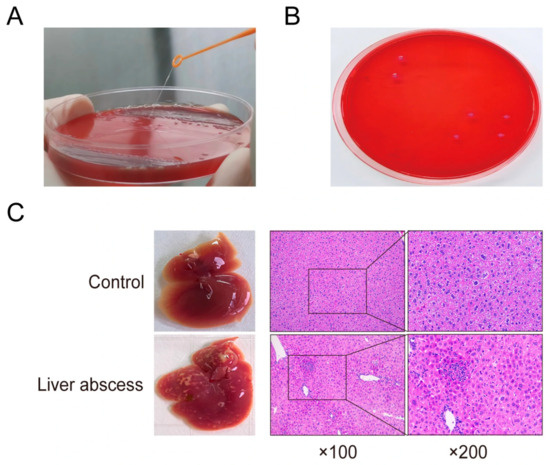
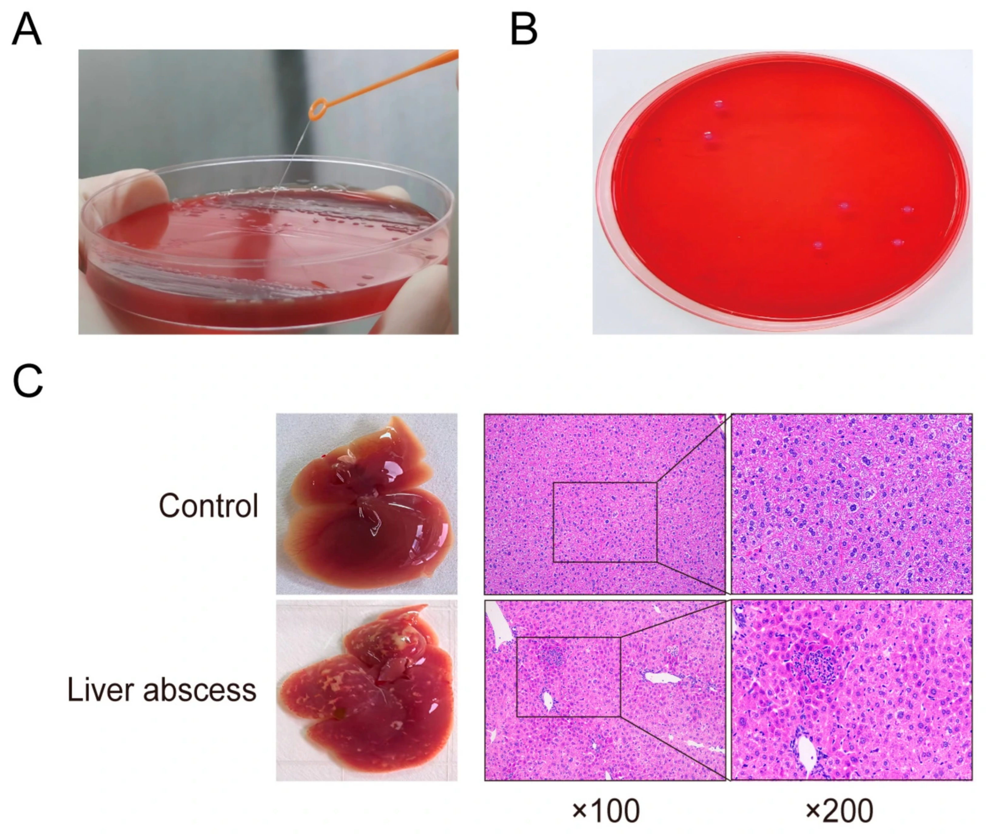
LA-Kpn006 exhibited a positive string test (mucus strand length > 10 mm), which is a typical phenotype of hypervirulent Klebsiella pneumoniae (Figure 1A). Molecular characterization further confirmed that this strain belonged to capsular serotype K1 and multilocus sequence type ST23, the two most clinically dominant and epidemiologically prevalent genotypes associated with severe invasive infections. Moreover, PCR detection verified that LA-Kpn006 harbored all six virulence genes known to be closely associated with the high pathogenicity, hyperinvasiveness, and enhanced virulence of hvKp strains.
Figure 1.
Phenotypic characteristics of LA-Kpn006 and pathological changes in liver tissues of KPLA mice. (A) String test showing hypermucoviscous phenotype of strain LA-Kpn006; (B) Bacterial colonization in liver homogenates of KPLA model mice; (C) Histopathological changes (H&E staining) in liver tissues from control and KPLA mice.
3.2. Establishment of the KPLA Model
At 3 days post-inoculation, viable hvKp was successfully detected in liver homogenates, with a bacterial load of approximately 1 × 10³ CFU/g, confirming stable colonization of the liver by the strain (Figure 1B). Meanwhile, serum levels of ALT and AST, key indicators of hepatocellular injury, were significantly increased in the infected group compared with the control group (p < 0.05), indicating severe liver damage induced by hvKp infection. Gross morphological examination showed obvious hepatomegaly and typical macroscopic abscess lesions on the liver surface. Histological examination via H&E staining further demonstrated complete disruption of the normal hepatic lobule structure, extensive hepatocyte necrosis, massive inflammatory cell infiltration, and distinct abscess formation in the liver tissue, which were consistent with the typical pathological features of KPLA (Figure 1C).
3.3. Phosphoproteomic Profiling
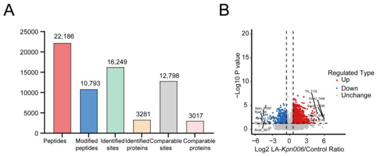
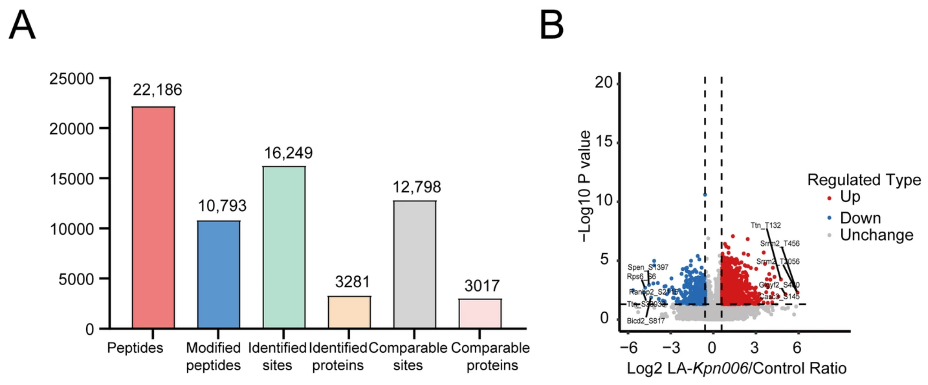
The results showed that a total of 16,249 phosphorylation sites and 3281 phosphorylated proteins were identified, among which 12,798 sites and 3017 proteins were quantified (Figure 2A). Among all phosphorylation sites, 13,921 (85.68%) were serine (Ser, S) residues, 1966 (12.10%) were threonine (Thr, T) residues, and 362 (2.22%) were tyrosine (Tyr, Y) residues. Differentially expressed phosphorylated proteins were screened by fold change and t-test. A p value < 0.05 and a fold change > 1.5 were set as the thresholds for significant upregulation, while a fold change < 1/1.5 was set as the threshold for significant downregulation. According to the above criteria, 1723 phosphorylated proteins (3176 sites) were significantly upregulated, and 425 phosphorylated proteins (491 sites) were significantly downregulated in the liver tissues of the KPLA mouse model (Figure 2B).
Figure 2.
Identification and quantitative distribution of phosphorylation sites and proteins in mouse liver tissues. (A) Numbers of identified and quantifiable phosphorylation sites and phosphoproteins; (B) Volcano plot showing significantly upregulated and downregulated phosphorylation sites in KPLA liver tissues.
3.4. Gene Ontology Annotation of Dysregulated Phosphoproteins
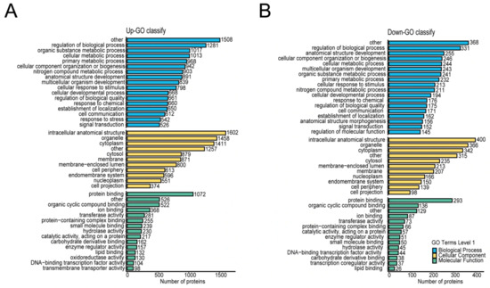
Based on the phosphoproteomic sequencing data, GO annotation was performed on 2148 differentially expressed phosphorylated proteins. As shown in Figure 3, for cellular components, the differentially phosphorylated proteins were mainly enriched in classic cellular compartments, including intracellular organelles, cytoplasm, cytosol, membrane structures, endomembrane system, nucleoplasm, and cell protrusions, suggesting that Kpn infection triggers global remodeling of protein phosphorylation in diverse subcellular structures of liver tissue, extensively affecting membrane functions, cytoplasmic signaling, and intranuclear biological processes. In terms of molecular functions, the differentially phosphorylated proteins were widely involved in protein–protein interaction, small molecule and ion binding, catalytic activity, transcriptional regulation, lipid and carbohydrate derivative binding, transmembrane transport, and other functions. Most core molecular functions were significantly enriched in both up- and downregulated proteins, indicating a global alteration of the molecular functional network in liver tissue after infection. Regarding biological processes, the differentially phosphorylated proteins participated in the regulation of biological processes, metabolic processes, cellular component organization, developmental processes, response to external stimuli, and signal transduction. Among them, downregulated phosphorylated proteins were specifically involved in anatomical structure morphogenesis and molecular function regulation, whereas upregulated phosphorylated proteins were significantly enriched in stress response-related biological processes, indicating functional differentiation mediated by distinct phosphorylation patterns in the KPLA model.
Figure 3.
GO enrichment analysis of differentially phosphorylated proteins in KPLA mouse liver tissues. Distribution of differentially phosphorylated proteins in cellular components, molecular function, and biological process categories. (A) Upregulated phosphoproteins; (B) Downregulated phosphoproteins.
3.5. KEGG Enrichment of Dysregulated Phosphoproteins
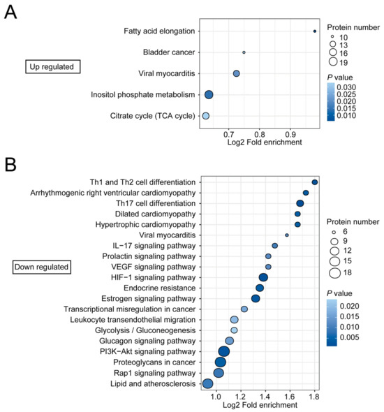
KEGG pathway analysis was performed on the differentially phosphorylated proteins in KPLA liver tissues. The results revealed distinct regulatory differences in signaling pathways between up- and down-regulated phosphorylated proteins. Up-regulated phosphorylated proteins were mainly enriched in lipid and energy metabolism-related pathways, including fatty acid elongation, inositol phosphate metabolism, and the tricarboxylic acid cycle (Figure 4A). In contrast, down-regulated phosphorylated proteins were predominantly concentrated in immune and inflammatory pathways such as Th1/Th2 and Th17 cell differentiation, IL-17 signaling, and leukocyte transendothelial migration. In addition, pathways including phosphatidylinositol-3-kinase/protein kinase B (PI3K/Akt), Ras-related protein 1 (Rap1), Hypoxia-inducible factor 1 (HIF-1), Vascular endothelial growth factor (VEGF), glycolipid metabolism, and endocrine regulation were also significantly down-regulated (Figure 4B). These findings suggest that differentially phosphorylated proteins participate in liver injury induced by KPLA mainly through pathways related to lipid metabolism, energy metabolism, inositol signaling, immune inflammation, and cell survival and differentiation.
Figure 4.
KEGG pathway enrichment analysis of differentially phosphorylated proteins. (A) Upregulated pathways; (B) Downregulated pathways.
3.6. Protein Domain Enrichment of Dysregulated Phosphoproteins
Protein domain enrichment analysis demonstrated that upregulated domains were mainly enriched in Post-synaptic density protein 95, Drosophila disk large tumor suppressor 1, Zonula occludens-1 protein (PDZ), Variant Src Homology 3 domain (SH3), Filamin/Actin-Binding Protein 280 (ABP280), deleted Differentially Expressed in Normal and Neoplastic (dDENN), Sterile Alpha Motif (SAM), 4.1-Ezrin-Radixin-Moesin (FERM) family, Pleckstrin Homology (PH), and Ras Guanine Nucleotide Exchange Factor (RasGEF) domains, which are involved in cytoskeleton remodeling, protein–protein interaction, G protein signaling, membrane trafficking, and inflammatory transcriptional regulation (Table 1). Downregulated domains were dominated by Phosphotyrosine Binding (PTB), SH2, Chromo, AT-Rich Interaction Domain (ARID)/Binding of Retinoblastoma protein, Inhibitor of cell proliferation, and Global transcription regulator (BRIGHT), Phosphatase and Tensin Homolog (PTEN)-C2, Sec16 vesicle trafficking, Signal Transducer and Activator of Transcription (STAT) function, and zinc finger transcription domains, which are mainly implicated in tyrosine phosphorylation signaling, chromatin transcriptional regulation, vesicular transport, tumor suppression, and inflammatory homeostasis (Table 1). Notably, PDZ and FERM central domains were significantly enriched in both upregulated and downregulated proteins, suggesting that they may participate in the pathological processes of inflammatory activation, hepatocyte injury, and tissue homeostasis imbalance through bidirectional phosphorylation modification [25].
Table 1.
Protein domain enrichment analysis of differentially phosphorylated proteins.
3.7. Core Proteins in the PPI Network
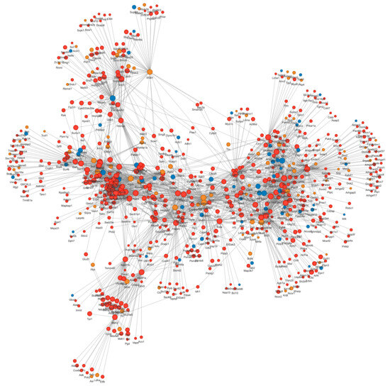
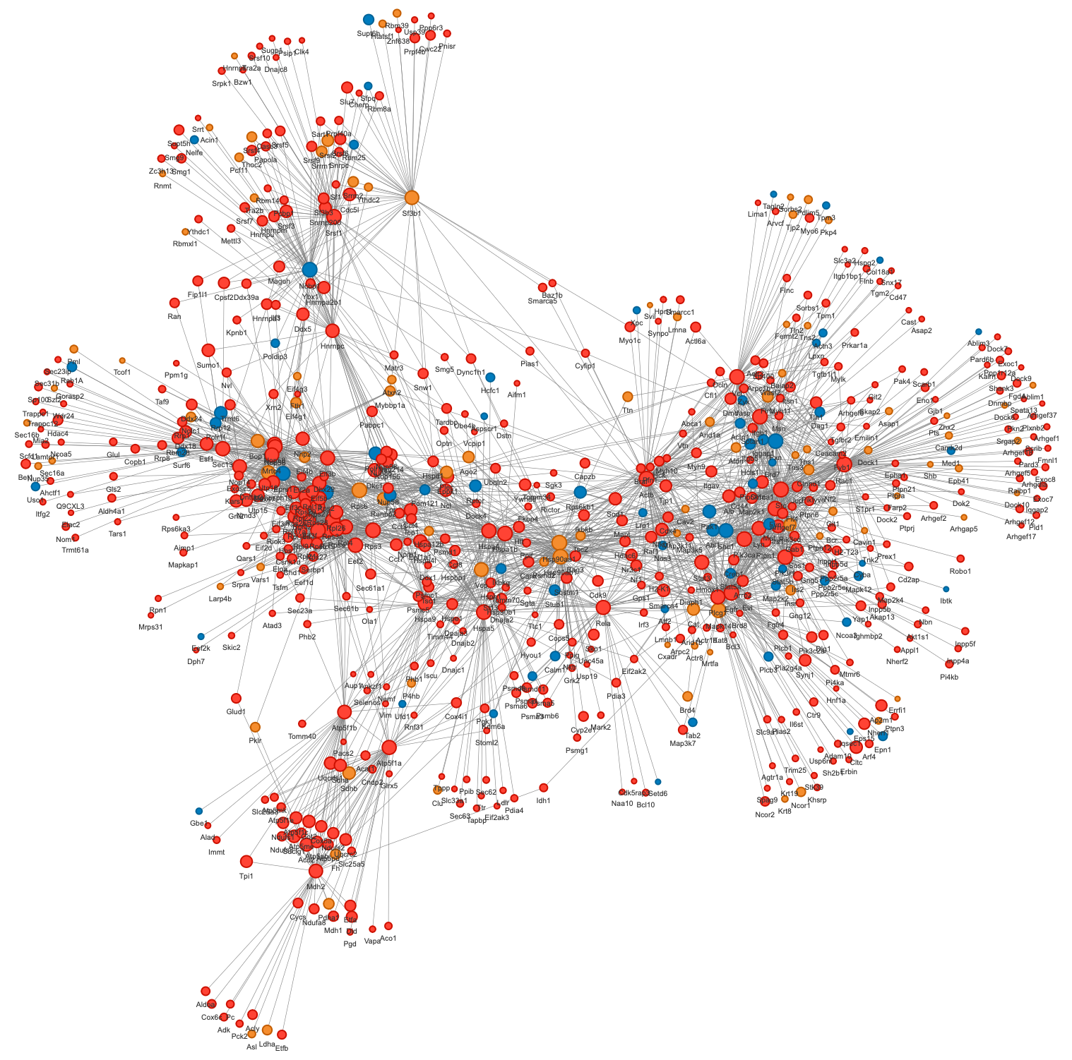
The top 50 phosphorylated proteins with the closest interactive relationships were selected to map the PPI network. The full set of 2148 differentially phosphorylated proteins was used as the input for STRING network construction. The final network includes both the input differential proteins and their predicted interacting partners. The “top 50” proteins are ranked by node degree from the completed network as the core subset for hub analysis. The results showed that the network consisted of 1593 nodes and 8573 edges, and most nodes possessed more than two interactive relationships (Figure 5). The top 10 proteins ranked by node degree were identified as core proteins (Table 2). Among them, Proto-oncogene tyrosine-protein kinase Src, Ras-related C3 botulinum toxin substrate 1 (Rac1), Beta-actin (Actb), Epidermal growth factor receptor (EGFR), Phosphatidylinositol-4,5-bisphosphate 3-kinase catalytic subunit alpha (Pik3CA), integrin gene (Itgb1) and Tyrosine-protein kinase Fyn were significantly upregulated; Heat shock protein (Hsp90) aa1, Hsp90ab1 and Ribosomal protein S6 (Rps6) exhibited bidirectional regulatory patterns. The node degree of these proteins ranged from 62 to 108. Collectively, these proteins possess high connectivity and serve as central regulators within the phosphorylation-mediated regulatory network of liver injury in KPLA.
Figure 5.
Protein–protein interaction network of differentially phosphorylated proteins. Nodes represent phosphorylated proteins; red = upregulated, blue = downregulated, yellow = bidirectionally regulated. Circle size indicates node degree. The interactive HTML file and raw node/link text files for this network are provided in Supplementary Materials for detailed browsing and analysis.
Table 2.
Top 10 hub proteins in the PPI network of differentially phosphorylated proteins.
4. Discussion
This study presents the first in-depth phosphoproteomic landscape of hvKp-induced liver abscess, revealing widespread phosphorylation-dependent remodeling of host metabolism, immunity, and cytoskeletal architecture. Our data establish that protein phosphorylation is a central regulatory pattern in KPLA pathogenesis and a key point for mechanistic dissection and therapeutic development.
The experimental strain exhibited a positive string test, with capsular serotype K1 and MLST ST23, and carried six major virulence genes, including rmpA, rmpA2, allS, iroN, iutA, and mrkD. These characteristics are typical of hypervirulent Klebsiella pneumoniae and are closely associated with high pathogenicity, hypermucoviscosity, iron acquisition, and adhesion ability [22,23,26]. Such genotypic and phenotypic features enable the strain to efficiently colonize, invade, and induce severe inflammatory damage in the liver, thereby contributing to the formation and progression of liver abscess.
Furthermore, we successfully established a murine model of KPLA via intragastric administration. Typical pathological changes were clearly observed in infected liver tissues, including structural destruction of hepatic lobules, massive inflammatory cell infiltration, hepatocellular necrosis, and obvious abscess formation. These results confirmed that the model was reliable and suitable for exploring the molecular mechanisms underlying hvKp-induced liver injury and pathological progression [24].
Notably, the culturable bacterial load is relatively low at day 3. Four factors explain this discrepancy. First, some bacteria enter a viable non-culturable state. Plate CFU underestimates the total bacterial burden. Second, liver abscesses distribute unevenly in the tissue. Single-site sampling may miss high-burden lesions. Third, innate immunity clears part of the culturable bacteria. Tissue damage persists longer than live bacteria. Fourth, our strain is a hypervirulent K. pneumoniae, which causes severe damage at low bacterial loads, and its virulence factors drive strong inflammatory responses. In addition, intragastric inoculation crosses the gut barrier, which filters most inoculated bacteria and naturally lowers liver colonization levels.
Phosphoproteomic quantification revealed 3667 dysregulated phosphosites in total, among which 3176 were significantly upregulated, and 491 were downregulated, demonstrating that hvKp disrupts host phosphorylation signaling to drive pathological progression. The predominance of serine phosphorylation implies that phosphorylation preferentially remodels the backbone conformational characteristics of serine over threonine residues during hvKp-induced liver injury [27].
Functional enrichment of phosphorylated proteins indicated that infection triggers a pro-inflammatory metabolic change; upregulated pathways strengthen lipid and energy metabolism to maintain inflammatory signaling and oxidative stress, while downregulated pathways suppress immune response, cell survival, and tissue repair. These changes exacerbate an inflammatory response, hepatic tissue injury, and subsequent abscess development. Notably, repression of PI3K–Akt and IL-17 signaling suggests impaired adaptive immunity and compromised tissue homeostasis, which may explain the persistence of infection and severity of liver injury. Mechanistically, the PI3K–Akt axis is central to regulating the development, differentiation, metabolic reprogramming, and effector functions of both myeloid and lymphoid immune cells, including macrophages, dendritic cells, and neutrophils, while the lymphoid lineage encompasses T cells, B cells, NK cells, and multiple innate lymphoid cell subsets, as well as innate-like lymphocytes, such as NKT cells and MAIT cells [28]. In parallel, IL-17 signaling is essential for host defense against extracellular bacteria and for maintaining hepatic immune tolerance; its inhibition disrupts local inflammatory control and tissue repair circuits, favoring persistent bacterial colonization and progressive parenchymal damage [29]. Consistent with our findings, prior studies have demonstrated that PI3K–Akt repression exacerbates liver injury by dysregulating macrophage polarization and impairing hepatocyte survival. As a key bioactive component of Scutellaria baicalensis Georgi, wogonin ameliorates hepatic damage and acetaminophen-related hepatotoxicity in mice by suppressing macrophage inflammatory activation via inhibiting the PI3K/AKT signaling pathway [30]. While IL-17 pathway impairment is closely linked to uncontrolled inflammatory disorders and increased host susceptibility to bacterial infection [31]. Collectively, these data support that co-suppression of PI3K–Akt and IL-17 signaling acts as a key molecular driver underlying ineffective adaptive immunity, disrupted hepatic homeostasis, and the severe, persistent infection observed in this KPLA model. This apparent discrepancy can be explained from three perspectives. First, bulk liver tissue is dominated by hepatocytes. The detected phosphorylation signals mainly reflect hepatocyte status rather than infiltrating immune cells, with a low proportion. Second, the downregulated pathways belong to adaptive immunity. The acute inflammatory lesions at day 3 are driven by innate immune cells. These two patterns act on separate immune branches. Third, pathway downregulation may represent host negative feedback. It is triggered by strong acute inflammation to avoid excessive tissue damage.
Protein domain analysis highlighted structural modules that govern protein–protein interaction, cytoskeleton dynamics, and transcriptional control. The bidirectional enrichment of PDZ and FERM domains implies context-dependent phosphorylation switches that coordinate inflammatory activation and cellular disorganization [32,33,34].
PPI network analysis prioritized 10 high-value hub proteins that integrate upstream signals and regulate downstream effector functions. Src, Rac1, Pik3CA, and EGFR control kinase signaling, cytoskeleton rearrangement, and immune cell infiltration [35,36,37,38]; Actb and Itgb1 mediate cellular structural integrity [39,40]; Hsp90 paralogs and Rps6 govern protein homeostasis and translation [41,42]. Among them, as an E3 ubiquitin ligase, NEDD4L negatively modulates PIK3CA protein abundance through ubiquitination and is essential for maintaining PI3K-AKT signaling homeostasis [37]. EGFR–HSP90 complex synergistically mediates acquired resistance to EGFR tyrosine kinase inhibitors and underpins the pathogenesis of EGFR-addicted tumors [38]. These hubs likely function as master regulators of KPLA pathogenesis and represent ideal candidates for targeted inhibition.
Several limitations should be noted: the sample size is modest, a single time point was analyzed, and in vivo functional validation is lacking. Future studies will expand animal cohorts, profile phosphorylation dynamics over time, and use genetic and pharmacological approaches to validate hub protein function.
5. Conclusions
Hypervirulent Klebsiella pneumoniae infection elicits extensive and functionally coordinated phosphoproteomic remodeling in mouse liver tissue. Dysregulated phosphorylation pathways drive metabolic reprogramming, immune dysregulation, and cytoskeletal disruption, collectively promoting liver abscess and tissue injury. The identified core proteins and signaling modules illuminate the molecular basis of KPLA and provide promising targets for novel diagnostic and therapeutic strategies against severe hvKp infection.
Supplementary Materials
The following supporting information can be downloaded at: https://www.mdpi.com/article/10.3390/pathogens15070739/s1, Interactive PPI network files: HTML visualization page, node text file, link text file and log file for detailed network browsing.
Author Contributions
Conceptualization, J.Y.; methodology, C.Y., X.L. and Y.C.; software, C.Y. and X.L.; validation, C.Y., X.L., Y.C., A.S., X.R., T.Z.; formal analysis, C.Y., X.L., Y.C., A.S., X.R., T.Z.; investigation, C.Y., X.L., Y.C., A.S., X.R., T.Z.; resources, J.Y.; data curation, C.Y., X.L., Y.C.; writing—original draft preparation, C.Y., X.L., Y.C.; writing—review and editing, C.Y., X.L., Y.C., A.S., X.R., T.Z., J.Y.; visualization, C.Y., X.L., Y.C., T.Z.; supervision, A.S., X.R., T.Z., J.Y.; project administration, J.Y.; funding acquisition, J.Y. All authors have read and agreed to the published version of the manuscript.
Funding
This research was funded by the National Natural Science Foundation of China, grant numbers 82472300, 82130065, and 82372270; Capital’s Funds for Health Improvement and Research, grant number CFH 2026-1Q-1081; Beijing Hospitals Authority’s Ascent Plan, grant number DFL20241301; Beijing Natural Science Foundation, grant number 7264238.
Institutional Review Board Statement
The bacterial strain Klebsiella pneumoniae LA-Kpn006 was adopted in the present research. It was isolated from leftover pus aspirate of pyogenic liver abscess patients during routine clinical microbial detection at Capital Institute of Pediatrics. All samples were collected as part of standard clinical diagnosis and treatment procedures. This study only used the leftover samples after clinical testing. No additional invasive procedures were performed on patients, and no personally identifiable information of patients was collected or retained. In accordance with the relevant regulations of the Ethics Committee of Capital Institute of Pediatrics on the scientific use of residual clinical samples, this type of research is exempt from separate human ethics review and approval. The study was conducted in compliance with the principles of the Declaration of Helsinki. The animal study protocol was approved by the Ethics Committee of Capital Institute of Pediatrics (protocol code DWLL2025010, approval date: 13 March 2025).
Informed Consent Statement
Not applicable.
Data Availability Statement
The mass spectrometry proteomics data have been deposited in the OMIX repository of the China National Center for Bioinformation (CNCB), with the accession number OMIX018608.
Acknowledgments
The authors thank all contributors to the studies cited in this review.
Conflicts of Interest
The authors declare no conflicts of interest. The funders had no role in the design of the study; in the collection, analyses, or interpretation of data; in the writing of the manuscript; or in the decision to publish the results.
Abbreviations
The following abbreviations are used in this manuscript:
| ABP280 | Filamin/Actin-Binding Protein 280 |
| Actb | Beta-actin |
| ALT | Alanine transaminase |
| ARID | AT-Rich Interaction Domain |
| AST | Aspartate transaminase |
| BRIGHT | Binding of Retinoblastoma protein, Inhibitor of cell proliferation, and Global transcription regulator |
| CFU | Colony Forming Unit |
| dDENN | deleted Differentially Expressed in Normal and Neoplastic |
| Egfr | Epidermal growth factor receptor |
| FERM | 4.1-Ezrin-Radixin-Moesin |
| GO | Gene Ontology |
| H&E | Hematoxylin and eosin |
| HIF-1 | Hypoxia-inducible factor 1 |
| Hsp90aa1 | Heat shock protein HSP 90-alpha |
| Hsp90ab1 | Heat shock protein HSP 90-beta |
| hvKp | Hypervirulent Klebsiella pneumoniae |
| Itgb1 | Integrin beta-1 |
| KEGG | Kyoto Encyclopedia of Genes and Genomes |
| Kpn | Klebsiella pneumoniae |
| KPLA | Klebsiella pneumoniae liver abscess |
| LC–MS/MS | Liquid chromatography with tandem mass spectrometry |
| MLST | Multilocus Sequence Typing |
| PBS | Phosphate-Buffered Saline |
| PDZ | Post-synaptic density protein 95, Drosophila disc large tumor suppressor 1, Zonula occludens-1 protein |
| PH | Pleckstrin Homology |
| PI3K/Akt | Phosphatidylinositol-3-kinase/protein kinase B |
| Pik3ca | Phosphatidylinositol 4,5-bisphosphate 3-kinase catalytic subunit alpha |
| PPI | Protein–protein interaction |
| PSMs | Peptide spectrum matches |
| PTB | Phosphotyrosine Binding |
| PTEN | Phosphatase and Tensin Homolog |
| Rac1 | Ras-related C3 botulinum toxin substrate 1 |
| Rap1 | Ras-related protein 1 |
| RasGEF | Ras Guanine Nucleotide Exchange Factor |
| Rps6 | Ribosomal protein S6 |
| SAM | Sterile Alpha Motif |
| SH2 | Src Homology 2 |
| SH3 | Src Homology 3 |
| SPF | Specific Pathogen-Free |
| STAT | Signal Transducer and Activator of Transcription |
| VEGF | Vascular endothelial growth factor |
References
- Shah, A.A.; Alwashmi, A.S.S.; Abalkhail, A.; Alkahtani, A.M. Emerging Challenges in Klebsiella pneumoniae: Antimicrobial Resistance and Novel Approach. Microb. Pathog. 2025, 202, 107399. [Google Scholar] [CrossRef] [Scilit] [PubMed]
- Liu, Y.C.; Cheng, D.L.; Lin, C.L. Klebsiella pneumoniae Liver Abscess Associated with Septic Endophthalmitis. Arch. Intern. Med. 1986, 146, 1913–1916. [Google Scholar] [CrossRef] [Scilit] [PubMed]
- Chung, D.R.; Lee, S.S.; Lee, H.R.; Kim, H.B.; Choi, H.J.; Eom, J.S.; Kim, J.S.; Choi, Y.H.; Lee, J.S.; Chung, M.H.; et al. Emerging Invasive Liver Abscess Caused by K1 Serotype Klebsiella pneumoniae in Korea. J. Infect. 2007, 54, 578–583. [Google Scholar] [CrossRef] [Scilit] [PubMed]
- Tsai, F.-C.; Huang, Y.-T.; Chang, L.-Y.; Wang, J.-T. Pyogenic Liver Abscess as Endemic Disease, Taiwan. Emerg. Infect. Dis. 2008, 14, 1592–1600. [Google Scholar] [CrossRef] [Scilit] [PubMed]
- Zhang, Y.; Zhao, C.; Wang, Q.; Wang, X.; Chen, H.; Li, H.; Zhang, F.; Li, S.; Wang, R.; Wang, H. High Prevalence of Hypervirulent Klebsiella pneumoniae Infection in China: Geographic Distribution, Clinical Characteristics, and Antimicrobial Resistance. Antimicrob. Agents Chemother. 2016, 60, 6115–6120. [Google Scholar] [CrossRef] [Scilit] [PubMed]
- Rossi, B.; Gasperini, M.L.; Leflon-Guibout, V.; Gioanni, A.; de Lastours, V.; Rossi, G.; Dokmak, S.; Ronot, M.; Roux, O.; Nicolas-Chanoine, M.-H.; et al. Hypervirulent Klebsiella pneumoniae in Cryptogenic Liver Abscesses, Paris, France. Emerg. Infect. Dis. 2018, 24, 221–229. [Google Scholar] [CrossRef] [Scilit] [PubMed]
- Fazili, T.; Sharngoe, C.; Endy, T.; Kiska, D.; Javaid, W.; Polhemus, M. Klebsiella pneumoniae Liver Abscess: An Emerging Disease. Am. J. Med. Sci. 2016, 351, 297–304. [Google Scholar] [CrossRef] [Scilit] [PubMed]
- Angeles-Solano, M.; Tabashsum, Z.; Chen, L.; Rowe, S.E. Klebsiella pneumoniae Liver Abscesses: Pathogenesis, Treatment, and Ongoing Challenges. Infect. Immun. 2025, 93, e0050824. [Google Scholar] [CrossRef] [Scilit] [PubMed]
- Al Ismail, D.; Campos-Madueno, E.I.; Donà, V.; Endimiani, A. Hypervirulent Klebsiella pneumoniae (hvKp): Overview, Epidemiology, and Laboratory Detection. Pathog. Immun. 2024, 10, 80–119. [Google Scholar] [CrossRef] [Scilit] [PubMed]
- Fang, C.-T.; Lai, S.-Y.; Yi, W.-C.; Hsueh, P.-R.; Liu, K.-L.; Chang, S.-C. Klebsiella pneumoniae Genotype K1: An Emerging Pathogen That Causes Septic Ocular or Central Nervous System Complications from Pyogenic Liver Abscess. Clin. Infect. Dis. 2007, 45, 284–293. [Google Scholar] [CrossRef] [Scilit] [PubMed]
- Martínez-Hernandez, L.; Alvarado-Delgado, A.; Rodríguez-Medina, N.; Garcia-Peniche, J.; Donis-Hernández, J.J.; Perez-Rezendiz, O.A.; Nava-Domínguez, N.; Duarte-Zambrano, L.; Tamayo-Legorreta, E.M.; Garza-Ramos, U. Pyogenic Liver Abscess Caused by an Atypical Hypervirulent Klebsiella pneumoniae K1-ST23 in Mexico. IDCases 2024, 36, e01987. [Google Scholar] [CrossRef] [Scilit] [PubMed]
- Russo, T.A.; Marr, C.M. Hypervirulent Klebsiella pneumoniae. Clin. Microbiol. Rev. 2019, 32, e00001-19. [Google Scholar] [CrossRef] [Scilit] [PubMed]
- Nguyen, T.N.T.; Howells, G.; Short, F.L. How Klebsiella pneumoniae Controls Its Virulence. PLoS Pathog. 2025, 21, e1013499. [Google Scholar] [CrossRef] [Scilit] [PubMed]
- Stanton, T.D.; Wyres, K.L. What Defines Hypervirulent Klebsiella pneumoniae? EBioMedicine 2024, 108, 105331. [Google Scholar] [CrossRef] [Scilit] [PubMed]
- Hung, C.-H.; Kuo, C.-F.; Wang, C.-H.; Wu, C.-M.; Tsao, N. Experimental Phage Therapy in Treating Klebsiella pneumoniae-Mediated Liver Abscesses and Bacteremia in Mice. Antimicrob. Agents Chemother. 2011, 55, 1358–1365. [Google Scholar] [CrossRef] [Scilit] [PubMed]
- Humphrey, S.J.; James, D.E.; Mann, M. Protein Phosphorylation: A Major Switch Mechanism for Metabolic Regulation. Trends Endocrinol. Metab. 2015, 26, 676–687. [Google Scholar] [CrossRef] [Scilit] [PubMed]
- Munk, S.; Refsgaard, J.C.; Olsen, J.V.; Jensen, L.J. From Phosphosites to Kinases. Methods Mol. Biol. 2016, 1355, 307–321. [Google Scholar] [CrossRef] [Scilit] [PubMed]
- Zhu, H.; Sydor, A.M.; Yan, B.-R.; Li, R.; Boniecki, M.T.; Lyons, C.; Cygler, M.; Muise, A.M.; Maxson, M.E.; Grinstein, S.; et al. Salmonella Exploits LRRK2-Dependent Plasma Membrane Dynamics to Invade Host Cells. Nat. Commun. 2025, 16, 2329. [Google Scholar] [CrossRef] [Scilit] [PubMed]
- Yang, Y.; Fang, H.; Xie, Z.; Ren, F.; Yan, L.; Zhang, M.; Xu, G.; Song, Z.; Chen, Z.; Sun, W.; et al. Yersinia Infection Induces Glucose Depletion and AMPK-Dependent Inhibition of Pyroptosis in Mice. Nat. Microbiol. 2024, 9, 2144–2159. [Google Scholar] [CrossRef] [Scilit] [PubMed]
- Cheng, X.; Kang, L.; Liu, J.; Wang, Q.; Zhang, Z.; Zhang, L.; Xie, Y.; Chang, L.; Zeng, D.; Tian, L.; et al. Proteomics and Phosphoproteomics Revealed Dysregulated Kinases and Potential Therapy for Liver Fibrosis. Mol. Cell Proteom. 2025, 24, 100991. [Google Scholar] [CrossRef] [Scilit] [PubMed]
- Wu, X.; Poulsen, K.L.; Sanz-Garcia, C.; Huang, E.; McMullen, M.R.; Roychowdhury, S.; Dasarathy, S.; Nagy, L.E. MLKL-Dependent Signaling Regulates Autophagic Flux in a Murine Model of Non-Alcohol-Associated Fatty Liver and Steatohepatitis. J. Hepatol. 2020, 73, 616–627. [Google Scholar] [CrossRef] [Scilit] [PubMed]
- Liu, C.; Guo, J. Hypervirulent Klebsiella pneumoniae (Hypermucoviscous and Aerobactin Positive) Infection over 6 Years in the Elderly in China: Antimicrobial Resistance Patterns, Molecular Epidemiology and Risk Factor. Ann. Clin. Microbiol. Antimicrob. 2019, 18, 4. [Google Scholar] [CrossRef] [Scilit] [PubMed]
- Candan, E.D.; Aksöz, N. Klebsiella pneumoniae: Characteristics of Carbapenem Resistance and Virulence Factors. Acta Biochim. Pol. 2015, 62, 867–874. [Google Scholar] [CrossRef] [Scilit] [PubMed]
- Lin, Y.-T.; Tseng, K.-Y.; Yeh, Y.-C.; Yang, F.-C.; Fung, C.-P.; Chen, N.-J. TREM-1 Promotes Survival during Klebsiella pneumoniae Liver Abscess in Mice. Infect. Immun. 2014, 82, 1335–1342. [Google Scholar] [CrossRef] [Scilit] [PubMed]
- Zhuang, Y.; Ortega-Ribera, M.; Thevkar Nagesh, P.; Joshi, R.; Huang, H.; Wang, Y.; Zivny, A.; Mehta, J.; Parikh, S.M.; Szabo, G. Bile Acid-Induced IRF3 Phosphorylation Mediates Cell Death, Inflammatory Responses, and Fibrosis in Cholestasis-Induced Liver and Kidney Injury via Regulation of ZBP1. Hepatology 2024, 79, 752–767. [Google Scholar] [CrossRef] [Scilit] [PubMed]
- Bezdicek, M.; Jakubickova, M.; Bitusikova, V.; Holubova, E.; Vitkova, H.; Kocmanova, I.; Vitkova, I.; Zdrazilova Dubska, L.; Lengerova, M. Emergence and Genomic Characterization of Hypervirulent ST23/K1 Klebsiella pneumoniae: Local Epidemiology and Global Context. Front. Microbiol. 2026, 17, 1758288. [Google Scholar] [CrossRef] [Scilit] [PubMed]
- Kim, S.-Y.; Jung, Y.; Hwang, G.-S.; Han, H.; Cho, M. Phosphorylation Alters Backbone Conformational Preferences of Serine and Threonine Peptides. Proteins 2011, 79, 3155–3165. [Google Scholar] [CrossRef] [Scilit] [PubMed]
- Deane, J.A.; Fruman, D.A. Phosphoinositide 3-Kinase: Diverse Roles in Immune Cell Activation. Annu. Rev. Immunol. 2004, 22, 563–598. [Google Scholar] [CrossRef] [Scilit] [PubMed]
- Pan, N.; Su, X.; Meng, Y.; Liang, Y.; Chen, X.; Zhang, H.; Wang, X. Gut Microbiota-Driven IL-17A Production by Hepatic Γδ T Cells Enhances Neutrophil Defense against Systemic Staphylococcus Aureus Infection. Virulence 2026, 17, 2629132. [Google Scholar] [CrossRef] [Scilit] [PubMed]
- Zhao, W.; Luo, H.; Lin, Z.; Huang, L.; Pan, Z.; Chen, L.; Fan, L.; Yang, S.; Tan, H.; Zhong, C.; et al. Wogonin Mitigates Acetaminophen-Induced Liver Injury in Mice through Inhibition of the PI3K/AKT Signaling Pathway. J. Ethnopharmacol. 2024, 332, 118364. [Google Scholar] [CrossRef] [Scilit] [PubMed]
- Agerholm, R.; Bekiaris, V. Evolved to Protect, Designed to Destroy: IL-17-Producing Γδ T Cells in Infection, Inflammation, and Cancer. Eur. J. Immunol. 2021, 51, 2164–2177. [Google Scholar] [CrossRef] [Scilit] [PubMed]
- Manjunath, G.P.; Ramanujam, P.L.; Galande, S. Structure Function Relations in PDZ-Domain-Containing Proteins: Implications for Protein Networks in Cellular Signalling. J. Biosci. 2018, 43, 155–171. [Google Scholar] [PubMed]
- Liu, X.; Fuentes, E.J. Emerging Themes in PDZ Domain Signaling: Structure, Function, and Inhibition. Int. Rev. Cell Mol. Biol. 2019, 343, 129–218. [Google Scholar] [CrossRef] [Scilit] [PubMed]
- Borkúti, P.; Kristó, I.; Szabó, A.; Kovács, Z.; Vilmos, P. FERM Domain-Containing Proteins Are Active Components of the Cell Nucleus. Life Sci. Alliance 2024, 7, e202302489. [Google Scholar] [CrossRef] [Scilit] [PubMed]
- Kasahara, K.; Re, S.; Nawrocki, G.; Oshima, H.; Mishima-Tsumagari, C.; Miyata-Yabuki, Y.; Kukimoto-Niino, M.; Yu, I.; Shirouzu, M.; Feig, M.; et al. Reduced Efficacy of a Src Kinase Inhibitor in Crowded Protein Solution. Nat. Commun. 2021, 12, 4099. [Google Scholar] [CrossRef] [Scilit] [PubMed]
- Ravi, L.I.; Tan, T.J.; Tan, B.H.; Sugrue, R.J. Virus-Induced Activation of the Rac1 Protein at the Site of Respiratory Syncytial Virus Assembly Is a Requirement for Virus Particle Assembly on Infected Cells. Virology 2021, 557, 86–99. [Google Scholar] [CrossRef] [Scilit] [PubMed]
- Zheng, S.; He, S.; Liang, Y.; Tan, Y.; Liu, Q.; Liu, T.; Lu, X. Understanding PI3K/Akt/mTOR Signaling in Squamous Cell Carcinoma: Mutated PIK3CA as an Example. Mol. Biomed. 2024, 5, 13. [Google Scholar] [CrossRef] [Scilit] [PubMed]
- Sabbah, D.A.; Hajjo, R.; Sweidan, K. Review on Epidermal Growth Factor Receptor (EGFR) Structure, Signaling Pathways, Interactions, and Recent Updates of EGFR Inhibitors. Curr. Top. Med. Chem. 2020, 20, 815–834. [Google Scholar] [CrossRef] [Scilit] [PubMed]
- Cruz-González, G.; Meschia, J.F.; Madden, B.J.; Prudencio, M.; Polania-Sandoval, C.A.; Hartwell, J.; Oyefeso, E.; Benchaaboune, R.; Brigham, T.; Sandhu, S.J.S.; et al. Recent Advances in Proteomic Analysis to Study Carotid Artery Plaques. JVS Vasc. Sci. 2024, 5, 100215. [Google Scholar] [CrossRef] [Scilit] [PubMed]
- Guo, S.S.; Au, T.Y.K.; Wynn, S.; Aszodi, A.; Chan, D.; Fässler, R.; Cheah, K.S.E. Β1 Integrin Regulates Convergent Extension in Mouse Notogenesis, Ensures Notochord Integrity and the Morphogenesis of Vertebrae and Intervertebral Discs. Development 2020, 147, dev192724. [Google Scholar] [CrossRef] [Scilit] [PubMed]
- Chiosis, G.; Digwal, C.S.; Trepel, J.B.; Neckers, L. Structural and Functional Complexity of HSP90 in Cellular Homeostasis and Disease. Nat. Rev. Mol. Cell Biol. 2023, 24, 797–815. [Google Scholar] [CrossRef] [Scilit] [PubMed]
- Ruvinsky, I.; Sharon, N.; Lerer, T.; Cohen, H.; Stolovich-Rain, M.; Nir, T.; Dor, Y.; Zisman, P.; Meyuhas, O. Ribosomal Protein S6 Phosphorylation Is a Determinant of Cell Size and Glucose Homeostasis. Genes. Dev. 2005, 19, 2199–2211. [Google Scholar] [CrossRef] [Scilit] [PubMed]

Disclaimer/Publisher’s Note: The statements, opinions and data contained in all publications are solely those of the individual author(s) and contributor(s) and not of MDPI and/or the editor(s). MDPI and/or the editor(s) disclaim responsibility for any injury to people or property resulting from any ideas, methods, instructions or products referred to in the content. |
© 2026 by the authors. Licensee MDPI, Basel, Switzerland. This article is an open access article distributed under the terms and conditions of the Creative Commons Attribution (CC BY) license.