Article

Regulation of PBP2x Surface Localization by aliD and clpL Alters β-Lactam Resistance in Pneumococcus


1
Department of Cell and Molecular Biology, University of Mississippi Medical Center, Jackson, MS 39216, USA
2
Center for Immunology and Microbial Research, University of Mississippi Medical Center, Jackson, MS 39216, USA
*
Author to whom correspondence should be addressed.
Pathogens 2026, 15(7), 738; https://doi.org/10.3390/pathogens15070738
Submission received: 17 June 2026 / Revised: 10 July 2026 / Accepted: 10 July 2026 / Published: 14 July 2026
(This article belongs to the Section Bacterial Pathogens)

Abstract

Streptococcus pneumoniae is a leading cause of respiratory infections and is often treated with β-lactam antibiotics despite frequent resistance. The β-lactam resistance is through mutations in penicillin-binding proteins (PBPs), but the impact of PBP regulation remains poorly understood. Here, we investigate the role of gene regulation by the oligopeptide-binding protein aliD on β-lactam susceptibility. aliD-expressing strains exhibit elevated minimum inhibitory concentrations (MICs) to the β-lactam antibiotics amoxicillin and cefdinir compared to isogenic aliD mutants. In contrast, aliD had minimal effects on susceptibility to vancomycin, suggesting a mechanism specific to β-lactam antibiotics. We demonstrate that aliD increases clpL expression with or without antibiotics present. Inducible expression of clpL resulted in stepwise increases in amoxicillin MIC, demonstrating that elevated clpL expression directly contributes to decreased β-lactam susceptibility. Furthermore, we show that increased clpL expression enhances PBP2x surface exposure in a dose-dependent manner. Additionally, aliD-expressing strains exhibited significantly increased cell wall cross-linking relative to aliD mutants. Together, these findings identify a novel regulatory pathway in which aliD enhances clpL expression, promotes PBP2x surface exposure, and increases peptidoglycan cross-linking, ultimately reducing β-lactam susceptibility. This work expands current understanding of pneumococcal antibiotic resistance and suggests that peptide sensing through oligopeptide transport systems may influence antibiotic susceptibility in host-specific environments.

1. Introduction

Streptococcus pneumoniae, also known as the pneumococcus, is the leading cause of lower respiratory tract infections (LRTIs) [1], which include pneumonia and bronchitis. Annually, S. pneumoniae causes almost 50% (~1,100,000 out of 2,300,000) of deaths associated with LRTIs, with the highest mortality rate being in children under the age of five [2]. The pneumococcus accounts for more deaths than the next three most common causes of LRTIs combined: Haemophilus influenzae, influenza, and respiratory syncytial virus [3].
This high mortality is due to a number of S. pneumoniae virulence factors, such as the highly variable pneumococcal capsule, PspA, and pneumolysin [4]. Clinicians use antibiotics and vaccines to treat and prevent S. pneumoniae infections. The advent of the pneumococcal vaccine is one of the most effective tools used to combat pneumococcal infections. Current vaccines target a subset of the more than 107 pneumococcal capsules currently known [5]. Each capsule serotype has an antigenically distinct carbohydrate structure that produces no cross-reactive antibodies to different serotypes. Current vaccines contain the most clinically relevant serotypes, as creating a vaccine targeting all serotypes would not be practical.
Pneumococci-expressing serotypes included within the vaccine are subjected to heightened immunological pressure. Conversely, pneumococcal strains expressing non-vaccine serotypes remain unimpeded by this immunological constraint. Consequently, the immunological landscape sculpted by vaccination preferentially facilitates the proliferation of non-vaccine serotypes, a phenomenon termed serotype replacement [6]. This shift permits the ascendance of previously low-prevalence serotypes, which may now exploit the vacated ecological niche to establish colonization and lead to disease development. This epidemiological observation is further increased through strain-level serotype switching [7]. Serotype switching refers to the change from one pneumococcal serotype to another through genetic exchange. Both serotype switching and replacement have limited the effectiveness of pneumococcal vaccination.
This selective pressure changes the epidemiology of pneumococcal strains present in the population. A notable change in the pneumococcal population is the rise of nonencapsulated S. pneumoniae (NESp). NESp lack a capsule and are divided into either group 1 or group 2 NESp. Group 1 NESp contain capsular polysaccharide synthesis (cps) genes but are not functional due to mutations. Group 2 NESp contain novel genes in the cps locus not related to the production of capsules. Group 2 is further divided into null capsule clades (NCCs), dependent on the genes present. NCC1 contains the surface protein pspK, while NCC2 and NCC3 contain aliC and aliD or only aliD, respectively [8]. The aliC and aliD genes encode substrate-binding proteins (SBP) used for oligopeptide importation through the Ami oligopeptide transport system [9,10]. The Ami system was the first oligopeptide permease discovered in Gram-positive bacteria and plays an important role in bacterial metabolism, as it directly imports various polypeptides and polyamines [10]. Oligopeptide transporters utilize a surface-bound SBP that binds exogenous compounds and imports them through an ABC transporter. This importation into the cell also impacts gene expression in multiple pathways, such as the metabolism of specific carbohydrates, cell signaling, competence, and colonization factors [11,12]. All pneumococci contain the SBPs AmiA, AliA, and aliB, while NESp can contain the additional aliC and aliD SBPs. Interestingly, while aliD is typically found only in NESp strains, there are three encapsulated serotypes that also express aliD: 25A, 25F, and 38. Of note is serotype 38, which has been increasing in prevalence in certain populations [13]. SBPs have a broad range of substrate specificity with overlapping substrates along with unique substrates. Changes in gene expression depend on the SBP and the substrate being imported [14].
Selective pressure against specific populations has also changed the prevalence of antibiotic resistance [15]. Antibiotic resistance generally occurs in one of two ways: extrinsic or intrinsic acquisition. Extrinsic is the acquisition of a new gene to perform a specific function, such as using efflux pumps to directly remove antibiotics from the cell or utilizing proteins like β-lactamases to inactivate antibiotics. For intrinsic acquisition, the bacteria can experience mutational resistance that modifies bacterial proteins, such as gyrase or topoisomerase mutations, leading to quinolone resistance [16]. Additionally, changes in gene expression levels can impact antibiotic susceptibility [17]. Since oligopeptide transporters can impact gene expression, adding additional SBPs—as seen in NESp and certain serotypes—may change antibiotic susceptibility.
Previous work examined the role of aliD on gene regulation and the impact on bacterial virulence. One of the genes regulated by the oligopeptide transporter aliD is the chaperone clpL [9]. However, the possible interaction between aliD and ClpL has not been explored. ClpL is an HSP100 chaperone protein in the caseinolytic protease system. Its most well-known function is assisting in protein refolding due to incorrect conformation or trafficking proteins to the ClpP protease for degradation. An additional function of ClpL is the transportation of penicillin-binding protein 2x (PBP2x) to the cell surface [18].
Penicillin-binding proteins (PBPs) are integral to the creation of the cell wall. The bacterial cell wall is formed from a mesh-like lattice of peptidoglycan. PBPs form cross-links in the peptidoglycan stands to give structure and stability to the cell wall. All strains of S. pneumoniae contain six PBPs: 1a, 1b, 2x, 2a, 2b, and 3 [19]. PBPs are the target for β-lactam antibiotics, which contain a β-lactam ring that will allosterically bind to the PBP, resulting in irreversible loss of function [19] and an inability of the bacteria to form cross-links between the peptidoglycan strands. This will destabilize the cell wall and render the bacteria vulnerable to outside threats such as osmotic pressure, leading to cell death. The primary mechanism for β-lactam resistance in S. pneumoniae is via mutations to the PBPs that inhibit β-lactam binding and can be transferred to other strains to increase rates of resistance [19]. This differs from many bacteria that employ β-lactamases to break the β-lactam ring [19,20]. In the last decade, rates of antibiotic resistance have varied, with resistance to some antibiotics increasing while others have decreased [21].
It is important to have a complete understanding of resistance mechanisms to more effectively combat changes in antibiotic resistance. Due to the role of PBPs on β-lactam resistance and the regulation of clpL expression by aliD, this study aimed to further characterize the relationship between aliD and antibiotic susceptibility. Additionally, this study examined how these pneumococcal regulatory mechanisms may contribute to the fitness, survival, and potential antibiotic resistance of the bacteria. Through analysis of antibiotic minimum inhibitory concentrations (MICs), gene expression, and cell wall composition, the following study provides insight into how altering clpL expression impacts β-lactam antibiotic susceptibility through altered cell wall cross-linking mediated by PBP2x.

2. Materials and Methods

2.1. Bacterial Strains and Growth Conditions

Streptococcus pneumoniae strains used in the current study are detailed in Table 1. All pneumococci were grown at 37 °C with 5% CO2 in Todd Hewitt broth supplemented with 0.5% yeast extract (THY) or on Columbia blood agar supplemented with 5% defibrinated sheep blood (BA). Both sourced from ThermoFisher, Waltham, MA, USA. Selection with antibiotics was done where appropriate, and concentrations are indicated on Table 1.

2.2. Strain Creation

Transformation of constructs followed the protocol described in Thompson et al. [26]. All constructs were created using GoldenGate assembly, and primers used for strain construction are detailed in Table 2, with restriction sites underlined and italicized. LRC1 was created by transforming R36A with the LacI and TetR repressor downstream of the prs1 locus amplified from strain PLT2-2 using KLO_448 and KLO_449 [27]. ClpL deletion was made through allelic replacement of DNA containing clpL flanking regions amplified from R36A gDNA (KLO_426 and KLO_427; KLO_430 and KLO_431) and a spectinomycin marker amplified (KLO_428 and KLO_429) from the plasmid pPEPX. Complementation of inducible clpL was created by inserting the clpL gene (amplified with KLO_256 and KLO_257) into the linearized pPEPY-Plac vector (KLO_254 and KLO_255) and transforming into R36A. For LRC2, PBP2x tagged with SmBiT was created through amplifying the flanking region from R36A gDNA (KLO_201 and KLO_202; KLO_205 and KLO_206), and erm resistance marker (KLO_203 and KLO_204) from pPEPZ was added to LRC1 [28]. This created a SmBiT tag, which was used as the template to create the HiBiT-tagged PBP2x using primer pair KLO_201 and KLO_362 along with KLO_206 and KLO_363. The resulting fragments were ligated and transformed into LRC1, creating LRC2. Construct-containing ClpL-luciferase transcriptional fusion was created through amplification of flanking region (KLO_280 and KLO_281; KLO_286 and KLO_287), luciferase gene (KLO_282 and KLO_283) from PEP1-LGZ [29], and kanamycin marker (KLO_284 and KLO_285) from pPEPY [28]. LRC2 was created through amplifying the PBP2x-HiBiT fusion from LRC5 and transformed into LRC4. LRC4 was made through transforming CDT11 with the ClpL-luciferase translational fusion amplified from LRC3 using KLO_201 and KLO_206.

2.3. Antibiotic Growth Curves

S. pneumoniae strains were grown in THY to an OD600 0.2, then diluted 1:100 in 96 well plates containing antibiotics. Ninety-six well plates contained either amoxicillin, cefdinir, or vancomycin at a maximum concentration of 1 μg/mL or 10 μg/mL for vancomycin. Antibiotics were diluted 1:3 with no antibiotic or bacteria controls added. Inoculated plates were read at OD600 in a BioTek Synergy HTX BioTek Instruments, Inc., Winooski, VT, USA plate reader every ten minutes for 16 h at 37 °C. The average OD for replicates was calculated, and background signal was removed. MIC was calculated using Prism (v11) using the Gompertz equation following the protocol detailed in Lambert and Pearson (2000) [29]. In brief, the MIC was defined as the concentration corresponding to the inflection point of the fitted Gompertz curve, representing the transition between bacterial growth and inhibition. Goodness-of-fit was assessed by the coefficient of determination (R2) and visual inspection of residuals. MIC values are reported as the mean ± standard deviation from at least three independent biological experiments, each containing technical replicate wells.

2.4. ClpL Gene Expression

LRC3 and LRC4 were grown in THY and the 96-well-plate setup as described in antibiotic growth curve methods with some variations. THY used in the experiment contained 340 µg/mL of luciferin (Thermo Fisher Scientific, Waltham, MA, USA). The plate reader measured OD600 and RLU at a 5 min interval. Luminescence was standardized by dividing the RLU value with the corresponding OD600 value.

2.5. Antibiotic Susceptibility of Inducible ClpL

LRC2 was grown in THY and the 96-well-plate setup and analyzed, as described in antibiotic growth curve methods with some variations. Antibiotics were added to the plate as described above, with the addition of IPTG at a max concentration of 256 µM followed by a 1:1 dilution in triplicate for each antibiotic concentration tested. Dilution was made across two 96-well plates, and the final three columns had no IPTG added.

2.6. PBP2x Surface Exposure

Detection of HiBiT tagged PBP2x was done in a 96-well-plate reader following the protocol described in antibiotic susceptibility of inducible ClpL, with some modifications. Media used for HiBiT tag detection followed the manufacturer’s protocol for Nano-Glo HiBiT Extracellular Detection (Promega Corporation, Madison, WI, USA). In brief, THY is diluted 1:1 with complete detection reagent, which includes buffer, 1:100 dilution of LgBiT fragment, and 1:50 Nano-Glo substrate. LRC2 was diluted to an OD600 of 0.1 in 96-well plates, and luminescence and OD600 read in the plate reader (BioTek Synergy HTX) every 5 min for 2 h.

2.7. Cell Wall Cross-Linking

The S. pneumoniae strains MNZ41 (WT) and JLB01 (MNZ41 ΔaliDΔaliC) were grown in THY to an OD600 of 0.05. Strains were pelleted and suspended in a 50 mM sodium phosphate buffer containing 200 µM HADA followed by 10 min incubation at 37 °C. The strain was then fixed with 2% glutaraldehyde in 0.1 M of phosphate buffer for 15 min at room temperature. The washed strain was then stained with BODIPY FL vancomycin (2 µg/mL) for 5 min at 37 °C followed by washing and fixed again as above. Cells were then visualized on a Nikon C2 scanning confocal microscope. (Nikon Instruments Inc., Melville, NY, USA) Quantification of fluorescence intensity from each stain allows for estimation of the relative proportion of cross-linked versus newly synthesized peptidoglycan, providing insight into cell wall alterations induced by aliDΔaliC deletions. The MicrobeJ plugin was used to calculate HADA and vancomycin fluorescence for each cell, and the ratio of the signal was calculated.

3. Results

3.1. Antibiotic Susceptibility Due to the Presence of aliD

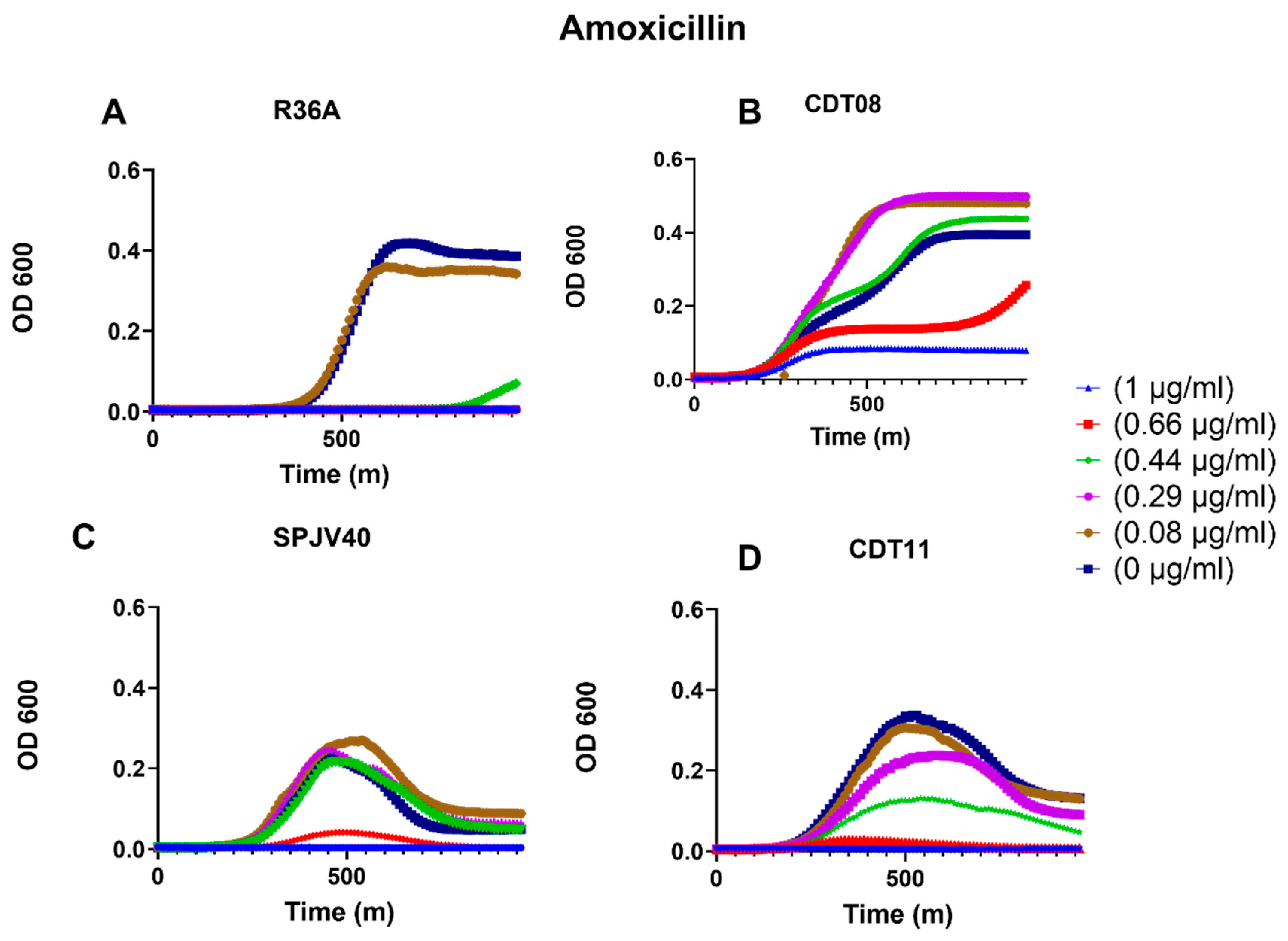
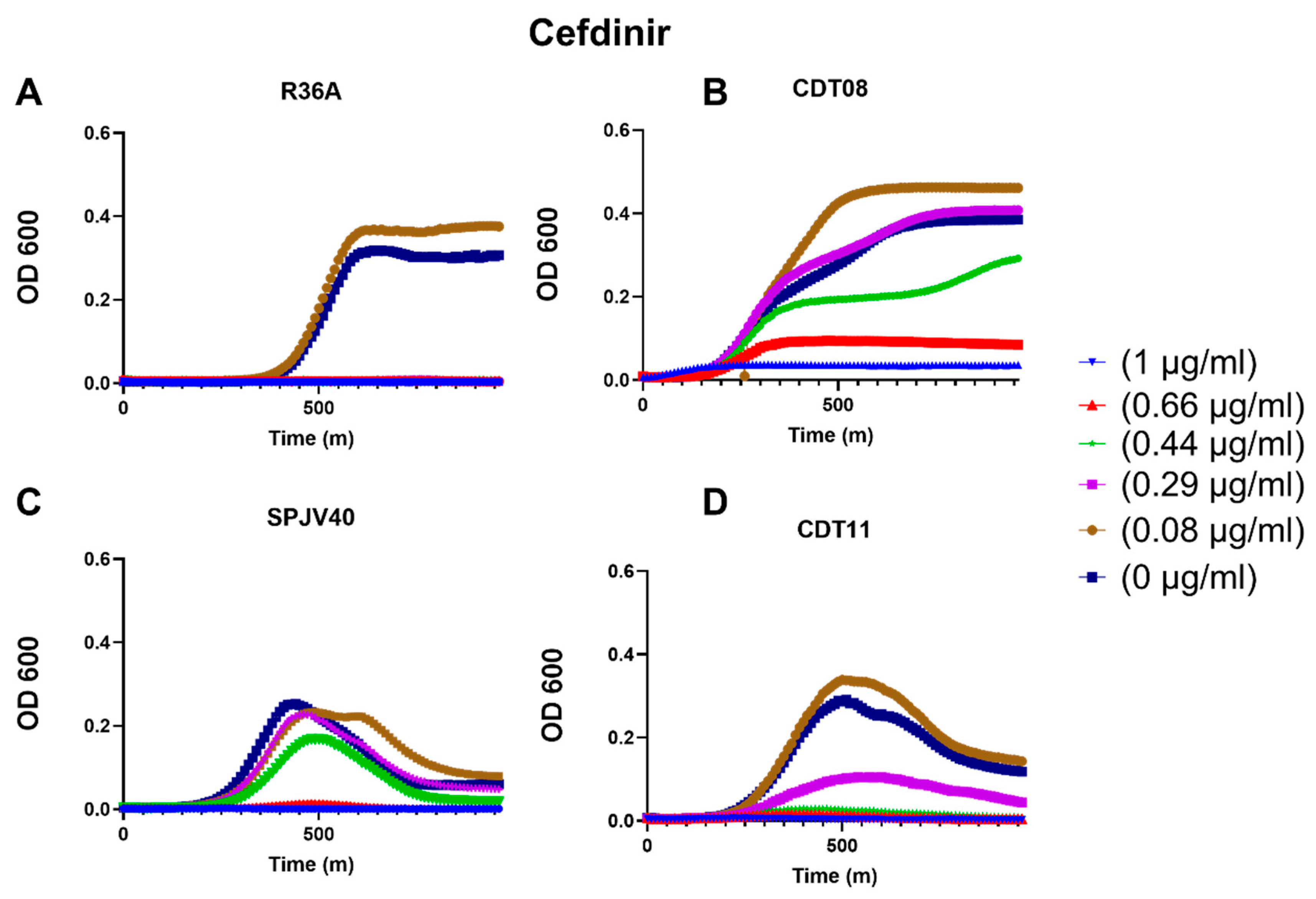
First, we wanted to determine if gene regulation by aliD impacts antibiotic resistance. To do this, we examined how growth is impacted in the presence of different antibiotics with and without the oligopeptide transporter aliD. The strains R36A, which does not natively express aliD, and CDT08 (R36A::aliD) were grown in the presence of amoxicillin (Figure 1A,B). Compared to R36A, we observed increased growth of CDT08 at all antibiotic concentrations tested, with R36A growth only observed at the lowest concentration (0.08 μg/mL). A more modest difference was noted between SPJV40 (WT serotype 38 with aliD) and CDT11 (SPJV40ΔaliD), with growth of WT occurring at 0.44 μg/mL compared to minimal growth at the same concentration when aliD is deleted in CDT11 (Figure 1C,D). Similar to growth in the presence of amoxicillin, CDT08 exhibited enhanced growth at higher cefdinir concentrations compared to its aliD-deficient parental strain R36A. CDT11 (SPJV40ΔaliD) showed no growth at either 0.44 μg/mL or 1.0 μg/mL, while the parental strain SPJV40 still grew at 0.44 μg/mL (Figure 2C,D). These observations suggest that the presence of aliD may confer a growth advantage under β-lactam antibiotic stress, indicating a potential role in β-lactam sensitivity.
Furthermore, calculation of cefdinir MIC values shows that SPJV40 has an MIC of 0.5909 ± 0.1 µg/mL, and CDT11 presents an MIC of 0.3666 ± 0.09 µg/mL; this implies that aliD has an effect on the MIC of β-lactams. This remains consistent with the strains R36A having an MIC of 0.2237 ± 0.06 µg/mL and CDT08 having an MIC of 1.017 ± 0.09 µg/mL (Table 3).
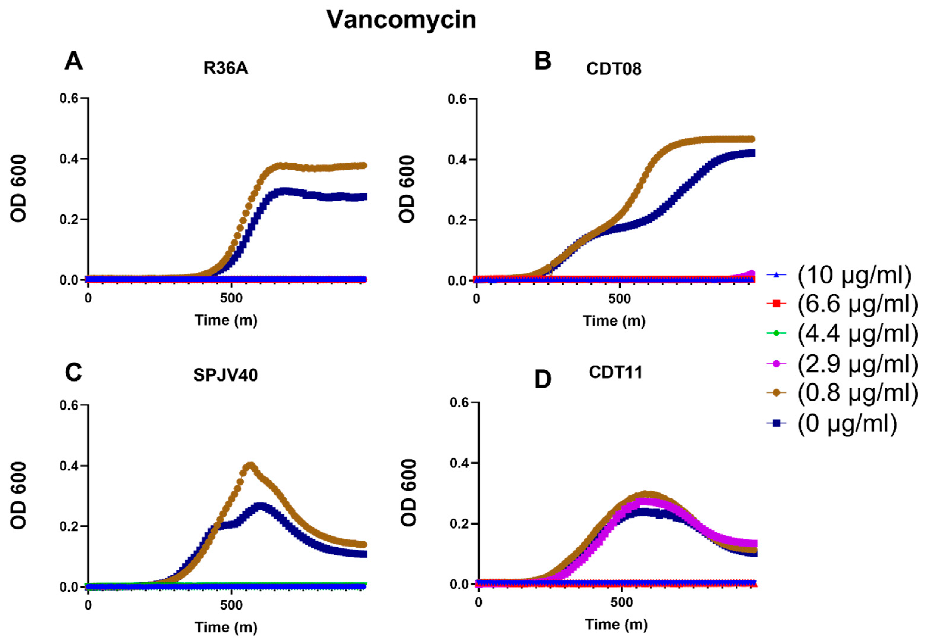
Next, we wanted to examine if other antibiotics that target the cell wall would also change the growth rates based on aliD expression. We tested growth in vancomycin, which targets the cell wall but has a different mechanism of action than the β-lactam antibiotics initially tested. We observed no notable change between CDT08 and R36A growth in the presence of vancomycin (Figure 3A,B). Surprisingly, a difference was observed between CDT11 and SPJV40, with the aliD deletion seemingly having increased fitness to vancomycin exposure (Figure 3C,D).
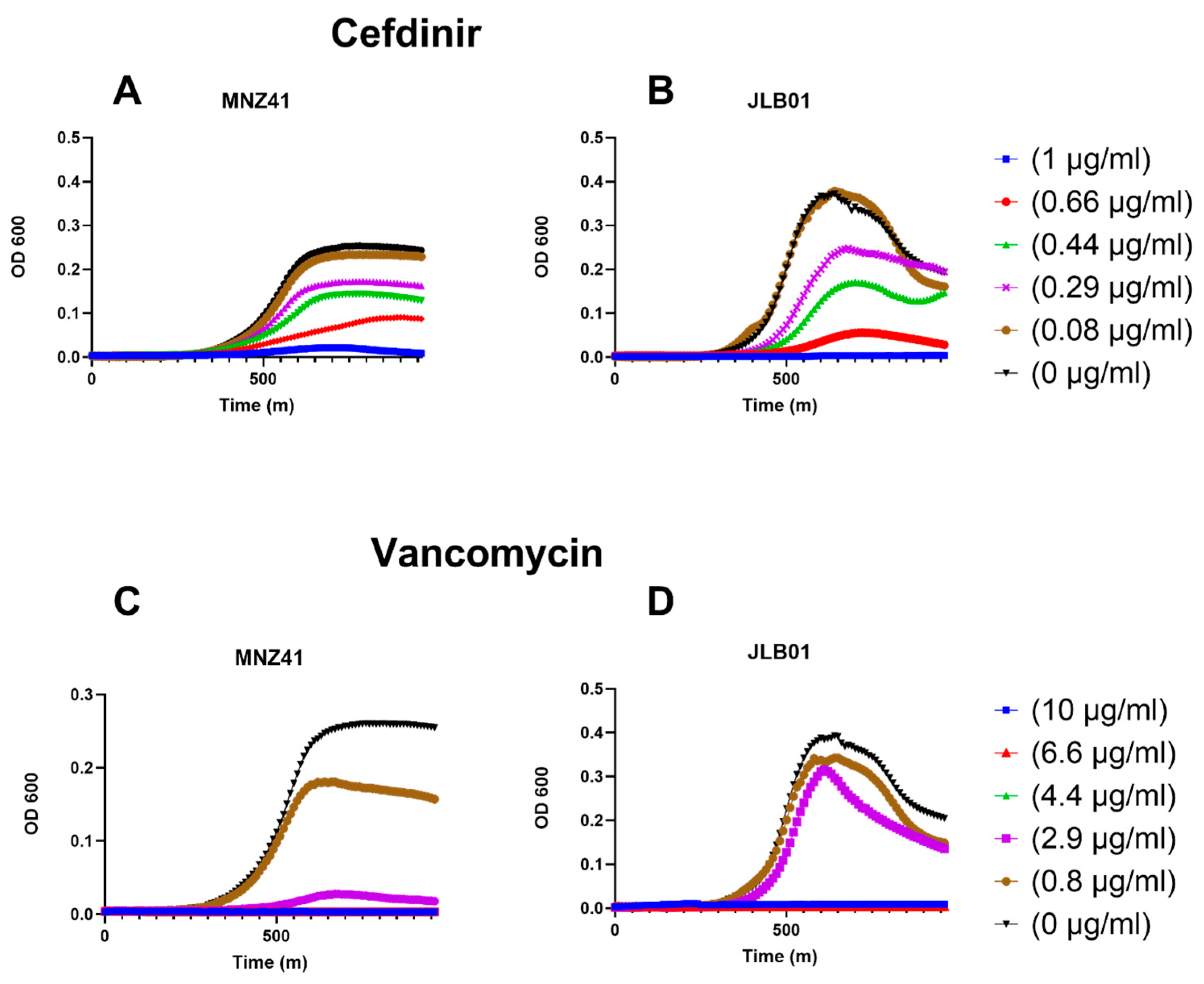
In conjunction, we also examined the effect aliD has on NESp resistance. aliD was first characterized from NESp strains, so the WT MNZ41 and JLB01 (MNZ41ΔaliCΔaliD) growth was tested in the presence of both cefdinir and vancomycin (Figure 4). A similar trend was observed when examining NESp antibiotic susceptibility, with WT MNZ41 displaying increased growth in the presence of cefdinir than the isogenic mutant JLB01.
Examining growth in these different antibiotics indicates that aliD impacts susceptibility to β-lactam antibiotics. To determine a possible mechanism explaining this difference in susceptibility, available transcriptomic and proteomic data was examined. β-lactam antibiotics specifically target PBPs, while vancomycin targets peptidoglycan subunits [10,22]. Of the genes regulated by aliD that may impact PBP activity, the gene clpL is a likely candidate for a possible mechanism. ClpL has been previously shown to mediate PBP2x transport to the surface. PBP2x is a transpeptidase that facilitates the creation of cross-links between peptidoglycan strands, and alterations in its expression can impact the structure and stability of the cell wall.

3.2. Changes in clpL Expression Due to aliD

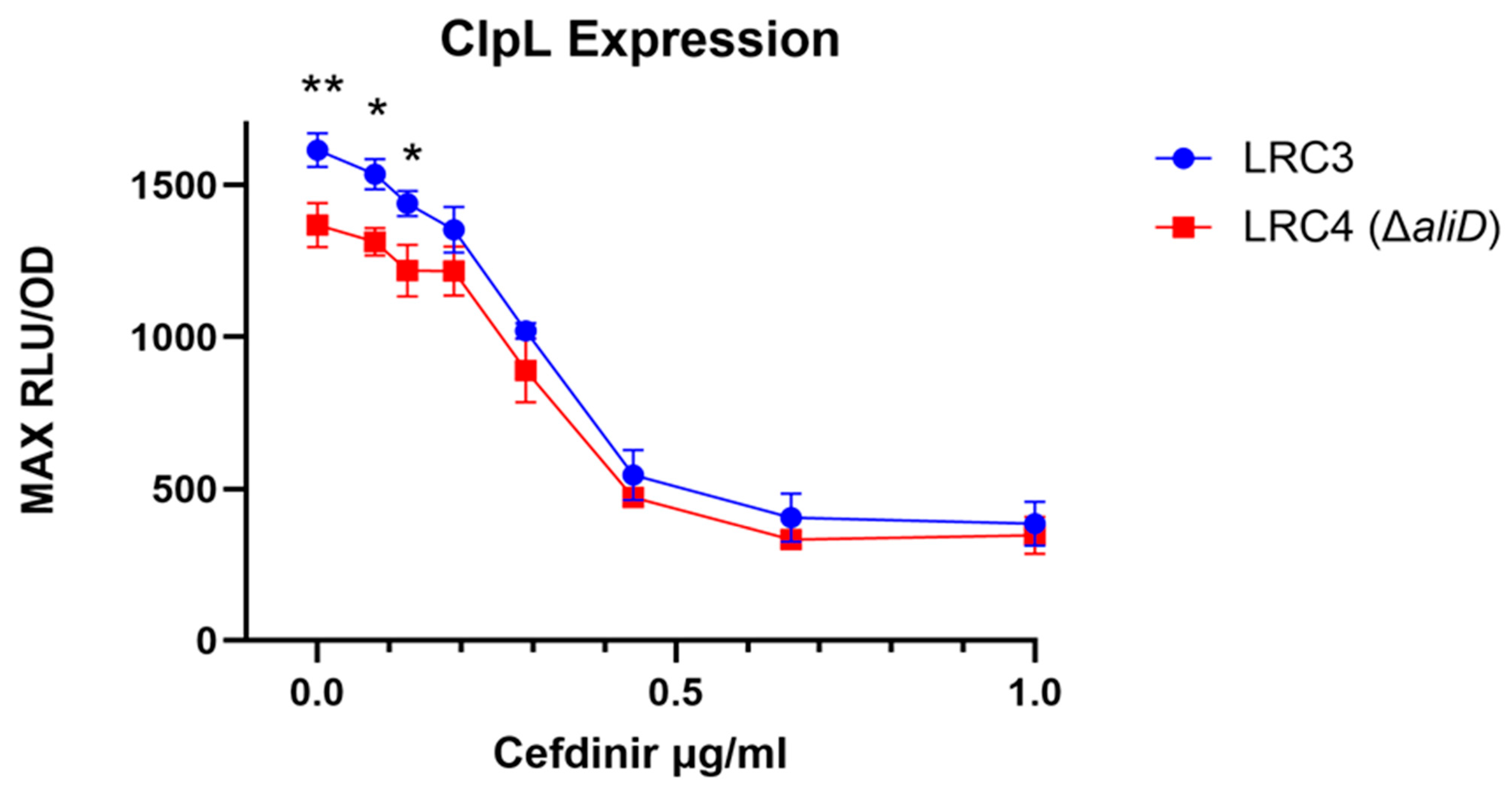
We wanted to further characterize the relationship between clpL expression and aliD. To test this, a transcriptional fusion of clpL and a luciferase reporter were created in the aliD containing SPJV40 (LRC3) and its mutant lacking aliD (LRC4). This allows for quantification of clpL expression based on luminescence intensity. With no antibiotics present, we observed a standardized luminescence value of ~1614 RLU in LRC3 and a significantly lower (~20%) clpL expression in LRC4, suggesting the presence of aliD increases clpL expression (p = 0.0075, Figure 5). Furthermore, with the addition of cefdinir, this trend is consistent at 0, 0.08, and 0.125 µg/mL, with LRC3 exhibiting significantly higher standardized RLU than LRC4 (Figure 5). No difference in clpL expression is observed at higher antibiotic concentrations, likely due to reduced bacterial growth in these conditions.

3.3. Amoxicillin Susceptibility Due to clpL Expression

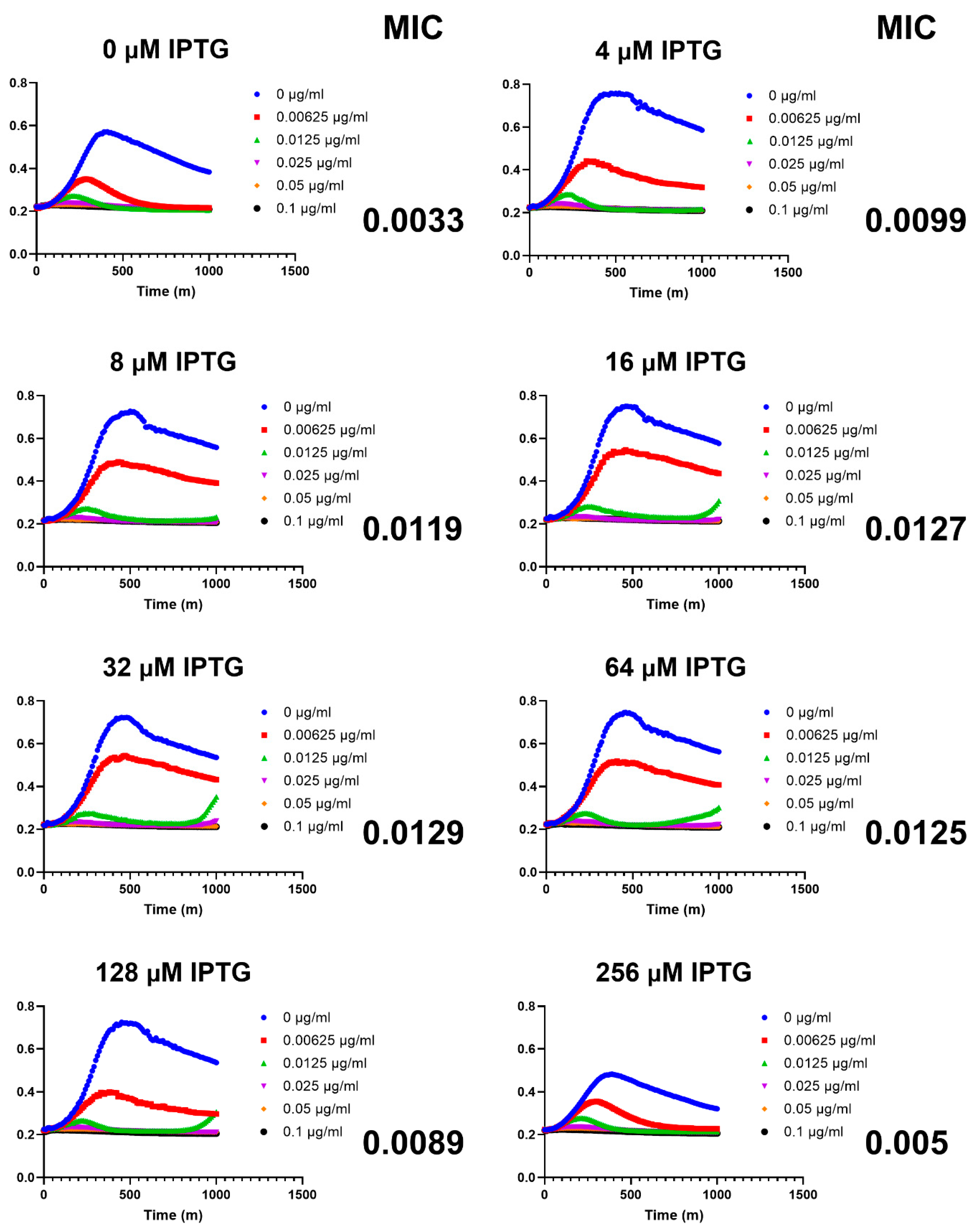
To further validate that changes in clpL expression impact β-lactam susceptibility, an inducible clpL expression system was created in the susceptible R36A background (LRC1). Induction of clpL with 4 µM IPTG resulted in a threefold increase in MIC to amoxicillin compared to the uninduced condition. Further increases in IPTG concentration led to incremental MIC elevation, reaching a plateau between 32 and 64 µM IPTG. Notably, at 256 µM IPTG, bacterial growth was impaired, likely due to proteotoxic stress associated with overexpression of clpL, a component of the caseinolytic protease (Clp) system (Figure 6).

3.4. Surface Exposure of PBP2x

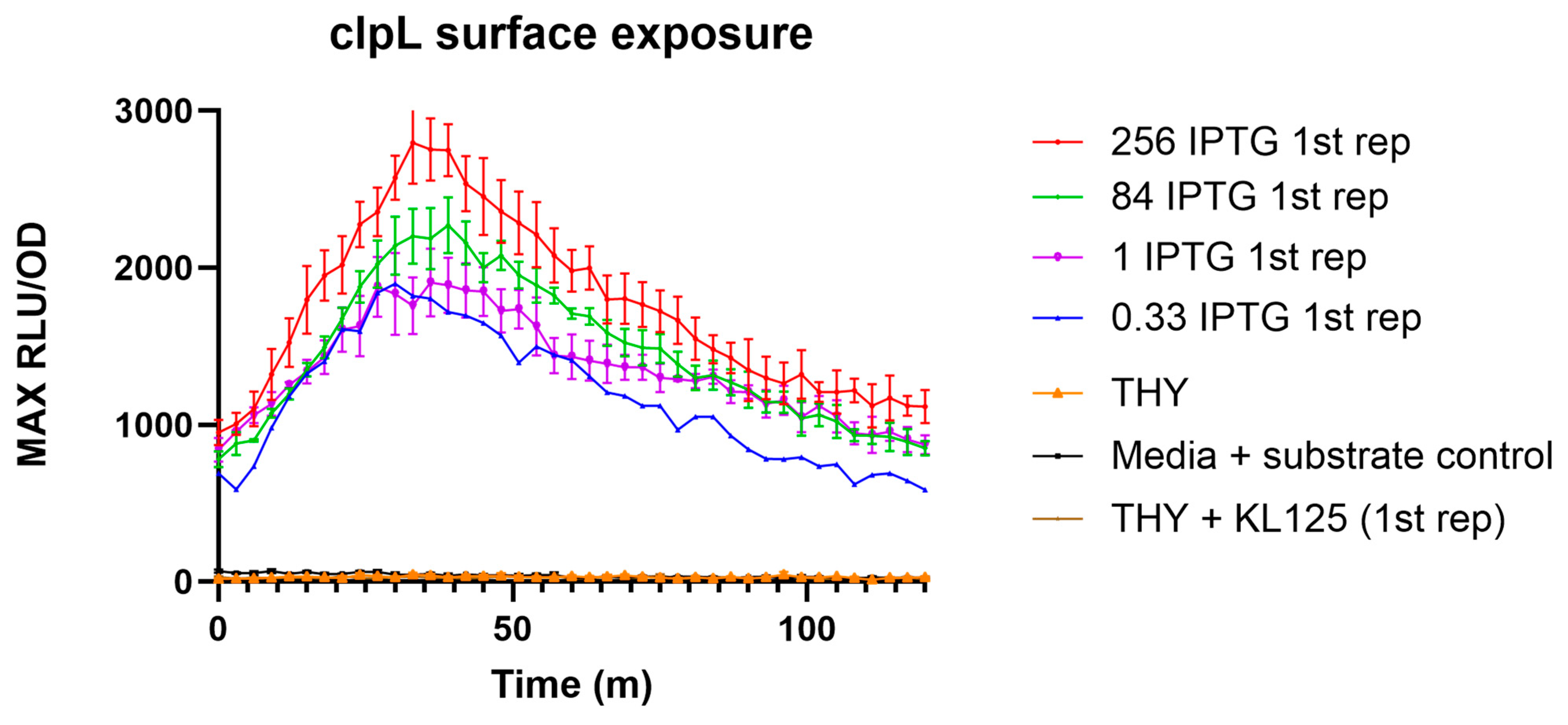
Since ClpL transports PBP2x to the bacterial surface, which in turn alters cell wall stability and antibiotic susceptibility, we wanted to determine the amount of PBP2x on the cell surface. To do this, we engineered PBP2x with a HiBiT tag under native expression in the LRC1 (R36A::lacI; ΔclpL; Plac-clpL) background, creating LRC2. Upon addition of the LgBiT fragment and the luciferase substrate, the amount of surface-exposed PBP2x can be determined through luminescence intensity. We observed an increase in luminescence based on the quantity of IPTG-inducing clpL expression with our high concentrations of 256 µM IPTG expressing a maximum luminescence of 2795, while our concentration of 0.33 µM IPTG had a luminescence of only 1898 (Figure 7). This indicates that, by increasing the amount of ClpL, a corresponding increase in surface-exposed PBP2x occurs.

3.5. Quantification of Cell Wall Cross-Linking

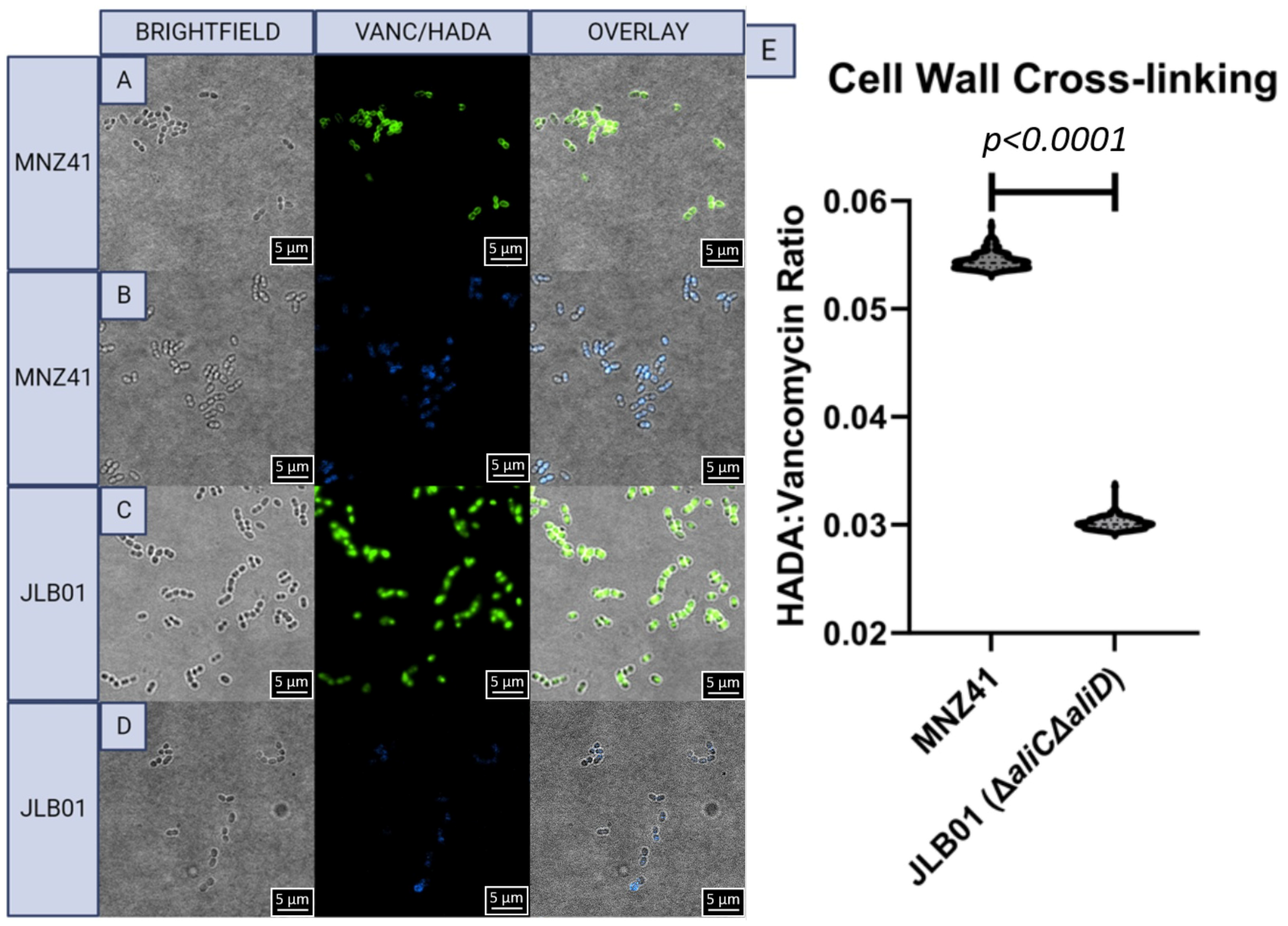
Since PBP2x mediates cross-linking of the cell wall, we wanted to examine if there is a correlation between the presence of aliD and cell wall cross-linking. To test this, we used two fluorescent dyes to compare the amount of new cell wall that is being synthesized to the amount of cross-linking between these segments. The newly synthesized cell wall was quantified using fluorescent vancomycin, and cross-linking was determined through incorporation of the fluorescent HADA probe. These two dyes allow us to quantify the ratio of newly created cell wall to the amount of cross-linking. Comparison of WT S. pneumoniae strain MNZ41 to a mutant lacking aliC and aliD (JLB01) demonstrated a significantly higher ratio of new cell wall to cross-linking in MNZ41 than the mutant JLB01, p < 0.0001 (Figure 8).

4. Discussion

Bacteria utilize multiple mechanisms to respond to antibiotic stress, and here we demonstrate that the oligopeptide transporter aliD alters antibiotic susceptibility to β-lactam antibiotics through regulation of clpL expression. Growth curve analysis revealed measurable differences between tested strains, suggesting that the presence of aliD is associated with decreased susceptibility to β-lactam antibiotics. In the antibiotic growth curves, we also observed no growth differences following treatment with vancomycin, which is significant because vancomycin targets the D-Ala-D-Ala terminus of peptidoglycan rather than penicillin-binding proteins (PBPs) recognized by β-lactam antibiotics. This suggests that the observed phenotype is limited to β-lactam antibiotics and the functioning of PBPs. We demonstrate that aliD increases clpL expression and PBP2x recruitment to the surface. This allows for increased cross-linking in the cell wall and is the likely cause of the reduced β-lactam susceptibility.
Prior work examining the role of aliD in virulence used transcriptomic and proteomic data to identify 42 genes regulated by aliD [23]. Many of these genes are associated with virulence, carbohydrate metabolism, or stress responses. Among these genes, clpL was of particular interest as a possible gene influencing β-lactam susceptibility. ClpL is a chaperone that transports misfolded proteins to the Clp protease system. ClpL is also a chaperone that assists in the folding, stability, and transport of PBP2x. This second function of ClpL suggests a role in cell wall structure, and other published work has shown that clpL deletions result in altered cell wall structure and increased stress sensitivity [18]. While all pneumococcal strains express clpL, the aliD gene is usually found in the capsular polysaccharide synthesis locus of nonencapsulated strains. However, there are three pneumococcal serotypes that express aliD: 25A, 25F, and 38. For this study, encapsulated strains expressing aliD were prioritized for investigation due to the clinical relevance of encapsulated pneumococci, which are responsible for the majority of pneumococcal disease. This is particularly important in the context of the emerging serotype 38, which has shown increased prevalence and may possess a selective advantage due to aliD-mediated peptide sensing and regulation of virulence-associated genes [25].
To more directly link ClpL to antibiotic resistance, we engineered a construct that enables controlled clpL expression and tested growth across increasing concentrations of β-lactam antibiotics. We observed that increased clpL expression correlated with improved growth under antibiotic stress, indicating decreased susceptibility. However, this positive effect plateaued and reversed at high ClpL levels, where bacterial growth was impaired. This suggests that while moderate clpL expression improves protein handling and cell wall synthesis under stress, excessive ClpL may be detrimental due to dysregulation of protease and chaperone systems. This highlights the importance of proper regulation of proteostasis systems for normal bacterial growth and cell wall synthesis.
Since ClpL is a chaperone, we next examined how clpL expression affects the amount of PBP2x present on the cell surface. Using an HiBiT-tagged PBP2x, we observed that increased clpL expression produced a corresponding increase in luminescence signal, indicating greater surface abundance of PBP2x. Interestingly, induction of clpL with 256 µM IPTG had the highest amount of PBP2x transported to the surface but reduced overall pneumococcal growth. Therefore, the amount of PBP2x is the limiting factor to the amount of PBP2x able to be transported to the surface. This supports the model that ClpL promotes PBP2x surface localization or stability, which in turn increases cell wall cross-linking and reduces susceptibility to β-lactam antibiotics. With increased clpL expression regulated by aliD, if other factors increase the expression of PBP2x, even more cross-linking in the cell wall could occur, resulting in a more pronounced difference in β-lactam susceptibility.
We were able to demonstrate that the presence of aliD increases clpL expression and PBP2x recruitment to the cell surface. PBPs perform partially redundant functions but produce different types and amounts of peptidoglycan cross-links [26]. Changes in the relative abundance of specific PBPs can significantly impact cell wall structure. PBP1a is typically the most highly expressed PBP, followed by PBP2x, and shifts in the ratio of these proteins can alter cell wall cross-link structure and impact antibiotic susceptibility [27].
To verify that aliD impacts cell wall structure, we used fluorescent vancomycin and HADA to measure new cell wall synthesis and cross-link formation. Due to the increased presence of PBP2x on the cell surface, increases in cross linking were expected. Vancomycin labeling was used alongside HADA as a normalization method to account for differences in peptidoglycan synthesis. Using this approach, we determined that deletion of aliD in a NESp substantially reduced cross-link formation relative to total new cell wall production, indicating that aliD contributes to efficient cell wall cross-linking activity.
This study details a novel role of the oligopeptide transporter aliD. Oligopeptide transporters are important regulators that influence gene expression based on the peptide pools available in the local environment. This suggests that the type of infection may influence antibiotic susceptibility, with certain tissues increasing clpL expression more than other tissues. The differences in peptide compositions could alter aliD signaling and downstream gene regulation of additional genes and impact virulence variability based on the tissue type. This may have important clinical implications, as MIC values determined in laboratory media may not accurately reflect antibiotic susceptibility in the human body, where peptide availability differs substantially from standard laboratory growth media. Therefore, peptide availability and environmental conditions may play an underappreciated role in regulating antibiotic susceptibility in S. pneumoniae.
The Ami transporter has previously been shown to decrease susceptibility to various antibiotics by acting as a pore for antibiotic removal [30]. This is unlikely to affect the mechanism we propose in this paper, as aliD is located on the cell wall but further demonstrates the need to increase research on the role of oligopeptide transporters on antibiotic resistance. Clinical susceptibility testing is dependent on bacterial growth, either on agar or using broth microdilutions. Each of these will have different peptide pools available, and growth in dilutions a single step above can change an intermediate resistance strain to susceptible. This can lead to treatment that may not be as effective as other antibiotic choices. This is particularly important for meningitis treatment since the difference from susceptible to resistance is a small change in MIC. Along with this, not all clinics will use the same method for quantifying resistance, which can impact not only treatment but reporting of resistance patterns in pneumococcal strains across different geographical areas.
Overall, this study advances our understanding of how aliD and clpL contribute to β-lactam resistance, likely through effects on cell wall synthesis machinery, protein handling, and PBP2x localization. Understanding the interactions regulating cell wall synthesis can help us learn how to better and more accurately treat pathogens such as the pneumococcus with antibiotics. This research could be used to better inform a physician on the choice of treatment based on whether the pneumococcal strain expresses aliD.

5. Conclusions

We were able to demonstrate that β-lactam susceptibility decreased when aliD was present in multiple pneumococcal strains. Along with this, clpL expression is reduced in an aliD mutant strain, and increased expression of clpL decreases amoxicillin susceptibility. Furthermore, by increasing clpL expression, the amount of PBP2x on the bacterial surface increases, likely resulting in increased peptidoglycan cross-linking. We were unable to show a direct link between peptide pools imported by aliD and regulatory pathways leading to clpL expression. Increased intracellular peptide pools could activate proteolytic systems indirectly and not through direct interaction with transcription factors for clpL. This remains unresolved and requires further investigation, particularly in clinically relevant encapsulated strains. Another possibility is that the presence of an additional oligopeptide transporter alters the available peptide pool, which can impact peptidoglycan structure [31]. Others have demonstrated in other organisms that altered peptide pools can decrease β-lactam susceptibility by increasing β-lactamase expression [32]. Future work will try and identify a direct link between aliD-imported peptides and transcription factors that regulate downstream gene expression and determine function in a broader range of strains [33]. This is of particular importance since the largest change in MIC was observed in the lab strain R36A. With these studies, it would be possible to determine if there is a direct interaction between aliD peptides and clpL regulation and have a better understanding of resistance mechanisms.

Author Contributions

L.R.G.C.: Data acquisition, conceptualization, writing—original draft, writing—review and editing, formal analysis. M.F.K.: Data acquisition, formal analysis, review and editing. L.S.M.: conceptualization, writing—review and editing, formal analysis. L.E.K.: conceptualization, funding acquisition, writing—original draft, writing—review and editing, formal analysis. This work was supported by the Department of Cell and Molecular Biology, University of Mississippi Medical Center. All authors have read and agreed to the published version of the manuscript.

Funding

Funding was provided through startup funds given to LEK.

Institutional Review Board Statement

Not applicable. All studies were performed following Institutional Biosafety Committee (IBC) guidelines and approved in protocol IBC-00001075.

Informed Consent Statement

Not applicable.

Data Availability Statement

No datasets were created during this study, but raw data is available upon request.

Acknowledgments

Department of Cell and Molecular Biology, University of Mississippi Medical Center. Center for Immunology and Microbial Research, University of Mississippi Medical Center. Figures made on Graphpad prism.

Conflicts of Interest

The authors declare no conflicts of interest.

References

  1. Feldman, C.; Anderson, R. Recent Advances in the Epidemiology and Prevention of Streptococcus Pneumoniae Infections. F1000Research 2020, 9, F1000 Faculty Rev-338. [Google Scholar] [CrossRef]
  2. Wang, G.-M.; Tao, W.; Pang, X.-Y.; Xin, Y.; Gou, Z.-H.; Wang, Y. Global, regional, and national burden of pneumococcal disease among children and adolescents aged <20 years from 1990 to 2021: A predictive analysis. Front. Public Health 2025, 13, 1675854. [Google Scholar] [CrossRef] [PubMed]
  3. Troeger, C.; Blacker, B.; Khalil, I.A.; Rao, P.C.; Cao, J.; Zimsen, S.R.M.; Albertson, S.B.; Deshpande, A.; Farag, T.; Abebe, Z.; et al. Estimates of the global, regional, and national morbidity, mortality, and aetiologies of lower respiratory infections in 195 countries, 1990–2016: A systematic analysis for the Global Burden of Disease Study 2016. Lancet Infect. Dis. 2018, 18, 1191–1210. [Google Scholar] [CrossRef]
  4. Shenoy, A.T.; Orihuela, C.J. Anatomical site-specific contributions of pneumococcal virulence determinants. Pneumonia 2016, 8, 7. [Google Scholar] [CrossRef] [PubMed]
  5. Ganaie, F.A.; Beall, B.W.; Yu, J.; van der Linden, M.; McGee, L.; Satzke, C.; Manna, S.; Lo, S.W.; Bentley, S.D.; Ravenscroft, N.; et al. Update on the evolving landscape of pneumococcal capsule types: New discoveries and way forward. Clin. Microbiol. Rev. 2025, 38, e0017524. [Google Scholar] [CrossRef] [PubMed]
  6. Atıcı, S.; Güneşer, D.; Kepenekli, E.; Söyletir, G.; Soysal, A. Serotypes distribution and antibiotic susceptibility of Streptococcus pneumoniae strains: Five-year surveillance results of post-PCV-13. BMC Pediatr. 2025, 25, 244. [Google Scholar] [CrossRef] [PubMed]
  7. Croucher, N.J.; Kagedan, L.; Thompson, C.M.; Parkhill, J.; Bentley, S.D.; A Finkelstein, J.; Lipsitch, M.; Hanage, W.P. Selective and Genetic Constraints on Pneumococcal Serotype Switching. PLoS Genet. 2015, 11, e1005095. [Google Scholar] [CrossRef] [PubMed]
  8. Park, I.H.; Kim, K.-H.; Andrade, A.L.; E Briles, D.; McDaniel, L.S.; Nahm, M.H. Nontypeable Pneumococci Can Be Divided into Multiple cps Types, Including One Type Expressing the Novel Gene pspK. mBio 2012, 3. [Google Scholar] [CrossRef] [PubMed]
  9. Nasher, F.; Förster, S.; Yildirim, E.C.; Grandgirard, D.; Leib, S.L.; Heller, M.; Hathaway, L.J. Foreign peptide triggers boost in pneumococcal metabolism and growth. BMC Microbiol. 2018, 18, 23. [Google Scholar] [CrossRef] [PubMed]
  10. Nasher, F.; Heller, M.; Hathaway, L.J. Streptococcus pneumoniae Proteins AmiA, AliA, and AliB Bind Peptides Found in Ribosomal Proteins of Other Bacterial Species. Front. Microbiol. 2018, 8, 2688. [Google Scholar] [CrossRef] [PubMed]
  11. Monnet, V. Bacterial oligopeptide-binding proteins. Cell. Mol. Life Sci. 2003, 60, 2100–2114. [Google Scholar] [CrossRef] [PubMed]
  12. Claverys, J.-P.; Grossiord, B.; Alloing, G. Is the Ami-AliA/B oligopeptide permease of Streptococcus pneumoniae involved in sensing environmental conditions? Res. Microbiol. 2000, 151, 457–463. [Google Scholar] [CrossRef] [PubMed]
  13. Keller, L.E.; Robinson, D.A.; McDaniel, L.S. Nonencapsulated Streptococcus pneumoniae: Emergence and Pathogenesis. mBio 2016, 7, e01792-15. [Google Scholar] [CrossRef] [PubMed]
  14. Alloing, G.; de Philip, P.; Claverys, J.P. Three highly homologous membrane-bound lipoproteins participate in oligopeptide transport by the Ami system of the gram-positive Streptococcus pneumoniae. J. Mol. Biol. 1994, 241, 44–58. [Google Scholar] [CrossRef] [PubMed]
  15. Mascarenhas, N.M.; Gosavi, S. Understanding protein domain-swapping using structure-based models of protein folding. Prog. Biophys. Mol. Biol. 2017, 128, 113–120. [Google Scholar] [CrossRef] [PubMed]
  16. Didi, S. Antibiotic Resistance Mechanisms in Bacteria: Biochemical and Genetic Aspects. Food Technol. Biotechnol. 2008, 46, 11. [Google Scholar]
  17. Ventola, C.L. The Antibiotic Resistance Crisis. Pharm. Ther. 2015, 40, 277–283. [Google Scholar]
  18. Tran, T.D.-H.; Kwon, H.-Y.; Kim, E.-H.; Kim, K.-W.; Briles, D.E.; Pyo, S.; Rhee, D.-K. Decrease in penicillin susceptibility due to heat shock protein ClpL in Streptococcus pneumoniae. Antimicrob. Agents Chemother. 2011, 55, 2714–2728. [Google Scholar] [CrossRef] [PubMed]
  19. Hakenbeck, R.; Grebe, T.; Zähner, D.; Stock, J.B. β-Lactam resistance in Streptococcus pneumoniae: Penicillin-binding proteins and non-penicillin-binding proteins. Mol. Microbiol. 1999, 33, 673–678. [Google Scholar] [CrossRef] [PubMed]
  20. Fani, F.; Leprohon, P.; Zhanel, G.G.; Bergeron, M.G.; Ouellette, M. Genomic Analyses of DNA Transformation and Penicillin Resistance in Streptococcus pneumoniae Clinical Isolates. Antimicrob. Agents Chemother. 2014, 58, 1397–1403. [Google Scholar] [CrossRef] [PubMed]
  21. Mohanty, S.; Johnson, K.D.; Yu, K.C.; Watts, J.A.; Gupta, V. A Multicenter Evaluation of Trends in Antimicrobial Resistance Among Streptococcus pneumoniae Isolates From Adults in the United States. Open Forum Infect. Dis. 2022, 9, ofac420. [Google Scholar] [CrossRef] [PubMed]
  22. McCurdy, S.P.; Sheets, A.J.; Cammarata, S.K.; Vidal, J.E. Serotype distribution and antimicrobial susceptibility of Streptococcus pneumoniae isolates from a Phase III community-acquired bacterial pneumonia (CABP) trial. JAC-Antimicrob. Resist. 2021, 3, dlab057. [Google Scholar] [CrossRef] [PubMed]
  23. Thompson, C.D.; Bradshaw, J.L.; Miller, W.S.; Vidal, A.G.; Vidal, J.E.; Rosch, J.W.; McDaniel, L.S.; Keller, L.E. Oligopeptide Transporters of Nonencapsulated Streptococcus pneumoniae Regulate CbpAC and PspA Expression and Reduce Complement-Mediated Clearance. mBio 2023, 14, e0332522. [Google Scholar] [CrossRef] [PubMed]
  24. Avery, O.T.; MacLeod, C.M.; McCarty, M. Studies on the Chemical Nature of the Substance Inducing Transformation of Pneumococcal Types: Induction of Transformation by a Desoxyribonucleic Acid Fraction Isolated from Pneumococcus Type III. J. Exp. Med. 1944, 79, 137–158. [Google Scholar] [CrossRef] [PubMed]
  25. Bradshaw, J.L.; Pipkins, H.R.; Keller, L.E.; Pendarvis, J.K.; McDaniel, L.S. Mucosal Infections and Invasive Potential of Nonencapsulated Streptococcus pneumoniae Are Enhanced by Oligopeptide Binding Proteins AliC and AliD. mBio 2018, 9, e02097-17. [Google Scholar] [CrossRef] [PubMed]
  26. Thompson, C.D.; Khan, F.; Crosby, L.R.G.; Holcomb, S.G.; Vidal, A.G.J.; Vidal, J.E.; McDaniel, L.S.; Keller, L.E. AliC and AliD of nonencapsulated Streptococcus pneumoniae enhance virulence in a Galleria mellonella model of infection by contributing to reactive oxygen species resistance. Front. Cell. Infect. Microbiol. 2025, 15, 1583375. [Google Scholar] [CrossRef] [PubMed]
  27. Keller, L.E.; Rueff, A.-S.; Kurushima, J.; Veening, J.-W. Three New Integration Vectors and Fluorescent Proteins for Use in the Opportunistic Human Pathogen Streptococcus pneumoniae. Genes 2019, 10, 394. [Google Scholar] [CrossRef] [PubMed]
  28. Howie, R.R.; McKinney, M.M.; Tataryn, N.M.; Cole, A.L.; Dupont, W.D.; Yang, T.S.; Gibson-Corley, K.N. Determination of Postmortem Interval in Mice. J. Am. Assoc. Lab. Anim. Sci. 2024, 63, 428–436. [Google Scholar] [CrossRef] [PubMed]
  29. Lambert, R.J.W.; Pearson, J. Susceptibility testing: Accurate and reproducible minimum inhibitory concentration (MIC) and non-inhibitory concentration (NIC) values. J. Appl. Microbiol. 2000, 88, 784–790. [Google Scholar] [CrossRef] [PubMed]
  30. Leshchiner, D.; Rosconi, F.; Sundaresh, B.; Rudmann, E.; Ramirez, L.M.N.; Nishimoto, A.T.; Wood, S.J.; Jana, B.; Buján, N.; Li, K.; et al. A genome-wide atlas of antibiotic susceptibility targets and pathways to tolerance. Nat. Commun. 2022, 13, 3165. [Google Scholar] [CrossRef] [PubMed]
  31. Zhao, H.; Roistacher, D.M.; Helmann, J.D. Aspartate deficiency limits peptidoglycan synthesis and sensitizes cells to antibiotics targeting cell wall synthesis in Bacillus subtilis. Mol. Microbiol. 2018, 109, 826–844. [Google Scholar] [CrossRef] [PubMed]
  32. Sverak, H.E.; Yaeger, L.N.; Worrall, L.J.; Vacariu, C.M.; Glenwright, A.J.; Vuckovic, M.; Al Azawi, Z.-D.; Lamers, R.P.; Marko, V.A.; Skorupski, C.; et al. Cryo-EM characterization of the anydromuropeptide permease AmpG central to bacterial fitness and β-lactam antibiotic resistance. Nat. Commun. 2024, 15, 9936. [Google Scholar] [CrossRef] [PubMed]
  33. Gooderham, W.J.; Hancock, R.E.W. Regulation of virulence and antibiotic resistance by two-component regulatory systems in Pseudomonas aeruginosa. FEMS Microbiol. Rev. 2009, 33, 279–294. [Google Scholar] [CrossRef] [PubMed]
Figure 1. Growth curves comparing susceptibility to amoxicillin with or without the oligopeptide transporter aliD. R36A, a serotype two capsule mutant naturally deficient in aliD grown in the presence of amoxicillin (A), expresses reduced fitness compared to R36A with genetically added aliD, CDT08 (B). Serotype 38 aliD-expressing WT strain SPJV40 (C) grown in the presence of beta lactam amoxicillin with concentrations ranging from 1 to 0 µg/mL, expressing greater fitness than SPJV40 aliD mutant CDT11 (D).
Pathogens 15 00738 g001
Figure 2. Growth curves comparing susceptibility to cefdinir with or without the oligopeptide transporter aliD. R36A (A) serotype two capsule mutant naturally deficient in aliD grown in the presence of cefdinir, expresses reduced fitness compared to R36A with genetically added aliD, CDT08 (B). Serotype 38 aliD-expressing WT strain SPJV40 (C) grown in the presence of beta lactam cefdinir with concentrations ranging from 1 to 0 µg/mL, expressing greater fitness than SPJV40 aliD mutant CDT11 (D).
Pathogens 15 00738 g002
Figure 3. Growth curves comparing susceptibility to vancomycin with or without the oligopeptide transporter aliD. R36A (A) serotype two capsule mutant naturally deficient in aliD grown in the presence of vancomycin, expresses reduced fitness compared to R36A with genetically added aliD CDT08 (B). Serotype 38 aliD-expressing WT strain SPJV40 (C) grown in the presence of antibiotic vancomycin with concentrations ranging from 10 to 0 µg/mL, expressing greater fitness than SPJV40 aliD mutant CDT11 (D).
Pathogens 15 00738 g003
Figure 4. Growth curves comparing susceptibility to vancomycin and cefdinir with or without the oligopeptide transporter aliD in nonencapsulated pneumococcus. NESp aliD-expressing WT strain MNZ41 (A) grown in the presence of beta lactam cefdinir with concentrations ranging from 1 to 0 µg/mL, expressing greater fitness than MNZ41 aliD mutant JLB01 (B). JLB01 (D) grown in the presence of vancomycin, grown in concentrations ranging from 10 to 0 µg/mL, expresses reduced fitness compared to MNZ41 with WT aliD (C).
Pathogens 15 00738 g004
Figure 5. ClpL expression compared via luminescence based on the presence of aliD and antibiotics. LRC3 (SPJV40::clpL-luciferase) is a serotype 38 construct with clpL expression measured through luciferase activity. LRC4 is an aliD deletion of LRC3. Both strains were grown in increasing concentration of the β-lactam cefdinir. We observed that aliD results in more clpL expression in our zero concentration and in the presence of cefdinir (* = p < 0.05, ** = p < 0.005).
Pathogens 15 00738 g005
Figure 6. Antibiotic susceptibility to amoxicillin under varying levels of clpL expression. Growth kinetics of the LRC1 strain were assessed under varying levels of clpL induction controlled by IPTG introduction in varying levels of antibiotic exposure. Antibiotic susceptibility was quantified by calculating the MIC using the Gompertz growth model fit to the area under the curve acquired from optical density data.
Pathogens 15 00738 g006
Figure 7. Measurement of PBP2x on the cell wall following increased levels of clpL expression. PBP2x contains a HiBiT tag that luminesces in the presence of LgBiT protein fragment and substrate. Increased induction of clpL through IPTG addition led to greater amounts of PBP2x observed on the bacterial surface, with 256 µM IPTG induction leading to the highest surface exposure of PBP2x. Intermediate levels of PBP2x are observed as IPTG concentration is reduced.
Pathogens 15 00738 g007
Figure 8. Quantification of cell wall cross-linking due to the presence of aliD. The S. pneumoniae strains MNZ41 (WT) (A,B) and JLB01 (MNZ41 ΔaliCΔaliD) (C,D) were labeled with fluorescent vancomycin (green) to monitor the amount of new cell wall formed, and HADA (blue) indicates the quantity of cross-links created. Fluorescent intensity in the blue and green channels was measured for each cell, and the ratio to the total new cell wall and cross-linking per cell was calculated. This ratio is displayed in a violin plot, which indicates significantly higher cross-linking occurring when aliD is present. (E) Cell wall cross-linking.
Pathogens 15 00738 g008
Table 1. Construct list.
Strain DescriptionAntibiotic MarkerReference
SPJV40Serotype 38N/A[22]
CDT11SPJV40ΔaliDErm (0.3 µg/mL)[23]
R36AD39 capsule mutantN/A[24]
CDT08R36A::aliDKan (300 µg/mL)[25]
LRC1R36A::prs1-lacI-tetR;ClpL-deletion;CIL::Plac-clpLGent (40 µg/mL), Spec (300 µg/mL), Kan (300 µg/mL)This Study
LRC2R36A::prs1-lacI-tetR::ClpL-deletion; Plac_clpL::PBP2x-HiBiTGent (40 µg/mL), Spec (300 µg/mL), Kan (300 µg/mL), Erm (0.3 µg/mL)This Study
LRC3SPJV40; clpL-luciferaseKan (300 µg/mL)This Study
LRC4SPJV40ΔaliD; clpL-luciferaseKan (300 µg/mL), Erm (0.3 µg/mL)This Study
LRC5D39::PBP2x-HibitErm (0.3 µg/mL)This Study
Table 2. Construct Gene list.
Primer NameSequenceRegion Amplified
KLO_448GACTCGTGCTCGTAAGTTGGprs1 Locus
KLO_449GGCGATTACCAACAATGGACprs1 Locus
KLO_426ACGGTGTTGACGGTTTAGclpL Upstream Flank
KLO_427CAAGCAGGTCTCCAGATTCTTTACCTCTTTTTGTTATTTATTATTACclpL Upstream Flank
KLO_428CAAGCAGGTCTCCATCTTTGGATTTTTGTGAGCTTGSpec Marker
KLO_429CAAGCAGGTCTCCTGTTCTAGCAAAAAACTGGACGSpec Marker
KLO_430CAAGCAGGTCTCCAACAGAATTTTGAGGATAAAAAAGAAGGclpL Downstream Flank
KLO_431AAGAACTCGGCAACCTCAclpL Downstream Flank
KLO_254CATCTCGCTCTTCGCTCGAGAAAGTGTAAGCAATTPEPY-Plac Linearization
KLO_255CATCTCGCTCTTCGGTTCATTAATTTTCCTCCTTATTTATTTAGATCTCAPEPY-Plac Linearization
KLO_256CATCTCGCTCTTCGAACAACAATTTTAATAATTTTAACAACATGclpL gene
KLO_257CATCTCGCTCTTCGGAGTTAGACTTTCTCACGAATAACCAclpL gene
KLO_201TTGGGATGACCCTCCTTGpbp2x Upstream Flank
KLO_202TAAGGGCACCTGCTCGTACGCCTTTTGCTGCTGCTTCGCCGCCTGAGCCTGAGCCGTCTCCTAAAGTTAATGTAATTTTTTTAATpbp2x Upstream Flank
KLO_203TAAGGGCACCTGCATCGGCGTGACCGGCTACCGGCTGTTCGAGGAGATTCTGTAAAGGAGGAAAATTAATGAACAAAAATATErm Marker
KLO_204TAAGGGCACCTGCATCGCTAAATTATTTCCTCCCGTTAAATAATAGATErm Marker
KLO_205TAAGGGCACCTGCAACGTTAGGAGACTAATATGTTTATTTCCATpbp2x Downstream Flank
KLO_206ATTGTGTATTTAAACACAAAAACACCpbp2x Downstream Flank
KLO_362ACGTCACACCTGCTCCAACAGCCGCCAGCCGCTCACGCCTTTTGCTGCTGCTTC
KLO_363ACGTCACACCTGCGTTGCTGTTCAAGAAGATTAGCTAAAGGAGGAAAATTAATGAACAAAAATATAAAA
KLO_280CACGCATTGCAGAATTGGclpL Upstream Flank
KLO_281GAAGGTCGTCTCCTTAGACTTTCTCACGAATAACCAclpL Upstream Flank
KLO_282GAAGGTCGTCTCCCTAAAAGGAGGAATAATGAGATCCGLuciferase Gene
KLO_283GAAGGTCGTCTCCCTTTACAATTTGGGCTTTCCGLuciferase Gene
KLO_284GAAGGTCGTCTCGAAAGGAGGAAAATTAATGAACAAAAATATKan Marker
KLO_285GAAGGTCGTCTCGGTCTTATTTCCTCCCGTTAAATAATAGATKan Marker
KLO_286GAAGGTCGTCTCGAGACAGAATTTTGAGGATAAAAAAGAclpL Downstream Flank
KLO_287TCAAATGAAATCCTGATTGCAclpL Downstream Flank
Table 3. MIC calculations.
StrainaliDAntibioticMIC μg/mLStandard Dev
R36AAmoxicillin0.17420.0214
CDT08+Amoxicillin1.0120.3093
SPJV40+Amoxicillin0.26250.4413
CDT11Amoxicillin0.3140.3065
R36ACefdinir0.22370.0569
CDT08+Cefdinir1.0170.7257
SPJV40+Cefdinir0.59090.1035
CDT11Cefdinir0.36660.0892
R36AVancomycin0.24870.0383
CDT08+Vancomycin0.2580.0336
SPJV40+Vancomycin0.2070.01
CDT11Vancomycin0.34650.0477
Disclaimer/Publisher’s Note: The statements, opinions and data contained in all publications are solely those of the individual author(s) and contributor(s) and not of MDPI and/or the editor(s). MDPI and/or the editor(s) disclaim responsibility for any injury to people or property resulting from any ideas, methods, instructions or products referred to in the content.