


Abstract
Shigella spp., and Escherichia coli exhibit notable genomic and phenotypic similarities, including serologically and genetically related somatic antigens. For example, the relationship among pathogenic strains E. coli 64474, O179, O188, and S. boydii O16 suggests a shared clonal origin. To evaluate their genomic proximity, a comparative genomics study was conducted using whole-genome sequencing. Comparative genomics involved rfb gene cluster regions and whole-genome comparisons. Phylogenomic inferences were performed using the virtual genome fingerprint (VGF) method with bootstrap support. The results revealed a high degree of genomic similarity and a close evolutionary relationship among E. coli strains, which also demonstrated genetic associations with clinically relevant pathotypes through the presence of virulence genes. Furthermore, serogroups 64474, O188, and S. boydii O16 exhibited close genetic relationships, suggesting that serotype 64474 could represent a novel serogroup, although its similarity to O188 indicates the influence of divergent factors. These findings support the hypothesis that these E. coli strains originated from a common clonal lineage, enhancing our understanding of serogroup diversity and the evolutionary dynamics within enteric pathogens.
1. Introduction
Escherichia coli is a facultative anaerobic bacterium that includes both commensal and pathogenic strains; these pathogens are capable of causing a wide variety of diseases in different animal species Liu, 2020 [1]. Eight pathotypes that cause human diseases have been described, six of which are intestinal pathogens: enteropathogenic E. coli (EPEC), enterohemorrhagic E. coli (EHEC), enterotoxigenic E. coli (ETEC), enteroaggregative E. coli (EAEC), enteroinvasive E. coli (EIEC) and diffusely adherent E. coli (DAEC). The other two cause extraintestinal infections and are therefore called ExPEC; uropathogenic E. coli (UPEC) and meningitis-associated E. coli (MNEC) (Kaper, 2004; Pokharel, 2023) [2,3]. Shigella was recognized in 1890 as Bacillus dysenteriae, a facultative anaerobe intracellular pathogen that causes bacillary dysentery or shigellosis (Lan, 2002; Stenhouse, 2023) [4,5]. E. coli and Shigella spp., are closely related species that were part of the same genus (then called Bacillus) until 1950, when Shigella was assigned to its own genus and classified into four species: S. boydii, S. dysenteriae, S. flexneri, and S. sonnei, also known as subgroups A, B, C, and D, respectively (Stenhouse, 2023; Pupo, 2000) [5,6]. Since this taxonomic separation, classification systems for each genus have been developed independently, with somatic (O) antigen serotyping established to identify Shigella species, as they lack surface (K) and flagellar (H) antigens. In contrast, E. coli classification is mainly genotypic, although serotyping is also used, based on distinctions between O, K and H antigens [1,7]. Most genes involved in O-antigen biosynthesis are grouped in the rfb cluster [8], which is typically located between the galF and gnd genes in both E. coli and Shigella [1].
Strain identification of E. coli and Shigella spp., can be performed using biochemical and molecular tools; however, differentiation depends on the level of genetic relatedness and the purpose of the study. A high degree of genotypic and phenotypic similarity may complicate differentiation. It is common to find E. coli and Shigella spp. strains with similar O-antigen structures, which may lead to a misdiagnosis, particularly in infections causing bacillary dysentery—commonly associated with both Shigella spp., and EIEC due to their genetic similarity [9]. This is evident in the strains studied here: E. coli 64474, O179, O188, and S. boydii type 16 (S. boydii O16), which share epitopes, particularly E. coli 64474, O188 and S. boydii O16. These strains express highly similar O-antigen phenotypes and share some rfb genes, such as wzx (O-antigen flippase) and wzy (O-antigen polymerase) [10,11].
It is worth noting that E. coli 64474 strains have undefined O-antigen and were isolated from fecal specimens of pediatric patients with diarrhea in different countries [10]. Meanwhile, E. coli O188 has been associated with the antibiotic resistance genes mcr-9.1, blaVIM-1, blaKPC-3, which confer resistance to colistin and beta-lactams, respectively. These findings are relevant given the high agglutination similarity of these serotypes with the O-antigen of S. boydii O16, which may also harbor or acquire these genes [12]. Lastly, E. coli O179 is a Shiga toxin-producing strain (STEC) [13], and although it shows lower agglutination levels, it remains relevant to this study.
The aim of this study is to explore the genomic relatedness and evolutionary relationships among these strains through comparative genomics and phylogenomic analysis, to assess the existence of clonality.
2. Materials and Methods
2.1. Strains
E. coli 64474, O179:H8 (E43478) and S. boydii O16 (G1219) strains were obtained from a previous study by the laboratory [10] and E. coli O188:H10 was obtained from the Statens Serum Institut (Copenhagen, Denmark).
2.2. Serotyping
The E. coli 64474, O179, E. coli O188 and S. boydii O16 strains were serotyped by agglutination assays [14] using 96-well microtiter plates with rabbit antisera (SERUNAM) obtained against 188 somatic antigens and 53 flagellar antigens for E. coli, and against 45 somatic antigens for Shigella species. Rabbit serum against the E. coli O188 strain was also prepared, and the anti-E. coli 64474, O179, and S. boydii O16 sera were previously obtained from the study referenced above.
2.3. Absorption Assays
Rabbit sera prepared against E. coli 64474, O179, O188 and S. boydii O16 strains were absorbed with homologous and heterologous antigens according to the method described by Ewing [15].
2.4. DNA Extraction and Sequencing
Genomic libraries were prepared using the Illumina TruSeq DNA Nano protocol, with an average insert size of approximately 550 bp. Library quality was assessed using a High Sensitivity Bioanalyzer chip. Sequencing was performed in a paired-end 2 × 300 bp format. The i7 index sequences assigned to the four libraries were ATCACG, CGATGT, TTAGGC, and TGACCA, respectively. In addition, A-tailing was performed prior to adapter ligation. The Index 1 (i7) adapter sequence was (A)GATCGGAAGAGCACACGTCTGAACTCCAGTCAC[i7]ATCTCGTATGCCGTCTTCTGCTTG, where the base in parentheses corresponds to the additional A incorporated during A-tailing. For adapter trimming, the following sequences were used: Read 1, AGATCGGAAGAGCACACGTCTGAACTCCAGTCA; Read 2, AGATCGGAAGAGCGTCGTGTAGGGAAAGAGTGT. Genomic sequencing was performed by the Genomic Services Laboratory of the Center for Research and Advanced Studies of the IPN (CINVESTAV) Irapuato, Mexico.
2.5. Genome Assembly
The four genomes of E. coli 64474, O179, O188, and S. boydii O16 strains were sequenced using paired-end reads on an Illumina HiSeq 1500 platform. Raw reads (R1/R2) were evaluated with FastQC v0.12.1 and summarized with MultiQC v1.17. Reads were trimmed using Trimmomatic v0.39 to remove adapters/primers (ILLUMINACLIP), trim low-quality bases at read ends (LEADING:30, TRAILING:30), apply sliding-window trimming (SLIDINGWINDOW:4:15), and discard reads shorter than 75 bp (MINLEN:75). Post-trimming quality was re-assessed with FastQC v0.12.1. Genome assembly was performed with SPAdes v3.15.5 [16] using the “careful” and “cov-cutoff” options. Annotation was carried out with Prokka v1.14.6 [17], specifying genus, species, kingdom, and the selected database.
2.6. Core Genome and Pan-Genome Analysis
Comparative genomic analysis was performed using Roary v3.13.0 to identify core and accessory genes across all genomes included in this study. A total of n complete genome assemblies representing Escherichia, Shigella, and Salmonella enterica lineages were retrieved from the NCBI RefSeq database (accession numbers listed in Supplementary Table S1) to provide phylogenetic context. These publicly available reference genomes were downloaded in FASTA format and combined with four newly sequenced isolates generated in this study.
Protein clustering was performed using BLASTp with a minimum sequence identity threshold of 95%. The core gene alignment generated by Roary v3.13.0 was used to reconstruct a maximum-likelihood phylogeny using IQ-TREE2, with the best-fit substitution model selected according to the Bayesian Information Criterion (BIC). Branch support was assessed using the SH-like approximate likelihood ratio test (SH-aLRT) and ultrafast bootstrap approximation (UFBoot) with 1000 replicates. Salmonella enterica subsp. enterica serovar Enteritidis strain LA5_775 was used as the outgroup for tree rooting.
2.7. Virulence Genes
Specific virulence genes were identified by manually inspecting the annotation files generated with Prokka [17]. This process was guided by previously reported associations in the literature. In particular, the study by Navarro-García et al. [18] was used as a reference for the presence and characterization of virulence factors, including genes related to the Type VI secretion system [19,20]. The genes of interest included stx2a, stx2b, sigA, pic, and iha.
3. Results
3.1. Serotyping and Antigenic Cross-Reactions
Serotyping of the E. coli strains with 188 anti-O E. coli and 45 anti-O Shigella sera, showed positive agglutination reactions with E. coli 64474, O179, O188 and S. boydii O16 rabbit antisera prepared against the homologous O antigens.
3.2. Absorption Assays
To evaluate the presence of common epitopes among E. coli 64474, O179, O188, and S. boydii O16 strains, serum samples prepared against E. coli 64474, O179, O188, and S. boydii O16, were absorbed with homologous and heterologous antigens. The agglutination reactions were determined using the OX188 antiserum against E. coli O179, 64474, OX188 and S. boydii O16 antigens, the registered titers were 1:100, 1:800, 1:1600 and 1:400 respectively (Table 1). Absorption of OX188 antiserum with E. coli O179 antigen removed the reaction against the O179 antigen only. In contrast, when OX188 antiserum was absorbed with either S. boydii O16 or E. coli 64474 antigens, agglutination was completely removed for E. coli O179, S. boydii 16 and E. coli 64474 antigens, which means that OX188 shares a somatic antigen with E. coli 64474 and S. boydii O16.
Table 1.
Agglutination Titers of Absorbed and Unabsorbed E. coli O179, 64474:H32, S. boydii O16 and OX188 Sera.
Regarding to the E. coli 64474 strain, we previously reported that it shares a common O antigen with S. boydii O16 [10]. In that study, we proposed that E. coli 64474 serotypes belong to a new pathogenic serogroup showing a somatic antigen identical to that of S. boydii O16. The absorption assays of anti-E. coli 64474, anti-S. boydii O16, and anti-E. coli OX188 sera with 64474 and S. boydii O16 antigens showed that the agglutination reactions were completely removed against these antigens (Table 1). The results suggest that the E. coli OX188 strain shares a common O antigen with E. coli 64474 and S. boydii O16, and shares an antigenic fraction with O179, since the absorption assay of the antiserum of E. coli OX188 with O179 antigen removed only the reaction against the O179 antigen.
3.3. Comparative Genomics Analysis of rfb
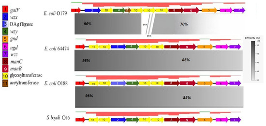
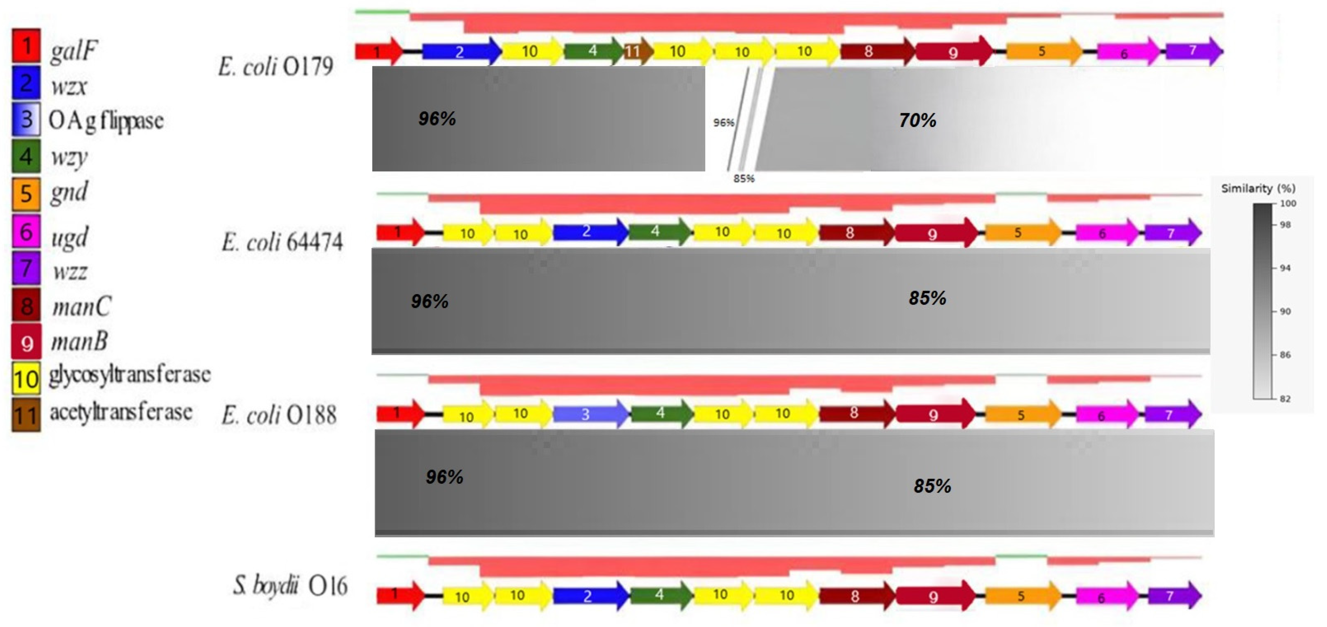
The results identified three distinct regions within the rfb cluster when comparing of E. coli O179 and E. coli 64474 (Figure 1). The first region, which contains the galF gene, showed similarity levels above 96%. The second region, located in the central part of the cluster, showed scattered short segments with similarity over 80%; however, these are not displayed in the figure due to their limited length. The third region extends from the glycosyltransferase gene upstream of maC to the wzz at the end of the cluster and shows a similarity greater than 70%. Three additional loci corresponding to S. boydii O16, E. coli 64474, and O188 shared similarity levels greater than 85%. In O188, the flippase gene matched the wzx gene, and it was arranged in the same order as in S. boydii O16 and E. coli 64474.
Figure 1.
Comparative analysis of the rfb gene clusters in the four strains and their GC content. Each gene alignment is represented by a colored arrow as indicated in the legend on the left side. The percentage of similarity scale between aligned regions is shown in the lower right corner, while GC content is depicted above each gene cluster (green: positive values; red: negative values). Alignments were performed using BLAST v. 2.14.1 from Easyfig v. 2.2.2.
3.4. The Comparative Genomics Analysis
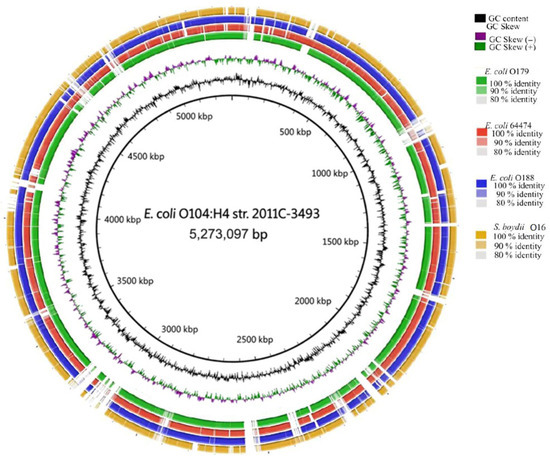
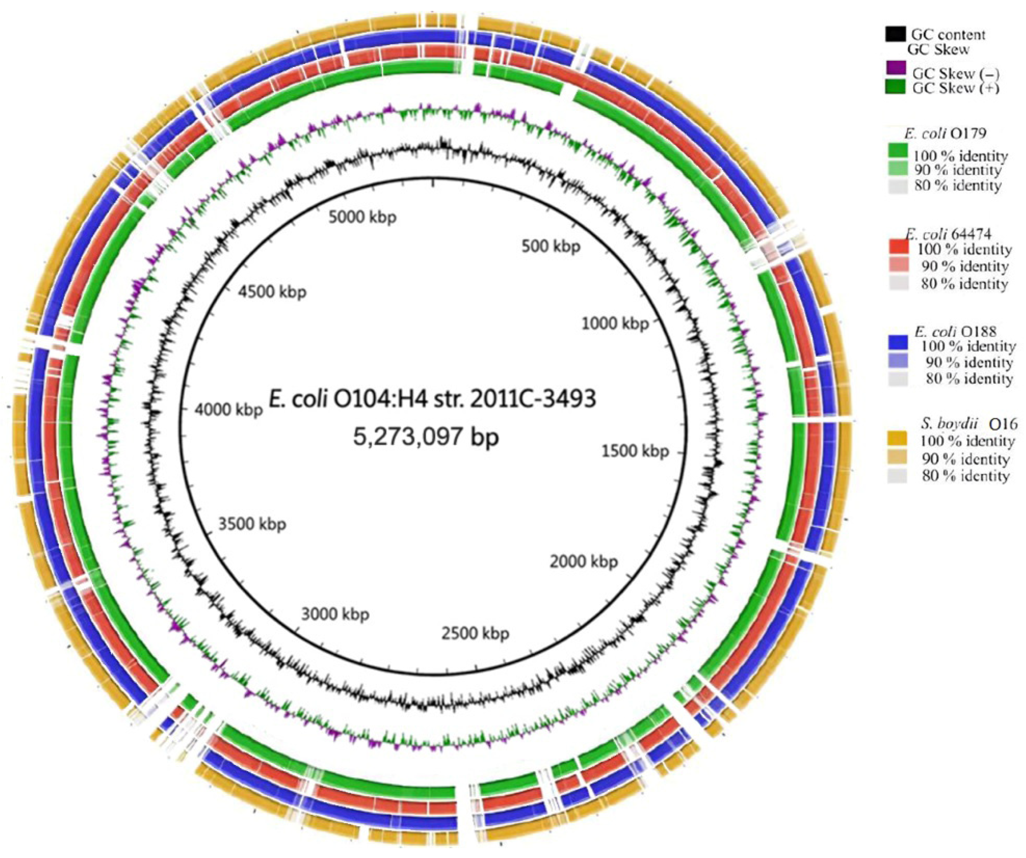
Comparison of the whole genome of E. coli O104:H4 strain 2011C-3493 with the study strains revealed regions with sequence similarity above 80%. Regions with 90% and 100% similarity are highlighted using distinct color labels. Additionally, GC content and GC skew are represented graphically (Figure 2).
Figure 2.
Comparative genomics of strains against a reference genome of E. coli O104:H4 strain 2011C-3493 (GCF_000299455.1). The analyses and their corresponding color codes are shown in the upper-right corner. The position along the reference genome is indicated in kilobase pairs (kbp) in the center pf the circular map. Alignments were performed using BLAST v. 2.14.1 via BRIG v. 0.95.
3.5. Pangenome Structure and Core Genome Phylogeny
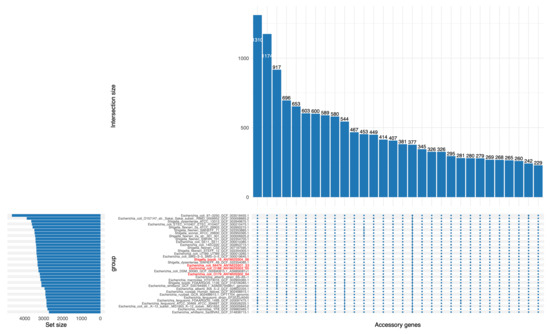
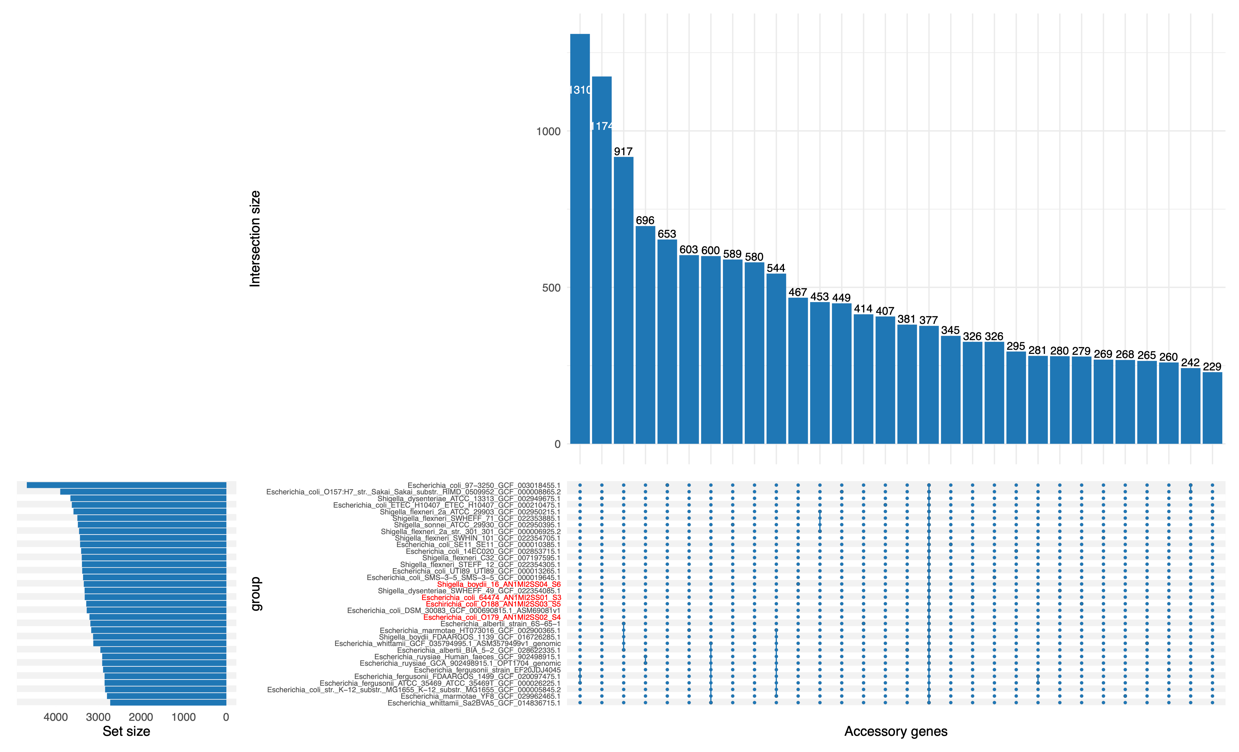
Analysis of the pangenome of E. coli 64474, O179, O188, and S. boydii O16 was performed using Roary v3.13.0 with a 95% BLASTp identity threshold. A total of 5199 gene clusters were identified across the four genomes. Of these, 3583 (68.9%) were classified as core genes, defined as those present in ≥99–100% of the genomes analyzed (i.e., all four strains), while the remaining 1616 (31.1%) constituted the accessory genome.
Figure 3 illustrates gene presence–absence patterns across the four genomes using an UpSet plot. Most accessory gene clusters are shared by only one or two genomes, indicating substantial genomic variability among strains. Notably, although 3583 gene clusters were classified as core genes, only a subset (n = 377) is represented as shared across all genomes in the UpSet plot. This discrepancy reflects the structure of the visualization, which does not display the full set of core genes but rather the subset captured in the plotted presence–absence matrix. Overall, the high proportion of core genes indicates the presence of a conserved genomic backbone among the four strains, supporting their close evolutionary relationship. In contrast, the accessory genome reflects lineage-specific variation, likely driven by horizontal gene transfer, prophage integration, and other mobile genetic elements contributing to genomic diversification.
Figure 3.
Roary-based pangenome structure and identification of genes present or absent in different genome sets. The UpSet plot shows the occurrence of gene clusters in E. coli 64474, O179, O188, and S. boydii O16 using Roary software version 3.13.0 with a BLASTp identity threshold of ≥95%. The top bar chart displays the number of gene clusters found in each combination of genome sets, while the left bar chart shows the total number of genes in each individual genome. The pangenome contains 5199 gene clusters, including 3583 core genes (gene clusters present in ≥99–100% of genome sets, i.e., all four genomes) and 1616 accessory genes. An UpSet plot highlighting a subset of gene clusters found in all four genomes (n = 377) is included as a representative portion of the core genome.
The high proportion of shared core genes indicates a strong conserved genomic backbone among the four strains, supporting their close evolutionary relationship. In contrast, the accessory genome comprised genes variably distributed among strains, likely reflecting horizontal gene transfer events, prophage insertions, and other mobile genetic elements that contribute to genomic diversification.
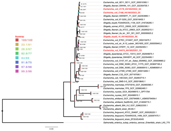
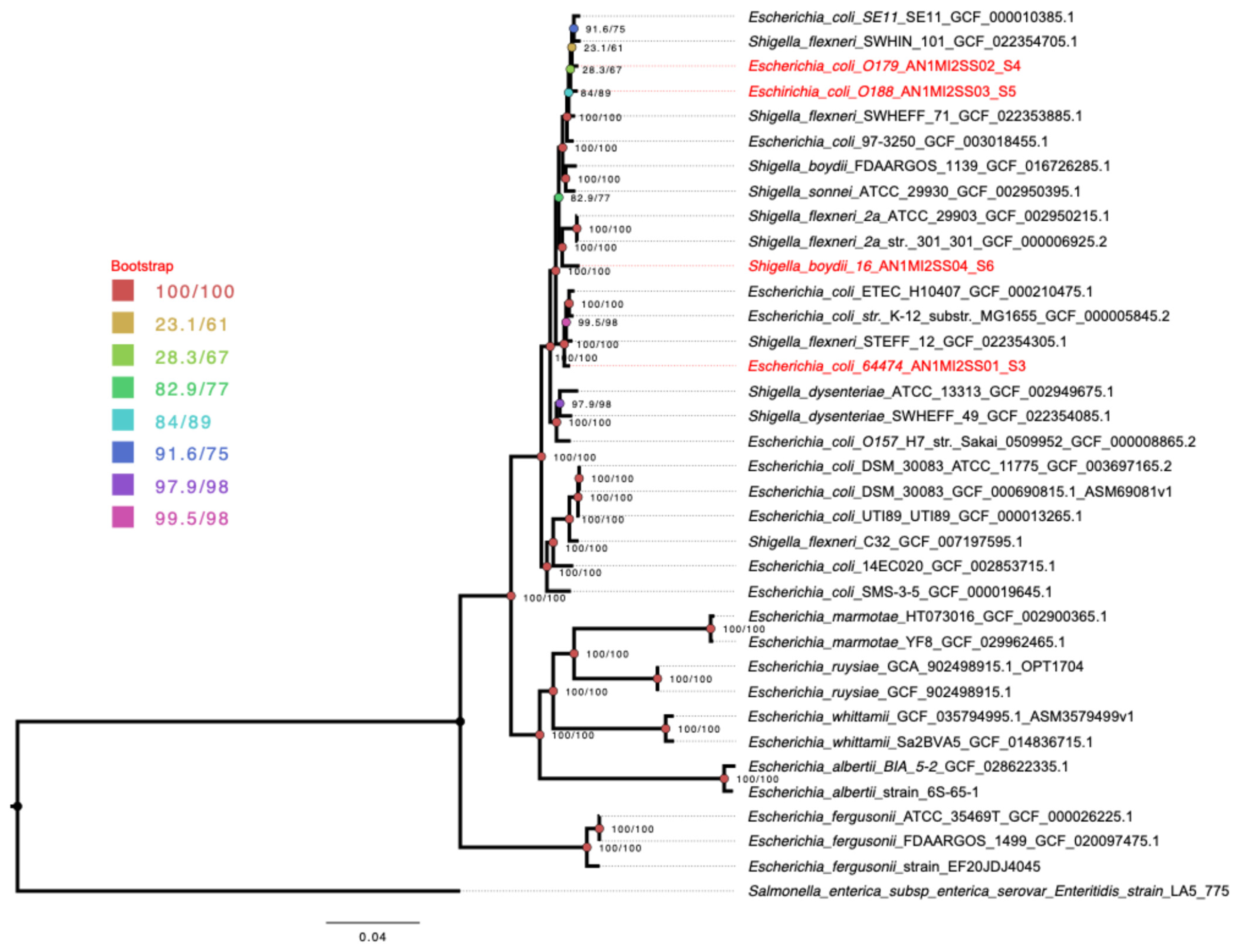
To contextualize these findings within a broader evolutionary framework, a core genome alignment generated by Roary was used to reconstruct a maximum-likelihood phylogeny using IQ-TREE2. The tree was rooted with Salmonella enterica subsp. enterica serovar Enteritidis strain LA5_775.
The phylogenetic tree reconstruction shows that E. coli and Shigella species are not differentiated into distinct, separate phylogenetic groups. Instead, they form a single, interspersed clade. This finding is consistent with the current genomic organization of these species and their relatedness. Within this clade, E. coli O179 and E. coli O188 are grouped together with E. coli SE11 and other related isolates. E. coli 64474 is also part of a single clade with other E. coli strains, indicating no differentiation from established E. coli lineages. In contrast, S. boydii O16 is grouped with S. boydii FDAARGOS_1139 and other related Shigella species, including S. flexneri and S. sonnei (Figure 4). In summary, all these isolates are part of a single clade with known species of the Escherichia/Shigella complex.
Figure 4.
Core genome maximum-likelihood phylogeny inferred from Roary alignment. Phylogenetic tree reconstructed from the core gene alignment generated by Roary v3.13.0 and inferred using IQ-TREE2 under the best-fit substitution model selected according to the Bayesian Information Criterion (BIC). Branch support was assessed using SH-like approximate likelihood ratio test (SH-aLRT) and ultrafast bootstrap approximation (UFBoot) with 1000 replicates, and support values (SH-aLRT/UFBoot) are indicated at the nodes. The tree was rooted with Salmonella enterica subsp. enterica serovar Enteritidis strain LA5_775. Our sequenced strains are highlighted in red, while reference strains are shown in black. The topology illustrates the interspersed distribution of E. coli and Shigella lineages within a single major clade.
3.6. Virulence Genes
To better contextualize the biological relevance of the analyzed strains, we screened a panel of virulence genes representative of different E. coli pathotypes. The resulting profiles showed that the strains harbor markers commonly associated with STEC, UPEC, EAEC, ETEC, and Shigella-related virulence schemes, suggesting a notable overlap of pathogenic traits among them. In particular, some strains carried combinations of genes that are typically linked to more than one pathotype, supporting the idea that these isolates may display a hybrid virulence profiles (Table 2). At the strain level, E. coli O179 carried stx2a/stx2b, iha, and sigA, consistent with virulence traits associated with STEC and UPEC. In contrast, E. coli O188 harbored sigA, pic, aggR, aatA, and aap, a profile mainly related to EAEC and also shared with virulence schemes reported in STEC. Likewise, E. coli 64474 carried eltA/eltB and aggR, indicating a combination of ETEC- and EAEC-associated markers. Overall, these findings support the presence of diverse and partially overlapping virulence repertoires among the analyzed strains.
Table 2.
Virulence genes identified in the analyzed strains and their association with pathogenic E. coli and Shigella spp.
4. Discussion
In a previous study conducted in our laboratory, we showed that E. coli 64474 and S. boydii O16 strains exhibited similar agglutination reactions with the anti-O sera of the these strains. They also share the wzx (flippase) and wzy (polymerase) genes, which are involved in biosynthesis of the O antigen of S. boydii O16 [10,25]. These results suggested that these strains could belong to the same clone. In the present study, we found that the anti-O sera of E. coli 64474 and anti-S. boydii O16 reacted with the O antigen of E. coli O188. To elucidate whether these strains belonged to the same clone due to their similarities at the O antigen level, we performed a comparative genomics and phylogenomic analyses among E. coli 64474, E. coli O179, E. coli O188 and S. boydii O16.
There are various types of analyses used to distinguish strains of enterobacteria; however, phylogenomic analyses can differentiate closely related strains because they use a larger amount of genetic information. The phylogenomic analysis revealed a monophyletic clade formed by E. coli strains, indicating a close clonal relationship. E. coli O188 and O179 were the most closely related, while E. coli 64474 was positioned third, contrary to previous serological studies, which showed a closer relationship between serotypes O188 and 64474 and between serotypes O179 and 64474. These discrepancies were supported by the comparative analysis of the rfb clusters, where distinct genes were only found in a section of the O179 O-antigen cluster.
4.1. rfb Cluster
Discrepancies in the high-similarity patters between the O-antigen and whole-genome sequences of the strains may result from the high rate of homologous and non-homologous recombination within O-antigen cluster [26]. These recombination events can lead to changes in the entire genes or gene sets within these genomic hotspots [27], which are associated with integration sites [28]. When comparing the rfb cluster of 64474 and O179 strains, more differences were observed than when comparing 64474 with O188 and S. boydii O16. Notably, mutations were found in housekeeping genes such as galF [29], over 96% similarity, gnd [30], and ugd [31], these last two genes with over 70% of similarity, which are common within these clusters due to frequent genetic rearrangement. The O179 rfb cluster consists of a different gene set compared to the other three rfb clusters, explaining the lower serological similarity. Despite these differences, certain loci within the O179 cluster retained high levels of similarity (over 70%), which may indicate a recombination event between O179 and other genomically similar strains [32].
Although S. boydii O16 did not exhibit a close clonal relationship with the E. coli strains, comparison of their O-antigen clusters revealed high similarity (over 85%) among the S. boydii O16, O188 and 64474 clusters. This genomic similarity supports the idea of S. boydii O16 is evolutionarily related strain to E. coli strains of the study. Furthermore, in addition to O antigen and whole-genome similarities, other studies, such as the structural analysis of O188 have revealed only minor differences from S. boydii O16, specifically in the N-acetylated residues of LPS [11]. Some reports also suggest that genome similarities with E. coli O188 may indicate that S. boydii O16 could act as a gene receptor or donor, potentially acquiring genes that increase virulence or drug resistance [12].
The high similarity among rfb clusters of 64474, O188, and S. boydii O16 is consistent with the serological findings. Nevertheless, O antigen molecular structures cannot be inferred solely from genome sequences; therefore, these results must be supported by further analyses. Due to the complexity of these polysaccharides, structural characterization using bioinformatics tools combined with NMR spectroscopy may help to determine whether 64474 carries a de novo O antigen [33].
4.2. Comparative Genomics
Although this analysis revealed similarity below 80%; these regions were located at conserved positions across the genomes, showing shared patterns that support the high similarities among the genomes of the study strains. These loci include accessory genes related to phages, mobile elements, or sequences acquired through horizontal gene transfer (HTG), such as genes involved in the mer operon (a mercury resistance system) [34], the sil operon (which promotes resistance to exogenous silver) [35], and the Type VI Secretion System (or T6SS) [20]. However, most of the genomic elements belong to the core genome (68.9% of genes), as computed from E. coli 64474, O179, O188 and S. boydii O16 strains, indicating a common evolutionary pattern. When comparing this analysis with broader studies, a report analyzing over 95,525 genome sequences from 14 phylogroups of E. coli and Shigella spp., revealed a core genome of only 1.96%, highlighting the close evolutionary relationship of the strains studied [36,37].
4.3. Virulence Genes
In addition, virulence genes should be considered when assessing the significance of this relationship. Particularly informative are the similarity levels detected among E. coli 64474, E. coli O179, E. coli O188, S. boydii O16, and E. coli O104:H4 strain 2011C-3493, this last strain a clinically relevant strain responsible for the 2011 outbreak in Europe, which resulted in 46 deaths, 782 cases of hemolytic uremic syndrome, and 3128 cases of acute gastroenteritis. This strain is genetically related to the EAEC and STEC pathotypes and harbors a broad repertoire of virulence genes. In the analysis, virulence genes recognized as critical for this pathogen’s virulence stx2a, stx2b, sigA, pic, and iha [18] were found in E. coli O179 and O188. All these genes, whether present or absent, are associated with horizontal gene transfer (HGT), which may explain the presence of unrelated loci between the strains of the study and E. coli O104:H4 strain 2011C-3493 because those are not core genes [38]. This information is relevant because the genome similarity observed between E. coli O179 and O188 includes shared high-virulence genes with E. coli O104:H4 strain 2011C-3493. Moreover, E. coli 64474 and S. boydii O16 strains, may acquire these genes via genetic recombination, due to their close genetic relationship.
5. Conclusions
E. coli 64474 may possess a de novo O-antigen and be a clone more closely related to E. coli O188 and O179. However, additional factors must be considered, such as the isolation date (1985), geographic origin (Mexico), and genome content similarities that could have led to divergence events over time, ultimately affecting phylogenomic interpretations and potentially indicating that this strain descends from an ancestor with the E. coli O188 antigen.
Similarity levels of rfb clusters should be interpreted alongside other factors, such as genomic plasticity, HGT, homologous recombination [39], and epigenetic effects [40], as well as the presence of virulence and drug resistance genes that may be transferred through HGT mechanisms. These characteristics, frequently observed in E. coli and Shigella spp., enhance their adaptability and, in some cases, their pathogenicity [41,42].
Supplementary Materials
The following supporting information can be downloaded at: https://www.mdpi.com/article/10.3390/pathogens15050462/s1, Table S1: Genomes analyzed in this study and their GenBank accession numbers.
Author Contributions
Conceptualization: A.N.-O., G.C.-E., E.O.D.-D., A.C. and H.G.C.-S.; Methodology, E.O.D.-D. and A.S.-P.; Formal analysis: E.O.D.-D., H.G.C.-S., A.N.-O. and A.C.; Investigation, E.O.D.-D. and A.S.-P.; Statistical analysis: H.G.C.-S.; Writing—original draft preparation, E.O.D.-D., A.N.-O. and H.G.C.-S.; Editing, A.N.-O. and H.G.C.-S. All authors have read and agreed to the published version of the manuscript.
Funding
The authors thank the Escuela Nacional de Ciencias Biológicas of Instituto Politécnico Nacional (Project 20232102-20240986) and the Dirección General de Asuntos del Personal Académico–Programa de Apoyo a Proyectos de Investigación e Innovación Tecnológica (DGAPA-PAPIIT, Project 1N22035) of Universidad Nacional Autónoma de México for their support. G.C.-E. received support from “Sistema Nacional de Investigadores (SNII)” from SECIHTI, Mexico also received support from “Estímulos al Desempeño de los Investigadores, Comisión de Operación y Fomento de Actividades Académicas (Instituto Politécnico Nacional)”.
Institutional Review Board Statement
Not applicable.
Informed Consent Statement
Not applicable.
Data Availability Statement
The data supporting the findings of this study are available from the corresponding authors upon reasonable request.
Acknowledgments
We would like to thank Gabriel Pérez, Liliana Cortes and Delia Licona (Faculty of Medicine, UNAM) for their technical assistance in the laboratory.
Conflicts of Interest
The authors declare no conflicts of interest.
References
- Liu, B.; Furevi, A.; Perepelov, A.V.; Guo, X.; Cao, H.; Wang, Q.; Reeves, P.R.; Knirel, Y.A.; Wang, L.; Widmalm, G. Structure and Genetics of Escherichia coli O Antigens. FEMS Microbiol. Rev. 2020, 44, 655–683. [Google Scholar] [CrossRef] [Scilit]
- Kaper, J.B.; Nataro, J.P.; Mobley, H.L.T. Pathogenic Escherichia coli. Nat. Rev. Microbiol. 2004, 2, 123–140. [Google Scholar] [CrossRef] [Scilit]
- Pokharel, P.; Dhakal, S.; Dozois, C.M. The Diversity of Escherichia coli Pathotypes and Vaccination Strategies against This Versatile Bacterial Pathogen. Microorganisms 2023, 11, 344. [Google Scholar] [CrossRef] [Scilit] [PubMed]
- Lan, R.; Reeves, P.R. Escherichia coli in Disguise: Molecular Origins of Shigella. Microbes Infect. 2002, 4, 1125–1132. [Google Scholar] [CrossRef] [Scilit] [PubMed]
- Stenhouse, G.E.; Keddy, K.H.; Bengtsson, R.J.; Hall, N.; Smith, A.M.; Thomas, J.; Iturriza-Gómara, M.; Baker, K.S. The Genomic Epidemiology of Shigellosis in South Africa. Nat. Commun. 2023, 14, 7715. [Google Scholar] [CrossRef] [Scilit] [PubMed]
- Pupo, G.M.; Lan, R.; Reeves, P.R. Multiple Independent Origins of Shigella Clones of Escherichia coli and Convergent Evolution of Many of Their Characteristics. Proc. Natl. Acad. Sci. USA 2000, 97, 10567–10572. [Google Scholar] [CrossRef] [Scilit]
- Li, Y.; Cao, B.; Liu, B.; Liu, D.; Gao, Q.; Peng, X.; Wu, J.; Bastin, D.A.; Feng, L.; Wang, L. Molecular Detection of All 34 Distinct O-Antigen Forms of Shigella. J. Med. Microbiol. 2009, 58, 69–81. [Google Scholar] [CrossRef] [Scilit]
- Coimbra, R.S.; Grimont, F.; Lenormand, P.; Burguière, P.; Beutin, L.; Grimont, P.A.D. Identification of Escherichia coli O-Serogroups by Restrictionof the Amplified O-Antigen Gene Cluster (Rfb-RFLP). Res. Microbiol. 2000, 151, 639–654. [Google Scholar] [CrossRef] [Scilit]
- Van Den Beld, M.J.C.; Reubsaet, F.A.G.; Pijnacker, R.; Harpal, A.; Kuiling, S.; Heerkens, E.M.; Hoeve-Bakker, B.J.A.D.; Noomen, R.C.E.A.; Hendriks, A.C.A.; Borst, D.; et al. A Multifactorial Approach for Surveillance of Shigella spp. and Entero-Invasive Escherichia coli Is Important for Detecting (Inter)National Clusters. Front. Microbiol. 2020, 11, 564103. [Google Scholar] [CrossRef] [Scilit]
- Navarro, A.; Eslava, C.; Perea, L.M.; Inzunza, A.; Delgado, G.; Morales-Espinosa, R.; Cheasty, T.; Cravioto, A. New Enterovirulent Escherichia coli Serogroup 64,474 Showingantigenic and Genotypic Relationships to Shigella boydii 16. J. Med. Microbiol. 2010, 59, 453–461. [Google Scholar] [CrossRef] [Scilit]
- Furevi, A.; Ståhle, J.; Muheim, C.; Gkotzis, S.; Udekwu, K.I.; Daley, D.O.; Widmalm, G. Structural Analysis of the O-Antigen Polysaccharide from Escherichia coli O188. Carbohydr. Res. 2020, 498, 108051. [Google Scholar] [CrossRef] [Scilit] [PubMed]
- Simoni, S.; Mingoia, M.; Brenciani, A.; Carelli, M.; Lleò, M.M.; Malerba, G.; Vignaroli, C. First IncHI2 Plasmid Carrying Mcr-9.1, blaVIM-1, and Double Copies of blaKPC-3 in a Multidrug-Resistant Escherichia coli Human Isolate. mSphere 2021, 6, e0030221. [Google Scholar] [CrossRef] [Scilit] [PubMed]
- Beutin, L.; Strauch, E. Identification of Sequence Diversity in the Escherichia coli fliC Genes Encoding Flagellar Types H8 and H40 and Its Use in Typing of Shiga Toxin-Producing E. coli O8, O22, O111, O174, and O179 Strains. J. Clin. Microbiol. 2007, 45, 333–339. [Google Scholar] [CrossRef] [Scilit]
- Ørskov, F.; Ørskov, I. 2 Serotyping of Escherichia coli * * The Terminology Used to Describe the Different Classes of Bacterial Antigens Is Explained in the Preface. However, the Authors Would like to Mention That a Different Convention is Used in Some Laboratories, for Example O:L, K:L, H:7 is Equivalent to O1:K1:H7. In Methods in Microbiology; Bergan, T., Ed.; Academic Press: Cambridge, MA, USA, 1984; Volume 14, pp. 43–112. [Google Scholar]
- Ewing, W.H. Edwards and Ewing’s Identification of Enterobacteriaceae; Elsevier: Amsterdam, The Netherlands, 1986. [Google Scholar]
- Bankevich, A.; Nurk, S.; Antipov, D.; Gurevich, A.A.; Dvorkin, M.; Kulikov, A.S.; Lesin, V.M.; Nikolenko, S.I.; Pham, S.; Prjibelski, A.D.; et al. SPAdes: A New Genome Assembly Algorithm and Its Applications to Single-Cell Sequencing. J. Comput. Biol. 2012, 19, 455–477. [Google Scholar] [CrossRef] [Scilit]
- Seemann, T. Prokka: Rapid Prokaryotic Genome Annotation. Bioinformatics 2014, 30, 2068–2069. [Google Scholar] [CrossRef] [Scilit] [PubMed]
- Navarro-Garcia, F.; Ruiz-Perez, F.; Cataldi, Á.; Larzábal, M. Type VI Secretion System in Pathogenic Escherichia coli: Structure, Role in Virulence, and Acquisition. Front. Microbiol. 2019, 10, 1965. [Google Scholar] [CrossRef] [Scilit]
- Melton-Celsa, A.R. Shiga Toxin (Stx) Classification, Structure, and Function. Microbiol. Spectr. 2014, 2, 10–1128. [Google Scholar] [CrossRef] [Scilit]
- Nesporova, K.; Steemans, B.; Govers, S.K. Large-Scale Genomic Analysis Reveals the Distribution and Diversity of Type VI Secretion Systems in Escherichia coli. mSystems 2025, 10, e00105-25. [Google Scholar] [CrossRef] [Scilit]
- Wang, X.; Yu, D.; Chui, L.; Zhou, T.; Feng, Y.; Cao, Y.; Zhi, S. A Comprehensive Review on Shiga Toxin Subtypes and Their Niche-Related Distribution Characteristics in Shiga-Toxin-Producing E. coli and Other Bacterial Hosts. Microorganisms 2024, 12, 687. [Google Scholar] [CrossRef] [Scilit]
- Johnson, J.R.; Jelacic, S.; Schoening, L.M.; Clabots, C.; Shaikh, N.; Mobley, H.L.T.; Tarr, P.I. The IrgA Homologue Adhesin Iha is an Escherichia coli Virulence Factor in Murine Urinary Tract Infection. Infect. Immun. 2005, 73, 965–971. [Google Scholar] [CrossRef] [Scilit]
- Al-Hasani, K.; Henderson, I.R.; Sakellaris, H.; Rajakumar, K.; Grant, T.; Nataro, J.P.; Robins-Browne, R.; Adler, B. The sigA Gene Which Is Borne on the shePathogenicity Island of Shigella flexneri 2a Encodes an Exported Cytopathic Protease Involved in Intestinal Fluid Accumulation. Infect. Immun. 2000, 68, 2457–2463. [Google Scholar] [CrossRef] [Scilit]
- Flores-Sanchez, F.; Chavez-Dueñas, L.; Sanchez-Villamil, J.; Navarro-Garcia, F. Pic Protein From Enteroaggregative E. coli Induces Different Mechanisms for Its Dual Activity as a Mucus Secretagogue and a Mucinase. Front. Immunol. 2020, 11, 564953. [Google Scholar] [CrossRef] [Scilit]
- Liu, B.; Senchenkova, S.N.; Feng, L.; Perepelov, A.V.; Xu, T.; Shevelev, S.D.; Zhu, Y.; Shashkov, A.S.; Zou, M.; Knirel, Y.A.; et al. Structural and Molecular Characterization of Shigella boydii Type 16 O Antigen. Gene 2006, 380, 46–53. [Google Scholar] [CrossRef] [Scilit] [PubMed]
- Didelot, X.; Méric, G.; Falush, D.; Darling, A.E. Impact of Homologous and Non-Homologous Recombination in the Genomic Evolution of Escherichia coli. BMC Genom. 2012, 13, 256. [Google Scholar] [CrossRef] [Scilit] [PubMed]
- Iguchi, A.; Iyoda, S.; Kikuchi, T.; Ogura, Y.; Katsura, K.; Ohnishi, M.; Hayashi, T.; Thomson, N.R. A Complete View of the Genetic Diversity of the Escherichia coli O-Antigen Biosynthesis Gene Cluster. DNA Res. 2015, 22, 101–107. [Google Scholar] [CrossRef] [Scilit]
- Denamur, E.; Clermont, O.; Bonacorsi, S.; Gordon, D. The Population Genetics of Pathogenic Escherichia coli. Nat. Rev. Microbiol. 2021, 19, 37–54. [Google Scholar] [CrossRef] [Scilit] [PubMed]
- Ebrecht, A.C.; Orlof, A.M.; Sasoni, N.; Figueroa, C.M.; Iglesias, A.A.; Ballicora, M.A. On the Ancestral UDP-Glucose Pyrophosphorylase Activity of GalF from Escherichia coli. Front. Microbiol. 2015, 6, 1253. [Google Scholar] [CrossRef] [Scilit]
- Nelson, K.; Selander, R.K. Intergeneric Transfer and Recombination of the 6-Phosphogluconate Dehydrogenase Gene (Gnd) in Enteric Bacteria. Proc. Natl. Acad. Sci. USA 1994, 91, 10227–10231. [Google Scholar] [CrossRef] [Scilit] [PubMed]
- Drummelsmith, J.; Amor, P.A.; Whitfield, C. Polymorphism, Duplication, and IS1-Mediated Rearrangement in the Chromosomal His-Rfb-Gnd Region of Escherichia coli Strains with Group IA and Capsular K Antigens. J. Bacteriol. 1997, 179, 3232–3238. [Google Scholar] [CrossRef] [Scilit]
- Iguchi, A.; Iyoda, S.; Ohnishi, M. Molecular Characterization Reveals Three Distinct Clonal Groups among Clinical Shiga Toxin-Producing Escherichia coli Strains of Serogroup O103. J. Clin. Microbiol. 2012, 50, 2894–2900. [Google Scholar] [CrossRef] [Scilit]
- Ståhle, J.; Widmalm, G. Lipopolysaccharides of Gram-Negative Bacteria: Biosynthesis and Structural Aspects. Trends Glycosci. Glycotechnol. 2019, 31, E159–E171. [Google Scholar] [CrossRef] [Scilit]
- Boyd, E.S.; Barkay, T. The Mercury Resistance Operon: From an Origin in a Geothermal Environment to an Efficient Detoxification Machine. Front. Microbiol. 2012, 3, 349. [Google Scholar] [CrossRef] [Scilit]
- Wang, Q.; Perepelov, A.V.; Wen, L.; Shashkov, A.S.; Wang, X.; Guo, X.; Knirel, Y.A.; Wang, L. Identification of the Two Glycosyltransferase Genes Responsible for the Difference between Escherichia coli O107 and O117 O-Antigens. Glycobiology 2012, 22, 281–287. [Google Scholar] [CrossRef] [Scilit]
- Abram, K.; Udaondo, Z.; Bleker, C.; Wanchai, V.; Wassenaar, T.M.; Robeson, M.S.; Ussery, D.W. Mash-Based Analyses of Escherichia coli Genomes Reveal 14 Distinct Phylogroups. Commun. Biol. 2021, 4, 117. [Google Scholar] [CrossRef] [Scilit]
- Chauhan, S.M.; Ardalani, O.; Hyun, J.C.; Monk, J.M.; Phaneuf, P.V.; Palsson, B.O. Decomposition of the Pangenome Matrix Reveals a Structure in Gene Distribution in the Escherichia coli Species. mSphere 2024, 10, e00532-24. [Google Scholar] [CrossRef] [Scilit] [PubMed]
- Costa, S.S.; Guimarães, L.C.; Silva, A.; Soares, S.C.; Baraúna, R.A. First Steps in the Analysis of Prokaryotic Pan-Genomes. Bioinform. Biol. Insights 2020, 14, 1177932220938064. [Google Scholar] [CrossRef] [Scilit] [PubMed]
- Shikov, A.E.; Savina, I.A.; Nizhnikov, A.A.; Antonets, K.S. Recombination in Bacterial Genomes: Evolutionary Trends. Toxins 2023, 15, 568. [Google Scholar] [CrossRef] [Scilit] [PubMed]
- D’Aquila, P.; De Rango, F.; Paparazzo, E.; Passarino, G.; Bellizzi, D. Epigenetic-Based Regulation of Transcriptome in Escherichia coli Adaptive Antibiotic Resistance. Microbiol. Spectr. 2023, 11, e04583-22. [Google Scholar] [CrossRef] [Scilit]
- Touchon, M.; Hoede, C.; Tenaillon, O.; Barbe, V.; Baeriswyl, S.; Bidet, P.; Bingen, E.; Bonacorsi, S.; Bouchier, C.; Bouvet, O.; et al. Organised Genome Dynamics in the Escherichia coli Species Results in Highly Diverse Adaptive Paths. PLoS Genet. 2009, 5, e1000344. [Google Scholar] [CrossRef] [Scilit]
- Touchon, M.; Perrin, A.; De Sousa, J.A.M.; Vangchhia, B.; Burn, S.; O’Brien, C.L.; Denamur, E.; Gordon, D.; Rocha, E.P. Phylogenetic Background and Habitat Drive the Genetic Diversification of Escherichia coli. PLoS Genet. 2020, 16, e1008866. [Google Scholar] [CrossRef] [Scilit]

Disclaimer/Publisher’s Note: The statements, opinions and data contained in all publications are solely those of the individual author(s) and contributor(s) and not of MDPI and/or the editor(s). MDPI and/or the editor(s) disclaim responsibility for any injury to people or property resulting from any ideas, methods, instructions or products referred to in the content. |
© 2026 by the authors. Licensee MDPI, Basel, Switzerland. This article is an open access article distributed under the terms and conditions of the Creative Commons Attribution (CC BY) license.