


Abstract
Brucella infections are associated with gestational complications in both humans and animals, including spontaneous abortion, premature delivery, and infertility. Successful pregnancy requires the proper invasion and functional integrity of extravillous trophoblasts (EVTs), specialized fetal cells that remodel the maternal decidua. However, the direct impact of the major human-pathogenic species on human EVT biology remains incompletely characterized. Here, we investigated in vitro the effects of B. abortus, B. melitensis, and B. suis infections on the fitness of a human EVT cell line (Swan71). Infection with all three species induced a proinflammatory profile in EVT, with increased CXCL8, CCL2 and IL-6 production. Infection markedly impaired EVT migration (wound closure) and reduced EVT invasive capacity in a matrix invasion assay. EVT tubulogenesis was affected by the infection, with all the species reducing master segments and B. suis and B. melitensis additionally reducing master junctions and meshes. Blastocyst-like spheroids derived from infected EVTs showed reduced outgrowth on decidualized endometrial cell monolayers, with B. suis exhibiting the most consistent effect. Antiproliferative effects were observed at 24 h for B. abortus and B. suis and at 48 h for B. melitensis, preceding marked cell death (late apoptotic features became pronounced only at 72 h post-infection). Collectively, our findings demonstrate that major human-pathogenic Brucella species induce an early dysfunctional state in human EVTs, affecting cellular processes involved in implantation and placental vascular remodeling.
1. Introduction
Brucellosis is a globally distributed zoonotic disease caused by Gram-negative bacteria of the genus Brucella. In their preferred animal hosts, these pathogens are responsible for significant reproductive losses, including abortion, premature delivery, and infertility [1,2]. Increasing clinical evidence also supports an association between human brucellosis and adverse pregnancy outcomes, with spontaneous miscarriage reported among the most frequent complications [2,3,4]. Notably, the three species most pathogenic for humans, B. melitensis, B. abortus, and B. suis, have been isolated from human placentas and aborted fetuses, underscoring their tropism for the maternal-fetal interface [5,6,7].
Successful pregnancy depends on the differentiation and proper function of extravillous trophoblasts (EVTs), specialized fetal cells that invade the maternal decidua and remodel uterine spiral arteries during early gestation [8]. This process is essential for establishing adequate uteroplacental blood flow and ensuring the delivery of oxygen and nutrients to the developing fetus [8,9,10]. Defective EVT invasion results in shallow placentation and is strongly associated with pregnancy disorders such as intrauterine growth restriction, preeclampsia, and spontaneous miscarriage [11].
Several intracellular pathogens, including members of the TORCH group, directly target EVTs and impair their biological functions [12]. Human Cytomegalovirus and Toxoplasma gondii, for example, reduce trophoblast migration and invasion through the disruption of signaling pathways involved in cell motility, adhesion, and extracellular matrix remodeling [13,14,15,16]. These observations highlight the susceptibility of EVTs to microbial infection and suggest that pathogen-mediated trophoblast dysfunction may represent a common mechanism contributing to adverse pregnancy outcomes.
In the context of brucellosis, Brucella spp. can invade and replicate within human trophoblasts, potentially contributing to pregnancy complications. In the case of B. abortus, intracellular survival in human trophoblasts is dependent on a functional VirB operon-encoded Type IV Secretion System (T4SS), which plays a critical role in intracellular trafficking and establishment of the replicative niche. Importantly, the intracellular compartment supporting Brucella replication varies according to the bacterial species and trophoblast subtype [17,18,19]. Infection of trophoblast cells triggers the production of proinflammatory mediators, including IL-6, CXCL8 (IL-8), and CCL2 (MCP-1), promoting an inflammatory environment that may compromise placental homeostasis [17]. Consistent with this notion, placentitis is a characteristic lesion observed in Brucella-infected pregnant animals and is considered a major contributor to reproductive failure [2,20]. Moreover, soluble factors released by Brucella-infected endometrial stromal cells have been shown to impair trophoblast migration and blastocyst-like spheroid adhesion [21].
Despite these observations, the consequences of direct infection by the three major pathogenic Brucella species on the viability and key biological functions of human EVTs remain poorly understood. In this study, we investigated the impact of B. abortus, B. melitensis, and B. suis in vitro infection on EVT survival, proliferation, migration, invasion, inflammatory responses, and blastocyst-endometrium interactions.
2. Materials and Methods
2.1. Bacterial Strains and Growth Conditions
This study included a representative virulent strain of each of the three Brucella species most frequently involved in human brucellosis: B. abortus, B. suis and B. melitensis. The preferred animal hosts for these species are bovines, pigs and goats, respectively. The wild-type strains Brucella abortus 2308, B. suis 1330, and B. melitensis 16M, obtained from our laboratory collection, have been widely used in studies about Brucella pathogenicity. Bacteria were routinely cultured in tryptic soy broth (TSB) at 37 °C under shaking conditions. Before infection assays, bacterial suspensions were washed twice with sterile phosphate-buffered saline (PBS) and adjusted to the required density according to optical density measurements. The viable bacterial count of each inoculum was verified by plating serial dilutions onto tryptic soy agar (TSA) plates and determining colony-forming units (CFU) after incubation at 37 °C. All procedures involving live Brucella organisms were conducted in a biosafety level 3 (BSL-3) laboratory.
2.2. Cell Culture
The Swan-71 trophoblast cell line was kindly provided by Dr. Gil Mor (Wayne State University). Swan-71 cells preserve key phenotypic and functional properties of primary trophoblasts [22] and were cultured in Dulbecco’s Modified Eagles Medium F-12 (DMEM/F-12, Gibco, Thermo Fisher Scientific, Waltham, MA, USA) supplemented with 10% fetal bovine serum (FBS, Internegocios, Mercedes, Argentina), 100 U/mL penicillin, 50 μg/mL streptomycin, 2 mM L-glutamine, and 50 mM sodium pyruvate.
The human endometrial stromal cell line T-HESC, kindly provided by Dr. Andrea Randi (University of Buenos Aires), was cultured in phenol red-free DMEM/F-12 supplemented with 10% FBS, 100 U/mL penicillin, 50 μg/mL streptomycin, 2 mM L-glutamine, 50 mM sodium pyruvate, and 500 ng/mL puromycin.
2.3. Cell Infection
Swan-71 cells were seeded in 24-well culture plates at a density of 5 × 10⁴ cells per well and maintained for 24 h in antibiotic-free medium at 37 °C in a humidified incubator containing 5% CO2. Cells were subsequently challenged with B. abortus, B. melitensis, or B. suis at multiplicities of infection (MOIs) of 50, 250, or 500 bacteria per cell. The MOI used varied according to the assay and is specified in the corresponding section.
To promote bacterial-cell contact, plates were centrifuged at 400× g for 10 min and incubated for an additional 2 h at 37 °C under 5% CO2. Following the infection period, extracellular bacteria were removed by repeated washing with culture medium. Cells were then incubated in medium containing gentamicin (100 μg/mL) and streptomycin (50 μg/mL) to eliminate any remaining extracellular bacteria. After antibiotic treatment, cultures were washed and maintained in antibiotic-free medium for the remainder of the experiment.
2.4. Decidualization of Endometrial Stromal Cells
T-HESC cells were seeded at a density of 5 × 10⁴ cells per well and induced to undergo decidualization by culture in DMEM/F12 supplemented with 2% FBS, 1 μM medroxyprogesterone acetate (MPA, Sigma-Aldrich, St. Louis, MO, USA), and 0.5 mM dibutyryl cyclic AMP (db-cAMP, Santa Cruz Biotechnology, Dallas, TX, USA). To sustain the decidualization stimulus, the culture medium was refreshed every 48 h. Cells were maintained at 37 °C in a humidified incubator with 5% CO2.
After 8 days of hormonal treatment, successful decidualization was confirmed by quantifying prolactin secretion in culture supernatants using a commercial sandwich ELISA kit (R&D Systems, Minneapolis, MN, USA), according to the manufacturer’s protocol.
2.5. Cytokine Quantification
To assess the effect of Brucella infection on the trophoblast inflammatory response, culture supernatants from infected Swan-71 cells were collected at 24, 48, and 72 h post-infection. The concentrations of IL-6, CXCL8 (IL-8), and CCL2 (MCP-1) were determined using commercially available sandwich ELISA kits (R&D Systems, Minneapolis, MN, USA) according to the manufacturer’s instructions.
2.6. Cell Migration Assay
The migratory capacity of Swan-71 cells was evaluated using a wound-healing assay [23]. Cells were seeded in 24-well plates at a density of 5 × 10⁴ cells per well and cultured until a confluent monolayer was formed. Then the monolayers were infected as described above. At 24 h post-infection, a linear scratch was made across the infected monolayer using a sterile 200 µL pipette tip. Detached cells were removed by gently washing the wells with DMEM/F-12 and cell wounds were photographed immediately (0 h) in pre-determined and marked locations. Cells were subsequently incubated in DMEM/F-12 containing 2% FBS for 18 h after which cell wounds were photographed in the exact previous locations (18 h). Swan-71 cells cultured in DMEM/F-12 supplemented with 10% FBS were included as a positive control of migration.
Wound areas were quantified in duplicate wells using ImageJ software (version 1.54p). Cell migration was expressed as the percentage of wound closure calculated according to the following equation:
All experiments were performed independently three times.
2.7. Cell Invasion Assay
The invasive capacity of Swan-71 cells was evaluated using a Matrigel-like Transwell invasion assay, as described by Zavattieri et al. [21]. Briefly, Transwell inserts with 8-μm pore-size membranes (Corning Costar, Corning, NY, USA) were coated with Geltrex matrix (Gibco) at a final protein concentration of 0.6 mg/mL in DMEM/F-12. Swan-71 cells harvested 24 h post-infection were seeded onto the coated inserts at a density of 3 × 10⁴ cells per insert. Non-infected cells were processed in parallel and used as controls. After 18 h of incubation at 37 °C in a humidified atmosphere containing 5% CO2, culture medium was removed and non-invading cells together with the remaining matrix on the upper surface of the membrane were carefully removed using a cotton swab. Inserts were washed with PBS and fixed with 4% paraformaldehyde for 15 min. Following an additional wash, cells that had migrated through the matrix and membrane were stained with 0.5% crystal violet for 15 min at room temperature. Excess stain was removed with distilled water. The membranes were examined using an EVOS microscope (Thermo Fisher Scientific), and the number of invading cells was quantified in 10 randomly selected fields per membrane at 20× magnification using ImageJ software. Each experiment was performed at least three times independently.
2.8. Tubule Formation Assay
Briefly, Geltrex matrix (9 mg protein/mL in DMEM/F-12) was added to 96-well plates and allowed to polymerize. Swan-71 cells harvested 24 h after infection, as well as non-infected control cells, were then seeded onto the matrix at a density of 2 × 10⁴ cells per well. After 6 h of incubation at 37 °C in a humidified atmosphere containing 5% CO2, images were acquired using an EVOS microscope (Thermo Fisher Scientific). The number of master junctions, master segments, and meshes was quantified using ImageJ software and the Angiogenesis Analyzer plugin.
2.9. Spheroid Adhesion to Decidual Cells
Blastocyst-like spheroids were generated as previously described [21]. Briefly, Swan-71 cells harvested at 24 h post-infection, as well as non-infected control cells, were labeled with Vybrant™ DiD (Thermo Fisher Scientific) according to the manufacturer’s instructions and seeded in low-binding Petri dishes at a density of 2 × 10⁴ cells per 20 μL drop of DMEM/F-12 supplemented with 2% FBS. Cells were incubated under agitation at 37 °C for 48 h to promote spheroid formation. The formation of compact spheroids was verified by light microscopy.
T-HESC cells were seeded in 96-well plates at a density of 1 × 10⁴ cells per well and decidualized as described above. Individual spheroids were carefully transferred onto the decidualized cell monolayers using a sterile pipette tip with a widened opening to avoid mechanical damage.
Images were acquired immediately after spheroid transfer (T0) and after 24 h of co-culture (T24) using an EVOS microscope (Thermo Fisher Scientific). Spheroid outgrowth was quantified using ImageJ software by measuring spheroid area at both time points. Results were expressed as the ratio between the area at T24 and the initial area at T0 (T24/T0).
2.10. Cell Proliferation Assay by Bromodeoxyuridine (BrdU) Incorporation
Cell proliferation was evaluated by measuring BrdU incorporation into newly synthesized DNA. Swan-71 cells were seeded in 96-well plates at a density of 3 × 10³ cells per well in DMEM/F-12 supplemented with 10% FBS and incubated for 24 h at 37 °C in a humidified atmosphere containing 5% CO2. Cells were then infected with different Brucella species at the indicated MOIs, and proliferation was assessed at 24, 48, and 72 h post-infection. Non-infected cells were processed in the same way in parallel.
Four hours before each time point, BrdU was added to the cultures at a final concentration of 10 μM. Cells were subsequently washed with PBS, fixed with 4% paraformaldehyde for 20 min at room temperature, and processed for BrdU detection by ELISA.
For BrdU detection, cellular DNA was denatured with 2 N HCl for 20 min and neutralized with 0.1 M sodium borate buffer (pH 9.0) for 15 min at 37 °C. Endogenous peroxidase activity was blocked with 3% H2O2 in methanol for 30 min. After blocking with 2% FBS and 0.1% Triton X-100 in PBS, cells were incubated overnight at 4 °C with a mouse anti-BrdU primary antibody, followed by incubation with an HRP-conjugated anti-mouse IgG secondary antibody for 2 h at room temperature. Bound antibodies were detected using tetramethylbenzidine (TMB)/H2O2 substrate, and the reaction was stopped with 4 N H2SO4. Absorbance was measured at 450 nm using a microplate reader (Multiskan, Thermo Fisher Scientific).
2.11. Cell Death
To determine whether Brucella infection induces trophoblast cell death, Swan-71 cells were seeded in 48-well plates at a density of 2.5 × 10⁴ cells per well and infected with B. abortus, B. suis, or B. melitensis as described above. At 24, 48, and 72 h post-infection, cells were stained with acridine orange/ethidium bromide (AO/EB; 1 μg/mL each) and immediately examined using an EVOS fluorescence microscope (Thermo Fisher Scientific). Fluorescence images were acquired separately using the GFP channel (LED excitation: 482/25 nm; emission filter: 524/24 nm) and the RFP channel (LED excitation: 542/20 nm; emission filter: 593/40 nm).
Live cells were identified by green nuclear fluorescence, whereas cells with compromised membrane integrity exhibited orange/red fluorescence. The number of dead cells was determined by counting stained cells in five randomly selected fields per well at 20× magnification.
2.12. Statistical Analysis
The number of samples and experimental replicates was established according to standard practice for in vitro studies of this type. No formal statistical power analysis was performed to determine the sample size. The number of independent experiments and technical replicates for each assay is indicated in the corresponding figure legends or in Section 3. Results are expressed as mean ± standard deviation (SD). Statistical comparisons were performed using one-way analysis of variance (ANOVA), followed by Tukey’s or Dunnett’s multiple-comparison test, as appropriate, using GraphPad Prism version 8.0.
3. Results
3.1. Infection with Virulent Brucella Species Induces a Proinflammatory Response
Previous studies from our group demonstrated that B. abortus induces a proinflammatory response in human trophoblastic cells [17]. Building on these findings, we expanded our investigation to include B. melitensis and B. suis, two additional Brucella species of major clinical relevance that, together with B. abortus, have been associated with reproductive disorders and pregnancy complications in humans and animals. In addition, extending the analysis up to 72 h post-infection (p.i.) allowed us to assess the kinetics of trophoblast responses and identify potential species-specific temporal patterns. Therefore, we aimed to determine whether B. melitensis and B. suis induce proinflammatory cytokine production in trophoblast cells and to compare their responses with those elicited by B. abortus. To this end, we evaluated the production of CXCL8, IL-6, and CCL2 following infection with B. abortus, B. melitensis, or B. suis.
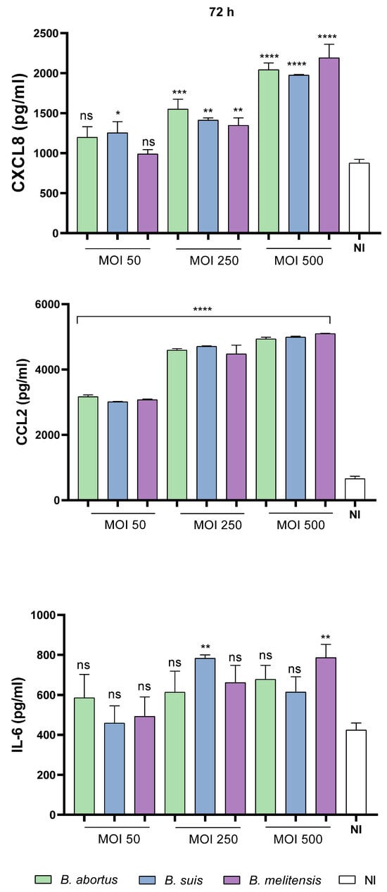
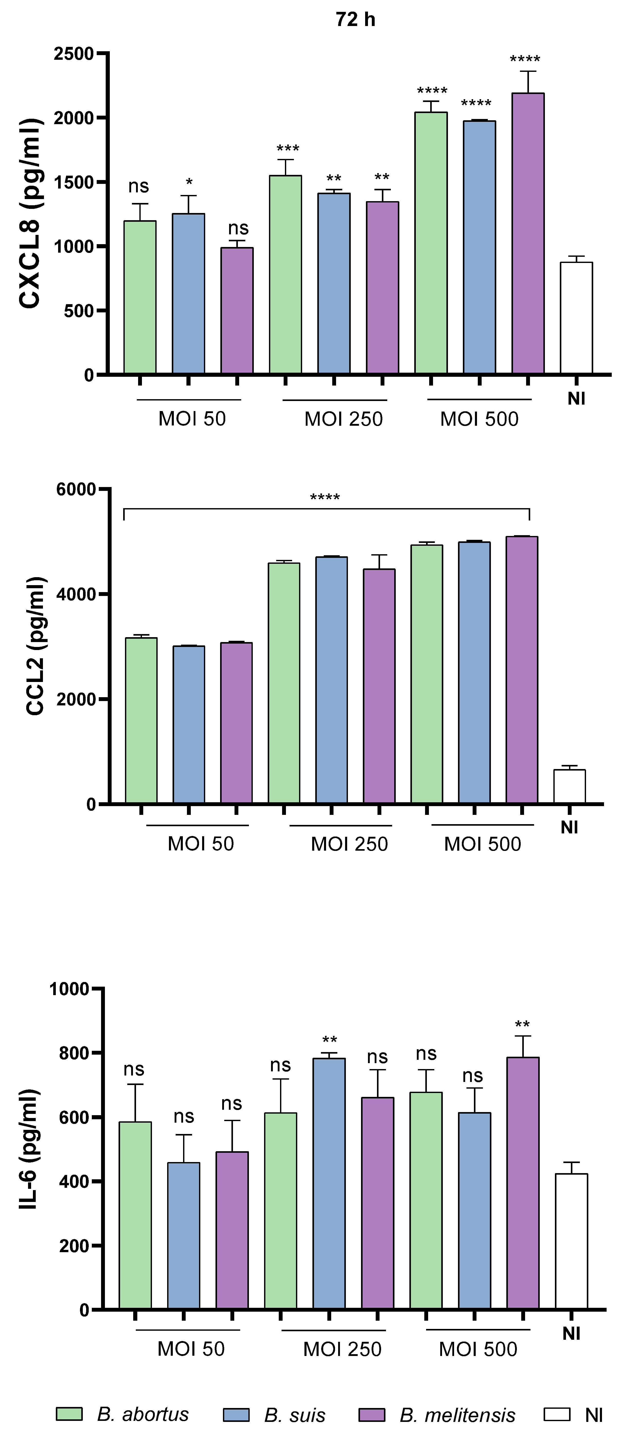
As shown in Figure 1 and Figure S1, CXCL8 and CCL2 production generally increased in an MOI-dependent manner following infection with B. abortus, B. suis, and B. melitensis. Increased cytokine levels were observed as early as 24 h post-infection (p.i.) for some Brucella species and MOIs, became more pronounced at 48 h p.i., (Figure S1) and were generally highest at 72 h p.i. across the species and MOIs tested (Figure 1). However, the statistical significance of these increases varied depending on the Brucella species, MOI, and time point.
Figure 1.
Trophoblast cells respond to infection with different Brucella species by secreting proinflammatory cytokines. Swan-71 cells were seeded in 24-well plates at a density of 5 × 10⁴ cells/well and infected with B. abortus, B. suis, or B. melitensis, or left uninfected. At 72 h post-infection (p.i.), culture supernatants were collected for subsequent quantification of CXCL8 CCL2 and IL-6 by ELISA. Data are presented as mean ± SD from three independent experiments performed in duplicate. Asterisks indicate significant differences (*, p < 0.05; **, p < 0.01; ***, p < 0.001; ****, p < 0.0001) compared with the non-infected (NI) control condition. ns: non-significant.
In contrast, IL-6 production showed a more variable response depending on the Brucella species, MOI, and time post-infection. Although IL-6 levels generally increased with increasing MOIs and time post-infection, no significant differences were observed at 24 h p.i. for any species or MOI tested (Figure S1). At 48 h p.i., B. abortus induced an MOI-dependent increase in IL-6 production, whereas significant increases were detected only at the highest MOI (500) following infection with B. suis or B. melitensis (Figure S1). At 72 h p.i., significant increases in IL-6 production were observed at an MOI of 250 for B. suis and at an MOI of 500 for B. melitensis (Figure 1).
3.2. Brucella Infection Impairs the Adhesion of Blastocyst-like Spheroids to the Decidua
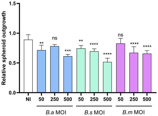
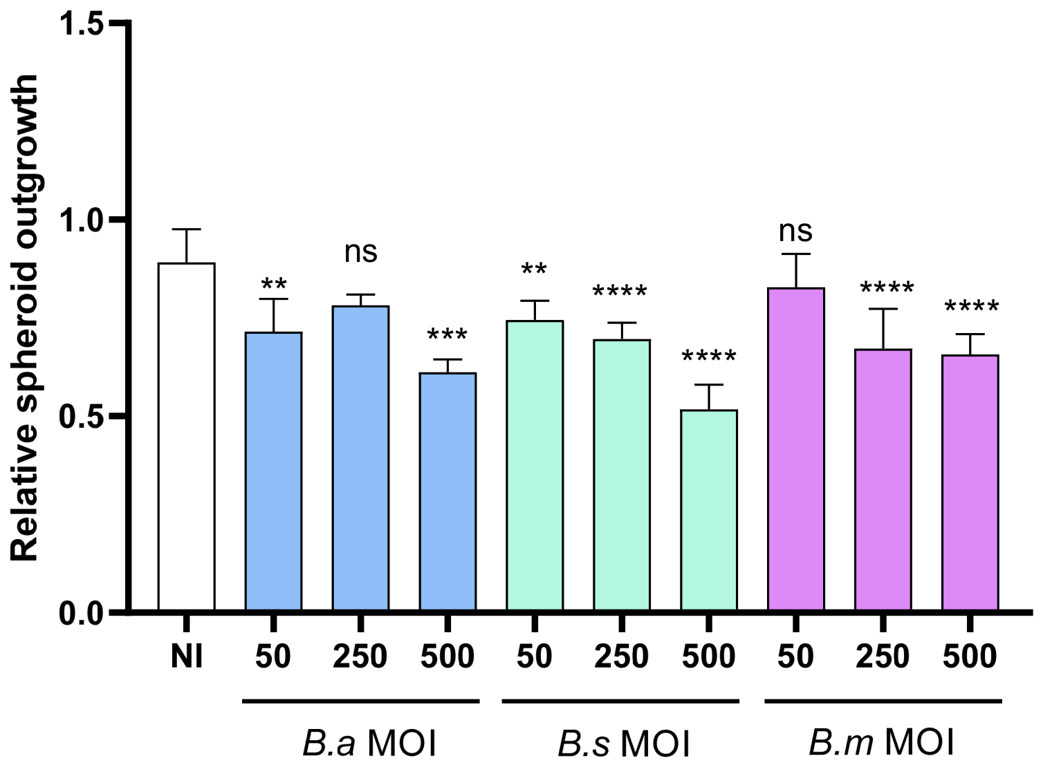
To evaluate whether Brucella infection affects the early stages of embryo implantation, we assessed the expansion of infected blastocyst-like spheroids on monolayers of non-infected decidualized endometrial cells over 24 h. The spheroid outgrowth area was quantified and normalized to both the initial spheroid area and the uninfected control. As shown in Figure 2 and Figure S2, infection with B. suis significantly reduced the outgrowth area of blastocyst-like spheroids at all MOIs tested compared with the non-infected (NI) control. B. abortus produced a significant reduction only at MOIs of 50 and 500, whereas B. melitensis significantly impaired spheroid outgrowth only at MOIs of 250 and 500, indicating a more consistent inhibitory effect of B. suis on blastocyst outgrowth.
Figure 2.
Brucella infection of trophoblast cells negatively affects spheroid adhesion to decidualized endometrial cells. T-HESC cells were seeded in 96-well plates at a density of 1 × 10⁴ cells/well and subsequently decidualized. Spheroids generated from infected trophoblast cells (2 × 10⁴ cells/drop) exposed to different Brucella strains at different MOIs were placed onto decidualized T-HESC cells. Images were acquired immediately after spheroid placement (0 h) and after 24 h of co-culture. Spheroid outgrowth was quantified and expressed as relative spheroid outgrowth normalized to that of spheroids generated from non-infected trophoblast cells. Data are presented as mean ± SD from three independent experiments performed in duplicate. Asterisks indicate significant differences (**, p < 0.01; ***, p < 0.001; ****, p < 0.0001) compared with the non-infected spheroid control. ns: non-significant.
3.3. Brucella Infection Impairs Trophoblast Migration
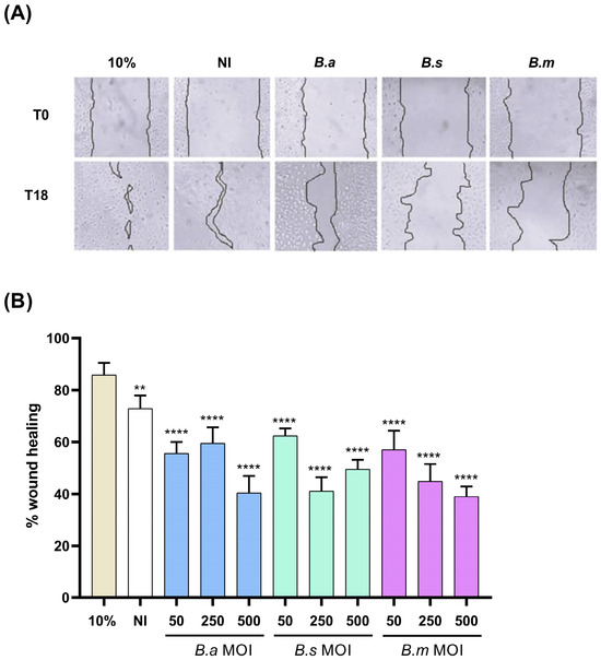
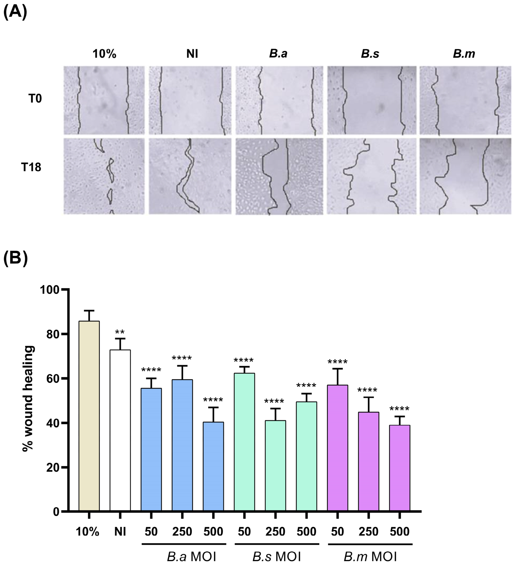
To assess the impact of Brucella infection on trophoblast migratory capacity, wound closure was quantified over time using a wound healing assay. As shown in Figure 3, infection with B. abortus, B. suis, and B. melitensis significantly reduced wound closure at all MOIs tested compared with the non-infected (NI) control. Cells cultured in medium supplemented with 10% FBS, used as a positive control for cell migration, exhibited significantly greater wound closure than infected cells. Collectively, these results demonstrate that infection with all three Brucella species impairs trophoblast wound closure in vitro.
Figure 3.
Infection with different Brucella species impairs trophoblast cell migration. A wound-healing assay was performed using Swan-71 cell monolayers infected with B. abortus (B.a), B. suis (B.s) or B. melitensis (B.m) at different MOI, or left uninfected (NI), in medium containing 2% FBS. Uninfected cells cultured in medium supplemented with 10% FBS were included as a positive control (10%) for maximal wound closure. Wound closure was evaluated by taking images at 0 h (T0) and 18 h (T18) from the same microscopic field. (A) Representative 40× magnification images of wound healing in Swan-71 cells infected with B.a, B.s or B.m at MOI 250 at T0 and T18. (B) Percentage of wound closure for Swan-71 cells infected with the different Brucella species at the indicated MOI. The percentage of wound healing was calculated as: [(area at 0 h–area at 18 h)/area at 0 h] × 100. Images were processed using ImageJ software (version 1.54p). Data are presented as mean ± SD from three independent experiments performed in duplicate. Asterisks indicate significant differences (** p < 0.01; **** p < 0.0001) compared with the NI control.
3.4. Brucella Infection Affects Trophoblast Invasion
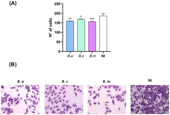
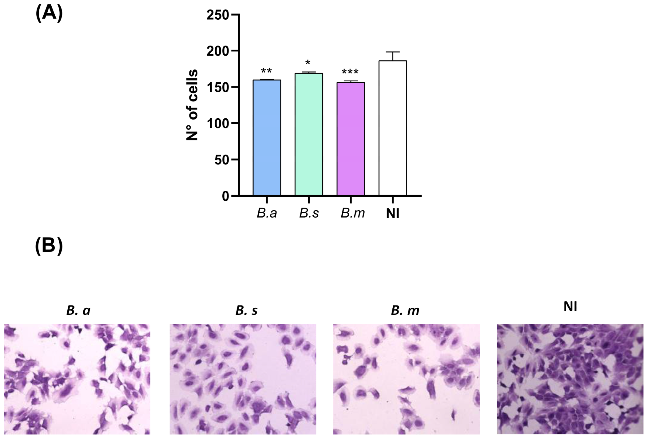
Since Brucella infection impaired blastocyst-like spheroid adhesion and trophoblast cell migration, we next investigated whether it also affects trophoblast invasion, another key process involved in successful placentation. To this end, we evaluated the effect of Brucella infection on trophoblast invasion using Geltrex-coated Transwell invasion assays. As shown in Figure 4A,B, infection of trophoblast cells with the virulent Brucella species at an MOI of 250 significantly reduced the number of cells invading through the Geltrex matrix compared with the non-infected (NI) control. These results indicate that infection with virulent Brucella species significantly compromises the invasive capacity of trophoblast cells.
Figure 4.
Brucella infection impairs the invasive phenotype of Swan-71 trophoblast cells. Swan-71 cells were infected with B. abortus (B.a), B. suis (B.s), or B. melitensis (B.m) at an MOI of 250 and subsequently seeded onto a commercial protein matrix (Geltrex) at a density of 3 × 10⁴ cells/insert. After 24 h, cells that had migrated to the basolateral side of the membrane were stained with crystal violet. Panel (A) shows the number of cells that migrated through the membrane under each experimental condition. Panel (B) shows representative images of crystal violet-stained cells that migrated to the basolateral side of the membrane. Images were acquired and analyzed using ImageJ software. Data are presented as mean ± SD from three independent experiments performed in duplicate. Asterisks indicate significant differences (* p < 0.05; ** p < 0.01; *** p < 0.001) compared with the non-infected (NI) control condition.
3.5. Brucella Infection Impairs Trophoblast Tubulogenesis
Following trophoblast invasion, extravillous trophoblasts acquire an endothelial-like phenotype and participate in the remodeling of maternal spiral arteries through a process known as pseudo-vasculogenesis. This tubulogenic process is essential for establishing adequate maternal–fetal blood flow and ensuring normal placental development. Therefore, we investigated whether Brucella infection affects the tubulogenic capacity of trophoblast cells.
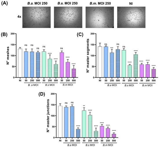
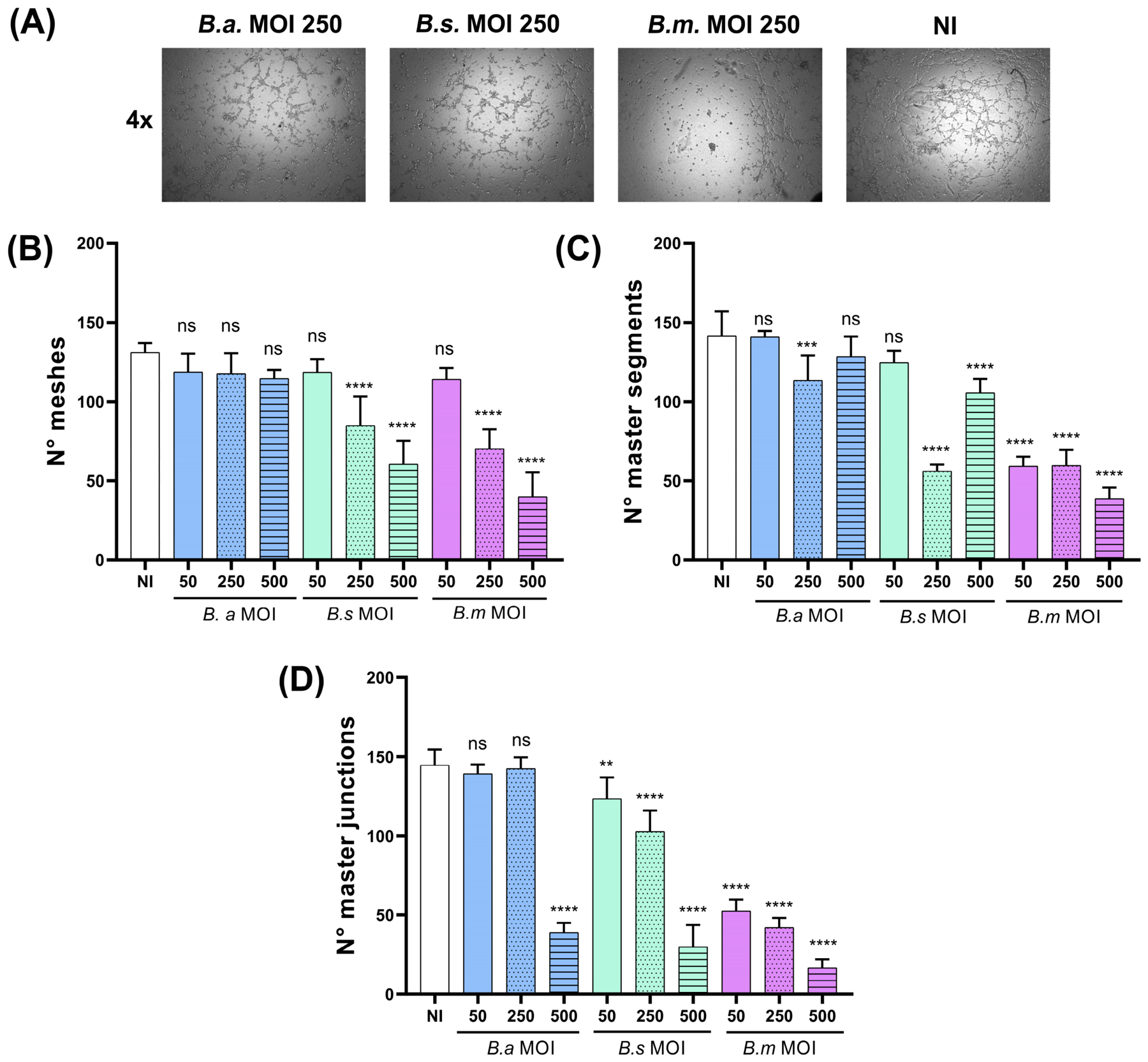
Swan-71 cells were infected with B. abortus, B. suis, or B. melitensis at different MOIs and subsequently seeded onto a Geltrex matrix. Cells were incubated for an additional 6 h, after which images were acquired by microscopy (Figure 5A) to quantify master segments, master junctions, and meshes, which are key parameters of tubulogenesis. As shown in Figure 5B, infection with B. abortus, B. suis, and B. melitensis significantly decreased the number of master segments compared with the non-infected control (NI). However, only B. suis and B. melitensis significantly reduced the number of master junctions and meshes (Figure 5C,D), whereas B. abortus did not significantly affect these parameters.
Figure 5.
Brucella spp. infection impairs trophoblast tubulogenesis. Swan-71 cells were infected with different Brucella species at different MOIs or left uninfected, and were subsequently seeded in 96-well plates coated with a protein matrix at a density of 2 × 10⁴ cells/well. After 6 h, images were acquired at 4× magnification from each well. Panel (A) depicts a representative experiment showing an interconnected network of elongated, tube-like structures forming enclosed spaces in the NI group, and an altered network with visible differences in its continuity and branching pattern in the infected groups. The numbers of segments (B), master junctions (C), and meshes (D) were quantified using ImageJ software. Data are presented as mean ± SD from three independent experiments performed in duplicate. Asterisks indicate significant differences (**, p < 0.01; ***, p < 0.001; ****, p < 0.0001) compared with the non-infected (NI) control condition. ns: non-significant.
3.6. Species- and Time-Dependent Effects of Brucella Infection on Trophoblast Cell Proliferation
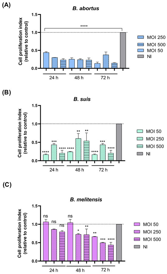
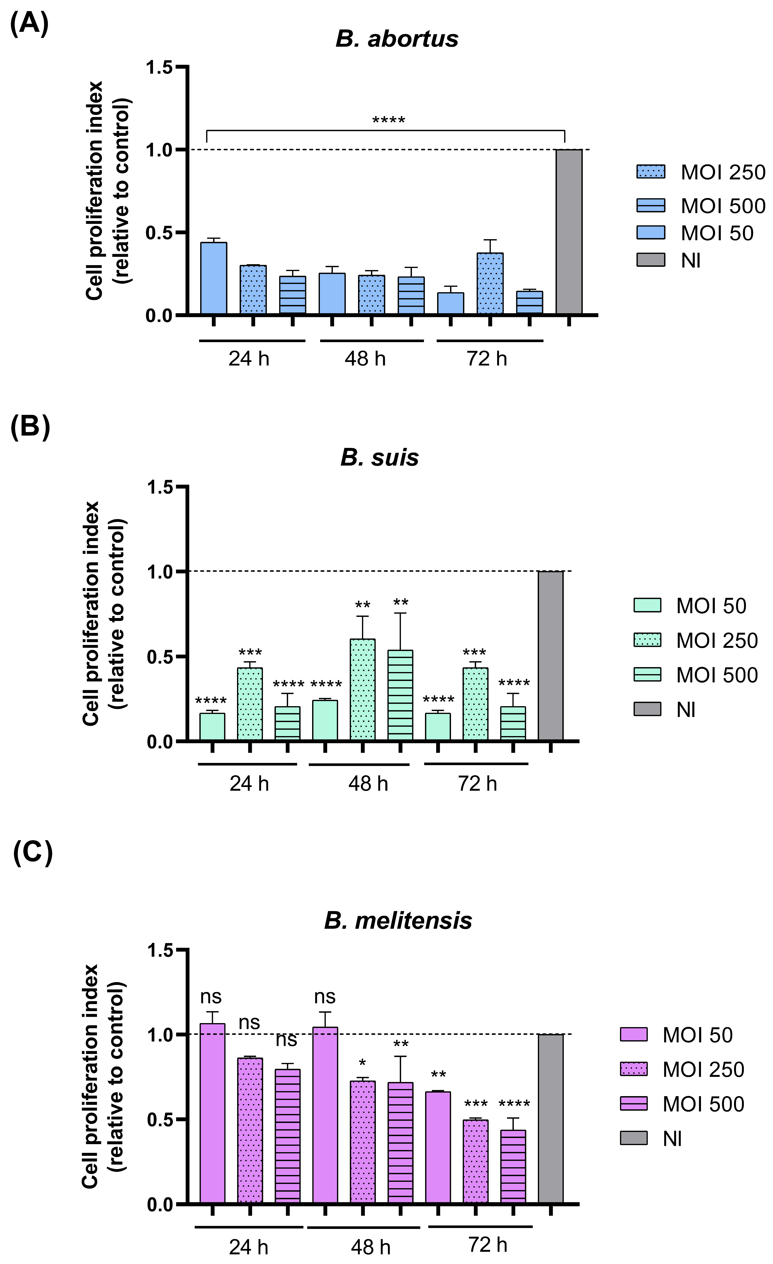
Trophoblast proliferation is essential for normal placental development, contributing to placental growth and the maintenance of an adequate trophoblast cell population. To determine whether Brucella infection affects trophoblast cell proliferation, DNA synthesis was assessed by BrdU incorporation at 24, 48, and 72 h p.i. As shown in Figure 6A,B, infection with B. abortus and B. suis significantly reduced trophoblast cell proliferation at all time points and MOIs tested compared with the non-infected control (NI). In contrast, B. melitensis did not significantly affect trophoblast cell proliferation at 24 h p.i. However, a significant reduction in proliferation became evident at 48 h p.i. at MOIs of 250 and 500, indicating a delayed inhibitory effect (Figure 6C). By 72 h p.i., B. melitensis significantly decreased cell proliferation at all MOIs tested in an MOI-dependent manner (Figure 6C). Overall, B. abortus, B. suis, and B. melitensis exhibited distinct temporal patterns in their effects on trophoblast cell proliferation. B. abortus and B. suis produced marked inhibitory effects on trophoblast proliferation, whereas B. melitensis exhibited a delayed response, with significant inhibition becoming evident only at 48 h p.i.
Figure 6.
Brucella infection alters trophoblast cell proliferation. Swan-71 cells were seeded at a density of 3 × 10³ cells/well and infected with B. abortus (A), B. suis (B), or B. melitensis (C) at different MOI. Cell proliferation was assessed at 24 h, 48 h and 72 h p.i. using a BrdU incorporation assay. Data are presented as mean ± SD of cell proliferation index relative to the non-infected (NI) control and were obtained from three independent experiments performed in duplicate. Asterisks indicate significant differences (*, p < 0.05; **, p < 0.01; ***, p < 0.001; ****, p < 0.0001) compared with the NI control. ns: non-significant.
3.7. Brucella Infection Induces Time-Dependent Apoptotic Features in Trophoblast Cells
To determine whether the reduction in trophoblast proliferation observed after infection was associated with changes in cell death, we evaluated apoptotic and necrotic features using acridine orange/ethidium bromide staining. This approach allowed the identification of viable, early apoptotic, late apoptotic, and necrotic cells based on fluorescence patterns and nuclear morphology.
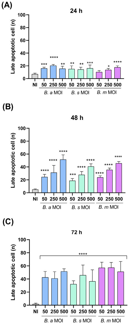
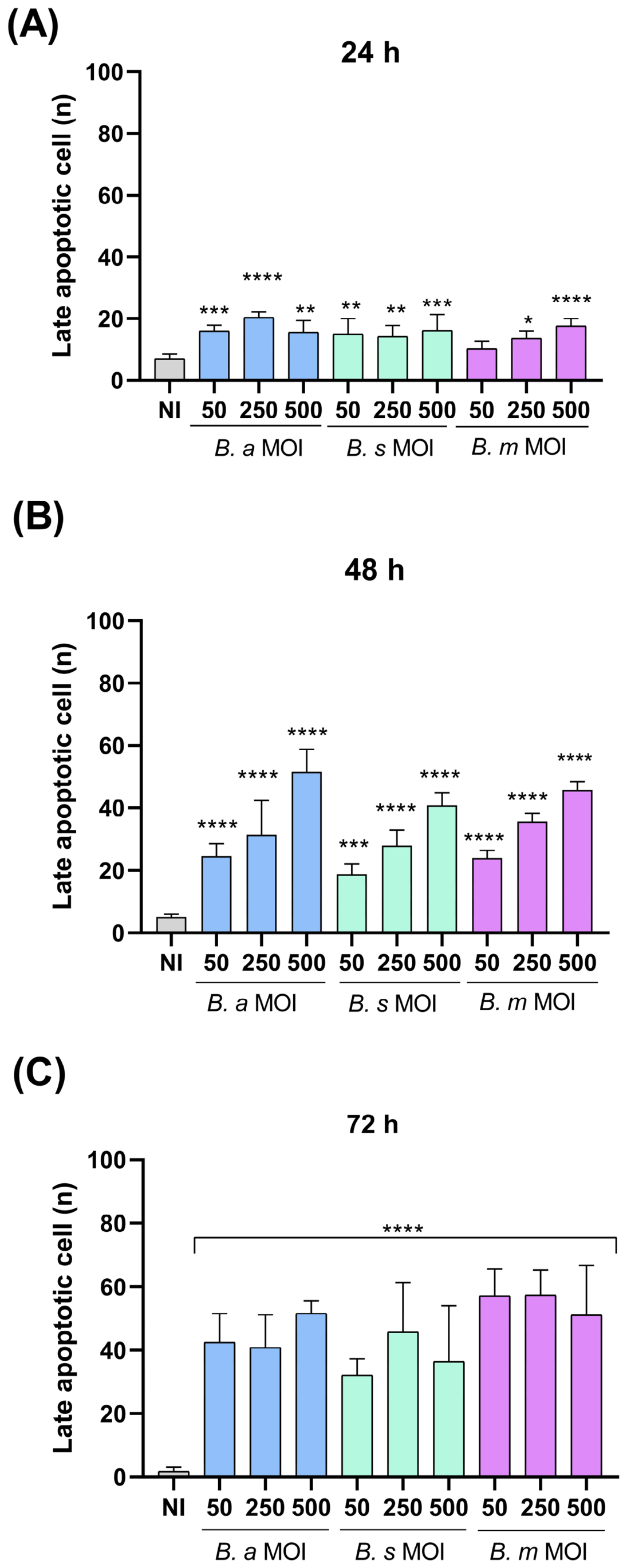
At 24 h p.i., infection with B. abortus, B. suis, and B. melitensis significantly increased the proportion of cells displaying late apoptotic features at all MOIs tested, except for B. melitensis at an MOI of 50 (Figure 7A). Nevertheless, the overall number of cells displaying late apoptotic features remained low compared with the total number of cells analyzed. This response was maintained at 48 h p.i., with all three Brucella species significantly increasing the proportion of late apoptotic cells (Figure 7B). At 72 h p.i., late apoptotic features were further increased, and the highest numbers of late apoptotic cells were observed across the infection conditions (Figure 7C). Overall, these findings indicate that infection with all three Brucella species induces late apoptotic features in trophoblast cells from early stages of infection, with the response becoming more pronounced over time and reaching its greatest magnitude at 72 h p.i.
Figure 7.
Brucella infection induces apoptotic features in trophoblast cells. Swan-71 cells were seeded in 48-well plates at a density of 2.5 × 10⁴ cells/well and infected with B. abortus, B. suis, or B. melitensis at MOIs of 50, 250, or 500. At 24 h (A), 48 h (B), and 72 h (C) post-infection (p.i.), cells were incubated with NaBrEt solution (1 µg/mL) in fresh medium and immediately examined by fluorescence microscopy (EVOS) using the red fluorescence channel at 20× magnification. Five randomly selected fields were analyzed per well. Data are presented as mean ± SD from three independent experiments performed in duplicate. Asterisks indicate significant differences (*, p < 0.05; **, p < 0.01; ***, p < 0.001; ****, p < 0.0001) compared with the non-infected (NI) control condition.
Conversely, early apoptotic cells were rarely detected across all species, MOIs, and time points evaluated, suggesting a rapid progression toward late apoptotic stages or primary clearance of early apoptotic events in infected cultures (Supplementary Figure S3).
The acridine orange/ethidium bromide assay also allowed the identification of necrotic cells. Overall, only a small number of necrotic events were observed across the experimental conditions, consistent with the absence of significant differences in lactate dehydrogenase (LDH) release compared with non-infected controls. Some increases in necrotic cells were detected at specific species, MOI, and time-point combinations, particularly at 48 and 72 h p.i.; however, these findings were based on relatively few events and are therefore presented as Supplementary Data (Supplementary Figure S4).
4. Discussion
In human pregnancy, the successful establishment of the maternal–fetal interface depends on a precisely orchestrated sequence of events that includes trophoblast proliferation, migration, and invasion, as well as remodeling of the maternal spiral arteries [24,25]. Infection with pathogens during early gestation can disrupt these critical processes, leading to severe obstetric complications [26]. Although the clinical association between human brucellosis and adverse pregnancy outcomes, such as spontaneous abortion and preterm birth, has been clinically documented [27,28], the specific cellular mechanisms through which Brucella species affect human trophoblast function remain incompletely understood. In this study, we demonstrate that direct infection with the three major human-pathogenic species (B. abortus, B. suis, and B. melitensis) impairs key functions of human EVTs including cell proliferation, migration, invasion, tubulogenesis, and adhesion/expansion of blastocyst-like spheroids onto decidualized cells, together with a marked proinflammatory response.
Early implantation requires coordinated communication and mechanisms between the blastocyst and the receptive endometrium [29]. Using a spheroid model of a blastocyst, we demonstrated that Brucella infection impairs trophoblasts’ ability to adhere to and expand onto decidualized endometrial stromal cells. These findings suggest that direct infection of trophoblasts may interfere with cellular processes required during the earliest stages of implantation, even before pregnancy is clinically recognized. Furthermore, our data show that direct infection with all three Brucella species markedly reduces migration and Transwell invasion of EVTs. Similar functional impairments were observed in trophoblasts in contact with soluble mediators released by Brucella-infected endometrial cells [21], but demonstrate here that direct infection of EVTs is sufficient to disrupt their invasive phenotype. Together, these findings suggest that both direct infection of trophoblasts and indirect effects mediated by infected maternal cells may contribute to impaired trophoblast motility and invasion. Similar alterations in trophoblast invasion have also been described for other congenital pathogens, including human cytomegalovirus and Toxoplasma gondi [15,16,30].
Following interstitial invasion into the maternal decidua, EVTs undergo endovascular differentiation, replacing maternal endothelial cells to transform the spiral arteries into high-capacity, low-resistance vessels [31]. This process is essential for ensuring adequate uteroplacental perfusion [32]. In our study, Brucella infection significantly impaired tubule formation on Geltrex matrix, markedly reducing master junctions, segments, and network tubes. Defective vascular remodeling and impaired trophoblast invasion are key features of placental insufficiency, which can lead to fetal growth restriction, preeclampsia, or early pregnancy loss [33,34]. Our in vitro evidence of impaired tubulogenesis in EVTs suggests that Brucella-induced placental damage is not solely a consequence of the local inflammatory environment (placentitis) [35], but could also involve functional alterations in trophoblast cells that may contribute to impaired placental vascular remodeling.
A notable aspect of this study is the identification of species-specific differences in the severity and temporal kinetics of trophoblast dysfunction. Although all three species induced marked proinflammatory responses and impaired cell migration and invasion, B. suis and B. melitensis had a broader and more pronounced impact on tubulogenesis and blastocyst-like spheroid adhesion compared with B. abortus. Likewise, B. abortus and B. suis rapidly inhibited EVT proliferation within 24 h after infection, whereas B. melitensis exhibited a delayed kinetic, requiring 48 to 72 h to reach comparable antiproliferative effects. These phenotypic differences may result from distinct virulence factor expression profiles, differences in intracellular replication rates, or differential modulation of host cell stress pathways [36,37].
Previous studies have shown that Brucella infection can induce trophoblast cell death in vivo and modulate apoptotic pathways in human trophoblast cell lines. In a murine pregnancy model, B. abortus induced trophoblast death through a T4SS- and VceC-dependent endoplasmic reticulum stress response [38], whereas infection of human HPT-8 trophoblasts implicated the T4SS effector VceA in the regulation of apoptosis [39]. Together, these findings indicate that Brucella can perturb trophoblast survival through multiple cell-death pathways. However, the limited apoptotic features observed in our model at 24 h, even for high MOIs, suggest that apoptosis alone is unlikely to account for the broad functional alterations observed at this early time point. Notably, the impairment of trophoblast proliferation, migration, invasion, and adhesion occurred in the context of limited apoptosis, suggesting that Brucella infection induces an early dysfunctional state that is not simply a consequence of cell death. The reduced proliferative capacity may therefore involve alterations in cell-cycle progression or other infection-induced cellular stress responses, while changes in cellular processes required for migration, adhesion, and invasion may similarly occur before extensive cell loss. Thus, apoptosis may contribute to the progressive trophoblastic dysfunction induced by Brucella but is unlikely to represent the sole mechanism underlying the early functional alterations observed.
In parallel with the functional alterations, infection with B. abortus, B. suis, and B. melitensis induced elevated levels of proinflammatory cytokines, including IL-6, CXCL8, and CCL2. Although an initial and localized inflammatory signal is required for early implantation [40], sustained or excessive secretion of chemokines and cytokines can disrupt immune tolerance at the maternal–fetal interface, recruit cytotoxic immune populations, and promote tissue damage [41,42]. Therefore, the inflammatory environment generated by infected EVTs may act in concert with direct impairment of trophoblast functions to compromise the cellular processes required for successful placentation, providing a potential mechanistic link between trophoblast infection and the adverse pregnancy outcomes associated with human brucellosis.
Some limitations of this study should be acknowledged. These results were obtained in vitro using a trophoblastic cell line and do not reproduce the complex cellular interactions that may occur in vivo. In addition, although the Swan71 cell line preserves most of the phenotypic and functional characteristics of primary trophoblasts, which is a strength of the study, it will be interesting to perform similar investigations using primary cells for comparison. Another potential limitation of the study is that results were obtained with only three Brucella species and only one strain of each. It must be noted, however, that the three species evaluated (B. abortus, B. suis, B. melitensis) are responsible for the vast majority of human brucellosis cases, and that the three strains employed are widely used as representative of the corresponding species. Finally, our study reveals functional alterations produced by Brucella on human EVTs but does not show that these effects are exclusive to Brucella infections. It cannot be ruled out that other intracellular pathogens may produce similar derangements in these cells.
5. Conclusions
In conclusion, our findings support a model in which direct infection of human trophoblasts by B. abortus, B. suis, and B. melitensis impairs key functions important for successful placentation, including proliferation, migration, invasion, vascular remodeling capacity, and blastocyst–endometrium interactions. These functional alterations, along with the proinflammatory response and limited apoptosis observed early in infection, suggest a potential cellular link between Brucella infection at the maternal–fetal interface and adverse pregnancy outcomes.
Supplementary Materials
The following supporting information can be downloaded at: https://www.mdpi.com/article/10.3390/pathogens15111102/s1, Figure S1: Trophoblast cells respond to infection with different Brucella species by secreting proinflammatory cytokines; Figure S2: Brucella infection affects the outgrowth of trophoblast spheroids on decidualized stromal cells; Figure S3: Early apoptotic features in Brucella-infected trophoblastic cells; Figure S4: Necrotic features in Brucella-infected trophoblastic cells.
Author Contributions
Conceptualization, L.Z., M.C.F. and P.C.B.; Methodology, L.Z., R.M., M.A.P., D.P., M.C.F., A.M.C. and P.C.B.; Formal Analysis, L.Z. and M.C.F.; Investigation, L.Z., M.C.F., M.A.P., D.P. and R.M.; Writing—Original Draft Preparation, L.Z., M.C.F. and P.C.B.; Writing—Review and Editing, P.C.B., M.C.F. and A.M.C.; Supervision, M.C.F. and P.C.B.; Project Administration, M.C.F. and P.C.B.; Funding Acquisition, P.C.B. All authors have read and agreed to the published version of the manuscript.
Funding
This research was funded by grants PICT-2021-I-A-00680 from Agencia Nacional de Promoción Científica, and UBACYT 20020220100012BA from Universidad de Buenos Aires.
Institutional Review Board Statement
Not applicable.
Informed Consent Statement
Not applicable.
Data Availability Statement
The raw data supporting the conclusions of this article will be made available by the authors on request.
Acknowledgments
We thank the staff of the INBIRS institute (UBA-CONICET) for expert technical assistance with BSL3 facilities.
Conflicts of Interest
The authors declare no conflicts of interest.
References
- Rossetti, C.A.; Maurizio, E.; Rossi, U.A. Comparative Review of Brucellosis in Small Domestic Ruminants. Front. Vet. Sci. 2022, 9, 887671. [Google Scholar] [CrossRef] [Scilit] [PubMed]
- Zavattieri, L.; Muñoz González, F.; Ferrero, M.C.; Baldi, P.C. Immune Responses Potentially Involved in the Gestational Complications of Brucella Infection. Pathogens 2023, 12, 1450. [Google Scholar] [CrossRef] [Scilit] [PubMed]
- Elshamy, M.; Ahmed, A.I. The effects of maternal brucellosis on pregnancy outcome. J. Infect. Dev. Ctries. 2008, 2, 230–234. [Google Scholar] [CrossRef] [Scilit] [PubMed]
- Madkour, M.M. Pregnancy and Brucellosis. In Madkour’s Brucellosis; Madkour, M.M., Ed.; Springer: Berlin/Heidelberg, Germany, 2001; pp. 187–192. [Google Scholar]
- IInan, A.; Erdem, H.; Elaldi, N.; Gulsun, S.; Karahocagil, M.K.; Pekok, A.U.; Ulug, M.; Tekin, R.; Bosilkovski, M.; Kaya, S.; et al. Brucellosis in pregnancy: Results of multicenter ID-IRI study. Eur. J. Clin. Microbiol. Infect. Dis. 2019, 38, 1261–1268. [Google Scholar] [CrossRef] [Scilit] [PubMed]
- Mujuni, F.; Andrew, V.; Mngumi, E.B.; Chibwe, E.; Mshana, S.E.; Mirambo, M.M. Predominance of Brucella abortus antibodies among women with spontaneous abortion in the city of Mwanza: Unrecognized link or coincidence? BMC Res. Notes 2018, 11, 792. [Google Scholar] [CrossRef] [Scilit] [PubMed]
- Guerrier, G.; Daronat, J.M.; Morisse, L.; Yvon, J.F.; Pappas, G. Epidemiological and clinical aspects of human Brucella suis infection in Polynesia. Epidemiol. Infect. 2011, 139, 1621–1625. [Google Scholar] [CrossRef] [Scilit] [PubMed]
- Ashton, S.V.; Whitley, G.S.J.; Dash, P.R.; Wareing, M.; Crocker, I.P.; Baker, P.N.; Cartwright, J.E. Uterine Spiral Artery Remodeling Involves Endothelial Apoptosis Induced by Extravillous Trophoblasts Through Fas/FasL Interactions. Arterioscler. Thromb. Vasc. Biol. 2005, 25, 102–108. [Google Scholar] [CrossRef] [Scilit] [PubMed]
- He, N.; van Iperen, L.; de Jong, D.; Szuhai, K.; Helmerhorst, F.M.; van der Westerlaken, L.A.J.; de Chuva Sousa Lopes, S.M. Human Extravillous Trophoblasts Penetrate Decidual Veins and Lymphatics before Remodeling Spiral Arteries during Early Pregnancy. PLoS ONE 2017, 12, e0169849. [Google Scholar] [CrossRef] [Scilit] [PubMed]
- Erlebacher, A. Mechanisms of T cell tolerance towards the allogeneic fetus. Nat. Rev. Immunol. 2012, 13, 23–33. [Google Scholar] [CrossRef] [Scilit] [PubMed]
- Kudo, Y.; Sugimoto, J. The Role of the Placental Enzyme Indoleamine 2,3-Dioxygenase in Normal and Abnormal Human Pregnancy. Int. J. Mol. Sci. 2024, 25, 4577. [Google Scholar] [CrossRef] [Scilit] [PubMed]
- Ostrander, B.; Bale, J.F. Congenital and perinatal infections. Handb. Clin. Neurol. 2019, 162, 133–153. [Google Scholar] [CrossRef] [Scilit] [PubMed]
- Fallahi, S.; Rostami, A.; Nourollahpour Shiadeh, M.; Behniafar, H.; Paktinat, S. An updated literature review on maternal-fetal and reproductive disorders of Toxoplasma gondii infection. J. Gynecol. Obstet. Hum. Reprod. 2018, 47, 133–140. [Google Scholar] [CrossRef] [Scilit] [PubMed]
- Almeida, M.P.O.; Ferro, E.A.V.; Briceño, M.P.P.; Oliveira, M.C.; Barbosa, B.F.; Silva, N.M. Susceptibility of human villous (BeWo) and extravillous (HTR-8/SVneo) trophoblast cells to Toxoplasma gondii infection is modulated by intracellular iron availability. Parasitol. Res. 2019, 118, 1559–1572. [Google Scholar] [CrossRef] [Scilit] [PubMed]
- Lebourg, A.; Leroux, L.P.; Chaparro, V.; Chagneau, S.; Vaillancourt, C.; Jaramillo, M. Toxoplasma gondii infection inhibits invasion and migration of human extravillous trophoblasts through dysregulation of FOXO1- and FOXO3a-dependent and -independent mechanisms. Front. Cell. Infect. Microbiol. 2025, 15, 1651142. [Google Scholar] [CrossRef] [Scilit] [PubMed]
- Warner, J.A.; Zwezdaryk, K.J.; Day, B.; Sullivan, D.E.; Pridjian, G.; Morris, C.A. Human cytomegalovirus infection inhibits CXCL12- mediated migration and invasion of human extravillous cytotrophoblasts. Virol. J. 2012, 9, 255. [Google Scholar] [CrossRef] [Scilit] [PubMed]
- Fernández, A.G.; Ferrero, M.C.; Hielpos, M.S.; Fossati, C.A.; Baldi, P.C. Proinflammatory response of human trophoblastic cells to Brucella abortus infection and upon interactions with infected phagocytes. Biol. Reprod. 2016, 94, 48. [Google Scholar] [CrossRef] [Scilit] [PubMed]
- García-Méndez, K.B.; Hielpos, S.M.; Soler-Llorens, P.F.; Arce-Gorvel, V.; Hale, C.; Gorvel, J.P.; O’Callaghan, D.; Keriel, A. Infection by Brucella melitensis or Brucella papionis modifies essential physiological functions of human trophoblasts. Cell. Microbiol. 2019, 21, e13019. [Google Scholar] [CrossRef] [Scilit] [PubMed]
- Salcedo, S.P.; Chevrier, N.; Lacerda, T.L.S.; Ben Amara, A.; Gerart, S.; Gorvel, V.A.; de Chastellier, C.; Blasco, J.M.; Mege, J.L.; Gorvel, J.P. Pathogenic brucellae replicate in human trophoblasts. J. Infect. Dis. 2013, 207, 1075–1083. [Google Scholar] [CrossRef] [Scilit]
- Poveda-Urkixo, I.; Ramírez, G.A.; Grilló, M.J. Kinetics of Placental Infection by Different Smooth Brucella Strains in Mice. Pathogens 2022, 11, 279. [Google Scholar] [CrossRef] [Scilit] [PubMed]
- Zavattieri, L.; Macchi, R.; Canellada, A.M.; Pibuel, M.A.; Poodts, D.; Ferrero, M.C.; Baldi, P.C. Brucella-Induced Impairment of Decidualization and Its Impact on Trophoblast Function and Inflammatory Profile. Int. J. Mol. Sci. 2025, 26, 8189. [Google Scholar] [CrossRef] [Scilit] [PubMed]
- Straszewski-Chavez, S.L.; Abrahams, V.M.; Alvero, A.B.; Aldo, P.B.; Ma, Y.; Guller, S.; Romero, R.; Mor, G. The isolation and characterization of a novel telomerase immortalized first trimester trophoblast cell line, Swan 71. Placenta 2009, 30, 939–948. [Google Scholar] [CrossRef] [Scilit] [PubMed]
- Rattila, S.; Kleefeldt, F.; Ballesteros, A.; Beltrame, J.S.; Ribeiro, M.L.; Ergün, S.; Dveksler, G. Pro-angiogenic effects of pregnancy-specific glycoproteins in endothelial and extravillous trophoblast cells. Reproduction 2020, 160, 737–750. [Google Scholar] [CrossRef] [Scilit] [PubMed]
- Knöfler, M.; Haider, S.; Saleh, L.; Pollheimer, J.; Gamage, T.K.J.B.; James, J. Human placenta and trophoblast development: Key molecular mechanisms and model systems. Cell. Mol. Life Sci. 2019, 76, 3479–3496. [Google Scholar] [CrossRef] [Scilit] [PubMed]
- Pollheimer, J.; Vondra, S.; Baltayeva, J.; Beristain, A.G.; Knöfler, M. Regulation of Placental Extravillous Trophoblasts by the Maternal Uterine Environment. Front. Immunol. 2018, 9, 2597. [Google Scholar] [CrossRef] [Scilit] [PubMed]
- Megli, C.J.; Coyne, C.B. Infections at the maternal-fetal interface: An overview of pathogenesis and defence. Nat. Rev. Microbiol. 2022, 20, 67–82. [Google Scholar] [CrossRef] [Scilit] [PubMed]
- Khan, M.Y.; Mah, M.W.; Memish, Z.A. Brucellosis in Pregnant Women. Clin. Infect. Dis. 2001, 32, 1172–1177. [Google Scholar] [CrossRef] [Scilit] [PubMed]
- Kurdoglu, M.; Adali, E.; Kurdoglu, Z.; Karahocagil, M.K.; Kolusari, A.; Yildizhan, R.; Kucukaydin, Z.; Sahin, H.G.; Kamaci, M.; Akdeniz, H. Brucellosis in pregnancy: A 6-year clinical analysis. Arch. Gynecol. Obstet. 2010, 281, 201–206. [Google Scholar] [CrossRef] [Scilit] [PubMed]
- Evans, J.; Salamonsen, L.A.; Winship, A.; Menkhorst, E.; Nie, G.; Gargett, C.E.; Dimitriadis, E. Fertile ground: Human endometrial programming and lessons in health and disease. Nat. Rev. Endocrinol. 2016, 12, 654–667. [Google Scholar] [CrossRef] [Scilit] [PubMed]
- Fisher, S.; Genbacev, O.; Maidji, E.; Pereira, L. Human Cytomegalovirus Infection of Placental Cytotrophoblasts In Vitro and In Utero: Implications for Transmission and Pathogenesis. J. Virol. 2000, 74, 6808–6820. [Google Scholar] [CrossRef] [Scilit] [PubMed]
- Whitley, G.S.J.; Cartwright, J.E. Cellular and Molecular Regulation of Spiral Artery Remodelling: Lessons from the Cardiovascular Field. Placenta 2010, 31, 465–474. [Google Scholar] [CrossRef] [Scilit] [PubMed]
- Hunkapiller, N.M.; Fisher, S.J. Chapter 12 Placental Remodeling of the Uterine Vasculature. Methods Enzymol. 2008, 445, 281–302. [Google Scholar] [CrossRef] [Scilit] [PubMed]
- Brosens, I.; Pijnenborg, R.; Vercruysse, L.; Romero, R. The “Great Obstetrical Syndromes” are associated with disorders of deep placentation. Am. J. Obstet. Gynecol. 2011, 204, 193–201. [Google Scholar] [CrossRef] [Scilit] [PubMed]
- Burton, G.J.; Jauniaux, E. Pathophysiology of placental-derived fetal growth restriction. Am. J. Obstet. Gynecol. 2018, 218, S745–S761. [Google Scholar] [CrossRef] [Scilit] [PubMed]
- Xavier, M.N.; Paixao, T.A.; den Hartigh, A.B.; Tsolis, M.R.; Santos, R.L. Pathogenesis of Brucella spp. Open Vet. Sci. J. 2010, 4, 109–118. [Google Scholar] [CrossRef] [Scilit]
- Pappas, G.; Akritidis, N.; Bosilkovski, M.; Tsianos, E. Brucellosis. N. Engl. J. Med. 2005, 352, 2325–2336. [Google Scholar] [CrossRef] [Scilit] [PubMed]
- Carvalho Neta, A.V.; Mol, J.P.S.; Xavier, M.N.; Paixão, T.A.; Lage, A.P.; Santos, R.L. Pathogenesis of bovine brucellosis. Vet. J. 2010, 184, 146–155. [Google Scholar] [CrossRef] [Scilit] [PubMed]
- Byndloss, M.X.; Tsai, A.Y.; Walker, G.T.; Miller, C.N.; Young, B.M.; English, B.C.; Seyffert, N.; Kerrinnes, T.; de Jong, M.F.; Atluri, V.L.; et al. Brucella abortus infection of placental trophoblasts triggers endoplasmic reticulum stress-mediated cell death and fetal loss via type iv secretion system-dependent activation of CHOP. MBio 2019, 10, e01538-19. [Google Scholar] [CrossRef] [Scilit] [PubMed]
- Zhang, J.; Li, M.; Li, Z.; Shi, J.; Zhang, Y.; Deng, X.; Liu, L.; Wang, Z.; Qi, Y.; Zhang, H.; et al. Deletion of the Type IV Secretion System Effector VceA Promotes Autophagy and Inhibits Apoptosis in Brucella-Infected Human Trophoblast Cells. Curr. Microbiol. 2019, 76, 510–519. [Google Scholar] [CrossRef] [Scilit] [PubMed]
- Mor, G.; Cardenas, I.; Abrahams, V.; Guller, S. Inflammation and pregnancy: The role of the immune system at the implantation site. Ann. N. Y. Acad. Sci. 2011, 1221, 80–87. [Google Scholar] [CrossRef] [Scilit] [PubMed]
- Romero, R.; Espinoza, J.; Gonçalves, L.; Kusanovic, J.; Friel, L.; Hassan, S. The Role of Inflammation and Infection in Preterm Birth. Semin. Reprod. Med. 2007, 25, 21–39. [Google Scholar] [CrossRef] [Scilit] [PubMed]
- Yockey, L.J.; Iwasaki, A. Interferons and Proinflammatory Cytokines in Pregnancy and Fetal Development. Immunity 2018, 49, 397–412. [Google Scholar] [CrossRef] [Scilit] [PubMed]
Disclaimer/Publisher’s Note: The statements, opinions and data contained in all publications are solely those of the individual author(s) and contributor(s) and not of MDPI and/or the editor(s). MDPI and/or the editor(s) disclaim responsibility for any injury to people or property resulting from any ideas, methods, instructions or products referred to in the content. |
© 2026 by the authors. Licensee MDPI, Basel, Switzerland. This article is an open access article distributed under the terms and conditions of the Creative Commons Attribution (CC BY) license.