


Abstract
Burkholderia pseudomallei, the causative agent of melioidosis, is endemic in Sarawak, Malaysian Borneo, where it is represented by a unique gentamicin-susceptible population. Despite trimethoprim-sulfamethoxazole (co-trimoxazole) being the cornerstone of eradication therapy, emerging reports of elevated minimum inhibitory concentrations (MICs) among Sarawak isolates have raised concerns over its clinical efficacy. We performed a retrospective and comprehensive antibiotic susceptibility assessment of clinical B. pseudomallei isolates from hospitals across Sarawak. Susceptibility to trimethoprim-sulfamethoxazole was determined using disk diffusion and the E-test, interpreted by both CLSI and EUCAST guidelines. Resistance to the individual components, trimethoprim and sulfamethoxazole, was characterized by broth microdilution. The results demonstrated a high prevalence of trimethoprim-sulfamethoxazole susceptibility, with 96.3% of isolates susceptible by CLSI criteria and 97.6% by EUCAST criteria. Interestingly, broth microdilution revealed that resistance to trimethoprim and sulfamethoxazole individually did not confer resistance to the synergistic combination. Our analysis validated CLSI guidelines as the most reliable standard for antimicrobial resistance surveillance in this region. This study provides evidence that trimethoprim-sulfamethoxazole remains effective for melioidosis treatment in Sarawak, offering crucial reassurance to clinicians. The paradoxical finding of susceptibility to the drug combination despite resistance to its individual components underscores the critical importance of the synergistic activity of trimethoprim-sulfamethoxazole and highlights the need for further investigation into the molecular basis of resistance in this distinct B. pseudomallei population.
1. Introduction
Melioidosis is a significant cause of community-acquired sepsis and pneumonia in its area of endemicity [1]. While historically endemic in Northern Australia and parts of Southeast Asia, its prevalence is rising with emerging foci in the Americas and Africa [2,3,4]. Recent models project an annual global burden of 165,000 affected individuals and 89,000 deaths [2]. In Malaysia, melioidosis is responsible for approximately 3700 cases and more than 2000 deaths yearly [5]. While Sarawak, the largest state in Malaysia, represents a significant hotspot, with the central regions reporting an average incidence of 12.3 per 100,000 and a case-fatality rate of up to 35% [6,7].
The causative agent of melioidosis is Burkholderia pseudomallei, a Gram-negative saprophyte classified as a Tier 1 select agent by the US Centers for Disease Control and Prevention [8]. This designation reflects its potential of easy dissemination, high fatality rate, intrinsic resistance to myriads of antibiotics, and lack of a licensed vaccine [2].
Standard melioidosis treatment employs biphasic therapy, with the initial parenteral intensive-phase of β-lactams such as ceftazidime or carbapenem, followed by the eradication-phase of oral trimethoprim-sulfamethoxazole (co-trimoxazole) regimen [9]. However, concern has grown recently regarding the efficacy of the trimethoprim-sulfamethoxazole regimen in Sarawak, following the emerging reports of an elevated minimal inhibitory concentration (MIC) of the clinical B. pseudomallei isolates [10,11]. This concern is particularly significant since the Sarawak isolates are predominantly characterized by a unique gentamicin-susceptible genotype unreported elsewhere [12], suggesting a distinct evolutionary lineage that may exhibit an atypical resistance profile. Additionally, there is a lack of standardized interpretive criteria for susceptibility testing for the Sarawak isolates that often complicates clinical reporting and optimal treatment guidance.
To address these critical gaps, this study aimed to evaluate the prevalence of trimethoprim-sulfamethoxazole resistance and to understand the mechanism of such phenomena in Sarawak’s distinct B. pseudomallei clinical isolates. We performed a retrospective analysis using recommended antibiotic susceptibility testing methods such as disk diffusion, the E-test, and broth microdilution for trimethoprim-sulfamethoxazole and its individual components. The study findings highlight crucial susceptibility data of the Sarawak isolates and reaffirm the clinical utility of trimethoprim-sulfamethoxazole for melioidosis management in Sarawak. The study also identified isolates with divergent susceptibility to the individual components, a finding that warrants further molecular characterization to elucidate the underlying genetic mechanism.
2. Materials and Methods
2.1. Ethics Statement
This study was approved by the Medical Research Ethics Committee and registered with the National Medical Research Registrar (NMRR-16-1029-31390) and the Clinical Research Centre, Ministry of Health Malaysia. Anonymized bacterial isolates were obtained from archival collections and routine laboratory diagnostic procedures; hence, the requirement for written patient consent was waived.
2.2. Bacterial Strains
A total 164 clinical B. pseudomallei isolates were included in this study, collected from melioidosis cases reported between 2011 and 2018 in six hospitals located in Bintulu, Sibu, Kapit, Miri, and Kuching in Sarawak, Malaysian Borneo (Table 1). The prototype strain SWK-C 001 (also coded as MSHR5078) was used for antibiotic susceptibility testing (AST) optimization. Escherichia coli ATCC 25922 and Escherichia coli ATCC 11775 were used as the quality control (QC) in the broth microdilution assays. All strains were cultured on modified Ashdown’s selective agar and incubated at 37 °C for 24 h. The medium contained 50 µg/mL colistin in place of gentamicin, based on previous findings [13]. Tryptic soy agar (TSA) (OXOID, Basingstoke, Hamsphire, UK) was used as an intermediary medium for all pure cultures prior to preparation for antimicrobial susceptibility testing and broth microdilution assays.
Table 1.
Number of Sarawak clinical B. pseudomallei isolates collected from the hospitals.
2.3. Antibiotic Susceptibility Testing (AST)
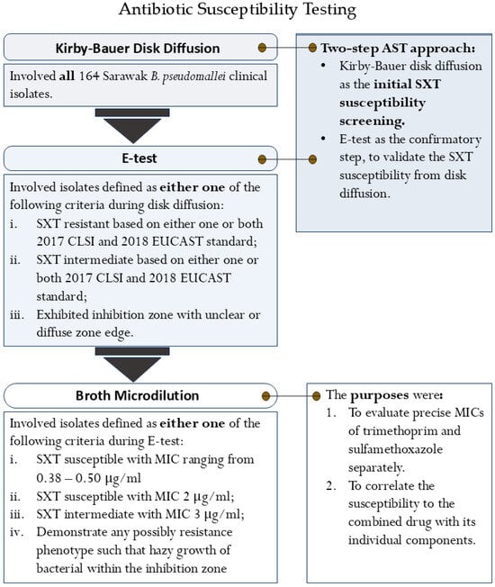
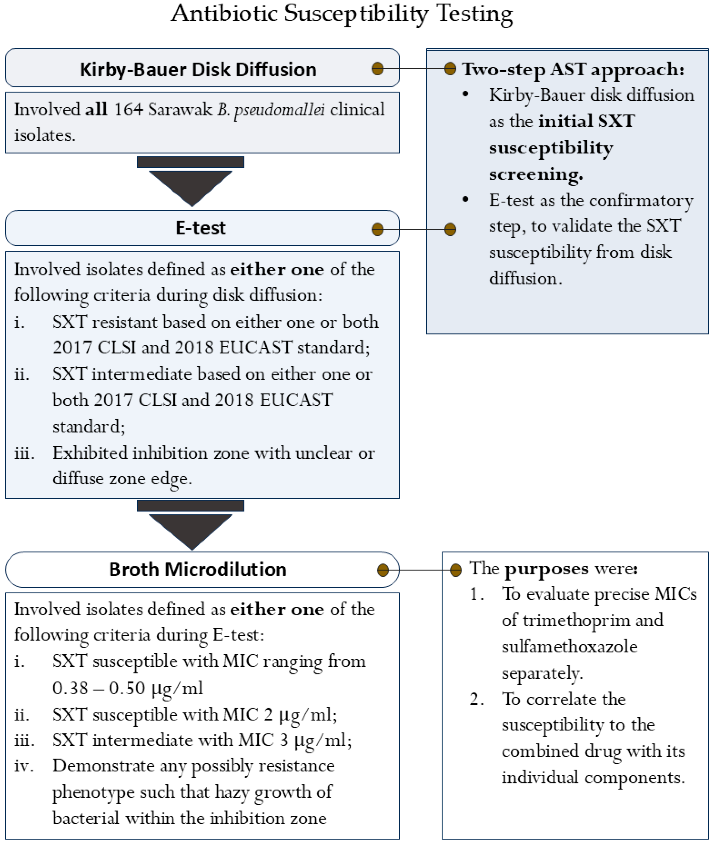
The trimethoprim-sulfamethoxazole susceptibility of the Sarawak B. pseudomallei clinical isolates was evaluated using a tiered testing strategy, performed in accordance with Clinical and Laboratory Standard Institute (CLSI) and European Committee on Susceptibility Testing (EUCAST) guidelines [14,15,16]. Initial screening was conducted with Kirby–Bauer disk diffusion, the routine method in most hospital settings in Sarawak. Selected isolates from this screen were then subjected to confirmatory E-testing. Finally, broth microdilution was employed to determine precise MICs for both sulfamethoxazole and trimethoprim separately. This final procedure was outlined to correlate the susceptibility to the combined drug with its individual components. The specific selection criteria of bacterial isolates for each procedure are detailed in Figure 1.
Figure 1.
The flowchart of the three standard antibiotic susceptibility testing procedures.
2.3.1. Kirby–Bauer Disk Diffusion Testing
The Kirby–Bauer disk diffusion method was performed according to the updated and modified protocols regulated by the 2017 CLSI guideline [14]. Bacterial suspensions were adjusted to a 0.5 McFarland standard and lawn-cultured onto Mueller-Hinton agar (MH) (HiMedia Laboratories, Mumbai, India). A 25 μg trimethoprim-sulfamethoxazole disk (OXOID, Basingstoke, Hamsphire, UK) was applied onto the agar plate, followed by incubation at 37 °C for 18 h. Inhibition zone diameters for susceptible, intermediate, and resistant according to CLSI were defined as ≥16 mm, 11–15 mm, and ≤10 mm; and for the EUCAST standard, they were ≥14 mm, 11–14 mm, and <11 mm, respectively [14,15].
2.3.2. E-Test
MICs for selected isolates against trimethoprim-sulfamethoxazole were determined using E-test strips (bioMérieux, Marcy-I'Étoile, France) according to the manufacturer’s instructions. Similarly, strains were grown overnight on TSA before the MIC determination. For isolates that were selected for broth microdilution (Figure 1), susceptibility to a panel of clinically relevant antibiotics (amoxicillin-clavulanate, ceftazidime, doxycycline, and meropenem) was further evaluated to assess their multidrug-resistant profiles. Gentamicin and azithromycin susceptibility was employed to determine the antibiogram phenotype of the isolates. The interpretation of MICs was according to CLSI and EUCAST standards for B. pseudomallei [14,15], with azithromycin interpreted using the manufacturer’s recommendation for aerobes (Table 2), as no standard breakpoints were established for the non-Enterobacteriaceae.
Table 2.
E-test MIC’s interpretation reference for the antibiotics tested in this study.
2.3.3. Broth Microdilution
The susceptibility of the selected isolates to trimethoprim (TMP) and sulfamethoxazole (SMX) was determined separately using the broth microdilution procedure outlined in CLSI documents M07-A11 and M100 [14,16]. Tests were performed using Mueller Hinton II broth (HiMedia Laboratories, Mumbai, India) at pH 7.0, using a final inoculum of 5 × 10⁵ CFU/mL, and incubated at 37 °C for 18 h. The inoculum was tested against antibiotic concentrations ranging from 0.25 µg/mL to 128 µg/mL for trimethoprim and 2 µg/mL to 1024 µg/mL for sulfamethoxazole. All tests were performed in a minimum of biological triplicate. The final adjusted optical density (OD) value was calculated using Equation (1), and the epidemiological MIC values were derived from Equation (2). Although growth inhibition was analyzed at both 50% (MIC50) and 80% (MIC80) thresholds, the MIC80 was used for reporting. MIC80 breakpoints interpretation for susceptible and resistant was defined as ≤8 µg/mL and ≥16 µg/mL and ≤256 µg/mL and ≥512 µg/mL for trimethoprim and sulfamethoxazole, respectively.
Quality Control (QC)
E. coli ATCC 25922 was initially employed as the primary QC strain. However, due to inconsistent and deranged MICs, E. coli ATCC 11775 was employed as an alternative QC strain. Since it is not standardized for AST, optimization and validation of its MICs against trimethoprim and sulfamethoxazole were carried out in triplicate against those of E. coli ATCC 25922 and a panel of B. pseudomallei with known susceptibility profiles. The alternative QC strain, E. coli ATCC 11775 demonstrated consistent MIC values within the expected ranges for B. pseudomallei and proved stable even with prolonged incubation (36 h), confirming its reliability for this assay (Table 3 and Table 4).
Table 3.
MIC values of the tested isolates and E. coli ATCC 11775 against trimethoprim-sulfamethoxazole based on disk diffusion and the E-test method.
Table 4.
MIC values of the tested isolates and E. coli ATCC 11775 against trimethoprim-sulfamethoxazole with prolonged incubation.
2.4. Statistical Data Analysis
All MIC definitions from the three antibiotic susceptibility testing methods were analyzed using SPSS Statistics version 23.0 (IBM, USA). The agreement between CLSI and EUCAST interpretive guidelines was assessed using the Cohen Kappa (κ) coefficient. The resulting Cohen’s Kappa value (κ) was interpreted as follows [17]:
- κ = 0.00–0.20 indicates slight agreement
- κ = 0.21–0.40 indicates fair agreement
- κ = 0.41–0.60 indicates moderate agreement
- κ = 0.61–0.80 indicates substantial agreement
- κ = 0.81–1.00 indicates almost perfect agreement
Subsequently, trimethoprim-sulfamethoxazole susceptibility was analyzed using descriptive statistics and a corrective mathematical estimation. First, the results from the initial disk diffusion testing were summarized descriptively. Next, a corrected susceptibility frequency was calculated by comparing these results against those from the confirmatory E-test. This involved identifying all isolates with susceptibility verified by the E-test. The final, corrected overall frequency was then derived by adjusting the initial disk diffusion data to reclassify isolates initially misclassified as resistant or intermediate.
2.5. Genomic Analysis
2.5.1. Primer Design
Oligonucleotide primers (Table 5) targeting the bpeEF-oprC efflux pump genes cluster (bpeT-llpE-bpeE-bpeF-oprC) were designed using Primer3plus version 2.6.1 [18], with B. pseudomallei K96243 (accession number: BX571996.1) as the reference strain [19]. This gene cluster has previously been associated with the trimethoprim-sulfamethoxazole resistance mechanism [20,21].
Table 5.
Oligonucleotide primers designed and used in this study.
2.5.2. Polymerase Chain Reaction (PCR)
Genomic DNA was extracted using Chelex® 100 Resin (Bio-Rad, Hercules, CA, USA). The PCR was performed in a 15 μL reaction volume containing 1× Taq Polymerase Buffer, 0.2 mM dNTPs, 0.08 µ/μL Taq Polymerase, primers, and 1 μL of a DNA template. Amplification parameters were set at 95 °C for 5 min of initial denaturation, 45 cycles of 95 °C for 30 s, 52–55 °C for 30 s, and 72 °C for 1 min followed by a final extension at 72 °C for 5 min [22].
For PCR product visualization, 5 μL of the PCR products were mixed with 1 μL of 6× loading dye and electrophoresed on a 0.7% agarose gel in 1× TBE buffer alongside a 100 bp DNA ladder (Vivantis, Shah Alam, Malaysia). Gels were run at 70 V, stained with SYBR® Safe DNA gel stain (Invitrogen, Carlsbad, CA, USA), and visualized using a Gel Doc XR+ System (Bio-Rad, Hercules, CA, USA).
3. Results
To accurately evaluate the trimethoprim-sulfamethoxazole susceptibility and to address the concerns for false-resistant results from disk diffusion, a two-step AST approach was implemented. Following the initial screening by disk diffusion, E-tests were used to validate the susceptibility results. The inhibition zone (IZ) and MIC from both methods were interpreted in parallel using CLSI and the EUCAST guidelines [14,15]. This approach allowed for the analysis of the interpretation variability between standards to determine the valid interpretive standard for B. pseudomallei in Sarawak.
3.1. Disk Diffusion Testing Revealed a High Prevalence of Trimethoprim-Sulfamethoxazole Susceptibility Among the Sarawak B. pseudomallei
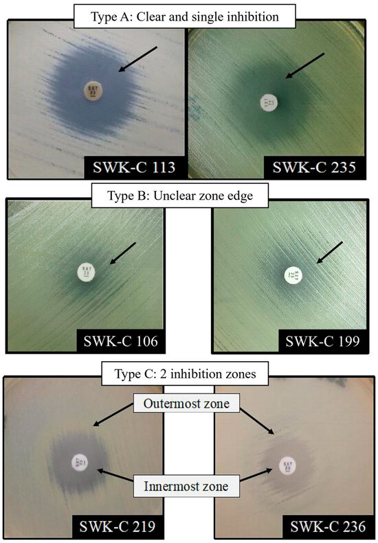
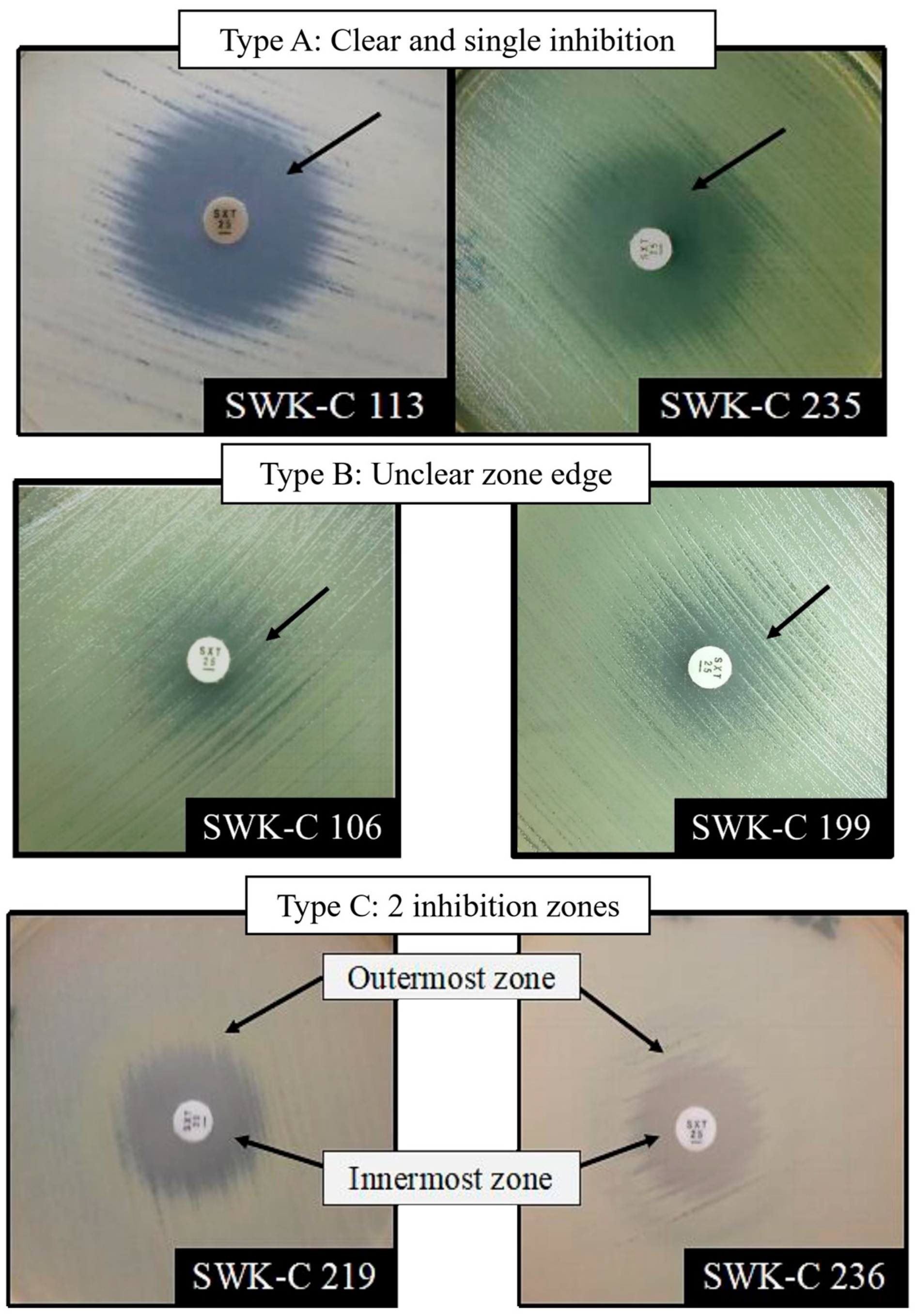
Of 164 isolates tested against a 25 μg trimethoprim-sulfamethoxazole disk (Oxoid, Basingstoke, Hamsphire, UK), 117 exhibited a single IZ, while the remaining 47 formed double IZs. Our assessment identified three distinct phenotypic IZ patterns (Figure 2). Isolates with a single IZ typically exhibited Type A or Type B patterns. Type A is a clear inhibition zone, while Type B was characterized by an unclear or diffuse edge that complicated IZ diameter measurement. All isolates with double IZs displayed the Type C pattern, characterized by an inner zone where 80% of growth was inhibited (MIC80) and an outer zone of 50% inhibition (MIC50). Geographically (see Appendix A Table A1), isolates from Bintulu and Kapit showed a notable tendency to form Type B and C IZs, which complicate the susceptibility definition. Most isolates exhibiting the diffused Type B pattern originated from Bintulu (21 isolates) and Kapit (5 isolates). Additionally, 8 isolates from these regions exhibited Type C IZ. The isolates were recorded with intermediate and resistant diameters ranging from 12 to 15 mm for Bintulu isolates and 9 to 15 mm for Kapit isolates.
Figure 2.
Type of inhibition zone(s) formed by Sarawak clinical B. pseudomallei isolates.
For all isolates, trimethoprim-sulfamethoxazole susceptibility was determined by measuring the IZ diameter at the MIC80 zone edge (see Table 6). The modal IZ diameter was 20 mm, observed in 25/164 isolates. The majority of the isolates (134/164) exhibited a zone diameter greater than 15 mm, while 30/164 isolates showed a diameter below this threshold. Regardless of whether single or double IZs formed, the distribution showed that most Sarawak isolates in this collection were susceptible to trimethoprim-sulfamethoxazole.
Table 6.
Trimethoprim-sulfamethoxazole (25 µg disk) inhibition zone diameter distributions for the Sarawak clinical B. pseudomallei isolates.
The analysis highlights the differences between the two guidelines (see Table 7). According to the CLSI guidelines, the results translated to 81.7% susceptibility and 18.3% reduced susceptibility (including intermediate and resistant isolates). In contrast, EUCAST criteria classified 131/164 isolates (79.9%) as susceptible and 33/164 (20.1%) as non-susceptible. The interpretative discrepancy was reflected in a Cohen’s Kappa value of κ = 0.458 (95% CI: 0.340–0.570), indicating only moderate agreement between the two guidelines. This observation was largely attributable to isolates with challenging IZ phenotypes (Type B in Figure 2) and compounded by the different interpretations by the two guidelines. CLSI criteria define the zone margin by disregarding slight (≤20%) inner-zone growth, whereas EUCAST advises ignoring minor growth only if a clear zone edge is visible. Therefore, while Sarawak isolates demonstrated high trimethoprim-sulfamethoxazole susceptibility, their susceptibility definition was dependent on both the observed IZ phenotype and the interpretive standard applied.
Table 7.
Comparisons of CLSI and EUCAST susceptibility interpretations for the Sarawak clinical B. pseudomallei isolates against trimethoprim-sulfamethoxazole (25 µg disk).
3.2. E-Test Indicates Evidence of a Trimethoprim-Sulfamethoxazole False-Resistance Profile Among the Sarawak B. pseudomallei Clinical Isolates
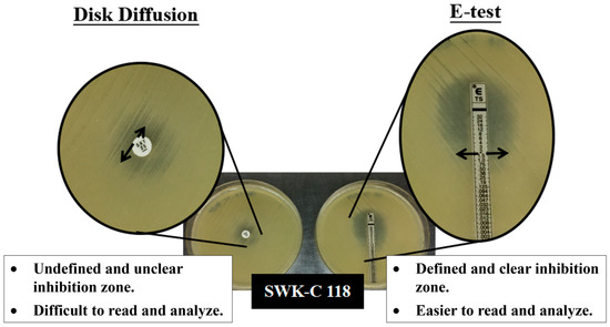
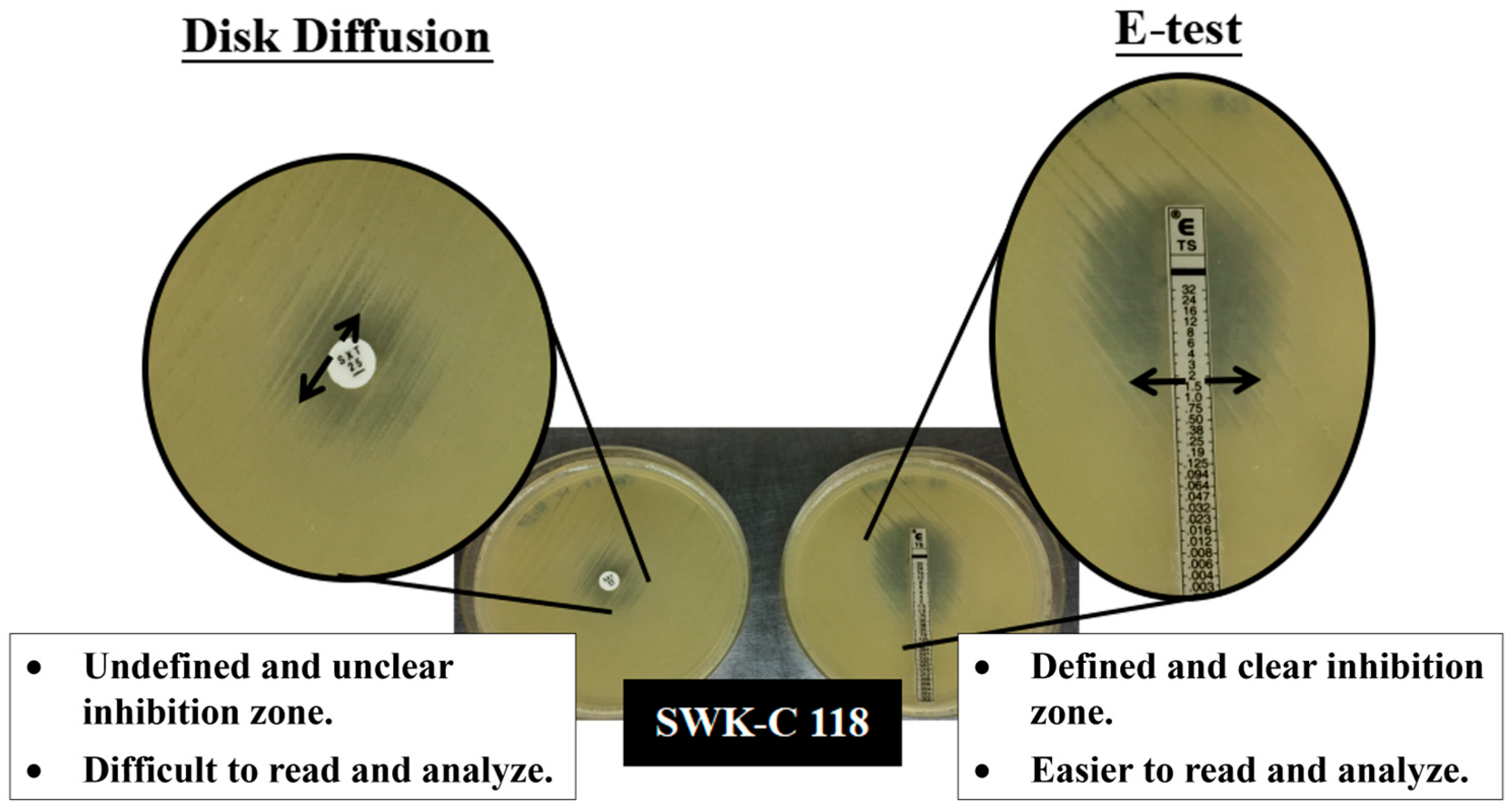
To validate the trimethoprim-sulfamethoxazole susceptibility by disk diffusion, the E-test was performed on 42 isolates previously defined as either intermediately susceptible or resistant (see bolded isolates in Appendix A Table A1). This confirmation was essential due to the observed challenges in interpreting IZs with hazy growth or unclear margins from the disk diffusion method (Figure 3).
Figure 3.
Inhibition zones of Sarawak B. pseudomallei clinical isolates by disk diffusion and E-test.
The E-test yielded clear and easily interpretable IZs for all isolates. The resulting MIC values indicated that the majority of these isolates were susceptible. The MICs for susceptible isolates ranged from 0.38 to 2 μg/mL, with a predominant MIC of 0.75 μg/mL (see Appendix A Table A2). Only one isolate, SWK-C 118, exhibited an MIC of 3 μg/mL, classifying it as intermediate.
Based on these definitive E-test results, the overall trimethoprim-sulfamethoxazole susceptibility prevalence for the studied Sarawak B. pseudomallei isolates was mathematically corrected. The estimated susceptibility was 96.3% (158/164) by CLSI standards and 97.6% (160/164) by EUCAST standards (Table 8).
Table 8.
Mathematically corrected susceptibility of trimethoprim-sulfamethoxazole among the Sarawak clinical B. pseudomallei isolates.
3.3. Broth Microdilution Demonstrated the Resistance of Sarawak B. pseudomallei Clinical Isolates to Trimethoprim and Sulfamethoxazole Despite Trimethoprim-Sulfamethoxazole Susceptibility
To further evaluate the susceptibility patterns observed in disk diffusion and the E-test, a subset of 14 isolates was selected for broth microdilution analysis. This selection included isolates with intermediate MIC values (i.e., 3 μg/mL), atypical inhibition zones such as Type B and C in Figure 2, and those representing comparatively low MIC values within the range of 0.38 to 0.50 μg/mL.
The initial characterization (Table 9) of this subset confirmed that 12/14 (85.7%) isolates were azithromycin- and gentamicin-susceptible (GenS), consistent with the established predominant and unique genotype of the B. pseudomallei isolates from Sarawak [12,13]. All 14 isolates were susceptible to the clinically relevant melioidosis antibiotics, including meropenem, ceftazidime, doxycycline, and amoxicillin-clavulanate. GenS isolates exhibited higher MIC values ranging from 0.50 μg/mL to 2 μg/mL, whereas the MIC values of the gentamicin-resistant (GenR) isolates ranged from 0.38 to 0.50 μg/mL.
Table 9.
Antibiotic susceptibility profile of 14 isolates selected for broth microdilution.
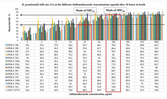
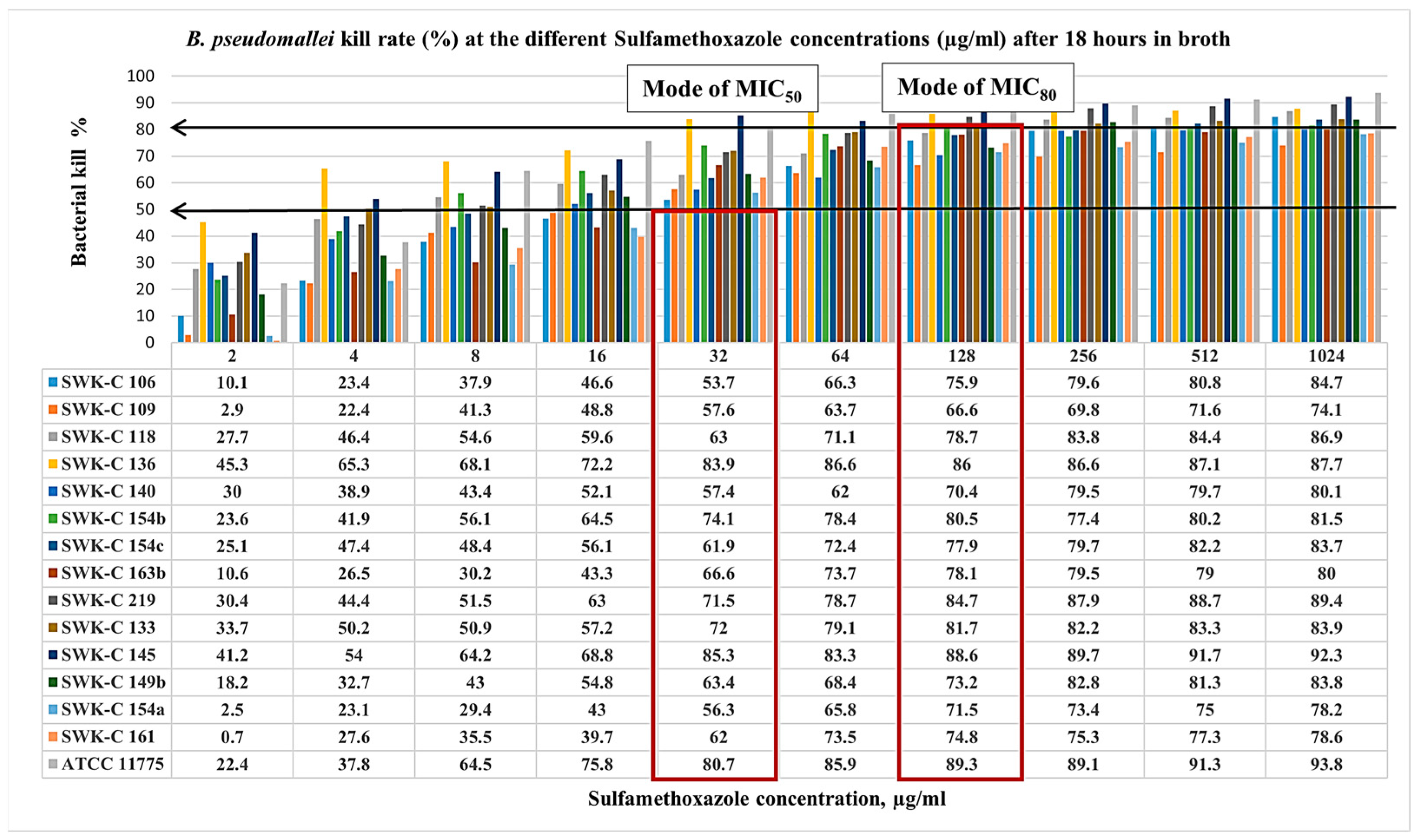
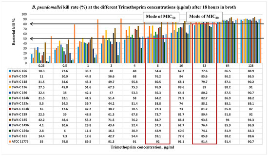
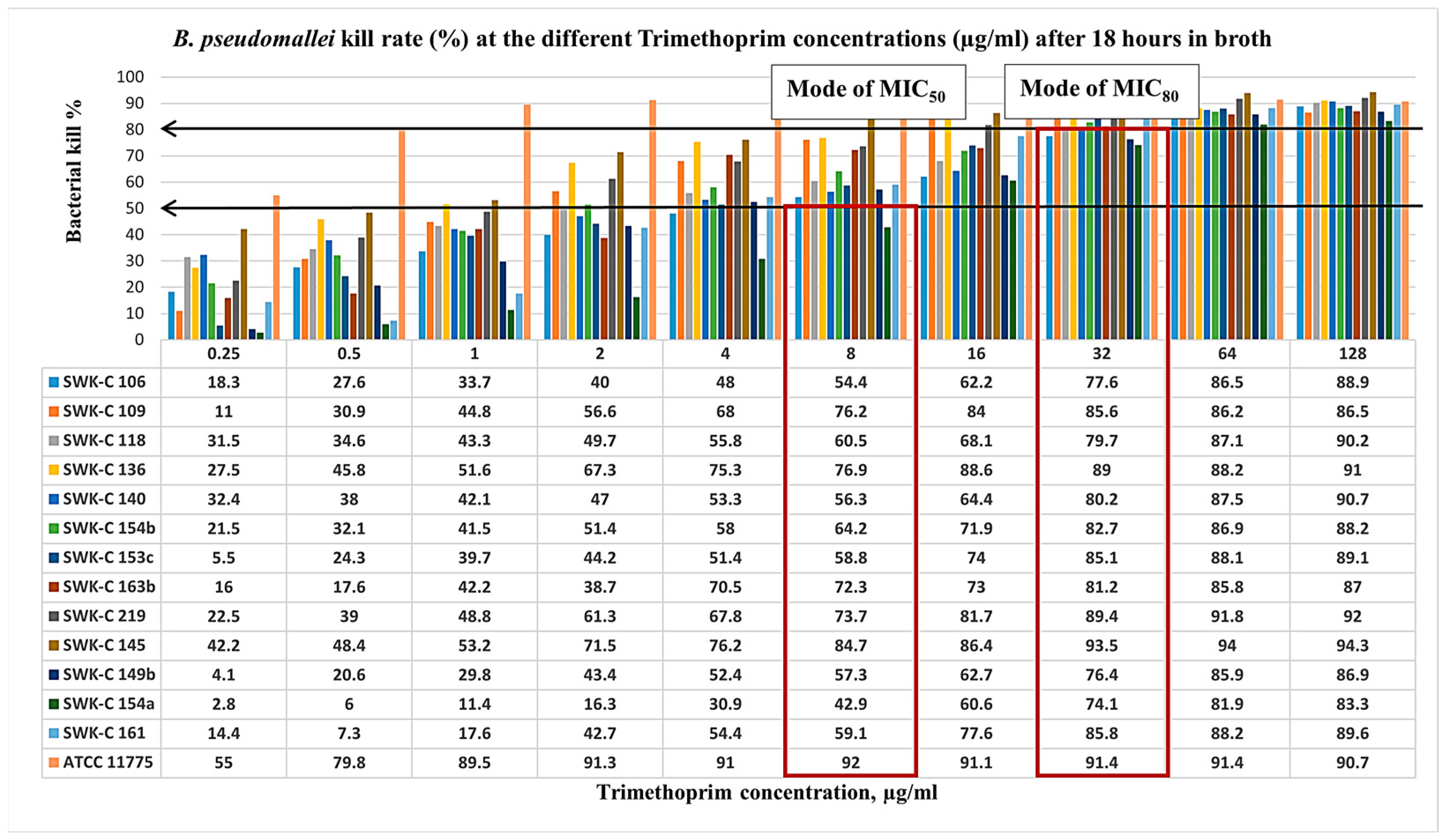
Following 18 h of broth microdilution testing with serial concentrations of sulfamethoxazole (SMX) and trimethoprim (TMP), the epidemiological MIC values of the isolates were analyzed (Figure 4 and Figure 5). Interestingly, the findings revealed that resistance to individual agents (SMX or TMP) did not confer resistance to the combined drug, trimethoprim-sulfamethoxazole. While all isolates were susceptible to SMX at MIC50 4 to 32 μg/mL, half of the isolates (7/14) were resistant at MIC80 (Figure 4). Among these, four isolates exhibited low-level resistance of MIC80, ranging from 512 to 1024 μg/mL, and three isolates demonstrated high-level resistance of MIC80 more than 1024 µg/mL. Susceptible isolates were inhibited at MIC80 values of 32 to 256 µg/mL, with a mode of 128 µg/mL. A similar trend was observed for TMP (Figure 5). All isolates were susceptible at MIC50 ranging from 1 to 8 μg/mL, but the majority (13/14) were resistant at MIC80 ranging from 16 to 128 μg/mL, with the modal MIC80 of 32 μg/mL.
Figure 4.
Sarawak clinical B. pseudomallei isolates kill rate (%) at the different sulfamethoxazole concentrations (µg/mL) after 18 h in broth. Isolates with the suffixes (b) and (c) represent subcultures or derivatives of the primary strain, denoted by suffix (a). For example, SWK-C 149 (b) and (c) were derived from SWK-C 149 (a). This naming convention was consistently applied to all isolates subcultured from their respective primary strains. The arrows indicate the bacterial kill rates at 50% and 80% whereas the boxes indicate the modes of MIC readings at MIC50 and MIC80.
Figure 5.
Sarawak clinical B. pseudomallei isolates kill rate (%) at the different trimethoprim concentrations (µg/mL) after 18 h in broth. Isolates with the suffixes (b) and (c) represent subcultures or derivatives of the primary strain, denoted by suffix (a). For example, SWK-C 149 (b) and (c) were derived from SWK-C 149 (a). This naming convention was consistently applied to all isolates subcultured from their respective primary strains. The arrows indicate the bacterial kill rates at 50% and 80% whereas the boxes indicate the modes of MIC readings at MIC50 and MIC80.
In Table 10, a comparison of the MIC values reveals that 7 trimethoprim-sulfamethoxazole-susceptible isolates were, in fact, resistant to both SMX and TMP at MIC80. Intriguingly, these isolates exhibited SMX MIC80 values higher than those of the trimethoprim-sulfamethoxazole-intermediate isolate SWK-C 118, and all were gentamicin-susceptible. Despite resistance to the individual agents, the synergistic combination of trimethoprim-sulfamethoxazole (co-trimoxazole) remained potent against all isolates, as confirmed by the E-test, with a predominant MIC of 2 µg/mL.
Table 10.
Comparisons of the MICs and susceptibility interpretation for 14 Sarawak clinical B. pseudomallei isolates.
3.4. PCR Analysis Suggests Divergence in the bpeEF-oprC Efflux Pump Gene Cluster in Sarawak B. pseudomallei Clinical Isolates
To investigate the molecular basis of the observed sulfamethoxazole resistance and its impact on the trimethoprim-sulfamethoxazole resistance, we targeted the bpeEF-oprC efflux pump gene cluster, a known determinant of trimethoprim-sulfamethoxazole resistance in B. pseudomallei. Thirteen primer sets were designed based on the alignment of reference strain B. pseudomallei K96243 (accession: BX571996.1) with sequences from closely related Thai and Malaysian isolates to ensure broad applicability.
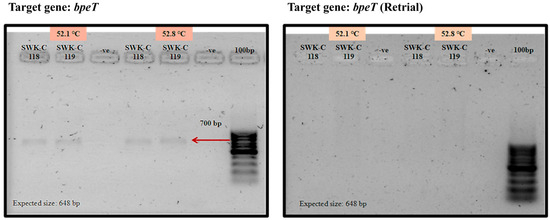
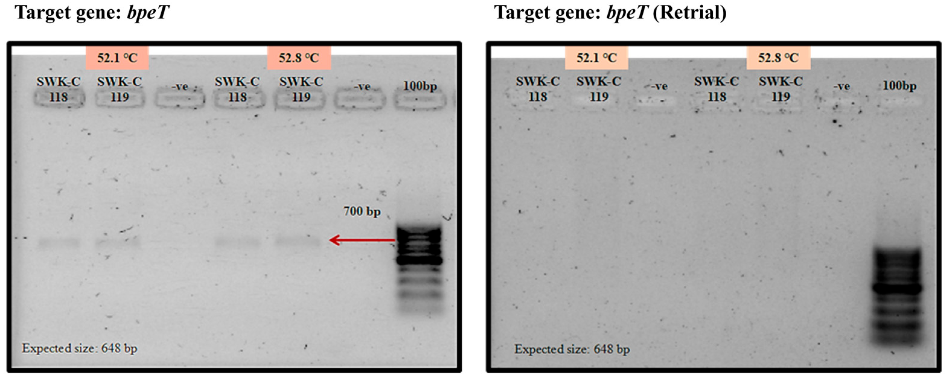
Despite extensive optimization efforts, including gradient PCR, we were unable to successfully amplify the bpeEF-oprC gene cluster (bpeT-llpE-bpeE-bpeF-oprC) from any of the Sarawak clinical isolates. Although a faint product (Figure 6) was initially observed for the bpeT gene, this result was not reproducible in subsequent trials.
Figure 6.
Analysis of the amplified bpeT from Sarawak B. pseudomallei clinical isolates.
4. Discussion
Sarawak, Malaysian Borneo, is endemic for a unique gentamicin-susceptible (GenS) population of B. pseudomallei belonging to the multilocus sequence type (ST)881 and its single-locus variant ST997 [12,13]. Comprehensive antibiotic susceptibility data for Sarawak clinical isolates remain scarce. Apart from the established GenS prevalence [12,13], only a single pediatric case of rare ceftazidime resistance has been reported [23]. Notably, some isolates from prior studies [12,13] were included in the present analysis (denoted in Appendix A Table A1 and Table A2).
This study demonstrates that the GenS genotype maintains high susceptibility to trimethoprim-sulfamethoxazole (SXT), reaffirming its clinical utility for melioidosis eradication therapy in this region. Our results indicated an estimated susceptibility prevalence of 96.3% (CLSI) and 97.6% (EUCAST). As confirmatory E-testing was limited to a subset of intermediate or resistant isolates, the estimated prevalence should be interpreted with caution. Despite that, this estimated prevalence aligns with the prevalence rates across Southeast Asia and Australia [24,25,26]. It is also consistent with a previous broader study of Malaysian isolates (which included 33/170 from Sarawak), where 90% of the isolates were SXT-susceptible [27]. The sustained in vitro susceptibility suggests SXT retains its bactericidal efficacy in Sarawak, which also corresponds to stable clinical treatment outcomes. Therefore, the findings of our present study resolve the concerns from previous reports of elevated MICs in Sarawak [10,11]. Our findings also recontextualize those reports to reflect methodological challenges in ASTs rather than a genuine loss or reduction in the clinical efficacy of SXT.
Another critical finding of our work is that disk diffusion testing significantly overcalled SXT resistance, a phenomenon previously documented in other endemic regions [22,23]. This overestimation resulted from visual ambiguity in some IZ phenotypes, particularly those with unclear or diffuse zone edges irrespective of haze growth (Figure 2). In addition, reader subjectivity is another major variable which impacts MIC estimation and the final susceptibility call [28]. This methodological challenge was attributed to the absence of a standardized, pathogen-specific AST methodology for B. pseudomallei at the time of the initiation of this study. The gap necessitated our local laboratories to adapt guidelines for Enterobacteriaceae and other non-Enterobacteriaceae. To ensure accurate reporting despite these constraints, we implemented a two-step AST algorithm by verifying non-susceptible disk diffusion results with an E-test. Our study offers a practical solution for AST reporting in cases where IZ cannot be confidently determined. Where resources permit, we recommend integrating a two-step AST into a standardized melioidosis diagnostic framework for Sarawak.
Our analysis further validated the CLSI breakpoints as the more reliable interpretive standard for these Sarawak isolates. While both 2017 CLSI and 2018 EUCAST criteria confirmed high susceptibility, EUCAST breakpoints yielded a substantially higher reported resistance rate (16.5% versus 2.4% by CLSI, Table 7). This higher rate defined by EUCAST, due in part to its more stringent susceptible breakpoint, could unnecessarily discourage the use of trimethoprim-sulfamethoxazole therapy.
Notably, our investigation coincided with EUCAST establishing its first standardized methodology for B. pseudomallei AST [28]. This new framework directly addresses our identified challenges, providing clear guidance for interpreting hazy or double inhibition zones via 80% growth inhibition measurement [28]. It also revises the ambiguous “intermediate” category to “susceptible, increased exposure” [29,30]. This update should reassure clinicians and bolster confidence without altering treatment regimens [9]. Therefore, while our data are aligned well with the CLSI standard, we recommend the gradual adoption of the pathogen-specific 2020 EUCAST guideline for future AST and surveillance works of B. pseudomallei in Sarawak. This will align local practices with the global standard, enhancing the consistency and clinical relevance of AST reporting.
Intriguingly, we found that the high in vitro efficacy of SXT persists despite intrinsic resistance to its individual components. Broth microdilution revealed that a substantial subset of SXT-susceptible isolates exhibited resistance to both SMX and TMP when assessed individually at MIC80. This finding aligns with observations from other endemic regions (northeast Thailand and northern Australia), where TMP resistance alone is common and does not compromise SXT efficacy [21]. In B. pseudomallei, such resistance is frequently attributed to the overexpression of the BpeEF-oprC efflux pump rather than target modification, indicating efflux as the predominant resistance mechanism [21]. The occurrence of SMX resistance, however, is rare. To our knowledge, this may be the first report of such resistance in GenS B. pseudomallei clinical isolates. In fact, the functional basis of SMX resistance in this pathogen remains largely uncharacterized. A previous study [31] suggested that BpeEF-OprC expression due to bpeT mutations or pump overexpression due to bpeS mutations, which is responsible for the TMP resistance, contributed to the reduced SMX susceptibility in regulatory mutants.
Additionally, this phenomenon occurred in isolates that otherwise maintained full susceptibility to clinically relevant antibiotics, including meropenem, ceftazidime, doxycycline, and amoxicillin-clavulanate. This suggests that their resistance to TMP and SMX is a specific anomaly, not part of a multidrug-resistant phenotype. This paradox underscores the paramount importance of drug synergy, whereby the combined formulation overcomes resistance for either drug alone. Hence, the unexpected resistance to the individual agents does not diminish the clinical utility of trimethoprim-sulfamethoxazole, which remains a cornerstone of melioidosis eradication therapy in Sarawak.
A key limitation of this study, however, is our inability to define the genetic basis for this phenotype. We were unable to amplify the bpeEF-oprC efflux pump gene cluster using primers designed against classic, gentamicin-resistant strains. The consistent failure to amplify the known resistance determinant [20] does not exclude the presence of an efflux-mediated resistance mechanism but rather suggests significant sequence divergence in this locus within the GenS Sarawak genotype. It is plausible that the unique lineage of ST881 and ST997 with distinguished mutation in the amrB efflux pump gene [12] also harbors distinct variations within the bpeEF-oprC operon. This is a compelling hypothesis that warrants future investigation through whole-genome sequencing.
In conclusion, our work provides strong evidence that SXT remains an effective eradication therapy for melioidosis in Sarawak. We recommend the use of CLSI guidelines, supplemented with the E-test for non-susceptible disk diffusion results, to ensure accurate AST reporting. The difference between individual and combination drug susceptibility, coupled with the genetic uniqueness of these isolates, highlights that the resistance mechanisms in this endemic population may be distinct. Future genomic studies utilizing next-generation sequencing of the GenS Sarawak B. pseudomallei clinical isolates are essential to elucidating these underlying mechanisms. These methods generate continuous sequence reads spanning the entire operons. This capability will allow for the comprehensive detection of structural variation or novel mutations within the complex resistance loci like the bpeEF-oprC efflux pump gene cluster, which could not be amplified with only PCR.
Author Contributions
Conceptualization, L.L.S., J.-S.W., M.-H.O., and Y.P.; methodology, L.L.S. and Y.P.; formal analysis, L.L.S. and Y.P.; investigation, L.L.S., Y.P., T.L.-L.S., M.-H.O., A.M., and J.-S.W.; resources, Y.P., T.L.-L.S., M.-H.O., A.M., and J.-S.W.; data curation, L.L.S., T.L.-L.S., and Y.P.; writing—original draft preparation, L.L.S., Y.P., T.L.-L.S., M.-H.O., A.M., and J.-S.W.; writing—review and editing, L.L.S., Y.P., and M.-H.O.; supervision, Y.P. and M.-H.O.; project administration, L.L.S., Y.P., and M.-H.O.; funding acquisition, Y.P. All authors have read and agreed to the published version of the manuscript.
Funding
This research was supported by internal funding from the Institute of Health and Community Medicine, Universiti Malaysia Sarawak (UNIMAS) and received no external funding.
Institutional Review Board Statement
This study was approved by the Malaysian Medical Research Ethics Committee of NMRR-16-1029-31390 on 25 January 2017, and registered with the National Medical Research Registrar and the Clinical Research Centre, Ministry of Health Malaysia.
Informed Consent Statement
Anonymized bacterial isolates were obtained from archival collections and routine laboratory diagnostic procedures; hence, the requirement for written patient consent was waived.
Data Availability Statement
Further information and data are available from the corresponding author upon reasonable request.
Acknowledgments
We acknowledge the contribution of doctors and nurses of Bintulu Hospital, Sibu Hospital, Kapit Hospital, Miri Hospital, Sarawak General Hospital, and Borneo Medical Centre for their involvement in the samples collection. We are grateful to our fellow colleagues in Institute of Health and Community Medicine, UNIMAS, especially the Melioid Team for their laboratory assistance and support throughout the study. We would also like to express our gratitude to the Melioidosis Research Team (Melioid Mob) at the Menzies School of Health Research, Northern Territory, Australia for their guidance and sharing of annotated isolates.
Conflicts of Interest
The authors declare no conflicts of interest.
Abbreviations
The following abbreviations are used in this manuscript:
| MIC | Minimal inhibitory concentration |
| CLSI | Clinical Laboratory Standards Institute |
| EUCAST | European Committee on Antimicrobial Susceptibility Testing |
| QC | Quality Control |
| IZ | Inhibition zone |
| AST | Antimicrobial Susceptibility Testing |
| MIC50 | Minimal inhibitory concentration at 50% inhibited growth |
| MIC80 | Minimal inhibitory concentration at 80% inhibited growth |
| OD | Optical density |
| GenS | Gentamicin-susceptible |
| TMP | Trimethoprim |
| SMX | Sulfamethoxazole |
| PCR | Polymerase Chain Reaction |
| ST | Sequence type |
Appendix A
Table A1 in this section showed the overall disk diffusion results of the trimethoprim-sulfamethoxazole susceptibility of the Sarawak isolates, detailed with the source location. Some of the isolates were previously studied in [12], characterizing the Gentamicin susceptibility profile (see Table A1). The bolded isolates in Table A1 then selected for the E-test against trimethoprim-sulfamethoxazole, which results were tabulated in Table A2.
Table A1.
Disk diffusion results of 164 Sarawak clinical B. pseudomallei isolates when tested against the 25 µg Trimethoprim-sulfamethoxazole disk.
Table A2.
Minimum inhibitory concentration results of Sarawak clinical B. pseudomallei isolates against the trimethoprim-sulfamethoxazole E-test strip.
References
- Currie, B.J.; Kaestli, M. A global picture of melioidosis. Nature 2016, 529, 290–291. [Google Scholar] [CrossRef] [Scilit]
- Limmathurotsakul, D.; Golding, N.; Dance, D.A.B.; Messina, J.P.; Pigott, D.M.; Moyes, C.L.; Rolim, D.B.; Bertherat, E.; Day, N.P.J.; Peacock, S.J.; et al. Predicted global distribution of Burkholderia pseudomallei and burden of melioidosis. Nat. Microbiol. 2016, 1, 15008. [Google Scholar] [CrossRef] [Scilit]
- Wiersinga, W.J.; Virk, H.S.; Torres, A.G.; Currie, B.J.; Peacock, S.J.; Dance, D.A.B.; Limmathurotsakul, D. Melioidosis. Nat. Rev. Dis. Primers 2018, 4, 17107. [Google Scholar] [CrossRef] [Scilit]
- Sanchez-Villamil, J.I.; Torres, A.G. Melioidosis in Mexico, Central America, and the Caribbean. Trop. Med. Infect. Dis. 2018, 3, 24. [Google Scholar] [CrossRef] [Scilit] [PubMed]
- Nathan, S.; Chieng, S.; Kingsley, P.V.; Mohan, A.; Podin, Y.; Ooi, M.-H.; Mariappan, V.; Vellasamy, K.M.; Vadivelu, J.; Daim, S.; et al. Melioidosis in Malaysia: Incidence, Clinical Challenges, and Advances in Understanding Pathogenesis. Trop. Med. Infect. Dis. 2018, 3, 25. [Google Scholar] [CrossRef] [Scilit]
- Sia, T.L.L.; Mohan, A.; Ooi, M.-H.; Chien, S.-L.; Tan, L.-S.; Goh, C.; Pang, D.C.L.; Currie, B.J.; Wong, J.-S.; Podin, Y. Epidemiological and Clinical Characteristics of Melioidosis Caused by Gentamicin-Susceptible Burkholderia pseudomallei in Sarawak, Malaysia. Open Forum Infect. Dis. 2021, 8, ofab460. [Google Scholar] [CrossRef] [Scilit] [PubMed]
- Mohan, A.; Podin, Y.; Tai, N.; Chieng, C.-H.; Rigas, V.; Machunter, B.; Mayo, M.; Wong, D.; Chien, S.-L.; Tan, L.-S.; et al. Pediatric melioidosis in Sarawak, Malaysia: Epidemiological, clinical and microbiological characteristics. PLoS Neglected Trop. Dis. 2017, 11, e0005650. [Google Scholar] [CrossRef] [Scilit] [PubMed]
- White, N. Melioidosis. Lancet 2003, 361, 1715–1722. [Google Scholar] [CrossRef] [Scilit]
- Dance, D. Treatment and prophylaxis of melioidosis. Int. J. Antimicrob. Agents 2014, 43, 310–318. [Google Scholar] [CrossRef] [Scilit]
- Vima, V.; Ling, H.-W.; Cho, W.-M.; Norhuzaimah, J. Incidence, Risk Factors and Clinical Epidemiology of Melioidosis in Miri Hospital, Sarawak, Malaysia. Sarawak Health J. 2016, 3, 22–23. [Google Scholar]
- Yong, K.-Y.; Tang, A.S.-O.; Teh, Y.-C.; Fam, T.-L.; Chua, H.-H. Prevalence of Antibiotic Resistance in Burkholderia pseudomallei Cases Presented to Miri General Hospital. Sarawak Health J. 2016, 3, 26. [Google Scholar]
- Podin, Y.; Kaestli, M.; McMahon, N.; Hennessy, J.; Ngian, H.U.; Wong, J.S.; Mohana, A.; Wong, S.C.; William, T.; Mayo, M.; et al. Reliability of Automated Biochemical Identification of Burkholderia pseudomallei Is Regionally Dependent. J. Clin. Microbiol. 2013, 51, 3076–3078. [Google Scholar] [CrossRef] [Scilit]
- Podin, Y.; Sarovich, D.S.; Price, E.P.; Kaestli, M.; Mayo, M.; Hii, K.; Ngian, H.; Wong, S.; Wong, I.; Wong, J.; et al. Burkholderia pseudomallei Isolates from Sarawak, Malaysian Borneo, Are Predominantly Susceptible to Aminoglycosides and Macrolides. Antimicrob. Agents Chemother. 2013, 58, 162–166. [Google Scholar] [CrossRef] [Scilit]
- CLSI. Performance Standards for Antimicrobial Susceptibility Testing 27th ed. CLSI Supplement M100; Clinical Laboratory Standards Institute: Wayne, PA, USA; Available online: https://www.google.com/url?sa=t&source=web&rct=j&opi=89978449&url=https://www.researchgate.net/profile/Nikolaos_Andritsos/post/Where_to_find_MICs_of_antimicrobial_agents/attachment/59d659e279197b80779af319/AS%253A544238144323584%25401506767893696/download/2017_CLSI_M100_Performance%2BStandards%2Bfor%2BAntimicrobial%2BSusceptibility%2BTesting_27th%2Bed..pdf&ved=2ahUKEwjwu9rQ-paSAxVp2TQHHZHCOzcQFnoECBoQAQ&usg=AOvVaw1tGIX4d98p2DVfRY_2WrOh (accessed on 12 November 2018).
- European Committee on Antimicrobial Susceptibility Testing. Breakpoint Tables for Interpretation of MICs and Zone Diameters. Version 8.1, Valid from 2018-05-15. 2018. Available online: https://www.eucast.org/ast_of_bacteria/previous_versions_of_documents/ (accessed on 12 November 2018).
- CLSI. Methods for Dilution Antimicrobial Susceptibility Tests for Bacteria That Grow Aerobically 11th ed CLSI Standard M07; Clinical Laboratory Standards Institute: Wayne, PA, USA, 2018; Available online: https://clsi.org/shop/standards/m07/ (accessed on 20 January 2019).
- Warrens, M.J. Five Ways to Look at Cohen’s Kappa. J. Psychol. Psychother. 2015, 5, 1000197. [Google Scholar] [CrossRef]
- Untergasser, A.; Cutcutache, I.; Koressaar, T.; Ye, J.; Faircloth, B.C.; Remm, M.; Rozen, S.G. Primer3—New capabilities and interfaces. Nucleic Acids Res. 2012, 40, e115. [Google Scholar] [CrossRef] [Scilit] [PubMed]
- Holden, M.T.G.; Titball, R.W.; Peacock, S.J.; Cerdeño-Tárraga, A.M.; Atkins, T.; Crossman, L.C.; Pitt, T.; Churcher, C.; Mungall, K.; Bentley, S.D.; et al. Genomic plasticity of the causative agent of melioidosis, Burkholderia pseudomallei. Proc. Natl. Acad. Sci. USA 2004, 101, 14240–14245. [Google Scholar] [CrossRef] [Scilit]
- Biot, F.V.; Valade, E.; Garnotel, E.; Chevalier, J.; Villard, C.; Thibault, F.M.; Vidal, D.R.; Pagès, J.-M. Involvement of the Efflux Pumps in Chloramphenicol Selected Strains of Burkholderia thailandensis: Proteomic and Mechanistic Evidence. PLoS ONE 2011, 6, e16892. [Google Scholar] [CrossRef] [Scilit]
- Podnecky, N.L.; Wuthiekanun, V.; Peacock, S.J.; Schweizer, H.P. The BpeEF-OprC Efflux Pump Is Responsible for Widespread Trimethoprim Resistance in Clinical and Environmental Burkholderia pseudomallei Isolates. Antimicrob. Agents Chemother. 2013, 57, 4381–4386. [Google Scholar] [CrossRef] [Scilit]
- Payne, G.W.; Vandamme, P.; Morgan, S.H.; LiPuma, J.J.; Coenye, T.; Weightman, A.J.; Jones, T.H.; Mahenthiralingam, E. Development of a recA Gene-Based Identification Approach for the Entire Burkholderia Genus. Appl. Environ. Microbiol. 2005, 71, 3917–3927. [Google Scholar] [CrossRef] [Scilit]
- Sia, T.L.-L.; Lai, C.D.; Manan, K.; Khiu, F.-L.; Bakhtiar, S.Z.; Chor, Y.-K.; Chien, S.-L.; Tan, L.-S.; Ooi, M.-H.; Mohan, A. Ceftazidime-resistance in pediatric melioidosis: A case report and literature review. IDCases 2025, 39, e02149. [Google Scholar] [CrossRef] [Scilit] [PubMed]
- Dance, D.; Davong, V.; Soeng, S.; Phetsouvanh, R.; Newton, P.; Turner, P. Trimethoprim/sulfamethoxazole resistance in Burkholderia pseudomallei. Int. J. Antimicrob. Agents 2014, 44, 368–369. [Google Scholar] [CrossRef] [Scilit] [PubMed]
- Saiprom, N.; Amornchai, P.; Wuthiekanun, V.; Day, N.P.; Limmathurotsakul, D.; Peacock, S.J.; Chantratita, N. Trimethoprim/sulfamethoxazole resistance in clinical isolates of Burkholderia pseudomallei from Thailand. Int. J. Antimicrob. Agents 2015, 45, 557–559. [Google Scholar] [CrossRef] [Scilit]
- Dutta, S.; Haq, S.; Hasan, M.R.; Haq, J.A. Antimicrobial susceptibility pattern of clinical isolates of Burkholderia pseudomallei in Bangladesh. BMC Res. Notes 2017, 10, 299. [Google Scholar] [CrossRef] [Scilit] [PubMed]
- Ahmad, N.; Hashim, R.; Noor, A.M. The In Vitro Antibiotic Susceptibility of Malaysian Isolates of Burkholderia pseudomallei. Int. J. Microbiol. 2013, 2013, 121845. [Google Scholar] [CrossRef] [Scilit] [PubMed]
- Karatuna, O.; Dance, D.; Matuschek, E.; Åhman, J.; Turner, P.; Hopkins, J.; Amornchai, P.; Wuthiekanun, V.; Cusack, T.-P.; Baird, R.; et al. Burkholderia pseudomallei multi-centre study to establish EUCAST MIC and zone diameter distributions and epidemiological cut-off values. Clin. Microbiol. Infect. 2021, 27, 736–741. [Google Scholar] [CrossRef] [Scilit]
- Dance, D.A.; Wuthiekanun, V.; Baird, R.W.; Norton, R.; Limmathurotsakul, D.; Currie, B.J. Interpreting Burkholderia pseudomallei disc diffusion susceptibility test results by the EUCAST method. Clin. Microbiol. Infect. 2021, 27, 827–829. [Google Scholar] [CrossRef] [Scilit]
- Kahlmeter, G.; Giske, C.G.; Kirn, T.J.; Sharp, S.E. Point-Counterpoint: Differences between the European Committee on Antimicrobial Susceptibility Testing and Clinical and Laboratory Standards Institute Recommendations for Reporting Antimicrobial Susceptibility Results. J. Clin. Microbiol. 2019, 57. [Google Scholar] [CrossRef] [Scilit]
- Podnecky, N.L.; Rhodes, K.A.; Mima, T.; Drew, H.R.; Chirakul, S.; Wuthiekanun, V.; Schupp, J.M.; Sarovich, D.S.; Currie, B.J.; Keim, P.; et al. Mechanisms of Resistance to Folate Pathway Inhibitors in Burkholderia pseudomallei: Deviation from the Norm. mBio 2017, 8, e01357-17. [Google Scholar] [CrossRef] [Scilit]

Disclaimer/Publisher’s Note: The statements, opinions and data contained in all publications are solely those of the individual author(s) and contributor(s) and not of MDPI and/or the editor(s). MDPI and/or the editor(s) disclaim responsibility for any injury to people or property resulting from any ideas, methods, instructions or products referred to in the content. |
© 2026 by the authors. Licensee MDPI, Basel, Switzerland. This article is an open access article distributed under the terms and conditions of the Creative Commons Attribution (CC BY) license.