


Simple Summary
We report for the first time a case of skin lesions due to active feeding of the ant Pheidole pallidula on a human corpse found in a suburban area of Cosenza (Region Calabria, Southern Italy). The autopsy revealed only major and minor workers of the species on the body, but no other insects. Ants are known to cause post-mortem skin damage on human and animal corpses and these damages may hinder pathologists’ forensic investigations into the cause, location and time of death. As this is the first case of skin artifacts caused on a human body by ants of the genus Pheidole, an overview of the relevance of the genus in forensic entomology is also provided.
Abstract
Many ant species play a relevant role in the successional patterns of insects colonizing human and animal corpses. Although feeding behavior in ants depends on the trophic preferences of each taxon, these insects are often reported in several studies as predators of eggs, larvae, and adults of necrophagous insects. Among ants, some species are known to cause damage on human and animal dermis, known as post-mortem skin artifacts. The extensive activity of worker ants on corpses may hinder forensic pathological investigations aimed at determining the cause and location of death, as well as the assessment of entomologists in estimating the minimum post-mortem interval. We report for the first time a case of skin lesions on a human corpse found in a suburban area of the city of Cosenza (Calabria, Southern Italy), caused by the ant Pheidole pallidula (Hymenoptera: Formicidae). During the autopsy, numerous individuals (major and minor workers) were observed feeding on the corpse, but no other insects were found on it. We discuss the appearance of skin artifacts caused by P. pallidula and provide an overview of the genus Pheidole in forensic entomology.
1. Introduction
Some insects accelerate the decay process of corpses by feeding directly on the remains, while others, on the contrary, slow it down by preying on necrophagous insects [1]. Ants (Hymenoptera belonging to the family Formicidae) are widely distributed throughout the world [2] and are capable of both feeding on decaying remains and preying on other insects [3,4]. Thanks to their foraging strategies, efficient chemical recruitment and trail communication systems, many ant species can rapidly find, colonize and exploit new food resources from plants or animals (living or dead) [5,6,7,8]. These abilities enable ants to colonize animal carcasses and human remains, as confirmed by forensic evidence showing that ants may consume corpses, and interfere with dipteran colonizers used to estimate the minimum post-mortem interval by slowing the decomposition rate of corpses [3,9,10,11,12]. Feeding activity of ants may cause the formation of irregular superficial skin damages, which can be misinterpreted as ante-mortem injuries [3,10,13,14,15]. In Italy, the species of the family Formicidae reported to have caused post-mortem damages or artifacts on human bodies are Tapinoma nigerrimum (Nylander, 1856) in rural and suburban areas [10,13,15] and Crematogaster scutellaris (Olivier, 1791) in indoor areas [16]. Workers of C. scutellaris have also been observed preying on eggs and dipteran adults, causing damage to the skin of a pig carcass [9].
Here are reported for the first time skin lesions caused by the ant Pheidole pallidula (Nylander, 1849) on a human corpse found in a suburban area of the city of Cosenza (Calabria, Southern Italy). The appearance of skin artifacts caused by P. pallidula and their possible role in forensic crime scene alterations are also discussed, together with an overview of the involvement of the genus Pheidole in forensic entomology.
2. Case Report
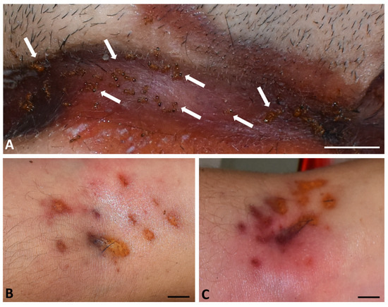
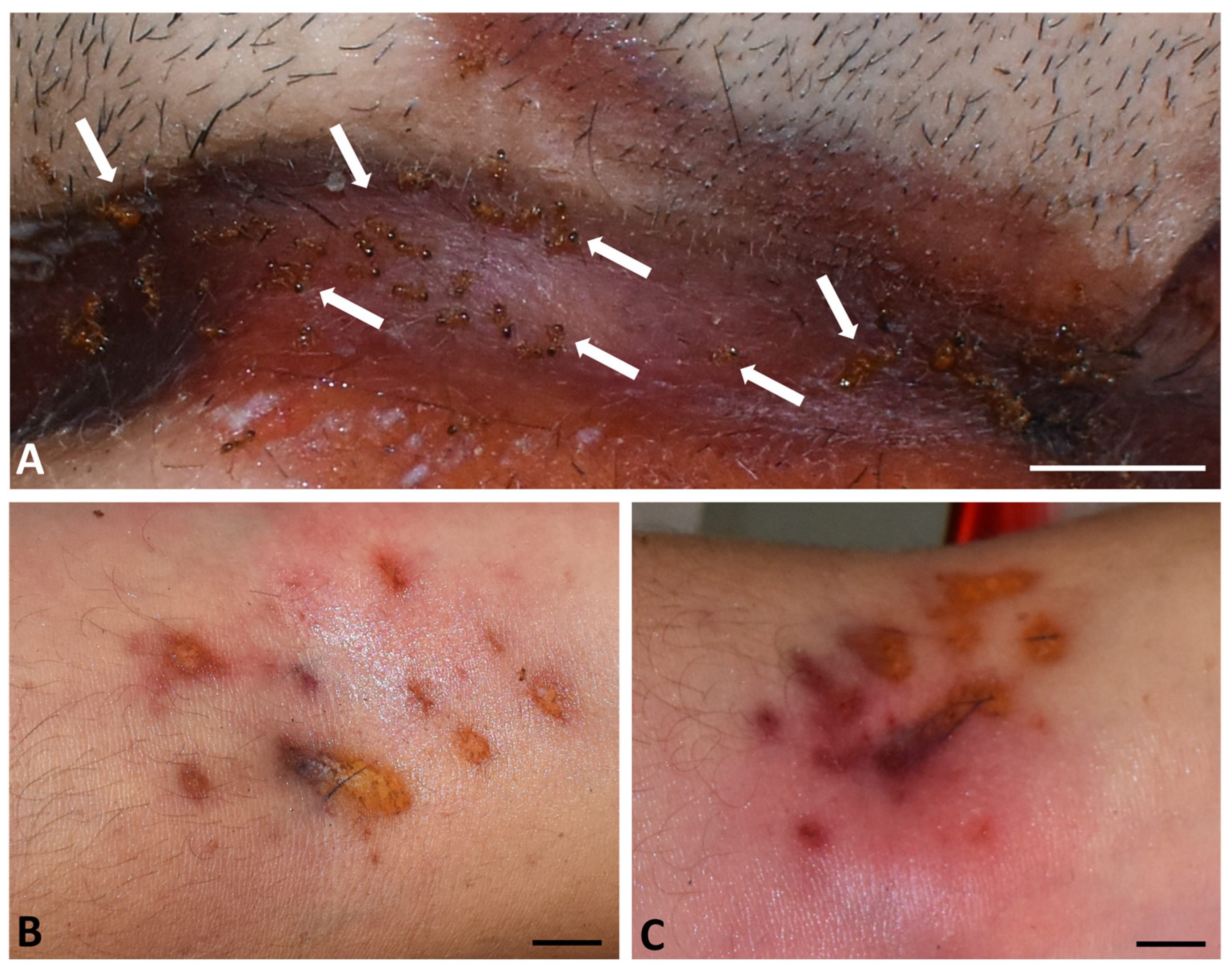
A 40-year-old man was found dead in a suburban area of Cosenza in August 2021. His body was discovered in the early morning in his home, hanging in a cellar that was locked from the inside. The man was wearing a T-shirt, shorts, and slippers. The autopsy, conducted three days after the body was discovered, determined the cause of death to be acute neurological and respiratory failure resulting from hanging. The presence of a hyoid bone fracture confirmed the hanging. Based on the findings and investigations conducted by the State Police, the post-mortem interval was estimated to be around 10–12 h, meaning the man died during the night. The case was officially declared a suicide. During the autopsy, live ant activity was observed post-mortem on the face, torso, back and arms, as well as on eyes and eyelids. The ants were observed feeding and moving on the skin surface (Figure 1). Macroscopic examination revealed irregular yellow-orange and brown lesions (0.5–5 cm long), scattered across the skin of the neck at the level of the nylon string used for hanging. No fluid or blood loss was observed on the damaged skin. Ants were observed feeding near the lesions and along the edges of the neck wounds (Figure 1A). Additional reddish-brown and roundish skin lesions (0.2 to 1.1 cm long) were scattered across the skin surface of the elbow crease of both arms (Figure 1B,C).
Figure 1.
Autopsy of the deceased, external view. (A) Neck area: live ants (arrows) feeding in the lesions caused by hanging. Scale bar = 1.0 cm. (B) Skin lesions on the elbow crease of the left arm caused by ants. Scale bar = 0.5 cm. (C) Skin lesions on the elbow crease of the right arm caused by ants. Scale bar = 0.5 cm.
A total of 13 live ants from all those found alive on the body were collected during the autopsy with entomological tweezers and preserved in 70% alcohol for species identification. During the ant sampling, no larvae or dipteran eggs were found on the body, probably because the death occurred at night and in a cellar with limited access to flies.
Individuals were observed and photographed using a stereomicroscope (Meiji Techno RZ, Meiji Techno America, Santa Clara, CA, USA), equipped with an image acquisition system (Delta Pix camera, Smoerum, Denmark), and then identified using taxonomic articles and books [17,18,19,20]. All collected ants were preserved in 70% alcohol at the Department of Biology, Ecology and Earth Sciences of the University of Calabria (Rende, Italy).
Histological analyses were performed on skin lesions caused by live ants on the right arm of the deceased (Figure 1C), according to a previously published protocol [15]. Sections were observed under a light microscope (Leica DM4, Leica Microsystems, Wetzlar, Germany) and acquired with a digital camera (Leica Microsystems, Wetzlar, Germany).
3. Bibliographic Methods
The bibliographic search on ants in forensic entomology was initially based on a previous study [15] and on publications indexed on PubMed, and was subsequently extended through web engines using the keywords “ants, artifacts, Formicidae, forensic entomology, animal and human corpses, Pheidole, Pheidole pallidula”. In addition to case reports, this search led to another review [21], which included a list of carrion-associated ant species and their references. The search about the role of Pheidole pallidula and the genus Pheidole in forensic entomology was conducted through November 2025.
4. Results and Discussion
4.1. Species Identification
Among European ants, identifying the genus Pheidole is relatively simple, based on the morphology of the mesosoma, node, and head. Females have antennae composed of 12 segments, with the last three antennomeres forming a club. Workers are dimorphic, with majors (also known as soldiers) possessing a massive head to produce an increased mandibular force [18,19,22].
In Italy, two species of the genus Pheidole have been reported outdoors. The first species, present in all Italian territory, is P. pallidula, belonging to the P. pallidula complex, including P. pallidula, P. balcanica Seifert, 2016, P. koshewnikovi Ruzsky, 1905 and P. cicatricosa Stitz, 1917 [17]. The second species is the non-native Pheidole indica (Mayr 1879), known mainly from urban areas of Sicily and the nearby island of Pantelleria [23,24,25]. Two other non-native species have been reported, Pheidole megacephala (Fabricius, 1793), found in 2001 in greenhouses of plants imported from Asia in Parabiago (Milan) and in 2006 inside a storage hangar in Cargo City (Milan Malpensa Airport, Ferno, Varese, Italy), and Pheidole nodus Smith, 1874, found in 2001 in greenhouses of plants imported from Asia in Parabiago (Milan). Neither species has been confirmed as established [25,26,27].
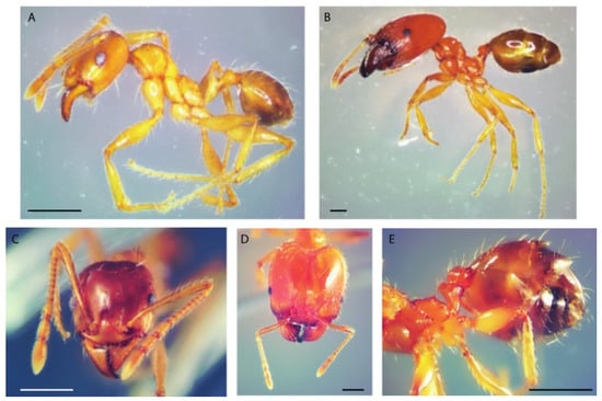
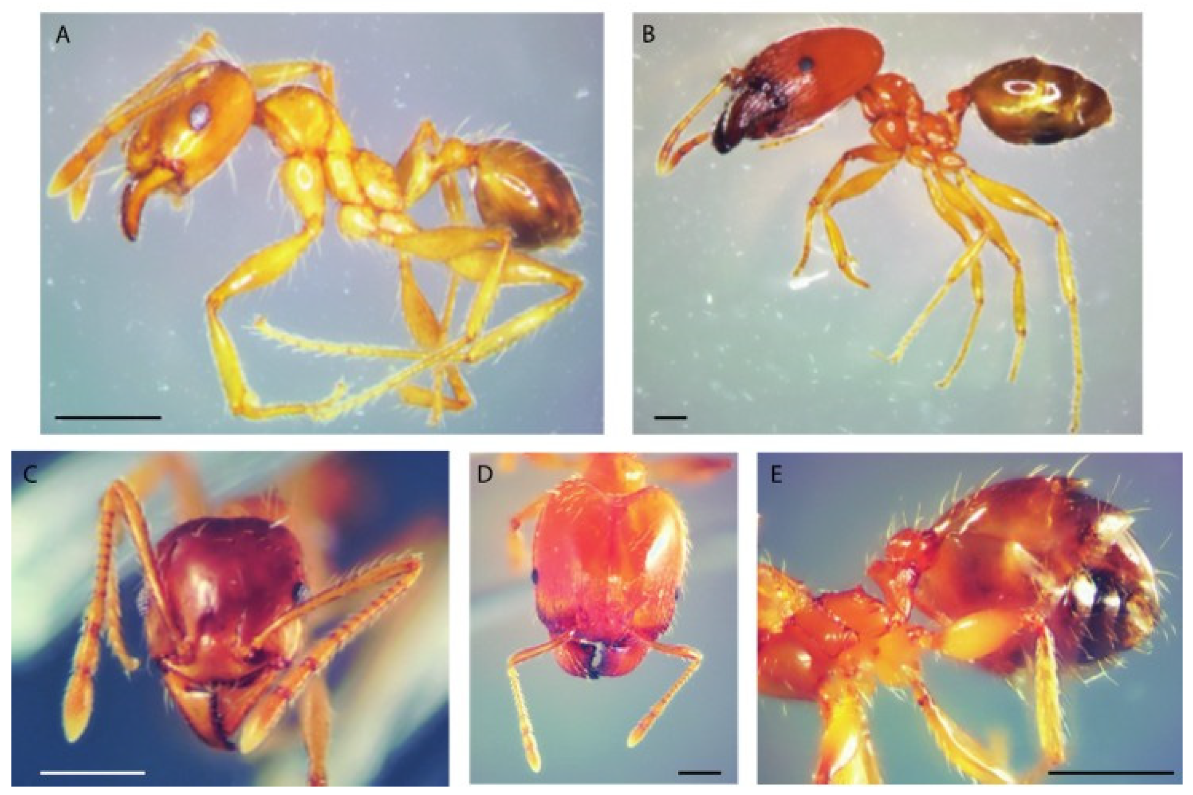
Workers of the P. pallidula complex are easily distinguishable from P. indica by the lack of a deep promesonotal groove and by the smoother head sculpture of soldiers, and from P. megacephala by the different ventral profile of the postpetiole. Morphological analysis of the collected ants confirmed that all specimens belonged to the Pheidole pallidula complex (Figure 2). Since P. pallidula is the only species of the complex recorded in Italy, the collected specimens are attributed to this species.
Figure 2.
Individuals of Pheidole pallidula collected on the deceased. (A) Minor worker, lateral view. (B) Major worker, lateral view. (C) Head of a minor worker, frontal view. (D) Head of a major worker, frontal view. (E) Major worker. From left to right: mesosoma, petiole, postpetiole and gaster. Scale bars = 0.5 mm.
Pheidole pallidula is an opportunistic, often dominant, omnivorous species, known to feed on invertebrates, seeds, and honeydew; it generally nests in ground or under stones in colonies of tens of thousands of individuals [18,19].
4.2. Case Interpretation and Literature Overview
Ants have been documented as playing a role in sarcosaprophagous insect communities and have been found associated with animal and human corpses in various habitats across several geographical areas, both outdoors and indoors.
The role of ants in sarcosaprophagous insect communities has been documented, and they have been found associated with animal and human corpses in various habitats across different geographic areas, both indoors and outdoors. A recent extensive study reviews in detail the role of ants in vertebrate carrion decomposition [21]. Like other insects, ants can influence the decay process of dead bodies by feeding on remains, preying on carrion arthropods, or using corpses as nesting sites or refuges [21,28,29]. Ant feeding activities on corpses can produce post-mortem artifacts on the skin of humans and animals [3,10,11,13,15,21,30].
These post-mortem artifacts typically appear as serpiginous lesions on the skin surface of many anatomical areas [3,10,30]. Post-mortem damage inflicted by unidentified species of the genera Atta and Cephalotes have been reported in Brazil [31], with the former genus found in rural and urban areas and the latter genus in forest areas. Other unidentified Formicidae taxa have been reported to inflict post-mortem damage on human skin [15].
In Italy, the ant species reported to have caused post-mortem damages or artifacts on human bodies are T. nigerrimum in rural and suburban areas [10,13,15], now considered a species complex [25] and C. scutellaris indoors [16].
Among the Formicidae taxa feeding on corpses, the cosmopolitan genus Pheidole Westwood, 1839, belonging to the subfamily Myrmicinae, has historically been reported as occasionally present on carcasses, with an unclear ecological role [32]. This genus currently includes over 1160 described and more than 1500 estimated species, thus representing one of the most species-rich ant genera, together with the genus Camponotus Mayr, 1861 [18,33,34].
Within the genus Pheidole, 20 species and 1 subspecies of the genus have been associated with vertebrate carcasses, ranging from small animals, such as chicks and mice, to large ones, such as pigs, deer and American alligators (Table 1). Other reports mention the genus Pheidole in association with vertebrate carcasses, but without species identification (Table 2). Individuals belonging to the genus, with or without species identification, have also been captured in necrophagous insect traps loaded with pig and cow liver, chicken meat and sardines (Table 1 and Table 2). In Italy, the only report of possible necrophagous activity of the genus Pheidole concerned pig carcasses periodically exhumed on the succession of arthropod fauna in experimental studies on decomposition stages in Calabria [35].
The ecological role of species of the genus Pheidole apparently ranges from necrophagous to predatory and omnivorous. In Brazil, Pheidole radoszkowskii Mayr, 1884, has been reported to cause skin lesions on rat carrions [36], and an unidentified species of the genus Pheidole was reported to cause small lesions on nose and external ear of a rabbit [37]. In Malaysia, P. megacephala was found associated with exposed rat and macaque carcasses and was thought to be responsible for slowing the decomposition rate of the examined animals [12]. Environments where the genus Pheidole was found associated with carcasses and entomological traps were forests, urban, rural and suburban areas (Table 1 and Table 2). To date, the genus Pheidole has never been reported actively feeding on human corpses anywhere in the world. The case reported here in Southern Italy is therefore the first involving necrophagous activity on a human corpse of the genus Pheidole and specifically of P. pallidula. This species had previously been mentioned as associated with animal carcasses and traps in open environments (Table 1), so this is also the first case reporting active feeding of P. pallidula on a human corpse indoors.
Table 1.
Cases involving species and subspecies of the genus Pheidole detected on animal corpses, on animal meat in entomological traps, and on a human corpse. NR, not reported.
Table 2.
Cases involving the genus Pheidole (without species identification) detected on animal corpses and in entomological traps. NR, not reported.
Injuries caused by ants and other arthropods [15] could be mistaken for those resulting from chemical burns, cigarette scars, nail abrasions or intravenous drug use [3], making it crucial to distinguish them for accurate crime scene reconstruction. In the reported case, the first interpretation of the injuries on the arms by investigators was that they were repeated intravenous injections.
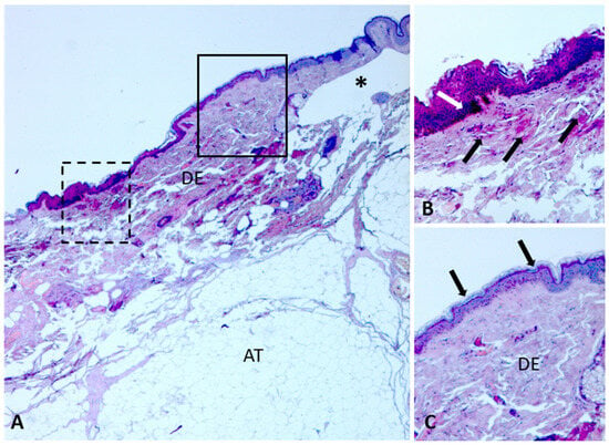
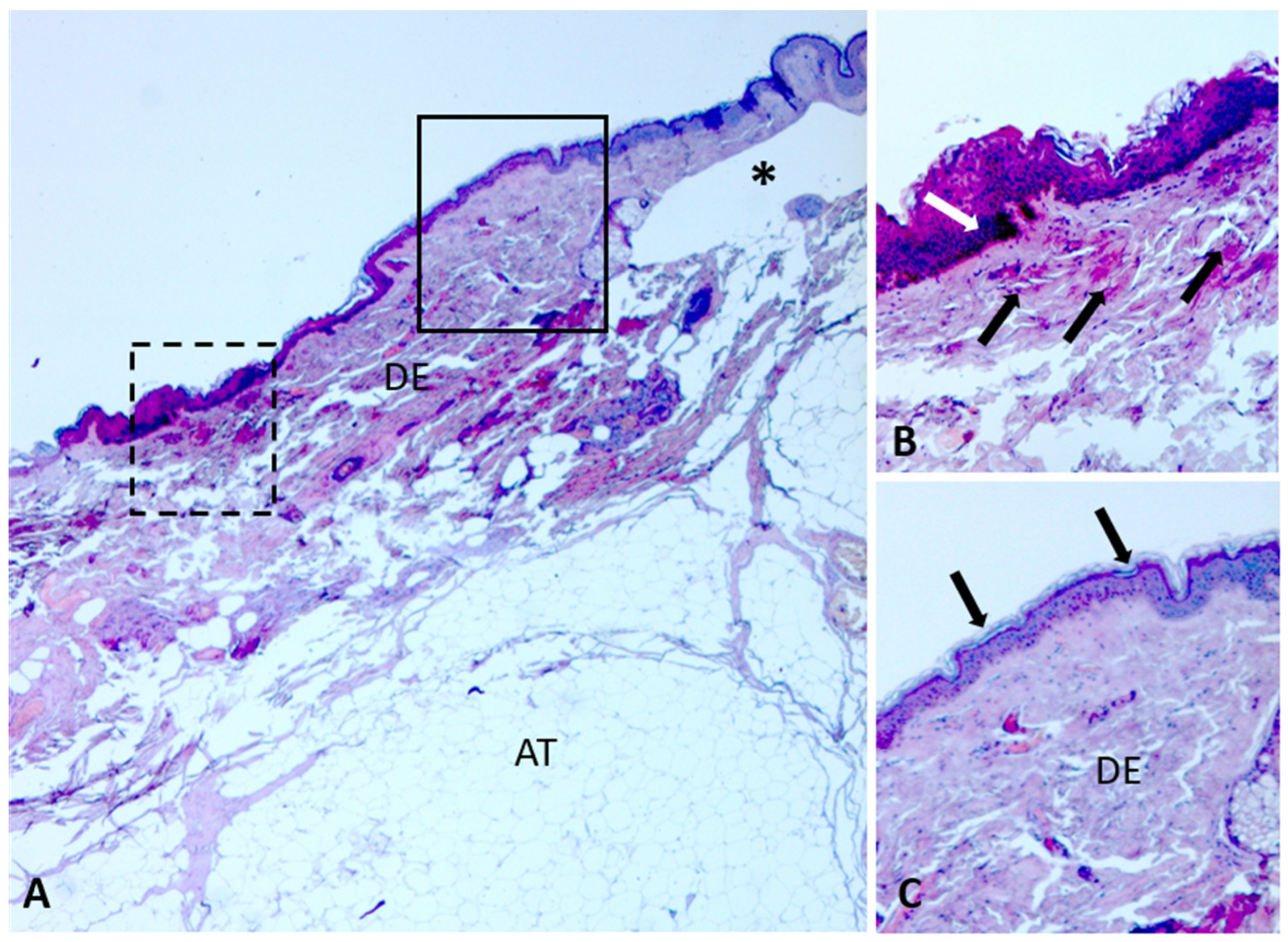
Although the morphology of the lesions cannot yet be considered species-specific because the data are still indicative, some differences can be appreciated by comparing the images of lesions caused by P. pallidula with those produced by T. nigerrimum complex [13,15]. In P. pallidula, the lesions appear roundish-elliptical in shape, with smooth and non-serpiginous margins, while those caused by T. nigerrimum complex appear irregular and serpiginous. Preliminary histological analyses revealed that the lesions inflicted by P. pallidula and T. nigerrimum complex [15] are both characterized by anomalous blister formation (Figure 3A). The blisters caused by P. pallidula appear located in the upper dermis and those by T. nigerrimum complex in the lower dermis. Moreover, the lesions caused by P. pallidula apparently include areas of hyperkeratosis, hypereosinophilia, and vascular activation (Figure 3B), compared with adjacent intact skin (Figure 3C), while these areas are apparently absent in lesions induced by T. nigerrimum complex [15].
Figure 3.
Histological analyses on post-mortem lesions caused by Pheidole pallidula on skin of the right arm of the deceased. Sections stained with Masson’s trichrome. (A) Section showing parts with lesions caused by feeding activity of P. pallidula. The asterisk indicates an anomalous dermal blister. (B) Detail of the dashed area in (A), showing skin surface with hyperkeratosis and areas with hypereosinophilia (white arrow) and vascular activation (black arrows). (C) Detail of the outlined area in (A), showing intact skin and regular inter-papillary ridges (arrows). Abbreviations: AT, adipose tissue; DE, dermis.
Although the case involving P. pallidula is a single one, the preliminary results suggest a different way of skin consumption in the two species. Further studies will be needed to evaluate the chemical composition of P. pallidula glands and the potential role of enzymes in pre-oral digestion.
In addition to their importance for studies on the biology of the species, investigations of post-mortem skin damage caused by P. pallidula can help avoid misinterpretations of these lesions during forensic investigations. Although ants have been clearly recognized as part of the sarcosaprophagous community, their role in producing skin artifacts is still poorly understood and often underestimated in forensic practice.
Our data suggest the need for in-depth investigations to identify the origin of any injuries that may be caused by macro- and microfauna after death. Studies that integrate entomological expertise with forensic pathology will be essential to establish diagnostic criteria for differentiating ant-induced lesions and improve the accuracy of estimating the post-mortem interval and reconstructing the death scene. Research involving experiments with animal models in controlled indoor and outdoor environments will also be useful to obtain more details on differences in feeding activity and the morphology of lesions caused by ants on corpses, in order to develop more standardized diagnostic criteria.
Author Contributions
Conceptualization, M.P. and T.B.; methodology, M.P. and T.B.; validation, M.P., D.A.G. and T.B.; formal analysis, M.P. and T.B.; investigation, M.P., E.D.B., F.F., D.B., F.M., F.C., E.S., D.A.G., M.C., V.V. and T.B.; data curation, M.P., M.C. and T.B.; writing—original draft preparation, M.P. and T.B.; writing—review and editing, M.P., M.C., E.S., D.A.G. and T.B.; visualization, M.P. and T.B.; supervision, M.P. and T.B.; All authors have read and agreed to the published version of the manuscript.
Funding
This research received no external funding.
Data Availability Statement
The original contributions presented in this study are included in the article. Further inquiries can be directed to the corresponding author.
Acknowledgments
Activities of E.S. for this article were supported by the “Juan de la Cierva” program (Grant No. JDC2024-054485-I) financed by the MICIU/AEI/10.13039/501100011033 and FSE+.
Conflicts of Interest
The authors declare no conflicts of interest.
References
- Smith, K.G.V. A Manual of Forensic Entomology; Trustees of the British Museum (Natural History): London, UK, 1986. [Google Scholar]
- Gibb, H.; Dunn, R.R.; Sanders, N.J.; Grossman, B.F.; Photakis, M.; Abril, S.; Agosti, D.; Andersen, A.N.; Angulo, E.; Armbrecht, I.; et al. A global database of ant species abundances. Ecology 2017, 98, 883–884. [Google Scholar] [CrossRef] [Scilit] [PubMed]
- Campobasso, C.P.; Marchetti, D.; Introna, F.; Colonna, M.F. Postmortem artifacts made by ants and the effect of ant activity on decompositional rates. Am. J. Forensic Pathol. 2009, 30, 84–87. [Google Scholar] [CrossRef] [Scilit] [PubMed]
- Byrd, J.H.; Castner, J.L. Forensic Entomology: The Utility of Arthropods in Legal Investigations, 2nd ed.; CRC Press: Boca Raton, FL, USA, 2010. [Google Scholar]
- Grasso, D.A.; Mori, A.; Le Moli, F. Chemical communication during foraging in the harvesting ant Messor capitatus (Hymenoptera, Formicidae). Insectes Sociaux 1998, 45, 85–96. [Google Scholar] [CrossRef] [Scilit]
- Grasso, D.A.; Mori, A.; Le Moli, F. Recruitment and trail communication in two species of Messor ants (Hymenoptera, Formicidae). Ital. J. Zool. 1999, 66, 373–378. [Google Scholar] [CrossRef] [Scilit]
- Visicchio, R.; Mori, A.; Grasso, D.A.; Castracani, C.; Le Moli, F. Glandular sources of recruitment, trail, and propaganda semiochemicals in the slave-making ant Polyergus rufescens. Ethol. Ecol. Evol. 2001, 13, 361–372. [Google Scholar] [CrossRef] [Scilit]
- Scarano, F.; Giannetti, D.; Trenti, F.; Giacomazzi, F.; Vigna, J.; Guella, G.; Grasso, D.A.; Haase, A. Trail pheromone identification in the ant Crematogaster scutellaris. Sci. Rep. 2024, 14, 7883. [Google Scholar] [CrossRef] [Scilit]
- Bonacci, T.; Zetto Brandmayr, T.; Brandmayr, P.; Vercillo, V.; Porcelli, F. Successional patterns of the insect fauna on a pig carcass in southern Italy and the role of Crematogaster scutellaris (Hymenoptera, Formicidae) as a carrion invader. Entomol. Sci. 2011, 14, 125–132. [Google Scholar] [CrossRef] [Scilit]
- Bonacci, T.; Benecke, M.; Scapoli, C.; Vercillo, V.; Pezzi, M. Severe post mortem damages by ants on a human corpse. Rom. J. Leg. Med. 2019, 27, 269–271. [Google Scholar] [CrossRef] [Scilit]
- Ramón-Cabrera, G.; Donoso, D.A. The role of ants (Hymenoptera: Formicidae) in forensic entomology. Rev. Ecuat. Med. Y Cienc. Biológicas 2015, 36, 19–26. [Google Scholar]
- Ahmad, A.; Omar, B. A report of Pheidole megacephala (Fabricius 1793) (Hymenoptera: Formicidae) colonisation on small and medium-sized mammal carcasses: Its implications in forensic entomology. Int. J. Trop. Insect Sci. 2023, 43, 1601–1623. [Google Scholar] [CrossRef] [Scilit]
- Bonacci, T.; Vercillo, V. Outdoor post-mortem bite injuries by Tapinoma nigerrimum (Hymenoptera, Formicidae) on a human corpse: Case report. J. Forensic Leg. Med. 2015, 33, 5–8. [Google Scholar] [CrossRef] [Scilit]
- Paula, M.C.; Morishita, G.M.; Cavarson, C.H.; Gonçalves, C.R.; Tavares, P.R.A.; Mendonça, A.; Súarez, Y.R.; Antonialli-Junior, W.F. Action of ants on vertebrate carcasses and blow flies (Calliphoridae). J. Med. Entomol. 2016, 53, 1283–1291. [Google Scholar] [CrossRef] [Scilit] [PubMed]
- Pezzi, M.; Benecke, M.; Vercillo, V.; Marino, F.; Marino, S.; Scapoli, C.; Marchetti, M.G.; Leis, M.; Bonacci, T. Histochemical and immunohistochemical investigations on postmortem injuries caused by ants and literature review. Rom. J. Leg. Med. 2020, 28, 223–228. [Google Scholar] [CrossRef] [Scilit]
- Bugelli, V.; Forni, D.; Bassi, L.A.; Di Paolo, M.; Marra, D.; Lenzi, S.; Toni, C.; Giusiani, M.; Domenici, R.; Gherardi, M.; et al. Forensic entomology and the estimation of the minimum time since death in indoor cases. J. Forensic Sci. 2015, 60, 525–531. [Google Scholar] [CrossRef] [Scilit] [PubMed]
- Seifert, B. Inconvenient hyperdiversity–the traditional concept of “Pheidole pallidula” includes four cryptic species (Hymenoptera: Formicidae). Soil Org. 2016, 88, 1–17. [Google Scholar]
- Seifert, B. The Ants of Central and North Europe; Lutra Verlags–und Vertriebsgesellschaft: Tauer, Germany, 2018; p. 408. [Google Scholar]
- Lebas, C.; Galkowski, C.; Blatrix, R.; Wegnez, P. Ants of Britain and Europe: A Photographic Guide; Bloomsbury Wildlife: London, UK, 2019; p. 415. [Google Scholar]
- Lenoir, A.; Perdereau, E.; Dupont, S.; Libbrecht, R.; Picquet, P.; Galkowski, C.; Mercier, J.-L. The tropical ant Pheidole megacephala (Fabricius, 1793) in France and discrimination with the local Pheidole pallidula (Nylander, 1849). Naturae 2025, 4, 35–43. [Google Scholar] [CrossRef] [Scilit]
- Eubanks, M.D.; Lin, C.; Tarone, A.M. The role of ants in vertebrate carrion decomposition. Food Webs 2019, 18, e00109. [Google Scholar] [CrossRef] [Scilit]
- Wilson, E.O. Pheidole in the New World: A Dominant, Hyperdiverse Ant Genus; Harvard University Press: Cambridge, MA, USA, 2003; p. 794. [Google Scholar]
- Mei, M. Hymenoptera Formicidae (con diagnosi di due nuove specie). Nat. Sicil. 1995, 19, 753–772. [Google Scholar]
- Schifani, E.; Alicata, A. Exploring the myrmecofauna of Sicily: Thirty-two new ant species recorded, including six new to Italy and many new aliens (Hymenoptera, Formicidae). Pol. J. Entomol. 2018, 87, 323–348. [Google Scholar] [CrossRef] [Scilit]
- Schifani, E. The new checklist of the Italian Fauna: Formicidae. Biogeographia 2022, 37, ucl006. [Google Scholar] [CrossRef] [Scilit]
- Limonta, L.; Colombo, M. Record of Pheidole megacephala (F.), Pheidole nodus Smith and Tetramorium bicarinatum Nylander (Hymenoptera Formicidae), tropical species, in nursery imported plants. Boll. Zool. Agrar. Bachic. 2003, 35, 287–289. [Google Scholar]
- Jucker, C.; Rigato, F.; Regalin, R. Exotic ant records from Italy (Hymenoptera, Formicidae). Boll. Zool. Agrar. Bachic. 2008, 40, 99–107. [Google Scholar]
- Payne, J.A. A summer carrion study of the baby pig Sus scrofa Linnaeus. Ecology 1965, 46, 592–602. [Google Scholar] [CrossRef] [Scilit]
- Forbes, S.L.; Carter, D.O. Processes and Mechanisms of Death and Decomposition of Vertebrate Carrion; CRC Press: Boca Raton, FL, USA, 2015. [Google Scholar]
- Erkol, Z.; Hösükler, E. Postmortem animal attacks on human corpses. In Post Mortem Examination and Autopsy—Current Issues from Death to Laboratory Analysis; Doga, K.H., Ed.; InTechOpen: London, UK, 2018. [Google Scholar]
- de Souza, A.A.D.F.; de Rosa, C.T.A.; Arantes, L.C.; Pujol-Luz, J.R. Artifacts caused by leaf-cutting ants of the genus Atta (Hymenoptera: Formicidae): Postmortem bite injuries and the tearing of clothes. J. Forensic Sci. 2020, 65, 1012–1015. [Google Scholar] [CrossRef] [Scilit] [PubMed]
- Fuller, M.E. The Insect Inhabitants of Carrion: A Study in Animal Ecology; Council for Scientific and Industrial Research, H.J. Green, Government Printer: Melbourne, Australia, 1934; p. 62. [Google Scholar]
- Bolton, B. An Online Catalog of the Ants of the World. 2025. Available online: https://antcat.org (accessed on 22 November 2025).
- Oberski, J.T.; Griebenow, Z.H.; Adams, R.M.M.; Andersen, A.; Andrade-Silva, J.; Barden, P.; Borowiec, M.; Brady, S.; Casadei-Ferreira, A.; Csősz, S.; et al. Ant systematics: Past, present, and future. Insect Syst. Divers. 2025, 9, 11. [Google Scholar] [CrossRef] [Scilit]
- Bonacci, T.; Mendicino, F.; Bonelli, D.; Carlomagno, F.; Curia, G.; Scapoli, C.; Pezzi, M. Investigations on arthropods associated with decay stages of buried animals in Italy. Insects 2021, 12, 311. [Google Scholar] [CrossRef] [Scilit]
- de Sales, T.A.; Daemon, E.; Lopes, J.F.S. Necrophagous or predators? The role of Pheidole radoszkowskii Mayr (Hymenoptera: Formicidae) on Rattus norvegicus (Berkenhout) carcasses (Rodentia: Muridae). J. Nat. Hist. 2015, 50, 971–974. [Google Scholar] [CrossRef] [Scilit]
- de Souza, A.S.B.; Kirst, F.D.; Krüger, R.F. Insects of forensic importance from Rio Grande do Sul state in southern Brazil. Rev. Bras. Entomol. 2008, 52, 641–646. [Google Scholar] [CrossRef] [Scilit]
- Castillo Miralbés, M. Estudio de la entomofauna asociada a cadáveres en el alto Aragón (España). Monogr. Soc. Entomológica Aragonesa 2002, 6, 1–94. [Google Scholar]
- Martínez, M.D.; Arnaldos, M.I.; Romera, E.; García, M.D. Los Formicidae (Hymenoptera) de una comunidad sarcosaprófaga en un ecosistema mediterráneo. Ann. Biol. 2002, 24, 33–44. [Google Scholar]
- Arnaldos, M.I.; Romera, E.; Presa, J.J.; Luna, A.; García, M.D. Studies on seasonal arthropod succession on carrion in the southeastern Iberian Peninsula. Int. J. Leg. Med. 2004, 118, 197–205. [Google Scholar] [CrossRef] [Scilit]
- Pérez-Marcos, M.; López-Gallego, E.; Arnaldos, M.I.; Martínez-Ibáñez, D.; García, M.D. Formicidae (Hymenoptera) community in corpses at different altitudes in a semiarid wild environment in the southeast of the Iberian Peninsula. Entomol. Sci. 2020, 23, 297–310. [Google Scholar] [CrossRef] [Scilit]
- Payne, J.A.; Mason, W.R.M. Hymenoptera associated with pig carrion. Proc. Entomol. Soc. Wash 1971, 73, 132–141. [Google Scholar]
- Braack, L.E.O. Arthropods associated with carcasses in the northern Kruger National Park. S. Afr. J. Wildl. Res. 1986, 16, 91–98. [Google Scholar]
- Watson, E.J.; Carlton, C.E. Insect succession and decomposition of wildlife carcasses during fall and winter in Louisiana. J. Med. Entomol. 2005, 42, 193–203. [Google Scholar] [CrossRef] [PubMed]
- Reed, H.B. A study of dog carcass communities in Tennessee, with special reference to the insects. Am. Midl. Nat. 1958, 59, 213–245. [Google Scholar] [CrossRef] [Scilit]
- Scott, M.P.; Traniello, J.F.A.; Fetherston, I.A. Competition for prey between ants and burying beetles (Nicrophorus spp): Differences between Northern and Southern temperate sites. Psyche 1987, 94, 325–332. [Google Scholar] [CrossRef] [Scilit]
- Park, S.-H.; Moon, T.-Y. Positive and negative impact to decomposition of pig cadavers by ants (Hymenoptera: Formicidae) at the Islet Yeongdo in Busan, South Korea. Entomol. Res. 2020, 50, 124–127. [Google Scholar] [CrossRef] [Scilit]
- Park, S.-H.; Moon, T.-Y. Forensic implication of cadaveric behaviors of the Japanese pavement ant (Tetramorium tsushimae Emery) attending to rabbit cadavers. Entomol. Res. 2020, 50, 177–181. [Google Scholar] [CrossRef] [Scilit]
- Leong, C.M.; Shelomi, M.; Lin, C.C.; Shiao, S.-F. Necrophilous ants (Hymenoptera: Formicidae) in diverse habitats in Taiwan. Sociobiology 2019, 66, 209–217. [Google Scholar] [CrossRef] [Scilit]
- Valdes-Perezgasga, M.T.; Sanchez-Ramos, F.J.; Garcia-Martinez, O.; Anderson, G.S. Arthropods of forensic importance on pig carrion in the Coahuilan semidesert, Mexico. J. Forensic Sci. 2010, 55, 1098–1101. [Google Scholar] [CrossRef] [Scilit] [PubMed]
- Bala, M.; Kaur, P. Insect faunal succession on buried piece of pork in the State of Punjab (India): A preliminary study. J. Forensic Res. 2015, 5, 252. [Google Scholar]
- Chen, C.D.; Nazni, W.A.; Lee, H.L.; Hashim, R.; Abdullah, N.A.; Ramli, R.; Lau, K.W.; Heo, C.C.; Goh, T.G.; Izzul, A.A.; et al. A preliminary report on ants (Hymenoptera: Formicidae) recovered from forensic entomological studies conducted in different ecological habitats in Malaysia. Trop. Biomed. 2014, 31, 381–386. [Google Scholar] [PubMed]
- Azwandi, A.; Nina Keterina, H.; Owen, L.C.; Nurizzati, M.D.; Omar, B. Adult carrion arthropod community in a tropical rainforest of Malaysia: Analysis on three common forensic entomology animal models. Trop. Biomed. 2013, 30, 481–494. [Google Scholar]
- Tantawi, T.I.; El-Kady, E.M.; Greenberg, B.; El-Ghaffar, H.A. Arthropod succession on exposed rabbit carrion in Alexandria, Egypt. J. Med. Entomol. 1996, 33, 566–580. [Google Scholar] [CrossRef] [Scilit]
- Shaalan, E.A.; El-Moaty, Z.A.; Abdelsalam, S.; Anderson, G.S. A preliminary study of insect succession in Al-Ahsaa Oasis, in the eastern region of the Kingdom of Saudi Arabia. J. Forensic Sci. 2017, 62, 239–243. [Google Scholar] [CrossRef] [Scilit]
- Al-Dakhil, A.A.; Alharbi, S.A. A preliminary investigation of the entomofauna composition of forensically important necrophagous insects in Al-Madinah Al-Munawwarah region, Kingdom of Saudi Arabia. J. Taibah Univ. Sci. 2020, 14, 1127–1133. [Google Scholar] [CrossRef] [Scilit]
- Early, M.; Goff, M.L. Arthropod succession patterns in exposed carrion on the island of O’ahu, Hawaiian Islands, USA. J. Med. Entomol. 1986, 23, 520–531. [Google Scholar] [CrossRef] [Scilit]
- Al-Ameri, D.T.; Hamza, A.K.; Alhasan, A.S. Relationship between ants Pheidole megacephala (Hymenoptera: Formicidae) and some dead animals tissue. Plant Arch. 2020, 20, 871–874. [Google Scholar]
- Hwang, W. Environmental variables affecting community composition and carcass utilization of necrophagous ant (Hymenoptera: Formicidae) in Southern and Northern Taiwan. BioFormosa 2011, 46, 21–31. [Google Scholar]
- Andrade-Silva, J.; Pereira, E.K.C.; Silva, O.; Santos, C.L.C.; Delabie, J.H.C.; Rebêlo, J.M.M. Ants (Hymenoptera: Formicidae) associated with pig carcasses in an urban area. Sociobiology 2015, 62, 527–532. [Google Scholar] [CrossRef] [Scilit]
- Pereira, E.K.C.; Andrade-Silva, J.; Silva, O.; Santos, C.L.C.; Moraes, L.S.; Bandeira, M.d.C.A.; Silva, C.R.R.; Rebêlo, J.M.M. Solenopsis saevissima (Smith) (Hymenoptera: Formicidae) activity delays vertebrate carcass decomposition. Sociobiology 2017, 64, 369–372. [Google Scholar] [CrossRef] [Scilit]
- Moretti, T.C.; Solis, D.R.; Godoy, W.A.C. Ants (Hymenoptera: Formicidae) collected with carrion-baited traps in Southeast Brazil. Open Forensic Sci. J. 2013, 6, 1–5. [Google Scholar] [CrossRef] [Scilit]
- Cruz, T.M.; Vasconcelos, S.D. Entomofauna de solo associada à decomposição de carcaça de suíno em um fragmento de Mata Atlântica de Pernambuco, Brasil. Biociências 2006, 14, 193–201. [Google Scholar]
- Viana, G.S.; de Paula, M.C.; de Moura Eulalio, A.D.; dos Santos, P.G.; Lima-Junior, S.E.; Antonialli-Junior, W.F. Formicidae fauna in pig carcasses contaminated by insecticide: Implications for forensic entomology. Rev. Bras. Entomol. 2022, 66, e20210085. [Google Scholar] [CrossRef] [Scilit]
- Wolff, M.; Uribe, A.; Ortiz, A.; Duque, P. A preliminary study of forensic entomology in Medellín, Colombia. Forensic Sci. Int. 2001, 120, 53–59. [Google Scholar] [CrossRef] [Scilit]
- Ekanem, M.S.; Dike, M.C. Arthropod succession on pig carcasses in Southeastern Nigeria. Pap. Avulsos Zool 2010, 50, 561–570. [Google Scholar] [CrossRef] [Scilit]
- Kyerematen, R.A.K.; Boateng, B.A.; Haruna, M.; Eziah, V.Y. Decomposition and insect succession pattern of exposed domestic pig (Sus scrofa L.) carrion. ARPN J. Agric. Biol. Sci. 2013, 8, 756–765. [Google Scholar]
- Gomes, L.; Gomes, G.; Desuó, I.C. A preliminary study of insect fauna on pig carcasses located in sugarcane in winter in south-eastern Brazil. Med. Vet. Entomol. 2009, 23, 155–159. [Google Scholar] [CrossRef] [Scilit]
- Mabika, N.; Masendu, R.; Mawera, G. An initial study of insect succession on decomposing rabbit carrions in Harare, Zimbabwe. Asian Pac. J. Trop. Biomed. 2014, 4, 561–565. [Google Scholar] [CrossRef] [Scilit]
- Monteiro-Filho, E.L.A.; Penereiro, J.L. Estudo da decomposição e sucessão sobre uma carcaça animal numa área do estado de São Paulo, Brasil. Rev. Bras. Biol. 1987, 47, 289–295. [Google Scholar]
- Feugang Youmessi, F.D.; Bilong Bilong, C.F.; Cherix, D.; Djiéto-Lordon, C. Biodiversity study of arthropods collected on rat carrion in Yaounde, Cameroon: First study on forensic entomology in Central Africa. Int. J. Biosci. 2012, 2, 1–8. [Google Scholar]
Disclaimer/Publisher’s Note: The statements, opinions and data contained in all publications are solely those of the individual author(s) and contributor(s) and not of MDPI and/or the editor(s). MDPI and/or the editor(s) disclaim responsibility for any injury to people or property resulting from any ideas, methods, instructions or products referred to in the content. |
© 2026 by the authors. Licensee MDPI, Basel, Switzerland. This article is an open access article distributed under the terms and conditions of the Creative Commons Attribution (CC BY) license.