


Simple Summary
Neonicotinoid insecticides have been extensively employed in agriculture. Recently, their use has decreased due to the development of resistance in the target insects but also because of restrictions imposed by regulatory bodies. However, neonicotinoids remain of interest due to their potential role in declining bee populations. Not all neonicotinoids have the same toxic effect on bees, which could be due to different abilities in detoxification by cytochrome P450 enzymes. These are involved in the detoxification of many insecticides, thus contributing to tolerance. Here, the role of one specific cytochrome P450 in the detoxification of two neonicotinoids of different classes, as well as a pyrethroid insecticide, was investigated. To this aim, the gene encoding the enzyme was successfully downregulated by the injection of double-stranded RNA into the brain of bees, to specifically target the protein in the head. The injected bees were then tested by bioassays and their mortality recorded. The results confirmed that this enzyme is responsible for the tolerance of bees to neonicotinoid thiacloprid and pyrethroid tau-fluvalinate, while no difference in mortality was observed with imidacloprid. These findings suggest that the selective tolerance of bees to some insecticides can be explained by the role of this specific enzyme.
Abstract
Neonicotinoid toxicity to honeybees varies among active ingredients, with the insects being tolerant to thiacloprid but sensitive to imidacloprid. The selective toxicity has been hypothesized to be due to differences in metabolic detoxification by cytochrome P450s. In vitro experiments revealed that CYP9Q3 efficiently metabolises thiacloprid but not imidacloprid, consistent with the toxicity data. Honeybee tolerance to tau-fluvalinate, a pyrethroid acaricide used against Varroa destructor, has also been linked to P450-mediated detoxification. To address the role of CYP9Q3 in vivo in Apis mellifera, we used an RNA interference approach based on double-stranded RNA delivery through the median ocellus of adults to transiently suppress expression of CYP9Q3 in the head. Following confirmation of CYP9Q3 suppression by qPCR, fixed-dose topical bioassays were conducted with the neonicotinoids imidacloprid and thiacloprid and the pyrethroid tau-fluvalinate. Suppression of CYP9Q3 was associated with increased mortality following exposure to thiacloprid and tau-fluvalinate, whereas no increase was observed with imidacloprid. These findings provide in vivo functional evidence supporting the role of CYP9Q3 in honeybee tolerance to thiacloprid and tau-fluvalinate. The RNAi approach offers a valuable tool for investigating the functions of cytochrome P450 enzymes and other genes expressed in the honey bee head that are involved in insecticide susceptibility.
Key Contribution:
Transient RNAi-mediated suppression of CYP9Q3 in honeybee head tissue increases susceptibility to thiacloprid and tau-fluvalinate but not to imidacloprid, providing in vivo evidence for the contribution of CYP9Q3 to the detoxification of thiacloprid and tau-fluvalinate in Apis mellifera.
1. Introduction
Neonicotinoid insecticides have been widely used in farming practices worldwide to the extent that they were the most widely used insecticide class by 2008, accounting for 24% of the total insecticide market and 80% of the global insecticide seed treatment market [1,2]. More recently, global insecticide usage patterns have shifted away from these insecticides due to evolving neonicotinoid resistance and restrictions on use by regulatory bodies such as the EU. Nevertheless, neonicotinoids remain an important research focus due to their widely discussed potential impact on declining wild bee populations [3,4,5,6,7,8].
The main target of neonicotinoids is the nicotinic acetylcholine receptor (nAChR), where they compete with the neurotransmitter acetylcholine for the agonist site [9]. Their potential disruptive effects may be mitigated in vivo by honey bee detoxification mechanisms [10,11]. Comprehensive studies of the honeybee transcriptome and proteome have revealed complex detoxification mechanisms involving multiple genes and pathways, with P450s playing a crucial role in the direct metabolism of xenobiotics [10].
Honey bees have different levels of tolerance to different classes of neonicotinoids, being tolerant to thiacloprid while sensitive to imidacloprid. Metabolic detoxification via cytochrome P450 has been found to contribute to the difference in sensitivity to these two neonicotinoids [12,13]; P450-mediated metabolism also contributes to honey bee tolerance of tau-fluvalinate, a pyrethroid acaricide widely used for Varroa control [14]. Several P450 enzymes involved in detoxification have been identified in A. mellifera, with members of the CYP9Q subfamily playing a prominent role [13,14,15]. Functional expression of all 27 honeybee P450s belonging to the CYP3 clan in an insect cell line showed that CYP9Q3 metabolised thiacloprid with high efficiency, whereas CYP6AS5, CYP9Q1 and CYP9Q2 exhibited at least tenfold lower activity. CYP9Q1, CYP9Q2 and CYP9Q3 also metabolised imidacloprid, but much less efficiently than thiacloprid [13]. Furthermore, all three CYP9Q enzymes metabolised the pyrethroid tau-fluvalinate and the organophosphate coumaphos in vitro [14]. CYP9Q1-3 P450s were also expressed in Drosophila melanogaster, revealing that CYP9Q3 gave the highest level of resistance to thiacloprid [13]. The CYP9Q3 enzyme was localized principally in the brain, Malpighian tubules and the midgut of A. mellifera [13]. Tissue-specific expression of the protein in the Malpighian tubules and neuronal cells of transgenic D. melanogaster led to increased levels of thiacloprid resistance. Based on this, the authors suggested that expression of CYP9Q3 in the brain had a secondary function in xenobiotic metabolism in the Drosophila model [13].
Here, we analysed the role of CYP9Q3 expressed in the bee head. We tested whether the transient reduction in CYP9Q3 transcripts in whole head samples following median ocellus dsRNA delivery was associated with altered mortality after exposure to imidacloprid, thiacloprid or tau-fluvalinate. Our results support a role of this enzyme in the detoxification of thiacloprid and tau-fluvalinate.
2. Results
2.1. Efficiency of Median Ocellus dsRNA Injections in Silencing CYP9Q3
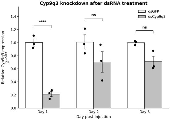
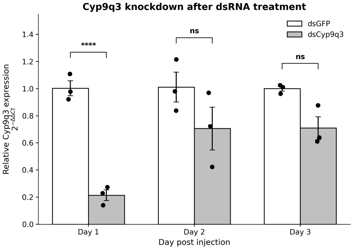
To test the hypothesis that CYP9Q3 expressed in the head has a role in insecticide detoxification, we injected dsRNA targeting the cognate gene in the median ocellus in the heads of adult bees. Three independent experiments were carried out. After the injections, the expression levels of CYP9Q3 mRNA transcripts were quantified in transcripts from dissected heads over a 3-day period using qPCR. Expression values were normalized to the two reference genes RpS18 and RpL32 and then calibrated against the corresponding dsGFP control group for each time point. This identified significant suppression of CYP9Q3 1 day post-injection in the dsCYP9Q3-injected bees, indicating efficient RNAi-mediated knockdown (Figure 1). Two-way ANOVA revealed a significant effect of treatment, day and treatment × day interaction on CYP9Q3 expression. Tukey’s post hoc analysis showed a significant reduction in the transcript in dsCYP9Q3-treated bees compared with dsGFP controls at day 1 post-injection: 0.214 ± 0.039 relative expression in dsCYP9Q3-treated bees compared with 1.003 ± 0.055 in dsGFP controls, p = 0.000066. At days 2 and 3, mean CYP9Q3 expression remained lower in the dsCYP9Q3 group, 0.705 ± 0.158 and 0.709 ± 0.085, respectively, but these differences were not statistically significant after Tukey correction.
Figure 1.
CYP9Q3 knockdown following dsRNA treatment. Relative CYP9Q3 expression was calculated using the 2−ΔΔCt method after normalization to RpS18 and RpL32, with the corresponding dsGFP-treated group used as calibrator for each day. Bars represent mean ± SEM from three biological replicates per treatment and time point, and individual points represent biological replicates. Statistical analysis was performed on ΔCt values using two-way ANOVA followed by Tukey’s multiple comparison test. Asterisks indicate Tukey-adjusted significance levels: **** p < 0.0001; ns, not significant. The complete dataset is shown in Supplementary Table S3, sheet 1.
Overall, these results indicate that dsCYP9Q3 injection induced a strong but transient reduction in CYP9Q3 transcript levels, with significant knockdown detected at day 1 post-injection and partial recovery by days 2 and 3.
2.2. Toxicity Assays
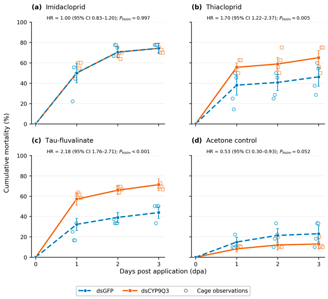
Having established that CYP9Q3 expression was downregulated after dsRNA injection, acute contact toxicity assays were conducted. The two neonicotinoids and the pyrethroid tau-fluvalinate were applied 24 h post-injection, correlating with the lowest levels of detected CYP9Q3 expression (Figure 2). Acetone served as the control. Mortality was followed for 3 days, and the experiment was repeated 4 times. Percentages given in the text are observed cumulative mortalities pooled across cages, whereas the lines in Figure 2 show model-estimated values.
Figure 2.
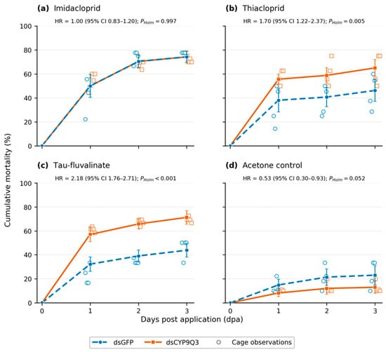
Effect of CYP9Q3 knockdown on cumulative honeybee mortality following chemical exposure. Cumulative mortality of bees injected with dsCYP9Q3 or control dsGFP following exposure to (a) imidacloprid, (b) thiacloprid, (c) tau-fluvalinate and (d) acetone. Each compound and dsRNA treatment combination comprised 4 cage replicates, with 6–13 bees per cage. Lines and filled symbols represent model-estimated cumulative mortality at each observation day, with vertical error bars indicating nominal, unadjusted 95% confidence intervals. Mortality was assessed at 24 h intervals over the 3-day period following topical application. Open symbols show the observed cumulative mortality in each cage and are presented descriptively. Interval-specific deaths among bees alive at the start of each interval were analysed using a discrete-time proportional hazards model with a complementary log-log link and cage-clustered robust standard errors. Hazard ratios (HRs) represent the mortality hazard in dsCYP9Q3-injected bees relative to dsGFP-injected bees; HR > 1 therefore indicates increased mortality following CYP9Q3 knockdown. Displayed p values were Holm-adjusted across the four compound-specific dsRNA contrasts. dpa, days post-application. The complete dataset is shown in Supplementary Table S3 sheet 2.
For imidacloprid, pooled cumulative mortality at 3 dpa was 72.1% in dsCYP9Q3-injected bees and 77.1% in dsGFP-injected bees (Figure 2a). Both groups showed substantial mortality by 1 dpa, followed by a further increase at day 2, with little additional mortality observed at 3 dpa. Taken together, no effect of CYP9Q3 silencing on the mortality hazard was detected over the complete observation period (HR = 1.00, 95% CI: 0.83–1.20; Holm-adjusted p = 0.997).
In contrast, CYP9Q3 silencing significantly increased the mortality hazard following thiacloprid and tau-fluvalinate exposure. For thiacloprid, pooled cumulative mortality at 3 dpa was 63.6% in dsCYP9Q3-injected bees and 47.2% in dsGFP-injected bees (Figure 2b). Across the complete observation period, dsCYP9Q3-injected bees had a 1.70-fold higher mortality hazard than dsGFP-injected bees (95% CI: 1.22–2.37; Holm-adjusted p = 0.005). Similarly, cumulative mortality following tau-fluvalinate exposure reached 69.4% in dsCYP9Q3-injected bees and 46.2% in dsGFP-injected bees by 3 dpa (Figure 2c). CYP9Q3 silencing was associated with a 2.18-fold higher mortality hazard over the observation period (95% CI: 1.76–2.71; Holm-adjusted p < 0.001).
In the acetone control, pooled cumulative mortality at 3 dpa was 12.8% in dsCYP9Q3-injected bees and 23.1% in dsGFP-injected bees (Figure 2d). Although the unadjusted comparison indicated a lower mortality hazard in dsCYP9Q3-injected bees (HR = 0.53, 95% CI: 0.30–0.93; raw p = 0.026), this difference did not remain statistically significant after correction for the four compound-specific comparisons (Holm-adjusted p = 0.052).
3. Discussion
The metabolic detoxification of insecticides in honeybees has been extensively studied primarily in vitro, with particular focus on the role of cytochrome P450s [16,17,18]. Specifically, of the bee P450s, CYP9Q3 has been identified as having the highest activity metabolising neonicotinoid thiacloprid [13], and it also metabolises the pyrethroid tau-fluvalinate [14]. In our study, we investigated the functional contribution of head CYP9Q3 expression in vivo to honey bee tolerance against these substances.
The decision to target CYP9Q3 in the head was based on its high expression in honey bee brain tissue [13]. In toxicity assays involving two neonicotinoids and one pyrethroid, bees injected with dsCYP9Q3 exhibited higher mortality than dsGFP-injected controls following exposure to thiacloprid and tau-fluvalinate, but not imidacloprid. This pattern is consistent with the efficient metabolism of thiacloprid by CYP9Q3 and its limited activity against imidacloprid in vitro [13], as well as the involvement of CYP9Q enzymes in tau-fluvalinate detoxification [14]. This result was, however, not completely expected, as experiments in transgenic D. melanogaster suggested that CYP9Q3 expression in the Malpighian tubules carried out the main detoxification while the brain-expressed enzyme was suggested to have a secondary role [13]. In contrast to this, our results suggest that the metabolic activity of CYP9Q3 in head tissues, which include the brain, may have a more prominent role than previously thought, although the specific tissue involved could not be resolved, and knockdown outside the head was not assessed in this study.
The compound-specific effect of CYP9Q3 knockdown is better explained by the substrate specificity of the enzyme than by the molecular targets of the tested compounds, which are the nAChR for neonicotinoids [9] and the voltage-gated sodium channel for pyrethroids: reducing the level of a detoxifying enzyme is expected to increase toxicity only for compounds that this enzyme metabolises at a relevant rate. CYP9Q3 metabolises the N-cyanoamidine neonicotinoid thiacloprid with high efficiency, whereas the N-nitroguanidine imidacloprid is metabolised much less efficiently by CYP9Q3 and by the other CYP9Q enzymes in vitro [13], and inhibition of P450 activity markedly increases the toxicity of cyano-substituted neonicotinoids to honeybees [12]. Tau-fluvalinate is metabolised by CYP9Q1, CYP9Q2 and CYP9Q3 [14]. Imidacloprid is metabolised by honeybees in vivo [19], but the enzymes responsible have not been identified in vivo, and determining the contribution of other P450s, such as CYP9Q1 and CYP9Q2, would require additional knockdown experiments. In addition, cumulative mortality after imidacloprid exposure exceeded 70% in both dsRNA groups, which may have limited the ability to detect a further increase in mortality following CYP9Q3 knockdown.
Injection through the median ocellus produced the greatest reduction in CYP9Q3 expression in whole head tissue at 24 h, but this effect weakened after 48 h. Since RNA was extracted from whole heads, the observed suppression cannot be assigned specifically to the brain or to another individual head tissue. This aspect should be further explored to clarify the role of the brain in detoxification. The dsGFP treatment controlled for the injection procedure and non-specific effects of dsRNA administration. Cumulative mortality in the acetone controls (12.8% and 23.1% at 3 dpa in dsCYP9Q3- and dsGFP-injected bees, respectively) exceeded the 10% control mortality accepted for non-injected bees in OECD Test Guideline 214 [20]. This background mortality probably reflects the combined stress of CO2 anaesthesia, handling, microinjection through the median ocellus, topical application and cage maintenance. Water-injected bees already showed 10.6% cumulative mortality within 3 days of injection, indicating that the injection and handling procedure itself contributed to background mortality. Because dsGFP- and dsCYP9Q3-injected bees underwent identical procedures and were compared within each compound, this background mortality is not expected to bias the comparison between dsRNA treatments, although it reduces the precision of the toxicity estimates. The absence of a significant treatment difference in the acetone controls nevertheless indicates that the increased mortality observed with thiacloprid and tau-fluvalinate was not accompanied by a general increase in background mortality. Overall, the results provide functional evidence for an association between reduced CYP9Q3 expression in whole head tissue and tolerance to thiacloprid and tau-fluvalinate, while the conclusions remain limited to the tested doses and the 3-day observation period.
Down-regulation of CYP9Q3 in the head was achieved using the methods for median ocellus dsRNA injections previously developed in honeybees [21,22]. While RNAi has limitations due to its stability and uptake [23], previous studies have found more sustained effects in insects. RNAi injection in a similar study in brain-targeted honeybees was also successful, with suppression lasting for 72 h [22]. The shorter duration of suppression in our study may be attributed to the particularly high expression levels of CYP9Q3 in A. mellifera, potentially overwhelming the RNAi machinery after 48 h [24]. This effect has been reported in honeybee brain injections, although the difference detected was fairly small in that study [22]. To account for the transient nature of the knockdown, insecticides were applied 24 h after injection, when transcript suppression was greatest, and the difference in mortality between dsCYP9Q3- and dsGFP-injected bees was already apparent at 1 dpa, i.e., within 48 h of injection (Figure 2). CYP9Q3 protein levels and enzyme activity need not follow transcript levels closely and were not measured in this study. The dsGFP control accounts for non-specific effects of dsRNA injection; however, sequence-specific off-target silencing of closely related genes, such as CYP9Q1 and CYP9Q2, cannot be excluded and should be examined in future studies.
This study demonstrates that head-specific RNAi delivery is a promising method for targeted P450 suppression in the honeybee, particularly valuable for investigating proteins with tissue-specific expression patterns. The technique successfully validated in vitro findings in vivo and could be adapted for similar studies in other insects. Although the temporal limitations of gene suppression must be considered in experimental design, this approach provides a valuable tool for investigating the role of cytochrome P450s in insect xenobiotic metabolism.
4. Materials and Methods
4.1. Insect Collection and Maintenance
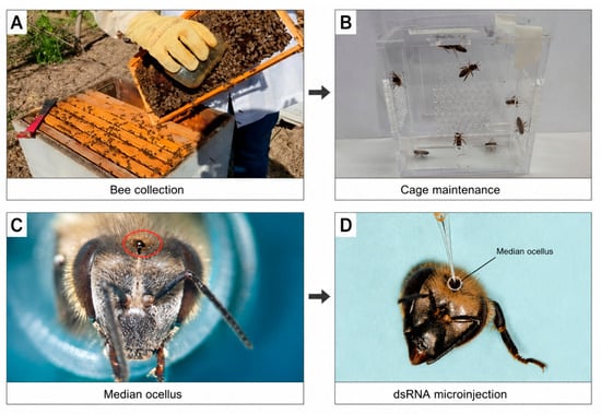
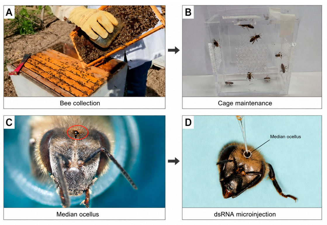
Adult Apis mellifera worker bees used in the study were collected from queen-right colonies with a known history and physiological status, located and established at the Agricultural University in Athens. Bees were collected from five such colonies maintained in a single apiary. They were foragers sampled from the outer honey-containing frames, and their exact age was not determined. Bees from the five colonies were pooled before being divided into cages. The colonies were not treated with any chemicals, such as antibiotics or anti-varroa agents, for at least 5 months prior to the study. The collected bees were divided randomly into groups of 20 and were placed in custom-made Plexiglas cages (7.1 × 12 × 12 cm) [25] (Figure 3B). The bees were first acclimatised at room temperature for 2 h with access to sucrose solution (50% w/v). Throughout the experiment, the bees were kept in darkness, at 25 ± 2 °C and 50–70% relative humidity.
Figure 3.
Experimental workflow for dsRNA delivery to adult workers. (A) Collection of adult worker bees from colony frames. (B) Maintenance of collected bees in Plexiglas cages under laboratory conditions. (C) Frontal view of the adult honeybee head under a stereoscope, showing the median ocellus selected as the injection site and indicated by a red circle. (D) Microinjection of dsRNA through the median ocellus using a glass capillary. Arrows indicate the sequence of the experimental procedure.
4.2. dsRNA Design and Synthesis
Double-stranded RNA (dsRNA), designed to target CYP9Q3 [13] and GFP, were synthesised by Genolution (Seoul, Republic of Korea) using AgroRNA’s Long Double-stranded RNA Synthesis Service (grade 3 purity). The dsRNA sequences are listed in Supplemental Table S1. Quality control was performed using gel electrophoresis and spectrophotometry.
4.3. Median Ocellus Injections
Median ocellus injections in Apis mellifera worker bees were based on the method described previously [21], with slight modifications. More specifically, A. mellifera workers were anaesthetised using CO2 until immobile (approximately 15 s), and individuals were secured in custom-made cages prior to injection (Figure 3C). Injections were performed using a Nanoject II Auto-Nanoliter Injector (Drummond™, Birmingham, AL, USA) fitted with a glass micropipette tip (Figure 3D). The individual injection volume was 345 nL, and the dsRNA concentration was 7.25 µg/µL. Each bee received 2.5 μg dsRNA through the median ocellus under stereoscopic guidance (Figure 3C,D). The non-targeting dsGFP construct was used as the procedural control for the RNAi experiments. Bees injected with dsGFP or dsCYP9Q3 underwent identical anaesthesia, handling, microinjection, post-injection maintenance and pesticide exposure procedures. Post-injection, bees were moved to Plexiglas cages with ad libitum access to sucrose solution (50% w/v) until sample collection. Mortality due to the injection and handling procedures was assessed in three water-injected control cages containing 12, 15 and 20 bees, respectively (47 bees in total), with survival recorded at 24-hour intervals for 3 days. Survival in the water-injected controls was 95.7% (45/47), 93.6% (44/47) and 89.4% (42/47) at days 1, 2 and 3 post-injection, respectively, corresponding to cumulative mortality of 4.3%, 6.4% and 10.6%.
4.4. RNA Extraction and qPCR
Knockdown was verified in bees sampled from separate cages that were injected in parallel with those used for the toxicity bioassays; because RNA extraction is destructive, transcript levels could not be measured in the individual bees used in the bioassays. Five bees were collected every 24 h from each cage and snap-frozen in liquid nitrogen; the heads were separated from the body and stored at −80 °C until further use. Total RNA was extracted from head tissues with Trizol according to the manufacturer’s instructions (Life Technologies, Carlsbad, CA, USA), after which DNase (Qiagen, Venlo, The Netherlands) treatment was applied. To analyse the expression levels of target genes, 2 µg total RNA in each sample was used for reverse transcription (SuperScript™ First-Strand, Thermo Fisher, Waltham, MA, USA) according to the manufacturer’s instructions. PCR amplification was conducted in STEP ONE PLUS (Applied Biosystems, Waltham, MA, USA) using SYBR Select Master-Mix (Thermo Fisher Scientific Inc.). The amplification procedure was initiated with 15 min incubation at 95 °C, followed by 40 cycles of 20 s at 95 °C, 20 s at 58 °C and 20 s at 72 °C. The primers for the sequences are provided in Supplemental Table S2. CYP9Q3 expression was normalized against the mean Ct of two reference genes, RpS18 and RpL32. These have previously been validated as reference genes in Apis mellifera RT-qPCR studies, including honeybee head tissue and dsRNA treatment conditions [26]. Relative expression was calculated using the 2−ΔΔCt method [27], with the corresponding dsGFP-treated group used as the calibrator for each day. Statistical analysis was performed on ΔCt values using two-way ANOVA with treatment, day and treatment × day as fixed factors, followed by Tukey’s multiple comparison test.
4.5. Toxicity Assays and Statistical Analysis
Test compounds were prepared in 100% acetone from technical-grade imidacloprid (≥98.0% purity; Sigma-Aldrich, St. Louis, MO, USA), thiacloprid (≥98.0%; Sigma-Aldrich) and tau-fluvalinate (≥95.0%; Sigma-Aldrich). Contact toxicity bioassays were conducted using a protocol adapted from OECD Test Guideline 214 [20]. Fixed challenge doses were selected a priori with reference to published topical exposure studies in adult honeybees. Imidacloprid was applied at 1 ng bee−1, corresponding to the sublethal topical dose evaluated by Guez et al. [28]. Thiacloprid was applied at 15 µg bee−1, approximating the reported 24 h topical LD50 of 14.6 µg bee−1 [13]. Tau-fluvalinate was applied at 0.4 µg bee−1, close to the lowest sublethal topical dose of 0.3 µg bee−1 used by Teeters et al. [29] and below previously reported topical toxicity thresholds [30]. These fixed doses were used to compare the susceptibility of dsCYP9Q3- and dsGFP-injected bees. For each exposure, 1 µL of the appropriate insecticide solution was applied to the dorsal thorax using a microsyringe. Solvent control bees received 1 µL of acetone. For each compound, four experimental replicate blocks were included, with each block comprising one dsGFP and one dsCYP9Q3 cage. Cages initially contained 6–13 bees. Bees injured during microinjection were excluded before insecticide or solvent application; consequently, cage sizes and group totals at the start of the toxicity assays were unequal, and the sample sizes below refer to the bees included at the start of exposure. Total initial sample sizes for the dsGFP and dsCYP9Q3 groups were 35 and 43 bees for imidacloprid, 36 and 33 for thiacloprid, 26 and 49 for tau-fluvalinate, and 39 and 39 for acetone, respectively (300 bees in total). The cage was considered the experimental unit, and a replicate block was included as a fixed effect in the statistical model. The number of surviving bees in each cage was recorded at days 1, 2 and 3 post-application (dpa), and mortality was calculated relative to the initial number of bees.
Cage-level interval mortality was analysed using a discrete-time proportional hazards model with a complementary log-log link. Fixed effects included dsRNA treatment, substance, observation interval, experimental replicate, and the dsRNA by substance and interval by substance interactions. Cage-clustered robust standard errors accounted for repeated observations within cages. Planned contrasts estimated hazard ratios for dsCYP9Q3 relative to dsGFP within each substance, with 95% confidence intervals. Raw and Holm-adjusted padj values across the four contrasts were reported, with significance defined as adjusted padj < 0.05. Analyses were performed in Python version 3.11 using NumPy, pandas and SciPy. This approach was chosen for three reasons. First, the discrete-time proportional hazards model with a complementary log-log link is the grouped-time analogue of the Cox model [31] and therefore uses the number of deaths in each daily interval among bees still alive rather than end-point mortality alone. Second, it treats the cage rather than the individual bee as the experimental unit, which avoids pseudoreplication, and it accommodates unequal numbers of bees per cage. Third, each replicate block, corresponding to one repetition of the experiment, comprised one dsGFP and one dsCYP9Q3 cage, so that variation between repetitions was accounted for by the block term. Because cluster-robust standard errors can be anti-conservative when the number of clusters is small [32], the model estimates are presented together with the individual cage observations in Figure 2, so that the consistency of the effects across cages can be assessed directly. For tau-fluvalinate, cumulative mortality in every dsCYP9Q3 cage exceeded that in every dsGFP cage at all three observation days, and for thiacloprid the cage-level observations were shifted in the same direction (Figure 2b,c).
The Graphical Abstract was created in BioRender. Vontas, J. (2027) https://BioRender.com/iskr6nl accessed on 3 November 2026.
Supplementary Materials
The following supporting information can be downloaded at https://www.mdpi.com/article/10.3390/insects17111186/s1, Table S1. The dsRNA sequences used for the A. mellifera RNAi experiments. Table S2. The primer sequences for quantitative PCR for both reference and target genes, along with the Tm and amplicon length information. Table S3: The complete dataset of Efficiency of RNAi knockdown (sheet 1), and of Toxicity testing (sheet 2).
Author Contributions
Conceptualisation, J.V.; methodology, E.K., S.V.; validation, E.K., I.S.-K.; formal analysis, J.V., E.K.; investigation, E.K., S.V., D.O., A.P., L.T., N.D., C.Z., K.C., H.T., L.D.; writing—original draft preparation, E.K., S.V., D.O., A.P., L.T., N.D., C.Z., K.C., H.T., L.D., J.V., I.S.-K.; writing—review and editing, J.V., E.K., I.S.-K.; visualisation, E.K.; supervision, J.V.; funding acquisition, J.V. All authors have read and agreed to the published version of the manuscript.
Funding
Funding was provided by Crop Protection AG (grant: 35481 to AUA). The funder had no role in study design, data collection and analysis, decision to publish, or preparation of the manuscript.
Data Availability Statement
The original contributions presented in this study are included in the article/Supplementary Material. Further inquiries can be directed to the corresponding author.
Conflicts of Interest
Authors Duncan Oliver, Nathalie Dewulf and Lies Degrave are employed at Syngenta Ghent Innovation Centre. Authors Adam Pym and Helen Thompson are employed at Syngenta Crop Protection Research. Author Kaat Cappelle is employed at both organisations. Authors Lea Talmann and Christoph T Zimmer are employed at Syngenta Crop Protection. The remaining authors declare that the research was conducted in the absence of any commercial or financial relationships that could be construed as a potential conflict of interest.
References
- Elbert, A.; Haas, M.; Springer, B.; Thielert, W.; Nauen, R. Applied Aspects of Neonicotinoid Uses in Crop Protection. Pest Manag. Sci. 2008, 64, 1099–1105. [Google Scholar] [CrossRef] [Scilit] [PubMed]
- Jeschke, P.; Nauen, R.; Schindler, M.; Elbert, A. Overview of the Status and Global Strategy for Neonicotinoids. J. Agric. Food Chem. 2011, 59, 2897–2908. [Google Scholar] [CrossRef] [Scilit] [PubMed]
- Smagghe, G.; Tirry, L. Insect Midgut as a Site for Insecticide Detoxification and Resistance. In Biochemical Sites of Insecticide Action and Resistance; Springer: Berlin/Heidelberg, Germany, 2001; pp. 293–321. [Google Scholar]
- Goulson, D.; Lye, G.C.; Darvill, B. Decline and Conservation of Bumble Bees. Annu. Rev. Entomol. 2008, 53, 191–208. [Google Scholar] [CrossRef] [Scilit] [PubMed]
- Goulson, D.; Nicholls, E.; Botías, C.; Rotheray, E.L. Bee Declines Driven by Combined Stress from Parasites, Pesticides, and Lack of Flowers. Science 2015, 347, 1255957. [Google Scholar] [CrossRef] [Scilit] [PubMed]
- Botías, C.; David, A.; Horwood, J.; Abdul-Sada, A.; Nicholls, E.; Hill, E.; Goulson, D. Neonicotinoid Residues in Wildflowers, a Potential Route of Chronic Exposure for Bees. Environ. Sci. Technol. 2015, 49, 12731–12740. [Google Scholar] [CrossRef] [Scilit] [PubMed]
- Cabirol, A.; Haase, A. The Neurophysiological Bases of the Impact of Neonicotinoid Pesticides on the Behaviour of Honeybees. Insects 2019, 10, 344. [Google Scholar] [CrossRef] [Scilit] [PubMed]
- David, A.; Botías, C.; Abdul-Sada, A.; Nicholls, E.; Rotheray, E.L.; Hill, E.M.; Goulson, D. Widespread Contamination of Wildflower and Bee-Collected Pollen with Complex Mixtures of Neonicotinoids and Fungicides Commonly Applied to Crops. Environ. Int. 2016, 88, 169–178. [Google Scholar] [CrossRef] [Scilit] [PubMed]
- Casida, J.E. Neonicotinoids and Other Insect Nicotinic Receptor Competitive Modulators: Progress and Prospects. Annu. Rev. Entomol. 2018, 63, 125–144. [Google Scholar] [CrossRef] [Scilit] [PubMed]
- Gong, Y.; Diao, Q. Current Knowledge of Detoxification Mechanisms of Xenobiotic in Honey Bees. Ecotoxicology 2017, 26, 1–12. [Google Scholar] [CrossRef] [Scilit] [PubMed]
- Johnson, R.M.; Ellis, M.D.; Mullin, C.A.; Frazier, M. Pesticides and Honey Bee Toxicity—USA. Apidologie 2010, 41, 312–331. [Google Scholar] [CrossRef] [Scilit]
- Iwasa, T.; Motoyama, N.; Ambrose, J.T.; Roe, R.M. Mechanism for the Differential Toxicity of Neonicotinoid Insecticides in the Honey Bee, Apis mellifera. Crop Prot. 2004, 23, 371–378. [Google Scholar] [CrossRef]
- Manjon, C.; Troczka, B.J.; Zaworra, M.; Beadle, K.; Randall, E.; Hertlein, G.; Singh, K.S.; Zimmer, C.T.; Homem, R.A.; Lueke, B.; et al. Unravelling the Molecular Determinants of Bee Sensitivity to Neonicotinoid Insecticides. Curr. Biol. 2018, 28, 1137–1143.e5. [Google Scholar] [CrossRef] [Scilit] [PubMed]
- Mao, W.; Schuler, M.A.; Berenbaum, M.R. CYP9Q-Mediated Detoxification of Acaricides in the Honey Bee (Apis mellifera). Proc. Natl. Acad. Sci. USA 2011, 108, 12657–12662. [Google Scholar] [CrossRef] [Scilit] [PubMed]
- Troczka, B.J.; Homem, R.A.; Reid, R.; Beadle, K.; Kohler, M.; Zaworra, M.; Field, L.M.; Williamson, M.S.; Nauen, R.; Bass, C.; et al. Identification and Functional Characterisation of a Novel N-Cyanoamidine Neonicotinoid Metabolising Cytochrome P450, CYP9Q6, from the Buff-Tailed Bumblebee Bombus Terrestris. Insect Biochem. Mol. Biol. 2019, 111, 103171. [Google Scholar] [CrossRef] [Scilit] [PubMed]
- Haas, J.; Glaubitz, J.; Koenig, U.; Nauen, R. A Mechanism-based Approach Unveils Metabolic Routes Potentially Mediating Chlorantraniliprole Synergism in Honey Bees, Apis mellifera L., by Azole Fungicides. Pest Manag. Sci. 2022, 78, 965–973. [Google Scholar] [CrossRef] [Scilit] [PubMed]
- Haas, J.; Zaworra, M.; Glaubitz, J.; Hertlein, G.; Kohler, M.; Lagojda, A.; Lueke, B.; Maus, C.; Almanza, M.T.; Davies, T.G.E.; et al. A Toxicogenomics Approach Reveals Characteristics Supporting the Honey Bee (Apis mellifera L.) Safety Profile of the Butenolide Insecticide Flupyradifurone. Ecotoxicol. Environ. Saf. 2021, 217, 112247. [Google Scholar] [CrossRef] [Scilit] [PubMed]
- Wang, X.; Shuai, J.; Kong, Y.; Li, Z.; Li, W.; Cheng, J. Mechanism of the Distinct Toxicity Level of Imidacloprid and Thiacloprid against Honey Bees: An in Silico Study Based on Cytochrome P450 9Q3. J. Mol. Graph. Model. 2022, 116, 108257. [Google Scholar] [CrossRef] [Scilit] [PubMed]
- Suchail, S.; Debrauwer, L.; Belzunces, L.P. Metabolism of Imidacloprid in Apis mellifera. Pest Manag. Sci. 2004, 60, 291–296. [Google Scholar] [CrossRef] [Scilit] [PubMed]
- OECD. Guidelines for the Testing of Chemicals Honeybees, Acute Contact Toxicity Test; OECD: Paris, France, 1998. [Google Scholar]
- Guo, X.; Wang, Y.; Sinakevitch, I.; Lei, H.; Smith, B.H. Comparison of RNAi Knockdown Effect of Tyramine Receptor 1 Induced by dsRNA and siRNA in Brains of the Honey Bee, Apis mellifera. J. Insect Physiol. 2018, 111, 47–52. [Google Scholar] [CrossRef] [Scilit] [PubMed]
- Zhang, Y.; Li, Z.; Wang, Z.-L.; Zhang, L.-Z.; Zeng, Z.-J. A Comparison of RNA Interference via Injection and Feeding in Honey Bees. Insects 2022, 13, 928. [Google Scholar] [CrossRef] [Scilit] [PubMed]
- Yu, N.; Christiaens, O.; Liu, J.; Niu, J.; Cappelle, K.; Caccia, S.; Huvenne, H.; Smagghe, G. Delivery of dsRNA for RNAi in Insects: An Overview and Future Directions. Insect Sci. 2013, 20, 4–14. [Google Scholar] [CrossRef] [Scilit] [PubMed]
- Mittal, V. Improving the Efficiency of RNA Interference in Mammals. Nat. Rev. Genet. 2004, 5, 355–365. [Google Scholar] [CrossRef] [Scilit] [PubMed]
- Lusebrink, I. Do It Yourself—Bee Hoarding Cages. 2018. Available online: https://www.researchgate.net/publication/323019151_Do_it_yourself_-_Bee_hoarding_cages (accessed on 3 November 2026). [CrossRef]
- Deng, Y.; Zhao, H.; Yang, S.; Zhang, L.; Zhang, L.; Hou, C. Screening and Validation of Reference Genes for RT-qPCR Under Different Honey Bee Viral Infections and dsRNA Treatment. Front. Microbiol. 2020, 11, 1715. [Google Scholar] [CrossRef] [Scilit] [PubMed]
- Livak, K.J.; Schmittgen, T.D. Analysis of Relative Gene Expression Data Using Real-Time Quantitative PCR and the 2-ΔΔCT Method. Methods 2001, 25, 402–408. [Google Scholar] [CrossRef] [Scilit] [PubMed]
- Guez, D.; Suchail, S.; Gauthier, M.; Maleszka, R.; Belzunces, L.P. Contrasting Effects of Imidacloprid on Habituation in 7- and 8-Day-Old Honeybees (Apis mellifera). Neurobiol. Learn. Mem. 2001, 76, 183–191. [Google Scholar] [CrossRef] [Scilit] [PubMed]
- Teeters, B.S.; Johnson, R.M.; Ellis, M.D.; Siegfried, B.D. Using Video-Tracking to Assess Sublethal Effects of Pesticides on Honey Bees (Apis mellifera L.). Environ. Toxicol. Chem. 2012, 31, 1349–1354. [Google Scholar] [CrossRef] [Scilit] [PubMed]
- Johnson, R.M.; Dahlgren, L.; Siegfried, B.D.; Ellis, M.D. Acaricide, Fungicide and Drug Interactions in Honey Bees (Apis mellifera). PLoS ONE 2013, 8, e54092. [Google Scholar] [CrossRef] [Scilit] [PubMed]
- Prentice, R.L.; Gloeckler, L.A. Regression Analysis of Grouped Survival Data with Application to Breast Cancer Data. Biometrics 1978, 34, 57. [Google Scholar] [CrossRef] [Scilit]
- Cameron, C.A.; Miller, D.L. A Practitioner’s Guide to Cluster-Robust Inference. J. Hum. Resour. 2015, 50, 317–372. [Google Scholar] [CrossRef] [Scilit]
Disclaimer/Publisher’s Note: The statements, opinions and data contained in all publications are solely those of the individual author(s) and contributor(s) and not of MDPI and/or the editor(s). MDPI and/or the editor(s) disclaim responsibility for any injury to people or property resulting from any ideas, methods, instructions or products referred to in the content. |
© 2026 by the authors. Licensee MDPI, Basel, Switzerland. This article is an open access article distributed under the terms and conditions of the Creative Commons Attribution (CC BY) license.