


Simple Summary
The evolution of social wasps in Madagascar has taken an interesting turn. Instead of bright black and yellow markings, wasps from the genus Ropalidia have developed a green body colour, blending with the environment to avoid visually driven predators. This is also seen in their nest architecture, which frequently includes green colours and other forms of concealment. This paper provides a taxonomic review by describing new species and establishing the key to species, which is critical for further taxonomic and biodiversity studies.
Abstract
A taxonomic revision of the social wasp genus Ropalidia from Madagascar is provided, based on 1543 examined specimens and 727 documented observations. In addition to 41 valid endemic species, 25 new species are described (R. andalana sp. nov., R. antoana sp. nov., R. aras sp. nov., R. beosi sp. nov., R. bevatana sp. nov., R. blommersi sp. nov., R. bongomaso sp. nov., R. janettae sp. nov., R. laureliniana sp. nov., R. mainty sp. nov., R. marolavaka sp. nov., R. miafina sp. nov., R. rafitra sp. nov., R. rampa sp. nov., R. seyrigi sp. nov., R. shattenbergi sp. nov., R. sonyae sp. nov., R. takona sp. nov., R. tapelaka sp. nov., R. tatsimo sp. nov., R. telperia sp. nov., R. tsiky sp. nov., R. vanga sp. nov., R. volontany sp. nov., R. zandry sp. nov.). Ropalidia obscurior Giordani Soika 1991 is a junior synonym of R. atra, syn. nov.; R. nilssoni Giordani Soika is a junior synonym of R. schulthessi (de Saussure), syn. nov.; R. venustula (de Saussure) is a junior synonym of R. ranavali (de Saussure), syn. nov. R. democratica (de Saussure) is treated as a nom. dub. COI analysis shows that the Malagasy species form a single lineage nested within the African mainland capensis group, indicating a single colonization of Madagascar followed by substantial adaptive radiation and a stunning demonstration of extant biodiversity. Green cuticular colour has evolved repeatedly within this radiation, and together with the concealed nests suggests that visual predation played a major role in the evolution of these species.
1. Introduction
Ropalidia Guérin-Méneville 1831 is a large Polistinae genus of the Old World social wasps, characterized by the merged or almost merged second metasomal tergum and sternum, with over 250 known species [1,2,3]. Despite a substantial interest in Asian and Australian species, the African species of this genus has been largely neglected. A recent revision of the mainland African species reported much greater diversity, by increasing the number of known species from 15 to 48 [3]. Two large groups were identified: the capensis and non-capensis groups. The distinctive morphological characteristics of the capensis group include clavate antenna in females and existence of tarsal I spur in males, alongside elevated male tyloids above flagellomere surface, features that were described in some Malagasy species [4,5,6]. In addition, several very interesting species were described, suggesting the existence of a third distinct phylogenetic lineage in mainland Africa, represented by Ropalidia chromis Polašek [7,8]. The African mainland species count was also supplemented by Ropalidia amabala Polašek, Bellingan and van Noort, a species that builds nests directly on tree trunks, which is another feature that was previously reported only in Madagascar [9] and absent in mainland African species [10]. Lastly, a recent study identified a new Malagasy species with a unique nest architecture, Ropalidia jemmae Polašek & de Beer, characterized by the inverse nest orientation. In contrast to all other Ropalidia species, the cell bottoms in R. jemmae nests are oriented outwardly, serving to visually conceal the nest in the lichen-covered tree trunk micro-environment [11].
Most of what is currently known on Malagasy Ropalidia taxonomy originates from three seminal papers by Henri de Saussure, published in 1853, 1890 and 1900, which collectively described about 40 of the currently accepted species [12,13,14]. Unfortunately, species descriptions were rather limited and included numerous colour-based differences, with some of the types eventually becoming mixed up or lost [2]. Studies on behaviour and social organization of these species were similarly scarce [9,15,16], with frequent misidentification of implied species [6] due to the lack of proper taxonomic revision. Several additional studies described isolated species [4,17,18], or reduced their numbers [19], until Blommers published an excellent comparative analysis, based on the analysis of nest series and nesting architecture [6]. Instead of the classical taxonomic approach, this study used a bottom-up, biological approach to species identification, and managed to describe several new species and re-describe some of the existing ones alongside describing their nests. However, the biggest remaining issue was the lack of a key to species that could provide the needed reference point for future work. Therefore, the aim of this study was to perform a taxonomic review of the entire Malagasy Ropalidia and develop a user-friendly key to species in an integrative taxonomic approach.
2. Materials and Methods
This revision was based on examination of 1543 specimens from various collections:
- American Museum of Natural History, New York, US [AMNH];
- California Academy of Science, San Francisco, US [CAS];
- Entomological Collection, ETH Zurich, Switzerland [ETH];
- Hungarian Natural History Museum, Budapest, Hungary [HNHM];
- Musée royal de l’Afrique centrale, Tervuren, Belgium [RMCA];
- Museo di Storia Naturale di Venezia Giancarlo Ligabue, Italy [MSNV];
- Museu de Historia Natural Maputo, Mozambique [MHNM];
- Muséum d’Histoire Naturelle, Geneva, Switzerland [MHNG];
- Muséum National d’Histoire Naturelle, Paris, France [MNHN];
- Museum für Naturkunde, Berlin, Germany [MFNB];
- National Museum of Natural History, Naturalis, Leiden, the Netherlands [NNM];
- National Museums Scotland, Edinburgh, UK [NMS];
- Natural History Museum London, UK [NHM];
- Naturhistorisches Museum Wien, Vienna, Austria [NMW];
- Oberösterreichisches Landesmuseum, Linz, Austria [OLM];
- Swedish Museum of Natural History, Stockholm, Sweden [NHRS];
- Zoologische Staatssammlung München, Munich, Germany [ZSM];
In addition, all of the available species occurrence records from online resources were examined and identified as precisely as possible, most notably from the community science biodiversity portal website iNaturalist (https://www.inaturalist.org, accessed on 17 April 2025). The search of iNaturalist was performed with the terms genus: Ropalidia and area: Madagascar. The search yielded 719 records that were entered into a database, which were all reviewed and species was assigned where possible. In addition, the same search was repeated with Hymenoptera as the keyword, yielding an additional 8 Ropalidia records that were added to the database. Species were assigned only in cases of clear support for determination; in other cases, only the genus level was retained in order to provide a reliable and meaningful account of the species distributions. Notably, all instances of the assumed observation species that were recorded outside of the area of confirmed specimens were revised and adjusted, and appropriately shown in the distribution maps in the category of “literature or observational data”, suggesting a degree of uncertainty associated with each record.
In line with our previous study [3], several morphological features proved useful in species separation. These include upes (upper portion of the lateral arm of epistomal suture) and oculo-clypeal angle (OCA). In all instances, the size of the specimens was measured as the longest length of the forewing (from outer tegula margin to the tip of the forewing) to offset the problems that might arise from a different angle or metasoma position in relation to the rest of the body. Head was measured as the widest eye distance divided by the distance from the anterior margin of the anterior ocellus to the tip of clypeus apex (“face measurement”). Clypeus was measured as the ratio of distance between oculo-clypeal angle and length between bottom of basal excavation and clypeus apex. Flagellomeres were measured as their length divided by the width measured at the middle of the same flagellomere (unless stated otherwise); the exception was the terminal flagellomere, which was measured as the length between the median of base and the tip, divided by the basal flagellomere width. All measurements were made in optimal alignment, in which a measurement was based on a photo that was made without stacking of the measured character, to provide a two-dimensional plane for more reliable measurement. Despite this, the measurements should be taken carefully into account, since most of these include circular structures that have a fair degree of inter-observer variation, on top of the degree of expected biological variation. In most instances, the descriptions and analyses were made with the range of distribution for measured traits, allowing for a better understanding of the differences and the degree of intra-specific variability. It should also be noted that such measurements were not intended as being exhaustive, but rather useful for species separation. In key to species, all wing measures were provided as mean [min-max] to provide the best possible source of information for species delimitation.
Photos were taken on a Leica s9i stereomicroscope with a built-in 10-megapixel camera. Stacking was based on Helicon Focus 6.8.0 software (Helicon Soft, Kharkiv, Ukraine; https://www.heliconsoft.com, accessed on 21 September 2026). Several abbreviations were used; metasomal terga were abbreviated as T (T1–T6/7), while sterna were abbreviated as S (S1–S6/7). Antennal flagellomeres were enumerated and marked by AF (AF1–AF10/11). Male tyloids are sometimes rudimentary or partly developed on the preceding flagellomere, and only visible fully on the next one; in these cases, tyloid origin was shown as AF(2)3, meaning that tyloids commonly originate on AF3, but in some specimens there can be modestly or partially developed tyloid on AF2. Notably, the flagellomere scheme in this study requires translation in comparison with other studies (such as [6]), in a way that segment III corresponds to AF1. Abbreviations in the key refer to females [f], males [m], forewing length [wl] and nest [n]. Nests were classified according to Jeanne [20].
Morphometric analysis was based on the same approach as in our previous study [3]. In short, 42 pre-defined landmarks were used to map head photographs [3]. The landmark assignment process was performed in tpsDig2 2.31 (Stony Brook University, NY, USA), while the landmark analysis was performed in tpsRelw 1.69. Corresponding landmarked images were averaged to get the “ancestral” image in tpsSuper 2.05.
Available specimens that were of interest and less than ten years post collection were genotyped. The process was similar to the previous study [3]. Most of genetic analyses were supplemented by additional specimens that were available in the BOLD database (http://www.boldsystems.org, accessed on 21 September 2026). In addition, a subset of analyses was also performed in Phylogeny.fr website [21], and visualized using SplitsTree version 4.15.1., built 18 June 2019 [22] and FigTree version 1.4.3 [23]. An UPGMA protocol was used to build the tree in SplitsTree, following MUSCLE alignment in Phylogeny.fr website (“One click” option).
3. Results
3.1. Key to Species
- Notes. Bracket tags are denoted as [f] = female diagnostic features, [m] = male diagnostic features, [n] = nest characteristics, [wl] = forewing length in mm (mean [minimum–maximum]). All ratios are dimensionless (e.g., 1.5× = one and a half times as long or as wide as the compared structure). Markings a, b, c, … in the key denote figures of each panel accompanying the corresponding couplet, unless a specific figure is cited, in which case the reference is to that figure. Detailed descriptions and comments are provided for new species; the existing information on previously described species was supplemented only where new and usable information was obtained. Note that subtle changes in colouration may cause some confusion, especially in unusual specimens; morphological characteristics always provide more reliable support, while colour should be viewed as a compound characteristic that generally falls within or deviates from a generalized colour pattern.
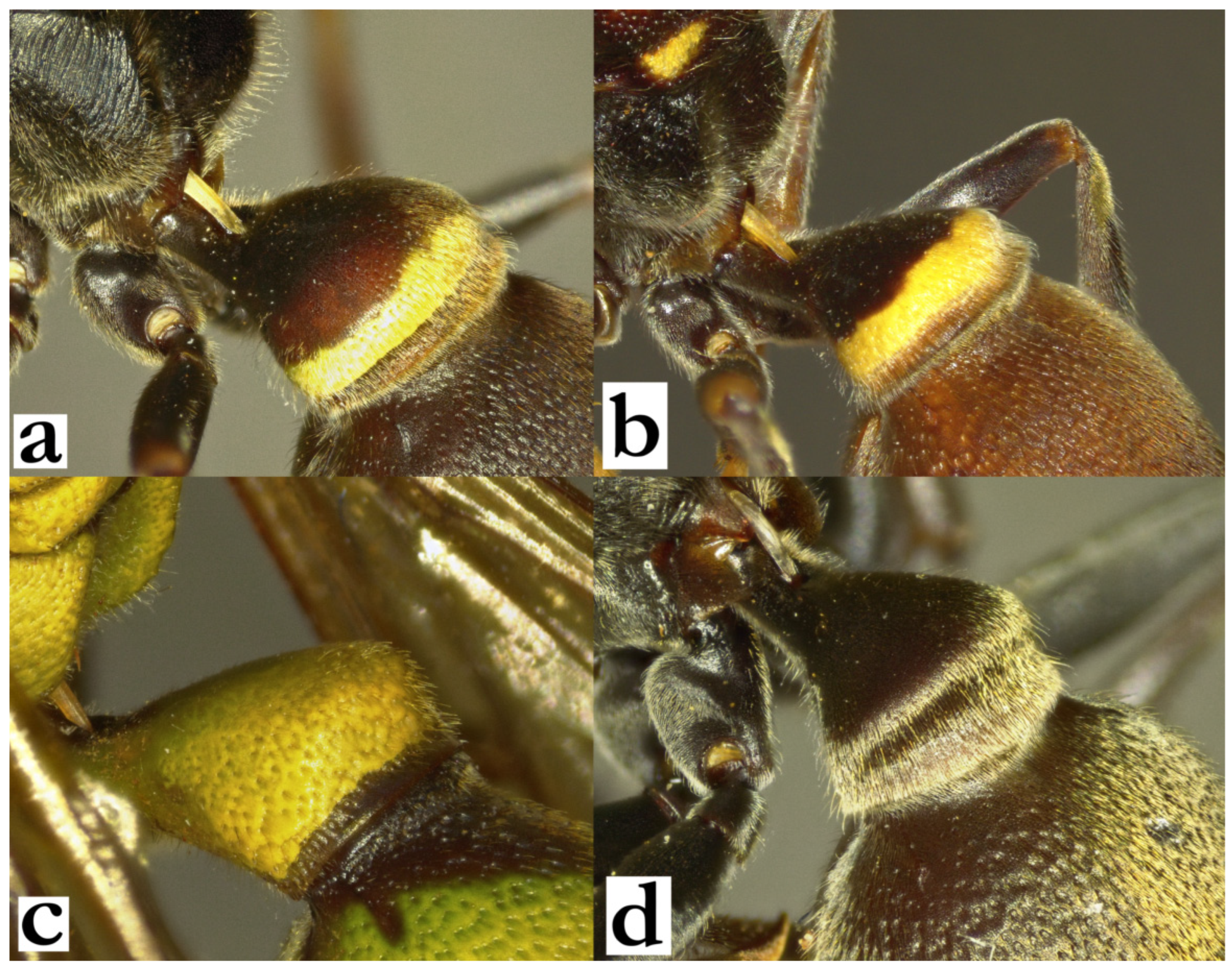
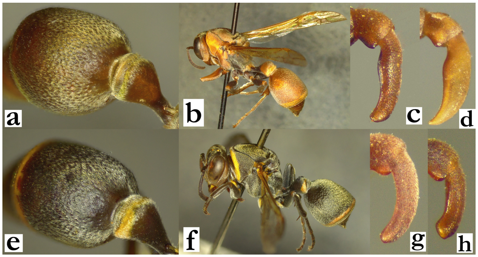
- 1. Forewing length over 11 mm; T1 globular or flattened-triangular (a), lacking or with only a very shallow posterior transverse sulcus (b). T1 basally brown to black, at most with a posterior yellow or green band (a, b)...............................................................................2
- - Forewing length under 11 mm; if approaching 11 mm then T1 predominantly yellow or green (c), or triangular with a flattened dorsal margin and a well-developed posterior sulcus (d).............................................................................................................................................10
Figure 1.
T1, oblique lateral view, female, (a) Ropalidia sakalava (de Saussure), (b) Ropalidia merina Blommers, (c) Ropalidia prasina (de Saussure), (d) Ropalidia atra (de Saussure).
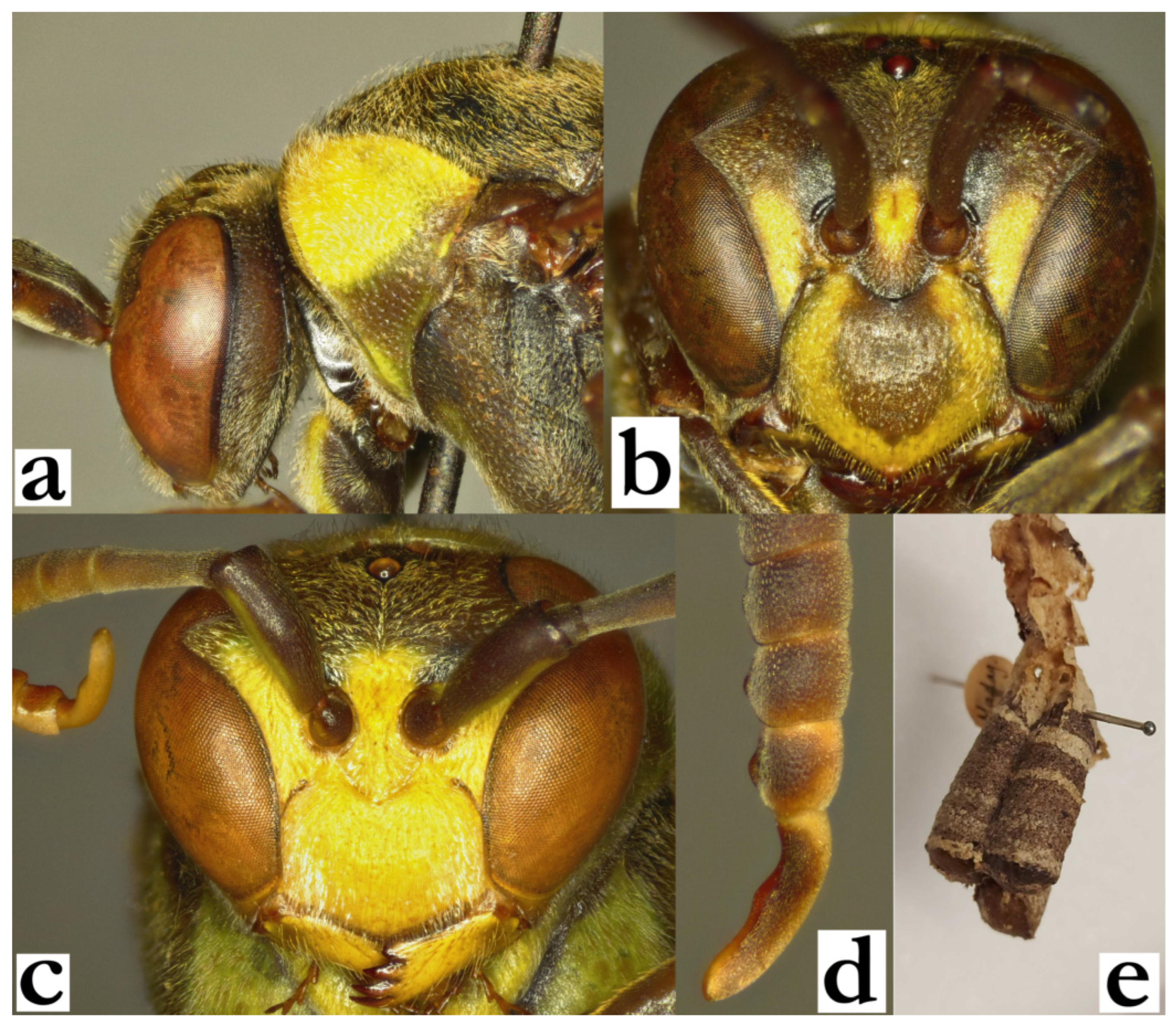
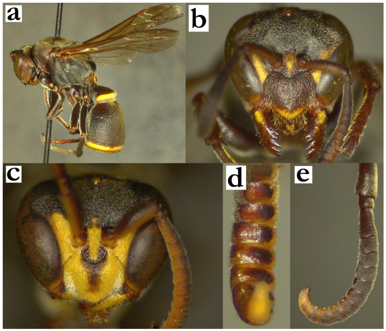
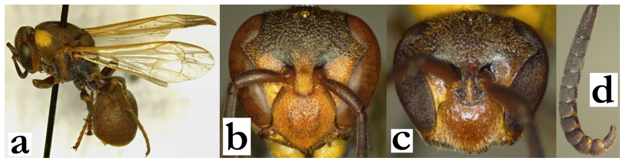
- 2. Basal body colour olivaceous-brown; pronotum with large yellow-greenish dorsal area (a), scutellum and metanotum with two large yellow spots. Entire body with long golden setae (a). Median carina of scutellum reaches over half of scutellum length.
[f] Clypeus pentagonal, yellow with basally attached olivaceous-brown spot; interantennal area and inner orbits yellow (b).
[m] Clypeus yellow (c); terminal flagellomere 3.8–4.1× as wide as long, apically obtuse; tyloids from AF4, elevated on AF(8)9–11 (d).
[wl] 13.1 [12.2–15.0] mm
[n] Small nest (5–15 cells), sessile (without petiole); wall striated in light and dark grey, cocoon caps dark brown (e) .........................................R. ducalis (de Saussure)
- - Basal body colour brown to black with fewer yellow or green markings (very xanthic specimens may have suffused markings on mesosoma); female inner orbits brown or with only small yellow marks; median carina of scutellum absent or weak (not reaching more than a third of scutellum length)..................................................................................................3
Figure 2.
Ropalidia ducalis (de Saussure), (a) head and mesosoma, lateral view, female, (b) head, frontal view, female, (c) head, frontal view, male, (d) terminal flagellomeres, dorsal view, male, (e) nest.
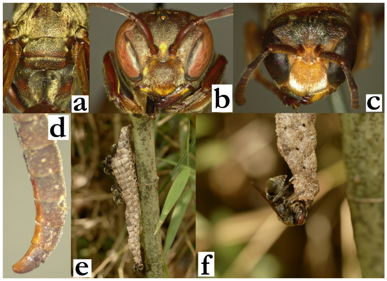
- 3. Basal body colour brown (a). Body with velutinous dense golden pubescence layer (a). Posterior ocelli at most a diameter apart (b).
[f] Yellow markings (if any) include patches on mandible, clypeal apex, latero-basal part of clypeus and interantennal area; gena with long protruding setae, 1.6–2.1× as long as AF1 base (b).
[m] Apical half of clypeus with dense and long silvery setae (c). Tyloids dark brown and very thin on AF(2)3, gradually widening and becoming yellow on AF8–11 (d); terminal flagellomere simple, about 1.8× as long as wide, apically rounded (d). Tarsal spur I short, no longer than first tarsal segment width.
[wl] 11.9 [11.4–13.9] mm
[n] Elongated, with apical petiole, up to 10 cells wide and 50 cells long (e); cell wall light grey, porous (f), cocoon caps dark grey and elevated from cell wall margin; suspended and aligned with a nearby branch (e)...............R. velutina (de Saussure)
- - Basal body colour black or dark brown. Body covered by substantially shorter and sparser pubescence layer. Posterior ocelli more than a diameter apart. Tarsal spur I in males longer than tarsal segment width.....................................................................................4
Figure 3.
Ropalidia velutina (de Saussure), (a) posterior mesosoma, dorsal view, female, (b) head, frontal view, female, (c) head, frontal view, male, (d) antenna, dorsal view, male, (e) nest, (f) nest, close-up [24].
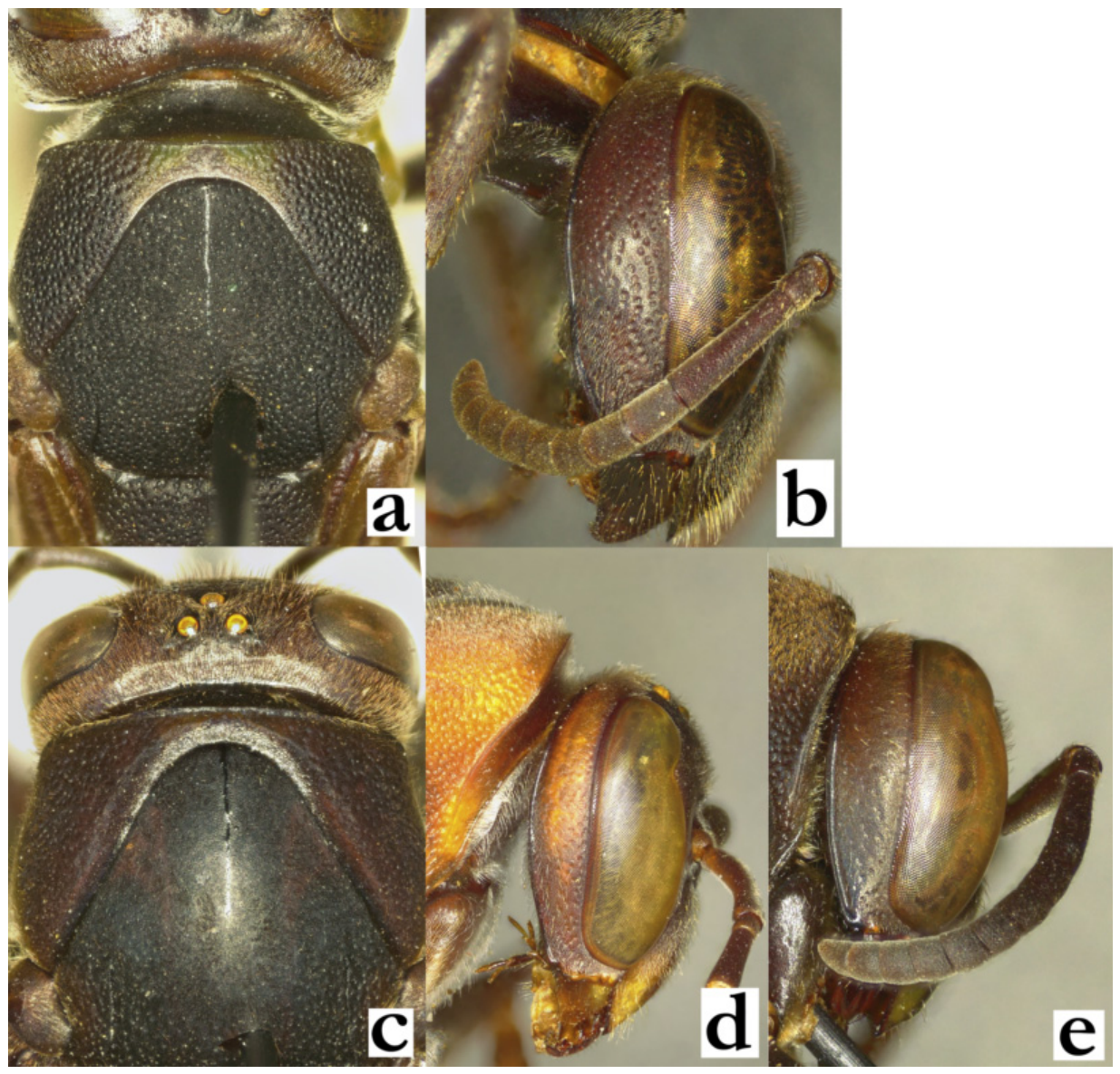
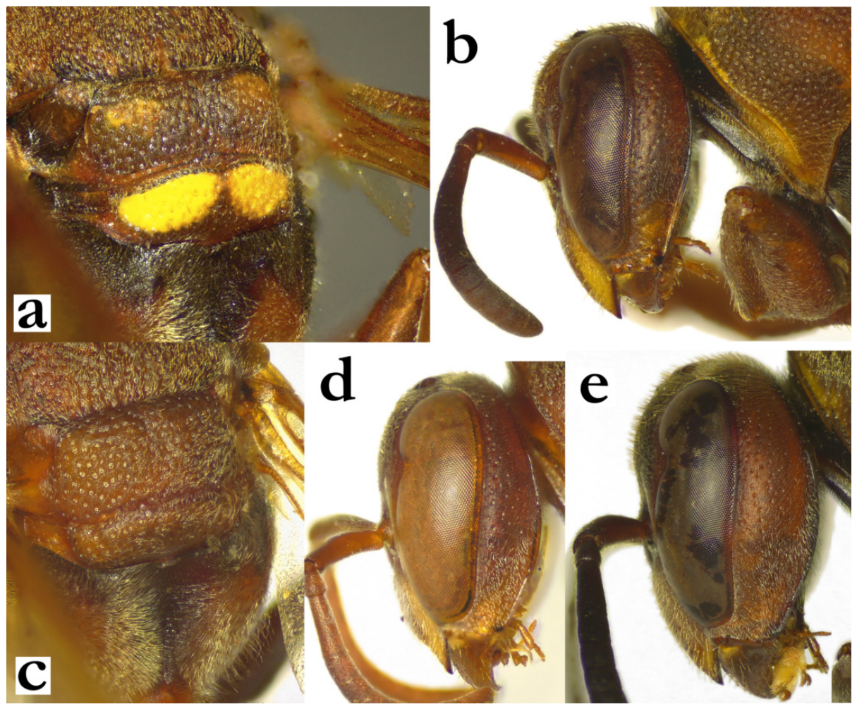
- 4. Anterior third of mesoscutum and dorsal margin of pronotum with discernible punctures (a). Female gena at least as wide as eye, with well-defined punctures (b)......................................................................................................................................................5
- - Anterior third of mesoscutum impunctate and shiny; dorsal margins of pronotum finely punctate or entirely impunctate (c). Female gena conspicuously narrower than eye (d), or wider with poorly defined craters and only an occasional well-defined puncture inferiorly (e)....................................................................................................................................................7
Figure 4.
(a) Ropalidia mainty sp. nov., anterior mesosoma, dorsal view, female; (b) Ropalidia bevatana sp. nov., head, lateral view, female; (c) Ropalidia sakalava (de Saussure), anterior mesosoma, dorsal view, female; (d) Ropalidia formosa (de Saussure), head, lateral view, female; (e) Ropalidia andalana sp. nov., head, lateral view, female.
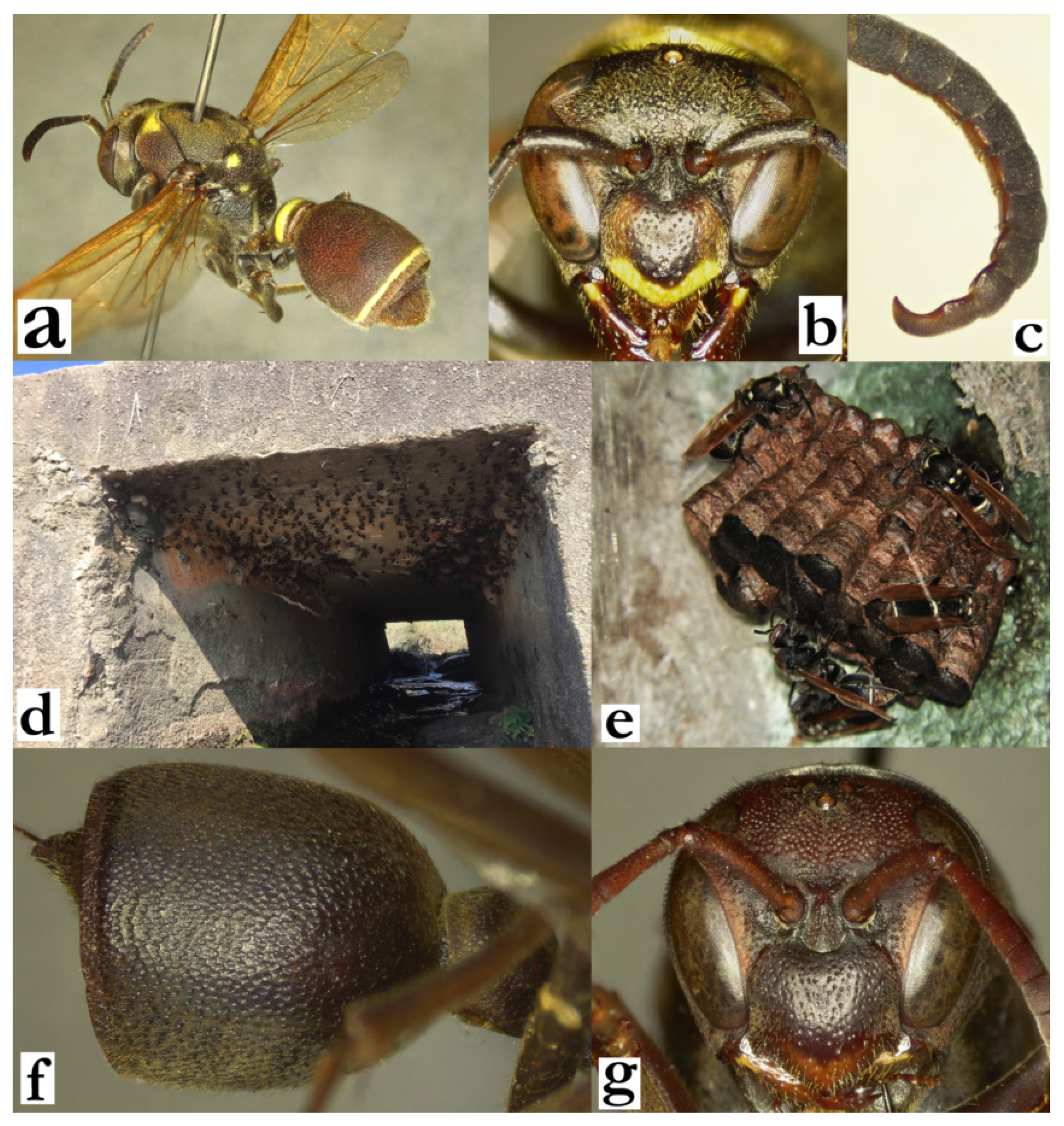
- 5. Body with whitish-green markings on pronotum, scutellum and metanotum (a). T1 dorsally flattened and low (a). T2 with longer yellow protruding hairs, extending over lamella (a).
[f] Clypeus commonly with green or yellow apical band (b). Frons covered by setae that are as long as or longer than ocelli diameter (b).
[m] AF2 1.3–1.4× as long as wide; AF4–10 about as long as wide. Tyloids brown and shiny, originating on AF2; tyloids setose; terminal flagellomere thick (c).
[wl] 11.2 [10.9–11.8] mm.
[n] Multiple discoid nests in colonies, usually located on underside of larger stones or in man-made objects, often close to water (d); up to five eggs can be present within a single cell; cocoon tops dark grey to black (e)................................R. merina Blommers
- - Body black with occasional green or yellow markings on mandible and T1; mesosoma exceptionally with suffused brownish, reddish or greenish markings. T1 globular, dorsally more developed (f). T2 with short black or brown setae or brissae that do not extend over lamella (f). Female clypeus rarely with green or yellow markings, at most with suffused reddish or orange apical area (g). Frons with setae or brissae that are shorter than anterior ocellus diameter (g)…………………………………………………………………….6
Figure 5.
Ropalidia merina Blommers, (a) habitus, oblique lateral view, female (b) head, frontal view, female, (c) distal flagellomeres, dorsal view, male, (d) nest colony [25], (e) nest, close-up [26]; Ropalidia bevatana sp. nov., (f) T2, lateral view, female, (g) head, frontal view, female.
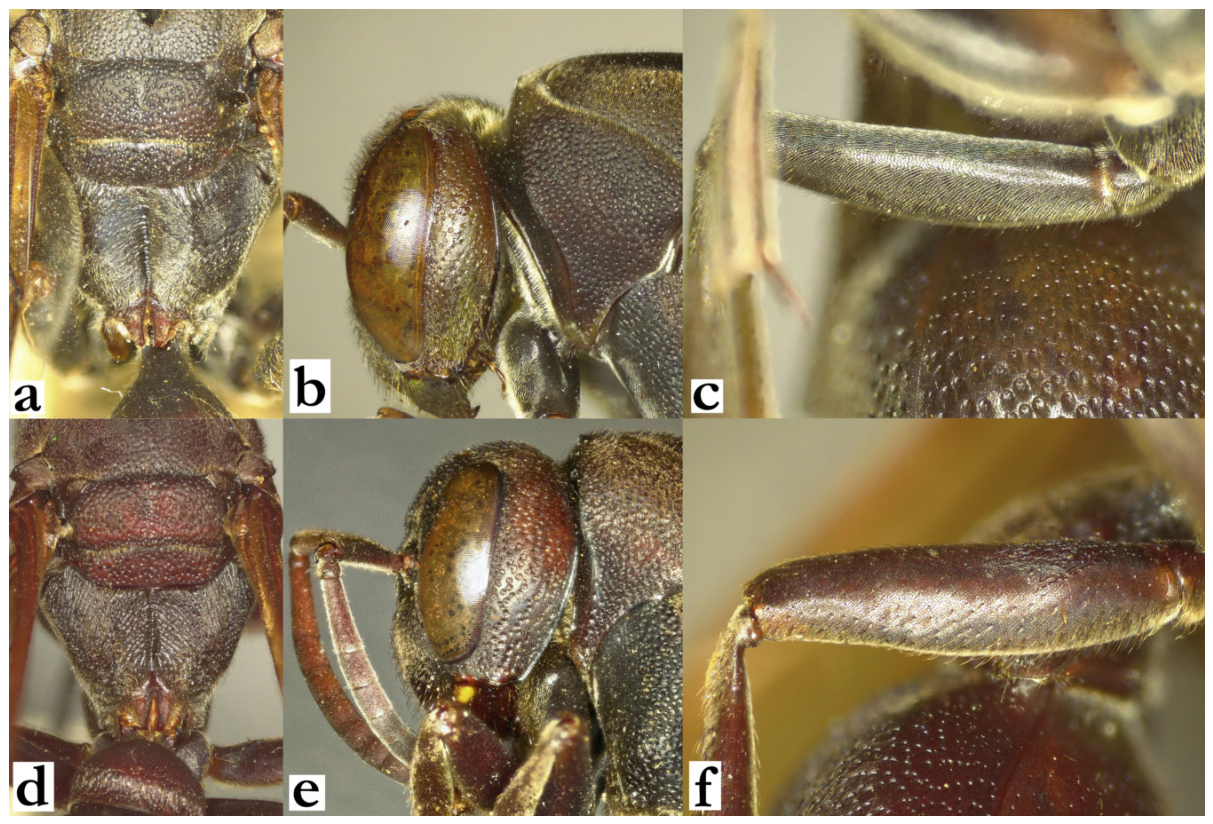
- 6. Metanotum posteriorly rounded; median depression of propodeum weakly striate, and shallowly and sparsely punctate (a).
[f] Head and mesosoma with short brownish-yellow setae (b). Femur III impunctate, with protruding setae of equal length proximally and distally (c)
[m] Unknown
[wl] 13.1 [12.7–13.5] mm
[n] Unknown............................................................................................R. mainty sp. nov.
- - Metanotum posteriorly flattened; median excavation of propodeum strongly striate and punctate (d).
[f] Head and mesosoma with short brown brissae (e). Femur III punctate, with proximal setae twice as long as distal (f).
[m] Unknown
[wl] 14.8 [12.7–17.9] mm
[n] Unknown.........................................................................................R. bevatana sp. nov.
Figure 6.
Ropalidia mainty sp. nov., female, (a) posterior mesosoma, dorsal view, (b) head, lateral view, (c) femur III, anterior view; Ropalidia bevatana sp. nov., female, (d) posterior mesosoma, dorsal view, (e) head, lateral view, (f) femur III, anterior view.
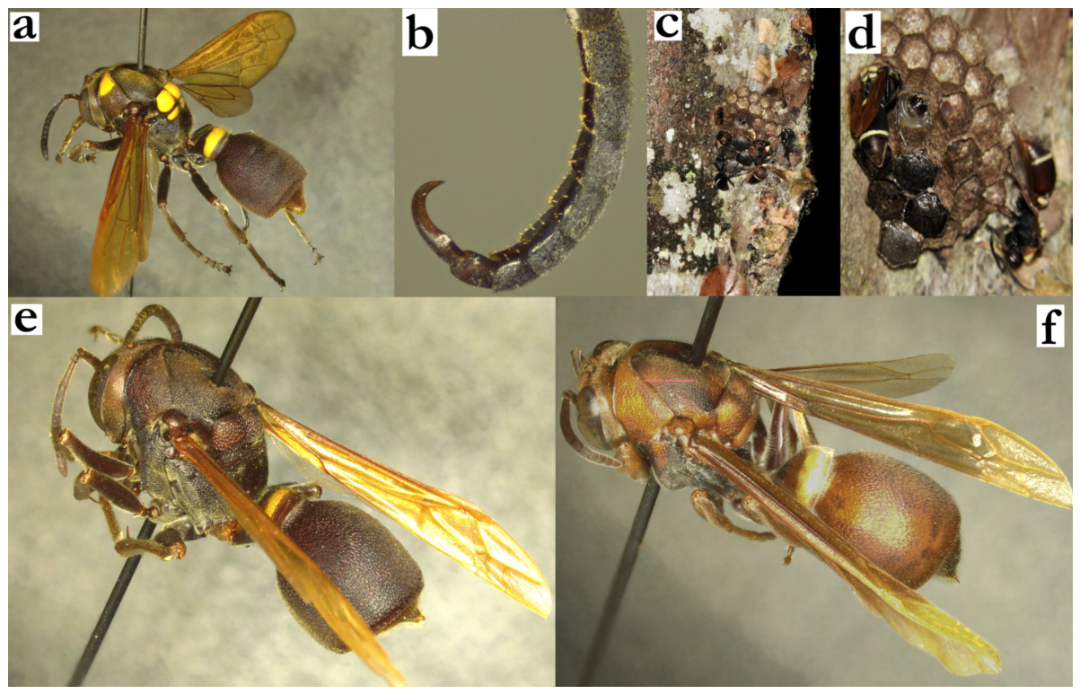
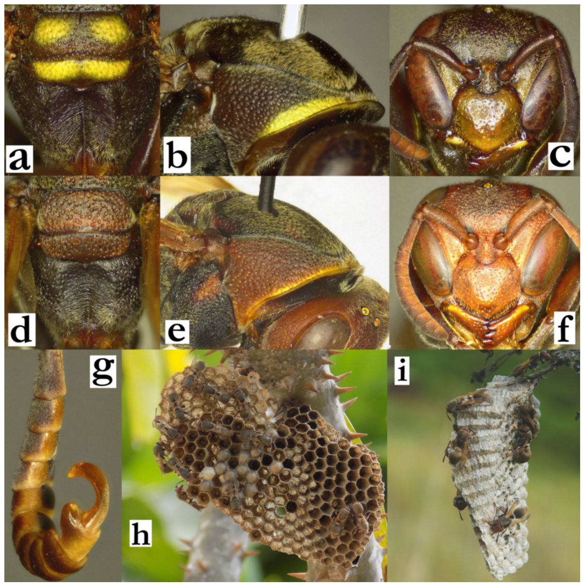
- 7. Mesosoma black with bright yellow markings; pronotum with large yellow area or entirely yellow, occasionally with reddish inferior angle (a). Scutellum and metanotum with yellow spots that occupy nearly entire surface (a). T1 (sometimes and T2) with a thick posterior yellow band (a).
[f] Mesoscutum pubescence yellowish-golden (a). Mesoscutum and inner orbit black; gena black, at most with greenish suffused hue (a).
[m] Tyloids dark-brown to black, originating of AF(2)3; terminal flagellomere elongated, curved and thin (b).
[wl] 12.3 [11.4–13.8] mm
[n] Located directly on tree trunk or similar vertical surfaces, concealed visually in diverse surroundings (c). Nest discoid, with up to 30 cells and a central petiole. Cell colour brownish, cocoon tops at later stages black (d)..................R. hova (de Saussure)
- - Mesosoma uniformly dark brown to black (e), at most with faintly reddish or yellowish suffused markings in very xanthic specimens (f); in these instances, mesoscutum frequently has two reddish or yellowish markings, while gena, frons and inner orbit can be suffused in reddish or yellow (f).
[f] Mesoscutum pubescence whitish-silvery or brown.
[m] Tyloids originate on AF1 or AF2..................................................................................8
Figure 7.
Ropalidia hova (de Saussure), (a) habitus, lateral oblique view, female, (b) terminal flagellomeres, dorsal view, male, (c) nest, (d) nest, close-up [27]; Ropalidia andalana sp. nov., lateral oblique view, female, (e) melanic specimen, (f) xanthic specimen.
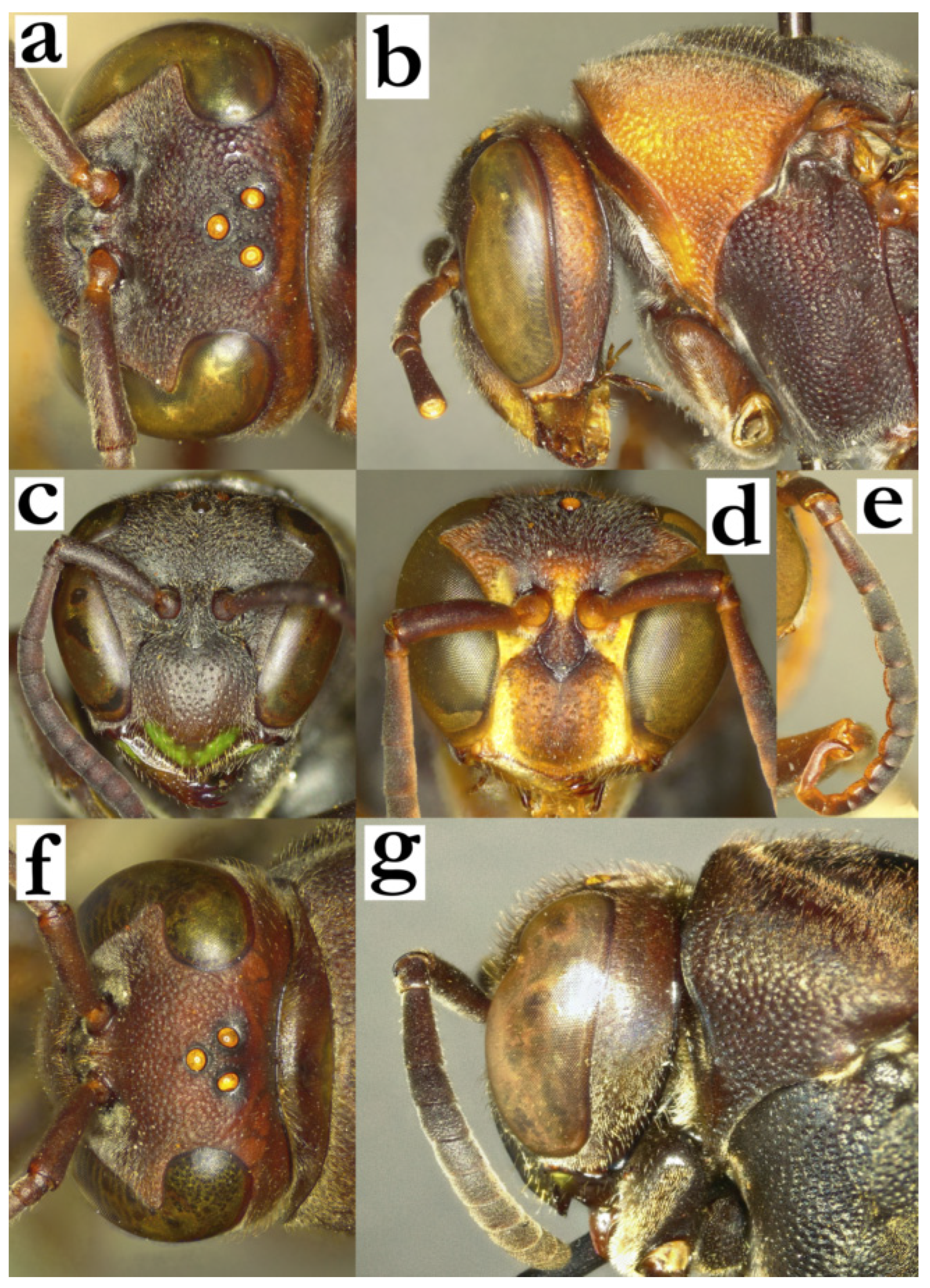
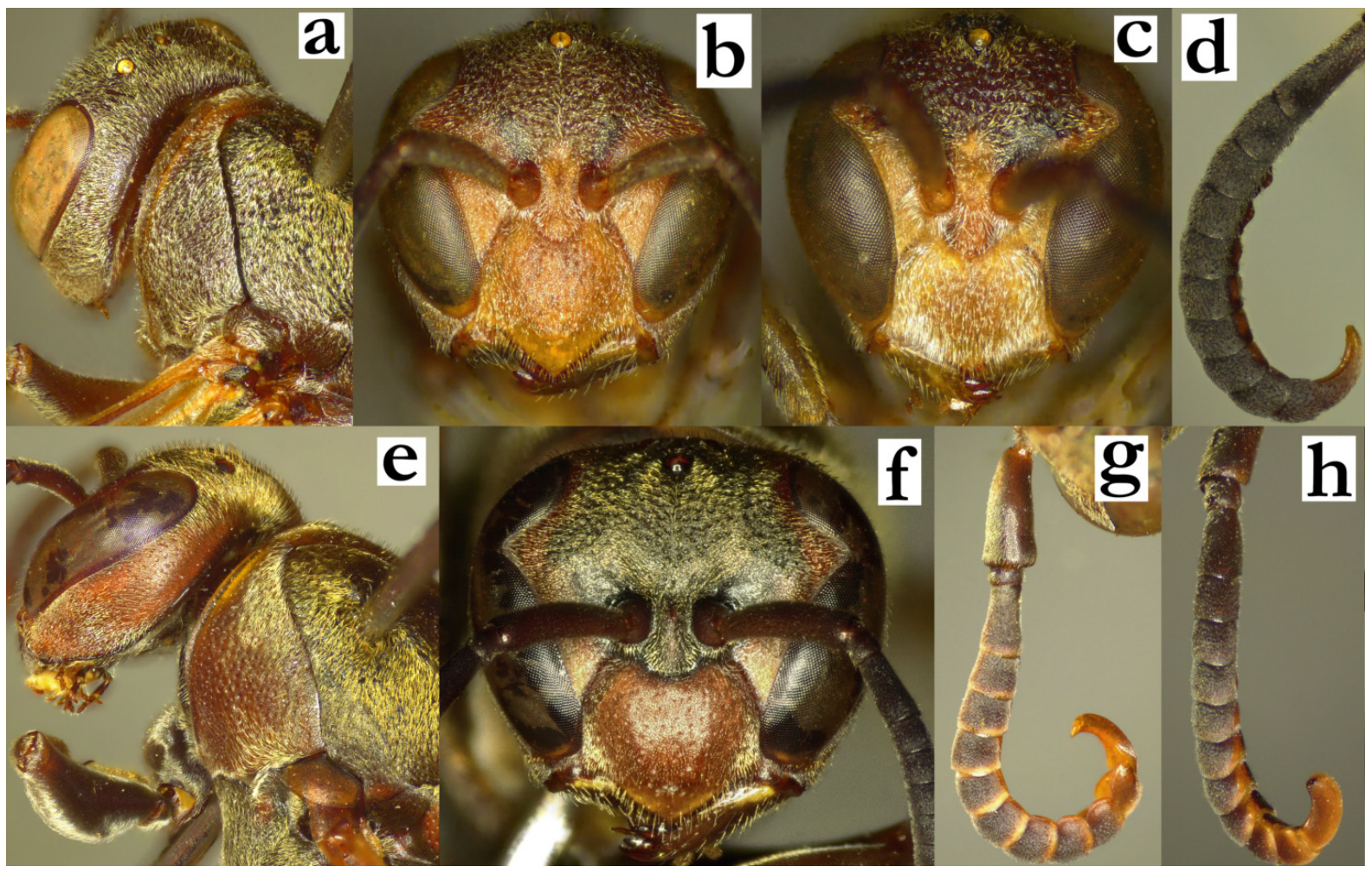
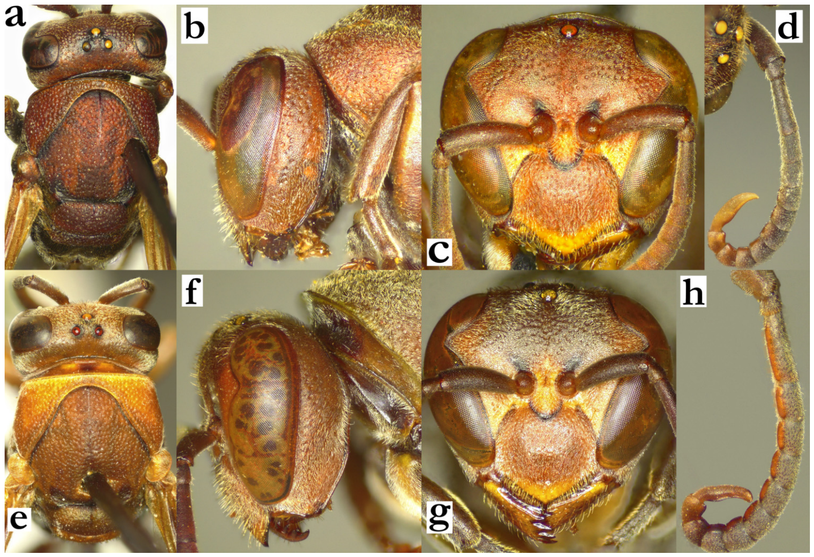
- 8. Posterior ocelli 1.8–2.0 diameters apart (a).
[f] Gena at midpoint half of eye width (b). Clypeus longer than wide, base and centre with well-defined punctures (c). Frons setae short, 0.5–0.8× as long as ocellus diameter.[m] Clypeus basally and centrally with well-defined punctures (d). AF2 2.1–2.4× as long as wide (e). Tyloids asetose, originate on AF2; terminal flagellomere strongly curved and thick (e).[wl] 14.6 [12.8–17.3] mm[n] Unknown................................................................................R. formosa (de Saussure)
- - Posterior ocelli 1.3–1.7 diameters apart (f).
[f] Gena at midpoint more than half of eye width (g). Clypeus about as long as wide or wider, centre impunctate or sparsely punctate. Frons setae longer than or equal to ocellus diameter.[m] AF2 1.4–2.1× as long as wide. Tyloid surface setose; tyloids originate on AF1 or AF2...........................................................................................................................................9
Figure 8.
Ropalidia formosa (de Saussure), (a) head, dorsal view, female, (b) head and anterior mesosoma, lateral view, female, (c) head, frontal view, female, (d) head, frontal view, male, (e) antenna, dorsal view, male; R. andalana sp. nov., (f) head, dorsal view, female, (g) head, lateral view, female.
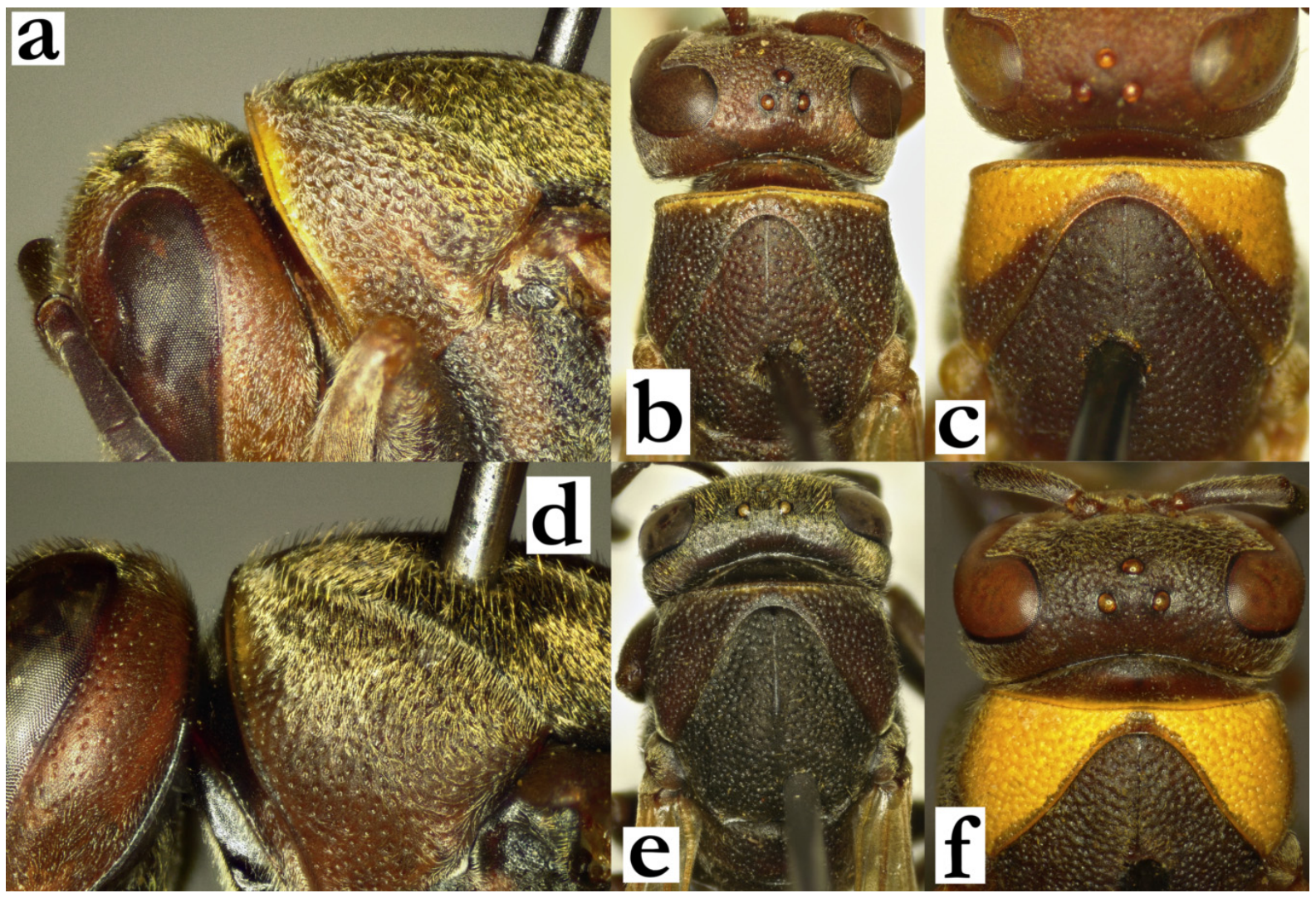
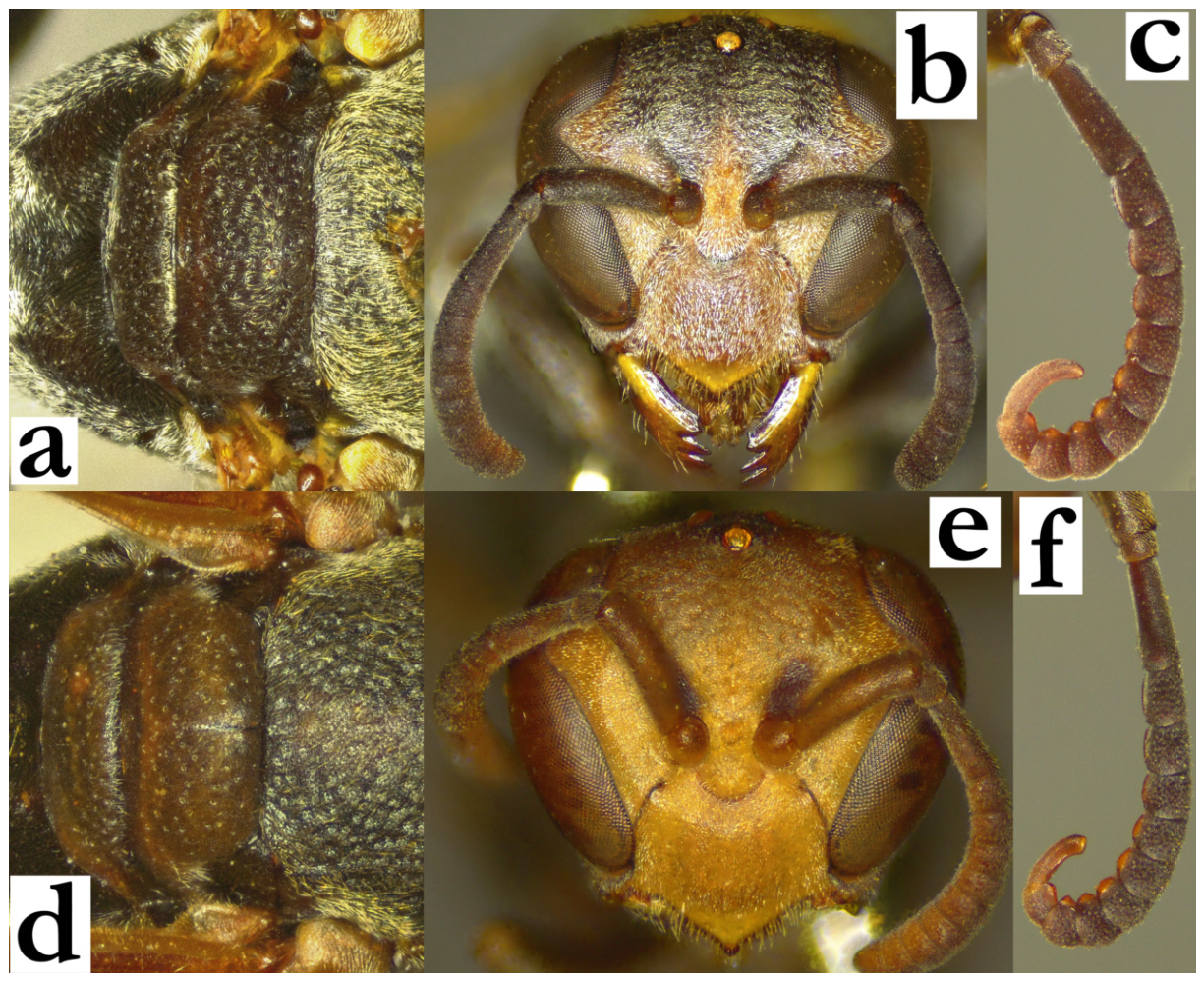
- 9. Median depression of propodeum striate, with defined lateral margins (a). Scutellum bituberculate, with a shiny median impunctate streak (a).
[f] Clypeus 1.2× as wide as long, with matte surface and scattered fine punctures (b), about 3–4 diameters apart basally (c). Interantennal slit absent or very poorly developed (c). Gena impunctate, sometimes with poorly defined craters close to mandible.
[m] Tyloids originate at AF1, yellow to brown (those on AF2-6 sometimes dark brown). Terminal flagellomere wider, with a flattened outer (anterior) margin (d). AF2 1.4–1.5× as long as wide.
[wl] 11.9 [11.4–13.6] mm
[n] Unknown.......................................................................................R. andalana sp. nov.
- - Median depression of propodeum finely striate and shiny, lateral margins obtuse (e). Scutellum with flattened surface and midline punctures (e).
[f] Clypeus as wide as long, with large, scattered punctures centrally (f), clustered basally, about one diameter apart (g). Interantennal slit developed, at least as long as half the antennal socket diameter (g). Gena at mid-height with some retained punctures.
[m] Tyloids brownish, originate on AF2; terminal flagellomere elongated and thin, with a minute anterior swelling (h). AF2 1.5–1.8× wide as long.
[wl] 12.1 [11.6–14.8] mm
[n] Unknown..............................................................................R. sakalava (de Saussure)
Figure 9.
Ropalidia andalana sp. nov., (a) posterior mesosoma, dorsal view, female, (b) head, frontal view, female, (c) interantennal area, oblique dorsal view, female, (d) distal flagellomeres, dorsal view, male; R. sakalava (de Saussure), (e) posterior mesosoma, dorsal view, female, (f) head, frontal view, female, (g) interantennal area, oblique dorsal view, female, (h) distal flagellomeres, dorsal view, male.
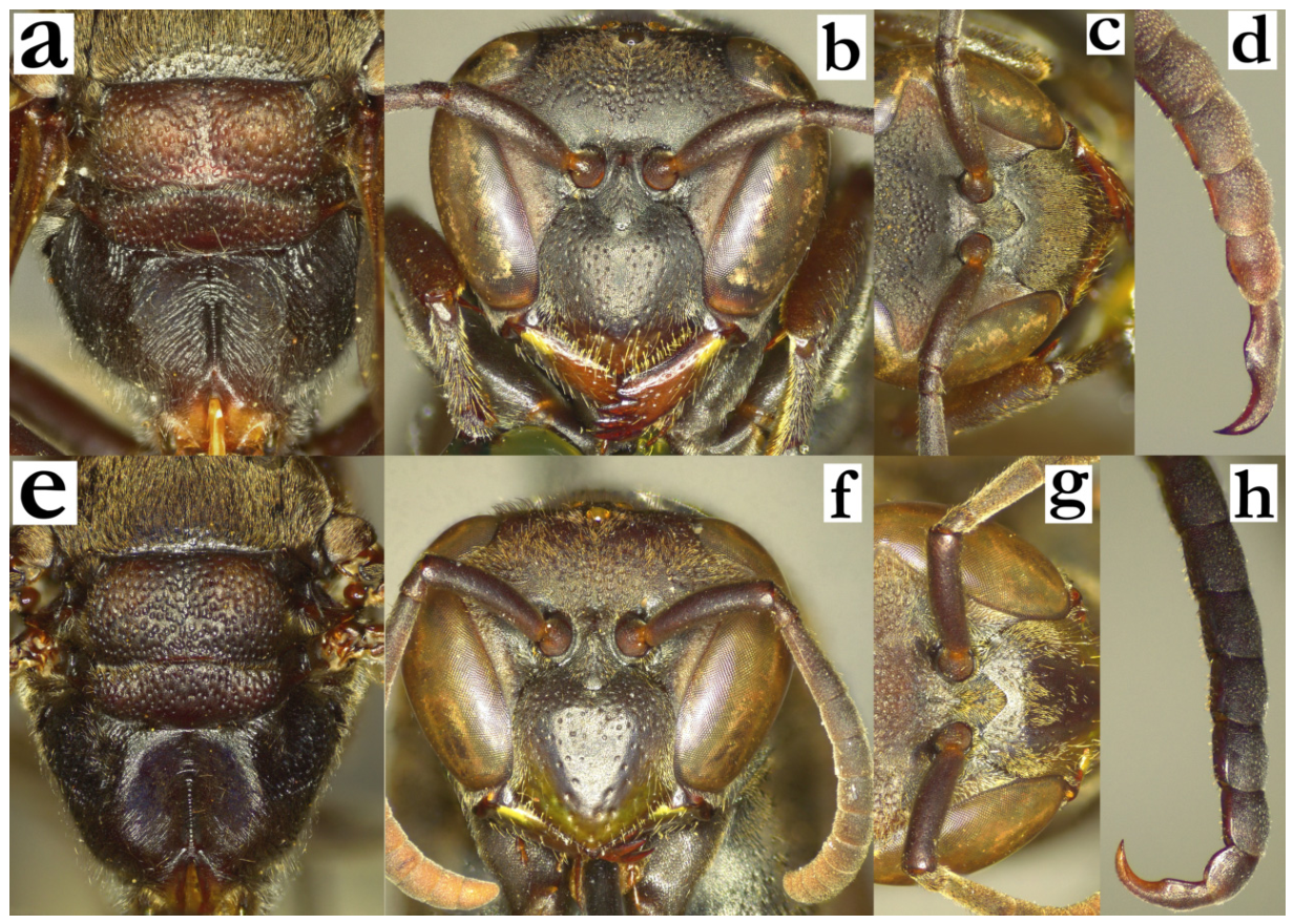
- 10. Larger species (fore wing length ≥ 11 mm), with predominantly green (and yellow) body colour. Gena strongly developed; at least 1.5× as wide as eye in females and as wide as eye in males; occipital carina dorsally evanescent or barely developed (a). Scutellum bituberculate, with visible median sulcus (b). Clypeus at least 1.4× as wide as long. Terminal flagellomere in males claw-liked and elongated..........................................................11
- - Body size smaller (fore wing length < 11 mm); if wing length is approaching 11 mm and body has numerous green markings, then occipital carina is conspicuous medio-dorsally (c) and scutellum is dorsally rounded (d).................................................................................12
Figure 10.
Ropalidia regina (de Saussure), (a) head and mesosoma, dorsal view, female, (b) posterior mesosoma, oblique view, female; Ropalidia dubia (de Saussure), (c) head and mesosoma, dorsal view, female, (d) posterior mesosoma, oblique view, female.
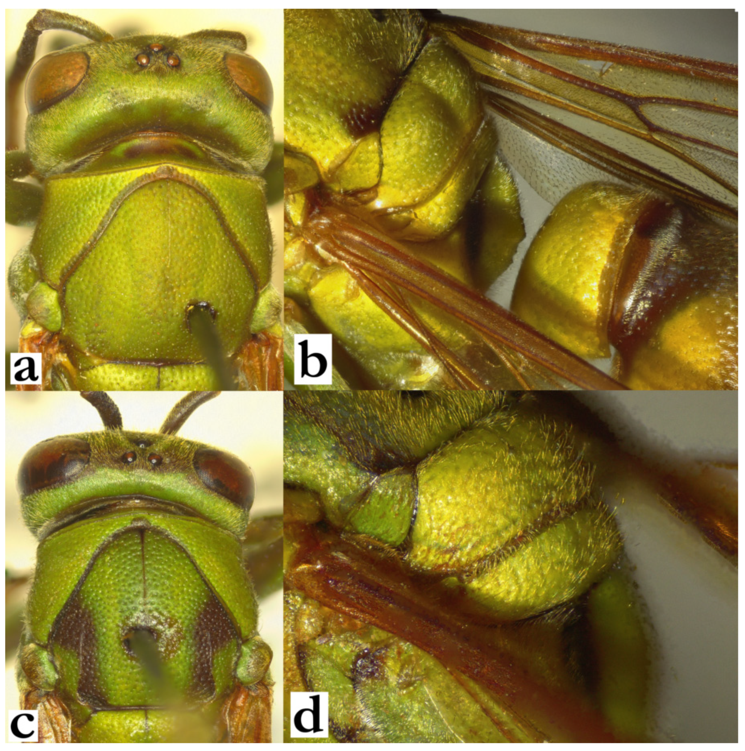
- 11. T1 posteriorly globular and rounded, weakly punctate; median depression of propodeum shallowly punctate and shiny (a). T2 shallowly punctate distally; T2 commonly with larger brownish cross-like area between two yellow(-green) basal spots and posterior yellow band (b). Antenna dorsally ferruginous or infuscate.
[f] Clypeus 1.3–1.4× as wide as long; upes sinuate. AF2 1.1–1.2× as long as wide.
[m] Clypeus with strong juxtamandibular excavations and depressed and pointy apex (c). Antenna stockier; scape 4× as long as wide, AF2 1.3× as long as wide. Tyloids originate on AF(4)5. Terminal flagellomere 2× as long as wide, with acute tip (d).
[wl] 11.4 [10.9–13.2] mm
[n] Discoid, up to 70 cells (some nests can be more elongated in shape). Cell wall compact, light grey with subtle darker streaks; cocoon caps slightly brownish, not extending above cell wall margin [28]............................R. regina (de Saussure)
- - T1 angular, dorsally coarsely punctate (e). Median depression of propodeum punctate (f). T2 shallowly punctate; T2 commonly dorsally yellow(-green), with just a remaining semi-circular dark-brown area (g). Antenna blackened or black dorsally.
[f] Clypeus 1.2–1.3× as wide as long; upes straight. AF2 1.2–1.4× as long as wide.
[m] Clypeus with less developed juxtamandibular excavation and obtuse tip (h). Antenna more slender; scape 4.8–5.0× as long as wide, AF2 2.4× as long as wide (h). Tyloids originate on AF(5)6. Terminal flagellomere 1.6–1.7× as long as wide, with subacute tip (h).
[wl] 12.1 [10.7–13.1] mm
[n] Elongated, two cells wide, up to 30 cells long [29]. Cell wall greenish-grey, cocoon caps brownish, with streaks of cell wall; nests commonly located in heavy bushes.....................................................................................R. prasina (de Saussure)
Figure 11.
Ropalidia regina (de Saussure), (a) propodeum, oblique view, female, (b) T2, dorsal view, female, (c) head, frontal view, male, (d) distal flagellomeres, dorsal view, male; Ropalidia prasina (de Saussure), (e) propodeum, oblique view, female, (f) T2, dorsal view, female, (g) head, frontal view, male, (h) distal flagellomeres, dorsal view, male.
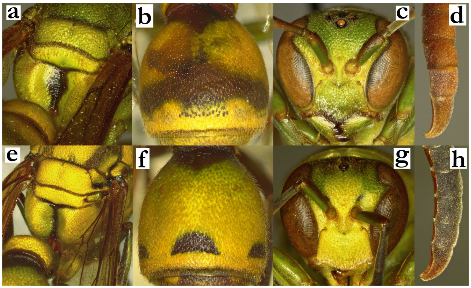
- 12. Head conspicuously developed, wider than mesosoma and tegulae in dorsal view (a). Female gena 1.5–2.0× as wide as eye (b). Entire body with very short pubescence; setae on frons shorter than anterior ocellus diameter (b). Mesosoma with very short setae; mesoscutum without protruding setae (b). Head and mesosoma punctures weakly defined and shallow, especially on gena, pronotum and mesoscutum....................................13
- - Head less developed, not wider than mesosoma (c). Female gena less developed, rarely more than 1.5× as wide as eye (in these cases, pilosity is much longer and head and mesosoma punctures more developed and coarser (d)...................................................................14
Figure 12.
Ropalidia vitripennis (de Saussure), (a) head and anterior mesosoma, dorsal view, female; (b) Ropalidia nitidula (de Saussure), head, lateral view, female; (c) Ropalidia grandidieri (de Saussure), head and anterior mesosoma, dorsal view, female; (d) Ropalidia cincinnata Blommers, head, lateral view, female.
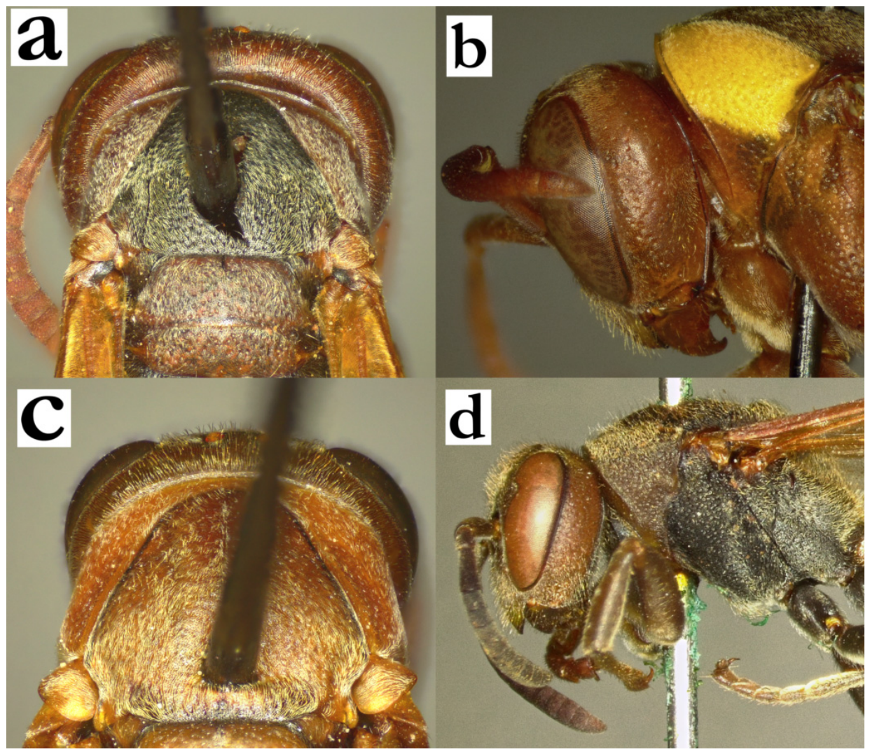
- 13. Smaller body size; wing length under 10 mm. Yellow markings minimal or suffused (a).
[f] Body basally brown or black, with reddish, ferruginous or brown areas on mandible, clypeus apex, inner orbit and pronotum; head and pronotum commonly reddish, mesoscutum and metasoma darker (a). Occipital carina less developed and thin, occasionally missing in lowest third (a). Frons, gena and mesoscutum with short whitish pubescence (a). Pubescence on posterior third of mesoscutum directed caudally. Pronotal carina equally wide laterally and dorsally (a).
[m] Not examined. Reported as apically (sub)acute terminal flagellomere, with several distally visible tyloids ([5], b).
[wl] 8.6 [7.6–9.4] mm
[n] Elongated, up to five cells wide apically and 30 cells long, with “tail” of low cells towards nest bottom; cell wall solid, greyish; nest suspended from branches (c).......................................................................................R. vitripennis (de Saussure)
- - Larger body size; wing length over 10 mm. Dorsal half or pronotum yellow (d).
[f] Occipital carina well developed in its entire length (d). Frons, mesoscutum and gena with short, golden pubescence (d); mesonotum pubescence almost entirely directed anteriorly. Pronotal carina twice as tall dorsally as wide laterally (d).
[m] Not examined. Reported as elongated terminal flagellomere with an obtuse tip and tyloids originating from AF5 ([5]; e).
[wl] 10.7 [10.3–11.1] mm
[n] Droplet-shaped, apically suspended, up to 200 cells, with 20 × 60 formula. Cell wall porous, grey with light-grey streaks; cocoon tops whitish to light-brown; nest suspended from branches, often in hardly accessible locations (above water or in heavy thicket), occasionally in clusters of multiple nests [30,31]…………………....................................................….R. nitidula (de Saussure)
Figure 13.
Ropalidia vitripennis (de Saussure), (a) head, oblique view, female, (b) antenna, dorsal view, male, (c) nest [32]; Ropalidia nitidula (de Saussure), (d) head, oblique view, female, (e) antenna, dorsal view, male.
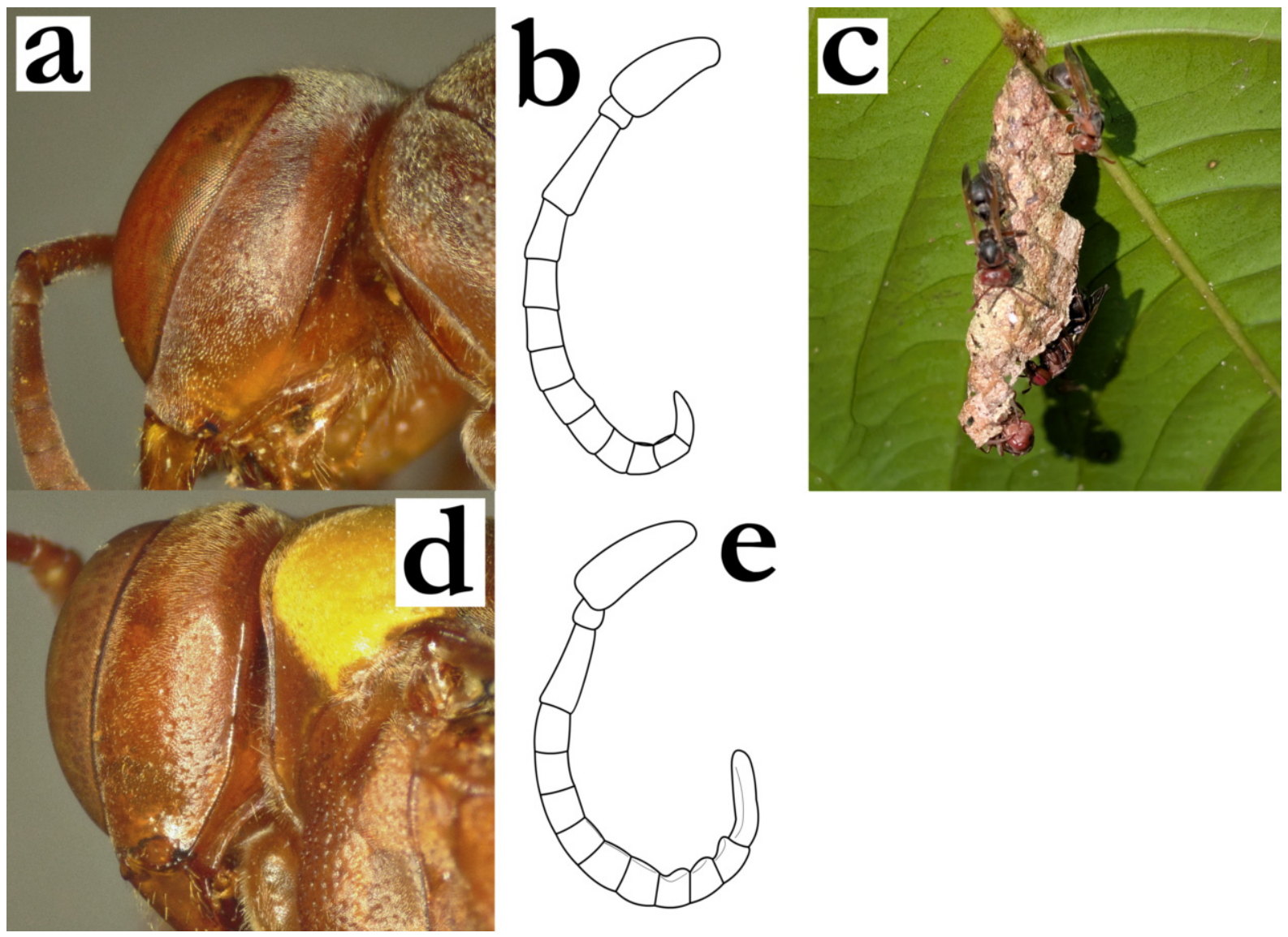
- 14. T1 triangular, posteriorly globular and wide, with small angular lateral processus (a). Body black and reddish, with bright yellow markings on head, mesosoma and legs; metanotum commonly with two quadratic bright yellow spots (b). Metanotum posteriorly flattened (b).
[f] Pronotal carina projecting antero-medially (c). Mesoscutum coarsely punctate (c).
[m] Tyloids originate at AF(6)7; terminal flagellomere elongated, about 4× as long as wide, with strong longitudinal carina an obtuse tip; outer margin with numerous short setae (d).
[wl] 7.8 [7.3–8.4] mm
[n] Discoid or slightly elongated, with up to 180 cells (sometimes irregular in shape, defined by surroundings); located on tree trunk patch between coarser bark, lichen or moss, less commonly on stones; nest is concealed visually and by texture; cell wall brownish-grey, with green streaks [33,34], cocoon caps silky white, later in the cycle brownish (e) ………………………………………….R. minor (deSaussure)
- - T1 without lateral processes (f). Body colour pattern different; specimens of R. volontany sp. nov. have general appearance similar to R. minor, but then T1 has developed posterior sulcus (f), metanotum is projecting posteriorly (g), pronotal carina straight anteriorly and mesoscutum punctures finer and shallower (h).......................................................................15
Figure 14.
Ropalidia minor (de Saussure), (a) habitus, anterior oblique view, female, (b) posterior mesosoma, dorsal view, female, (c) head and anterior mesosoma, dorsal view, female, (d) antenna, dorsal view, male, (e) nest [35]; Ropalidia volontany sp. nov., (f) habitus, anterior oblique view, female, (g) posterior mesosoma, dorsal view, female, (h) head and anterior mesosoma, dorsal view, female.
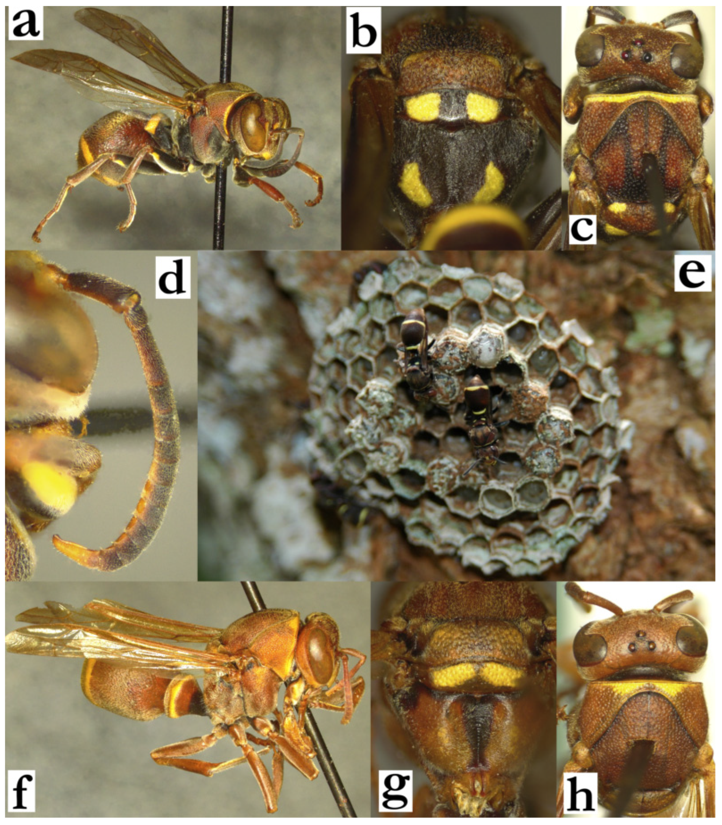
- 15. T1 wide, 0.5–0.8× as wide as T2, dorsally infundibular, triangular, with rounded posterior margins (a); posterior sulcus of T1 well developed and symmetrical, of similar depth and width along its circumference (b). T1 without bright green markings (a, b), or exceptionally with a subtle greenish tint. Male tyloids wide and shiny, black or at least partly black (c); except in R. schulthessi and some specimens of R. takona sp. nov., which have brown tyloids (see couplets 16 and 24) .................................................grandidieri group—16
- - T1 narrower, no more than half of T2 width, pyriform (d) or globular (e); if T1 is wider than 0.5× of T2 width, then T1, head and mesosoma have numerous green markings (f). Posterior sulcus of T1 absent (d) or asymmetrical, with two deeper sections in the mid-lateral part of its course and shallower dorso-medially and laterally (e). Proximal male tyloids light brown (some species can have darkened tyloids on two distal flagellomeres).............................................................................................................................................26
Figure 15.
Ropalidia grandidieri (de Saussure), (a) T1, dorsal view, female, (b) T1, oblique view, female, (c) antenna, dorsal view, male; (d) Ropalidia ranavali (de Saussure), T1, dorsal view, female; (e) Ropalidia galimatia (de Saussure), T1, oblique view, female; (f) Ropalidia constitutionalis (de Saussure), T1, dorsal view, female.
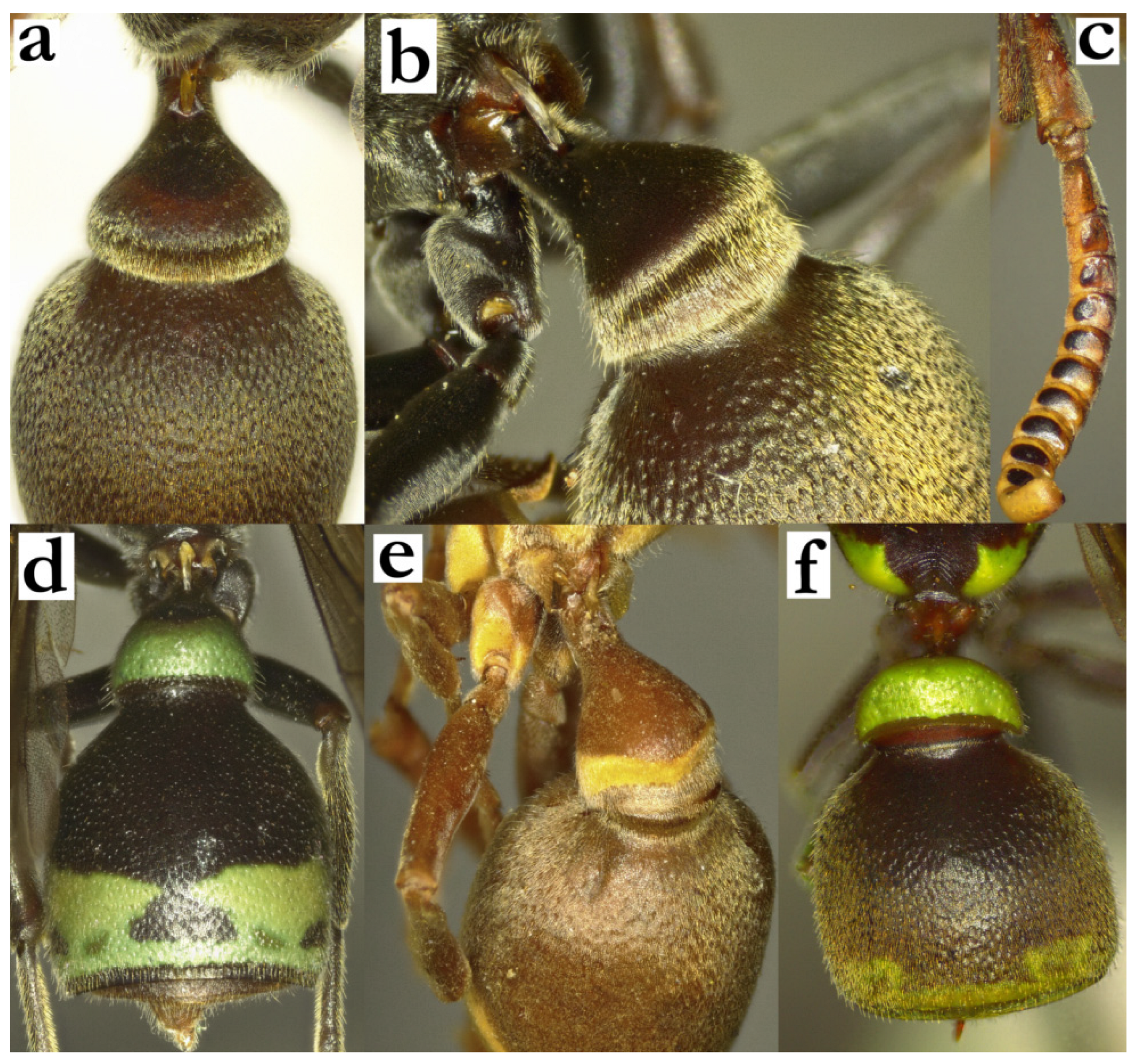
- 16. Small species, with wing length rarely exceeding 7 mm. T1 0.7–0.8× as wide as T2 (a); mesosoma comparatively slender; mesoscutum 1.4× as long as wide. Body pubescence very short; setae on frons much shorter than ocellus diameter (b).
[f] Clypeus with deep juxtamandibular excavation and strongly projecting apex; face ferruginous (b).
[m] Clypeus with obtuse apex (c). Tyloids brown and shiny, originate at AF2, those on AF9–11 elevated; terminal flagellomere elongated, curved and apically obtuse (d).
[wl] 6.6 [6.2–7.1] mm
[n] Small and discoid, up to 35 cells. Often with broadend or with multiple supporting petioles, located on the underside of coconut, palms or banana leaves nest [36]............................................................................R. schulthessi (de Saussure)(=R. nilssoni Giordani Soika, syn. nov.)
- - Body larger (forewing length rarely under 7 mm). If wing size is approaching 7 mm, then female clypeus has shallower juxtamandibular notches and body pilosity is more developed (as in R. linearecta Blommers, see couplet 18). Mesoscutum at most 1.2× as long as wide. At least some tyloids on male antenna black (except R. takona sp. nov., see couplet 24)...................................................................................................................................................17
Figure 16.
Ropalidia schulthessi (de Saussure), (a) habitus, lateral view, female, (b) head, frontal view, female, (c) head, frontal view, male, (d) distal flagellomeres, dorsal view, male.
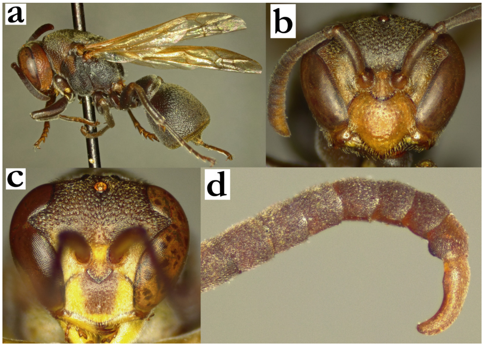
- 17. Highly contrasting colour pattern: dark brown to black body with broad yellow posterior bands on T1 and T2; mesosoma without yellow markings, at most with suffused reddish area on pronotum or scutellum (a). Smaller size; wing length under 8 mm. Metepisternum shiny, minutely and sparsely punctate anteriorly, posterior part impunctate.
[f] Head with yellow markings on inner orbits, interantennal area, clypeal apex and mandible (b).
[m] Head with a similar coloured pattern to female, but with more extensive yellow marks, clypeus nearly completely yellow (c). Tyloids originate on AF2, ventrally black, dorsally reddish-brown; dorsal margin of tyloid on AF6–AF11 elevated (d, e). Terminal flagellomere beak-like, with obtuse tip (e).
[wl] 7.4 [7.0–7.9] mm
[n] Elongated and sometimes very large, with up to 150 cells (20 cells wide and up to 200 cells long), located on palm leaves, sometimes with multiple petioles. Cell wall uniformly grey .....................................................R. carinata (de Saussure)
- - Less contrasting colour pattern: body entirely black, or mesosoma with yellow(ish) markings; metasoma at most with faint yellowish posterior bands or without any yellow markings. If wing length is close to 8 mm, then basal body colour is light brownish in R. takona sp. nov. (see couplet 24), or body pilosity much more developed in R. linearecta (key couple 18). Metepisternum coarsely punctate and less shiny................................................18
Figure 17.
Ropalidia carinata (de Saussure), (a) habitus, lateral view, female, (b) head, frontal view, female, (c) head, frontal view, male, (d) tyloids, median view, male, (e) antenna, dorsal view, male.
- 18. Body with a dense under-layer of silvery-yellowish pubescence, which almost completely obscures cuticular surface and punctures (a). Occipital carina placed high on postero-dorsal head margin (top of head curvature; a). Pronotal carina twice as wide dorsally than laterally (a).
[f] Clypeus pentagonal, with narrower base and longer upes that continue to lateral clypeal margin (almost) uninterrupted; OCA absent (b).
[m] Clypeus with long silvery setae (c). Tyloids originating at AF1, those on AF2–AF5 rounded and elevated above segment surface; more distal tyloids flat (d). Terminal flagellomere slender and elongated, evenly curved, with tyloid that reaches the tip (d).
[wl] 7.6 [7.1–7.9] mm
[n] Elongated and thin, resembling a dried branch, with a formula of 2×n [two cells wide and n cells long], with the longer nests up to 70 cells long [37]; most cells belong to type III [38], but some nests are type I, with only one cell width at sections of the nest or a part overlap of the adjacent cells [39]. Cell wall light brown or greyish, cocoon caps grey....................................R. linearecta Blommers
- - Body without dense lower layer of pubescence—rotating the specimen can easily reveal pronotum punctures; some specimens of R. grandidieri (de Saussure) can have denser pubescence, but in these cases pronotal carina is equally wide laterally and dorsally (e) and occipital carina is placed lower than postero-dorsal head margin (e).
[f] Clypeus with wider base and obtuse OCA (f).
[m] Clypeus with less developed or less dense setae. Antenna without elevated tyloids on proximal flagellomeres (g, h). Terminal flagellomere more developed and differently shaped, with an acute (g) or obtuse tip (h)....................................19
Figure 18.
Ropalidia linearecta Blommers, (a) mesosoma and head, oblique view, female, (b) head, frontal view, female, (c) head, frontal view, male, (d) antenna, dorsal view, male; R. grandidieri (de Saussure), (e) mesosoma and head, oblique view, female, (f) Ropalidia grandidieri (de Saussure), head, frontal view, female; (g) Ropalidia shestakowi (Schulthess), antenna, dorsal view, male; (h) Ropalidia grandidieri (de Saussure), antenna, dorsal view, male.
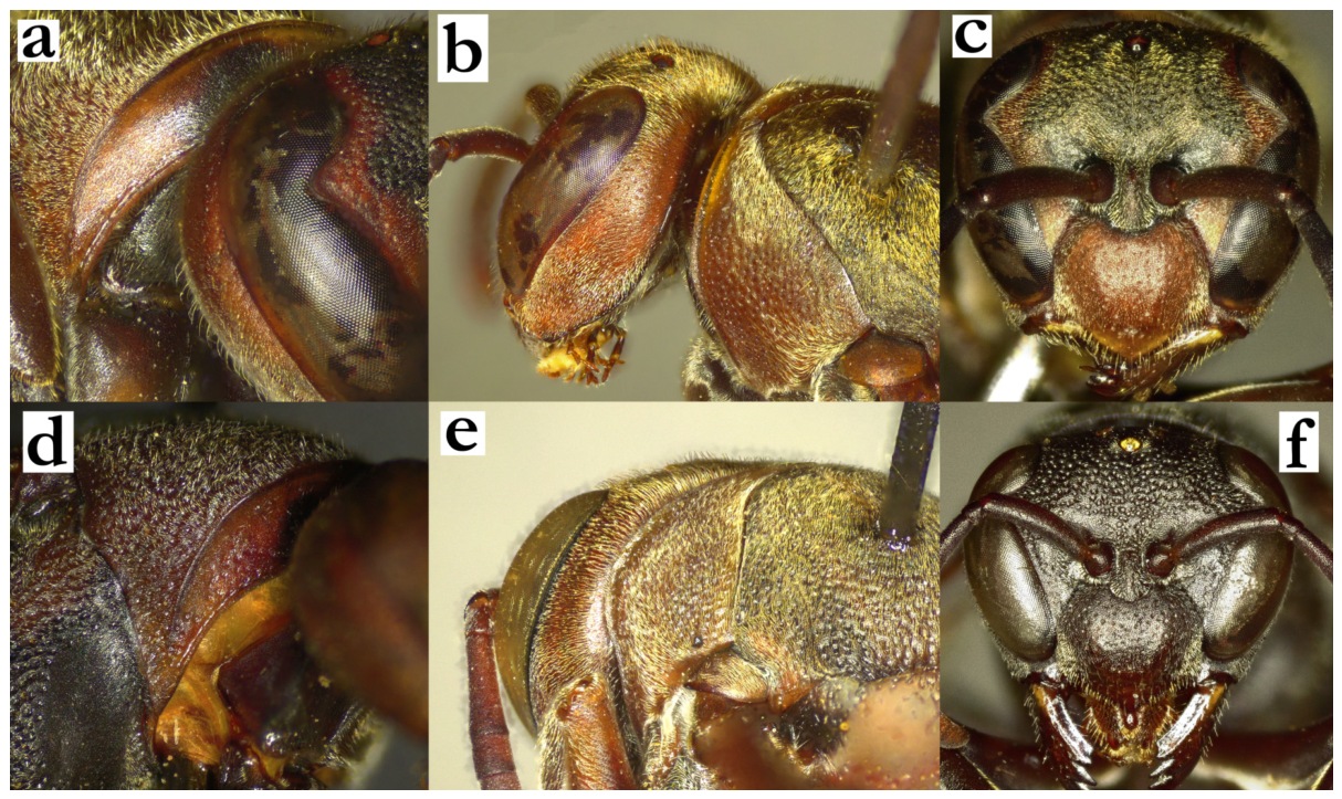
- 19. Mesoscutum with short pubescence and no protruding setae in lateral view (except superimposition from pronotum), or exceptionally with barely visible protruding setae (a). Mesonotum anteriorly sparsely punctate (b); some specimens of R. tsiky sp. nov. can have almost equally dense punctures pattern (see couplet 20). Pronotum with yellow or greenish line underneath carina, extended towards inferior pronotal angle (a, b); in very xanthic specimens, yellow area originates from underneath carina, and does not reach dorsal (mesonotal) margin (c).....................................................................................................20
- - Mesoscutum with underlayer of pubescence and protruding setae (d); some specimens of R. volontany sp. nov. can have shorter protruding setae, but in these cases T1 is about 0.8× of T2 width (see couplet 22). Mesoscutum anteriorly with similar puncture density (e). Pronotum with minimal yellow markings dorsally (d), or with yellow area near dorsal (mesoscutal) margin, that does not extend inferiorly towards pronotal angle (f)..............21
Figure 19.
Ropalidia shestakowi (Schulthess), (a) mesosoma, lateral view, female, (b) mesoscutum, dorsal view, female, melanic specimen, (c) mesoscutum, dorsal view, female, xanthic specimen; (d) Ropalidia grandidieri (de Saussure), mesosoma, lateral view, female, (e) mesoscutum, dorsal view, female; (f) Ropalidia anarchica (de Saussure), mesoscutum, dorsal view, female.
- 20. Scutellum with two green-yellow spots, metanotum with complete or almost complete green-yellow line (a). Median depression of propodeum deep (a). Scutellum flattened dorsally (b).
[f] Clypeus shiny, with scattered punctures (c). Tibia and tarsi olivaceous. Gena at midpoint as wide as eye in lateral view.
[m] Unknown
[wl] 8.0 [7.9–8.2] mm
[n] Unknown................................................................................................R. tsiky sp. nov.
- - Scutellum and metanotum reddish or brownish (d). Median depression of propodeum shallow (d). Scutellum dorsally rounded (e). Basal body colour light-brown, without green markings (occasionally males have suffused greenish hue of pronotum and tibia); female head light-brown (d), mesosoma and metasoma darker.
[f] Clypeus matt (f). Tibia and tarsi brown or light-brown. Gena at midpoint wider than eye.
[m] Tyloids originate as AF2, they are black and shiny (first few occasionally brown and shiny, with only terminal ones always being black). Terminal flagellomere elaborated, with bi-phasic tyloids on AF7–AF11, which have both ventral and dorsal elevation from the segment surface; terminal segment very elongated and strongly curved (g).
[wl] 8.4 [7.3–9.5] mm
[n] Large colonies, with up to 300 cells in the nest size and over a hundred of adults in a colony. Nest shape initially shaped as elongated droplet with apical petiole (h), later in the colony development can have additional lobes and become less regular in shape (i). Cell wall porous (i). Nest located on branches, succulent plants, stones or man-made objects.........................................................R. shestakowi (von Schulthess)
Figure 20.
Ropalidia tsiky sp. nov., (a) posterior mesosoma, dorsal view, female, (b) anterior mesosoma, oblique view, female, (c) head, frontal view, female; Ropalidia shestakowi (Schulthess), (d) posterior mesosoma, dorsal view, female, (e) anterior mesosoma, oblique view, female, (f) head, frontal view, female, (g) distal flagellomeres, posterior view, male, (h) nest [40], (i) nest [41].
- 21. Apical fifth of the forewing with a subtle darkening, which is better visible if the wing is folded, sometimes reduced to only darker nervature and black trichome (a). Pronotum with a large yellow spot (a; sometimes entirely yellow, but even then inferior pronotal angle remains in basal colour); melanic specimens from the central highlands can have reduced yellow markings or be entirely without them, but in these cases pronotum and tibia are suffused with olivaceous green.
[f] Clypeus, mandible and inner orbit suffused ferruginous (b).
[m] Frons and clypeus shiny, sparsely and minutely punctate (c). Tyloids blackish and shiny, originate at AF(2)3. Terminal flagellomere beak-like, with obtuse tip and sharp tyloid (d). AF(7)8–11 with postero-median brown area dorsally (d). Mesosoma underside mostly black (at most coxa I with and proximo-median quadrant of coxa II with yellow markings).
[wl] 8.3 [7.6–9.8] mm (with strong queen-worker size dimorphism)
[n] Elongated, with up to 10 cells wide and 50 cells long; larger nests can be curved backwards, exposing cell openings more [42]. Cell wall uniformly light brown-grey [43], sometimes ferruginous with dark grey streaks; cocoon caps grey or reddish, with parts of cell wall. Nest petiole asymmetrically on top of the nest. Bi-phasic cell depth, with much lower cells in expanding parts of the nest and longer cells located centrally; nest on branches, palm or Agave leaves.................R. anarchica (de Saussure)
- - Forewing uniformly yellowish or transparent, without apical darkening (some specimens of R. takona sp. nov. can have modest darkening in marginal cell, see couplet 24). Pronotum with less yellow, usually just a thin line underlying pronotal carina or no yellow at all. Male antenna dorsally uniformly coloured, without postero-median dorsal brown area. In males of smaller size, thorax underside with numerous yellow markings: coxa I, II and III, femur I and II, and anterior face of mesepisternum yellow.....................22
Figure 21.
Ropalidia anarchica (de Saussure), (a) habitus, lateral oblique view, female, (b) head, frontal view, female, (c) head, frontal view, male, (d) antenna, dorsal view, male.
- 22. Mesosoma and metasoma with yellow markings; T1 and T2 with posterior bands, two yellow spots on metanotum (a). Superior margin of metanotum biphasic (a). Wing length over 8 mm.
[f] Head thin, 1.8× as high as long; eye 3.5× as high as long; frons projecting anteriorly; clypeus projecting inferiorly more than mandible width (b).
[m] Unknown
[wl] 8.0 [7.9–8.2] mm
[n] Unknown.......................................................................................R. volontany sp. nov.
- - Mesosoma and metasoma without yellow or with minimal yellow markings; metanotum reddish or brown (c); some specimens of R. takona can have multiple yellow markings on mesosoma and metasoma, but then wing length is under 8 mm. Superior margin of metanotum flattened, irregular or evenly convex (c).
[f] Head stockier, 1.6–1.7× as high as long; eye up to 3.1× as high as long; frons flattened anteriorly; clypeus projecting inferiorly less than mandible width (d, e)…......23
Figure 22.
Ropalidia volontany sp. nov., (a) posterior mesosoma, oblique view, female, (b) head, lateral view, female; Ropalidia takona sp. nov., (c) posterior mesosoma, oblique view, female, (d) head, lateral view, female; (e) Ropalidia grandidieri (de Saussure), head, lateral view, female.
- 23. Smaller size: wing length exceptionally over 9 mm. Pronotal collar impunctate or with a few poorly defined, small and shallow punctures (a). Pronotal carina evenly rounded laterally (b), or with modest angle in larger specimens. Pronotum punctures more than a diameter apart. Median depression of propodeum commonly impunctate and very finely striate.
[f] Clypeus base shallowly punctate (c).
[m] Tyloids originate at AF1 or AF2.................................................................................24
- - Larger size, wing length commonly over 9 mm. Pronotal collar punctate, with at least a few coarse and large punctures (d); pronotal carina with angled contour (e). Pronotum punctures about a diameter apart. Median depression of propodeum coarsely striate.
[f] Clypeus basally with large and well-defined punctures (f).
[m] Tyloids originate at AF3 or AF4..................................................................................25
Figure 23.
Ropalidia grandidieri (de Saussure), (a) pronotal collar, anterior oblique view, female, (b) pronotal carina, oblique view, female, (c) head, frontal view, female; Ropalidia atra (de Saussure), (d) pronotal collar, anterior oblique view, female, (e) pronotal carina, oblique view, female; (f) Ropalidia cocoscola Blommers head, frontal view, female.
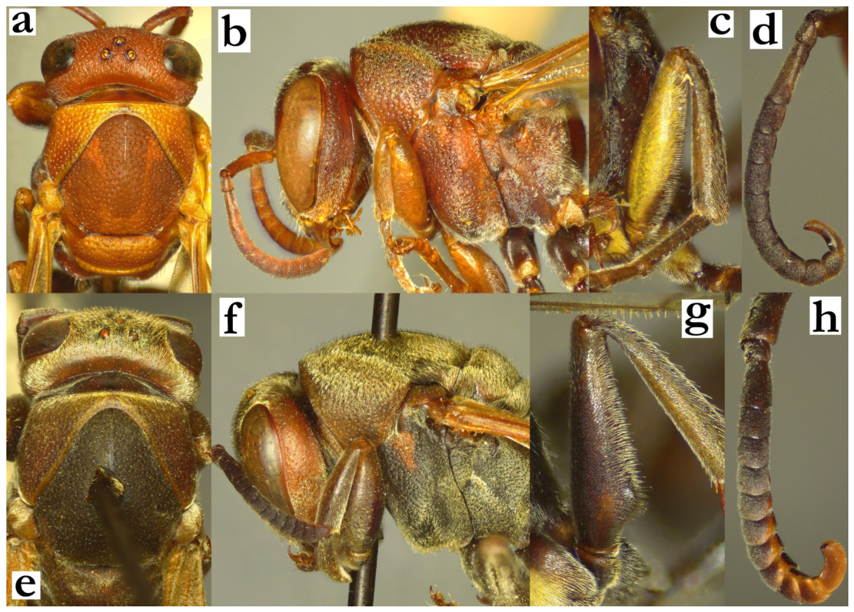
- 24. Smaller size: wing length rarely over 8 mm. Pilosity shorter, less conspicuous (a). Median carina of scutellum absent or developed anteriorly as a short black line (a). Body with light-brown markings (a).
[f] Frons setae 0.4–0.7× of ocellus diameter (b).
[m] Femur II with nearly parallel sides (c). Tyloids originate at AF2; AF2–AF7 longer than wide; AF11 > 4× as long as wide (d).
[wl] 7.6 [7.1–8.2] mm
[n] Unknown..............................................................................................R. takona sp. nov.
- - Larger size: wing length commonly over 8 mm. Pilosity longer and denser (e). Median carina of scutellum developed anteriorly and extending on dorsal scutellum surface (e). Body darker, dark brown or dark olivaceous (e).
[f] Frons setae 0.9–1.3× of ocellus diameter (f).
[m] Femur II with proximal protuberance (g). Tyloids originate at AF1; AF2 and more distal flagellomeres wider than long; AF11 3× as wide as long (h).
[wl] 8.8 [8.0–9.2] mm
[n] Elongated and irregular, up to 10 cells wide and 30 cells long; cell wall dense and thick; nests located on branches and trees............................R. grandidieri (de Saussure)
Figure 24.
Ropalidia takona sp. nov., (a) head and mesosoma, dorsal view, female, (b) head and mesosoma, lateral view, female, (c) femur II, anterior view, male, (d) antenna, dorsal view, male; Ropalidia grandidieri (de Saussure), (e) head and mesosoma, dorsal view, female, (f) head and mesosoma, lateral view, female, (g) femur II, anterior view, male, (h) antenna, dorsal view, male.
- 25. Pilosity shorter; gena with short pubescence and only a few protruding setae (a, b). T2 setae do not extend over lamella (c).
[f] Clypeus base finely and shallowly punctate (a). Frons with punctures over a diameter apart (a).
[m] Frons setae shorter than anterior ocellus diameter (b). Tyloids originate at AF(2)3, black and shiny; AF3 as long as wide (d). Terminal flagellomere terminally widened and blackened (d).
[wl] 8.9 [8.6–9.1] mm
[n] Small, quadratic with paracentral petiole, up to 8 cells wide and 7 cells long, in total to 40 cells per nest; nests located in coconut tree holes......R. cocoscola Blommers
- - Pilosity longer; gena with longer protruding setae inferiorly (e, f). T2 setae extend over lamella (g).
[f] Clypeus base largely and coarsely punctate (e). Frons punctures less than a diameter apart (e).
[m] Frons setae longer than anterior ocellus diameter (f). Tyloids brown, originate on AF3 (h). AF3 longer than wide (h). Terminal flagellomere elongated, with distally narrowed but obtuse tip (h).
[wl] 9.3 [8.9–10.8] mm
[n] Comparatively small, already with cocoon caps at size of 15 cells; the only observed nest was recorded early in the colony cycle, so information is limited; nest is discoid or elongated, suspended from a leaf, light-brownish, built by heavily masticated material......................................................................................R. atra (de Saussure)
Figure 25.
R. cocoscola Blommers, (a) head, frontal view, female, (b) head, frontal view, male, (c) T2 lamella, dorsal view, female, (d) antenna, dorsal view, male; R. atra (de Saussure), (e) head, frontal view, female, (f) head, frontal view, male, (g) T2 lamella, dorsal view, female, (h) antenna, dorsal view, male.
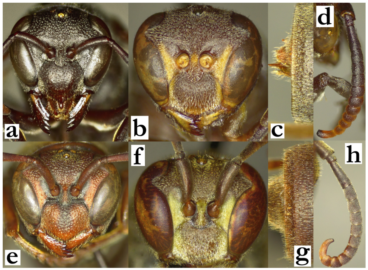
- 26. T1 globular (a), with marked posterior constriction seen dorsally (b); T1 posterior sulcus asymmetrical in dorsal view; shallower dorsally and laterally, and deeper in the oblique direction (b). Specimens of R. interjecta can sometimes have less globular T1, but in these cases the wing length is under 6.5 mm and there are only suffused green areas on the body (see couplet 31). Male tyloids thin and shiny, barely elevated, originate at AF1 (except R. zandry sp. nov., see couplet 30).................................................fraterna group—27
- - T1 pyriform (c) or strongly dorsally swollen (d); posterior sulcus not developed or minimal (c, d)......................................................................................................................................32
Figure 26.
Ropalidia fraterna (de Saussure), (a) habitus, oblique view, female, (b) T1, dorsal view, female; (c) Ropalidia perplexa Blommers, habitus, oblique view, female; (d) Ropalidia constitutionalis (de Saussure), habitus, oblique view, female.
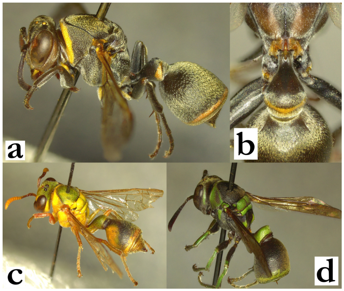
- 27. Body with numerous yellow or green markings on mesosoma (a, b); metanotum, metepisternum and propodeum commonly with green or yellow markings. Antenna ferruginous or orange dorsally (a, b).............................................................................................28
- - Body basally brown or dark brown, with suffused reddish, greenish or yellowish markings (c) or fewer yellow markings on mesosoma (d); metanotum, propodeum and metepisternum dark brown to black. Antenna dorsally infuscate (c) or dark brown (d)............29
Figure 27.
Habitus, lateral view, female, (a) Ropalidia laureliniana sp. nov.; (b) Ropalidia galimatia (de Saussure); (c) Ropalidia cauponae Blommers; (d) Ropalidia fraterna (de Saussure).
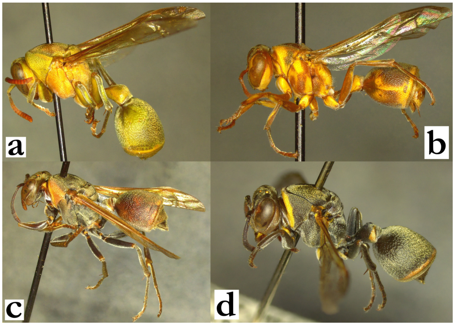
- 28. Body predominantly yellow or green; pronotum, mesoscutum, base of T2, tibia and femur with yellow or green markings (a); if mesonotum is entirely brown without yellow or green markings, then punctures are well-defined (b). Anterior margin of forewing not substantially darkened (b). Median depression of propodeum without black markings (c). Yellow and green areas with suffused margins (c).
[wl] f: 8.6 [8.0–9.3] mm; m: 7.8 [7.5–8.1] mm
[n] Initially discoid, at later colony stages large and irregular, sometimes with accessory lobes; cell wall brown-grey with reddish or dark-grey streaks; empty cells often filled with honey; cocoon caps grey or brown. Nest located directly on tree trunks [44]....................................................................................................R. laureliniana sp. nov.
- - Body ferruginous-brown with yellow markings (exceptionally with a greenish hue in males); mesoscutum, base of T2 and leg segments usually with minimal yellow markings (d); mesonotum finely and superficially punctate (e). Anterior margin of forewing blackened (f). Propodeum centrally with a dark streak (f). Yellow areas emarginated from ferruginous base (f).
[wl] 7.8 [7.0–9.9] mm
[n] Initially droplet-shaped, suspended from the apical cell (g); at later stages, nest and colony size can substantially increase by adding accessory lobes and totalling 550 cells; nest axis often backwardly curved (h), sometimes even expanding in an inverse globular form (i); nests sometimes located in smaller colonies. Cell wall light to dark grey, cocoon caps grey to black (g), often located in streaks or clusters (i); empty cells often filled with honey-like liquid. Nest located on branches, leaves, stones and man-made objects.................................................R. galimatia (de Saussure)
Figure 28.
Ropalidia laureliniana sp. nov., (a) habitus, dorsal view, female, (b) head and mesosoma, dorsal view, female, (c) posterior mesosoma, dorsal view, female; Ropalidia galimatia (de Saussure), (d) habitus, dorsal view, female, (e) head and mesosoma, dorsal view, female, (f) posterior mesosoma, dorsal view, female, (g) nest [45], (h) nest [46], (i) nest [45].
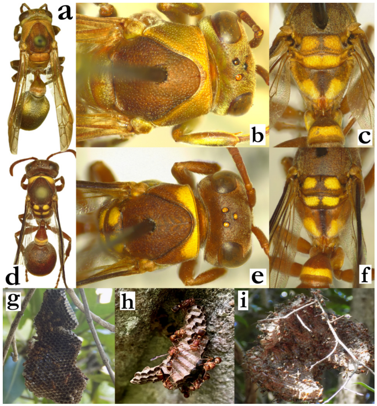
- 29. Wing length over 8 mm. Base of T2 coarsely punctate (a). Pronotum commonly without yellow markings, sometimes with a thin yellow line underneath carina or suffused reddish and yellow area (b). Coxa II and III in females commonly without yellow markings (sometimes suffused green or reddish in more xanthic specimens). Terminal flagellomere in males with a sharp proximal tyloid widening, with tyloid that extends to acute or subacute tip (c, d)...............................................................................................................................30
- - Wing length rarely over 8 mm; larger specimens (wing length 7–8 mm) have weak punctures at T2 base (e). Pronotum commonly with widened yellow line underneath carina (f). Coxa II and III in females commonly with yellow markings. Terminal flagellomere in males without proximal tyloid widening and with an obtuse tip (g, h)...............................31
Figure 29.
Ropalidia cauponae Blommers, (a) T1 and T2, anterior oblique view, female, (b) habitus, lateral view, female, (c) terminal flagellomere, dorsal view, male; (d) Ropalidia zandry sp. nov., terminal flagellomere, dorsal view, male; Ropalidia fraterna (de Saussure), (e) T1 and T2, anterior oblique view, female, (f) habitus, lateral view, female, (g) terminal flagellomere, dorsal view, male; (h) Ropalidia interjecta (de Saussure), terminal flagellomere, dorsal view, male.
- 30. Pronotum in dorsal view mildly curved anteriorly (a). Body black and reddish-brown, without green(ish) markings (a). Occipital carina streamlined, dark-brown to black (b).
[f] Clypeus base with coarse and well-defined punctures; frons with well-defined punctures (c). Gena with coarse punctures (b). Mesoscutum with very short pubescence (b), frons setae 0.7× as long as ocellus diameter (b).
[m] Tyloids originate at AF(4)5; terminal flagellomere apically acute (d).
[wl] f: 8.4 [8.2–9.0] mm, m: 8.2 [7.8–8.4] mm
[n] Elongated, initially 2–3 cells wide, later up to 10×60 cells; cell wall grey, cocoon caps grey to brownish; nests suspended from branches.....................R. zandry sp. nov.
- - Pronotum in dorsal view anteriorly straight (e). Body colour variable, often with yellowish, reddish or even greenish areas (f). Occipital carina brown, inferiorly mildly angulate (f).
[f] Clypeus base with fine and hardly discernible punctures; frons with similar punctures (g). Mesoscutum with discernible pubescence and short protruding setae (g), frons with setae as long as ocellus diameter (f).
[m] Tyloids originate at AF1; terminal flagellomere commonly apically subacute (h), but can be more acute in some specimens.
[wl] f: 8.5 [8.1–9.6] mm; m: 8.2 [7.6–9.1]
[n] Discoid, sometimes rhomboid with an apical petiole, reaching up to 500 cells; cell wall grey, sometimes with darker or lighter streak; cocoon tops darker, often located in streaks or clusters on the nest; empty cells often filled with honey; nests located on man-made objects...........................................................................R. cauponae Blommers
Figure 30.
Ropalidia zandry sp. nov., (a) head and mesosoma, dorsal view, female, (b) head, lateral view, female, (c) head, frontal view, female, (d) antenna, dorsal view, male; Ropalidia cauponae Blommers, (e) head and mesosoma, dorsal view, female, (f) head, lateral view, female, (g) head, frontal view, female, (h) antenna, dorsal view, male.
- 31. Wing length over 6 mm. Entire body covered by a dense silvery under-layer of pubescence (a). T1 globular in most females, commonly with two triangular yellow or greenish areas. Scutellum without median carina (a), exceptionally visible as short streak anteriorly in some specimens.
[f] Clypeus narrower, 1.1× as wide as long; face darker, with visible pubescence (b); frons and vertex darker (b).
[m] Terminal flagellomere elongated, with gradually narrowing tip; tyloid carina weak or missing (c).
[wl] f: 6.9 [6.4–8.0] mm; m: 6.6 [6.4–7.5] mm
[n] Discoid or droplet-shaped [47], but sometimes irregular and elongated [48]; larger nests can have up to 250 cells; nests usually located on branches, less commonly on man-made objects; cell wall longer; cocoon tops dark brown to blackish, often located in lines or streaks .........................................................................R. fraterna (de Saussure)
- - Wing length commonly under 6 mm. Body not covered by denser under-layer of pubescence (d); T1 commonly less globular, with shallower posterior sulcus and commonly with a complete posterior yellow-greenish band. Scutellum with developed median carina, which extends to more than half of its length (d).
[f] Clypeus wider, 1.3× as wide as long; frons and vertex lightly coloured (e).
[m] Terminal flagellomere with broadened tip and more developed tyloid carina (f).
[wl] 5.5 [5.3–6.4] mm.
[n] Elongated, smaller, up to 50 cells arranged in 2×n formula [49], sometimes irregular, with accessory streaks [50]; commonly located on broad leaves; cell wall shorter, with interjected long cells.........................................................R. interjecta (de Saussure)
Figure 31.
Ropalidia fraterna (de Saussure), (a) scutellum and metanotum, dorsal view, female, (b) head, frontal view, female, (c) antenna, dorsal view, male; Ropalidia interjecta (de Saussure), (d) scutellum and metanotum, dorsal view, female, (e) head, frontal view, female, (f) antenna, dorsal view, male.
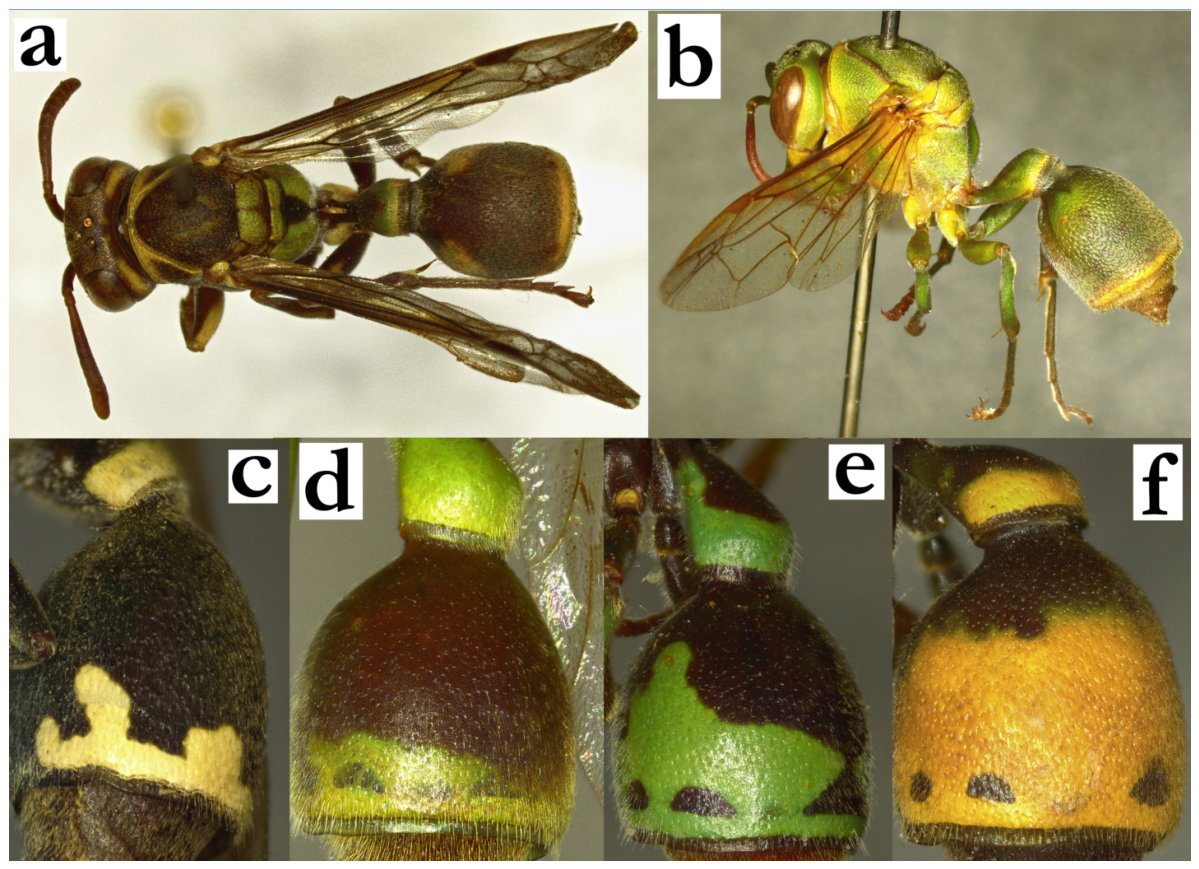
- 32. T2 basally with green or yellow markings: with a bilateral green or yellow basal spot (a) or entire T2 surface green or yellow (b). Mesoscutum commonly with green markings, ranging from thin green or yellow lines (a), to entirely green (b).........................................33
- - T2 basally without green or yellow markings; distal part of T2 can be without a posterior band or with a band of variable thickness that can occupy most of its surface (c–f). Mesoscutum without green or yellow markings (thin longitudinal green-yellowish lines can be present in R. sonyae sp. nov., see couplet 52, or R. miafina sp. nov., see couplet 60)...................................................................................................................................................40
Figure 32.
(a) Ropalidia vanga sp. nov., habitus, dorsal view, female; (b) Ropalidia flavoviridis Kojima, habitus, lateral view, female; T2, lateral view, female, (c) Ropalidia ranavali (de Saussure), cluster “venustula”, (d) Ropalidia sonyae sp. nov., (e) Ropalidia aras sp. nov., (f) Ropalidia ranavali (de Saussure).
- 33. Larger species, wing length at least 8 mm. Mesosoma robust, 1.3× as long as high in lateral view (a). If wing length is close to 8 mm, then posterior ocelli are more than their diameter apart and mesoscutum is coarsely punctate (b)......................................................34
- - Smaller species, wing length under 8 mm. Mesosoma more slender and elongate, 1.5× as long as high in lateral view (c). If wing length is close to 8 mm, then posterior ocelli are less than a diameter apart and mesoscutum is shallowly and finely punctate (d)............36
Figure 33.
Ropalidia flavoviridis Kojima, (a) habitus, lateral view, female, (b) head and mesosoma, dorsal view, female; Ropalidia bongomaso sp. nov., (c) habitus, lateral view, female, (d) head and mesosoma, dorsal view, female.
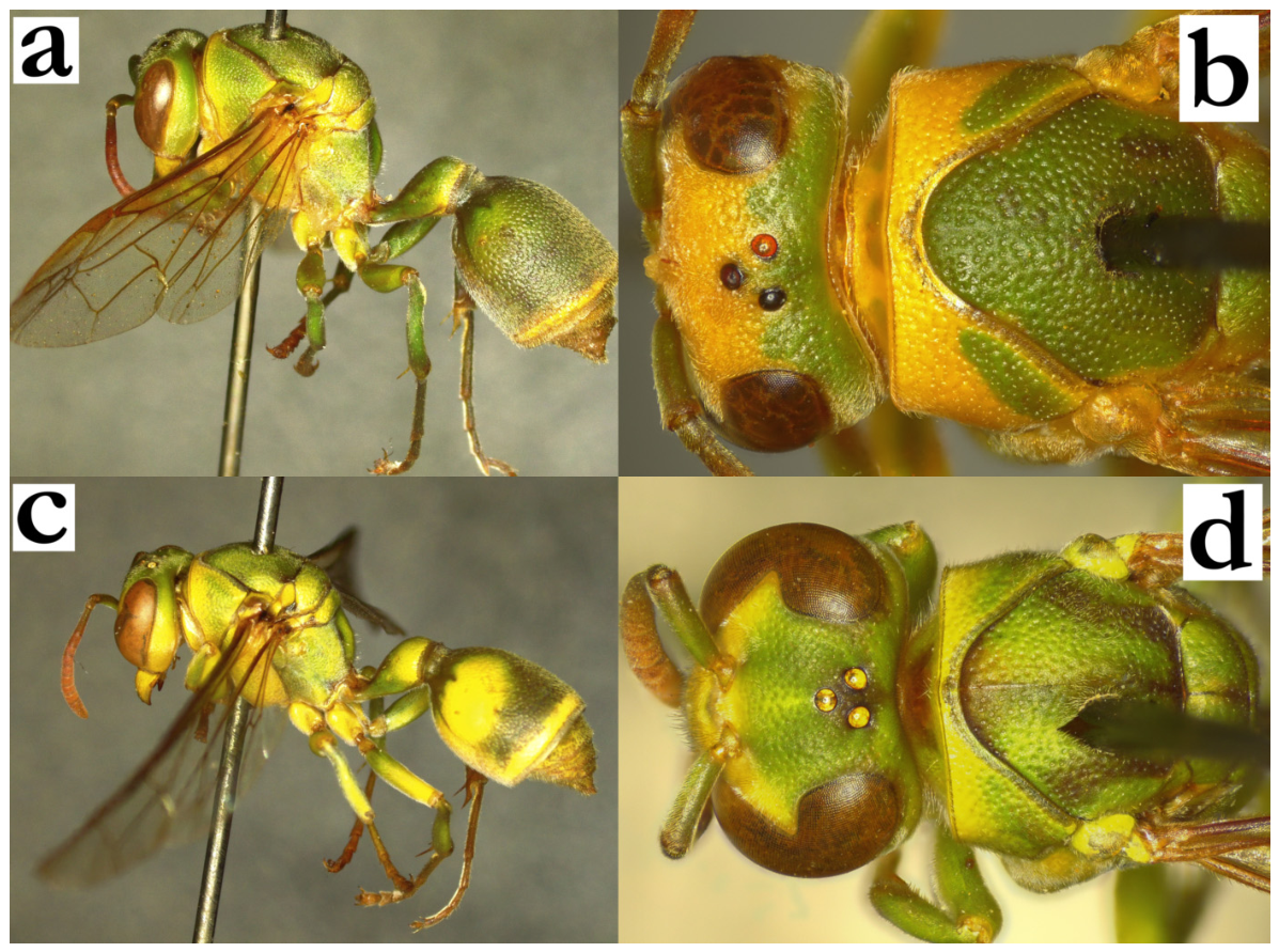
- 34. Mesoscutum with short pubescence, without protruding setae (a). Frons and occiput setae shorter than anterior ocellus diameter (b). T2 with short pubescence that barely extends over lamella. Mesoscutum and pronotum commonly unicolorous. Interantennal area with well-defined vertical slit (b, c).
[f] Clypeus 1.3× as wide as long or wider, centrally with large and shallow punctures (b). Inner orbit punctate, even in yellow areas (b).
[m] Clypeus 1.4–1.5× as wide as long (c). Tyloids originate at AF(5)6, those on AF(10)11 can be darkened or even black. Terminal flagellomere up to 1.5× as long as wide, broadened, with a developed median tubercle and acute tip (d).
[wl] 8.3 [8.0–9.5] mm
[n] Elongated and somewhat irregular in shape (e), up to 10 cells wide and 30 cells long, commonly about 60 cells in a nest (f); later in colony cycle, additional superior or accessory lobe can be added (g). Cell wall greyish and compact, cocoon caps dark brown; nest on leaves and tree ferns...........................................R. flavoviridis Kojima
- - Mesoscutum with underlying pubescence and longer protruding setae (h). Frons and occiput with protruding setae that are at least as long as the anterior ocellus diameter (i, j). T2 with longer protruding setae that extend over lamella (some specimens of R. perplexa can have shorter pilosity on head and mesosoma, but in these cases T2 setae are always protruding over lamella). Mesoscutum commonly bicolorous. Interantennal area elevated, with a weakly developed slit (i) or without it (j).
[f] Clypeus base impunctate or weakly punctate and shiny; inner orbit impunctate or punctate with poorly defined puncture margins (i).
[m] Clypeus narrower, no more than 1.3× as wide as long (j). Terminal flagellomere differently shaped and at least 1.5× as long as wide, or longer….................................35
Figure 34.
Ropalidia flavoviridis Kojima, (a) head and anterior mesosoma, lateral view, female, (b) head, frontal view, female, (c) head, frontal view, male, (d) antenna, dorsal view, male, (e) nest [51], (f) nest [52], (g) nest [53]; (h) Ropalidia saussurei Kojima, head and anterior mesosoma, lateral view, female; (i) Ropalidia dubia (de Saussure), head, frontal view, female; (j) Ropalidia saussurei Kojima, head, frontal view, female.
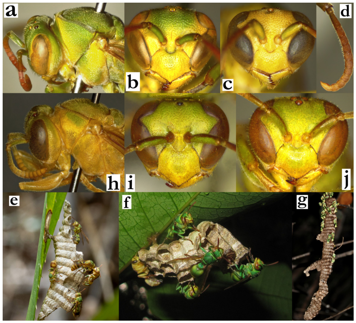
- 35. Femur and coxa II (and III) dorsally with dark green or black markings (a). T2 commonly with fused green or yellow area (a), less commonly with separate bilateral spot from the posterior band.
[f] Clypeus elongated, with flattened surface, deeper basal notch and straight apical margin; antenna strongly darkened or black dorsally; eyes asetose. Metanotum posteriorly weakly convex.
[m] Scape widened, about twice as wide as AF1 base (b). Terminal flagellomere elongated, apically curved with median tubercle and darkened or black tip (c). Tyloids originate at AF(2)3.
[wl] 9.9 [9.1–11.6] mm
[n] Discoid or irregular in shape with initially apical petiole [54], later sometimes paracentral [55]; up to 10 cells wide and 20 cells long; cell wall compact, greyish with suffused reddish streaks or yellow. Cocoon caps substantially elevated from the cell wall edge, dark brown, sometimes with extra pulp added to them. Nest suspended from a leaf underside, branch or directly on a tree trunk, concealed by the cleared zone in the surrounding moss [56]................................................R. dubia (de Saussure)
- - Femur and dorsal side of coxa II and III green or yellow, without darkening (d). T2 commonly with clearly separate anterior bilateral spot from the posterior spot and band, producing a cross-like brown area (d).
[f] Clypeus wider, with more convex surface, shallower basal notch and sinuate apical margin; antenna ferruginous or darkened dorsally. Eyes with dense setae of intermediate length. Metanotum posteriorly flattened or even centrally excavated.
[m] Scape no more than 1.5× as wide than AF1 base (e). Tyloids originate at AF(4)5 and never occupy more than half of inner segment surface. Eyes setose or asetose (e). Terminal flagellomere beak-like, elongated and brown, without median tubercle (f).
[wl] 9.8 [9.4–11.2] mm
[n] Discoid or somewhat elongated, with pedicel on the top of the nest and up to 150 cells [57]. Cell wall green or green with grey streaks, with very loose wall material and frequent holes in the wall structure [58]. Cells comparatively shorter in length; cocoon caps convex, built commonly in the line with the cell openings, less commonly above it, grey-brownish with green nodules. Nest suspended from the palm or tree trunk, located in or close to green lichen colonies...............R. saussurei Kojima
Figure 35.
Ropalidia dubia (de Saussure), (a) habitus, lateral view, female, (b) head, frontal view, male; (c) antenna, dorsal view, male; Ropalidia saussurei Kojima, (d) habitus, lateral view, female, (e) head, frontal view, male, (f) antenna, dorsal view, male.
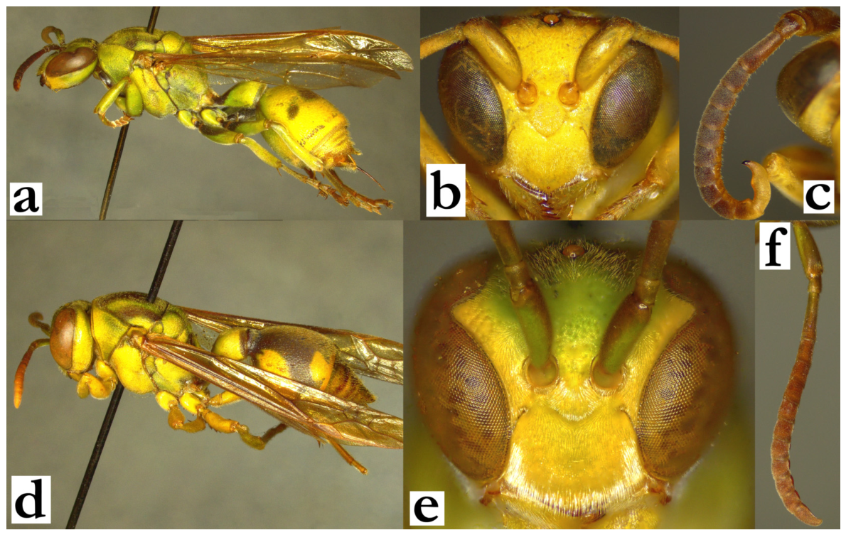
- 36. Entire body coarsely and densely punctate, especially mesoscutum (a) and frons (b). Mesoscutum 1.1× as wide as long (a). Median depression of propodeum with large and coarse punctures close to metanotum (c).
[f] Base of clypeus shiny and minutely punctate (b). Base of T2 punctate.
[m] Clypeus finely punctate (d). Tyloids yellowish and shiny, originate at AF(3)4. Terminal flagellomere elongated, beak-like, with acute tip (e).
[wl] 7.1 [6.9–7.8] mm
[n] Located on palm leaves, cell wall light grey [59]...................R. marolavaka sp. nov.
- - Punctures much less developed and less coarse, especially on green and yellow areas (f). Median depression of propodeum impunctate or with scattered and shallow, poorly defined punctures (g).......................................................................................................................37
Figure 36.
Ropalidia marolavaka sp. nov., (a) mesosoma, dorsal view, female, (b) head, frontal view, female, (c) median depression of propodeum, oblique dorsal view, female, (d) head, frontal view, male, (e) antenna, dorsal view, male; R. bongomaso sp. nov., (f) head, frontal view, female, (g) posterior mesosoma, dorsal view, female.
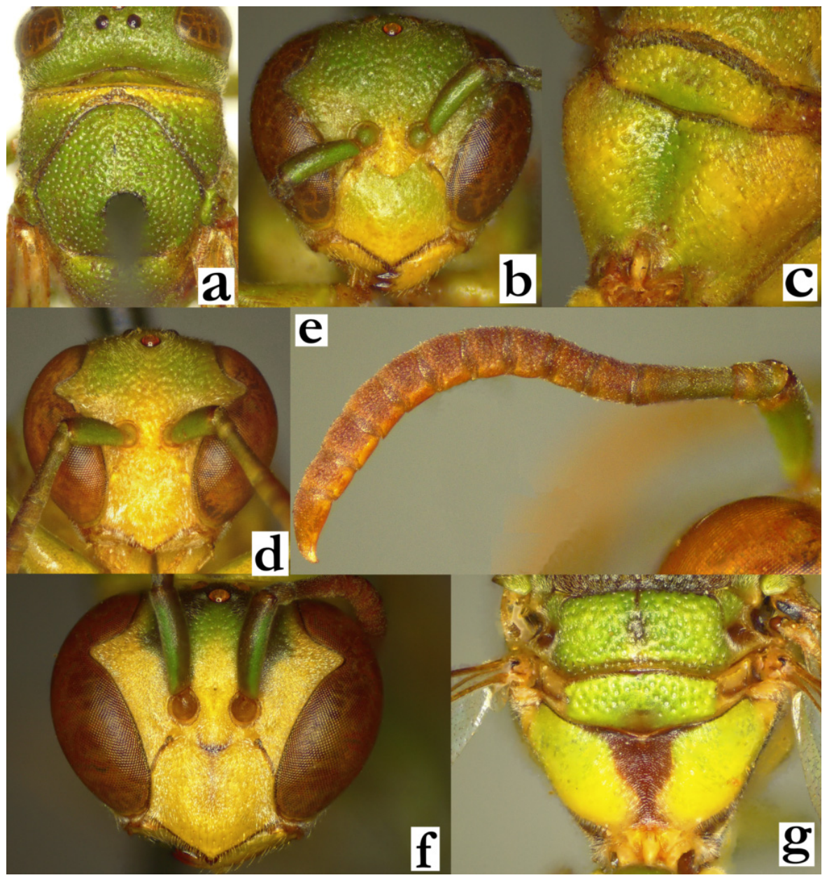
- 37. Head and mesosoma green and yellow, with minimal brownish areas. Body pilosity longer: mesoscutum with protruding setae, those on T2 extending over the lamella (a). Metepisternum impunctate (b).
[f] Antennal scape with numerous protruding setae; AF1 and AF2 with protruding setae.
[m] Tyloids originate at AF(6)7; terminal flagellomere elongated and evenly curved, with a very acute tip that can sometimes be darkened (c).
[wl] 7.3 [6.1–7.7] mm
[n] Circular, central petiole, up to 70 cells (6 cells wide and 14 cells long), located on Asplenium fern ..................................................................................R. perplexa Blommers
- - Body with numerous brown or brownish areas (d). Pilosity shorter; antenna without isolated setae or only with a few shorter ones on AF1; mesoscutum with short pubescence and barely longer protruding setae, setae on T2 not extending over lamella (d). Metepisternum with a few well-defined punctures (e). Male tyloids originate on AF1–5; terminal flagellomere differently shaped (f)............................................................................................38
Figure 37.
Ropalidia perplexa Blommers, (a) habitus, lateral view, female, paratype, (b) metepisternum, lateral view, female, (c) antenna, dorsal view, male; Ropalidia bongomaso sp. nov., (d) habitus, lateral view, female, paratype, (e) metepisternum, lateral view, female, (f) antenna, dorsal view, male.
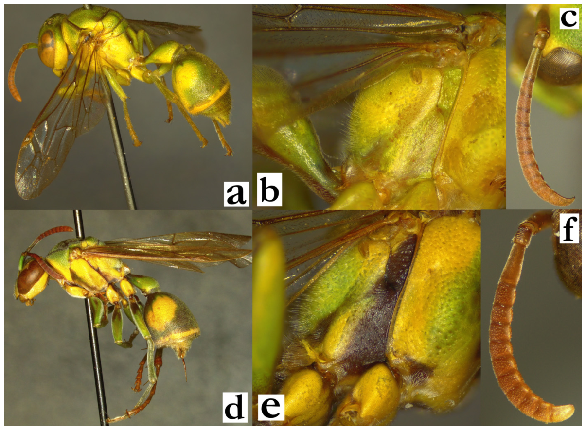
- 38. Mesonotum coarsely and densely punctate, punctures a diameter apart (a). Head behind eyes not substantially narrowing (a). Median carina of scutellum reaches half of its length (a). T2 coarsely punctate (b). Wing length exceptionally over 7 mm.
[f] Gena at midpoint almost as wide as eye; inferior part of gena wide (c).
[m] Tyloids originate at AF5 (d).
[wl] 6.3 [5.4–7.2] mm
[n] Small and discoid, with paracentral petiole, usually with cell openings facing downwards. Cell wall porous, light-grey, sometimes with streaks of darker grey or greenish material, located on leaves underside...............R. phalansterica (de Saussure)
- - Mesonotum shallowly and sparsely punctate, punctures 2 or more diameters apart (e). Head substantially narrowing behind eyes (e). Median carina of scutellum short, conspicuous only on anterior scutellum face (e). T2 finely punctate (f). Wing length over 7 mm.
[f] Gena at midpoint 0.5× as wide as eye; inferior part of gena narrowed (g).
[m] Tyloids originate at AF1 (h).........................................................................................39
Figure 38.
Ropalidia phalansterica (de Saussure), (a) head and mesosoma, dorsal view, female, (b) T2, oblique dorsal view, female, (c) head, lateral view, female, (d), antenna, dorsal view, male; Ropalidia bongomaso sp. nov., (e) head and mesosoma, dorsal view, female, (f) T2, oblique dorsal view, female, (g) head, lateral view, female, (h), antenna, median view, male.
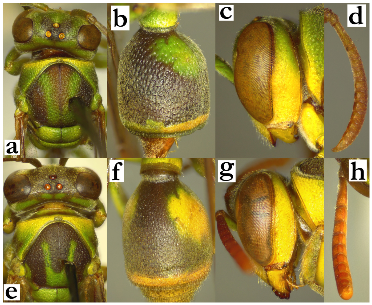
- 39. Body yellow and brown, with minimal green markings dorsally on mesosoma (a). Posterior ocelli distance equal to posterior ocellus diameter (a).
[f] AF2 as long as wide (b). Mesoscutum with short erect setae (c). Frons setae 0.9–1.4× as long as anterior ocellus diameter (c).
[m] Unknown
[wl] 7.5 [7.4–7.6] mm
[n] Unknown...............................................................................................R. vanga sp. nov.
- - Body with numerous green markings (d). Posterior ocelli distance less than a diameter apart (d).
[f] AF2 1.2× as long as wide (e). Mesoscutum without erect setae (f). Frons setae 0.6–0.9× as long as anterior ocellus diameter (f).
[m] Tyloids originate at AF1(2).
[wl] 7.7 [7.1–7.9] mm
[n] Elongated and very thin, usually with one vertical row of cells, sometimes slightly wider; re-use of the material was proposed as the mechanism how these cell-less streaks appear [6]; larva can be very exposed in such shallow cells; nest suspended from a branch or a mango tree leaf................................................R. bongomaso sp. nov.
Figure 39.
Ropalidia vanga sp. nov., (a) head and mesosoma, dorsal view, female, (b) antenna, dorsal view, female, (c) head and anterior mesosoma, lateral view, female; Ropalidia bongomaso sp. nov., (d) head and mesosoma, dorsal view, female, (e) antenna, dorsal view, female, (f) head and anterior mesosoma, lateral view, female.
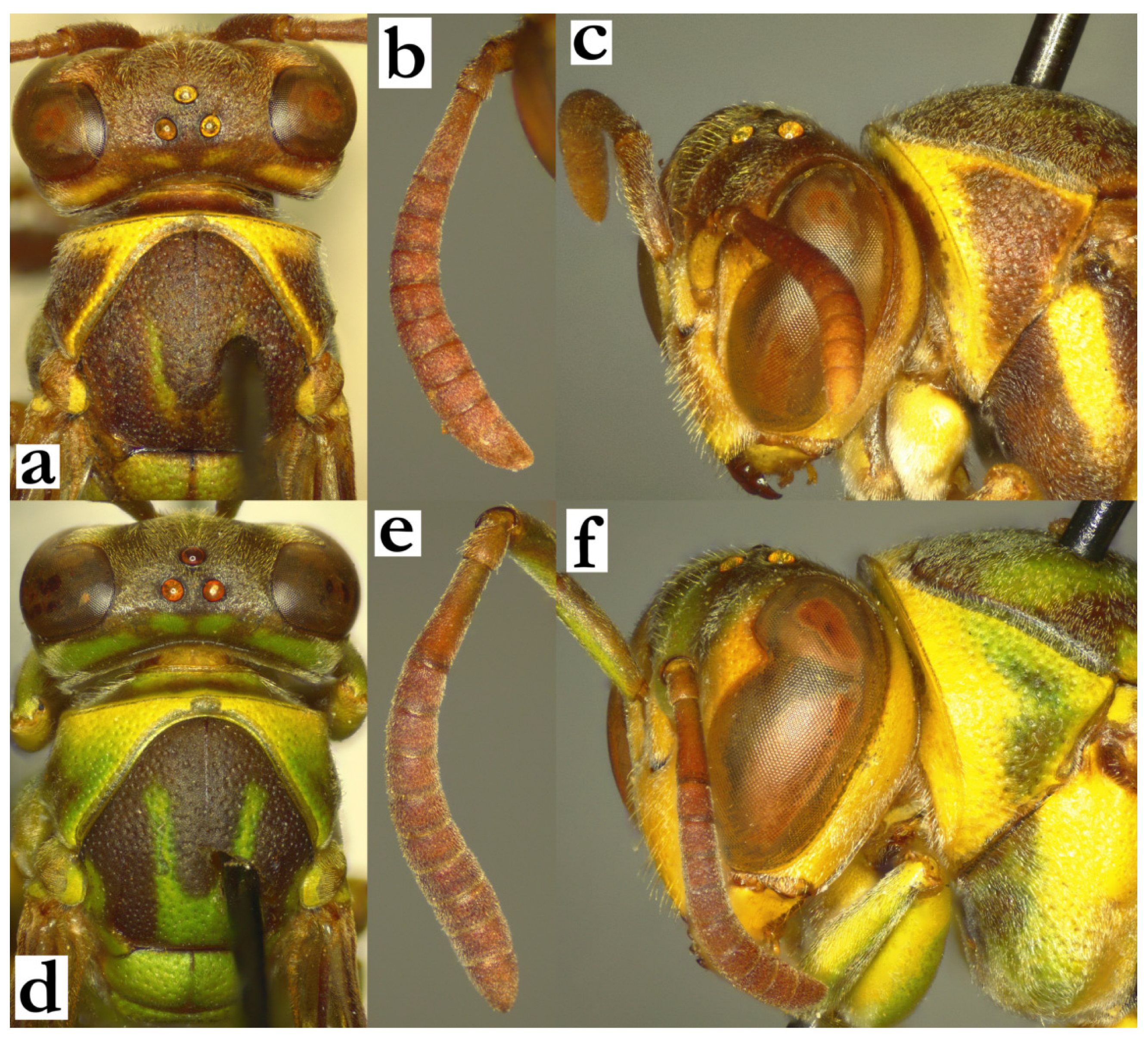
- 40. T1 elongate and slender, at least 2.8× as long as its maximal height in lateral view; T2 about 3.2× taller than T1; T1 dorsally flattened (a) or weakly rounded (b). All coxa and femora with green or yellow markings. Female clypeus yellow with a central olivaceous-brown(ish) spot.............................................................................................................................41
- - T1 in lateral view less elongated, no more than 2.6× longer than its maximal height (c); some specimens of R. miafina sp. nov. can have elongated T1 and yellow markings on coxa and femora; in that case, female clypeus is entirely yellow and upes is about 0.2× as long as lateral clypeus margin (d; see couplet 60); some specimens from the bicinta group can also resemble elongated T1, but then main body colour is black or dark green, coxa and femora have no yellow markings and T1 and T2 have yellow or whitish posterior bands (see couplet 47). T2 at most 2.8× as high as T1 (c)....................................................................42
Figure 40.
(a) Ropalidia telperia sp. nov., metasoma, lateral view, female; (b) Ropalidia beosi sp. nov., metasoma, lateral view, female; (c) Ropalidia miafina sp. nov., metasoma, lateral view, female, (d) head, frontal view, female.
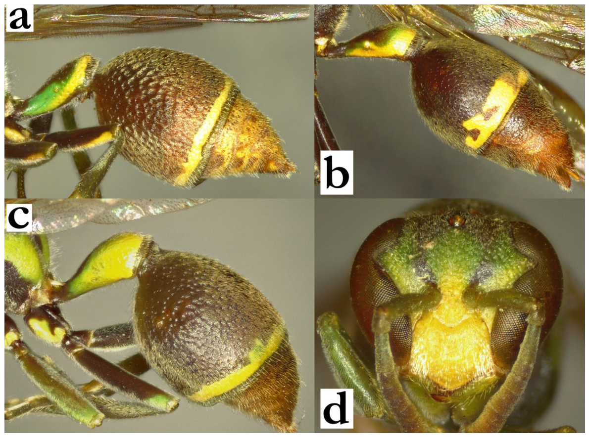
- 41. Smaller size, wing length under 8 mm. Body covered by a dense layer of silvery-yellowish pubescence (which is silvery on parts of the body that have green basal colour and yellowish on the body parts that have blackish colour; a). Posterior ocelli at least 2 diameters apart (a). T2 with dense layer of pubescence, which follows the coarse and deep punctures texture and creates rugged pattern (Figure 40a).
[f] OCA dull, clypeus commonly with suffused olivaceous central spot attached to the base, frons with a denser layer of pubescence (b). Femora I-III with yellow markings; either a longitudinal line, or as little as a small apical spot. T2 with a thin posterior yellow line (a). Scutellum commonly in basal colour (or only with a faint greenish marking), metanotum with a green line and propodeum with two large green rhomboid spots (c). T1 0.5–0.7× as wide as T2 (c).
[m] Unknown
[wl] 6.8 [6.4–7.2] mm
[n] Unknown..........................................................................................R. telperia sp. nov.
- - Larger body size, wing length over 8 mm. Body covered by less developed layer of pubescence; T2 basally shiny, with scattered and minute punctures (Figure 40b).
[f] OCA sharp, clypeus yellow with a faint brownish unattached spot; frons pubescence less developed (e). All femora without yellow markings, at most with (light) green apical spot. T2 with thicker posterior yellow band. Scutellum with two large green spots, propodeum black (f). T1 0.4× as wide as T2.
[m] Unknown
[wl] 8.3 mm
[n] Unknown................................................................................................R. beosi sp. nov.
Figure 41.
Ropalidia telperia sp. nov., (a) head and anterior mesosoma, dorsal view, female, (b) head, frontal view, female, (c) posterior mesosoma, dorsal view, female; Ropalidia beosi sp. nov., (d) head and anterior mesosoma, dorsal view, female, (e) head, frontal view, female, (f) posterior mesosoma, dorsal view, female.
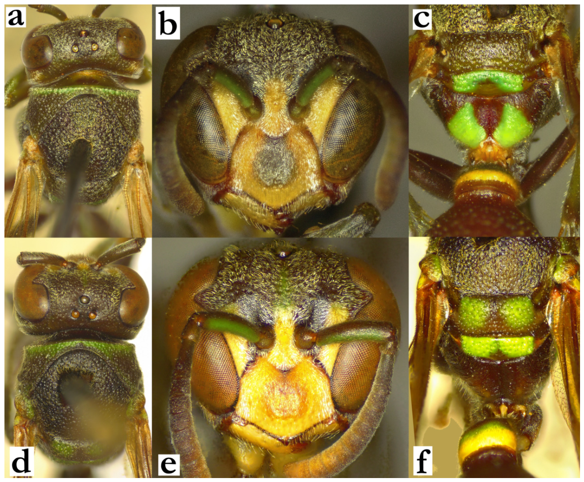
- 42. Clypeus longer than wide, without OCA; upes continue directly into lateral clypeal margin; clypeus entirely yellow (a) or dark green, with suffused olivaceous area apically (b). Clypeus and interantennal area with fine pubescence (c); antennal slit weak or absent (c)....................................................................................................................................................43
- - Clypeus with visible OCA (d), sometimes dull and very weak (e). Clypeus and interantennal area with simple yellowish pubescence (f). Interantennal slit developed. ............44
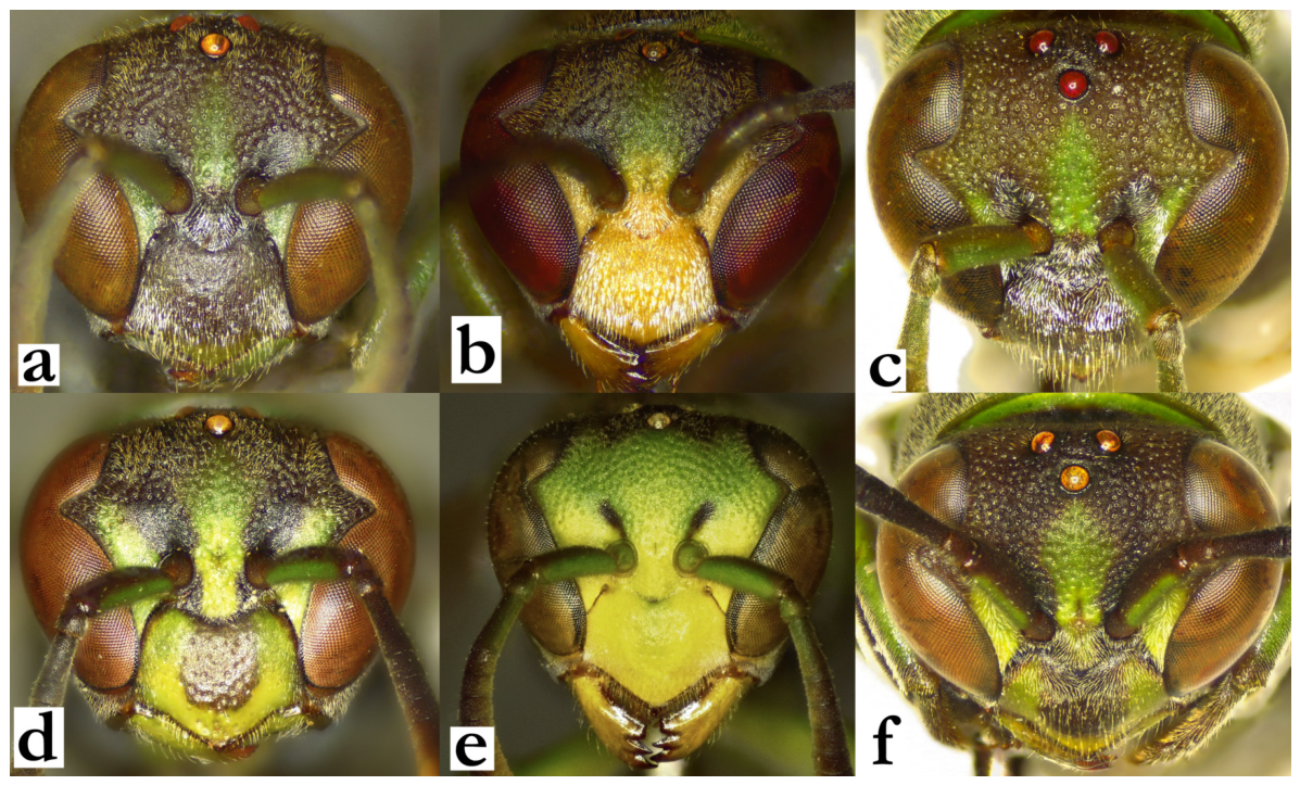
Figure 42.
(a) Ropalidia seyrigi sp. nov., head, frontal view, female; (b) Ropalidia rafitra sp. nov., head, frontal view, female, (c) Ropalidia seyrigi sp. nov., head, oblique dorsal view, female; (d) Ropalidia favulorum Blommers, head, frontal view, female, (e) Ropalidia phalansterica (de Saussure), head, frontal view, female; (f) Ropalidia favulorum Blommers, head, oblique dorsal view, female.
- 43. Body black and green: coxa black, propodeum with small green spot or entirely black (a; body exceptionally with suffused minimal yellow markings).
[f] Antenna dorsally infuscate (a). All coxa black (a). Clypeus without yellow markings (Figure 42a).
[m] Clypeus dark green (b). AF2 and AF3 longer than wide (c). Terminal flagellomere barely excavated medially (c).
[wl] 6.9 [6.6–7.4] mm
[n] Unknown..............................................................................................R. seyrigi sp. nov.
- - Face and mesosoma underside with numerous yellow markings (d).
[f] Antenna very dark or black dorsally (d). Coxa I and II with yellow, coxa III commonly with green markings (d). Clypeus entirely yellow (Figure 42b).
[m] Clypeus, inner orbit and interantennal area merged in yellow (e). AF2 and more distal flagellomeres wider than long (f). Terminal flagellomere very excavated (f).
[wl] 6.4 [6.1–7.0] mm
[n] Small nests in colonies, suspended from the leaf underside; cell wall dark brown with some yellowish or light-brown streaks..........................................R. rafitra sp. nov.
Figure 43.
Ropalidia seyrigi sp. nov., (a) head and mesosoma, lateral view, female, (b) head, frontal view, male, (c) antenna, dorsal view, male; Ropalidia rafitra sp. nov., (d) head and mesosoma, lateral view, female, (e) head, frontal view, male, (f) antenna, dorsal view, male.
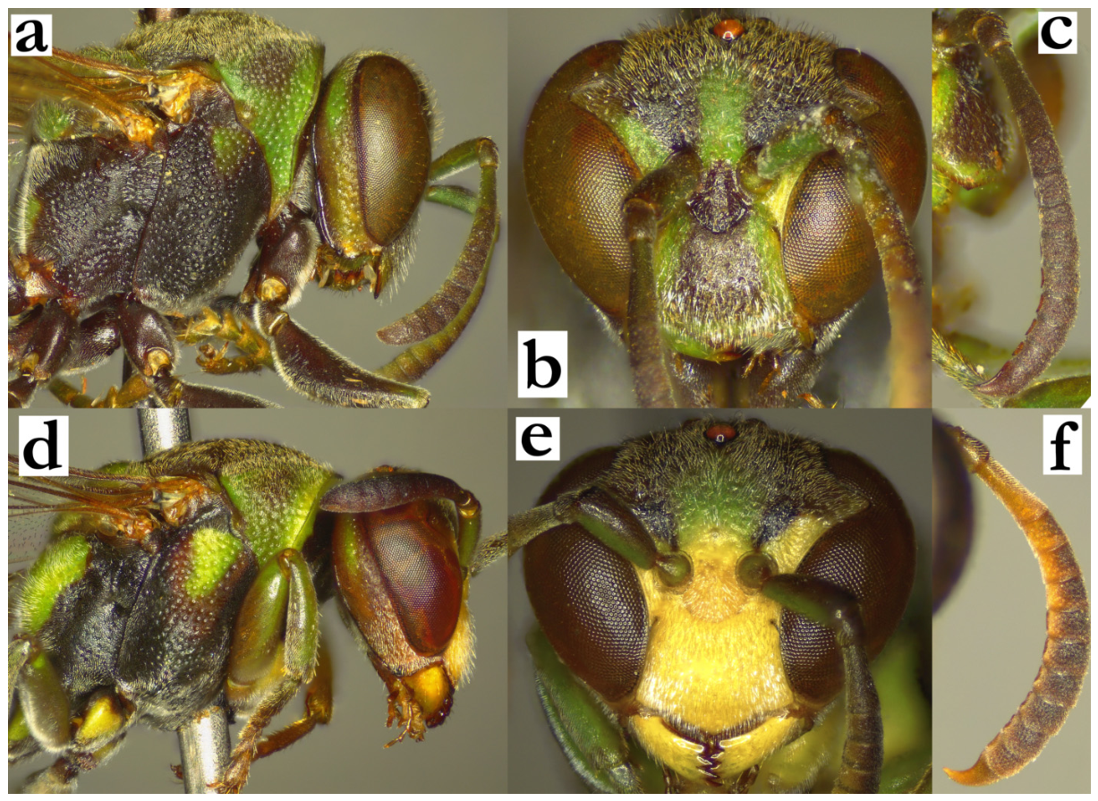
- 44. Gena posteriorly excavated close to mandible (a). T1 posteriorly with acute angle between dorsal and posterior plane (b). T2 conspicuously shorter than S2, yielding angled lateral contour of second metasomal segment (b)...................................................................45
- - Gena posteriorly convex or straight (c). T1 at most with the right angle between dorsal and posterior plane (d). T2 and S2 approximately equally long or T2 somewhat longer (d)....................................................................................................................................................47
Figure 44.
Ropalidia constitutionalis (de Saussure), (a) head and mesosoma, lateral view, female, (b) T2, lateral view, female; (c) Ropalidia subclavata (de Saussure), head and mesosoma, lateral view, female; (d) Ropalidia blommersi sp. nov., T2, lateral view, female.
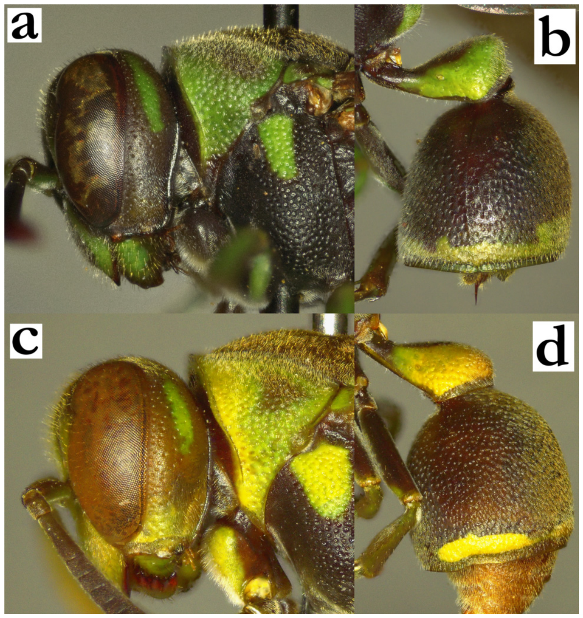
- 45. Small species, with wing length up to 7 mm. S2 with angular contour near base (a). Femora green, tarsi yellowish (a).
[f] Gena weakly excavated, lower part yellow (a). Clypeus, mandible and inner orbit yellow; antenna dorsally brown (b).
[m] Unknown
[wl] 6.6 mm
[n] Small nest, located on the underside of broad leaves.......R. pulchella (de Saussure)
- - Larger body size, wing length rarely under 7 mm. Second metasomal segment with symmetric contour in lateral view. Femora black, tarsi green, head and mesosoma without yellow colour.................................................................................................................................46
Figure 45.
Ropalidia pulchella (de Saussure), female, holotype, (a) habitus, lateral view, (b) head, frontal view.
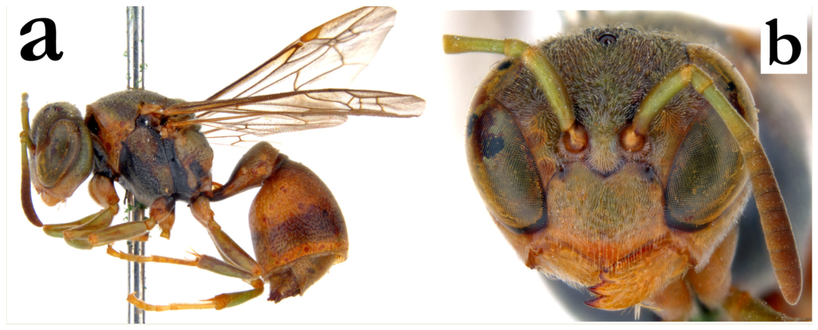
- 46. General body colour pattern with more green; pronotum mainly green, mesepisternum with large green patch, propodeum with green rhomboid spot; all markings bright green (a); head with numerous green markings (b).
[f] Gena moderately excavated (c). Head rounded in frontal view; clypeal base narrow, juxtamandibular excavation deep, with strongly projecting apex, OCA weak or absent (b). Ocellar triangle nearly equidistant or with a barely wider base. Median depression of propodeum striate and impunctate.
[m] Unknown
[wl] 8.0 [7.9–8.2] mm
[n] Unknown...................................................................R. constitutionalis (de Saussure)
- - General body colour pattern darker, with less green markings (d); head with fewer green markings (e).
[f] Gena posteriorly with stronger excavation (f). Head conspicuously triangular in frontal view; clypeal base wider, with shorter and more horizontally placed upes, weak juxtamandibular excavation and less projecting apex (e); ocellar triangle base 1.7–2.0× as wide as side. Median depression of propodeum superiorly punctate.
[m] Unknown
[wl] 8.1 [7.9–8.3] mm
[n] Unknown.............................................................................................R. rampa sp. nov.
Figure 46.
Ropalidia constitutionalis (de Saussure), (a) habitus, oblique posterior view, female, (b) head, frontal view, female, (c) head, lateral view, female; Ropalidia rampa sp. nov. (d) habitus, oblique posterior view, female, (e) head, frontal view, female, (f) head, lateral view, female.
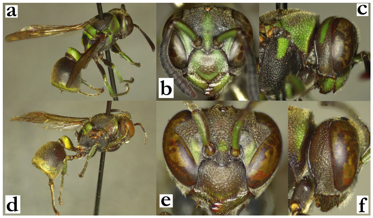
- 47. Body basally black or dark brown, with sharply defined and simple, green, yellow or whitish posterior band on T2, of equal or nearly equal width to posterior band on T1 (T1 exceptionally with somewhat broader posterior band than T2) (a). T1 short, pyriform and moderately developed dorsally (b). If there are suffused greenish markings on body, then female gena is 1.0–1.4× as wide as eye and head is conspicuously emarginate posteriorly (c), and male tyloids originate at AF6–7.....................................................bicincta group—48
- - Posterior band on T2 more developed, thick and occupying large T2 surface (d), or if thin, then with small integrated spots or merged areas (e); in these cases, bands on T1 and T2 with conspicuously dissimilar width (e). Body frequently with green markings. T1 longer and more developed dorsally, angular or posteriorly globular (e). If body has faintly suffused greenish markings, with T1 and T2 with bands of similar width, then female gena is 0.6–0.9× as wide as eye, posteriorly narrowing (f) and male tyloids originate at AF5 (in R. kojimai Blommers, see couplet 66)..........................................................................................51
Figure 47.
Ropalidia antoana sp. nov., (a) habitus, dorsal view, male, (b) habitus, lateral view, male; (c) Ropalidia janettae sp. nov., head and mesosoma, dorsal view, female; (d) Ropalidia scottiana (de Saussure), habitus, oblique dorsal view, female; (e) Ropalidia tatsimo sp. nov., habitus, lateral view, female, (f) Ropalidia kojimai Blommers, head and mesosoma, dorsal view, female.
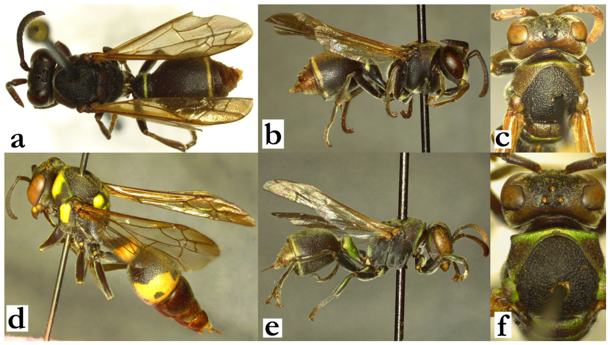
- 48. T1 shorter, 0.5–0.6× as wide as long, posteriorly straight; T1 petiole missing or very short (a).
[f] Posterior occiput and coxa I with long and wavy setae (b); occipital carina commonly evanescing ventrally or entirely missing in the lowest third of gena (b).
[m] Male tyloids originate on AF1–2................................................................................49
- - T1 longer, 0.6–0.7× as wide as long, posteriorly rounded; T1 petiole visible and parallel-sided (c).
[f] Posterior occiput and coxa I covered by short setae (d); occipital carina complete (c), sometimes widened close to mandible.
[m] Male tyloids originate on AF3–7................................................................................50
Figure 48.
(a) Ropalidia bicincta (de Saussure), T1 and T2, oblique dorsal view, female; (b) Ropalidia cincinnata Blommers, head and anterior mesosoma, lateral view, female; Ropalidia janettae sp. nov., (c) T1 and T2, oblique dorsal view, female, (d) head and anterior mesosoma, lateral view, female.
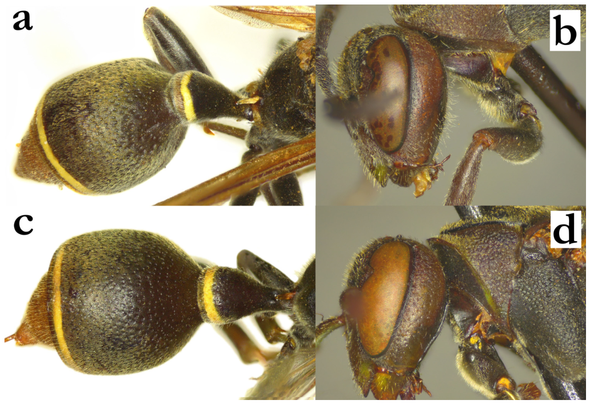
- 49. Body with shorter setae and weaker punctures (a). Upes continuity broken by anterior tentorial pit (b, c).
[f] Head and mesosoma dorsally with short setae, not longer than anterior ocellus diameter (a); pronotum inferiorly impunctate (a); gena close to eye impunctate or with an occasional poorly defined puncture (a).
[m] Tyloids originate on AF1; terminal flagellomere elongated, with darkened and obtuse tip (d). Tarsal I spur elongated.
[wl] 7.9 [7.0–8.2] mm
[n] Unknown..................................................................................R. bicincta (de Saussure)
- - Body with longer setae and coarser punctures (e). Upes uninterrupted by anterior tentorial pits (f, g).
[f] Head and mesosoma dorsally with long and wavy setae, 2× or more as long as anterior ocellus diameter (a); pronotum with retained puncture contour (a); gena close to eye punctate (a).
[m] Tyloids originate on AF2; terminal flagellomere short, with acute tip (h). Tarsal I spur absent
[wl] 8.3 mm [7.8–9.1] mm
[n] Elongated, up to 70 cells long and up to 10 cells wide (frequently with more than 100 cells in total); sometimes wider and triangular. Cell wall grey, cocoon caps grey-brown; located on branches....................................................... R. cincinnata Blommers
Figure 49.
Ropalidia bicincta (de Saussure), (a) head and mesosoma, lateral view, female, (b) head, frontal view, female, syntype, (c) head, frontal view, male, (d) antenna, dorsal view, male; Ropalidia cincinnata Blommers, (e) head and mesosoma, lateral view, female, (f) head, frontal view, female, (g) head, frontal view, male, (h) antenna, dorsal view, male.
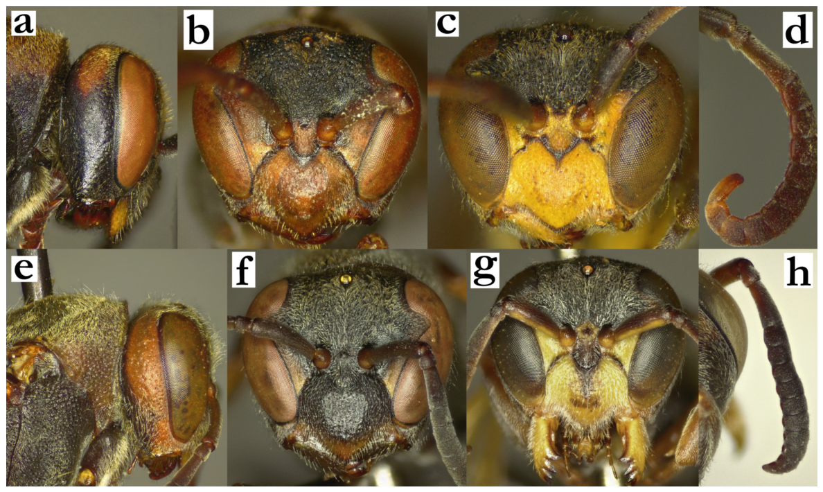
- 50. Mesepisternum punctures twice larger dorsally than ventrally (a). T2 finely and shallowly punctate, setae do not extend over lamella (b). Green markings minute or absent (except male coxa).
[f] Juxtamandibular notches shallow, base of clypeus completely impunctate; clypeus apex mildly projecting (c).
[m] Clypeus convex, wider than long (d). Tyloids originate as AF(3)4.
[wl] 8.6 [8.4–9.1] mm
[n] Unknown..........................................................................................R. antoana sp. nov.
- - Mesepisternum dorsally and ventrally similarly punctate (e). T2 more coarsely punctate, setae extend over lamella (f). Body with green of suffused greenish areas.
[f] Juxtamandibular notches deep, base of clypeus with poorly defined punctures; clypeus apex more projecting (g).
[m] Clypeus concave apically, with more projecting apical margin (h). Tyloids originate on AF(6)7.
[wl] 8.4 [8.1–8.6] mm
[n] Unknown..........................................................................................R. janettae sp. nov.
Figure 50.
Ropalidia antoana sp. nov., (a) mesepisternum, lateral view, female, (b) T2, lateral view, female, (c) head, frontal view, female, (d) head, frontal view, male; Ropalidia janettae sp. nov., (e) mesepisternum, lateral view, female, (f) T2, lateral view, female, (g) head, frontal view, female, (h) head, frontal view, male.
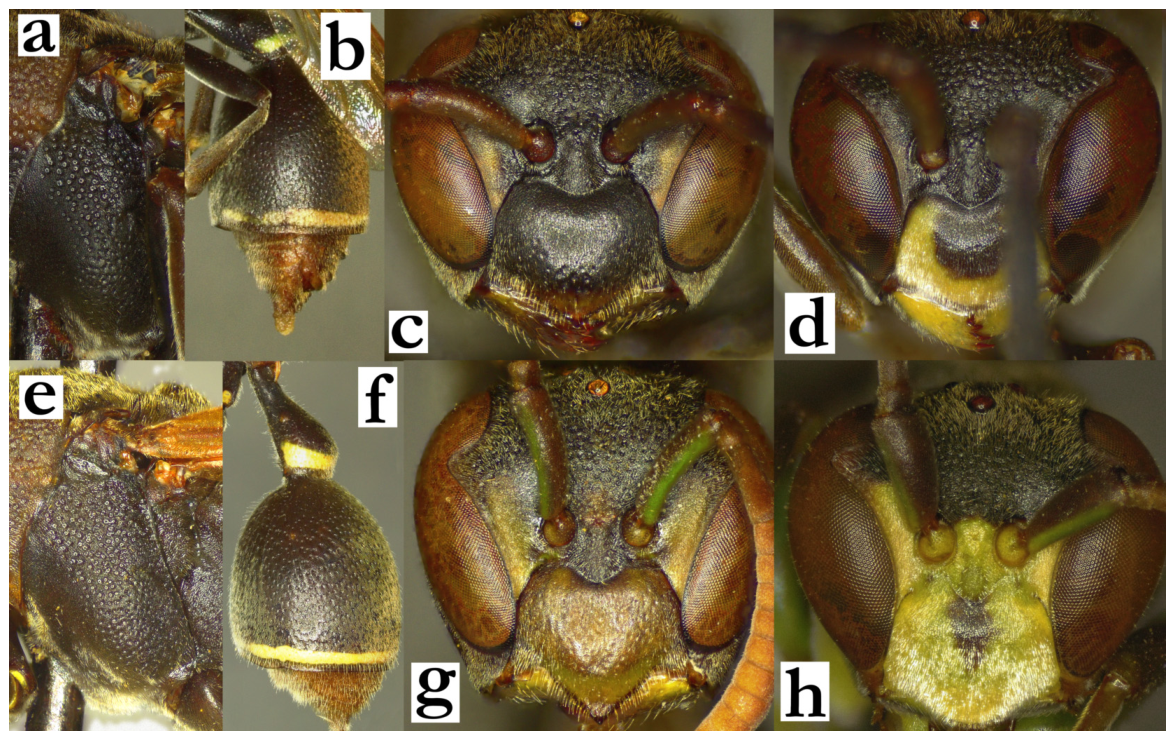
- 51. Head and mesosoma almost entirely green or dark-green, T2 ferruginous-brown, with a thin posterior green band; mesoscutum without green or yellow markings (a) or exceptionally with a barely visible two longitudinal green lines. T1 developed and rounded dorsally (a). Head and mesoscutum with microscopic pubescence, and very short protruding setae (b).
[f] Clypeus very wide, 1.4–1.5× as wide as long, with impunctate base (c). Gena impunctate or barely punctate, inferiorly wider than at mid-height (b).
[m] Tyloids rounded and very short, extending only on proximal half of flagellomere length (d).
[wl] 8.5 [8.2–8.8] mm
[n] Unknown.........................................................................................R. tapelaka sp. nov.
- - Colour pattern dissimilar, head and mesosoma with similar amount of green or yellow markings; if there are numerous green body markings (e), then T2 has broadened posterior band, and frons and mesoscutum have longer setae (f).
[f] Clypeus rarely entirely green or yellow, and in these cases not more than 1.3× as wide as long (g), and gena more coarsely punctate and narrower inferiorly (f).
[m] Tyloids, where present, extend along entire flagellomere length (h)...................52
Figure 51.
Ropalidia tapelaka sp. nov., (a) habitus, oblique posterior view, female, (b) head, lateral view, female, (c) head, frontal view, female, (d) distal flagellomeres, median view, male; (e) Ropalidia sonyae sp. nov., habitus, oblique posterior view, female; Ropalidia phalansterica (de Saussure), (f) head, lateral view, female, (g) head, frontal view, female; (h) Ropalidia variabilis (de Saussure), distal flagellomeres, median view, male.
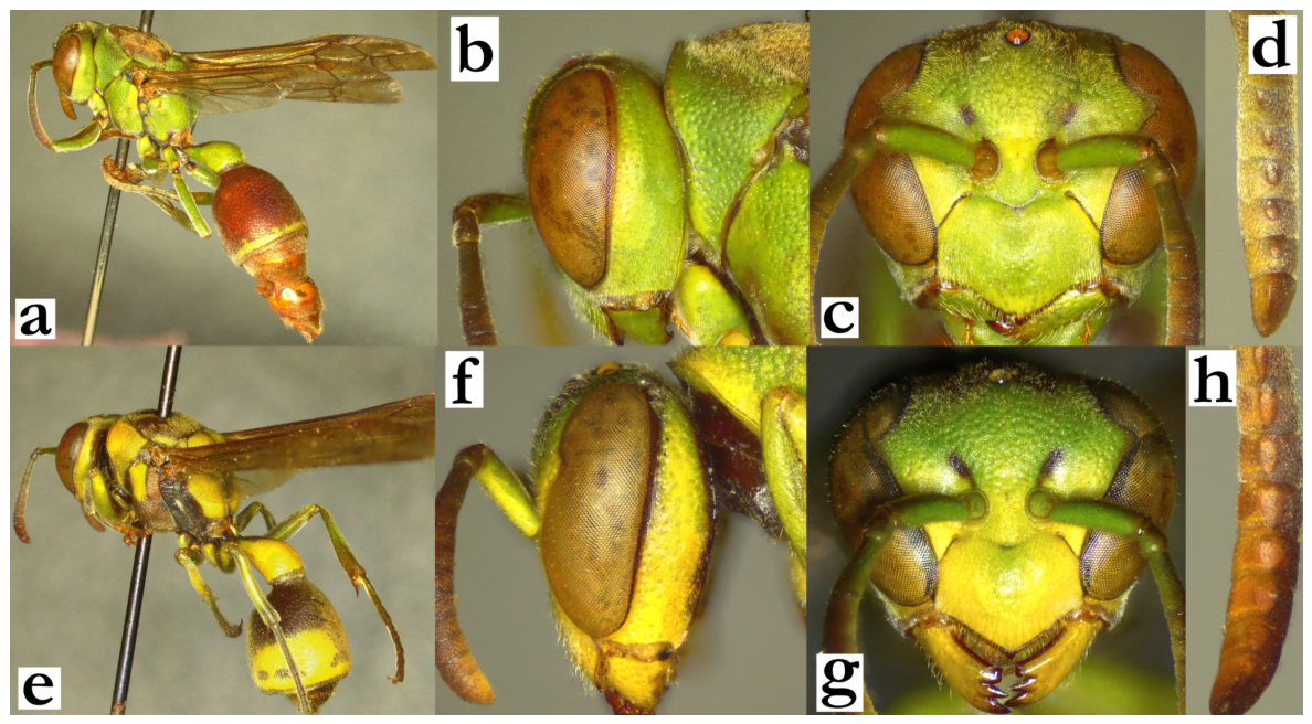
- 52. Large and colourful species, with numerous green and yellow markings (a). T1 streamlined and evenly dorsally rounded (a). Mesoscutum with two longitudinal green lines, sometimes also with area next to tegula (b).
[f] Clypeus yellow; upes short, 0.5× as long as lateral clypeal margin (c).
[m] Tyloids originate on AF(3)4; terminal flagellomere 1.6× the AF10 length, evenly curved, with developed proximal tyloid and acute tip (d).
[wl] 9.9 [8.8–10.6] mm
[n] Elongated, with a downward facing thin extension originating from a wider base with an apical attachment to the surface. Cell wall porous, built from heavily masticated material. Nest located on branches..........................................R. sonyae sp. nov.
- - Body smaller or not that colourful; if there are numerous markings on similarly sized body, these are bright emerald green and T1 is obtusely angled dorsally (e). Mesoscutum exceptionally with green longitudinal lines (f; except R. miafina sp. nov., see couplet 60, but then wing length is under 7 mm).
[f] Clypeus variably coloured; if wing length is similar, then clypeus is mainly green with central spot or spots (g). Upes long, of similar length to lateral clypeal margin (g).
[m] Terminal flagellomere without proximally developed tyloid (h)..........................53
Figure 52.
Ropalidia sonyae sp. nov., (a) habitus, lateral view, female, (b) head and mesosoma, dorsal view, female, (c) head, frontal view, female, (d) distal flagellomeres, dorsal view, male; Ropalidia aras sp. nov., (e) habitus, lateral view, female, (f) head and mesosoma, dorsal view, female, (g) head, frontal view, female; (h) Ropalidia variabilis (de Saussure) distal flagellomeres, dorsal view, male.
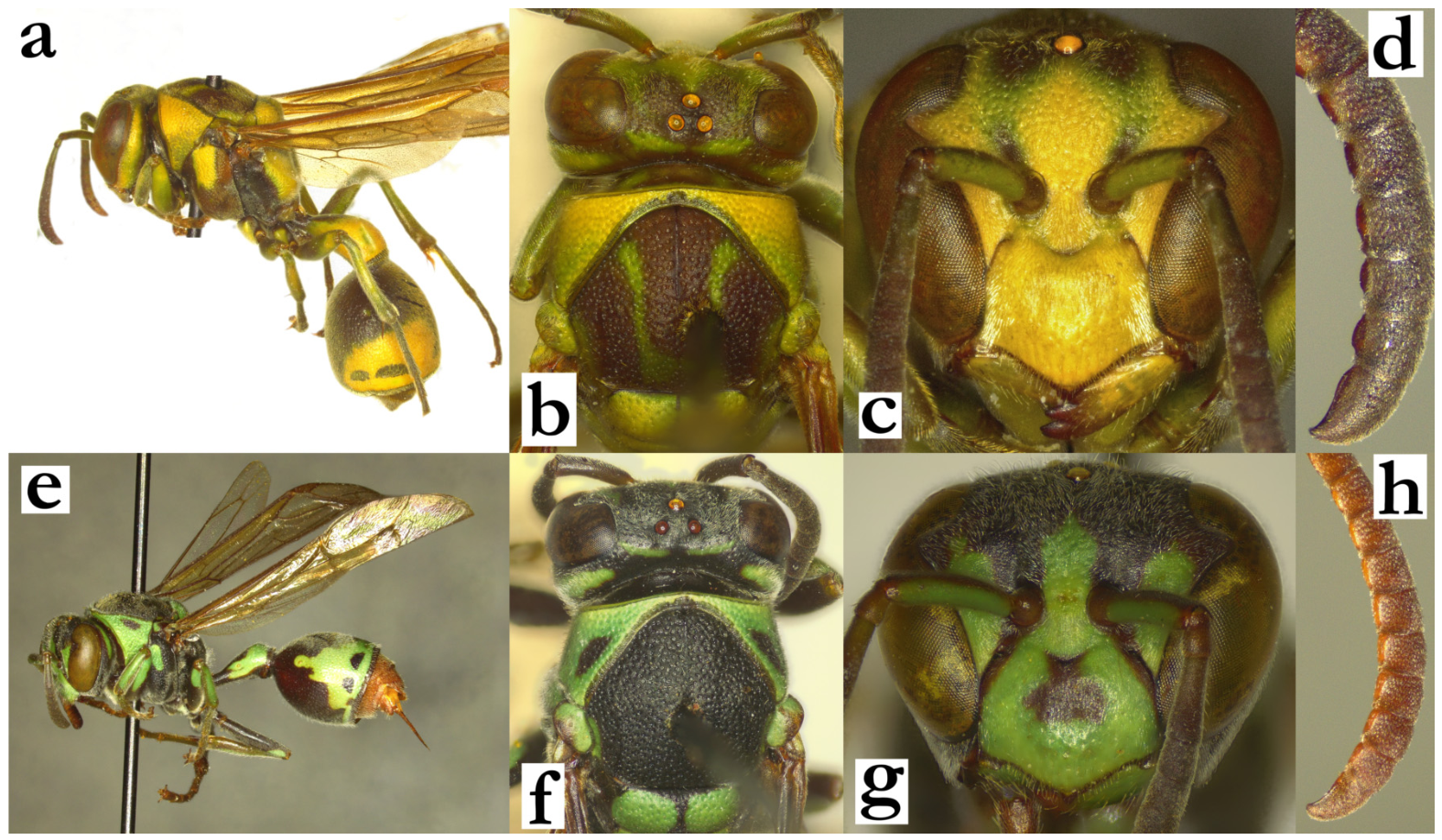
- 53. Dorsal and posterior face of T1 at right angle in lateral view, or even more obtuse (a); posterior part of T1 coarsely punctate (b). Body with numerous green(-yellow) markings (b). T2 with a simple or barely widened posterior green or yellow band (a, b).................54
- - Dorsal part of T1 at an obtuse angle to posterior surface, pyriform in shape and posteriorly weakly punctate (c); some specimens of R. variabilis can have angulated and coarsely punctate posterior part of T1, but in those cases body is mainly black and suffused greenish or reddish, with minimal green or yellow markings (d); some specimens of R. scottiana can have more dorsally developed T1, but in these cases T2 has a broadened posterior yellow band; couplet 63...............................................................................................................57
Figure 53.
(a) Ropalidia subclavata (de Saussure), T2, lateral view, female; (b) Ropalidia mysterica Blommers, habitus, lateral view, female; (c) Ropalidia ranavali (de Saussure), T2, lateral view, male; (d) Ropalidia variabilis (de Saussure), habitus, oblique lateral view, female.
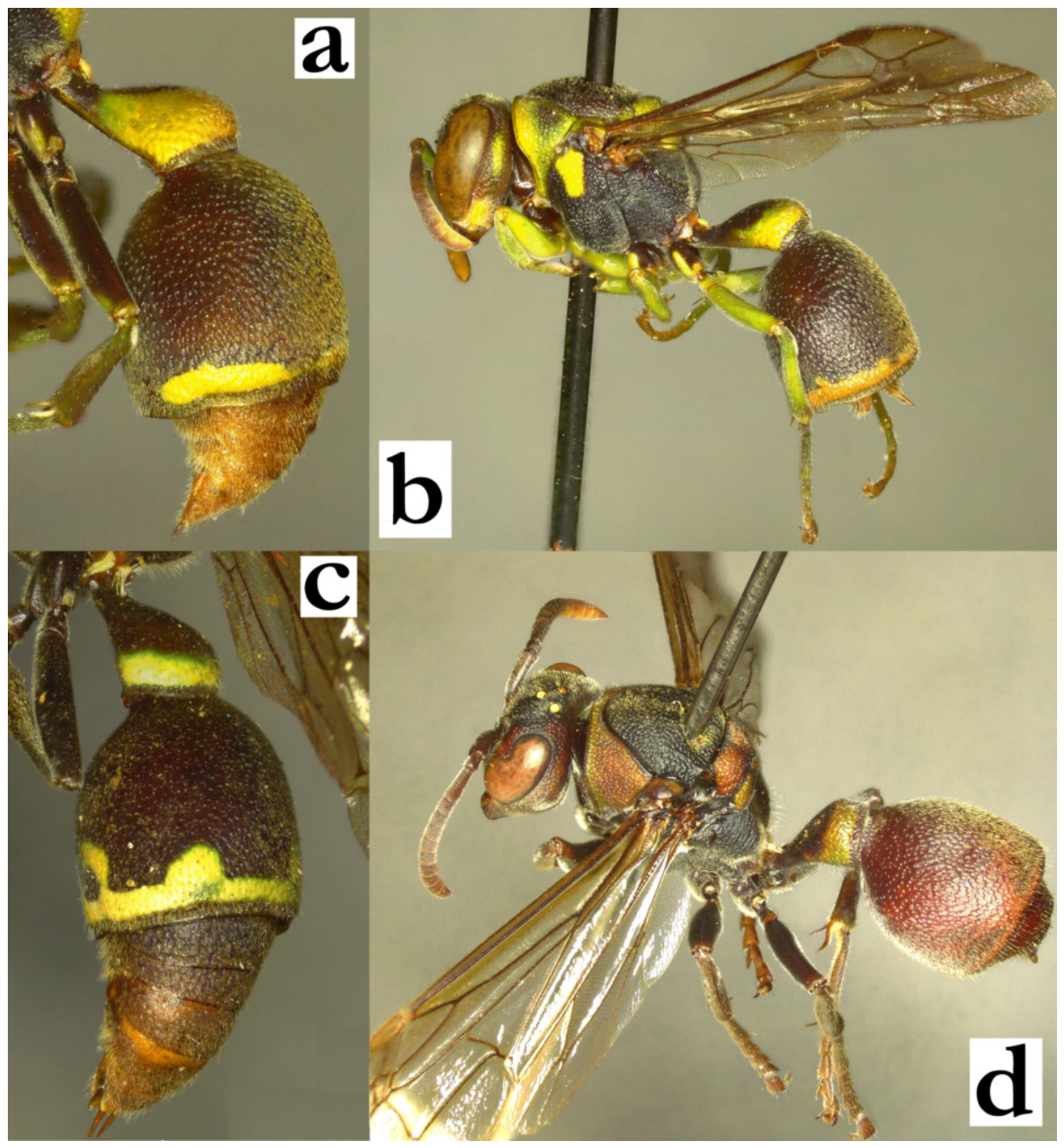
- 54. Occipital carina angulated, with correspondingly sinuate pronotal carina (a). Face yellow in both sexes (b, c). Smaller size; wing length commonly 6.5 mm.
[f] Clypeus shallowly punctate, upes short and curved (b).
[m] Scape 1.9–2.0× as wide as AF1 base; AF2 wider than long (c). Tyloids originate at AF3; terminal flagellomere elongated and acute, with developed median tubercle (d).
[wl] 6.5 [5.8–6.8] mm
[n] Elongated and somewhat irregular in shape, with up to 30 cells; nesting in forested areas, on mango and cardamom leaves.............................R. mysterica Blommers
- - Occipital and pronotal carina with a streamlined contour (e). Face with green markings, at least a suffused central green (-yellow) spot in both sexes; upes straight and eyes conspicuously setose (f, g). Larger size; wing length commonly over 6.5 mm.
[f] Clypeus coarsely punctate, upes straight and long (f).
[m] Scape 1.3–1.7× as wide as AF1 base; AF2 longer than wide (g). Tyloids originate at AF4–7, terminal flagellomere shorter, without median tubercle (h)........................55
Figure 54.
Ropalidia mysterica Blommers, (a) head, oblique view, female, (b) head, frontal view, female, (c) head, frontal view, male, (d) distal flagellomeres, dorsal view, male; (e) Ropalidia blommersi sp. nov., head, oblique view, female; Ropalidia subclavata (de Saussure), (f) head, frontal view, female, (g) head, frontal view, male, (h) distal flagellomeres, dorsal view, male.
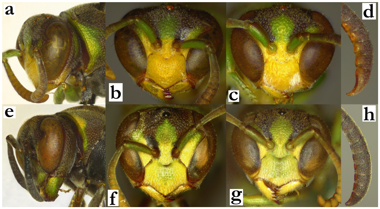
- 55. T1 posterior translucent area impunctate or barely punctate proximally (a). Distance between posterior ocelli over 3× as wide as their diameter (b). Mesoscutum as long as wide in front of tegula (b).
[f] Clypeus with narrower base and weakly developed or absent OCA; upes of similar length to lateral clypeus margin (c).
[m] Tyloids originate at AF4, terminal flagellomere 1.8–2.0× as long as wide (d).
[wl] 6.9 [6.6–7.3] mm
[n] Discoid, 11×10 cells, with very shallow cells and infundibular base; cell wall grey(ish) and compact (e).................................................................... R. tatsimo sp. nov.
- - T1 posterior translucent area coarsely punctate (f). Posterior ocelli no more than two diameters apart (g). Mesoscutum 1.2× as wide as long (g).
[f] Clypeus with wider base and developed OCA; if OCA is weak, than upes conspicuously shorter than lateral clypeus margin (h, i).
[m] Clypeus without substantially longer pubescence apically. Tyloids less elevated above flagellomere, originate at AF5–AF7. Terminal flagellomere at most 1.7× as long as wide...................................................................................................................................56
Figure 55.
Ropalidia tatsimo sp. nov., (a) T1, oblique dorsal view, female, (b) head and mesosoma, dorsal view, female, (c) head, frontal view, female, (d) antenna, dorsal view, male, (e) nest; Ropalidia subclavata (de Saussure), (f) T1, oblique dorsal view, female, (g) head and mesosoma, dorsal view, female, (i) head, frontal view, female; (h) Ropalidia blommersi sp. nov., head, frontal view, female.
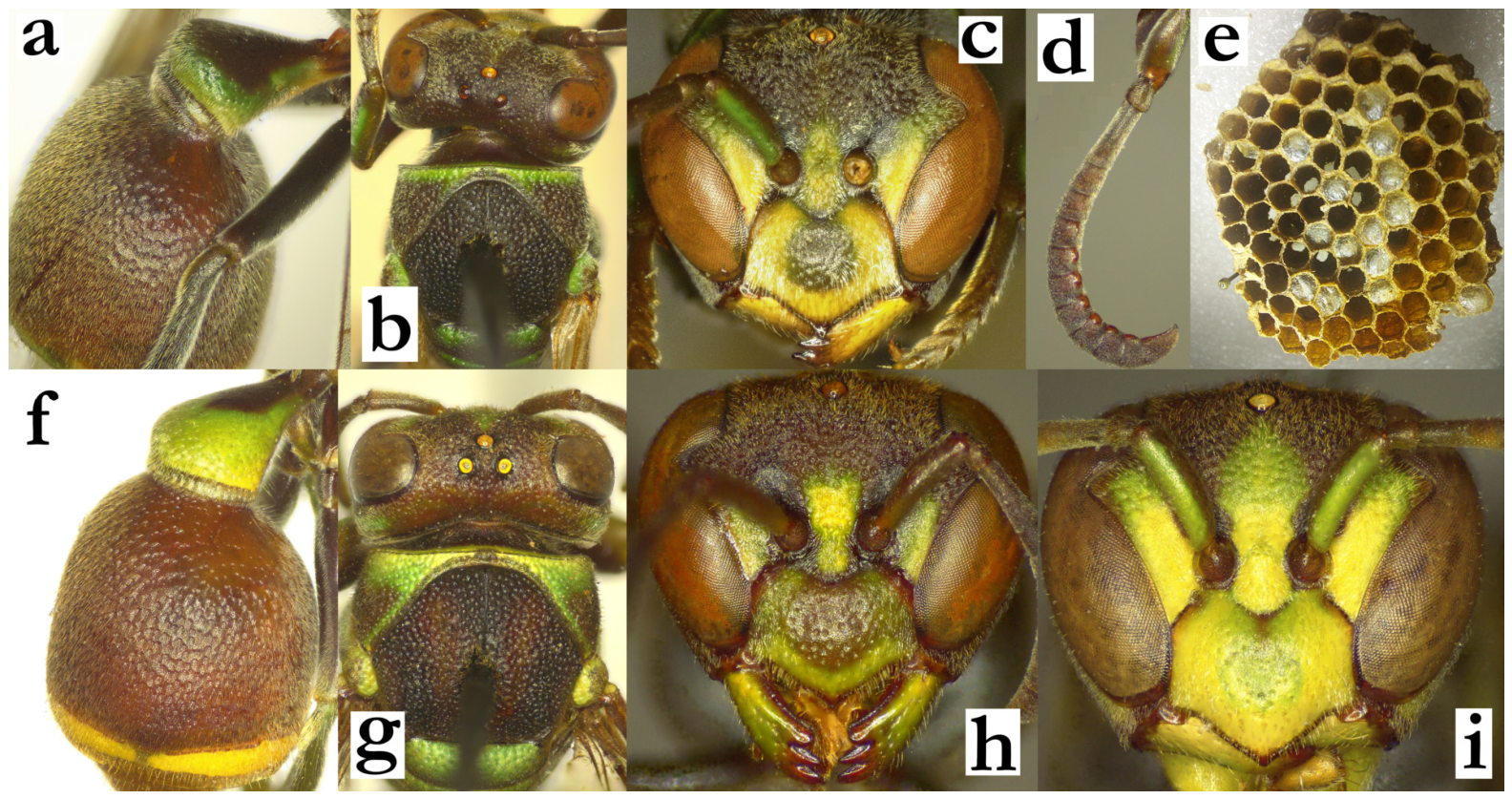
- 56. Median carina of scutellum absent or barely developed as thin streak (a). Median depression of propodeum punctate, with visible oblique striations (b). Metanotum follows scutellum contour (c). Eyes asetose.
[f] Clypeus with basal suffused green spot. Mesosoma side without green or yellow markings. Metanotum and scutellum with smaller green spots (a). Gena 0.8–1.0× as wide as eye.
[m] Tyloids originate at AF(5)6 and occupy no more than half of flagellomere surface. Anterior surface of mesepisternum black, coxa at most with smaller green quadrant.
[wl] 6.9 [6.7–7.5] mm
[n] Nest irregular in shape, with multiple projecting branches; cell walls black, with greyish streaks and cocoon tops; located on branch [60]................R. blommersi sp. nov.
- - Median carina of scutellum developed as thin streak (e). Median depression of propodeum impunctate and without substantial striation in the lower half (f). Metanotum slightly elevated from the metanotum posterior surface (f).
[f] Clypeus commonly entirely green or yellow. All coxa with green markings, mesepisternum with large green spot, propodeum mainly green (f). Scutellum and metanotum mostly green (f). Gena 1.0–1.3× as wide as eye.
[m] Tyloids originate at AF(6)7 and occupy more than half of flagellomere surface. Mesepisternum with a large green or yellow area anteriorly, all coxa pairs commonly green.
[wl] 6.9 [6.7–7.5] mm
[n] Discoid and small, with up to 40 cells at the end of the colony cycle; cell wall most commonly light-grey, with thin black streaks, but sometimes entirely grey; paper supple [61]................................................................................R. subclavata (de Saussure)
Figure 56.
Ropalidia blommersi sp. nov., (a) scutellum, dorsal view, female, (b) posterior mesosoma, dorsal view, female, (c) posterior mesosoma, oblique view, female; Ropalidia subclavata (de Saussure), (d) scutellum, dorsal view, female, (e) posterior mesosoma, dorsal view, female, (f) posterior mesosoma, oblique view, female.
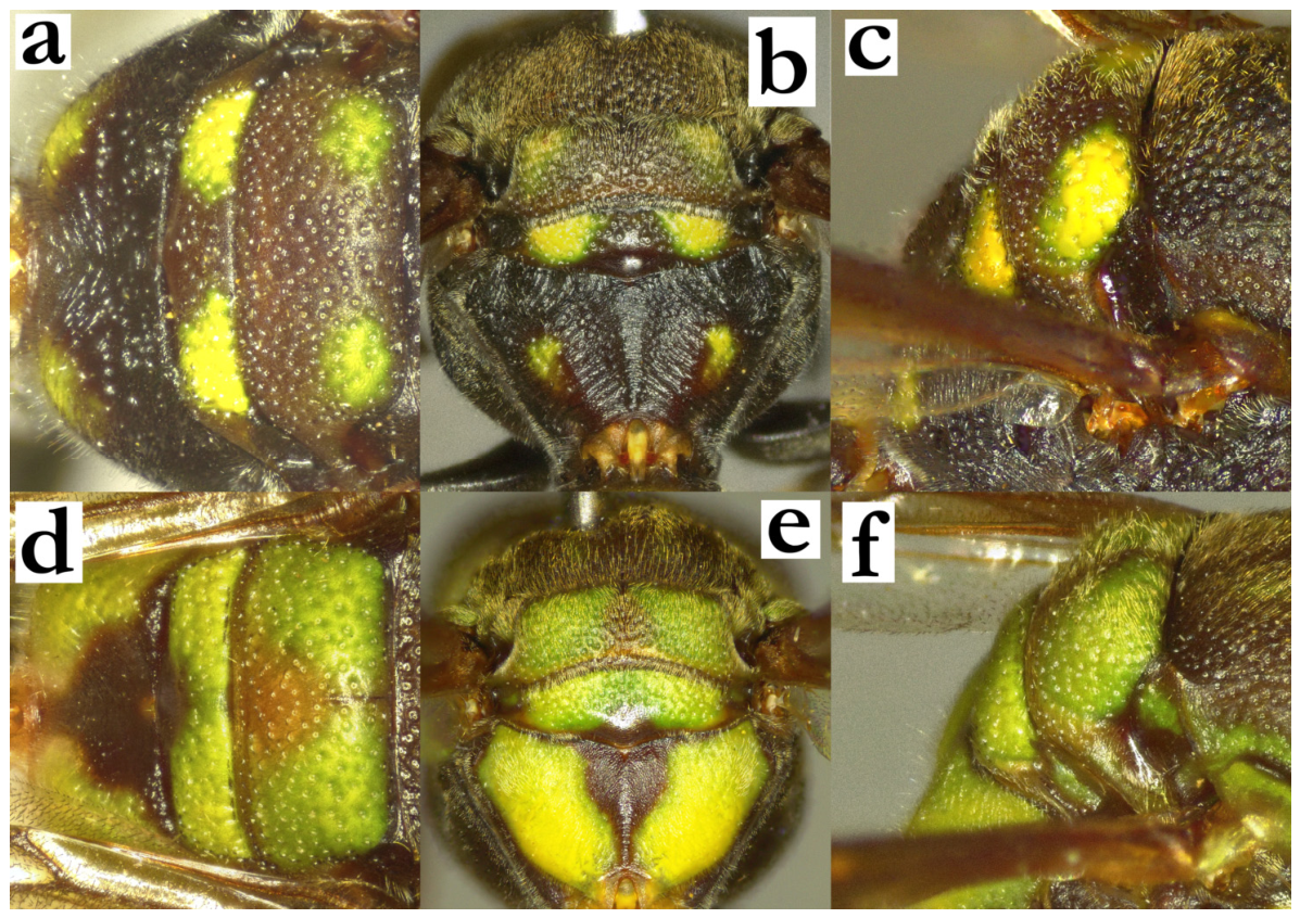
- 57. Wing length under 7 mm. Mesosoma commonly with plenty green or yellow markings, T2 with a thin posterior band (a, b)...........................................................................................58
- - Wing length over 7 mm. Mesosoma commonly with less bright markings, T2 with broadened posterior band (c), but sometimes thin or almost entirely missing (d).......................62
Figure 57.
Habitus, oblique lateral view, female, (a) Ropalidia favulorum Blommers, (b) Ropalidia miafina sp. nov., (c) Ropalidia ranavali (de Saussure), (d) Ropalidia ranavali (de Saussure), melanic form from the central highlands.
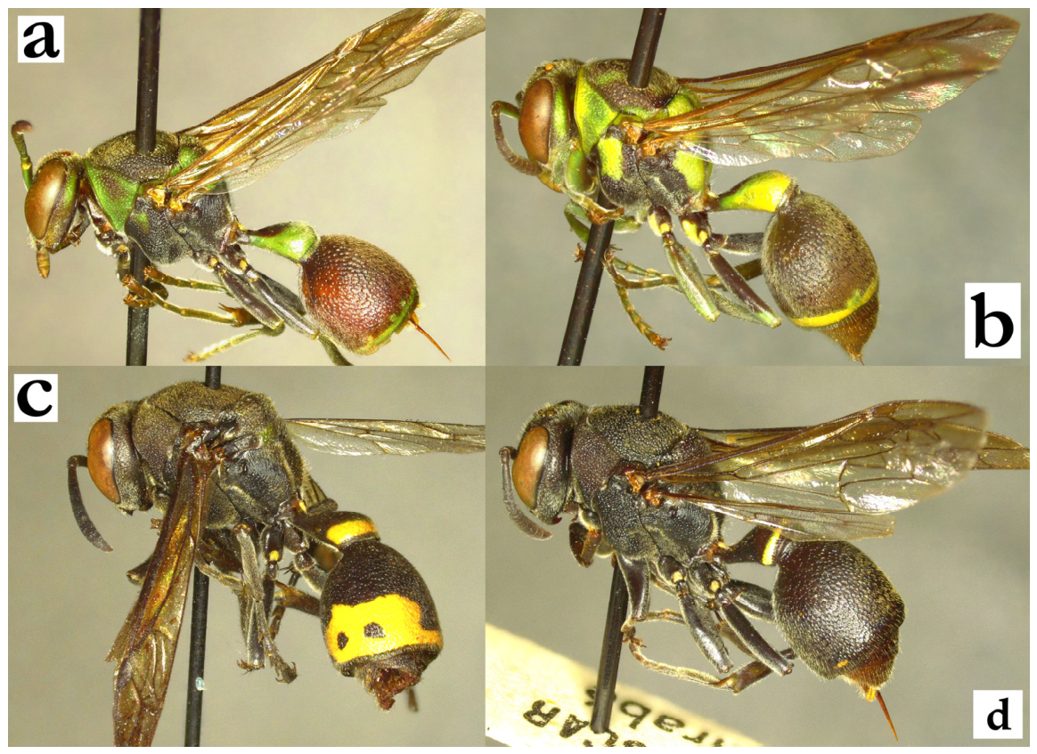
- 58. Body brown and yellow (a), green and black colours absent or at most with a faint olivaceous tint at pronotum, scutellum or tibia (b).................................................................59
- - Body basally black with numerous green markings (c, d)...................................................60
Figure 58.
Habitus, oblique lateral view, (a) Ropalidia shattenbergi sp. nov., female, (b) Ropalidia rosae Blommers, female, (c) Ropalidia phalansterica (de Saussure), male, (d) Ropalidia favulorum Blommers, male, reddish colour pattern.
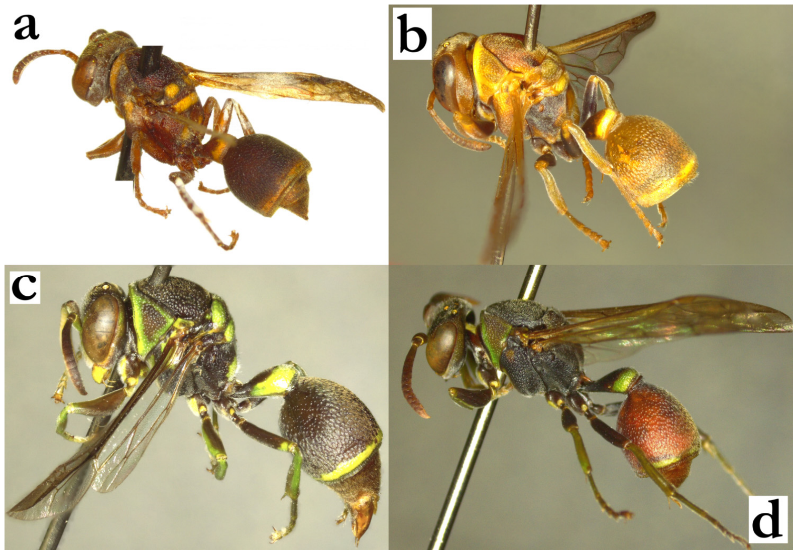
- 59. Very small species, with wing length barely exceeding 5 mm. Body bicolorous: basally ferruginous with distinct yellow markings on pronotum, scutellum and metanotum; mesoscutum in basal colour; propodeum with two bright yellow spots (Figure 58a). T1 posteriorly globular, T2 finely punctate (a).
[f] Head frontally wider than long, clypeus yellow (b).
[m] Clypeus 1.4× as wide as long (c). Tyloids brownish and shiny, originate at AF5; terminal flagellomere 1.2× as long as wide, beak-like, with an acute tip (d).
[wl] 5.0 [4.9–5.1] mm
[n] Unknown....................................................................................R. shattenbergi sp. nov.
- - Somewhat larger species, with wing length over 5 mm. Body with more than two colours, with numerous suffused areas ranging from almost black to yellow; mesoscutum in all examined specimens with three or four colours, brownish or greenish central area, reddish lateral thirds and remaining two basal dark brown areas close to scutellum; more xanthic specimens can also have yellow markings close to tegulae; propodeum with faint yellowish suffused spots (Figure 58b). T1 pyriform and more slender, T2 coarsely punctate with swollen interspaces (e).
[f] Head frontally longer than wide; clypeus with suffused brown spot (f).
[m] Clypeus convex, with a wide base, about 1.3× as wide as long (g). Tyloids yellowish, originate at AF(5)6; terminal flagellomere 1.1× as long as wide, with a sub-acute tip (h).
[wl] 5.4 [5.1–5.9] mm
[n] Colonies of small and fragmented nests, each with only a few cells; nests commonly located in mango trees..............................................................R. rosae Blommers
Figure 59.
Ropalidia shattenbergi sp. nov., (a) T1, oblique dorsal view, female, (b) head, frontal view, female, (c) head, frontal view, male, (d) antenna, dorsal view, male; Ropalidia rosae Blommers, (e) T1, oblique dorsal view, female, (f) head, frontal view, female, (g) head, frontal view, male, (h) antenna, dorsal view, male.
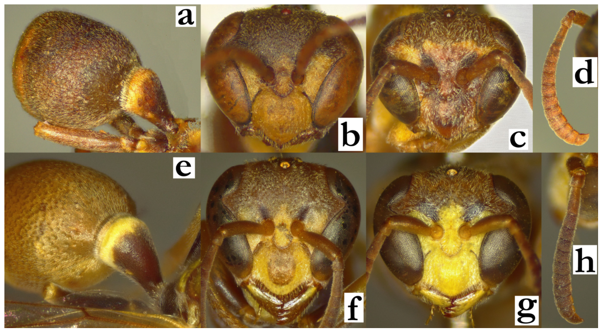
- 60. Mesoscutum shallowly punctate, with punctures more than a diameter apart (a) Mesoscutum with two thin green longitudinal lines (a), in combination with T2 basally without lateral spots (b). T1 elongated and streamlined (b). T2 shallowly punctate (b).
[f] Clypeus longer than wide, upes short, 0.3–0.4× as long as lateral clypeal margin; clypeus, mandible and interantennal area merged in yellow, without green markings.
[m] Clypeus longer than wide, upes 0.1–0.2× as long as lateral clypeal margin. Tyloids originate at AF(4)5, terminal flagellomere beak-like and acute, with median excavation.
[wl] 6.4 [5.9–6.7] mm
[n] Initially discoid, hanging from the leaf underside, with heavily masticated cell wall; cell wall light-grey with greenish and blackish streaks and spots; nest used with cocoon caps already in the early stage, with approximately 15 cells, suggesting a very quick colony cycle...................................................................................R. miafina sp. nov.
- - Mesoscutum coarsely punctate, punctures large; mesonotum entirely black (c). T1 less elongated; mesosoma commonly with fewer green markings (d). T2 coarsely punctate (d).
[f] Clypeus wider than long, upes at least 0.5× as long as lateral clypeal margin; face with lesser amount of yellow (which is commonly combined with central green markings).
[m] Clypeus wider than long, upes at least 0.3× as long as lateral clypeal margin......61
Figure 60.
Ropalidia miafina sp. nov., (a) mesoscutum, dorsal view, female, (b) habitus, lateral view, female; Ropalidia favulorum Blommers, (c) mesoscutum, dorsal view, female, (d) habitus, lateral view, female.
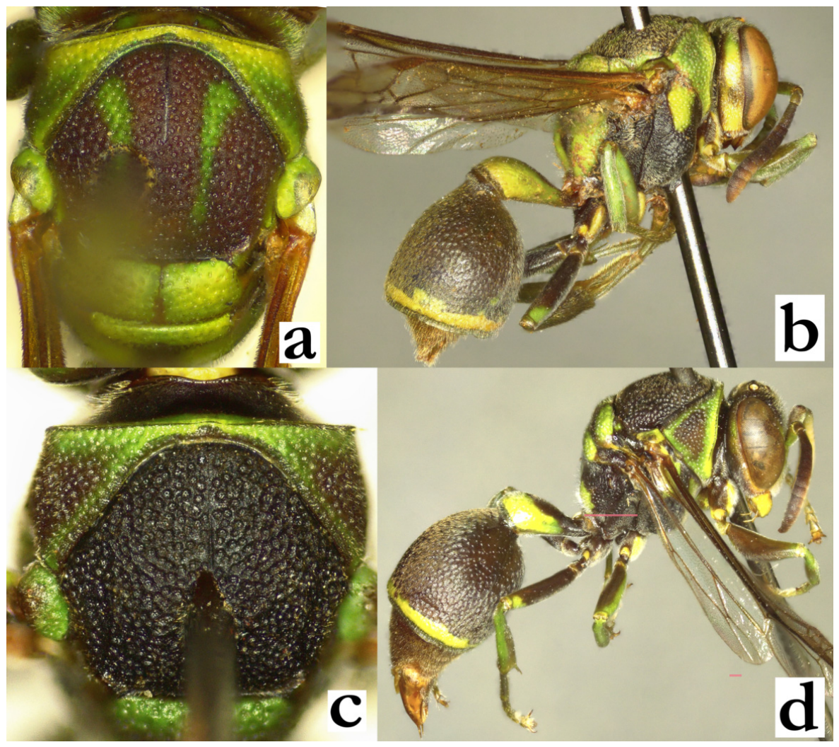
- 61. Body lightly coloured; T2 basally brown; femora, frons, propodeum and mesepisternum with green markings (a).
[f] Clypeus, inner orbit and interantennal area commonly entirely yellow; upes straight, OCA developed (b). Coxa I yellow, coxa II (and III) with green markings. Frons setae 0.3–0.6× as long as ocellus diameter (c).
[m] Antenna more slender; AF1 3.5× as long as basally wide, AF2 longer than wide (d).
[wl] 6.3 [5.4–7.0] mm
[n] see couplet 39....................................R. phalansterica (de Saussure), melanic form
- - Body darkly coloured; basal colour black(ish), femora, frons, propodeum and mesepisternum with smaller green markings or entirely in basal colour (e).
[f] Clypeus with central dark spot (f), occasionally black with remaining apical transversal green line; upes curved, OCA less developed (f). Frons setae 0.7–1.0× as long as anterior ocellus diameter (g).
[m] Antenna more clavate; AF1 3.0× as long as basally wide, AF2 wider than long (h).
[wl] 6.4 [5.6–7.0] mm
[n] Multiple small nests in colonies on leaves or palm stem, each nest consisting of up to five cells; cell wall black or streaked with grey transversal lines. Cocoon tops grey, later darker...........................................................................R. favulorum Blommers
Figure 61.
Ropalidia phalansterica (de Saussure), melanic form, (a) habitus, oblique lateral view, female, (b) head, frontal view, female, (c) head, lateral view, female, (d) antenna, dorsal view, male; Ropalidia favulorum Blommers, (e) habitus, oblique lateral view, female, (f) head, frontal view, female, (g) head, lateral view, female, (h) antenna, dorsal view, male.
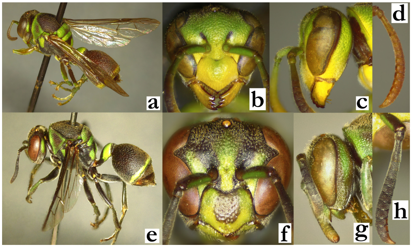
- 62. T1 posteriorly coarsely punctate, commonly angulated (a). Body with reddish or dark green markings (a). Pronotum pubescence silvery, mesoscutum yellowish (b).
[f] Clypeus commonly dark, interantennal area and inner orbits with faint yellow markings (c).
[m] Clypeus bell-shaped, curved upes and blackish basal area. Tyloids originate at AF2 (Figure 51h); terminal flagellomere 1.7× as long as wide, curved, apically subacute.
[wl] f: 8.7 [8.6–9.1] mm; m: 8.5 [8.2–8.7] mm
[n] Oval or triangular, with apical pedicel. Cell wall compact, without holes, light brown to brown, sometimes striated by brown-greenish layers. Cocoon tops grey to brownish. Nests located on branches or man-made objects, with 50–60 cells in a yearly cycle, and up to 120 cells in bi-annual nests................R. variabilis (de Saussure)
- - T1 posteriorly shallowly punctate to impunctate, rounded (e). Pronotum and mesoscutum pubescence similarly coloured (f). Head and mesosoma commonly with numerous green markings (f) or entirely black in dark specimens of R. ranavali (see couplet 64). T2 commonly with elaborate green or yellow posterior band (e), except R. kojimai, in which cases female gena is impunctate and male tyloids originate from AF5 (see couplet 66). Male tyloids originate on AF4–5................................................................................................63
Figure 62.
Ropalidia variabilis (de Saussure), (a) habitus, oblique lateral view, female, (b) head and mesosoma, lateral view, female, (c) head, frontal view, female, (d) antenna, dorsal view, male; Ropalidia aras sp. nov., (e) habitus, oblique lateral view, female, (f) head and mesosoma, lateral view, female; (g) Ropalidia ranavali (de Saussure), head, frontal view, female; (h) Ropalidia kojimai Blommers, antenna, dorsal view, male.
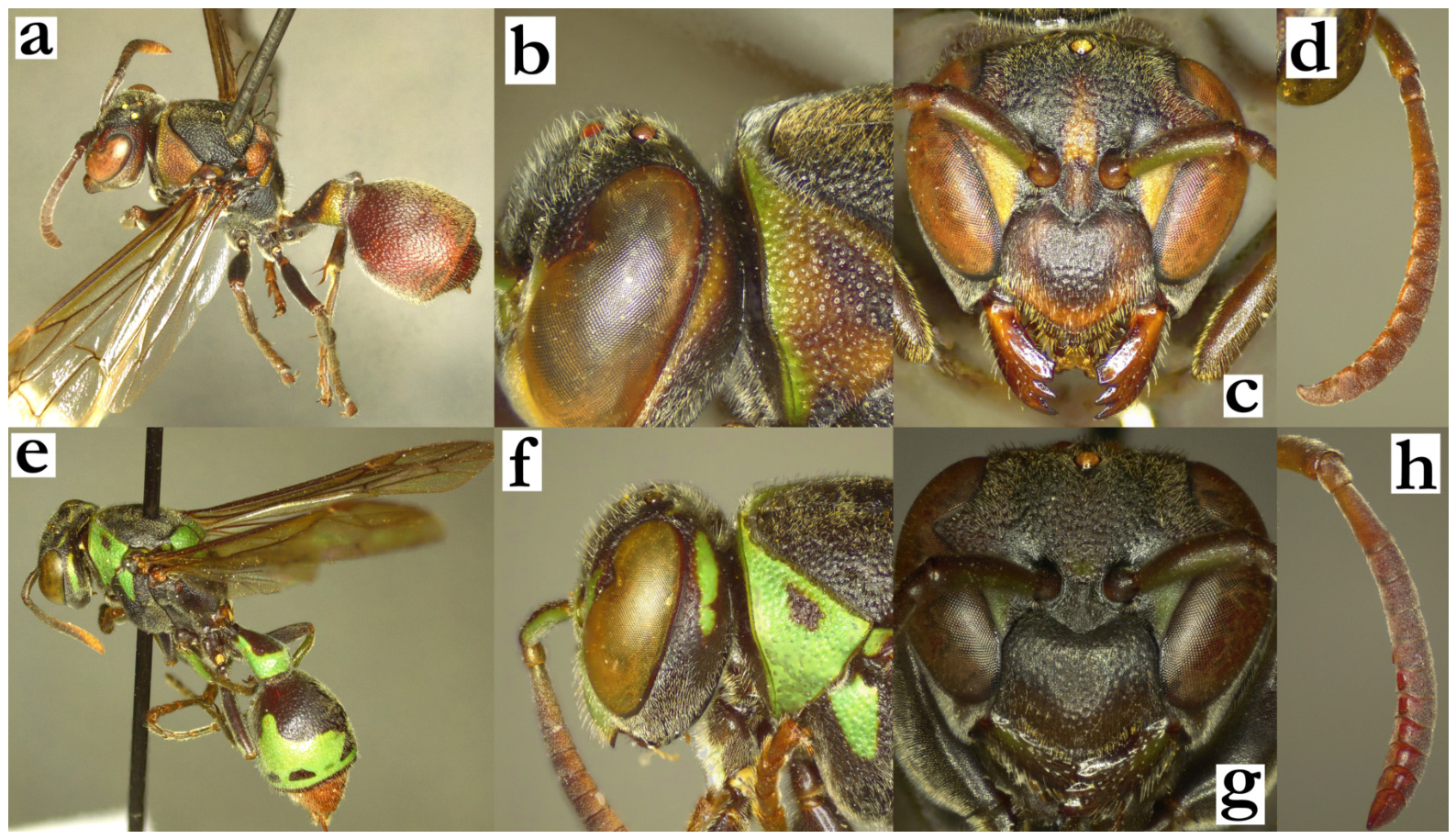
- 63. Pronotum lobe developed, laterally projecting in front of tegula in dorsal view (a). Terminal metasomal segment dorsally with yellow spot, ventrally with two yellow spots (b).
[f] Clypeus pentagonal, 1.3–1.5× as wide as long, with straight upes, angled lateral clypeal margins and weak OCA; most of face yellow (c).
[m] Tyloids occupy more than half of inner flagellomere surface (d).
[wl] 7.9 [7.5–8.3] mm; notably, the species description suggests 9.5 mm of the wing length, while the holotype examination suggested 8.3 mm.
[n] Two cells wide (initially directed at 90-degree angle) with apical petiole; cell wall greenish, located on branch.......................................................R. scottiana (de Saussure)
- - Pronotum lobe less developed, without lateral projections in dorsal view (e). Terminal metasomal segment without yellow markings (f).
[f] Clypeus 1.1–1.4× as wide as long; yellow markings on face suffused or absent (g).
[m] Tyloids occupy about a third of flagellomere surface (h)........................................64
Figure 63.
Ropalidia scottiana (de Saussure), (a) anterior mesosoma, dorsal view, female, (b) metasoma, caudal view, female, (c) head, frontal view, female, (d) distal flagellomeres, median view, male; Ropalidia aras sp. nov., (e) anterior mesosoma, dorsal view, female, (f) metasoma, caudal view, female; (g) Ropalidia kojimai Blommers head, frontal view, female; (h) Ropalidia ranavali (de Saussure), distal flagellomeres, median view, male.
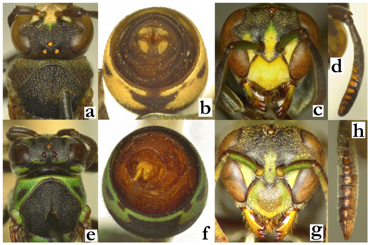
- 64. Punctures coarse; occiput punctate similarly to frons (a). Mesoscutum coarsely and densely punctate, punctures less than a diameter apart (a). Gena coarsely punctate (b). Head and mesosoma with minimal green and yellow markings (Figure 57c), metasoma variable, commonly with very broad yellow band (Figure 32f), but in some specimens can be reduced (Figure 32c).
[f] Clypeus coarsely punctate, punctures large and well-defined (c). Head with minimal suffused greenish or yellowish markings (c).
[m] Tyloids originate on AF3; flagellomeres wider than long (d).
[wl] 8.4 [7.8–9.8] mm.
[n] Two distinct forms. Discoid: up to 20×20 cells, suspended from the leaf underside, with a central petiole; brownish cocoon tops, irregularly distributed on the nest; cell wall grey or brown with darker streaks. Elongated: very large colonies, nests with up to 250 cells long and 15 cells wide; the tip of the nest gradually becomes more pointy; light-grey to greenish cell wall, with occasional transverse black or dark green streaks; cocoon caps dark grey to black, ordered in long lines; nests suspended from the thick tree branches.................................................................................R. ranavali (de Saussure)=R. venustula (de Saussure), syn. nov.
- - Punctures less coarse; occiput punctures smaller and less defined than frons (e). Mesoscutum with fine and shallow punctures, commonly a diameter apart (e). Gena anteriorly with ill-defined punctures, posteriorly impunctate (f). Head and mesosoma commonly with numerous green (-yellowish) markings (g).
[f] Clypeus base shallowly punctate or almost impunctate (g). Face with more green and yellow markings (g).
[m] Tyloids [where males are known] originate at AF5; proximal flagellomeres longer than wide (h)........................................................................................................................ 65
Figure 64.
Ropalidia ranavali (de Saussure), (a) head and mesosoma, dorsal view, female, (b) head and anterior mesosoma, lateral view, female, (c) head, frontal view, female, (d) antenna, dorsal view, male; Ropalidia kojimai Blommers, (e) head and mesosoma, dorsal view, female, (f) head and anterior mesosoma, lateral view, female, (g) head, frontal view, female, (h) antenna, dorsal view, male.
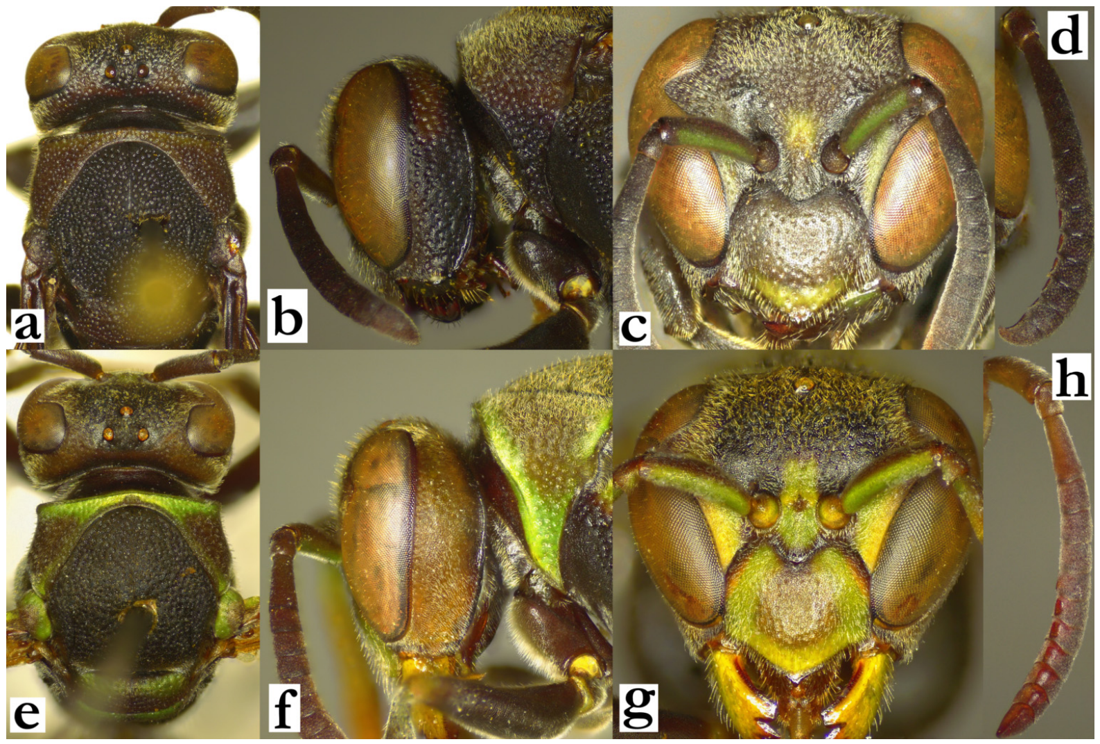
- 65. Posterior ocelli at most a diameter apart (a).
[f] Gena at mid-height 0.7–0.9× as wide as eye (b). Clypeus about as wide as long, with elongated base and parallel lateral sides (c). Face darker; clypeus black with apical green line, interantennal area and inner orbits with minimal greenish suffused markings (c).
[m] Unknown
[wl] 7.9 [7.8–8.0] mm
[n] Specific, with invert cells openings, extremely well concealed by exposing the cell bottoms outwardly; in later stages elongated, up to 30 cells wide and 50 cells long, located directly on tree trunks.........................................R. jemmae Polašek and de Beer
- - Posterior ocelli more than a diameter apart (d).
[f] Gena at mid-height as wide as or wider than eye (e). Clypeus wider than long, angled lateral margins (f). Face with numerous green-yellow areas; clypeus green with central large or multiple olivaceous-brownish spots (f).........................................66
Figure 65.
Ropalidia jemmae Polašek and de Beer, holotype, (a) head and mesosoma, dorsal view, female, (b) head and anterior mesosoma, lateral view, female, (c) head, frontal view, female; Ropalidia aras sp. nov., holotype, (d) head and mesosoma, dorsal view, female, (e) head and anterior mesosoma, lateral view, female, (f) head, frontal view, female.
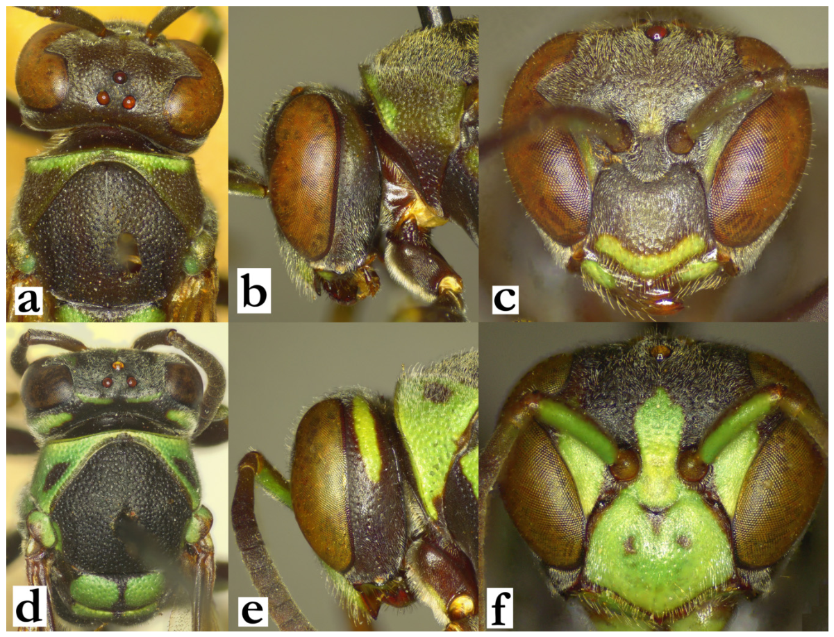
- 66. Body multi-coloured, including green, reddish, brownish and/or yellowish areas; all markings thin (a). Mesoscutum setose, median suture of mesoscutum hardly discernible (b).
[m] Scape 1.4–1.7× as wide as AF1 base; tyloids originate as AF5, terminal flagellomere apically acute.
[wl] 9.4 [8.8–9.9] mm
[n] Discoid, with paracentral petiole; cell wall grey and compact, with streaks of different grey hues; cocoon caps brownish or grey nest [62]; up to 50 cells in a nest, nests always in colonies, located on underside of rocky structures and water canal culverts or similar man-made objects............................................................R. kojimai Blommers
- - Body bicoloured, with numerous emerald-green markings on basally black(ish) head and mesosoma; T2 with elaborate and wide posterior band; T3–6 and S3–6 ferruginous (c). Median suture of mesoscutum conspicuous (d).
[m] Unknown
[wl] 9.1 [8.7–9.4] mm
[n] Discoid, up to 40 cells, with a central petiole; cell wall light grey, sturdy and comparatively thicker, made from heavily masticated material; located on the leaf underside, in proximity to the leaf central vein [63].........................................R. aras sp. nov.
Figure 66.
Ropalidia kojimai Blommers, (a) habitus, oblique lateral view, female, (b) head and anterior mesosoma, oblique view, female; Ropalidia aras sp. nov., (c) habitus, oblique lateral view, female, (d) head and anterior mesosoma, oblique view, female.
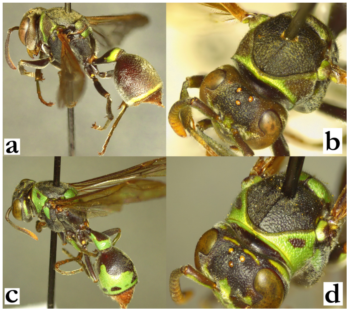
3.2. Taxonomy
- Ropalidia anarchica (de Saussure, 1853)Icaria anarchica de Saussure, 1853; “Madagascar” (MNHN).Plazi: https://tb.plazi.org/GgServer/html/03AE106DFFDD396225D7FBC9842EF119 (accessed on 21 September 2026).Number of examined specimens: 27 ♀♀, 12 ♂♂.
Type material. MNHN holds four females of Icaria anarchica de Saussure, 1853, with labels “MUSEUM PARIS, MADAGASCAR, JACQUINOT 1841”, with another label “TYPE”; one specimen has additional label “Icaria anarchica de Saussure, Type, H. de Saussure DET” and “1059 41” in blue circular label, as reported previously, treated as holotype [19].
Notes. This species is a member of the grandidieri group, characterized by the intermediate body size, longer pilosity and protruding setae on the mesoscutum, mainly yellow pronotum and a distal darkening of the forewing (Figure 21a), with a specific morphology and colour of the male antenna (Figure 21d). The common body colour pattern with large yellow areas on the pronotum usually presents no substantial taxonomic problems, but specimens from the highlands around the Perinet region can be much darker, even without any yellow markings on the pronotum; in these cases, the pronotum and tibia are suffused with green (Figure 67a).
Description. Male. Wing length 7.6–8.4 mm.
Head. Head 1.2× as wide as high in frontal view, clypeus 1.4–1.5× as wide as long, with curved upes, shallow juxtamandibular notches and acute apex (Figure 21c); clypeus base and centre shiny and sparsely punctate; mandible shiny, nearly impunctate; frons coarsely punctate (Figure 21c); gena posteriorly about 0.4–0.6× of eye width (Figure 67a); scape barely widened, 1.3–1.5× as wide as AF1 base width, longer than AF1; AF1 3.4× as long as wide, AF2 1.1–1.5× as long as wide (Figure 21d); tyloids dark brown to black, originate at AF2 (Figure 67b); terminal flagellomere 1.9–2.1× as long as wide, with weakly developed basal tubercle (Figure 21d), and subacute tip (Figure 67b).
Mesosoma. Similar to females morphologically, but dorsal side of mesosoma more coarsely punctate (Figure 19f) and fore tarsal spur I developed, longer than wide (Figure 67b).
Metasoma. T1 triangular, dorsally flattened, with a strong posterior sulcus; S7 weakly concave, with scattered shallow punctures.
Pilosity. Clypeus, frons and gena with long golden setae (Figure 21c); eyes asetose; mesosoma dorsally with setae of similar length or shorter than those on frons; metasoma with similar protruding setae, those on T2 extend over posterior lamella.
Colour. Dark brown with greenish-suffused tint, except following yellow parts: clypeus (Figure 21c; sometimes with basal brown spot), inner orbit and interantennal area (sometimes ferruginous), basal spot on mandible (Figure 21c); large spot on pronotum, except in dark cluster from central highlands that has pronotum entirely suffused in green (Figure 67a); coxa I and median half of coxa II, sometimes anterior surface of femur I with minimal yellow markings or without them entirely (in contrast to predominantly yellow mesosoma underside in R. takona sp. nov.); forewing with distal darkening, sometimes reduced to blackish trichome distally (Figure 67a); antenna dorsally dark brown, ventrally orange (Figure 21d); tyloids blackish and shiny (first few sometimes brownish); those on AF(7)8-11 with a posterior brownish area (Figure 67b).
Similar species. Some confusion is possible with R. takona sp. nov.; basal colour in females is darker than R. takona sp. nov., while males have predominantly black or olivaceous mesosoma underside (which is mainly yellow in R. takona sp. nov.). The black cluster of R. anarchica from the central highlands can be somewhat problematic, especially in comparison to an entirely black cluster of R. ranavali (de Saussure); the shape of T1 is useful in separation (Figure 1d shows the grandidieri group shape of T1). Very xanthic R. shestakowi (Schulthess) can be separated by the origin of the yellow colour on pronotum, originating from underneath the pronotal carina in R. shestakowi (Figure 19c), as opposed to dorsal parts of the pronotum in R. anarchica (Figure 19f and Figure 21a). Lastly, this species could also be confused with R. nitidula, based on the large yellow areas on the pronotum; basal body colour is light brown in R. nitidula (Figure 12b), and there are numerous morphological differences like thicker gena, shorter pilosity and less developed punctures.
Distribution. Most frequently in north-eastern parts of the islands, less common in the south (Figure 67c); also previously reported from Nosy Boraha [64].
Nest. The low risk of misidentification and similarity with the confirmed nests suggests that two previous literature sources may be referring to this species. This species is reported to prefer forested regions around Perinet; the same publication also suggests that cocoon caps are initially white, and later become dirty coloured and pulp is added to them [9]. Nests can reportedly be large, with as many as 200 [16] or even up to 340 cells per nest [9]. Nest congregations were reported on Tamarindus indicus trees, referring to hundreds of nests [16].
Genetics. One specimen was successfully sequenced (BOLD:GARO229-25/AG3G4), confirming closeness to other members of the grandidieri group.
Figure 67.
Ropalidia anarchica, (a) habitus, oblique anterior view, male, dark cluster, (b) tarsal spur I and antenna, median view, male, (c) distribution map.
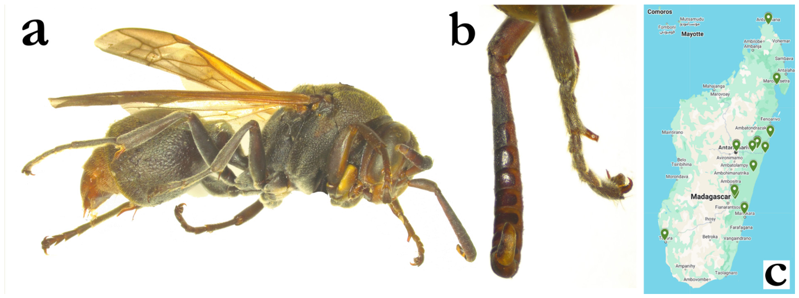
- Ropalidia andalana sp. nov. PolašekZooBank: 141E4A7D-F74C-4D87-B1F1-88D6936E0516Material examined. Holotype: “Mandena/II 2004, leg. Bulirsh”, ♂ (OLM).Paratypes: same location as holotype, 1♀ (OLM); “Rogez”, 1♂ (ETH); “Nosy Mangabe/3 January 2004, Dolin&Andreeva&Andreev”, 1♀ (OLM; DNA.AG3J8); “Ranamofana/leg. Kantner”, 1♀; “Fort Dauphin/Seyrig, XII 1936”, 1♂ (MSNV); “Rogez”, 2♀♀ (MSNV); “Perinet”, 1♀ (MSNV); “Ivandry/Seyrig, II 1940”, 6♀♀, 7♂♂; “Ivandry/Seyrig, XII 1936”, 1♀; “Fort Dauphin/Seyrig, XII 1936”, 1♀, 1♂; “Fort Dauphin/Seyrig, I 1933”, 2♀♀; “Ligabue”, 1♀, 1♂; “Ivandry/Seyrig, I 1939”, 4♀♀ (nest “P”); “Ivandry/Seyrig, I 1939”, 1♀; “Rogez”, 3♀♀; “Perinet/Seyrig, II 1936”, 2♀♀; “Fenerive/Oberthur 1897”, 1♀; “Manambato (Anove)”, 1♀; “Baie D’Antogil/ Mocquerys 1898”, 1♀; “Diego-Suarez/1893 coll Allaud”, 1♀ (all in MSNV); “Coll. MUS. TEREVUREN./MADAGASCAR:Tam/Foulpointe foret, ix. 1995, A. Pauly rec.”, 2♀♀, 1♂ (RMCA); “Coll. MUS. TEREVUREN./MADAGASCAR:Tam/Foulpointe foret, v. 1995, A. Pauly rec.”, 1♂ (RMCA); “COLL. MUS. TERVUREN/NE MADAGASCAR:Ambodivoangy, X.1961 J. Vadon” 2♀♀ (RMCA); “COLL. MUS. TERVUREN/NE MADAGASCAR:Fizono, IX.1959 J. Vadon” 1♀ (RMCA); “MADAGASCAR, Foulpointe, 31.xii.1991, A. Pauly rec.”, 1♂ (RMCA); “COLL. MUS. TERVUREN/MADAGASCAR:Fampanambo, X.1962 J. Vadon” 1♀ (RMCA); “MADAGASCAR, Foulpointe, xii.1996, A. Pauly rec.”, 1♀ (RMCA); “MADAGASCAR, Foulpointe, viii.1995, A. Pauly rec.”, 1♀ (RMCA); “FENERIVE/ 27 DEC 1955”, “McCallan/ Collector”, 1♀ (NHM); “IN FOREST/ PERINET/ 20 DEC 1955”, “McCallan/ Collector” 2♀♀ (NHM); “MADAGASCAR/ Fort Dauphin (Mandena 100m)/ 14-18.IV.1968/ K.M.G. & P.D.”, 1♀ (NHM); “MADAGASCAR/ Fort Dauphin 500m/ 15.IV.1968/ K. M. Guichard”, 1♂ (NHM).BOLD: https://portal.boldsystems.org/bin/BOLD:ADU6238 (accessed on 21 September 2026).Number of examined specimens: 34 ♀♀, 6 ♂♂.
Diagnosis. Species from formosa group, characterized by body basally brown to black, anterior part of mesoscutum impunctate, scutellum bituberculate, interantennal slit developed, female clypeus evenly punctate and male terminal flagellomere robust.
Description. Female. Wing length 11.9–14.8 mm.
Head. Head in frontal view 1.2× as wide as high; in dorsal view 2.9× as wide as long, shallowly emarginated posteriorly, with occipital carina retained dorsally (Figure 8f). Clypeus 1.2× as wide as long, with straight upes (Figure 9b); clypeus base and centre finely and sparsely punctate with matte interpuncture surface, with less defined craters apically (Figure 9b); mandible shiny, coarsely punctate; area around antennal sockets conspicuously impunctate, frons with shallow and defined punctures centrally (Figure 9b), eye sinus with smaller and more spaced punctures (Figure 9c); gena at midline 0.75 of eye width, mostly shiny, with occasional crater or puncture close to mandible (Figure 8g); posterior ocelli 1.1–1.4 ocelli diameters apart, ocelli to eye 2.3× ocelli diameter, ocelli to occipital carina 1.5 diameters apart (Figure 8f); scape longer than AF1; scape 4.3× as long as wide, AF2 1.0–1.1× as long as wide (Figure 8g).
Mesosoma. Mesosoma 1.5× as long as wide; mesoscutum anteriorly impunctate (similar to Figure 4c), posteriorly shallowly and sparsely punctate; pronotum dorsally impunctate, centrally with some larger punctures, inferior angle largely and coarsely punctate, with punctures less than a diameter apart (Figure 68a); mesepisternum superiorly coarsely punctate (with some reticulation), inferiorly with retained density, but smaller puncture size and more spacing; scutellum bituberculate, with a shiny median impunctate streak (Figure 9a); metepisternum coarsely punctate, anteriorly punctures about one diameter apart, posteriorly much sparser; median depression of propodeum striate (some rotation of specimen may be required for proper visualisation), with moderately defined lateral margins (Figure 9a).
Metasoma. T1 0.4–0.5× as wide as T2, dorsally impunctate, posteriorly with a few poorly defined punctures; T2 densely and minutely punctate, punctures on S2 proximally somewhat larger; T2 lamella punctate, with two or three rows of punctures similar in size to those on distal part of T2; remaining segments minutely punctate.
Pilosity. Body with yellowish pubescence and some protruding setae; apical third of clypeus with twice as long setae as basally; frons with yellowish setae with bent tips, equally long or longer than anterior ocellus diameter; eyes minutely setose; mesosoma with short yellowish-golden pubescence and equally coloured protruding setae; T2 with fine, yellowish setae that do extend over lamella; remaining segments with somewhat longer protruding setae.
Colour. Basal body colour dark brown to black (Figure 7e), while more xanthic specimens can have multiple suffused reddish areas on head, mesosoma and metasoma (Figure 7f); clypeus apex, inner orbits, antero-basal spot on mandible and gena variable, commonly suffused yellow-reddish-greenish, but sometimes with bright yellow spots, especially on mandible (Figure 9b); pronotum and scutellum suffused brownish (Figure 7e), metanotum black or black with bilateral yellow-green spot (Figure 7f); coxa and femora black, tibia and tarsi olivaceous-dark-green (Figure 7e); T1 black, with green(ish) posterior band, T2 in basal colour or with suffused reddish posterior band (Figure 7f); remaining metasomal segments in basal colour; wings infuscate, apical quarter yellowish, with corresponding yellowish trichome (Figure 7e,f); stigma light brown to transparent yellowish, without apical darkening or apical spot (Figure 7e,f); antenna in basal colour (Figure 9b).
Males. Resemble females in the general appearance. Wing length 11.4–12.2 mm.
Head. Head 1.3× as wide as high, clypeus 1.3× as wide as long, upes curved, without OCA, juxtamandibular lobes very weak, apex barely protruding (Figure 68b); clypeus basally and centrally shallowly and finely punctate, with few more developed punctures (Figure 68b); gena 0.4× as wide as eye, mandible shiny, nearly impunctate (Figure 68b); scape barely widened, 3x as long as wide, 1.4× as wide as AF1 base, longer than AF1 (Figure 68b); AF2 1.x as long as wide; tyloids originate at AF1, as shiny, yellow to brown (those on AF2-6 sometimes dark brown); AF2 1.4–1.5× as long as wide; terminal flagellomere very elongated, 4.5× as long as wide, with flattened dorsal margin, very developed median tyloid, distal excavation and very acute tip (Figure 9d).
Mesosoma. Similar to females. Tarsal spur I curved, 2× as long as wide.
Metasoma. S7 weakly concave, with shiny and irregular surface.
Pilosity. Similar to females, except clypeal surface with fine, equally long, silvery setae (Figure 68c; notably, these setae are much longer in very similar R. sakalava); frons setae longer than in females.
Colour. Clypeus yellow with basal black spot or vertical broadened stripe (Figure 68b), mandible yellow with remaining olivaceous postero-basal area (Figure 68b); interantennal area and inner orbits merged in yellow, sinus brownish (Figure 68b); remaining parts in basal colour; T1 sometimes with posterior yellow(-green) band, T2 with posterior reddish band; tarsi with a skipping colour pattern: proximal part black, distal light-green; antenna black dorsally, orange ventrally (Figure 68b).
Etymology. The name originates from the Malagasy word andalana, translated as “line”, referring to the median impunctate line of the scutellum, treated as a noun.
Similar species. Females of this species have a very high degree of resemblance to R. sakalava (de Saussure), and their separation is very problematic; males are easily recognizable by their antenna morphology.
Distribution. Eastern coast and central Madagascar (Figure 68e).
Genetics. One specimen was genotyped, confirming the position within formosa group. The closest BOLD distance was recorded for BOLD:ADR1779, which is Ropalidia hova (de Saussure), specimen SICOB1407-18, with 94.77% overlap.
Figure 68.
Ropalidia andalana sp. nov., (a) head and mesosoma, lateral view, female, (b) head, frontal view, male, (c) head, oblique view, male, (d) antenna, median view, male, (e) distribution map.
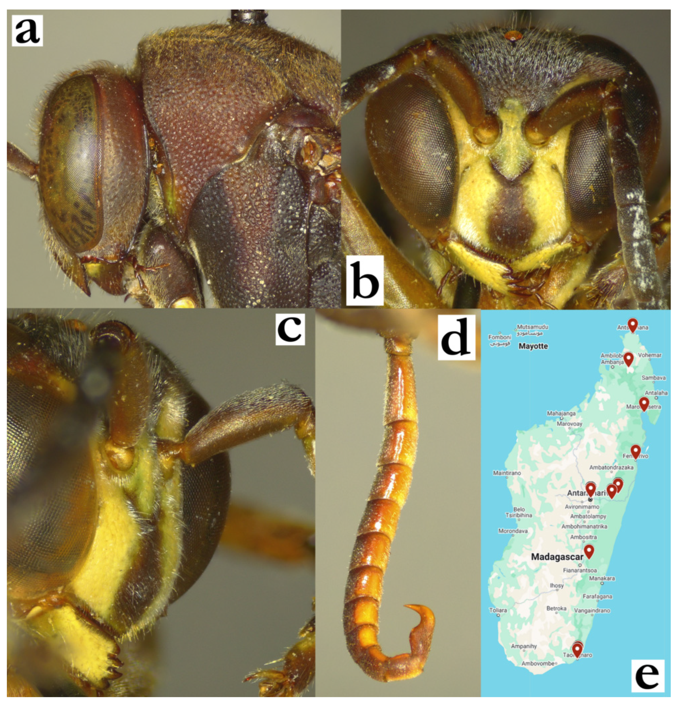
- Ropalidia antoana sp. nov. PolašekZooBank: 3DC9E778-D29A-4343-9A98-4CC854F58D4BMaterial examined. Holotype: “Andehahelo/ 1800 m/ 1.54 RP”, “INSTITUTE/ SCIENTIFIQUE/ MADAGASCAR”, ♂ (MSNV), Figure 47a,b and Figure 69d,e.Paratypes: “Andehahelo/ (1800 m)/ 1.54. (RP)”, “INSTITUTE/ SCIENTIFIQUE/ MADAGASCAR”, 1♀; “Madagascar/Sikora”, 1♀ (all in MSNV).Number of examined specimens: 2 ♀♀, 1 ♂.
Diagnosis. This species can be separated from other members of the bicincta group by mesosoma strong punctures gradient, T2 finely punctate, face in females dark and tyloids in males elevated.
Description. Females. Wing length 8.4–9.1 mm.
Head. Head frontally 1.15× as wide as high, in dorsal view 2.5× as wide as long, shallowly emarginated (Figure 50c). Clypeus 1.3× as wide as long, with straight upes and nearly absent OCA; juxtamandibular notches shallow, apex mildly pointy (Figure 50c); base and centre of clypeus impunctate and shiny, apical half with dense and poorly defined craters (Figure 50c). Frons shallowly and densely punctate, anterior third of gena with poorly defined craters, posterior impunctate; mandible punctate, occipital carina complete; posterior ocelli 1.6 diameters apart and 1.8 diameters apart from eye (Figure 69a); scape slightly longer than AF1, AF1 shorter than AF2+3+4; AF2 1.0–1.1× as long as wide.
Mesosoma. Mesosoma in lateral view 1.5× as long as high, in dorsal view head 1.1× as wide as mesosoma (Figure 69a). Disc of mesoscutum 1.05× as wide as long, with defined punctures about a diameter apart and velvety puncture interspaces (Figure 69a). Pronotum anteriorly projecting, with carina barely sinuate and evanescing at inferior angle (Figure 69b); punctures large and shallow, somewhat larger than those on mesoscutum, inferiorly more sparse and less defined, with more swollen interspaces (Figure 69b). Mesepisternum anteriorly impunctate, laterally with a specific gradient with superiorly densely punctate with punctures less a diameter apart, while inferiorly puncture size, definition and density diminish, superior punctures twice larger than inferior (Figure 69b). Metepisternum with velvety surface and occasional poorly defined craters. Propodeum distally conspicuously narrowing, with weakly defined margins; propodeum median depression with cuticular surface, shiny and impunctate, lateral sides regularly and finely striate (Figure 69c).
Metasoma. T1 pyriform, narrow and moderately developed dorsally; elongated laterally, with elongated petiole; T2 with shiny surface and dense minute punctures (Figure 47a); remaining segments shallowly and minutely punctate.
Pilosity. Body with yellowish pubescence and some protruding setae; apical third of clypeus with yellowish setae, twice as long as basally (Figure 50c); frons with straight golden setae with bent tips, equally long as anterior ocellus diameter; eyes minutely setose; mesosoma with short yellowish-golden pubescence and equally coloured protruding setae with bent tips; coxa anteriorly with short setae, coxa II and III posteriorly with somewhat longer setae; T2 with fine, yellowish setae that do not extend over lamella; remaining segments with somewhat longer protruding setae.
Colour. Basal body colour black; clypeus apex, inner orbits, antero-basal spot on mandible suffused yellow-reddish-greenish (Figure 50c); pronotum and scutellum suffused brownish (similar to male, Figure 47a), metanotum black or black with bilateral yellow-green spot; propodeum black (Figure 47b and Figure 69c); coxa and femora black, tibia and tarsi olivaceous-dark-green (Figure 47a); T1 black, with green(ish) posterior band, T2 brownish with a whitish posterior band (Figure 47a,b); remaining metasomal segments brown or ferruginous; wings yellowish, nervature brown, stigma ochre, semi-transparent, without apical darkening or apical spot (Figure 47a,b); antenna olivaceous-brown dorsally, yellowish-orange ventrally (Figure 50c).
Males. Resemble females in general appearance. Wing length 8.4 mm.
Head. Clypeus 1.25× as wide as long, upes straight, without OCA, apex rounded juxtamandibular lobes very weak (Figure 50d); clypeus basally and centrally shallowly and finely punctate, with an occasional more developed puncture (Figure 50d); gena 0.4× as wide as eye, mandible shiny, nearly impunctate; scape barely widened, 1.4× as wide as AF1 base, shorter than AF1; AF2 1.2× as long as wide; tyloids rounded, originate at AF(3)4, brownish and shiny (Figure 69d), elevated above flagellomere surface; terminal flagellomere elongated, 1.8× as long as wide, with proximally widened tyloid and beak-like, acute tip (Figure 69d).
Mesosoma. Mesoscutum coarsely punctate, mesepisternum like females; fore tarsal spur I developed, but thin (Figure 69e).
Metasoma. S7 weakly concave, with shiny and irregular surface.
Pilosity. Similar to females, except clypeal surface with fine, equally long, silvery setae, that become denser on juxtamandibular region (Figure 50d); frons setae comparatively longer than in females.
Colour. Clypeus yellow with basal black spot, mandible yellow with remaining darker postero-basal area (Figure 50d); remaining parts of the head black (except minute, suffused dark-olive area high on gena); mesosoma basally black, with suffused olive(-green) markings on pronotum and scutellum; coxa I with green-yellow distal half, coxa II quadratic green-yellow spot on lateral-distal triangle and a smaller medial marking of the same colour (Figure 47a); T1 with posterior green band, T2 with complete posterior yellow-green band, S2 with traces of green-yellow band (Figure 47a); remaining segments brownish; antenna black dorsally, orange ventrally (Figure 50d).
Etymology. The name originates from the Malagasy word antoana, translated as “steps, stairs, order, gradation”, referring to the gradual increase in puncture size along mesepisternum; the name is treated as indeclinable.
Note. The only examined male is stylopized, with a brownish female stylops between T5 and T6.
Figure 69.
Ropalidia antoana sp. nov., (a) head and anterior mesosoma, dorsal view, female, (b) head and mesosoma, lateral view, female, (c) posterior mesosoma, oblique view, female, (d) antenna, median view, male, (e) tarsal spur I, lateral view, male.
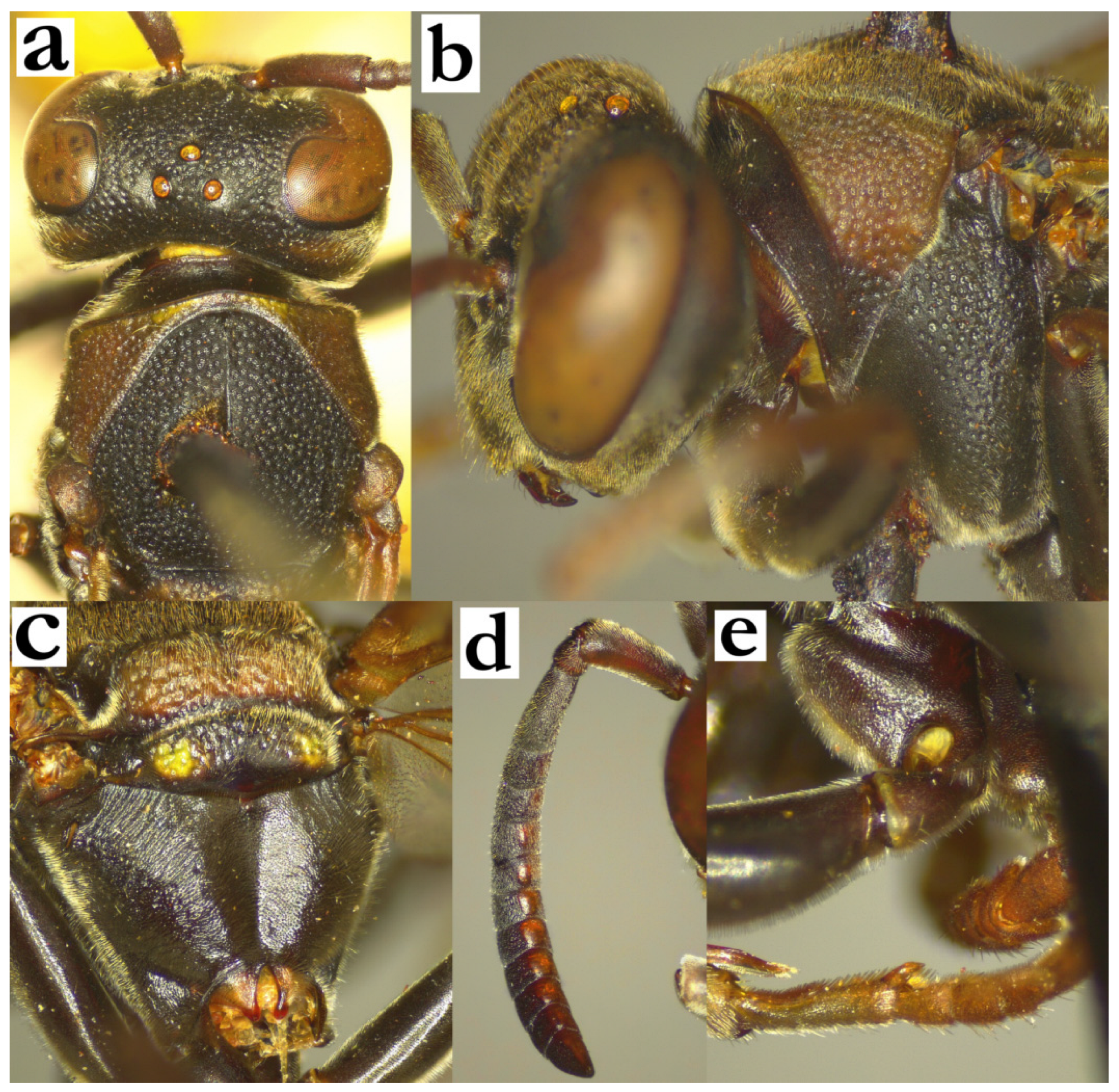
- Ropalidia aras sp. nov. PolašekZooBank: 4D5D899D-6CF3-4992-B2CE-5A4C9D70844BMaterial examined. Holotype: “Ambositra/XI 36 Seyrig”, ♀ (MSNV).Paratypes: 6♀♀, same label as holotype; “Ambositra/XI 40 Seyrig”, 1♀ (MSNV); “Fianarantsoa/III 38 Seyrig”, 1♀ (MSNV); “Ambondrombe, 1500–1850 m asl, 26 March 2001, leg. P. Burlisch”, 1♀ (OLM).Observations: Ranomafana [63]; Anjozorobe [65].Number of examined specimens: 8 ♀♀.
Diagnosis. Intermediate-sized species that resembles R. ranavali (de Saussure), and can be established by: basally black body with numerous emerald-green markings, female clypeus pentagonal, green with one to three black spots, non-confluxing punctures on pronotum, T1 posteriorly globular and T2 superficially punctate.
Description. Females. Wing length 8.7–9.4 mm.
Head. Head frontally 1.1× as wide as high, in dorsal view 2.9× as wide as long, barely narrowing behind eyes and posteriorly moderately emarginated; head 1.05× as wide as mesosoma (Figure 70a). Clypeus 1.3× as wide as long, with narrow basal part, straight upes and weak OCA, weak juxtamandibular excavations and a projecting apex; base and centre of clypeus shiny, with scattered and poorly defined small punctures, apex with denser and less defined craters; frons with moderately sized and very shallow punctures, with velvety interpuncture surface. Gena shallowly punctate close to eye, posteriorly impunctate; mandible punctate, occipital carina complete; posterior ocelli 1.45 diameter apart, 2.9 diameter apart from eye (Figure 70a). Scape 3.8× as long as wide and 1.35× as long as AF1; AF1 4.2× as long as wide; AF2 1.2–1.3× as long as wide, remaining flagellomeres wider than long.
Mesosoma. Mesosoma about 1.4× as long as high in lateral view, mesoscutum 1.05× as wide as long; entire mesosoma with shallow but well-defined punctures, denser on mesoscutum than pronotum (Figure 70a); mesepisternum shallowly punctate, punctures 1.5 diameters apart, interspaces flat and matte; metepisternum with cuticular surface and occasional shallow puncture (Figure 70b); scutellum dorsally rounded, with shallow punctures and swollen interspace; puncture 2–3 diameters apart; median carina developed, reaches half of scutellum length (Figure 70a). Metanotum coarsely punctate, especially on green areas, with shiny posterior impunctate area; metanotum surface weakly convex (Figure 70c). Propodeum distinctly narrowing posteriorly, median propodeal depression superiorly shallow, inferiorly deeper, regularly striate and impunctate; lateral surface equally striate (Figure 70c).
Metasoma. T1 pyriform, posteriorly globular, with visible terminal constriction; posterior part of T1 weakly punctate; T2 shiny, finely punctate, punctures about three diameters apart, or even four diameters apart on S2 (Figure 62e).
Pilosity. Body covered by silvery setae, except that those on T2, dorsal side of mesoscutum, and clypeus can be golden in some specimens (Figure 62e); apical third of clypeus covered by longer setae, more than twice as long as apically (Figure 52g); frons setae as long as anterior ocellus diameter; mandible and gena with short silvery pubescence, except mandibles also with longer protruding setae; eyes barely setose or asetose; mesosoma with silvery-whitish pubescence, with equally coloured protruding longer setae (commonly yellowish in R. ranavali); metasoma with longer whitish setae, which extend well over T2 lamella (Figure 70d).
Colour. Basal colour nearly black, with many emerald-green markings (Figure 62e), including clypeus (except one to three spots basally and two lines under upes), inner orbit, rhomboid patch on frons, most of mandible (except a postero-basal remaining blackish area), line high on gena, most of pronotum (sometimes with remaining postero-dorsal black spot), small marking on mesoscutum adjacent to tegula, a spot on mesepisternum, scutellum with two large spots, metanotum with two triangular spots that can be merged, ventral half of coxa I, coxa II with ventro-lateral green square, coxa III sometimes with a small lateral line (Figure 62e); femur dorsally black, ventrally green or with a smaller green patch near the distal end; tibia dorsally green, ventrally olivaceous, tarsi olivaceous (Figure 70b). T1 basally black, with substantially widened posterior green band; T2 with green pattern that resembles R. ranavali, with expanded posterior band, and remaining six black half-circles; remaining metasomal segments ferruginous; T2 lamella dark green to blackish (Figure 70d); antenna black or blackish dorsally, scape ventrally green, remaining segments ventrally orange (Figure 62e); wings faintly yellowish, nervature brown, stigma transparent and yellow, apical spot absent (Figure 62e).
Males. Unknown.
Distribution. Central highlands (Anbomdrobme, Ambositra, Anjozorobe, Ranomafana).
Etymology. Sanskrit word for “loud roar”, referring to emerald green markings, which are sufficient for a species determination already upon initial inspection; treated as a noun.
Note. Giordani Soika had labelled three specimens from the type series with white labels, bearing the unpublished name of Ropalidia nigroviridis.
Figure 70.
Ropalidia aras sp. nov., (a) head and mesosoma, dorsal view, female, (b) mesosoma, lateral view, female, (c) posterior mesosoma, posterior view, female, (d) T2 lamella, lateral view, female, (e) distribution map (red marker—specimen, grey marker—observations and literature reports).
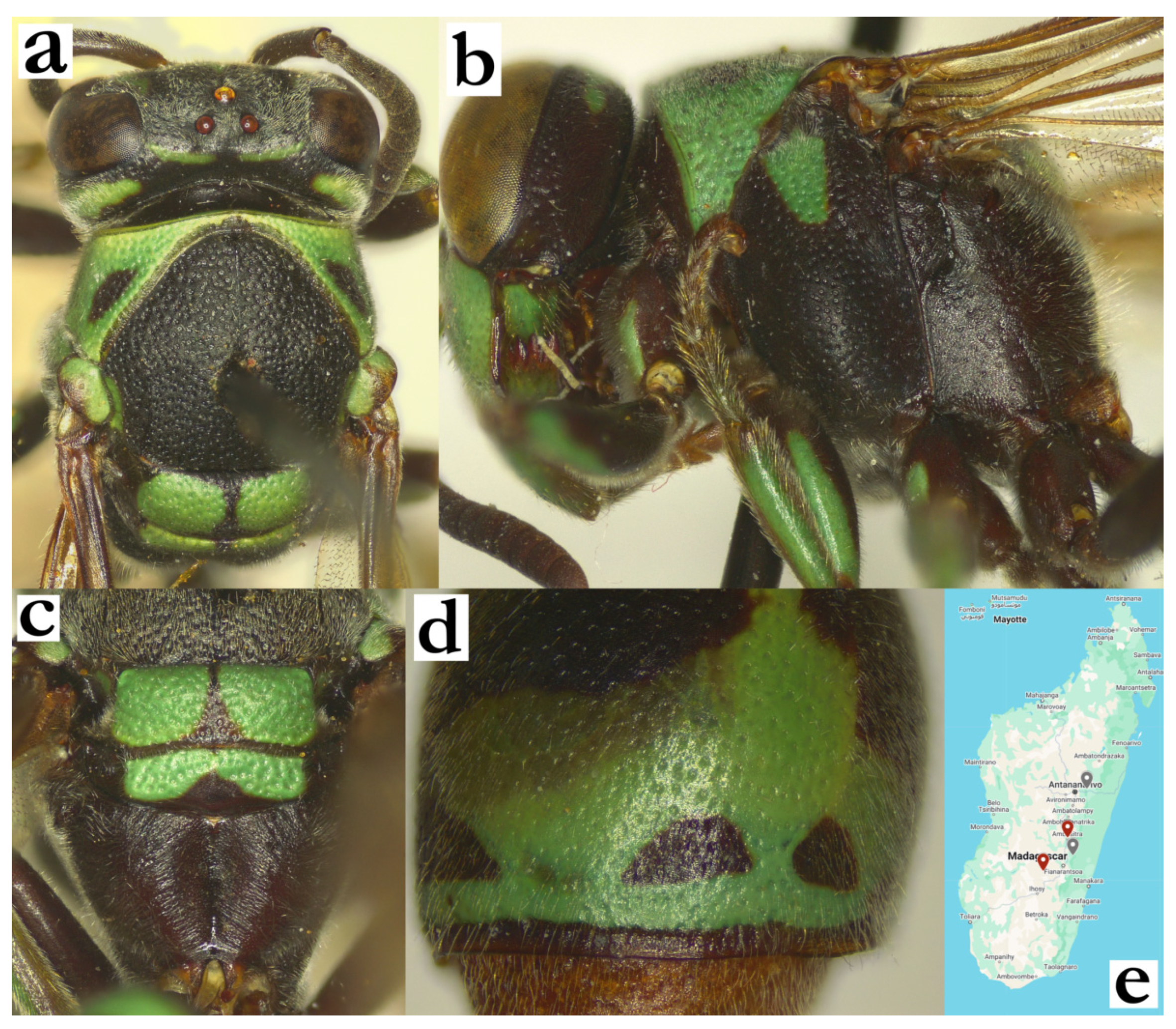
- Ropalidia atra (de Saussure, 1890)Icaria atra (de Saussure), 1890; “Madagascar/Elliot” (MHNG), Figure 71a,b.Plazi: https://tb.plazi.org/GgServer/html/03AE106DFFDC396225D7FE5D842EF37C (accessed on 21 September 2026).Ropalidia obscurior Giordani Soika 1991, syn. nov.Plazi: https://tb.plazi.org/GgServer/html/03AE106DFFD5396B25D7FC64842EF414 (accessed on 21 September 2026).Number of examined specimens: 21 ♀♀, 1 ♂.
Type material. Icaria atra (de Saussure), 1890 (Figure 71a,b)
A female from MHNG, previously examined by Blommers, who reported the holotype wing length of 14 mm [6]; re-examination suggest a somewhat smaller specimen, with wing length of 11 mm and labels “Madagascar/Elliot” on a green label, followed by a rugged paper label “486”, a blue label in de Saussure’s handwriting “Icaria atra Sss/female sign”, later designated as MHNG ENTO 00089385 (Figure 71b).
Ropalidia obscurior Giordani Soika, 1991 (Figure 71c,d)
The entire series of type specimens was examined. A single male paratype was already identified as R. cocoscola Blommers [6], suggesting that the male of R. obscurior Giordani Soika is unknown. Blommers provided an indirect comparison of R. atra (de Saussure) and R. obscurior Giordani Soika holotypes, with R. cocoscola Blommers, suggesting substantial morphological similarities, and a minimal extent of morphometric differences in these species.
The taxonomic status of this species was somewhat confusing, as Giordani Soika considered this to be the largest Ropalidia; several specimens from MSNV are labelled as R. atra, when in fact these belong to R. bevatana sp. nov. In conjunction with several more problematic taxonomic situations, it seems that Giordani Soika did not examine a collection in MHNG, thus leading to several misidentifications and a description of R. obscurior. The remaining type series females are hereby designated as synonyms of R. atra (de Saussure); one specimen designated as paratype of R. obscurior (labelled as 04945.5) is actually a female of R. grandidieri. Therefore, the male of R. atra (de Saussure) is described for the first time.
Description. Male. Wing length 7.6–8.4 mm.
Head. Head 1.3× as wide as high in frontal view, clypeus 1.5× as wide as long, with straight upes, very shallow juxtamandibular notches and obtuse apex (Figure 25f); clypeus base and centre matte and sparsely punctate by shallow and poorly defined punctures (Figure 25f); mandible shiny, nearly impunctate; frons more coarsely punctate; gena posteriorly about 0.8–1.0x of eye width. Scape widened, 1.8× as wide as AF1 base width, longer than AF1; AF1 2.9× as long as wide, AF2 1.2–1.3× as long as wide. Tyloids originate at AF3, first few brown, distally black; terminal flagellomere 2.8× as long as wide, curved, with an obtuse and dorsally darkened tip (Figure 25h).
Mesosoma. Similar to females (Figure 71e), except for developed fore tarsal spur I, which is longer than wide; femur II proximally without tubercle.
Metasoma. T1 triangular, dorsally flattened, with strong posterior sulcus (Figure 1d); S7 weakly concave.
Pilosity. Long, golden setae present on entire clypeus, frons (longer than anterior ocellus diameter; Figure 25f) and gena; eyes with short setae; mesosoma dorsally with setae of similar length or shorter than those on frons; metasoma with similar protruding setae, those on T2 extend over posterior lamella (Figure 71e).
Colour. Brown with greenish-suffused tint, except following greenish-yellow parts (Figure 71e): clypeus except basal brown spot, inner orbit and interantennal area (sometimes ferruginous), basal spot on mandible (Figure 25f); large spot on pronotum (Figure 71e), coxa I and median half of coxa II, sometimes anterior surface of femur I (in contrast to predominantly yellow mesosoma underside in R. takona sp. nov.); anterior wing with a distal darkening, which is sometimes reduced to blackish trichome (Figure 71e); antenna dorsally dark brown, ventrally orange; tyloids blackish and shiny (first few sometimes brownish); those on AF(7)8-11 with a posterior brownish area (Figure 25h).
Nest. There were no confirmed nests of this species observed at later colony stage; one observation from Nosy Be suggested an early stage nest, with simple discoid form, underneath a broad leaf [66].
Figure 71.
Ropalidia atra (de Saussure), (a) habitus, lateral view, female, holotype, (b) labels, holotype; (c) R. obscurior Giordani Soika, syn. nov., habitus, lateral view, female, (d) labels, holotype, (e) habitus, lateral view, male.
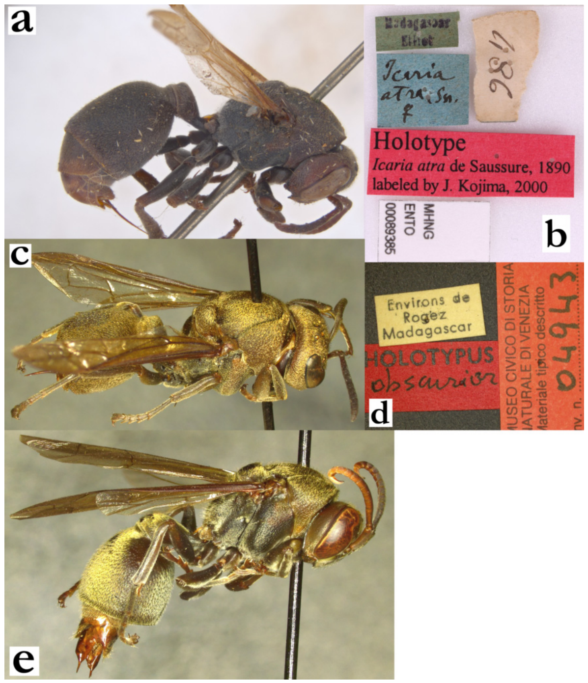
Diagnosis. This species is characterized by the elongated and slender T1, sparse pubescence and T2 minutely and scarcely punctate.
Description. Female. Wing length 8.3 mm.
Head. Head 1.2× as wide as long, clypeus 1.3× as wide as long, with straight upes and well developed OCA (Figure 41e); juxtamandibular excavation weak; base and centre of clypeus finely punctate; frons shallowly and sparsely punctate, similarly to anterior part of gena, while posterior part of gena becomes almost impunctate; mandible punctate, occipital carina complete; gena 0.5× as wide as eye (Figure 72a); ocellar triangle equidistant, posterior ocelli 1.2 diameters apart (Figure 41d); scape 1.3× as long as AF1, AF1 4× as long as wide, AF2 1.1× as wide as long.
Mesosoma. Mesosoma shallowly but coarsely punctate; punctures commonly one diameter apart (Figure 41d); pronotum inferiorly with somewhat sparser punctures; scutellum with much sparser punctures and elevated interspaces, weakly bituberculate, median carina absent (notably, there may be a subtle malformation in the holotype specimen, therefore the exact status of median scutella carina remains unclear); metanotum flattened, coarsely punctate, with comparatively smaller posterior impunctate shiny area (smaller than in R. telperia sp. nov.); metepisternum shiny and very weakly and shallowly punctate with an occasional puncture; median depression of propodeum very shallow, finely striate and impunctate; propodeum contour distally narrowing (Figure 41f), in contrast to wider contour in R. telperia sp. nov. (Figure 41c).
Metasoma. T1 slender, nearly 3× as long as high (Figure 40b), with parallel petiole, posteriorly rounded (Figure 72b); dorsally 0.3× of T2 width (Figure 41f); T2 shiny, with shallow, small and sparse punctures, about 3–5× puncture diameters apart (Figure 40b); punctures on S2 similar, but sparse, and comparatively weaker in the remaining metasomal segments.
Pilosity. Clypeus covered by fine silvery pubescence and some protruding whitish setae, which are twice longer apically; silvery pubescence also present on interantennal area and inner orbits, and converts to golden further on frons; frons also with golden protruding bent setae, which are about as long as ocellus diameter (Figure 41e). Mesosoma with short yellowish-golden pubescence and equally coloured protruding bent protruding setae (Figure 72a); T2 with fine and somewhat long setae, which barely extend over the lamella (Figure 40b); similar setae are present on remaining metasomal segments; eyes with very short setae.
Colour. Head and metasoma basally brown, mesosoma basally black (Figure 72a,b); except yellow: clypeus (with a central suffused light-brownish area), interantennal area and inner orbits, mandible (Figure 41e), lower gena (Figure 72a), dorsal part of T2 and broadened band on T2; green or greenish areas include scape underside, area close to tegula, two spots on scutellum, two merged spots on metanotum (Figure 72a,b); femora black, tibia olivaceous, tarsi brownish (Figure 72a); coxa in basal colour; ventral surface of coxa I entirely yellow, coxa II and III with lateral yellow area. Femora in basal colour (except small distal green patch on femora II and III), tibiae and tarsi olivaceous. Forewing faintly yellowish, nervature brown, stigma light brown, apical spot not developed. Anterior half of T1 in basal colour, followed by a green transitional area to posterior yellow band; lateral sides of T1 with more yellow (Figure 40b); T2 and S2 in basal colour, with widened posterior light-yellow band, which is interrupted on middle part of S2. Remaining metasomal segments brown, gradually turning ferruginous distally; antenna dorsally brown (several distal segments somewhat lighter), flagellomeres ventrally orange (Figure 72a).
Males. Unknown.
Distribution. Central highlands.
Etymology. The name originates from the name of the cryptic Malagasy group of people who inhabited the central highlands, the Beosi; the name is treated as indeclinable.
Figure 72.
Ropalidia beosi sp. nov., (a) habitus, lateral view, female, holotype, (b) habitus, dorsal view, female, holotype, (c) distribution map.
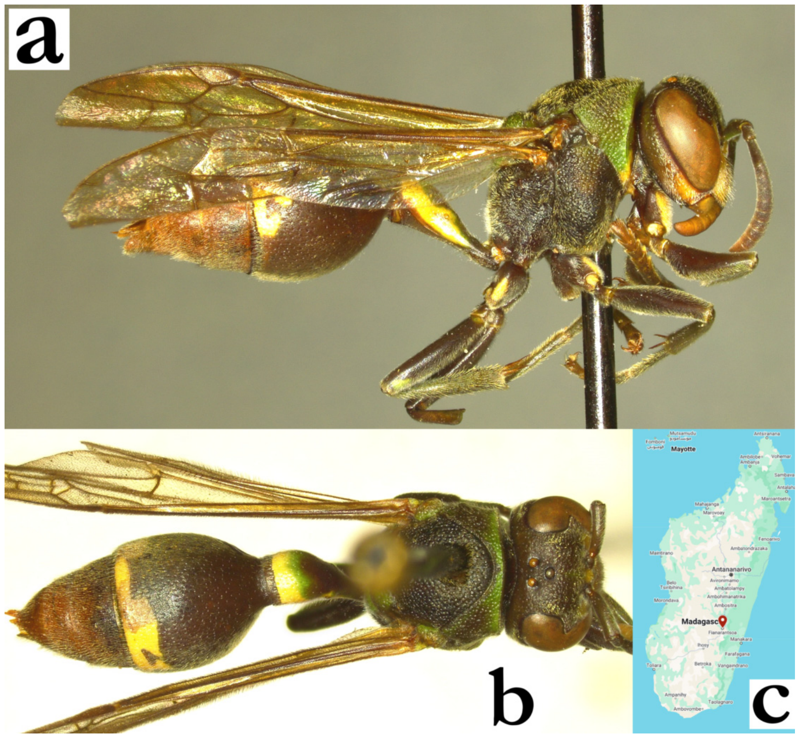
- Ropalidia bevatana sp. nov. PolašekZooBank: F6618F97-067C-446A-86E3-1B5B3006DE0BMaterial examined. Holotype: “Rogez/ O. Madagascar”, (ETH), Figure 73a–e.Paratypes: 1♀, same label as holotype; “Berafia/ Nossibe/ (Melis)”, 4♀♀ (2 ETH, 2 MSNV); “Madagascar/ V Sombirano/ A. Seyrig 33”, 2♀♀ (MSNV); “Tananarivo, Madag”, 1♀ (NNM); “MADAGASCAR/ Tulear/ Andranovory 200m/ 24.11.1968/ K.M.G.&P.D.”, 1♀ (NHM); “Coll. MUS. TERVUREN/ MADAGASCAR:Tam/ Foulpointe, foret, iv.1995/ /i.1995/ /v.1995, 5♀♀ (RMCA).Number of examined specimens: 15 ♀♀.
Diagnosis. Largest Malagasy Ropalidia, characterized by basal colour black(ish), punctures coarse, head with short brissae and gena broad and coarsely punctured.
Description. Females. Wing length 12.7–17.9 mm.
Head. Head frontally 1.2× as wide as long, in dorsal view 2.5× as wide as long (Figure 5g), broadened behind eyes and mildly posteriorly emarginate (Figure 73c). Clypeus 1.1× as wide as long, evenly and strongly convex, commonly with straight upes, weak OCA and developed juxtamandibular excavation (Figure 73b); centre and base with well-defined and coarse scattered punctures, with retained density basally; frons coarsely and densely punctate (Figure 73b); mandible coarsely punctate; gena coarsely punctate next to the eye, less defined close to occipital carina; occipital carina complete and well developed (Figure 4b). Posterior ocelli 1.7× radius apart, 4.5× diameters apart from eye and 4.3× diameters apart from occipital carina (Figure 73c). Scape very long, commonly over 5× as long as wide basally, almost twice longer than AF1; anterior surface densely punctate; AF1 4.1× as long as wide, AF2 1.5–1.6× as long as wide (Figure 73b).
Mesosoma. Mesosoma in dorsal view about as wide as long, 1.05× as wide as head; disc of mesoscutum 1.2× as wide as long; mesoscutum anteriorly shallowly punctate, puncture definition very weak (Figure 73c). Pronotum punctures coarse (Figure 73c). Mesepisternum reticulated superiorly, inferiorly with dense and coarse punctures, less than a diameter apart (Figure 73d). Scutellum densely punctate, metanotum posteriorly flattened with upwardly oriented puncture openings (Figure 6d). Median depression of propodeum deep, obliquely striate; upper part of median depression of propodeum with large punctures and striae (Figure 6d). Femora III coarsely punctate, punctures setose (Figure 6f).
Metasoma. T1 globular (larger specimens have somewhat developed posterior sulcus and thus resemble species from grandidieri group); T2 elongated, densely punctate; T2 lamella punctate by two or even three layers of punctures, with gradual and extended transition to T2 surface (Figure 5f); S2 with larger and sparser punctures and a shiny appearance.
Pilosity. Entire body covered by short black brissae, sometimes with longer protruding brownish setae (Figure 6e); clypeus with fine yellowish pubescence and protruding brownish brissae, twice as long apically (Figure 6e); frons with brown or blackish brissae, shorter than anterior ocellus diameter (Figure 6e); eyes barely setose (Figure 5g); scape with strong brissae (Figure 6e); entire flagellum setose (Figure 73b); mesosoma with short black brissae, that turn to longer setae posteriorly; metasoma covered by underlying yellowish pubescence and brown protruding brissae; brissae on T2 barely extend over posterior lamina (Figure 5f).
Colour. Basal body colour dark brown and black (Figure 73a); head and anterior mesosoma reddish; head in combination of reddish and brown, with small yellow spot on mandible (Figure 73b); mesosoma brown(-reddish), metasoma somewhat darker (Figure 73a). Wings basally light-brown, tips whitish (Figure 73a); antenna brown(-blackish) dorsally, brown-reddish ventrally (Figure 73a).
Males. Unknown.
Similar species. Some difficulties may arise in separation from R. mainty sp. nov.; in addition to characteristics provided in the key to species, there is an additional feature. The mesepisternum is densely punctured in R. bevatana sp. nov., with punctures less than a diameter apart, while in R. mainty sp. nov. punctures are more than a diameter apart.
Distribution. North-Eastern parts of the islands close to Nosy Be, the central highlands.
Etymology. The name originates from the Malagasy adverb be, translated as “large, huge”, and tana, referring to the “body, self”, treated as a noun, reflecting the largest body size of Malagasy Ropalidia.
Figure 73.
Ropalidia bevatana sp. nov., female, holotype, (a) habitus, oblique lateral view, (b) head, frontal view, (c) head and mesosoma, dorsal view, (d) mesepisternum, lateral view, (e) label.
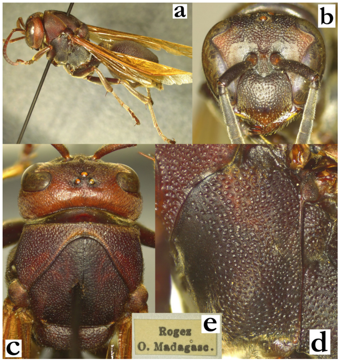
- Ropalidia bicincta (de Saussure, 1890)Icaria bicincta de Saussure, 1890; “Madagascar …Nosibe” (MHNG).Plazi: https://tb.plazi.org/GgServer/html/03AE106DFFDC396225D7FCF3842EF501 (accessed on 21 September 2026).Number of examined specimens: 2 ♀♀, 1 ♂.
Type material. De Saussure mentioned six males and females from the Robillard Collection, with another mention of Nosy Be, deposited in Hamburg Museum. Subsequently, a number of specimens were suggested as types [2], but only two specimens can be considered as such, and these are a female from Geneva and a newly assigned syntype with the same label, deposited in MSNV (Figure 49b and Figure 74a–c). Remaining specimens fail to meet the type criteria, and are therefore not considered as types [2].
Nest. No confirmed nests were observed; Vesey-FitzGerald reported two nests, one under the overhang of an earth bank in Ambanja, and the other on a coconut leaf underside in Nosy Be [9], and provided an account of a small nest with only three cells; notably, a certain caution is needed in interpretation of this claim, as the exact species determination remains uncertain, since several species were often misidentified as R. bicincta in examined collections. One possible observation on iNaturalist might indicate a nest that is inhabited by what seems to be several adult members of the bicincta group [67]. The general colour and pilosity pattern might suggest R. bicincta, but this remains speculative.
Distribution. Entire island.
Group notes. This species belongs to the bicincta group, a group of four species characterized by contrasting bands on T1 and T2 of equal or seemingly equal width and weakly developed T1 dorsally. There are some minor issues in separation of these four species, listed in the key to species. However, there are several instances of problems in separating the entire group, especially in three instances. These include melanic R. ranavali (de Saussure), which can have reduced posterior band on T2. R. ranavali has coarsely punctate gena and occiput (Figure 57d and Figure 64a), which can be very useful in separation of more finely punctate areas in all species from the bicincta group (Figure 47c). Some problems may arise in separation of R. janettae sp. nov. from R. kojimai Blommers; this is represented by the last claim in the couplet 47, with wider gena and less developed pronotal carina in R. janettae sp. nov. (Figure 47c vs. Figure 47f), while male tyloids originate on AF(6)7 in R. janettae sp. nov., vs. AF5 in R. kojimai Blommers. Unfortunately, there were no fresh specimens from this group for genetic analysis, so their relationship with other groups remains unclear. This is of special interest since some isolated species might belong to this group, most notably R. ducalis, R. nitidula or R. vitripennis, which share some characteristics of this group, and other of the fraterna group.
Figure 74.
Ropalidia bicincta (de Saussure), female, syntype, (a) habitus, lateral view, (b) habitus, dorsal view, (c) labels.
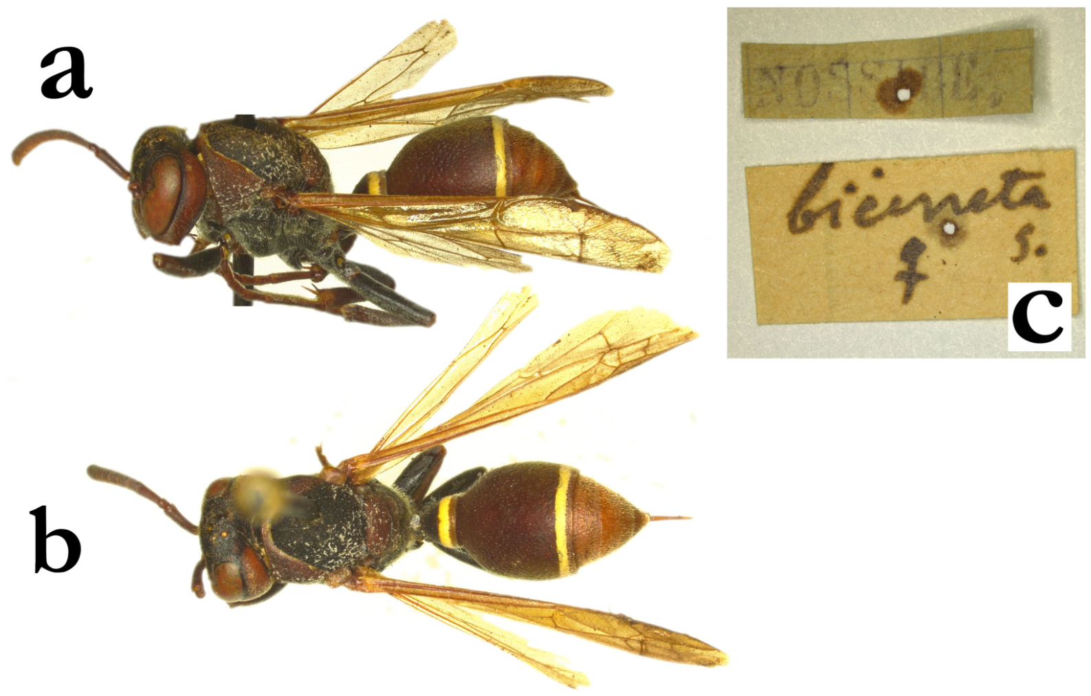
- Ropalidia blommersi sp. nov. PolašekZooBank: 4DB69CC7-F57E-4CA9-AEB9-4467AB2DC3FFMaterial examined. Holotype: “MADAGASCAR. Tam./Perinet, 1000m/2.XII.57 F. KEISER”, ♀ (NNM); Figure 75a–c.Paratypes: 5♀♀, same labels as holotype; “MADAGASCAR. Tam./Perinet/9.IV.58, F. KEISER”, 1♀ (NNM); “MADAGASCAR.D.S./Mtge d’Ambre/12.V.85, F. KEISER”, 2♂♂ (all in NNM).Observations: Iaroka Forest, Ambohimanatrika [60].Number of examined specimens: 7 ♀♀, 2 ♂♂.
Diagnosis. Darkest species from the phalansterica group, characterized by combination of T1 dorsally developed, body punctures coarser, metanotum flattened and body with reduced green-and-yellow markings.
Description. Females. Wing length 6.8–7.5 mm.
Head. Head frontally 1.25× as wide as long, in dorsal view 2.4× as wide as long, narrowing behind eyes and weakly emarginate posteriorly (Figure 75c). Clypeus 1.4× as wide as long, with mildly curved upes and weak OCA; juxtamandibular lobe well developed, apex pointy; base and centre of clypeus with large and shallow punctures, which turn to less defined craters apically (Figure 55h); frons coarsely punctate, punctures one diameter apart, anterior part of gena with even larger and coarser punctures, posterior part with smaller punctures, but in most cases punctures are shallower but retained even close to occipital carina. Gena at midpoint 1.1–1.25× as wide as eye (Figure 75a); mandible punctate, occipital carina complete, ocellar triangle equidistant or with barely wider base; gena 0.8–1.0× as wide as eye; scape 4.8× as long as wide, 1.1–1.2× as long as AF1; AF1 4.7× as long as wide; AF2 1.0–1.1× as long as wide (Figure 75b).
Mesosoma. Mesosoma punctures coarse and dense, with velvety interpuncture surface; mesoscutum with coarse and dense punctures; pronotum with similarly sized punctures dorsally and twice larger puncture in inferior angle (similarly to R. subclavata); mesepisternum coarsely punctate, metepisternum with sparse but even larger punctures; metanotum and scutellum dorsally flattened (Figure 56c), median carina of scutellum absent or barely developed in some specimens (Figure 56a); propodeum strongly striate, with retained punctures between striae; median suture dorsally widened and impunctate (Figure 56b).
Metasoma. T1 dorsally developed, about 0.5× as wide as T2 dorsally, posteriorly coarsely punctate, with long T1 lamella; T2 coarsely and directionally punctate, T2 lamella short, proximally punctate; remaining segments shallowly punctate.
Pilosity. Body with yellowish pubescence and some whitish protruding setae; apical third of clypeus with yellowish setae, twice as long as basally; frons with straight golden setae, equally long or slightly longer than anterior ocellus diameter; eyes asetose; mesosoma dorsally with yellowish-golden setae, laterally with whitish; T2 with fine, yellowish pubescence that extends over lamella, remaining segments with somewhat longer setae.
Colour. Basal body colour olivaceous-black; green area include clypeus apically and basally (commonly with a remaining dark-olivaceous central spot), inner orbits, interantennal area, mandible (Figure 55h), gena (olivaceous, sometimes suffused green inferiorly), line underneath pronotal carina (sometimes yellowish anteriorly; inferior pronotal angles yellowish or reddish; Figure 56c); yellowish-green bilateral spot on scutellum and metanotum, smaller bilateral spot on propodeum (Figure 56b), broad yellow-green band on T1 and several areas of bright yellow marks on T2 band (Figure 75a); remaining metasomal segments brownish; coxa and femur black (Figure 75a), tibia and tarsi olivaceous, terminal tarsi segments brownish; scape dorsally dark olivaceous, ventrally greenish, flagellum black; wings transparent with some lightly brownish apical spot; nervature brown, stigma brownish (Figure 75a).
Males. Resemble females in the general appearance (Figure 75d). Wing length 8.4 mm.
Head. Clypeus 1.4× as wide as long, upes straight, without OCA, apex rounded juxtamandibular lobes very weak (Figure 75e); clypeus basally and centrally shallowly and largely punctate (Figure 75e); gena 0.4–0.6× as wide as eye, mandible shiny, nearly impunctate; scape barely widened, 1.4× as wide as AF1 base (Figure 75e), shorter than AF1; AF2 1.2× as long as wide; tyloids originate at A6, brownish and shiny (Figure 75g); terminal flagellomere elongated, 1.8× as long as wide, with proximally widened tyloid and beak-like, acute tip (Figure 75g).
Mesosoma. Fore tarsal spur I developed, but thin (Figure 75f).
Metasoma. S7 nearly straight, with shiny and irregular surface.
Pilosity. Similar to females, except clypeal surface with fine, equally long, silvery setae, that become denser on juxtamandibular region (Figure 75e).
Colour. Clypeus yellow with basal greenish spot, mandible yellow with remaining darker postero-basal area. Mesosoma basally black, with suffused olive(-green) markings on pronotum and scutellum; coxa I with green-yellow distal half, coxa II quadratic green-yellow spot on lateral-distal triangle and a smaller medial marking of the same colour. T1 with posterior green band, T2 with complete posterior yellow-green band, S2 with traces of green-yellow band; antenna black dorsally, orange ventrally (Figure 75d).
Distribution. North part of the island, from river Ambre to Perinet.
Etymology. The name is given after Leo Blommers, who substantially contributed to the overall understanding of Malagasy Ropalidia.
Figure 75.
Ropalidia blommersi sp. nov., (a) habitus, oblique view, female, (b) head, lateral view, female, (c) head and anterior mesosoma, dorsal view, female, (d) habitus, lateral view, male, (e) head, frontal view, male, (f) tarsal I spur, lateral view, male, (g) antenna, median view, male.
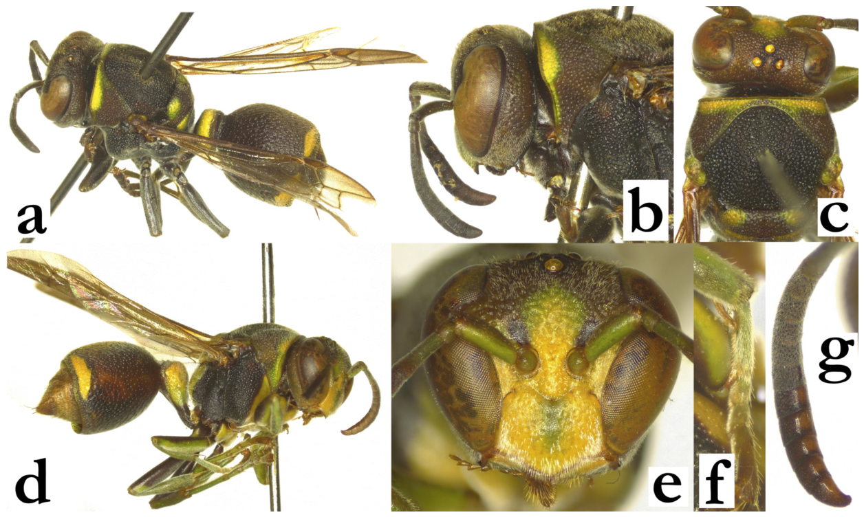
- Ropalidia bongomaso sp. nov. Polašek and DagleyZooBank: E0893A73-E9DC-477F-840D-FE3D95EB8BBBRopalidia dubia [6]; misidentification.Material examined. Holotype: “MADAGASCAR”, “Ft Dauphin, III 40”, “MUSEUM PARIS, A. SEYRIG”, “h”, ♀ (MSNV), Figure 76a–c.Paratypes: 17♀♀, 6♂♂, same label as holotype; “MADASCAR, Ivandro”, “MUSEUM PARIS, XII 38, A. SEYRIG”, 1♀ (MSNV); “MADAGASCAR”, “Ft Dauphin, II 37”, “MUSEUM PARIS, A. SEYRIG”, 4♀♀, 2♂♂ (MSNV); “MUSEUM PARIS, MADAGASCAR, BAIE D’ANTOGIL, A. MOCQUERYS 1898”, 1♀ (MSNV); “Madagascar, Special Reserve Nosy Mangabe, 3.-6.1.2004., leg. Dolin&Andreeva”, 9♀♀, 4♂♂ (OLM); “N-MADAGASKAR, Marotaonala, mastiff Ankaratra, Moravec leg. 2000”, 1♀ (OLM); “COLL. MUS. TERVUREN/ MADAGASCAR:Fampanambo/ X.1962/ J. Vadon” 1♀ (RMCA); “Coll. MUS. TERVUREN/ MADAGASCAR: TAM/ Foulpointe, foret/ iii. 1995/ A Pauly rec”, 1♀ (RMCA).Number of examined specimens: 34 ♀♀, 12 ♂♂.
Diagnosis. Small, predominantly green and yellow species, with T2 with two basal spots, eyes bulging, head and mesosoma pilosity short, posterior ocelli less than a diameter apart, simple terminal flagellomere in males and tyloids originating on AF1, exceptionally first fully developed tyloid present on AF2. Considered here as a member of the dubia group, but with a special position within it, shared with R. vanga sp. nov.
Description. Females. Wing length 7.6–7.9 mm.
Head. Head frontally 1.2–1.3× as wide as long, with bulging eyes (Figure 36f); clypeus 1.1–1.2× as wide as long, with straight upes and developed OCA, moderate juxtamandibular excavations and a projecting apex (Figure 36f); base and centre of clypeus very sparsely and shallowly punctate, with scattered and minute punctures; frons with scattered and shallow punctures, more than a diameter apart (Figure 36f); gena impunctate, occipital carina complete; oculo-malar space very short, about as long as or less than AF1 base width (Figure 38g). Ocelli large, posterior ocelli distance less than a diameter apart (Figure 39d). Scape 1.2–1.4× as long as AF1, AF1 shorter than AF2+3+4; AF2 about 1.1–1.2× as long as wide (Figure 39e).
Mesosoma. Entire mesosoma shallowly punctate; mesoscutum sparsely punctate, sparser than pronotum (Figure 39d); mesepisternum similarly punctate, metepisternum with even more spaced and shallow punctures (Figure 37e); scutellum dorsally rounded, with shallow punctures and median carina that reaches third of its length (Figure 36g); metanotum with coarser punctures and large shiny posterior impunctate area. Median depression of propodeum impunctate and shiny, with horizontally developed weak and barely visible striae (Figure 36g).
Metasoma. T1 pyriform, posteriorly somewhat globular, with longer lamella, but without visible terminal constriction (Figure 37d); posterior part of T1 with occasional puncture; T2 with minute directional punctures, about a diameter apart; distal half of T2 lamella impunctate and asetose.
Pilosity. Entire body covered by short whitish pubescence and protruding setae (Figure 33d). Clypeus with a fine and dense layer of silvery pubescence and about twice longer yellowish protruding setae; silvery pubescence continues on most of interantennal area, frons and gena, while mandible is covered by some longer yellow protruding setae; eyes asetose (Figure 36f); AF1-3 covered by some protruding setae, which are no longer than a third of the AF1 base width. Dorsal surface of mesosoma short yellowish pubescence, except dorsal margin of pronotum, with longer whitish protruding setae and propodeum, with longer white protruding setae; T1 with somewhat longer whitish setae, T2 with yellowish setae that extend over the lamella (Figure 37d).
Colour. Body commonly in green and yellow combination, predominantly green dorsally (Figure 33c and Figure 37d); clypeus, mandible, inner orbit and interantennal area fused in yellow, frons commonly green with dark brown markings around ocelli, vertex green to dark brown (Figure 33d); scape dorsally olivaceous, ventrally green; flagellum orange-ferruginous. Mesosoma with green-yellow colour pattern (Figure 38e); area under pronotal carina most commonly yellow, dorsal part of pronotum green; mesoscutum basally dark brown, with two or four thin green lines, which can sometimes be merged into larger areas (Figure 39d); mesoscutum rarely entirely green; both mesepisternum and metepisternum most commonly anteriorly brown, posteriorly yellow, but can be completely green or yellow; scutellum and metanotum entirely green (except median carina); propodeum with large green areas and only a thin remaining dark brown perpendicular median area (Figure 36g). All coxa green or yellow, femora sometimes proximally with dark brown area, distally green or with suffused yellow antero-distal patches; tibia olivaceous with dorsal green patches, tarsi gradually converting from olivaceous into ferruginous. Wings completely transparent, except minimal yellowing anteriorly; nervature brown, stigma yellowish, apical spot faintly yellowish, occupying no more than the anterior fourth of the marginal cell (Figure 37d). T1 mostly green, with remaining dorsal dark brown elongated patch; T2 brown to olivaceous, with two large basal yellow (or green) spots and a moderately widened posterior band, which is no more than twice wider than the T2 lamella; remaining metasomal segments with suffused yellow of green bands.
Males. Resemble females in the general appearance. Wing length 7.5–7.9 mm.
Head. Head frontally 1.35× as wide as long, clypeus 1.15× as wide as long, upes straight and short, OCA well developed, juxtamandibular lobes very weak, apex rounded (Figure 76a); clypeus very finely punctate, with an occasional conspicuous puncture underneath pubescence (Figure 76a); gena 0.3–0.4× as wide as eye, mandible shiny and impunctate; scape barely widened, 1.3× as wide as AF1 base, about 1.3–1.4× as long as AF1; AF1 3.0–3.2× as long as wide, AF2 1.3× as long as wide (Figure 76b); tyloids originate on AF1 (occasionally very weak on AF1, just a thin shiny streak), first few more strongly elevated and narrow, distal ones widened (Figure 76c); terminal flagellomere 1.4–1.5× as long as wide, apically obtuse (Figure 76b).
Mesosoma. Fore tarsal spur I weakly developed, small and thin.
Metasoma. S7 mildly concave.
Pilosity. Similar to females, except clypeal surface with fine, short, silvery setae and weakly protruding setae apically.
Colour. Similar to females, with head and mesosoma underside with more yellow areas.
Notes. Blommers [6] reported that male tyloids originate on AF6 (segment VIII in his classification), but this is an error; the same publication [6] shows Figure 84, where tyloids are clearly visible on AF4 and more distal flagellomeres, while flagellomeres AF1-AF3 have subtle widenings, suggesting a present of tyloids on these, as well. One examined male from Blommers’ collection has tyloids originating on AF1, although that exact specimen has the first tyloid incomplete, extending only along half of flagellomere length.
Nest. The nest of this species was already described [6], suggesting an elongated shape, up to 30 cells, in a nest formula of 2×n (Figure 88 in [6]), and other examples [68]. The same article also reported that some cells were removed from the nest, and suggested that this is probably a reflection of the lack of locally suitable nesting material [69]. The additional observed nests are in line with this report, suggesting the same general form (although some nests seem to have an apical widening and have more than two cells of width [70]. The nest petiole is heavily applied with the glandular excretion, due to the glazed and shiny surface; the commonest nest location is the leaf underside, but some nests are built on branches [69].
Etymology. The name originates from the Malagasy word bongo, translated as “swollen, bulging, mound”, and maso “eye”, treated as a noun.
Figure 76.
Ropalidia bongomaso sp. nov., (a) head, frontal view, male, (b) antenna, dorsal view, male, (c) antenna, median view, male.
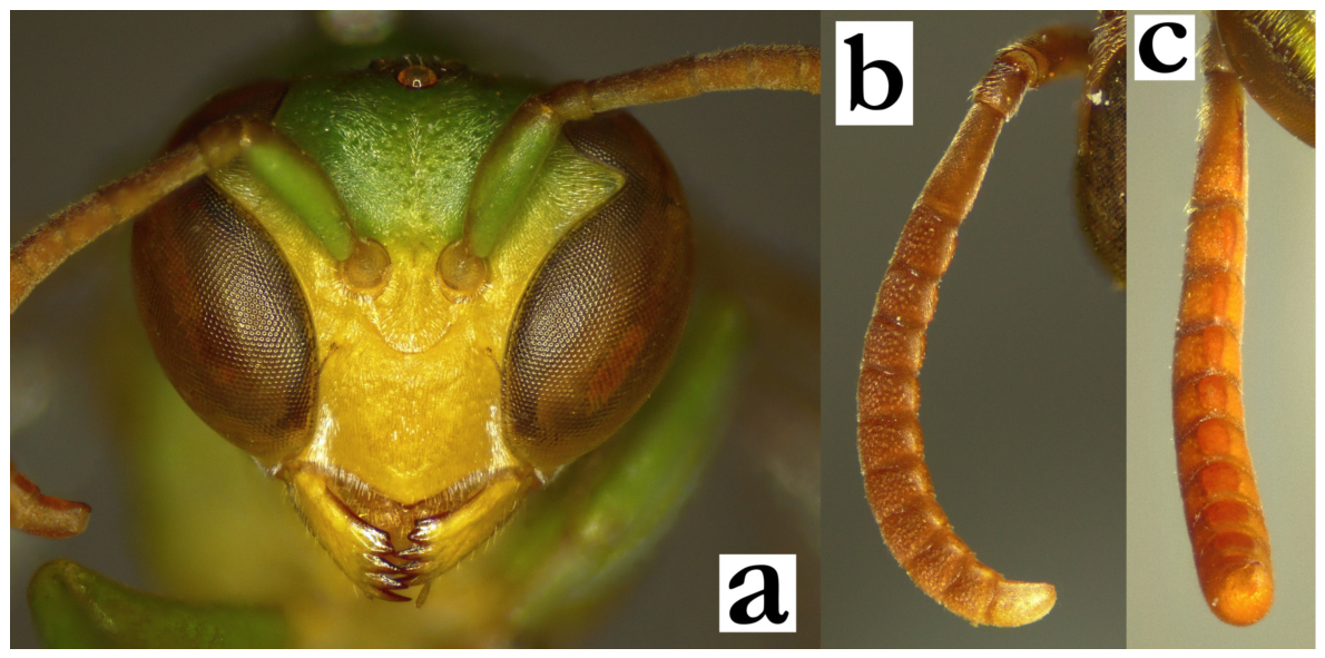
- Ropalidia carinata (de Saussure, 1890)Icaria carinata de Saussure, 1890; “Madagascar”, Collection de Saussure (MHNG)Plazi: https://tb.plazi.org/GgServer/html/03AE106DFFDC396525D7FA65842EF29D (accessed on 21 September 2026).Number of examined specimens: 10 ♀♀, 5 ♂♂.
Notes. Easily distinguishable by the wide T1 (Figure 77a), with shallow posterior sulcus (Figure 77b), basally black and brown body, with two bright, nearly equally wide posterior bands on T1 and T2 (Figure 17a). Both females and males share the specific six-spotted yellow patterns on the face (Figure 17b,c), with more yellow in males. In addition, males have bicolorous tyloids, offering sufficient determination features (Figure 17d). Males have yellow markings on coxa I and II (Figure 77d) and weakly developed tarsal I spur (Figure 77c).
Similar species. The colour pattern may resemble any of the bicincta group species, especially in case of unusually shaped T1. In these cases the T2 lamella may serve to separate the species; while species of the bicincta group have impunctate or barely punctate lamella, it is punctate by two or even three rows of punctures in R. carinata (Figure 77e). Blommers established that this species is easily recognizable and that there are no substantial determination problems [6].
Nest. The nest was reported located in Casuaria foliage at Majunga, with only four cells in a nest, three of which had already hosted an egg [9]. Subsequent reports suggests nests on Aframomum or Pandanus, often with multiple petioles, implied as the additional protection of the nest in more windy environments [6].
Distribution. All of the examined specimens were collected in the eastern coastal areas, while literature reports and observations also suggest that this species may be present elsewhere (Figure 77f).
Figure 77.
Ropalidia carinata (de Saussure), (a) T1, dorsal view, female, (b) T1, lateral view, female, (c) habitus, lateral view, male, (d) tarsal I spur, lateral view, male, (e) T2 lamella, lateral view, female, (f) distribution map (red marker—specimen, grey marker—observations and literature reports).
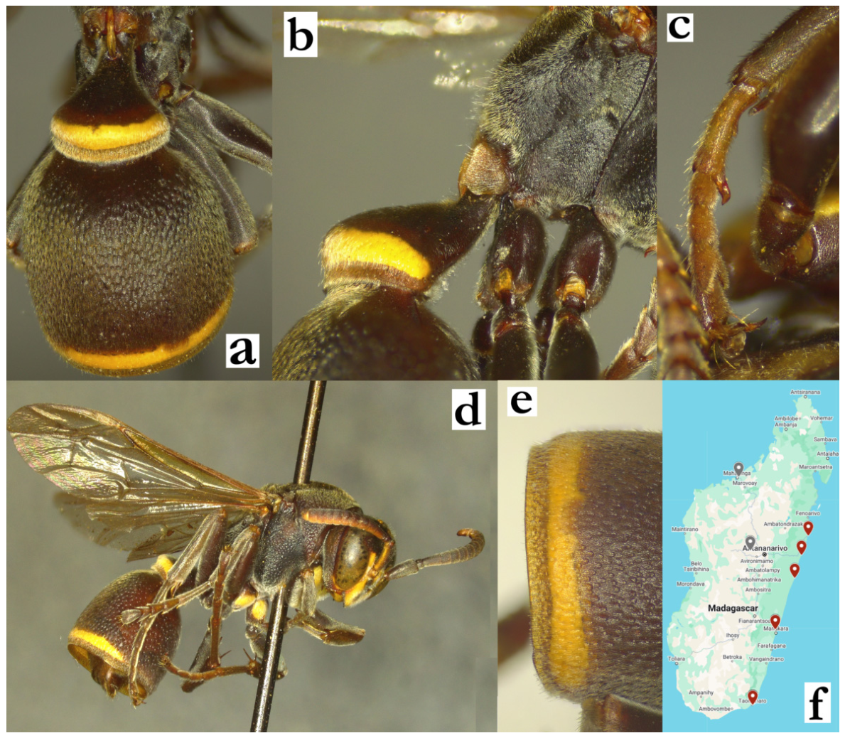
- Ropalidia cauponae Blommers, 2012Ropalidia cauponae n. sp., Blommers 2012: 146; ♂♀, nest, biology, Madagascar; holotype NNM.Number of examined specimens: 59 ♀♀, 14 ♂♂.
Type material. Two paratypes of this species were examined, from nests 71.33 and 71.34 (Figure 78a,b). The entire type series is represented by nearly uniformly basally greenish colour pattern.
Notes. One of the recently described species, similar to R. fraterna (de Saussure) (Figure 31), but with larger body size. Specimens of this species in the existing collections were often determined as R. fraterna or R. madecassa (=galimatia), even by de Saussure, reflecting similarities among them. This species exhibits a very diverse colour pattern. The commonest is the type series pattern (Figure 78a,c), but there are also more xanthic specimens (Figure 27c and Figure 29b). Males have clypeus base coarsely punctate and setose eyes (Figure 78d).
Nest. Blommers reported that nests are often placed on man-made objects, frequently in small colonies [6], which was confirmed in the observations of this species [71]. In later stages the nest becomes droplet-shaped, with petiole placed at the top, and cocoon caps placed in order [72], similar to R. galimatia. Storage of honey was reported in the oldest cells of this species [6]. This was also recorded in R. galimatia and R. laureliniana sp. nov., suggesting that more species from the fraterna group may be involved in honey storing.
Similar species. The species description suggests similarity with R. grandidieri, but general appearance of this species may resemble other species even more. The specimens of R. cauponae can easily be misidentified as R. shestakowi, since both species have a similar body size and colour pattern. The defining criterion in these cases is T1 shape, which is globular in the entire fraterna group (Figure 27c,d) vs. infundibular and triangular in grandidieri group (Figure 15a,b). Some problems can arise in separation from R. fraterna, with features useful in separation listed in key couplet 29. However, a substantial problem is separation of females from very similar R. zandry sp. nov. There are several features listed in the key to species, but some specimens can be very challenging due to mixture of characteristics from both species. This includes the colour pattern, which can be misleading, having in mind an extreme variability of R. cauponae. For example, one colony exhibited a large female (presumably queen) and one worker in the common colour pattern for R. cauponae, with three additional workers having the colour pattern of R. zandry sp. nov. [73].
Figure 78.
Ropalidia cauponae Blommers, (a) habitus, lateral view, male, paratype from the nest 71.34, (b) labels, the same paratype, (c) habitus, lateral view, female from the nest 71.35, (d) head, frontal view, male.
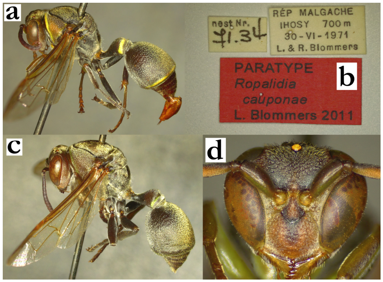
- Ropalidia cincinnata Blommers, 2012Ropalidia cincinnata n. sp., Blommers 2012: 172; ♂♀, nest, biology, Madagascar; holotype NNM.Number of examined specimens: 17 ♀♀, 2 ♂♂.
Type material. Two paratypes of this species were examined, one from the nest 73.27 and the other from the nest 73.26 [6].
Notes. A member of the bicincta group, characterized by the elongated and irregular setae on the head and mesosoma, complete occipital carina in females and absence of tarsal I spur in males, with elevated proximal tyloids. Some problems may arise in separation of females of R. bicincta, with characters provided in the key to species. In some instances, their separation may be difficult; in these cases characters that may help include posteriorly globular T1 (Figure 79a; more flattened in R. bicincta, Figure 48a).
The most interesting specimen is a male from the Sikora collection (“Madagaskar/Sikora 1896”), which has entirely short dorsal pubescence (Figure 79b). Although initially this specimen presented a substantial enigma, the morphological characteristics (antenna, tyloid and clypeus) unequivocally suggest this is R. cincinnata. This further warrants care in separation from R. bicincta, suggesting that some specimens may exhibit uncommonly short pilosity.
Distribution. Central highlands (Figure 79c).
Figure 79.
Ropalidia cincinnata Blommers, (a) T1, oblique view, female, (b) head and anterior mesosoma, oblique view, male from Sikora collection with very short head and mesosoma pubescence, with antenna shown, (c) distribution map (red marker—specimen, grey marker—observations and literature reports), (d) nest [74], (e) nest [75], (f) nest [75].
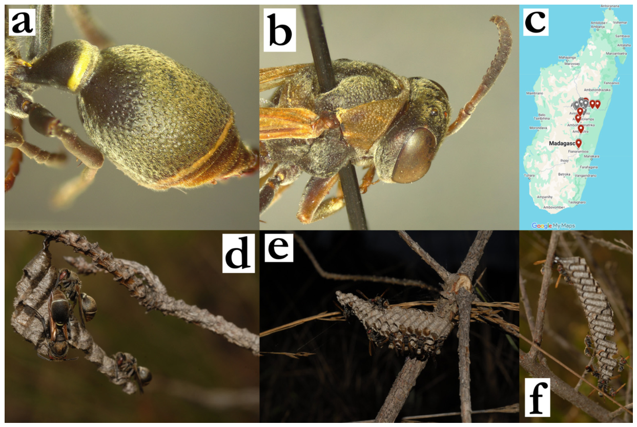
- Ropalidia cocoscola Blommers, 2012Ropalidia cocoscola n. sp., Blommers 2012: 144; ♂♀, nest, biology, Madagascar; holotype NNM.Number of examined specimens: 2 ♀♀, 1 ♂.
Type material. Two paratypes of this species were examined (Figure 80a,b), with another specimen from MSNV that was indicated as conspecific, but not labelled by Blommers.
Notes. An intermediate-sized member of the grandidieri group, characterized by the basally black body colour, silvery setae and coarse puncture pattern. Besides type specimens, only a few additional ones were examined, suggesting that it may be very rare. Blommers reports small nests concealed in palm tree holes [6].
Figure 80.
Ropalidia cocoscola Blommers, female, paratype, (a) habitus, lateral view, (b) labels.
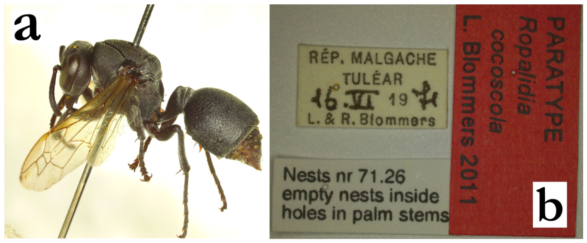
- Ropalidia constitutionalis (de Saussure, 1890)Icaria constitutionalis de Saussure, 1890; “MP/Madagascar/Goudot 1839” (MNHN).Plazi: https://tb.plazi.org/GgServer/html/03AE106DFFDB396525D7FCD2842EF527 (accessed on 21 September 2026).BOLD: https://portal.boldsystems.org/bin/BOLD:ADO7028 (accessed on 21 September 2026).Number of examined specimens: 2 ♀♀.
Notes. This species belongs to a group of species with posteriorly excavated gena and shorter T2 compared to S2, alongside R. rampa sp. nov. and R. pulchella (de Saussure). In addition, these three species have strongly dorsally developed T1 (Figure 26d and Figure 81a), widely placed posterior ocelli (Figure 81b) and translucent green T2 lamella (Figure 81c). The only account of a male suggests that males have a simple terminal flagellomere and a green scape underside [14]. This taxon remained rather obscure in the subsequent publications; the key to species by Giordani Soika [4] listed females, without males, and that publication provided no further account of this species. Blommers did not mention it at all [6], suggesting that this may be a very obscure taxon. In total, only two females of this species were examined.
Distribution. De Saussure reports it from the central highlands (Imerina), having examined three females [14], suggesting a wide distribution pattern.
Genetics. The closest BIN to this species is BOLD:ADI1183, one that belongs to assumed dark colour pattern of R. phalansterica (RNOCF371-17; photograph-based species determination). The UPGMA tree suggested a complex relationship with R. subclavata.
Figure 81.
Ropalidia constitutionalis (de Saussure), (a) T1 and T2, dorsal view, female, (b) head and anterior mesosoma, dorsal view, female, (c) T2 lamella, lateral view, female.
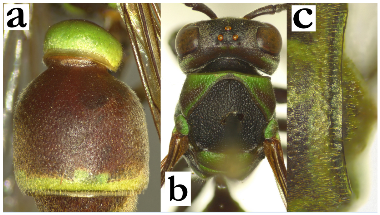
- Ropalidia dubia (de Saussure, 1890)Icaria dubia de Saussure, 1890; “MUSEUM PARIS/MADAGASCAR/BERNIER 4175-34” (MNHN).Plazi: https://tb.plazi.org/GgServer/html/03AE106DFFDA396725D7FE0C842EF160 (accessed on 21 September 2026).Photographs: https://science.mnhn.fr/institution/mnhn/collection/ey/item/ey6587 (accessed on 21 September 2026).Icaria pomicolor de Saussure, 1890; “Museum Paris/Madagascar/Goudot 1839” (MNHN).Photographs: https://science.mnhn.fr/institution/mnhn/collection/ey/item/ey6588 (accessed on 21 September 2026).Number of examined specimens: 19 ♀♀, 9 ♂♂.
Type material. The taxon named R. dubia (de Saussure) is the most commonly misclassified among all Malagasy Ropalidia species. The species was described in 1890 as Icaria dubia, head-to-head with I. pomicolor [13]; the description suggests that T1 is shorter in I. pomicolor. Both types were examined, confirming that previously established synonymy [17]. The holotype of R. dubia is deposited with a reference EY6587.
The key by Giordani Soika [4] listed R. dubia, but that is actually R. saussurei; scape and pedicel are reported as covered by longer setae in females and terminal flagellomere drawing resembles the shape of R. saussurei. Blommers managed to link a female and a male of a taxon that he considered R. dubia [6]. However, the original description reports a larger body size, with 11.5 mm of body length or even 12.5 mm for Icaria pomicolor, which is substantially larger than sizes reported by Blommers, who reported body length of 9.5 mm and wing length of 7.5 mm [6]. The comparison to R. dubia holotype suggests that the specimens determined by Blommers belong to a different species, described here as R. bongomaso sp. nov.
Notes. This species is characterized by the intermediate to larger body size, with strong green or yellow body colour (Figure 82a), longer setae, notably those on T2 that extend over the lamella (Figure 82b) and a specific dark green or even blackish dorsal markings on femora II and III (Figure 35a). In addition, males have very specific shape of terminal flagellomere and are described for the first time (Figure 35c).
Description. Males. Wing length 9.1–10.9 mm.
Head. Head frontally 1.2× as wide as long, clypeus 1.3× as wide as long, with elongated and curved upes, weak OCA, shallow juxtamandibular lobe and pointy apex (Figure 35b); clypeus with evenly distributed intermediate punctures on basally matte surface; frons with somewhat larger punctures, about 3–4 diameters apart; gena mostly impunctate and matte, sometimes with several poorly defined punctures close to inferior eye marking; gena at the widest 0.9–1.2× as wide as eye; mandible with poorly defined craters basally, on shiny surface; occipital carina complete, weakly sinuate laterally, and deeply excavated dorsally; ocelli large, equidistant or barely acute forwards; eyes asetose or with barely visible setae; scape widened, twice as wide as AF1 base, 3.1× as wide as long, 1.4× as long as AF1; AF2 1.2–1.5× as long as wide (Figure 35b). Tyloids originate on AF2 or AF3 (in that case, those on AF2 are only visible in the distal half of flagellomere), they are light brown and setose (Figure 35c and Figure 82d); terminal flagellomere 2.7–3.2× as wide as long, with very developed tyloid with median tubercle, and black and acute apex (Figure 35c); some specimens can also have median tubercle or even AF10 blackened.
Mesosoma. Mesosoma with shallow punctures pattern; pronotum with shallow punctures about a diameter apart; mesoscutum punctures more than a diameter apart; mesepisternum with sparse and shallow punctures, similar on metepisternum, but sparser. Scutellum dorsally rounded, shallowly punctate, with median carina that extends about one third of its length; metanotum shallowly and sparsely punctate; median depression of propodeum shiny, impunctate and densely striate; fore tarsal spur I developed, about 2.2–2.9× as long as wide.
Metasoma. T1 wide, dorsally triangular, laterally rounded, with weak posterior constriction; T2 and S2 shallowly punctate, about one diameter apart on T2 and further apart on S2; T2 lamella wider on T2 than S2, punctate; remaining metasomal segments with weakly shallowly punctate. S7 concave, with shiny surface and scattered poorly defined craters.
Pilosity. Clypeus with short whitish setae of equal length; frons with yellowish or whitish straight setae, about as long as ocellus diameter. Mesosoma with short yellowish pubescence and longer protruding setae; T2 with short yellowish pubescence and longer protruding setae that extend over lamella; remaining segments with longer pubescence than that on T2 and S2.
Colour. Basal body colour olivaceous-brown to black with numerous and large yellow areas (Figure 82c); face yellow (Figure 35b), area around lateral ocelli brown to black, gena yellow, dorsal area under occipital carina sometimes black; scape dorsally yellow, with brownish area distally and proximally, ventrally yellow to brownish; flagellum dorsally darkened or even blackish, ventrally orange to brownish (Figure 35c). Entire mesosoma commonly green, except narrow remaining area emarginating mesoscutum black, upper part of mesepisternum and a thin median line of propodeum black (Figure 82c). Coxa green-yellow, dorsal half of femur black, legs otherwise green or yellow, with brownish tarsi; wings barely yellowish, nervature brown, stigma yellow and translucent, apical spot absent. T1 distally green or yellow; T2 mostly green or yellow, with some remaining darker areas centrally; S2 variably coloured; remaining metasomal segments brown or suffused with green or yellow.
Nest. This species is building discoid or irregular nests, with petioles located apically in the early colony stages. In later colony development stage an additional lobe is added, leading to the paracentral petiole location. The nest are initially between 20 and 30 cells, in later stages close to 70. Cell wall is built from heavily masticated material, with minute wall fragments joined together by the salivary excretion. Cells are 3.5× as long as wide, with uneven opening margin. Cocoon caps are elevated high above the cell margin, initially light brown, later dark brown to black, sometimes with added cell wall material to their surface. Nests are located on branches, leaves or directly to the tree trunk, in the area of cleared moss [54,55,56].
Distribution. Eastern and central Madagascar.
Figure 82.
Ropalidia dubia (de Saussure), (a) head and mesosoma, dorsal view, female, (b) T2, lateral view, female, (c) habitus, dorsal view, male, (d) antenna, median view, male.
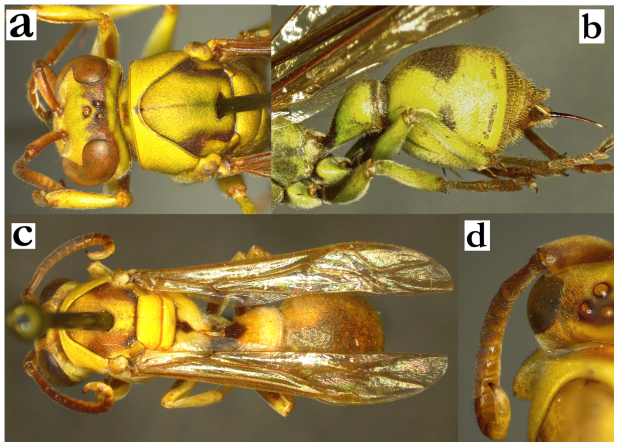
- Ropalidia ducalis (de Saussure, 1900)Icaria ducalis de Saussure, 1900; “Madagascar” (MHNG).Plazi: https://tb.plazi.org/GgServer/html/03AE106DFFD9396725D7FE87842EF23C (accessed on 21 September 2026).Number of examined specimens: 3 ♀♀, 2 ♂♂.
Type material. Two females from MHNG are considered as types [2].
Notes. A large species characterized by brownish-olivaceous-reddish basal colour (Figure 83a), large yellow areas, anteriorly impunctate mesoscutum (Figure 83b) and long golden pubescence. Males are described here for the first time.
Description. Male. Wing length 12.2–14.3 mm.
Head. Head 1.4× as wide as high, clypeus 1.5× as wide as long, with convex surface, parallel lateral sides, curved upes and an subacute apex; juxtamandibular excavations shallow (Figure 2c); entire clypeus finely punctate, obscured by the setae; frons shallowly punctate, gena finely, obscured by setae; mandible similarly punctate as clypeus; eyes asetose, occipital carina complete, ocelli large, acute forwards. Scape somewhat broadened, about 1.8× as wide as AF1 base width; AF1 shorter than scape, 3.8× as long as wide, AF2 1.5–1.8× as long as wide (Figure 2c). Tyloids shiny and brown, originate at AF(4)5 (Figure 83e), those on AF(8)9-11 dorsally elevated (Figure 2d); terminal flagellomere very elongated, 3.8–4.1× as long as wide, proximally curved, distally straight, with median tubercle and an obtuse tip (Figure 2d).
Mesosoma. Mesosoma shallowly and finely punctate; mesoscutum anteriorly impunctate (Figure 83b), pronotum dorsally shallowly punctate, inferiorly with more defined punctures (Figure 2a); median carina of scutellum developed (Figure 83c); median depression of propodeum shiny, upper half with an occasional and shallow puncture, lower half moderately striate (Figure 83c). Fore tarsal spur I well developed, wide and long, substantially darker than tarsal segment.
Metasoma. T1 globular, with marked posterior constriction (Figure 83d); S7 weakly concave.
Pilosity. Entire body covered by longer golden hairs, which obscure the observation of underlying structure on head and mesosoma (Figure 2c).
Colour. Basal body colour is brownish-olivaceous-reddish, with numerous yellowish-green markings on the body (Figure 83d). Clypeus, interantennal area and inner orbits yellow, mandible yellow with a remaining postero-basal, suffused olivaceous-green area; gena yellow (Figure 2c). Pronotum mostly greenish-yellow, with only infero-posterior, sometimes with a suffused remaining darker area (Figure 2a). Mesepisternum with patchy greenish-yellow markings, scutellum with two greenish spots that occupy the majority of its surface, except a thin complete line that separates them in two. Metanotum with two smaller greenish-yellow spots, that are not attached to its margins. Metepisternum and propodeum in basal colour. Coxa I and II with greenish-yellow markings, ventral surface of femora olivaceous-green, dorsal darker (Figure 83d). Tibia and first two tarsal segments olivaceous, remaining tarsal segments yellow with distal darkening, largest on terminal tarsal segment (Figure 83d). Metasoma entirely in basal body colour, except segments 3–7 sometimes ferruginous. Wings barely yellowish, nervature brown, stigma light brown and semi-transparent, apical spot not developed (Figure 83d). Scape brown dorsally, green ventrally; remaining segments dark brown dorsally, with a greenish-yellow-orange underside (Figure 2c).
Similar species. Some taxonomic problems are possible with R. hova, which is another member of the formosa group with large yellow areas. In these cases, yellow pubescence is the main characteristic (longer in R. ducalis), in addition to female clypeus colour (yellow with basal olivaceous spot in R. ducalis (Figure 2b) in comparison to mainly black, with apical green or yellow band in R. hova). T1 is posteriorly rounded in R. ducalis (Figure 83d), in comparison to a more triangular shape in R. hova (Figure 7a). Males are easily separated by the antenna morphology (Figure 2d).
Group membership. Based on the size, this species is considered as a member of the formosa group. Two more characteristics fit this proposal, namely gradual and punctate transition of T2 surface to T2 lamella, and an interesting male leg colour pattern, with femur, tibia and first tarsal segment olivaceous, and more distal segments proximally yellowish, distally brownish (Figure 83d). T1 shape is somewhat more globular, but posterior sulcus is wide and shallow, in disagreement with those in the grandidieri group (Figure 1d).
Distribution. Eastern parts of Madagascar.
Figure 83.
Ropalidia ducalis (de Saussure), (a) habitus, lateral view, female, (b) head and mesoscutum, dorsal view, female, (c) scutellum, dorsal view, female, (d) habitus, lateral view, male, (e) antenna, median view, male.
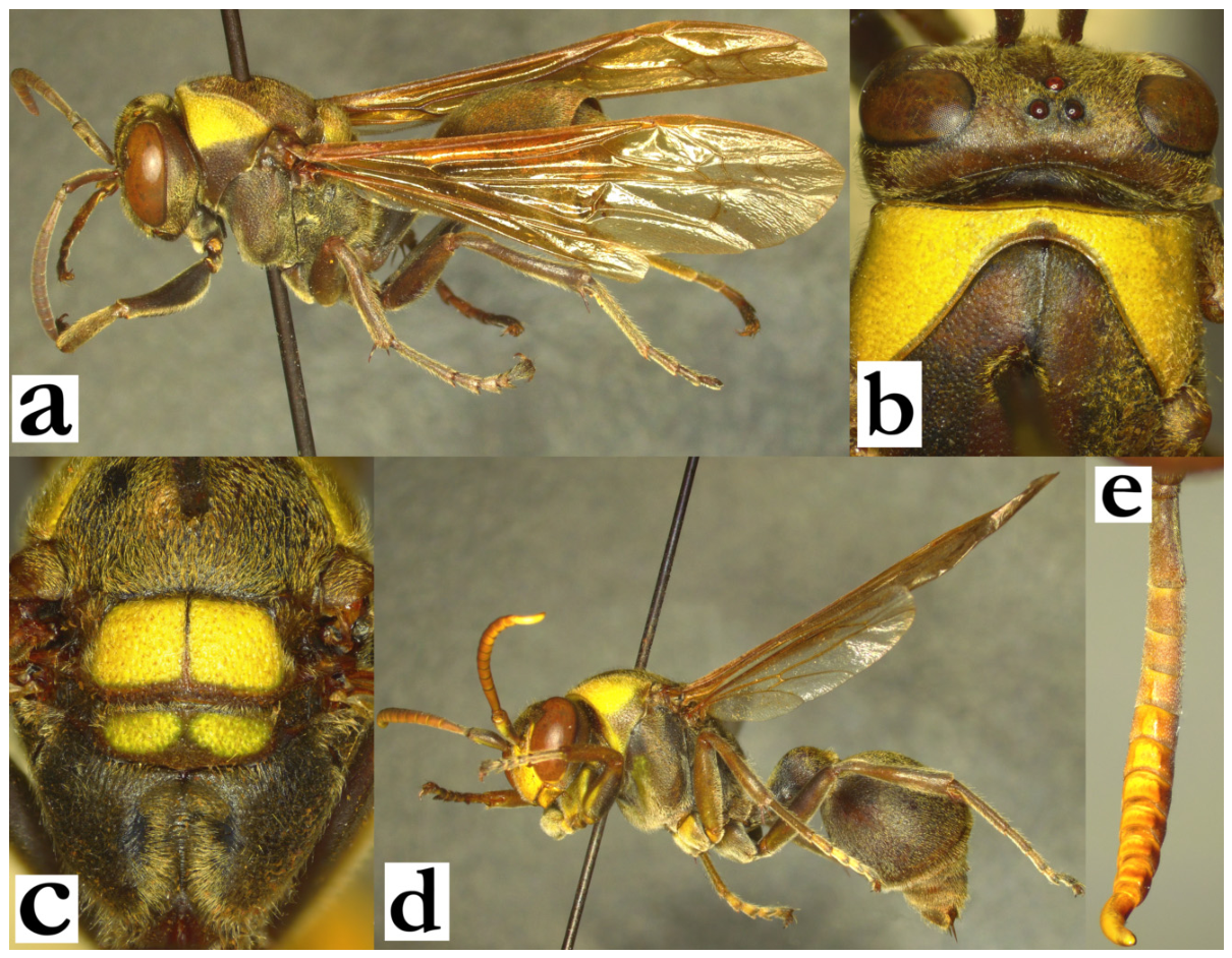
- Ropalidia favulorum Blommers, 2012Ropalidia favulorum n. sp., Blommers 2012: 161; ♂♀, nest, biology, Madagascar; holotype NNM.Number of examined specimens: 12 ♀♀, 18 ♂♂.
Type material. Male paratype from nest 72.48 (Figure 84a,b) and female paratype from nest 70.37 were examined.
Notes. A small dark-green species, with darker appearance (Figure 61e) and specific male antenna (Figure 61h); this is one of the species that builds minute-nested colonies, alongside R. rafitra sp. nov. Some difficulties may arise in separation from a few similar small dark-and-green species, most notably R. miafina sp. nov. and R. seyrigi sp. nov., which are listed in the key to species. In addition, this seems to be a complex of at least two taxa, with a somewhat separate cluster of specimens with reddish metasoma (Figure 58d); also, males from this cluster have a somewhat broadened scape, while remaining morphological differences were not observed; none of the reliable features were found in separating females from this cluster, suggesting that these two should be treated as the same taxon in the absence of additional information.
Nest. This species builds small black nests, consisting of a few cells each, sometimes in large nesting aggregations of hundreds of nests [6]. One female was implied to build multiple nests [6], which might have been confirmed in the observation record [76], in which one female apparently tends to two nests. Blommers also noted the lack of the basal cell opening for meconium removal [6], which was also seen in R. rafitra sp. nov. nests; one possible explanation is that these two species are building fragmented nests where meconium removal might not be feasible due to petiole proximity, causing them to build multiple nests, rather than removing meconium. Cells of R. favulorum Blommers nests are 2.2–3.9× as long as wide with closed cocoon caps, comparable to 3.1–4.6× as long as wide in R. rafitra sp. nov., possibly indicating that plenty of cell space gets freed for meconium deposit that remains unremoved. Interestingly, there are also instances of a single nest being built by two foundresses [77], possibly indicating a foundation of a new nests aggregation site and contributing to the idea that one queen may be involved in tending to more than one nest at a later colony stage, especially in line with the report that at any given time, the majority of nests seemed unattended [6]. Nests were also observed on similar elongated leaves [78], or even located circularly on petioles (Figure 84h,i). Dried nests apparently loose blackish colour and remain brown (Figure 84h–j).
Figure 84.
Ropalidia favulorum Blommers, (a) habitus, oblique lateral view, male, paratype (nest 72.48), (b) labels, paratype, (c) head, frontal view, female, melanic specimen, (d) head, frontal view, male, (e) antenna, median view, male, (f) nest [76], (g) nest [77], (h) nest 190318-3, (i) nest 190318-3, different angle, (j) nest 190318-2 (collected nests all from AMNH).
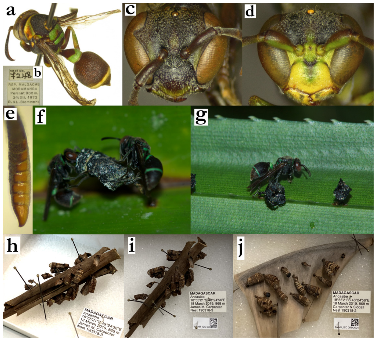
- Ropalidia flavoviridis Kojima, 1988Ropalidia flavoviridis Kojima, 1988; “Tulear Prov., Berenty, J. Wenzel”, Snow Entomological Museum, University of Kansas.Plazi: https://tb.plazi.org/GgServer/html/03E08B01FFFD761505D9FA6AFCD1F9F5 (accessed on 21 September 2026).Number of examined specimens: 31 ♀♀, 14 ♂♂.
Type material. One paratype female was examined (Figure 85a,c), with the labels “MADAGASCAR/ BEHARA/ FORET COTE EST]”, “MUSEUM PARIS/ III. 37/ A. SEYRIG” (Figure 85c). The specimen is mostly yellow, with partly green mesoscutum and brownish metasoma (Figure 85a).
Taxonomy. This species was described in 1988 [18]. However, the same year the taxon became synonymized with R. dubia, in a parallel split of R. saussurei Kojima from R. dubia (de Saussure). Giordani Soika [4] considered it as a separate species from R. dubia (de Saussure), but his R. dubia refers to R. saussurei, and R. pomicolor refers to R. dubia. Lastly, Blommers [6] utilized the apparent difference in male morphology and re-established R. flavoviridis Kojima as valid species.
Notes. Several morphological characteristics allow this species to be separated from similar mainly green species. These include wider and punctate female clypeus (Figure 34b) and predominantly green mesoscutum and T2 (Figure 32a), in combination with short pubescence on mesosoma dorsally. The most useful feature in determination of this species is the terminal flagellomere in males, which is usually yellowish-orange (Figure 34d), but in some specimens the tip, or even the distal two flagellomeres can have a black tip (Figure 85e). The nest of this species was described before [6], and it is in line with all the analysed observations.
Similar species. Some confusion may be possible with other mainly green species, but the biggest problem may arise in atypical morphology of R. laureliniana sp. nov. Useful features in this case include the shape of T1 (Figure 85g), which is more rounded or streamlined in R. laureliniana sp. nov. (Figure 28a,c), and rounded median depression of the propodeum (Figure 85f, as opposed to shallower depression in R. laureliniana sp. nov., Figure 28c). The general colour pattern may also be useful, but it may also vary in this species substantially, ranging from almost entirely yellow body (Figure 85h), to yellow with bright green areas, which is the commonest colour form (Figure 85i).
Figure 85.
Ropalidia flavoviridis Kojima, (a) habitus, lateral view, female, paratype, (b) head, frontal view, female, paratype, (c) labels, paratype, (d) antenna, median view, male, (e) antenna, dorsal view, male with black tyloids, (f) posterior mesosoma, dorsal, female, (g) T1 and T2, dorsal view, female, (h) habitus, lateral view, female with yellow colour pattern, (i) habitus, lateral view, female with green colour pattern.
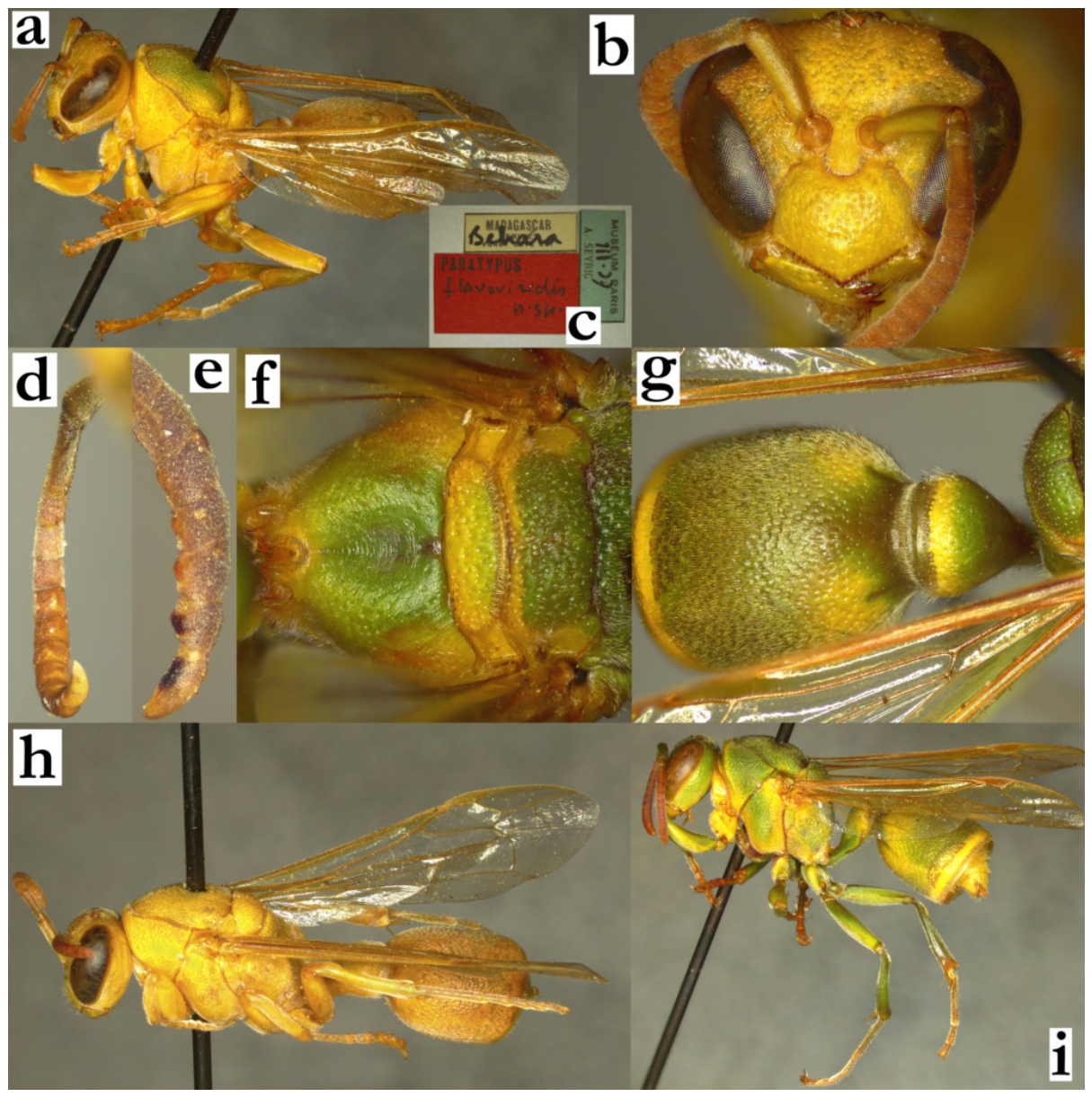
- Ropalidia formosa (de Saussure, 1853)Icaria formosa de Saussure, 1853; India/ Les Indes Orientales [erroneous]; “Madaga.” (Figure 86a,b) (NHM)(accessed on 21 September 2026).
- BOLD: https://portal.boldsystems.org/bin/BOLD:ACT6336 (accessed on 21 September 2026).Number of examined specimens: 7 ♀♀, 6 ♂♂.
Type material. A single female, erroneously considered to have originated from India/ “Les Indes Orientales”; the specimen has seven labels; the first one is the circular, red-bordered label that says “Type”, followed by a rectangular label “F. Sm. Coll. 79 22”, next is “India”, but after that one is the next label with multiple pin punctures that says “Madaga.” [notably, these two labels are written in different handwriting, the second corresponding to de Saussure’s from other examined types, while the first is of unknown origin]. In addition, there is a label that says “Icaria formosa [female sign] Sauss”, followed by label “B. M. TYPE HYM 18.858”, and finally an identification label with QR code and label NHM010636342 (Figure 86b).
This species can be established by the following combination of features: mesoscutum impunctate anteriorly, mesepisternum coarsely and densely punctate, with sharp angle between anterior and inferior surface (Figure 86c), widely placed posterior ocelli and thin gena. Males are described here for the first time.
Description. Males. Wing length 13.1–14.6 mm.
Head. Head frontally 1.2× as wide as high, in dorsal view 2.2× as wide as long, not narrowing behind eyes, minimally emarginate posteriorly (Figure 8d). Clypeus 1.1–1.2× as wide as long, upes curved, OCA absent, juxtamandibular excavation minimal, apex mildly projecting (Figure 8d); clypeus coarsely punctate basally, punctures large and grouped close to epistomal suture; centre with somewhat smaller punctures, apex mildly depressed and shiny (Figure 8d). Frons densely punctate, punctures 0.5 diameter apart, interspace shiny; inner orbit and vertex with smaller and less dense punctures, gena with even less defined, but retained punctures contour even close to occipital carina; gena 0.3–0.4× as wide as eye; mandible shiny, with deep ill-defined punctures, occipital carina complete; posterior ocelli 1.4 diameter apart, 2.15× diameters apart from compound eye and 1.9 diameters apart from occipital carina (Figure 86d). Scape 4.0–4.2× as long as wide, 1.4× as long as AF1; AF2 1.5× as long as wide, remaining flagellomeres longer than wide (Figure 8d). Tyloids shiny, originate at AF2 (Figure 86e), setose and mildly elevated above flagellomere surface (Figure 8e); terminal flagellomere elongated, 3.7× as long as wide and 2.6× as long as AF10; AF11 tyloid with developed median protuberance, mostly retained and reaches acute tip (Figure 8e).
Mesosoma. Mesosoma slender, 1.1× as wide as head in dorsal view (Figure 86d), 1.6× as long as wide in lateral; mesoscutum 1.05× as wide as long (Figure 86d); pronotum in dorsal view barely convex anteriorly, humeri weak, lateral margins barely excavated anteriorly, posteriorly finely rounded; pronotal carina complete, opaque and thin (Figure 86d), equally wide dorsally as laterally, or just barely wider dorsally (Figure 86f). Pronotum punctures shallow, one diameter apart, with swollen interspaces; punctures retained in yellow areas; mesoscutum anteriorly impunctate, posteriorly with some ill-defined punctures (Figure 86d). Mesepisternum without carina towards mesepimeron, but with noticeable transition of puncture pattern; mesepisternum reticulate, with puncture interspace creating a network; mesepimeron with smaller punctures more spaced apart than mesepisternum; anterior face of mesepisternum impunctate, lateral punctate; inferior face angled to anterior, creating a right angle and a minimal caudally directed depression (Figure 86c). Disc of scutellum flattened and bituberculate, without strongly developed median depression; median carina of scutellum incorporated into punctures and hardy discernible; punctures coarse and dense, about 1.5 diameters apart, interspaces mildly swollen and matte (Figure 86g). Metepisternum coarsely punctate on shiny cuticular surface; propodeum posteriorly widened (Figure 86g), median depression deep, striate and weakly punctate; lateral surface more strongly striate, separated from median depression by developed crenulated margin superiorly, less defined inferiorly.
Metasoma. T1 globular, with shallow posterior sulcus; anterior surface impunctate, posterior with some poorly defined craters. T2 1.2× as long as high, tergum and sternum similarly convex; punctures shallow, directional, 1.0–1.6 diameters apart, with weakly swollen matte interspace; punctures on S2 somewhat larger and sparser, especially proximally; transition between T2 surface and lamella gradual, lamella with three lines of punctures; remaining metasomal segments with shallow and large punctures.
Pilosity. Body with short pubescence, silvery on head and mesosoma, yellowish on metasoma (Figure 86f); clypeus with dense and fine silvery setae, 2.5× as long apically as basally; mandible with less dense protruding setae of similar length as clypeus apically; frons with slightly bent protruding setae, 1.4–1.7× as long as anterior ocellus diameter; gena and occiput with some short protruding setae; eyes minutely setose (Figure 8d). Pronotum and mesonotum without setae similar to those on frons, or slightly shorter; posterior mesosoma with some longer fine silvery setae; T1 anteriorly with fine pubescence, posteriorly with some yellowish setae that do not project over posterior margin; T2 with yellowish protruding setae that are projecting backwardly just to the level of lamella (Figure 86f); remaining metasomal segments with some longer protruding setae distally; coxa with fine silvery pubescence and an some silvery protruding setae with bent tips; femora with dense silvery pubescence, femur III also with longer setae.
Colour. Basal colour black(ish), with brownish, reddish or yellowish markings (Figure 86f); clypeus with central brownish area and remaining two vertical lateral yellow lines that are connected to yellow markings on inner orbits, yellow marking on interantennal area and yellow mandible, sometimes with dark yellow basal spot; frons reddish, vertex darker (Figure 8d); gena and vertex reddish, pronotum reddish to yellowish (Figure 86f), mesoscutum in basal colour (Figure 86d), exceptionally with two thin reddish longitudinal lines; scutellum and metanotum with two reddish spots, propodeum in basal colour (Figure 86g). Legs in basal colour, except tarsi with skipping pattern, proximal half yellowish, distal half dark brown (Figure 86f). Antenna black dorsally, reddish ventrally, tyloids brownish-orange (Figure 86e). Wings transparent, nervature brown, costa somewhat darker, stigma opaque ochre, apical spot or entire distal fifth of the wing with yellowish trichome (tip of wing lightly coloured; Figure 86f; notably, this is also visible in the holotype, but to a lesser degree; Figure 86a).
Nests of formosa group. Due to lack of confirmed specimens collected with nests and difficulties in establishing species from the formosa group based on observation photos, nests of all the species of this group are herewith treated together. The general pattern of their nests suggest smaller nests, with colony cycle that includes males even at the nest size of close to 10 cells [79]. One of these nests exhibits grey to dark-grey cocoon caps, that are very close to the cell wall margin, with solid cell wall and impregnated apical petiole [80].
Notes. There are a few important additions to the taxonomic work that can be made from these observations. The first one is the substantial difference in the proximal and distal wing colour, which seems to be much stronger in living animal than dried museum specimens [79]. The second interesting finding is a more vivid green colour, which might be caused by the improperly defined camera settings; nevertheless, it suggests that the colour of the living animal may be somewhat different from the dried museum specimen. One of the nest observations is more likely to belong to R. mainty sp. nov., but in the absence of a more reliable determination, this nest was also considered as a member of the formosa group, rather than being treated at the species level [81].
Distribution. Only one nest series marked “d” from Ivandry was examined (north part of Antananarivo).
Figure 86.
Ropalidia formosa (de Saussure), (a) holotype, dorsal, (b) holotype, labels [kindly provided by Gavin Broad from NHM], (c) mesepisternum, (d) head and anterior mesosoma, male, dorsal, (e) antenna, male, (f) habitus, male, (g) posterior mesosoma, dorsal, male.
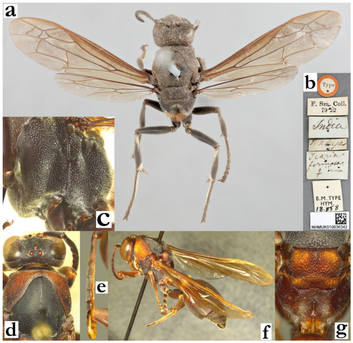
- Ropalidia fraterna (de Saussure, 1900)Icaria fraterna de Saussure, 1900; “Imerina” (MHNG).Ropalidia fraterna (de Saussure), Blommers 2012: 182; ♂, ♀ re-description, nest, biology, Madagascar; lectotype designated (♀, MHNG).(accessed on 21 September 2026).BOLD: https://portal.boldsystems.org/bin/BOLD:AFX3580 (accessed on 21 September 2026).Number of examined specimens: 105 ♀♀, 22 ♂♂.
Notes. A species characterized by T1 globular, T2 base coarsely punctate, median carina of scutellum absent and gradually narrowing and acute tip of terminal flagellomere in males. This is a complex of several taxa, confirmed by the separate genetic lineages, varying morphology and diverse nesting architecture. Saussure seemingly had problems with this taxon; among the specimens from MSNV that he determined as R. fraterna there were two specimens of R. shestakowi (Schulthess), two specimens of R. linearecta Blommers, and one specimen of R. cauponae Blommers.
Genetics. Two specimens were successfully genotyped, showing separate lineages, with R. galimatia and R. zandry nested within one of these lineages. The specimens showed most similarity with two identified BIN clusters related to this species, with the specimen AG7G1 expressing 97.26% concordance with BOLD:AFX3580 [LPGEO42856-24], which is R. fraterna, and 95.29% concordance with BOLD:ADH8057 [IPSIO524-22], which belongs to R. zandry sp. nov. Specimen AG3G8 showed an inverse pattern, with 95.50% concordance with BOLD:ADH8057 [IPSIO513-22] and 94.98% concordance with BOLD:AFX3580 [LPGEO42856-24]. These results suggest that two separate lineages are present in this species, which might explain observed patterns in the nest architecture. Notably, these lineages were carefully compared, yielding a reliable size difference (the AG7G1 specimen has a wing length of 6.8 mm, while AG3G8 has a wing length of 7.9 mm) and the mesosoma dorsal punctures pattern, with less defined punctures on a velvety interspace in AG3G8 (Figure 87a–e). These differences were not considered sufficient to establish a separate species, therefore both lineages were considered the same taxon.
Nest. The nest architecture of this species presents yet another interesting problem, reflected in substantial plasticity. There were two different nest shapes observed, one that was regular in form (discoid or triangular), and the other branched of the two-cell-wide linear form. This could also be interpreted as extreme nesting plasticity, suggesting a cryptic taxon or even possibly a misidentification with R. interjecta that seems to build similar nests. Unfortunately, the available data were insufficient to fully resolve this situation.
Distribution. The entire island.
Figure 87.
Ropalidia fraterna (de Saussure), in scale, specimen designated as AG3G8, female, (a) habitus, lateral view, (c) head, frontal view, (e) head and mesosoma, dorsal view; specimen designated as AG7G1, female, (b) habitus, lateral view, (d) head, frontal view, (f) head and mesosoma, dorsal view.
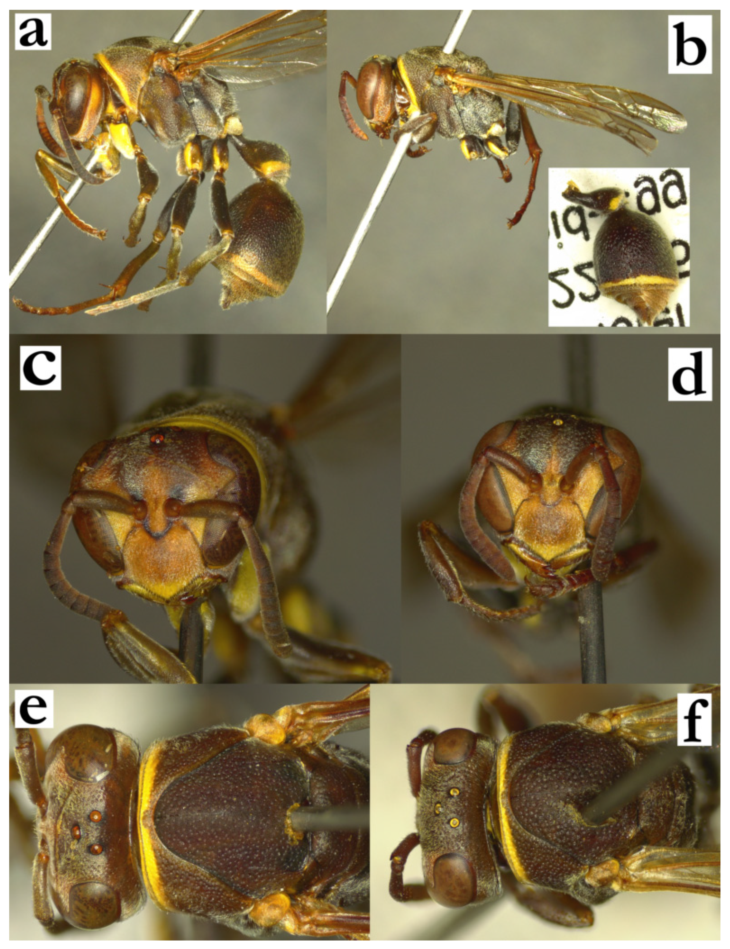
- Ropalidia galimatia (de Saussure, 1853)Icaria galimatia de Saussure, 1853; “MUSEUM PARIS, MADAGASCAR, JACQUINOT 1841” (MNHN).Plazi: https://tb.plazi.org/GgServer/html/03AE106DFFD8396925D7FC80840FF453 (accessed on 21 September 2026).BOLD: https://portal.boldsystems.org/bin/BOLD:AFX3584 (speculative, based on a photograph of the specimen LPGEO42768-24; accessed on 21 September 2026).Number of examined specimens: 71 ♀♀, 6 ♂♂.
Type material. De Saussure described this species in 1853, and provided a replacement name of Icaria madecassa that bears no valid reason for a change [2]. He also described Icaria ignobilis in 1890, which later became synonymized with the initial name, Icaria galimatia [19]. A female in MNHN is labelled as lectotype, with a male in MNHN and a female in MHNG labelled as paralectotypes [19] of Ropalidia galimatia (de Saussure).
Notes. This species is characterized by the basally brown body colour and bright yellow markings (Figure 88a); these markings may be variable and almost entirely missing in larger specimens. Female and male head or male antenna (Figure 88b–d) do not show many differences from very similar R. laureliniana sp. nov., which can sometimes present a taxonomic challenge.
Distribution. Entire island (Figure 88e). This species was reported to prefer dry areas of Madagascar [19].
Genetics. Sequenced specimens have exhibited the closest relationship with R. zandry sp. nov., followed by R. fraterna (de Saussure). Notably, the BOLD database holds a BIN designation of a specimen that resembles R. galimatia (specimen ID LPGEO42768-24), but the determination could not be confirmed. The corresponding BIN designation is BOLD:AFX3584, possibly belonging to this species.
Figure 88.
Ropalidia galimatia (de Saussure), (a) habitus, oblique lateral view, female, (b) head, frontal view, female, (c) head, frontal view, male, (d) antenna, dorsal view, male, (e) distribution map of Ropalidia galimatia and Ropalidia laureliniana sp. nov.
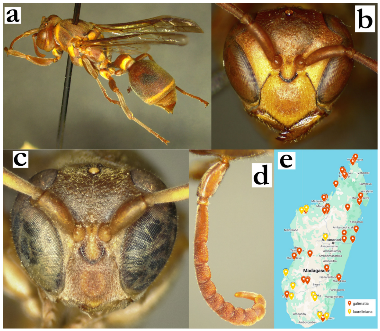
- Ropalidia grandidieri (de Saussure, 1890)Icaria grandidieri de Saussure, 1890; “Diverses [female sign] recoltées par A. Grandidier—Musee de Berlin. [female sign], [male sign] recolteés par Hildehrandt”.Plazi: https://tb.plazi.org/GgServer/html/03AE106DFFD7396825D7FB88842EF2BE (accessed on 21 September 2026).Ropalidia grandidieri (de Saussure), Blommers 2012: 141; ♂, ♀ re-description, nest, biology, Madagascar; lectotype designated (♀, MHNG).Number of examined specimens: 34 ♀♀, 13 ♂♂.
Type material. Blommers [6] assigned a lectotype status to a specimen at MHNG, with the labels “19.” (round blue label with handwritten), “S3” or “53”; “Madag.” in handwriting on the white rectangular label and “Icaria grandidieri Sss, type” in handwriting on the blue rectangle. There is another specimen from ETH that belongs to the type series and it is hereby treated as a paralectotype (Figure 89a–e).
Notes. This species is characterized by dense pilosity (Figure 23b), pronotal collar impunctate (Figure 23a), protruding setae on mesoscutum (Figure 24f) and a tubercle on the femur II in males (Figure 24g). Some problems may arise in separation from R. takona sp. nov., which are listed in the key to species. A sequenced specimen most closely resembles the sequence of R. tsiky sp. nov., close to other members of the grandidieri group.
Distribution. The entire island, possibly absent from western parts (Figure 89f).
Figure 89.
Ropalidia grandidieri (de Saussure), newly assigned female paralectotype from the ETH collection, (a) habitus, oblique lateral view, (b) habitus, dorsal view, (c) labels, (d) head and mesosoma, dorsal view, (e) head and mesosoma, lateral view, (f) distribution map (red marker—specimen, grey marker—observations and literature reports).
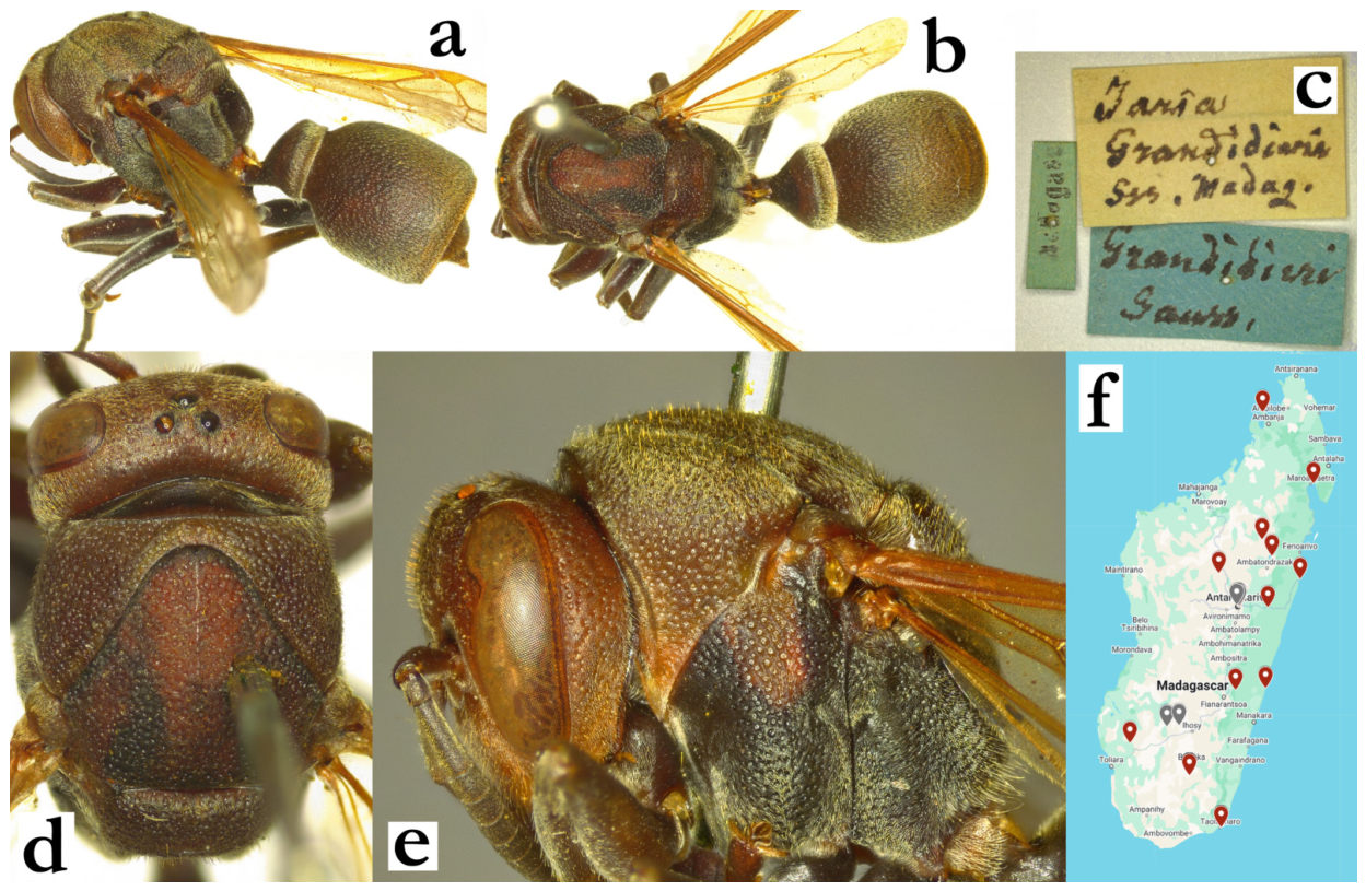
- Ropalidia hova (de Saussure, 1890)Icaria hova de Saussure, 1890; “Madagascar … aux environs d’Andrangoloaka (Imerina)” (MHNG).BOLD: https://portal.boldsystems.org/bin/BOLD:ADR1779 (accessed on 21 September 2026).Number of examined specimens: 11 ♀♀, 1 ♂.
Type material. Several specimens were previously implied as belonging to the type series, deposited in MHNG [2,6]. The description lists a number of specimens, namely “Madagascar—Deux [female sign] recoltees par Alfred Grandidier.—Plusieurs individus récoltés par Hildebrandt se trouvent au Musée de Berlin; d’autres récoltés aux environs d’Andrangoloaka (Imerina) par M. Fr. Sikora, ligurent dans ma collection”. There were no type specimens recorded in the Berlin collection. The specimens with label “Sikora/Madagascar” and the label “Madagasc./Annarivo” are R. merina (both deposited in MSNV).
Notes. Strongly contrasting colours species, basally black, with yellow areas on mesosoma (and metasoma). Some confusion may be possible with R. merina Blommers in terms of the general colour pattern. R. hova has yellow areas on pronotum, scutellum and metanotum, and T1 and/or T2 (Figure 7a). R. merina, follows a similar pattern, but with less surface or smaller markings on the same body parts (Figure 5a), sometimes missing entirely; notably, all the marks are bright yellow in R. hova vs. whitish-green markings in R. merina (Figure 7a vs. Figure 5a).
Female and male head morphology do not deviate substantially from other species of the formosa group (Figure 90a,b), except for overall longer pilosity. The conversion from T2 surface to T2 lamella is very weak, and in some specimens nearly absent (Figure 90c). Another useful taxonomic characteristic is entirely yellowish wing, useful in separation of xanthic specimens of few other members of the same species group (Figure 90d). Male tyloids originate on AF(2)3, and the first few are always somewhat less developed (Figure 90e).
Distribution. Eastern and central Madagascar.
Genetics. One specimen was successfully sequenced, and an additional one was included from the BOLD database, confirming membership in the formosa group and close proximity to R. formosa (de Saussure), BOLD:ACT6336.
Figure 90.
Ropalidia hova (de Saussure), (a) head, frontal view, female, (b) head, frontal view, male, (c) T2, lateral view, female, (d) wing, dorsal view, female, (e) antenna, median view, male.
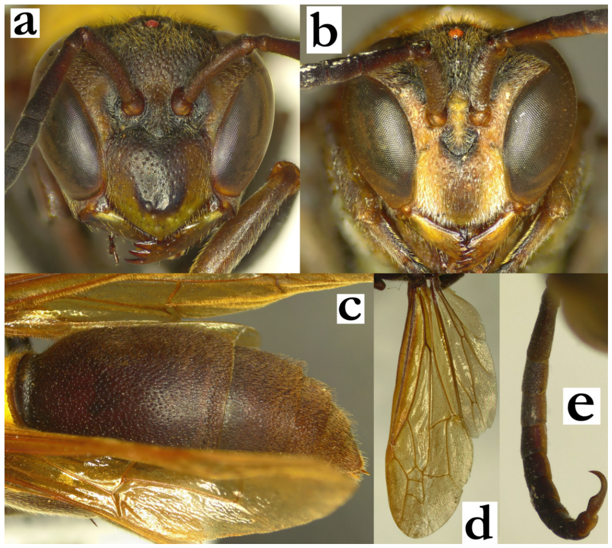
- Ropalidia interjecta (de Saussure), 1900Icaria interjecta de Saussure, 1900; “Madagascar—Nossi-Bé” (ETH).Plazi: https://tb.plazi.org/GgServer/html/03AE106DFFD6396825D7FD35842EF43E (accessed on 21 September 2026).Number of examined specimens: 9 ♀♀, 14 ♂♂.
Type material. Multiple males and females from the type series were examined (ETH, MSNV).
Notes. This species is characterized by a combination of weakly globular T1 and comparatively wider head in females (Figure 91a), developed median scutellar carina, and elongated and apically widened terminal flagellomere in males. Some taxonomic problems may occur in separation from R. fraterna (de Saussure); features for separation of these two species are provided in the key to species. Additionally, the nest architecture of this species shows a continuum from the strictly bi-cellular nest to multiple branches, which sometimes substantially resembles nests of R. fraterna. One more nest from Diana region was observed, located on the leaf; the nest was apparently broken in two parts, but the separate part did not drift away completely [49].
Distribution. Confirmed specimens originated from the Nosy Be island and the surrounding areas (Mahajanga, Boeny, Diana).
Figure 91.
Ropalidia interjecta (de Saussure), female and male, syntypes, (a) habitus, lateral view, female (ETH), (b) labels, (c) habitus, lateral view, female (MSNV), (d) habitus, lateral view, male, (e) label, male, (f) head and mesosoma, dorsal view, female, (g) head, frontal view, male, (h) antenna, dorsal view, male.
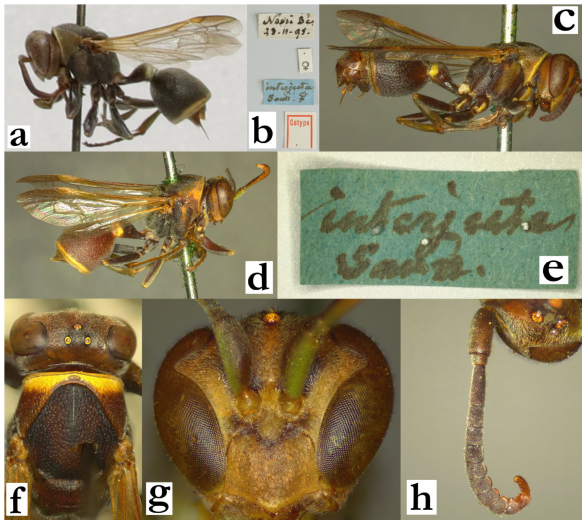
- Ropalidia janettae sp. nov. Polašek and de BeerZooBank: EF1A619C-3529-4164-8BE9-CFE178DCF4EBMaterial examined. Holotype: “Ankaratra/Manjakatompo”, “1.52 Rakotovao”, “INSTITUTE SCIENTIFIQUE MADAGASCAR”, ♀ (MSNV), Figure 92a–c.Paratypes: 1♀, 3♂♂, same labels as holotype (MSNV); “Andohahelo/(1800 m)/1.54 (R.P.)”, “INSTITUTE/ SCIENTIFIQUE/ MADAGASCAR”, 1♀ (MSNV); “Bekily/III-33”, “Madagascar/A. Seyrig”, 2♀♀ (MSNV); “MADAGASCAR/ Ambohimanga”, “MUSEUM PARIS/ XI 35, Larine?/ A. SEYRIG”, 1♀ (MSNV); “MADAGASCAR/AMBOSITRA”, “MUSEUM PARIS/ II 39/A. SEYRIG”, 1♀ (MSNV); MADAGASCAR/Antsirabe”, “MUSEUM PARIS/ XI 36/ A. SEYRIG”, 1♀ (MSNV); “MADAGASCAR/ Kalambatitra/I.1933 Leg. Vadon”, 1♂ (MSNV); “Inst. Scient. Madagascar/ Manjakatompo/22-XII-50 R. Benoist” 3♀♀ (MSNV); “Madagascar/65km S. of/Tananarive/XI-25-1959”, 1♀ (CAS); “MADAGASCAR: Tan./ Mt Angavokely/ 6.iv.1991/A. PAULY col.”, 4♀♀ (RMCA); “COLL. MUS. CONGO/ Madagascar: Masif An-/ karatra 1700/1800 Man-/jakatompo XII-51 Benoist”, 1♀ (RMCA); “Manjakatompo/1800 m/4 JAN 1956”, “E. McC. Callan”, “INSTITUTE/ SCIENTIFIQUE/ MADAGASCAR”, “STYLOPIZED”, 1♀, 2♂♂ (NHM).Number of examined specimens: 17 ♀♀, 6 ♂♂.
Diagnosis. Member of bicincta group, characterized by T1 longer and posteriorly globular, body with suffused green markings, juxtamandibular notches in female deep, and male antenna with acute tip.
Description. Female. Wing length 8.3–8.6 mm.
Head. Head 1.1–1.2× as wide as high in frontal view, clypeus 1.2–1.5× as wide as long, with straight upes, deep juxtamandibular notches and projecting apex (Figure 50g); clypeus base and centre shiny and finely punctate, with larger and less defined punctures apically (Figure 50g); mandible shiny, coarsely punctate; frons shallowly punctate, with shiny interpuncture area. Gena posteriorly 1.2–1.4× as wide as eye, mostly shiny, with occasional large and poorly defined puncture close to eye and entirely impunctate area close to occipital carina (Figure 48d). Inner orbit impunctate (Figure 50g); occipital carina complete (Figure 48d); posterior ocelli about 2.5 diameters apart (Figure 92b). Antenna clavate; scape 1.3–1.4× as long as AF1; AF1 4.3× as long as wide, AF2 1.5× as wide as long.
Mesosoma. Mesosoma 1.6× as long as wide in lateral view, head 1.05× as wide as mesosoma (Figure 92b). Mesoscutum anteriorly sparsely and shallowly punctate, pronotum dorsally with less defined punctures, that remain of similar definition inferiorly (Figure 48d); mesepisternum superiorly with large punctures, that slightly diminish in size and density inferiorly (Figure 50e); metepisternum with a few poorly defined punctures; scutellum with developed median carina, rounded, shiny, with some scattered punctures (Figure 92c); median depression of propodeum shallow, striate and impunctate (Figure 92c).
Metasoma. T1 dorsally developed and posteriorly globular, with some poorly defined punctures (Figure 50f); T2 coarsely punctate, with punctures 1.5–2 diameters apart and shiny interpuncture area (Figure 48c); T2 lamella dark and shiny; remaining segments minutely punctate.
Pilosity. Apical third of clypeus covered by yellowish setae, which are longer than those on the base (Figure 48d); frons with straight golden setae, 0.7–1.0x as long as anterior ocellus diameter; eyes minutely setose (Figure 92b); mesosoma dorsally with short setae, 0.5–0.7× as long as those on frons (Figure 48d); protruding yellowish setae on T2 extend over lamella (Figure 50f).
Colour. Basal body colour dark brown to black (Figure 92a); clypeus, inner orbits, antero-basal spot on mandible and gena suffused yellow-reddish-greenish (Figure 50g); pronotum and scutellum suffused brownish, reddish or greenish (Figure 92a), metanotum basally darker, with two suffused green spots (Figure 92c); propodeum in basal (blackish) colour; coxa and femora black, tibia olivaceous-dark-green, tarsi reddish (Figure 92a); T1 and T2 basally dark-brown to black, T1 with broadened posterior yellow or greenish band, T2 with thinner yellow band (Figure 92a); remaining metasomal segments brown or ferruginous; wings transparent, with yellowish anterior portion, nervature light brown, stigma ochre, semi-transparent, apical spot very faint (Figure 92a); antenna orange-ferruginous, scape dorsally dark green, ventrally green (Figure 50g).
Male. Resemble females in the general appearance, with more yellow and green markings (Figure 92d). Wing length 11.4–12.2 mm.
Head. Clypeus 1.25× as wide as long, upes slightly curved, without OCA, apex rounded juxtamandibular lobes very weak (Figure 50h); clypeus basally and centrally shallowly and finely punctate, occasionally with some more developed punctures (Figure 50h); gena 0.4× as wide as eye, mandible shiny, nearly impunctate; scape barely widened, 1.4× as wide as AF1 base, shorter than AF1 (Figure 50h); AF2 1.1× as long as wide (Figure 92e); tyloids originate on AF(6)7, ferruginous and matt (Figure 92f); terminal flagellomere elongated, 1.8× as long as wide, apically acute, with darkened tip (Figure 92e).
Mesosoma. Mesoscutum somewhat more coarsely punctate than females; fore tarsal spur I very developed.
Metasoma. S7 weakly concave, with shiny and irregular surface.
Pilosity. Similar to females, except clypeal surface with fine, equally long, silvery setae, that become denser on juxtamandibular region (Figure 50h); frons setae comparatively longer than in females.
Colour. Clypeus yellow with basal black spot, mandible yellow with remaining darker postero-basal area (Figure 50h); remaining parts of the head black (except minute, suffused dark-olive area on gena); mesosoma basally black, with suffused olive(-green) markings on pronotum and scutellum; coxa I with green-yellow distal half, coxa II quadratic green-yellow spot on lateral-distal triangle and a smaller medial marking of the same colour (Figure 92d); T1 with posterior green band, T2 with complete posterior yellow-green band, S2 with traces of green-yellow band; remaining segments brownish (Figure 92d); antenna infuscate dorsally, orange ventrally, scape green ventrally, dark green dorsally (Figure 50h).
Distribution. Central and south highlands (Figure 92g).
Etymology. The name is given in honour of Janette Rakotondranaivo, and the countless hours that her father, Toky gave for the advancement of Malagasy science in the Ambohitantely forest.
Note. A few examined specimens were stylopized.
Figure 92.
Ropalidia janettae sp. nov., (a) habitus, lateral view, female, (b) head and mesosoma, dorsal view, female, (c) propodeum, oblique dorsal view, female, (d) habitus, lateral view, male, (e) antenna, dorsal view, male, (f) antenna, median view, male, (g) distribution map (red markers denote the locations of examined specimens).
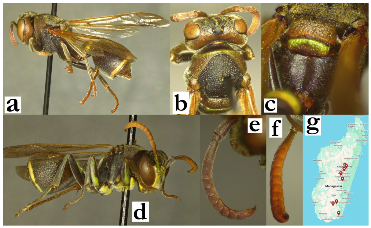
- Ropalidia jemmae Polašek and de Beer, 2023Plazi: https://tb.plazi.org/GgServer/html/EB2BCB57F8F15600BBCDAC166DCCCFB4 (accessed on 21 September 2026).Number of examined specimens: 3 ♀♀.
Notes. A recently described species, characterized by the interesting inverse, visually concealing nesting architecture (Figure 93a). Males remain undescribed. Slender black ants (genus Tetraponera) were observed close to one of the nests [82], suggesting a possible predation risk for the colony. A recent observation was added from the from Itasy region [83], showing a characteristic nest background of ornamented cell bottoms exposed outwardly; notably, the species was confirmed by closely placed posterior ocelli, used to separate this from a few more similarly coloured species (Figure 65a). The observation originates from the area with substantial forest devastation, suggesting that this species might tolerate certain degree of habitat loss.
Figure 93.
Ropalidia jemmae Polašek and de Beer, (a) abandoned nest, (b) distribution map (grey areas overlap locations for the three known museum collection specimens).
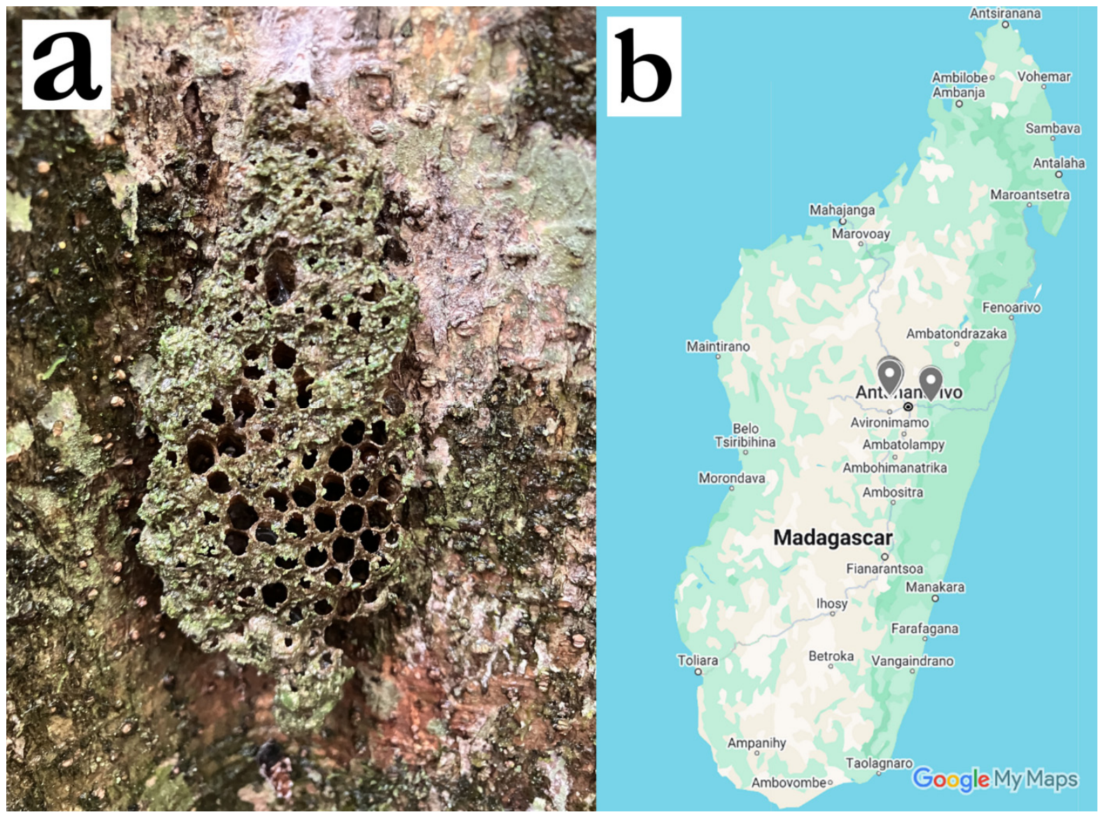
- Ropalidia kojimai Blommers, 2012Ropalidia kojimai n. sp., Blommers 2012: 164; ♂♀, nest, biology, Madagascar; holotype NNM.Number of examined specimens: 3 ♀♀, 2 ♂♂.
Type material. Three paratypes were examined, two males and a female from the nest 73.21 (Figure 94a,b), alongside one more (non-type) female from the nest 71.38.
Notes. In addition to examined types, only two more females were recorded, one from Antsirabe and the other with the label “Zool Mus Berlin/Madagascar”; the latter was determined by de Saussure as R. subclavata, bearing his label. The available evidence suggests a narrowly distributed species that is occurring around the Alaotra-Mangoro region; some specimens may be more demanding to determine due to similarity with several other species. This is also one of the few species that builds colonies of nests, most commonly in large stone hollows or man-made concrete objects [62].
Figure 94.
Ropalidia kojimai Blommers, male, paratype, (a) habitus, lateral oblique view, (b) labels, (c) head, frontal view, (d) propodeum, dorsal view, (e) antenna, median view.
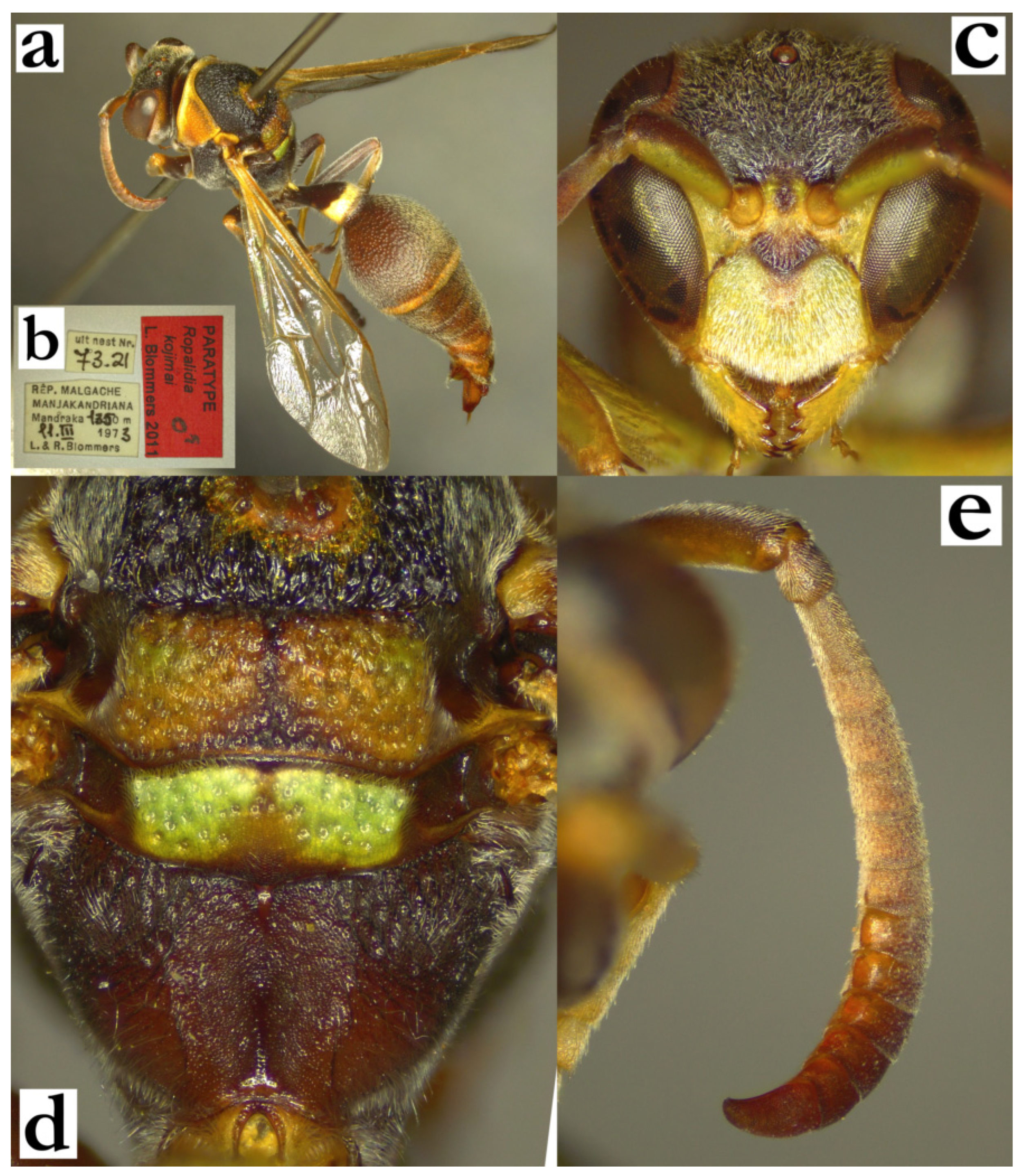
- Ropalidia laureliniana sp. nov. PolašekZooBank: 6749C4ED-B1BA-4AA8-B78A-7AFBA68C2D15Ropalidia ignobilis (de Saussure): [15]; at least in part, since the study mentions green colour, not mentioned in the R. ignobilis species description.Ropalidia galimatia [9]: cocoon caps white, in contrast to dark-grey to black in R. galimata.Material examined. Holotype: “MADAGASCAR/BEKILY/REG SUD DE L’ILE”, “MUSEUM PARIS/V. 37/A. SEYRIG”, “H2”, ♀ (MSNV).Paratypes: 6♀♀, 4♂♂, same labels as holotype (MSNV); “[female sign]”, 1♀ (ETH); “Station Agric/Bas Mangoky”, “INSTITUTE/ SCIENTIFIQUE/ MADAGASCAR” 27♀♀ (MSNV); “Madagascar/Fort Dauphin/Sikora”, 2♀♀ (MSNV); “MADAGASCAR/BEKILY/REG SUD DE L’ILE”, “MUSEUM PARIS/V. 37/A. SEYRIG”, 3♀♀, 1♂ (MSNV); “MADAGASCAR/BEKILY/REG SUD DE L’ILE”, “MUSEUM PARIS/IX. 36/A. SEYRIG”, 2♀♀ (MSNV); “MADAGASCAR/BEKILY/REG SUD DE L’ILE”, “MUSEUM PARIS/VIII. 36/A. SEYRIG”, 5♀♀ (MSNV); “MADAGASCAR/BEKILY/REG SUD DE L’ILE”, “MUSEUM PARIS/VII. 36/A. SEYRIG”, 16♀♀, 1♂ (MSNV); “MADAGASCAR/BEKILY/REG SUD DE L’ILE”, “MUSEUM PARIS/IV. 37/A. SEYRIG”, 4♀♀, 11♂♂ (MSNV); “Hera/Ankazoabo”, “INSTITUTE/ SCIENTIFIQUE/ MADAGASCAR”, 3♀♀, 1♂ (MSNV); “Tongobory/Sept-Lacs/A.K.” “INSTITUTE/ SCIENTIFIQUE/ MADAGASCAR”, 1♀ (MSNV); “MADAGASCAR/Bekara”, “MUSEUM PARIS/III. 37/A. SEYRIG”, 7♀♀, 4♂♂ (MSNV); “Ankazoabo (“Besalamyny”?)”, “INSTITUTE/ SCIENTIFIQUE/ MADAGASCAR”, 2♀♀ (MSNV); “Sa Dakoa [ineligible]/R.P. Cattola”, “INSTITUTE/ SCIENTIFIQUE/ MADAGASCAR”, 4♀♀ (MSNV); “Bekily/III 33”, 1♀ (MSNV); “Namoroka/Ralcotovano”, “INSTITUTE/ SCIENTIFIQUE/ MADAGASCAR”, 1♀ (MSNV); “Sept Laco/Area de Tulear/KM 54 vi 51 AR”, 2♀♀ (MSNV); “Tulear/28.3.53 (A.R.)”, “INSTITUTE/ SCIENTIFIQUE/ MADAGASCAR”, 1♀ (MSNV); “MADAGASCAR/BEKILY/REG SUD DE L’ILE”, “MUSEUM PARIS/X. 36/A. SEYRIG”, 2♀♀ (MSNV); “MADAGASCAR/Bekara”, “MUSEUM PARIS/XI. 38/A. SEYRIG”, 2♀♀ (MSNV); “MADAGASCAR/Bekara”, “MUSEUM PARIS/I. 38/A. SEYRIG”, 2♀♀ (MSNV); “MADAGASCAR/Bekara”, “MUSEUM PARIS/IV. 39/A. SEYRIG”, 5♀♀ (MSNV); “MADAGASCAR/ Tulear Pr/ St Augustin s.l./ 29.II.1968/ K.M.G.&P.D.”, 1♀ (NHM); “MADAGASCAR/ Tulear Pr./ Bevilany 800m/ 12.V.1968/ K.M.G.&P.D.”, 1♀ (NHM).Number of examined specimens: 111 ♀♀, 22 ♂♂.
Diagnosis. This species can be separated from other members of the fraterna group by predominantly yellow or green body colour, punctate mesoscutum, very short pubescence, lightly coloured anterior wing margin and elongated terminal flagellomere in males.
Description. Females. Wing length 8.0–9.3 mm.
Head. Head frontally 1.1× as wide as high, in dorsal view 2.6× as wide as long, shallowly emarginate posteriorly (Figure 28b). Clypeus 1.1–1.2× as wide as long, but with elongated base; upes straight, longer than lateral clypeus margin (Figure 95a); juxtamandibular excavation shallow, apex pointy (Figure 95a); clypeus finely punctate basally, with some poorly defined craters apically (Figure 95a); frons sparsely punctate, punctures 1.5–2.5 diameters apart, with matte interspaces (Figure 95a); gena mostly impunctate, only with few small punctures close to mandible and high on gena; mandible punctate, occipital carina complete (Figure 95a); posterior ocelli 1.3 diameters apart, 2.2× diameters apart from compound eye and 2.4 diameters apart from occipital carina (Figure 28b). Scape 3.0–3.3× as long as wide, 1.2× as long as AF1; AF2 1.05× as long as wide, remaining flagellomeres wider than long (Figure 95a).
Mesosoma. Mesosoma slender, 1.05× as wide as head in dorsal view; 1.4× as long as wide, in lateral view 1.8× as long as wide; mesoscutum 1.05× as long as wide (Figure 28b); pronotum in dorsal view straight anteriorly, humeri weakly angled in larger specimens, rounded in smaller ones; pronotal carina complete and thin, dorsally at most 1.5× as wide as laterally; pronotum punctures shallow, 1.5–2 diameters apart, with matte interspaces, retained even in yellow areas; mesoscutum with similar but sparser punctures (Figure 28b); mesepisternum without carina towards mesepimeron, sparsely punctured in lateral area, impunctate or with just a few punctures anteriorly; posterior surface with less dense and smaller punctures, similar to those on mesepimeron; metepisternum basally matte, with more spaced and well-defined punctures; propodeum posteriorly mildly widened (Figure 28c), with median depression shallow, with poorly defined lateral margins; median depression impunctate and matte, with variable short midline striae (Figure 28c); lateral face with very faint striae and poorly defined occasional puncture.
Metasoma. T1 globular, with asymmetric posterior sulcus (Figure 28b), 1.9× as long as high; anterior surface impunctate, posterior with some poorly defined craters. T2 1.15× as long as high, tergum more convex than sternum; punctures shallow, directional, 1.0–1.6 diameters apart, with weakly swollen matte interspace; punctures on S2 somewhat larger and sparser, especially proximally; transition between T2 surface and lamella gradual, lamella translucent and very weakly punctate proximally, distal two thirds impunctate; remaining metasomal segments shallowly punctate.
Pilosity. Body with short pubescence, silvery on head, yellowish on mesosoma and metasoma (Figure 28b and Figure 95a); clypeus with protruding setae, 2.0–2.5× as long apically as basally; mandible with protruding setae of similar length as clypeus apically; frons with straight protruding setae, 0.6–0.8× as long as anterior ocellus diameter; gena and occiput with some short protruding setae; eyes asetose (Figure 95a). Pronotum and mesonotum without protruding setae, or occasionally with some short setae on propodeum dorsally; propodeum with some longer whitish fine setae; T1 anteriorly with fine pubescence, posteriorly with some yellowish setae that do not project over posterior margin; T2 without protruding setae or with barely protruding ones that do not extend over lamella; remaining metasomal segments with some protruding setae distally; coxa with fine silvery pubescence and an some silvery protruding setae with bent tips; femora with dense silvery pubescence, femur III also with longer setae.
Colour. Very variable, ranging from almost entirely green (Figure 95f), combinations of green and yellow (Figure 28a) to very melanic specimens with ferruginous basal colour and yellow markings (Figure 95g); colours suffused, with wide margins between colours, even in melanic specimens (Figure 28c); anterior wing margin brown, stigma light-brown (Figure 28a).
Males. Resemble females in general appearance (Figure 95c). Wing length 7.5–8.1 mm.
Head. Head in frontal view 1.25× as wide as high, 2.4× as wide as long in dorsal view, shallowly emarginate posteriorly. Clypeus 1.35× as wide as long, upes barely curved, OCA absent or weak, apex rounded, juxtamandibular lobes very weak (Figure 95b); clypeus base with some large and sparse punctures, apex with less defined craters (Figure 95b). Gena 0.4× as wide as eye. Scape 2.4× as long as wide, 1.4× as wide as AF1 mid-width; AF2 1.25× as long as wide (Figure 95d). Tyloids shiny, originate at AF1, setose and mildly elevated above flagellomere surface (Figure 95d); terminal flagellomere elongated, 4.2× as long as wide and 2.25× as long as AF10; AF11 tyloid with weak median protuberance, evanescing towards slightly bent apical portion, with apically rounded tip (Figure 95d).
Mesosoma. Fore tarsal spur I developed, thin and straight (Figure 95e).
Metasoma. S7 weakly concave, with shiny and somewhat irregular surface.
Pilosity. Overall with longer head setae than in females; clypeus with fine silvery setae apically (Figure 95b); frons with setae 0.8–1.2× as long as anterior ocellus diameter (Figure 95b).
Colour. Very variable, similar to females, but more frequently with numerous green markings (Figure 95c).
Nest. A reference to R. ignobilis in Wenzel [15] is R. laureliniana sp. nov., at least in part. The publication explicitly mentions green colour, and Figure 3 [15] points to a brightly coloured species, which is in disagreement with bicolorous pattern observed in R. galimatia (Figure 28e).
The nests of R. laureliniana sp. nov. are rather simple discoid or slightly irregularly shaped nests in the initial colony cycle, which abruptly changes as the colony starts producing queens [15]. This is confirmed by one observation, in which a large nest is seen, with added lobe [84]. Cocoon caps on R. laureliniana sp. nov. nests are initially white, later on greyish, in contrast to initially grey and later black cocoon caps of R. galimatia (Figure 28i). This would suggest that yet another previous reference to R. galimatia [9], is probably R. laureliniana sp. nov., since cocoon caps in that study were described as white. In addition, the same study also suggests R. ignobilis as a separate species, probably reporting that species as R. galimatia (de Saussure).
Distribution. South Madagascar (Figure 88e).
Etymology. After Laurelin (laurë, gold and lindë song), the tree from J.R.R. Tolkien’s mythology that gave birth to the Sun. The name is based on the bright colour pattern of this species and treated as feminine gender.
Similar species. The problems may arise in separation from several species, notably R. galimatia, with several characteristics listed in the key to species. There can be some problems in separation of mainly green specimens from R. flavoviridis Kojima, which has differently shaped T1 (Figure 33a) and wider clypeus in males and female (Figure 34b,c).
Figure 95.
Ropalidia laureliniana sp. nov., (a) head, frontal view, female, (b) head, frontal view, male, (c) habitus, oblique lateral view, male with green colour pattern, (d) antenna, dorsal view, male, (e) tarsal I spur, lateral view, male, (f) habitus, lateral view, female with green colour pattern, (g) habitus, lateral view, female with brownish (melanic) colour pattern.
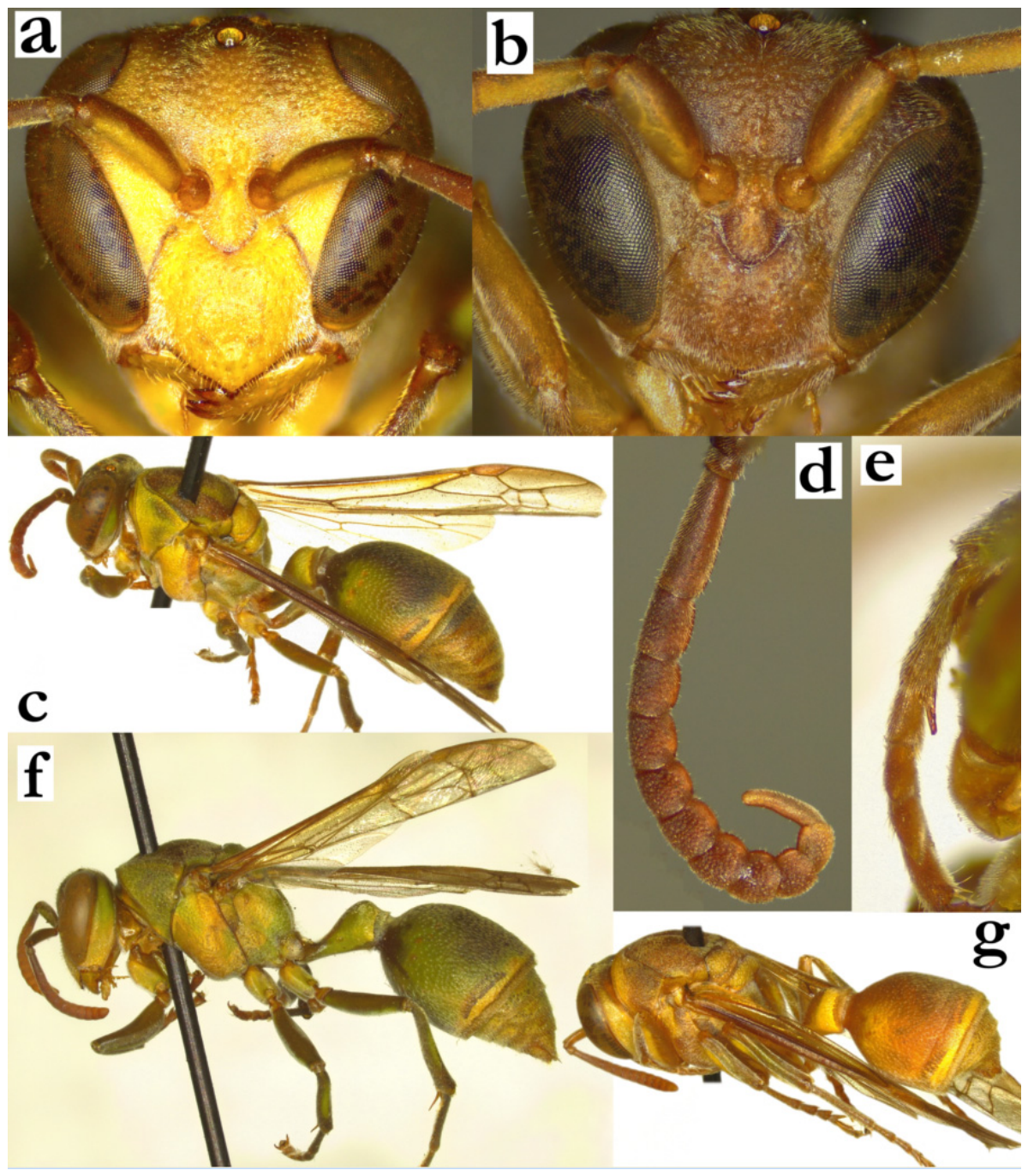
- Ropalidia linearecta Blommers, 2012Ropalidia linearecta n. sp., Blommers 2012: 184; ♂♀, nest, biology, Madagascar; holotype NNM.Number of examined specimens: 36 ♀♀, 17 ♂♂.
Type specimens. The description publication provides sufficient features to establish this species [6]; several specimens from the Blommers collection were examined.
Notes. A member of the grandidieri group, characterized by smaller size and very dense pubescence that obstructs the view of body surface (Figure 96a). It has a wide distribution across the entire island (Figure 96b).
Males have an inverse pattern of tyloids, making this a sufficient determination characteristic; more proximal flagellomeres have elevated tyloids, which become flattened in the distal ones (Figure 18d). Females may provide difficulties in separation from smaller specimens of R. grandidieri (de Saussure), which can have similar pilosity. The most useful criteria in that case are the occipital carina that is placed very high on postero-dorsal head margin (Figure 96c), in contrast to occipital carina that is placed lower than posterior margin of the head in other species of the grandidieri group. An additional useful criterion is the deep-green basal spot on mandible (Figure 96d), which is also uncommon in other species of this group.
Nest. This species has elongated nest shape, with 2×n formula, with up to 60 cells in length. However, a possible reuse of the cell wall was observed [85], a pattern that was also observed in R. bongomaso sp. nov.
Figure 96.
Ropalidia linearecta Blommers, (a) habitus, lateral view, female, (b) distribution map (red marker—specimen, grey marker—literature report or an observational record), (c) head, oblique dorsal view, female, (d) mandible, oblique inferior view, female.
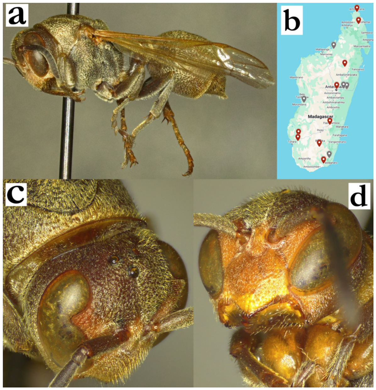
- Ropalidia mainty sp. nov. Polašek and DagleyZooBank: E77DEAF9-CF83-418F-8187-81A139F56D72Material examined. Holotype: “Manambato/ (Anove)”, “INSTITUT/ SCIENTIFIQUE/ MADAGASCAR”, ♀ (MSNV); Figure 97a–f.Paratypes: “MUSEUM PARIS/ MADAGASCAR/ BAIE D’ANTOGIL/ A. MOCQUERYS 1898”, 1♀ (MSNV).Number of examined specimens: 2 ♀♀.
Diagnosis. Member of the formosa group, characterized by anterior part of mesoscutum punctate, T1 globular, pilosity short, metanotum rounded posteriorly and femur impunctate.
Description. Females. Wing length 12.7–13.5 mm.
Head. Head frontally 1.1–1.2× as wide as high, in dorsal view 2.8× as wide as long, not narrowing behind eyes, barely emarginate posteriorly (Figure 97c); juxtamandibular excavation moderately developed, apex protruding; clypeus base convex, coarsely and sparsely punctate; inner orbit punctate (Figure 97a). Frons punctures larger and denser than those on clypeus, less than a diameter apart (Figure 97a); gena less coarsely punctate, with shiny surface (Figure 6b). Gena at midpoint as wide as eye, punctate superiorly near eye, inferiorly punctures less defined, area close to occipital carina impunctate (Figure 6b). Mandible shiny, with some coarse punctures basally. Occipital carina complete (Figure 6b). Posterior ocelli about a diameter apart, 3.2× diameters apart from eye and 2.7× diameters apart from occipital carina (Figure 97c). Eyes barely setose (Figure 97b); scape conspicuously longer than AF1, AF1 shorter than AF2+3+4; AF2 about 1.3–1.5× as long as wide (Figure 97a).
Mesosoma. Mesosoma slender, 0.95× as wide as head in dorsal view; 1.3× as long as wide (Figure 97c), in lateral view 1.6× as long as wide; mesoscutum 1.15× as wide as long (Figure 117c); pronotum in dorsal view mildly sinuate excavated medially, humeri rounded; carina of pronotum complete and thin, dorsally 1.5× as wide as laterally; pronotum punctures shallow, 1.5 diameters apart, with swollen and matte interspaces (Figure 97c). Mesoscutum with less defined and more spaced punctures compared to pronotum (Figure 97c). Mesepisternum sparsely and coarsely punctured in lateral area, with traces of reticulation towards mesepimeron (Figure 97d); impunctate or with just a few punctures anteriorly; posterior surface with less dense and punctures of same size, similar to those on mesepimeron; metepisternum basally matte, with somewhat more spaced and well-defined punctures. Scutellum dorsally rounded, mildly bituberculate, metanotum posteriorly rounded (Figure 6a). Propodeum posteriorly narrowing; median depression of propodeum developed, weakly horizontally striate and shallowly punctate (Figure 6a).
Metasoma. T1 pyriform, with minimal posterior sulcus (Figure 97a); anterior surface impunctate, posterior with some poorly defined craters. T2 1.3× as long as high, tergum and sternum similarly convex; punctures shallow and coarse, directional, about a diameter apart, with shiny interspaces; punctures on S2 somewhat larger and sparser, especially proximally; transition between T2 surface and lamella gradual, lamella translucent and very weakly punctate proximally, distal third impunctate; remaining metasomal segments shallowly punctate.
Pilosity. Body with short yellowish-white pubescence, clypeus with protruding setae, 2.0x as long apically as basally (Figure 6b); mandible with protruding setae of similar length as clypeus apically; frons with straight protruding setae, 0.9–1.3× as long as anterior ocellus diameter; gena and occiput with some protruding setae; eyes very finely setose (Figure 42a). Propodeum with some longer whitish fine setae; T2 with protruding setae that do not extend over lamella (Figure 117a); remaining metasomal segments with some protruding setae distally; coxa with fine silvery pubescence and some silvery protruding setae with bent tips; femora with dense silvery pubescence, femur III also with short pubescence and some silvery protruding setae with bent tips, of approximately same length proximally and distally (Figure 6c).
Colour. Basal colour uniformly black(ish) with following faint suffused green areas: clypeus, inner orbits, mandible basally (Figure 97a), gena, pronotum medio-dorsally, inferior pronotal angle, posterior band of T1 (Figure 97a,b); femur black, tibia and tarsi olivaceous or brown. Spot high on mesepisternum, posterior part of scutellum and lateral parts of metanotum reddish. Wings uniformly brownish, nervature brown, apical spot absent (Figure 97a). Antenna dark brown dorsally, reddish or lightly reddish ventrally (Figure 97a).
Males. Unknown.
Distribution. North-eastern part of the island.
Etymology. The name originated from a Malagasy word mainty, “black”, referring to basal body colour.
Figure 97.
Ropalidia mainty sp. nov., female, holotype, (a) habitus, oblique lateral view, (b) head, frontal view, (c) head and mesosoma, dorsal view, (d) mesepisternum, lateral view, (e) propodeum, dorsal view, (f) specimen labels.
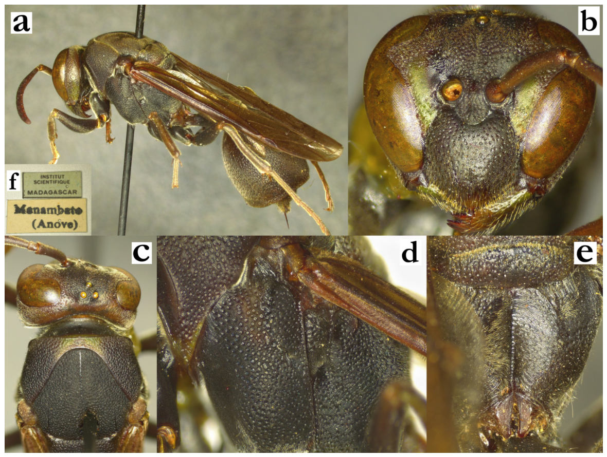
- Ropalidia marolavaka sp. nov. PolašekZooBank: 9E2CCF2E-A58E-4999-8686-E39FB4F76CF1Material examined. Holotype: “Hera/Ankazoabo”, “INSTITUT/ SCIENTIFIQUE/ MADAGASCAR”, ♀ (MSNV), Figure 98a–d.Paratypes: 15♀♀, 6♂♂, same label as holotype (MSNV); “MADAGASCAR:TAM/ Mahabo, 20°22 S–44°40 E/ Ipomoea arborescens /12.xii.1991, A. Pauly”, 1♀ (RMCA).Observations: District de Mahabo [59].Number of examined specimens: 17 ♀♀, 6 ♂♂.
Diagnosis. This species is characterized by body dorsally green, ventrally yellow, punctures coarse with swollen glassy interpuncture spaces and pubescence very short.
Description. Females. Wing length 7.2–7.8 mm.
Head. Head frontally 1.1–1.2× as wide as high; clypeus 1.25× as wide as long, with straight upes and developed OCA (Figure 36b); juxtamandibular lobe moderately developed, apex projecting (Figure 36b); clypeus basally shiny and impunctate, medially with scattered and shallow, poorly defined punctures and some apical craters (Figure 36b); frons coarsely punctate, with similar pattern extending posteriorly high on gena and occiput (Figure 36a), while lower part of gena with less defined punctures, almost impunctate (Figure 98c); mandibles punctate, occipital carina complete; eyes with short and scattered setae or asetose (Figure 36b); ocelli with wider base than sides; scape longer than AF1, AF2 as wide as long.
Mesosoma. Mesosoma comparatively shorter; mesoscutum 1.1× as wide as long (Figure 36a); mesosoma dorsally coarsely and densely punctate (Figure 36a); mesoscutum and pronotum similarly punctate, with swollen and partly shiny interspaces; punctures one diameter or more apart; mesepisternum sparsely punctate, metepisternum with a few scattered, weakly defined punctures; scutellum with weakly developed median carina obscured by glassy and translucent punctures interspaces; metanotum with postero-median impunctate shiny area (Figure 36c); propodeum with rounded contour, without strong carinae; entire surface of propodeum punctate, infero-lateral areas with additional striation (Figure 36c).
Metasoma. T1 elongated and pyriform with globular posterior part (Figure 98d); posterior third of T1 coarsely punctate with elevated glassy puncture interspaces (Figure 98d); T2 coarsely punctate, punctures proximally about 1.5× diameters apart, distally about a diameter apart (Figure 98d); lamella wide, proximal half with poorly defined micro-punctures, distally translucent and impunctate (Figure 98d); remaining metasomal segments shallowly punctate.
Pilosity. Microscopic in most body parts, except clypeus (Figure 98c) and propodeum; apical third of clypeus with twice longer setae than base; frons with short golden straight setae, about 0.5–0.7× as long as anterior ocellus diameter (Figure 98c); mesosoma dorsally with short golden or whitish setae (Figure 98c); T2 with some protruding yellowish setae, that do not extend over lamella (Figure 98d).
Colour. Head and mesosoma dorsally green, ventrally yellow, without strong margin between the two (Figure 98a); metasoma darker green, sometimes T2 with clear basal yellow spot (Figure 98a); clypeus basally green, apically yellow (Figure 36b), in some specimens entirely yellow; mandible yellow, inner orbit and interantennal area yellow, with gradual conversion to green frons (Figure 36b); gena high greenish, inferiorly yellow (Figure 98c); mesosoma with similar colour pattern, dorsally green, ventrally yellow (Figure 36a); leg colour pattern inverse; coxa and proximal part of femora yellow, distal part of femora, tibia and tarsi green; terminal two tarsal segments olive-green or brownish (Figure 98a); metasoma dark green; T1 green, sometimes with two suffused yellowish triangular postero-lateral areas; T2 with a posterior light-green band, sometimes with a basal spot (Figure 98a); S2 with posterior light-brown band, sometimes with two basal corresponding suffused yellowish spots; remaining terga mostly green, sterna yellowish; wings transparent, lightly yellowish; apical spot not visible at all, stigma light brown or yellowish (Figure 98a); scape dorsally green, distal segments variable (green to olive-green, sometimes even brownish), underside proximally green, distally yellow (Figure 36b).
Males. Resemble females in general appearance, but often have more yellow colour on the body. Wing length 7.0–7.4 mm.
Head. Clypeus 1.1–1.2× as wide as long, upes short and curved, OCA visible but weak, apex slightly bent outwards (Figure 36d); clypeus shallowly punctate, base sometimes impunctate and shiny; gena 0.5–0.7× as wide as eye; mandible shiny, nearly impunctate; scape moderately widened, 1.6–1.9× as wide as AF1 base width, shorter or just about as long as AF1; AF2 1.2–1.5 times longer than wide (Figure 36e); tyloids yellowish and shiny, originate at AF(2)3, flattened (Figure 98e); terminal flagellomere elongated, 1.9× as long as AF10, beak-like and acute (Figure 36e).
Mesosoma. Fore tarsal spur I well developed, with a wide base (Figure 98f).
Metasoma. S7 flattened, with shiny and somewhat irregular surface.
Pilosity. Clypeus surface with intermediate length, fine yellowish setae (Figure 36d);
Colour. Face mostly yellow, mesosoma with more yellow, green areas more lightly coloured than in females. T1 often with posterior yellow band and two lateral yellow areas, S2 sometimes entirely yellow. Antenna dorsally ferruginous, scape and pedicel greenish; ventrally ferruginous-orange (Figure 36e and Figure 98e).
Distribution. South-Western part of the island (Figure 98g).
Etymology. The name is a combination of two Malagasy words, an adjective maro, “many”, and the noun lavaka, “crater, hole”, referring to the strongly developed punctures, treated as indeclinable.
Figure 98.
Ropalidia marolavaka sp. nov., (a) habitus, lateral view, female, holotype, (b) labels, holotype, (c) head and anterior mesosoma, lateral view, female, holotype, (d) metasoma, lateral view, female, holotype, (e) antenna, dorsal view, male, (f) tarsal spur I, lateral view, male, (g) distribution map (red marker—specimen, grey marker—observations).
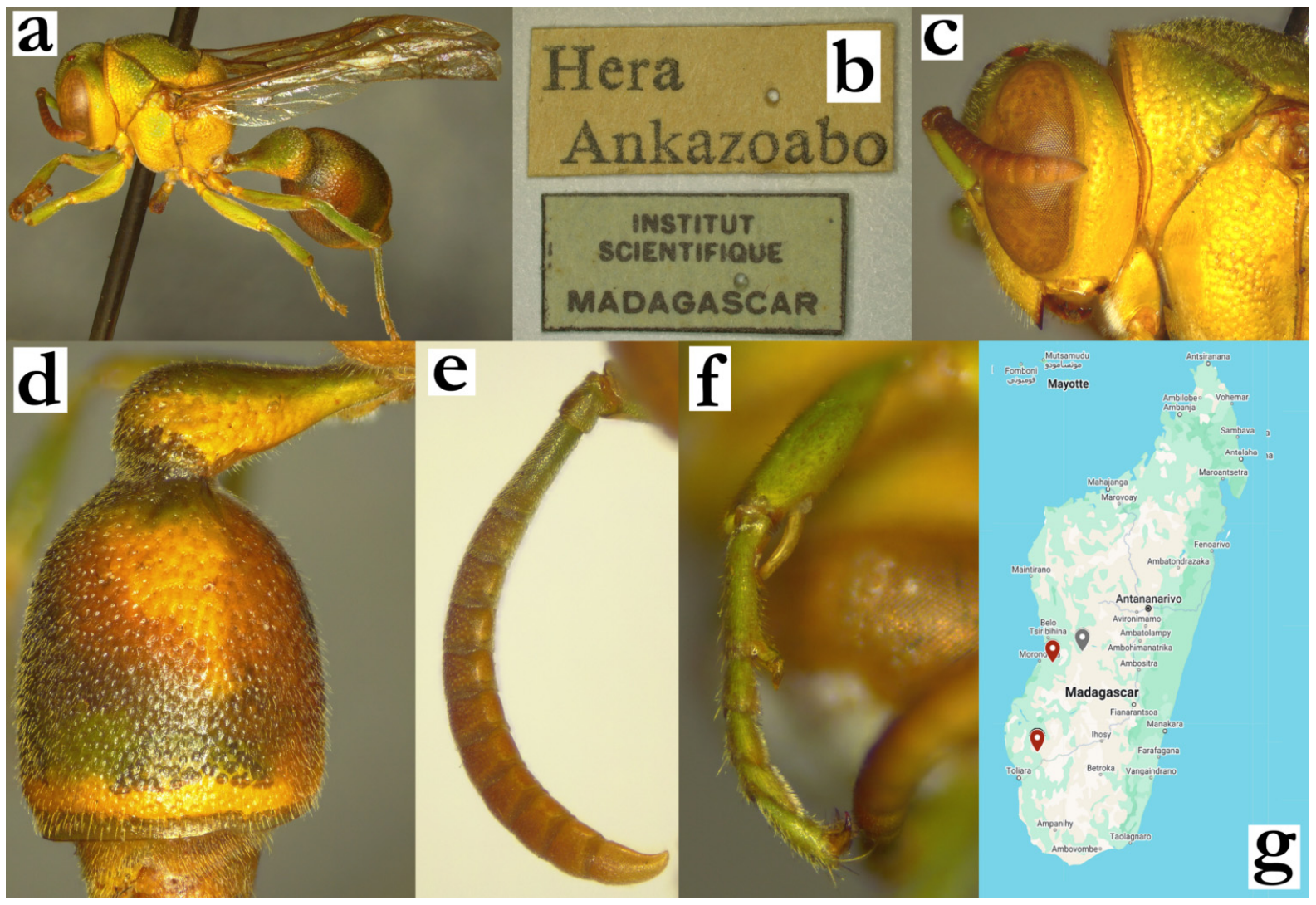
- Ropalidia merina Blommers 2012Ropalidia merina n. sp., Blommers 2012: 150; ♂♀, nest, biology, Madagascar; holotype NNM.Ropalidia hova var. minor [9]Ropalidia formosa [16]Number of examined specimens: 19 ♀♀, 3 ♂♂.
Type material. This taxon was described by Blommers, who resolved the relationships of several similar taxa that were often previously treated together. Male and female paratypes were examined; male originated from the nest 70.13 (Figure 99a,b), and female from the nest 72.14.
Notes. This species is easily separated from other species by comparatively larger body size, basal black colour with a characteristic set of greenish-yellow markings on mesosoma. These usually come in pairs, and this can provide grounds for colour-based determination; other members of the formosa group do not have greenish hue of the mesosoma markings that includes (almost mandatory) markings on scutellum and metanotum.
Nest. This species has the most commonly misidentified nests in previously publications, which was already reported before [6]. Nests were reported in larger aggregations, with up to 240 nests [16]. Blommers also reported that sometimes nests are reused in another season [6].
Distribution. Most specimens originated from the central highlands and the Eastern coast.
Figure 99.
Ropalidia merina Blommers, (a) habitus, oblique lateral view, male, paratype from the nest 70.13, (b) labels, (c) head and mesosoma, dorsal view, female paratype from the nest 72.14, (d) Abdomen, lateral view, male, (e) head, frontal view, male, (f) antenna, median view, male.
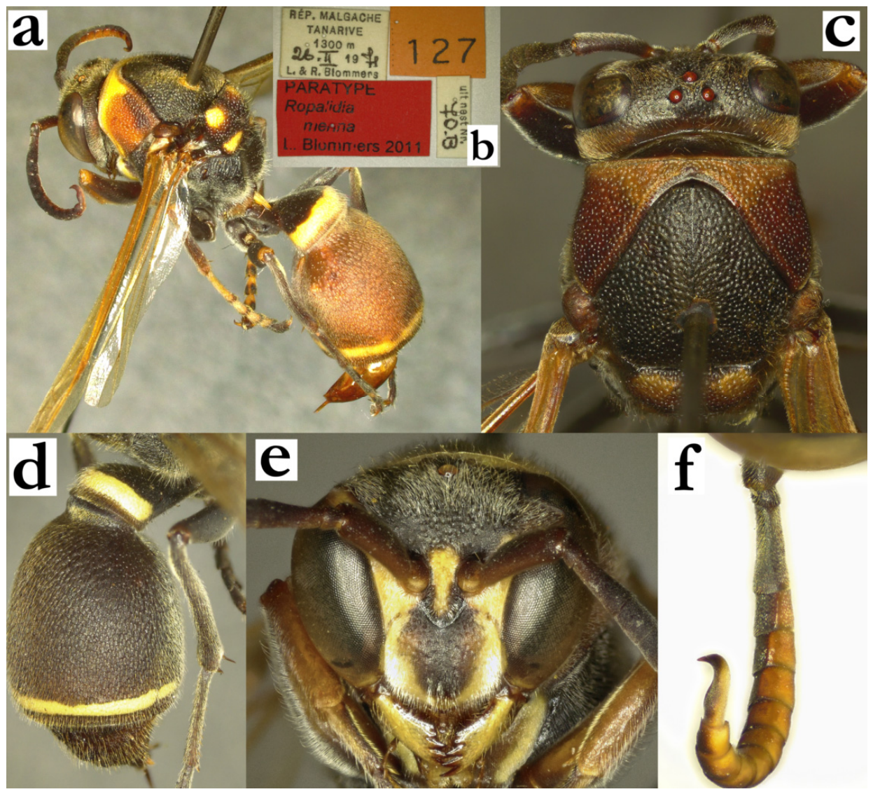
- Ropalidia miafina sp. nov., PolašekZooBank: 39D7FCC1-1D8E-46AD-B5C1-0BCAEAD401F0Material examined. Holotype: “MADAGASCAR/ ROGEZ/ FORET COTE EST”, “MUSEUM PARIS/ IV.37/ A. SEYRIG”, ♀ (MSNV), Figure 60a,b and Figure 100a–e.Paratypes: 4♀♀, 5♂♂, same labels as holotype; “MADAGASCAR/ ROGEZ/ FORET COTE EST”, “MUSEUM PARIS/ II. 34A. SEYRIG”, 1♀, 1♂; “Ivohibe/XI 50, AR”, 3♀♀ (all in MSNV); “COLL. MUS. CONGO/ Madagascar: Rogez for./ Analandraraka VI-3/ A. Seyrig”, 1♀ (RMCA).Observation: Ranomafana National Park, [86].Number of examined specimens: 9 ♀♀, 6 ♂♂.
Diagnosis. Characterized by small size, colour predominantly green and black colour, T1 elongate and impunctate, mesoscutum with thing green lines in combination with blackish base of T2, face mainly yellow, mesoscutum shallowly and sparsely punctate and T2 shallowly punctate.
Description. Females. Wing length 5.9–6.7 mm.
Head. Head frontally 1.25× as wide as high (Figure 100a), in dorsal view 2.75× as wide as long, narrowing behind eyes, emarginate posteriorly. Clypeus 1.15–1.2× as wide as long, with elongated shape; upes curved, OCA narrow (Figure 100a); juxtamandibular excavation shallow, apex very pointy (Figure 100a); clypeus with cuticular surface and some poorly defined punctures basally, apically less defined craters hardly discernible due to silvery pubescence (Figure 100a). Frons coarsely punctate, punctures one diameter apart, with swollen and matte interspaces (Figure 100a); gena at midpoint 1.8× as wide as eye, weakly punctate to impunctate, some punctures retained close to eye and mandible (Figure 100c); mandible punctate. Occipital carina complete and very developed (Figure 100c); posterior ocelli 1.15 diameters apart, 2.15 diameters apart from compound eye and 1.7 diameters apart from occipital carina. Scape 3.3–3.4× as long as wide, 1.45× as long as AF1; AF2 1.25× as wide as long, remaining flagellomeres conspicuously wider than long (Figure 100a).
Mesosoma. Mesosoma slender, 0.95× as wide as head in dorsal view; 1.05× as long as wide (Figure 60a), in lateral view as long as wide (Figure 100c); mesoscutum 1.1× as wide as long (Figure 60a); pronotum in dorsal view mildly sinuate anteriorly, humeri rounded and angled; carina of pronotum complete and thin, dorsally at most 2.6× as wide as laterally; pronotum punctures shallow and sparse, up to 2 diameters apart, with swollen and matte interspaces, retained even inferiorly (Figure 100c). Mesoscutum with similar punctures to those on pronotum, but somewhat less coarse (Figure 60a). Mesepisternum without carina towards mesepimeron, sparsely and coarsely punctured laterally, similar to pronotum; metepisternum with cuticular surface and with only a few scattered punctures anteriorly. Scutellum with shallow and scattered punctures, mildly swollen and matte interspaces (Figure 100d), median carina developed, reaches between a third and half of anterior scutellum surface (Figure 60a). Metanotum with somewhat larger and more spaced punctures than those on scutellum, with posterior shiny impunctate area (Figure 100d). Propodeum posteriorly narrowing; median depression of propodeum shallow and barely emarginated; surface with very fine horizontal striae, similar medially and laterally, impunctate (Figure 100d).
Metasoma. T1 pyriform and elongated, posteriorly shallowly punctate (Figure 100e). T2 with dorsally flattened surface, yielding asymmetrical second metasomal segment contour (Figure 100e); punctures shallow, directional, 1.5 diameters apart, with shiny and flat interspace; punctures on S2 somewhat larger and sparser, especially proximally; transition between T2 surface and lamella very gradual, lamella translucent and very weakly punctate proximally, about half a puncture wide, distally impunctate; remaining metasomal segments shallowly punctate.
Pilosity. Body with short pubescence, silvery on clypeus, interantennal area, gena and propodeum, yellowish on other body parts (Figure 100c); clypeus with protruding setae, 1.4× as long apically as basally; mandible with protruding setae of similar length as clypeus apically; frons with protruding setae with bent tips, 1.2–1.5× as long as anterior ocellus diameter; gena and occiput with a few short protruding setae; eyes finely setose (Figure 100c). T1 anteriorly with fine pubescence, posteriorly with some yellowish setae that project over posterior margin; T2 with protruding setae that extend over lamella (Figure 100e); remaining metasomal segments with some protruding setae distally; coxa with fine silvery pubescence and some silvery protruding setae with bent tips; femora with dense silvery pubescence, femur III also with longer setae.
Colour. Basally brownish, with numerous green and yellow areas; yellow areas include most of ventral body parts, including face, gena and mandible, coxa I(-III) (Figure 60b), and suffused yellow areas under pronotal carina, and at posterior bands of T1 and T2 (Figure 100e). Green areas include most of dorsal parts of body, including frons (Figure 100c), gena, pronotum, two lines on mesoscutum, tegula (Figure 60a), scutellum, metanotum and propodeum (Figure 100d), coxa, parts of femur and tibia; tarsi light brownish (Figure 60b). Antenna dorsally brown and infuscate, ventrally greenish-brown (Figure 60b). Wings translucent or barely yellowish at costa, nervature brown, stigma light-brown and partly translucent (Figure 60b).
Males. Resemble females in general appearance, except mesosoma underside mostly yellow and all leg segments green (Figure 100f). Wing length 6.1–6.4 mm.
Head. Head in frontal view 1.4× as wide as high (Figure 100g); clypeus barely punctate, apex depressed (Figure 100g). Scape 2.8× as long as wide, 1.5× as wide as AF1 base; AF1 2.7× as long as wide; AF2 1.25× as long as wide (Figure 100h). Tyloids shiny, originate at AF(4)5, brownish and matte (Figure 100i); terminal flagellomere elongated, 1.7× as wide basally as long in straight line, with thin tyloid that reaches subacute tip (Figure 100h).
Mesosoma. Fore tarsal spur I developed, but small (Figure 100f).
Metasoma. S7 barely concave, with shiny and irregular surface.
Pilosity. Overall with longer head setae than females; clypeus with fine silvery setae apically.
Nest. One nest was observed [86]. It was observed in the early colony cycle, with three adults and one or a few apparent cocoon caps [86]. The nest was hanging under an elongated leaf, with an apparent central petiole, centrally located cells with closed cocoon caps, and several rows of shorter, peripheral cells that already had laid eggs very early in the cell wall development.
Distribution. Eastern Madagascar (Rogez to Ivohibe).
Etymology. The name originates from the Malagasy word miafina, translated as “secret-keeper, hidden”, treated as a noun, referring to the similarity and initial difficulties in separation from R. phalansterica (de Saussure) and R. mysterica Blommers (Figure 54).
Figure 100.
Ropalidia miafina sp. nov., (a) head, frontal view, female, holotype, (b) labels, holotype, (c) head and mesosoma, lateral view, female, holotype, (d) posterior mesosoma, dorsal view, female, (e) T1 and T2, lateral view, female, (f) mesosoma underside, oblique inferior view, male, (g) head, frontal view, male, (h) antenna, dorsal view, male, (i) antenna, median view, male.
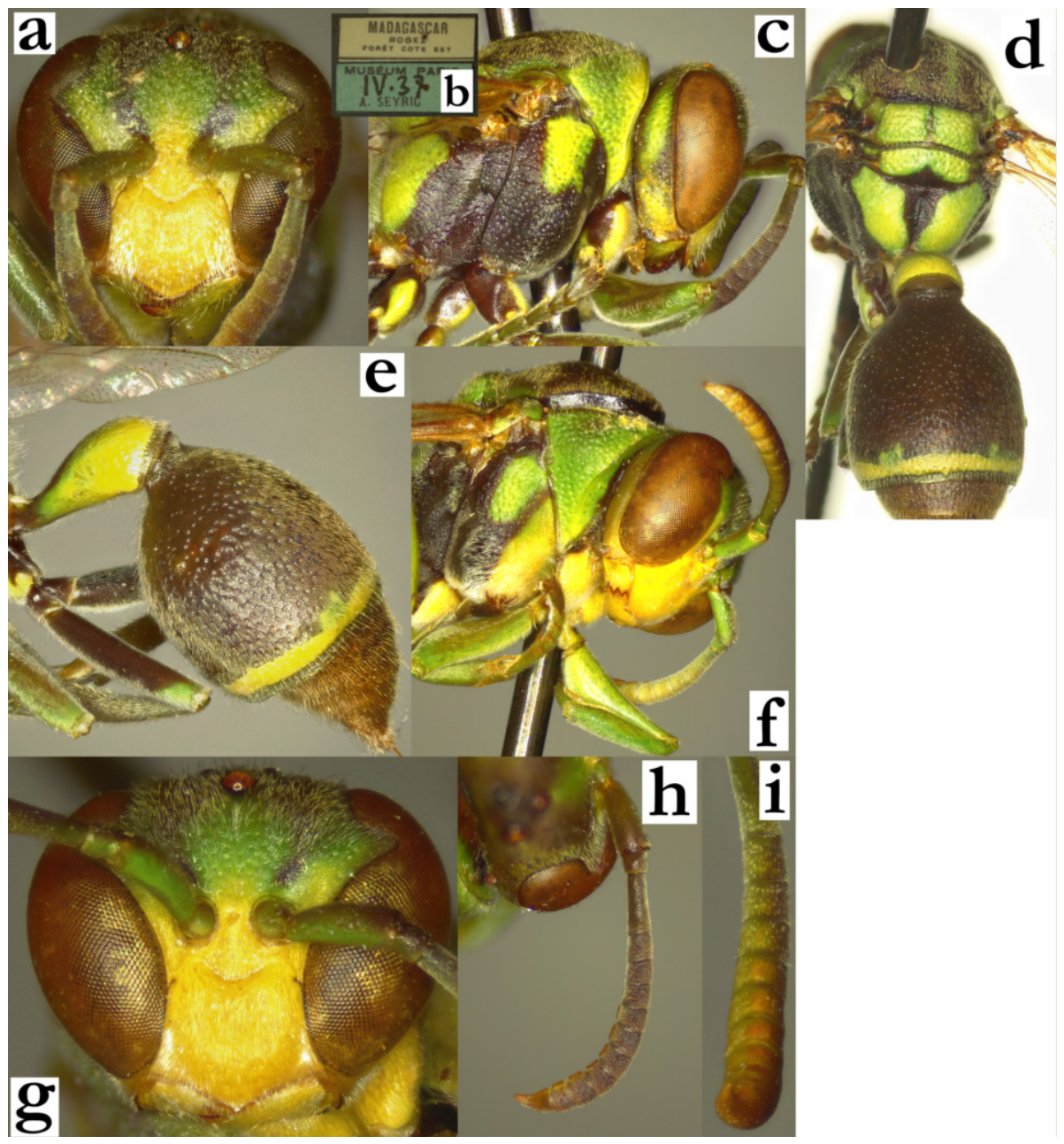
- Ropalidia minor (de Saussure, 1900)Icaria minor (de Saussure), 1900; “In Madagascar vulgaris” (MHNG).Number of examined specimens: 10 ♀♀, 5 ♂♂.
Type material. This taxon was originally described by de Saussure as a variety of Icaria hova [14]. Subsequent publications often repeated this status, until Giordani Soika listed it as a separate species [4]. The original description suggests a smaller species, with plenty of yellow markings, including those on femora. Several specimens from MHNG could be treated as types [2]; due to clear taxonomical situation and lack of similar species, no additional type status was assigned.
Notes. A very characteristic species, which does not seem to be a member of any species group. No other Malagasy Ropalidia has lateral projections on T1, making this a sufficient determination feature (Figure 14b). In addition, several colour-based patterns on the head are specific, including wide and apically blunt yellow spot on interantennal area (Figure 101a,b). The general colour pattern in females (Figure 14a) is very similar to males (Figure 101c), with the exception of more yellow markings on mesosoma underside (Figure 101d).
Similar species. The general appearance may sometimes be misleading in separation from R. carinata (de Saussure); this species has no yellow markings on the mesosoma, and has a differently shaped T1 (Figure 17a). Some specimens of R. volontany sp. nov. can resemble R. minor, but in these cases, there are numerous colour-based and morphological differences, listed in the key to species. Lastly, colonies of R. galimatia (de Saussure) with darker basal body colour may sometimes be slightly problematic [87,88]; that species usually has lighter basal body colour and multiple yellow markings on mesosoma, in combination with the fraterna group shape of T1, globular with two postero-lateral excavations (Figure 15e).
Genetics. Two specimens were sequenced (a male and a female), suggesting a basal position in the phylogenetic tree, unrelated to any other species.
Figure 101.
Ropalidia minor (de Saussure), (a) head, frontal view, female, (b) head, frontal view, male, (c) habitus, dorsal view, male, (d) habitus, lateral view, male.
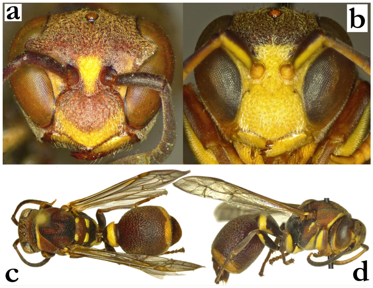
- Ropalidia mysterica Blommers 2012Ropalidia mysterica n. sp., Blommers 2012: 158; ♂♀, nest, biology, Madagascar; holotype NNM.Number of examined specimens: 41 ♀♀, 11 ♂♂.
Type material. Holotype photographs, deposited in NNM, are provided in the description of this species, exhibiting characteristic posterior T1 coarse punctures and punctate posterior lamella of T1 [6]. However, a female from the implied paratype nest 72.46 is actually a melanic form of R. phalansterica, meaning that the paratype status of specimens from the nest 72.46, listed in the description publication as “Nest 72.46 + 2 + 3 queens, 2 workers, 1 male + 5 females reared same place and date as holotype” is misidentified (Figure 102a). None of the characters fit the paratype status, including posteriorly impunctate T1, lack of angulated occipital carina or head colour. Notably, the holotype photographs (Figures 43–47 in [6]) exhibit these characteristics, suggesting that the holotype specimen status is not questioned.
Notes. This species is characterized by the coarser punctures pattern, especially on T1 (Figure 102b), with punctate T1 posterior lamella, absent in other species from this group. Head and mesosoma are coarsely punctate (Figure 102c). Some confusion is possible in separation of R. miafina sp. nov., but several morphological and colour characteristics are sufficient for their separation (see R. miafina sp. nov. description for detailed comparison). Terminal flagellomere of males provides sufficient singular characteristic for determination (Figure 102d); notably, there is a nest series from Ambodivoangy (Maroantsetra), which have smaller body size, overall darker appearance and a single examined male in this series has much less developed tyloid tubercle on terminal flagellomere (Figure 102d).
Nest. Elongated, up to 30 cells, irregular in shape, with a central pedicel; nesting in forested areas, on leaves of mango, cardamom or Hedychrysum [6]. One nest was examined, with 47 cells, arranged in the regular 4 × 11 frame (Figure 102e,f). In contrast to previous reports of paracentral petiole, this nest has an apical petiole, which is substantially glazed and shiny. The characteristics of the nest might be confounded, since the nest is broken in two parts across the middle (Figure 102f). The cells are deep, about 2.8× as long as wide, and apparently slightly angled downwards (Figure 102e). The nest is attached to a leathery dried leaf, possibly a palm leaf (Figure 102e). Notably, the description publication reported a paracentral petiole [6], suggesting a degree of plasticity in the nest development or a different nest stage (additional cells may be added later on, converting the petiole from apical to paracentral).
Figure 102.
Ropalidia mysterica Blommers, (a) contested paratype from the nest 72.46 which is actually a melanic form of R. phalansterica, (b) posterior mesosoma and metasoma, dorsal view, female, (c) head and anterior mesosoma, dorsal view, female, (d) antenna, dorsal view, male, (e) nest, lateral view, (f) nest fragment, dorsal view.
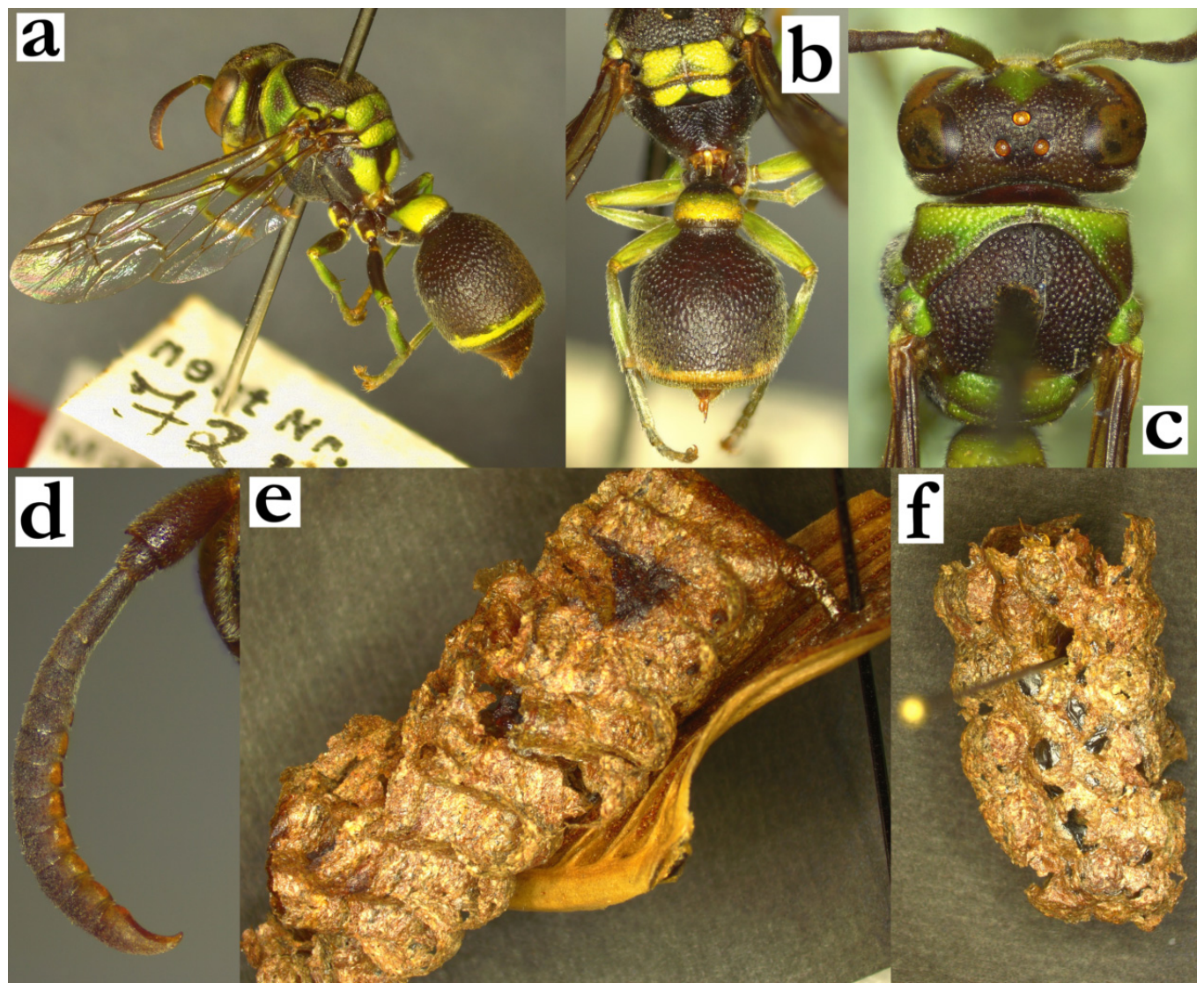
- Ropalidia nitidula (de Saussure) 1890Icaria nitidula de Saussure, 1890; “Madagascar” (MFNB).Plazi: https://tb.plazi.org/GgServer/html/03AE106DFFD5396B25D7FE63842EF30F (accessed on 21 September 2026).Number of examined specimens: 6 ♀♀.
Type material. A female specimen from MFNB was examined; the specimen fits the description well, prompting Kojima to label it as an (unpublished) holotype. The status is hereby confirmed.
Notes. A larger species, characterized by the light brown basal colour, large yellow areas on pronotum, (Figure 103a), wide female gena and shallowly punctate head and mesosoma (Figure 103b), while metasoma is darker than mesosoma and more coarsely punctate (Figure 103a); the entire body is covered by a microscopic pubescence (Figure 103c). This species is building very interesting, droplet-shaped nests, commonly located in clusters and above water [30,31].
Group membership. T1 of this species is quite unusual, resembling possibly R. ducalis or R. vitripennis. The gradual conversion of T2 to lamella surface suggests a similarity to formosa+grandidieri groups.
Distribution. Western Madagascar (Figure 103d).
Figure 103.
Ropalidia nitidula (de Saussure), (a) habitus, oblique lateral view, female, (b) head and mesosoma, dorsal view, female, (c) head, frontal view, female, (d) distribution map (red marker—specimen, grey marker—observations and literature reports).
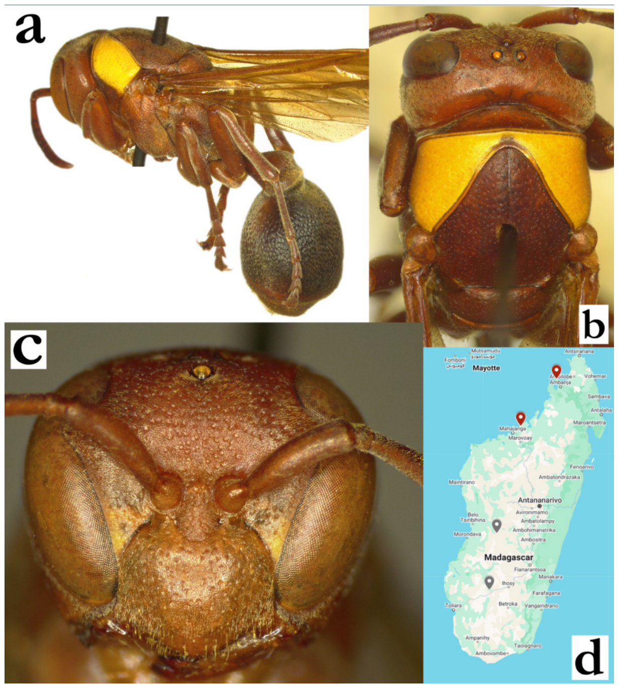
- Ropalidia perplexa Blommers, 2012Ropalidia perplexa n. sp., Blommers 2012: 178; ♂♀, nest, biology, Madagascar; holotype NNM.Number of examined specimens: 7 ♀♀, 1 ♂.
Type material. Two paratypes were examined from NNM collection, a female from the nest 72.11 (Figure 104a,b) and a male from the nest 72.8.
Notes. This is almost exclusively green-yellow coloured species with minimal brown(ish) markings (Figure 104c–f) and long golden protruding setae that are present on most body parts. Five females from Manambato and one from Maroantsetra (MSNV) were initially considered to be a separate species. These females are smaller, and have shorter pilosity; the examined paratypes of R. perplexa have longer setae on the head, clypeus and T2, while these females have shorter setae on all body parts, and also lack protruding setae on the antenna, while the remaining morphological and colour feature correspond to the paratypes.
Distribution. The northern part of the island.
Figure 104.
Ropalidia perplexa Blommers, (a) habitus, lateral view, female, paratype from the nest 72.11, (b) labels, paratype, (c) head, frontal view, female, (d) head and anterior mesosoma, dorsal view, female, (e) head and anterior mesosoma, lateral view, (f) head, frontal view, male.
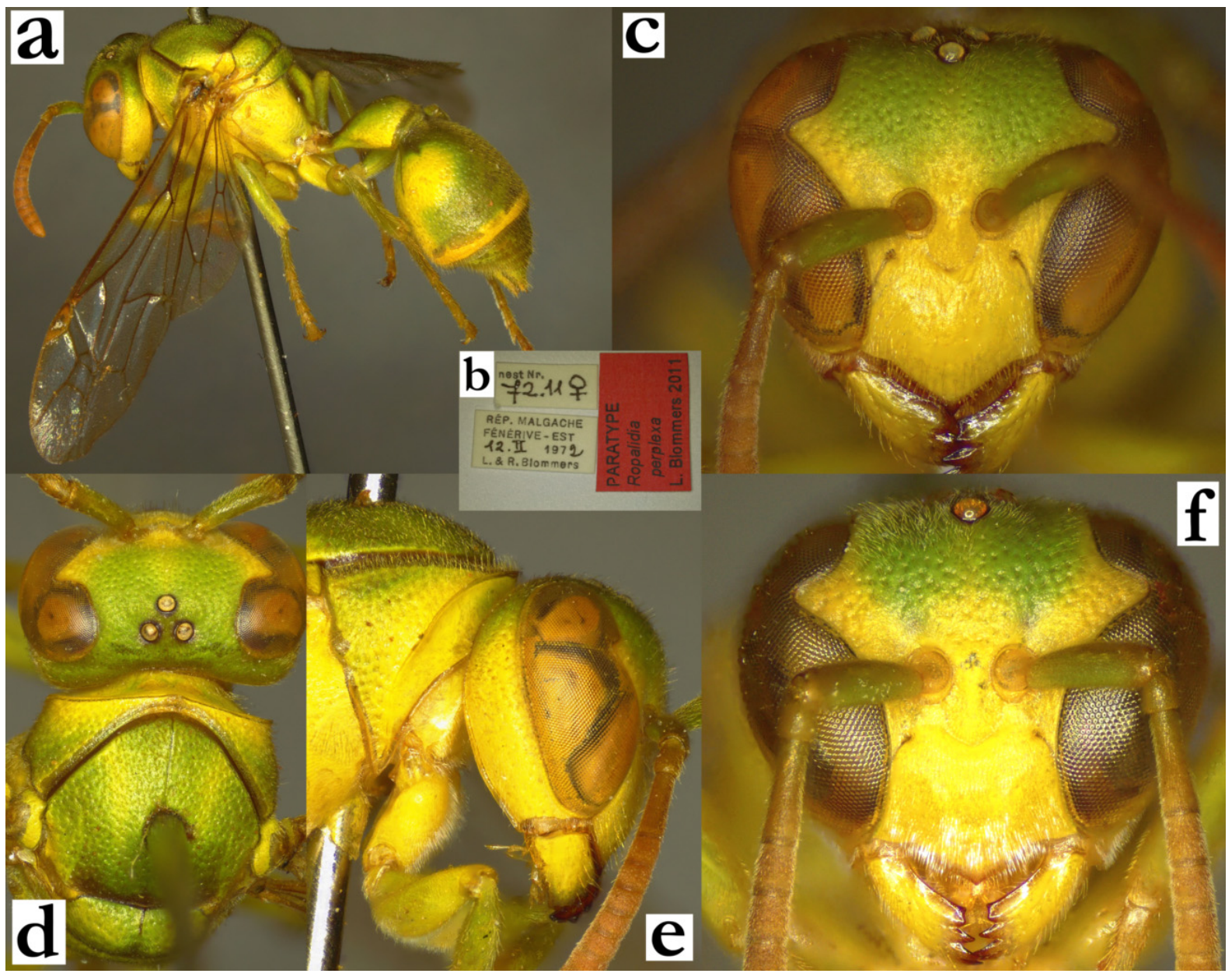
- Ropalidia phalansterica (de Saussure), 1853Icaria phalansterica de Saussure 1853; “Madagascar” (MNHN).Plazi: https://tb.plazi.org/GgServer/html/03AE106DFFD5396A25D7FB4B842EF253 (accessed on 21 September 2026).Holotype photographs: https://science.mnhn.fr/taxon/species/ropalidia/phalansterica (accessed on 21 September 2026).Number of examined specimens: 25 ♀♀, 13 ♂♂.
Type material. Multiple syntype specimens are deposited in MNHN, including one male (designated MNHN-EY-EY6014), belonging to the series of specimens with the teal labels “Museum Paris/ Madagascar/ Jacquinot 1841”. The specimen labelled as “Museum Paris/EY6013” was labelled by Kojima as lectotype, and the status is hereby confirmed. Additionally, there was another specimen identified from the type series, hereby designated as paralectotype, deposited in MSNV (Figure 105a–c).
Notes. This species is characterized by a combination of smaller size (wing length does not exceed 7 mm, in most specimens it is slightly above 6 mm), numerous dark brown areas on the body and bulging eyes in males. Two colour forms can be defined in this species, the melanic and the xanthic. The first corresponds to the holotype specimens and is characterized by the presence of a basal green or yellow spot on T2, while the melanic form lacks these spots.
Nest. This species builds small nests located on the underside of leaves. The cell wall is commonly uniformly grey [89], sometimes darker [90], or yellowish [91].
Similar species. Some confusion is possible in separation from smaller specimens of R. subclavata (de Saussure), which has a more robust body and overall more developed pilosity. The defining feature is dorsally developed and coarsely punctate T1 in R. subclavata (Figure 53a), compared to a pyriform and dorsally much less developed and finely punctate T1 in R. phalansterica (Figure 38a). The melanic form can be confused with R. miafina sp. nov., which has more elongated T1 (Figure 60b), and a strong pattern of lower face yellow (Figure 40d).
Figure 105.
Ropalidia phalansterica (de Saussure), (a) habitus, lateral view, female, syntype, (b) labels, female, syntype, (c) head, frontal view, female, syntype.
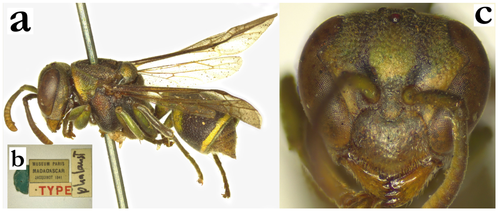
- Ropalidia prasina (de Saussure, 1900)Icaria prasina de Saussure, 1900; “Madagascar, Imerina” (MHNG).Plazi: https://tb.plazi.org/GgServer/html/03AE106DFFD4396A25D7FD88842EF3AE (accessed on 21 September 2026).Number of examined specimens: 8 ♀♀, 1 ♂.
Type material. De Saussure described this taxon in 1900 [14]. The original species description suggests Imerina as the type locality, meaning that the specimens with the label “Madagasc” that were presumed as types [2], cannot considered as such. In addition, the MHNG collection holds two such specimens, while de Saussure had at least four specimens, when he described multiple varieties of this taxon. In the absence of taxonomic uncertainties, the type status is not assigned.
Notes. A large, mainly green species, with a very broad female clypeus (Figure 106a) and mainly green body colour (Figure 106b,c) and slender antenna in males (Figure 106d). It is very similar to R. regina (de Saussure), with several characteristics listed in the key to species.
Distribution. Most examined specimens and literature reports indicated northeastern parts of the island (Figure 106e).
Figure 106.
Ropalidia prasina (de Saussure), (a) head, frontal view, female, (b) habitus, lateral view, female, (c) habitus, dorsal view, female, (d) antenna, median view, male, (e) distribution map (red marker—specimen, grey marker—observations and literature reports).
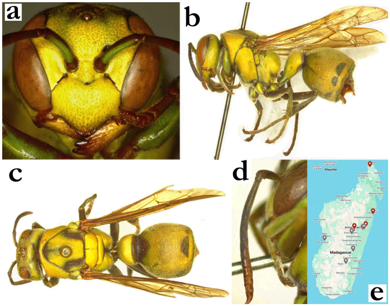
- Ropalidia pulchella (de Saussure, 1900)Icaria pulchella de Saussure, 1900; “Nosy Be” (MHNG).Plazi: https://tb.plazi.org/GgServer/html/03AE106DFFD4396A25D7FBC5842EF569 (accessed on 21 September 2026).Number of examined specimens: 1 ♀.
Type material. One female from MHNG (Figure 107a), with wide label, with multiple folds, that states “Hymenoptera with Nest”, “Nossi-Be 12 October 1891”, reddish label “pulchella”, [female sign] “SSS”, and an unpublished lectotype status by J. Kojima, 2000 (Figure 107b). The specimen is hereby designated as lectotype, with a collection designation MHNG ENTO 00089388.
A small species similar to R. constitutionalis, characterized by the dorsally developed T1 and excavated occipital carina. It was previously believed to be a synonym of R. phalansterica (de Saussure) [4], which was disputed by Carpenter and Blommers [2,6]. The examination of the type suggests a much more robust build than R. phalansterica (de Saussure), despite similar size. In addition, the holotype has a very specifically angled S2 contour, in contrast to more even and symmetric T2 contour in R. phalansterica. However, the species description mentions at least a few more specimens, characterized by the difference in colour and T1 shape; neither of these specimens were encountered in any of the collections, thus leaving only the holotype as the only examined specimen.
Nest. The species description is primarily based on the special nesting habit, with small nests located on the leaf, with cell openings directed downwardly [14].
Distribution. Previous publications list Nosy Be and Antsiranana [2,64], but only Nosy Be was confirmed in this study (Figure 107c).
Figure 107.
Ropalidia pulchella (de Saussure), (a) habitus, dorsal view, female, holotype, (b) labels, holotype, (c) distribution map (red marker—specimen, grey marker—literature report).
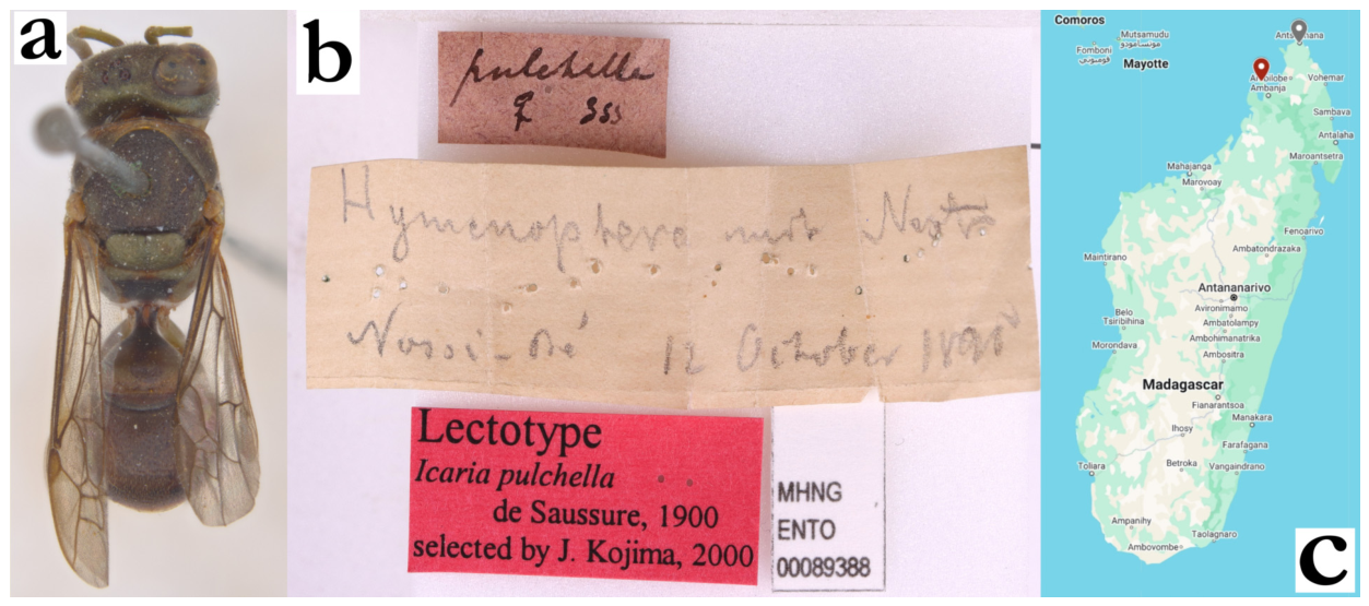
- Ropalidia rafitra sp. nov. PolašekZooBank: 025F0AE9-FE2F-461D-8A50-EEEC5AFABDE3Material examined. Holotype: “MADAGASCAR/Andasibe/18°55′21″ S 48°24′56″ E/20 March 2019, 868m/James M Carpenter/Nest 190320-1”, ♀ (AMNH_IZC 00341938); Figure 108a–e.Paratypes: 12♀♀, 2♂♂, same data as holotype (AMNH).Number of examined specimens: 13 ♀♀, 2 ♂♂.
Diagnosis. This species is characterized by small size, upes continuing to lateral clypeus margin, setose clypeus, elongate T1, medially excavated terminal flagellomere in males and elaborate fragmented nesting architecture.
Description. Females. Wing length 6.2–7.0 mm.
Head. Head frontally 1.25× as wide as high (Figure 42b), in dorsal view 2.1× as wide as long, narrowing behind eyes, barely emarginate posteriorly (Figure 108c). Clypeus 1.1× as wide as high, with short and curved upes that directly continue to lateral clypeus margin; OCA absent (Figure 42b); juxtamandibular excavation shallow, apex very pointy (Figure 42b); clypeus with cuticular surface and impunctate basally, apically punctures less defined and obscured by the pubescence (Figure 42b). Frons sparsely and shallowly punctate, punctures 1.5–2 diameters apart, with matte interspaces (Figure 42b); gena punctate close to eye, similarly but sparsely than frons, posteriorly less defined and nearly impunctate (Figure 43d); gena at midpoint 0.6× as wide as eye; mandible punctate, occipital carina complete (Figure 43d); posterior ocelli 1.9× diameters apart, 2.8× diameters apart from eye and 1.7× diameters apart from occipital carina (Figure 108c). Scape 3.3× as long as wide, 1.3× as long as AF1; AF2 1.15× as wide as long, remaining flagellomeres wider than long.
Mesosoma. Mesosoma stocky, 0.95× as wide as head in dorsal view; 1.2× as long as wide, in lateral view 1.4× as long as wide; mesoscutum 1.05× as wide as long (Figure 108c); pronotum in dorsal view mildly sinuate anteriorly, humeri rounded or barely angled; carina of pronotum complete and thin, dorsally at most 1.3× as wide as laterally; pronotum punctures shallow, 1.5 diameters apart, with mildly swollen and matte interspaces, retained even inferiorly, where they become more spaced apart (Figure 43d). Mesoscutum punctures more coarse than those on pronotum (Figure 108c). Mesepisternum without carina towards mesepimeron, sparsely and coarsely punctured in lateral area, impunctate or with just a few punctures anteriorly; posterior surface with less dense and punctures of same size, like those on mesepimeron; metepisternum basally matte, with somewhat more spaced and poorly defined punctures (Figure 43d). Propodeum posteriorly narrowing; median depression of propodeum finely and regularly striate and entirely impunctate.
Metasoma. T1 pyriform and elongated, without posterior sulcus (Figure 108a,d); anterior surface impunctate, posterior with some poorly defined craters; T1 with posterior translucent lamella (Figure 108a). T2 1.1× as long as high, tergum and sternum similarly convex; punctures large and shallow, directional, 1.0–1.5× diameters apart, with matte interspace; punctures on S2 somewhat larger and sparser, especially proximally; transition between T2 surface and lamella gradual, lamella translucent and very weakly punctate proximally, distal two thirds impunctate; remaining metasomal segments shallowly punctate.
Pilosity. Body with short pubescence, silvery on clypeus and mandible, yellowish on mesosoma and metasoma (Figure 43d); clypeus with protruding setae, 1.6–2.0× as long apically as basally; mandible with sparse protruding setae of similar length as clypeus apically; frons with straight protruding setae, 0.6–0.9× as long as anterior ocellus diameter; gena and occiput with some short protruding setae; eyes asetose (Figure 42d). Propodeum with some longer whitish fine setae; T1 anteriorly with fine pubescence, posteriorly with long yellowish setae that project over posterior margin; T2 with protruding setae with bent tips that extend over lamella (Figure 108d); remaining metasomal segments with sparse long protruding setae with bent tips distally; coxa with fine silvery pubescence and some silvery protruding setae with bent tips; femora with dense silvery pubescence, femur III also with longer setae.
Colour. Basally black, except yellow clypeus, interantennal area and low inner orbit, mandible (Figure 42b), coxa I and II (Figure 43d); green areas include frons, a line high on gena, most of pronotum, spot high on mesepisternum, two large spots on scutellum, one large, merged line on metanotum, two large spots on propodeum, coxa III, distal femora, wide posterior band on T1 and thin posterior band on T2 (Figure 108a). Wings translucent, nervature dark brown, stigma light brown and somewhat transparent, apical spot very faint yellowish (Figure 108a). Antenna dorsally black, ventrally olivaceous (Figure 43d).
Males. Resemble females in general appearance (Figure 108e), except antenna much more slender, apically acute and mesosoma underside with more yellow markings. Wing length 6.1–6.9 mm.
Head. Head in frontal view 1.25× as wide as high (Figure 43e); clypeus base impunctate, apex somewhat depressed and with less defined craters (Figure 43e); scape 2.6× as long as wide, 1.5× as wide as AF1 at midpoint; AF2 1.25× as long as wide (Figure 43f); tyloids shiny, originate at AF(4)5, brownish and matte (Figure 108f); terminal flagellomere elongated, 1.9× as wide basally as long in straight line, medially excavated, with an acute tip (Figure 43f).
Mesosoma. Fore tarsal spur I developed and long, as broad as tarsus base (Figure 108g).
Metasoma. S7 weakly concave, with shiny and somewhat irregular surface.
Pilosity. Somewhat longer head setae than in females; clypeus with denser silvery setae apically (Figure 43e).
Nest. Multiple small combs, each with up to six cells, located on a leaf underside (Figure 108h). The only examined colony consisted of 15 separate nests, with some signs of possible cell wall reuse (Figure 108h). The cell wall is dark brown with whitish streaks, although this may be the result of drying. Cocoon caps are grey, in contrast to black in R. favulorum Blommers, which exhibits a very similar nesting architecture, characterized by mainly black cell wall with blackish cocoon caps. Nests of R. rafitra sp. nov., in a limited number of examined cases, seem to be on average consisting of more cells and have a more rounded shape, while the nests of R. favulorum Blommers appear to have lesser number of cells per nest. A direct comparison of nests of these two species suggests moderately more colour layers in R. favulorum Blommers, as compared to a more uniform cell wall in R. rafitra sp. nov. The concomitant appearance of two species in the same location, both of which are building nesting aggregations, might indicate a case of coevolution, where both species benefit gain by collocating their nests. A similar occurrence was also seen for R. merina with R. cauponae, both of which are known to build nesting colonies [92].
Distribution. Only the type series was examined, originating from Andasibe. Notably, this species is sympatric with R. favulorum, which was collected in the close vicinity.
Etymology. From Malagasy rafitra, “carpenter’s work”, as a reference to the holotype collector, James M. Carpenter.
Genetics. One specimen was successfully genotyped, showing similarity with other smaller green species.
Figure 108.
Ropalidia rafitra sp. nov., (a) habitus, oblique lateral view, female, holotype, (b) labels, (c) head and mesosoma, dorsal view, female, holotype, (d) T1 and T2, dorsal view, female, (e) habitus, lateral view, male, (f) antenna, median view, male, (g) tarsal spur, lateral view, male, (h) nest cluster (AMNH).
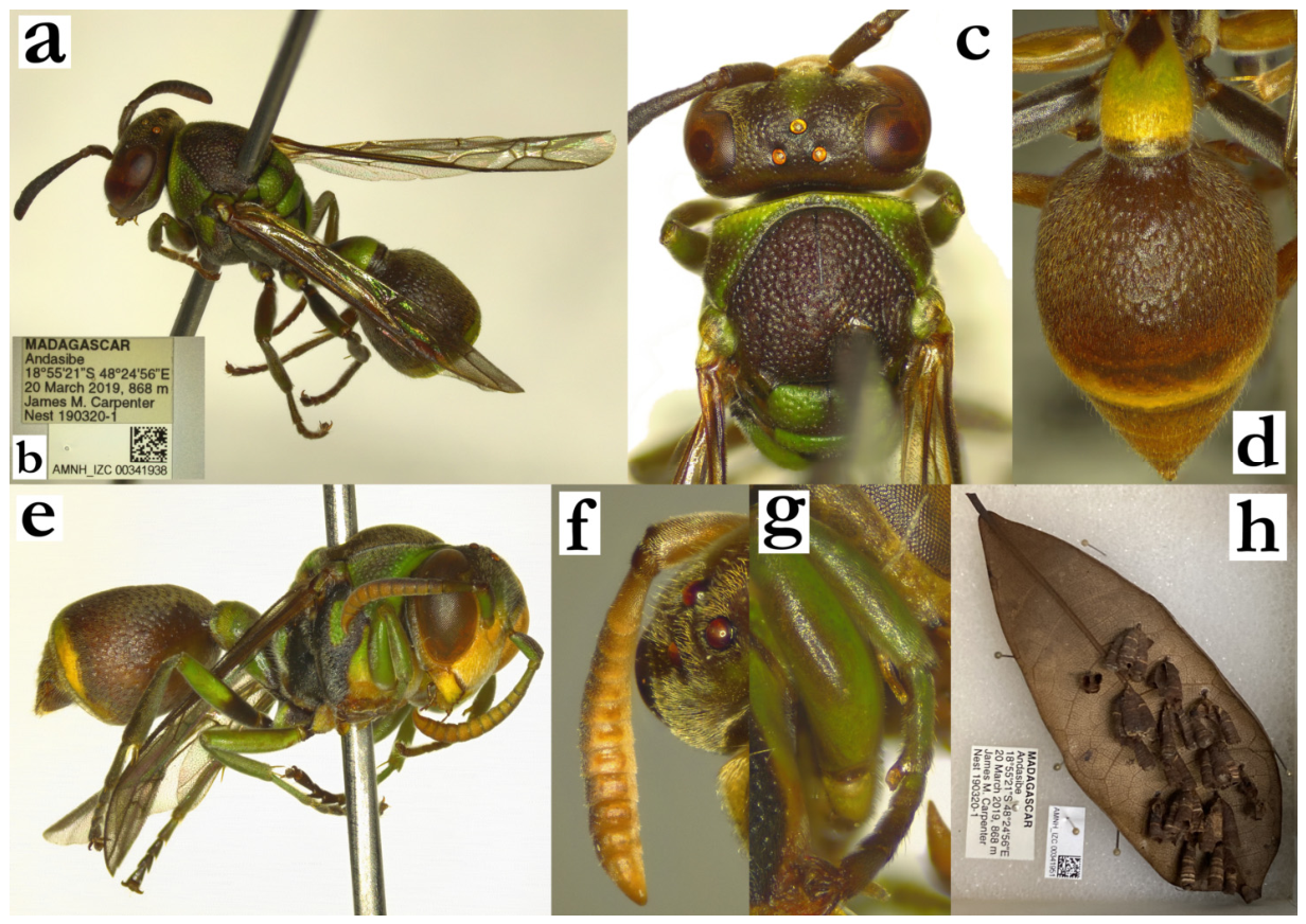
- Ropalidia rampa sp. nov. PolašekZooBank: F5AA781E-FE84-4F1F-88FE-43961A59C7B8Material examined. Holotype: “MADAGASCAR/ Fanovana/ (Olsonfitt?)”, “MUSEUM PARIS/ III.34/ A. SEYRIG”, ♀ (MSNV), Figure 46d–f and Figure 109a–d.Paratype: same label as holotype, 1♀ (MSNV).Number of examined specimens: 2 ♀♀.
Diagnosis. Member of the phalansterica group, characterized by T1 dorsally obtuse, posterior head margin and occipital carina deeply emarginate, and T2 shorter than S2.
Description. Females. Wing length 7.7–8.3 mm.
Head. Head frontally 1.3–1.4× as wide as high; clypeus about 1.3× as wide as long, with wide base, sinuate upes and conspicuous OCA, developed juxtamandibular excavations and a projecting apex (Figure 46e); base and centre of clypeus shiny, with scattered and poorly defined punctures, apex with denser and less defined craters; frons with moderately sized and very shallow punctures (Figure 46e); gena coarsely punctate close to eye, posteriorly with less defined punctures or nearly impunctate (Figure 46f); mandible punctate; occipital carina strongly sinuate close to mandible, creating a characteristic excavation (Figure 46f); ocellar triangle base 1.7–2.0x as wide as side; posterior ocelli over three diameters apart (Figure 109b); scape 3.9× as long as wide (Figure 46e) and 1.4× as long as AF1 length; AF2 1.2–1.4× as long as wide, remaining flagellomeres wider than long.
Mesosoma. Mesosoma 1.4× as long as high, mesoscutum 1.1× as long as wide; entire mesosoma shallowly punctate; puncture on mesoscutum less than one diameter apart, with matte interspaces (Figure 109b); density similar on mesoscutum and pronotum, but these on pronotum smaller and less defined, with more swollen and elevated interspaces (Figure 109b); mesepisternum and metepisternum coarsely and densely punctate; scutellum dorsally rounded, with shallow punctures and developed median carina that reaches half of its length; scutellum with moderate central depression (Figure 109c); metanotum punctures deeper and less defined than those on scutellum, with large posterior shiny impunctate area; propodeum posteriorly widened, median depression of propodeum shallow, weakly striate and superficially punctate in upper half (Figure 109c).
Metasoma. T1 infundibular and very developed dorsally (Figure 109d), 0.6–0.7× as wide as T2 dorsally; posterior part shallowly and sparsely punctate; T2 densely punctate with directional punctures, somewhat shorter than S2 (Figure 109d); remaining metasomal segments are missing in both holotype and paratype specimens.
Pilosity. Head with intermediate length setae (Figure 46f), silvery-yellowish on clypeus, interantennal area, inner orbit and gena, yellow on mandible and frons; clypeus apex setae 2.0–3.2× as long as basally; frons setae 1.1–1.4× as long as anterior ocellus diameter (as opposed to 0.8–1.0 in R. constitutionalis); pronotum dorsally with some whitish protruding setae, mesoscutum with yellowish pubescence and short protruding setae (Figure 46d); T1 with yellowish setae dorsally, whitish ventrally; T2 with yellowish pubescence and protruding setae that barely extend over lamella (Figure 109d); eyes minutely setose (Figure 46e).
Colour. Basal colour dark-olivaceous to black, except suffused green: clypeus (apically), inner orbit, rhomboid patch on interantennal area (Figure 46e), area under pronotal carina (pronotum posteriorly olivaceous-brown), a spot high on mesepisternum, tegula, scutellum with two large spots, metanotum with two merged spots, propodeum without green markings (Figure 46d); femur I black, femur II and III with small green patch distally; tibia dorsally green, ventrally olivaceous, tarsi olivaceous (Figure 46d); T1 with substantially widened posterior green-yellowish band; T2 with green pattern that resembles reduced pattern of the R. ranavali, with greenish-yellow band and several attached spots (Figure 109d); T2 lamella dark green and party translucent (Figure 109d); scape ventrally green, dorsally blackened, flagellum dorsally blackened, ventrally dark ferruginous (Figure 46e); wings translucent with minimal yellowing forwardly, nervature brown, stigma light brown, apical spot absent (Figure 46d).
Males. Unknown.
Etymology. The name originates from the Malagasy word rampa, translated as “chipped, as a knife or a pen blade”, referring to the excavated occipital carina, treated as a noun.
Similar species. This species mostly resembles R. constitutionalis (de Saussure), but numerous characteristics are useful in their separation. These include weakly punctate median depression of propodeum (impunctate in R. constitutionalis), eyes minutely setose (intermediate length in R. constitutionalis), fewer green body markings (propodeum with two green areas in R. constitutionalis, mesepisternum with large green spot, clypeus greener); several more characteristics are provided in the key to species.
Figure 109.
Ropalidia rampa sp. nov., female, holotype, (a) labels, (b) head and anterior mesosoma, dorsal view, (c) median depression of propodeum, oblique lateral view, (d) T1 and T2, lateral view.
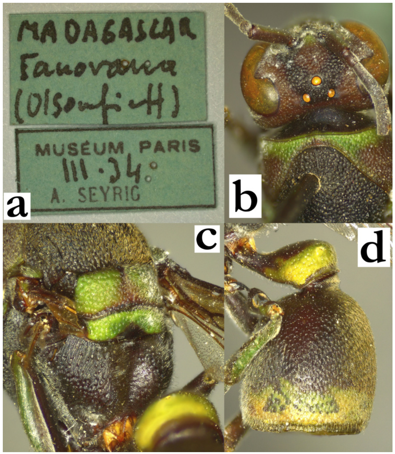
- Ropalidia ranavali (de Saussure), 1890Icaria ranavali de Saussure, 1890; “Madagascar” (MFNB).Plazi: https://tb.plazi.org/GgServer/html/03AE106DFFD4396D25D7FA8E842EF1BD (accessed on 21 September 2026).Icaria venustula de Saussure, 1900; “Madagascar; Imerina” (MHNG), syn. nov.Plazi: https://tb.plazi.org/GgServer/html/03AE106DFFD0396E25D7FE2E842EF370 (accessed on 21 September 2026).Number of examined specimens: 20 ♀♀, 7 ♂♂.
Type material. Ropalidia ranavali (de Saussure), 1890 (Figure 110a)
The species description suggests Berlin Museum as the depository, prompting Kojima to label and consider a specimen from MHNG as a lectotype (not published). The specimen is a somewhat xanthic example of this taxon, with suffused green markings on dorsal mesosoma and a bright broad yellow band on T2, characteristic for this species (Figure 110a). The two specimens from the ETH collection are also labelled as co-types, but they were not listed in the species description. As the examination of the Berlin collection did not yield any additional specimen, the (unpublished) lectotype proposed by Kojima is therefore designated.
Ropalidia venustula (de Saussure), 1900
The species description mentions numerous specimens, most of which are males. The examination of these specimens indeed shows a cluster of specimens with greenish-white markings and a thin posterior band on T2, with only two attached and connected posterior round spots (Figure 110b). In MHNG there is a specimen labelled as lectotype by J. Kojima (unpublished), but the specimen bears the hand-written label “Madagascar/Sikora” (in de Saussure’s handwriting), and therefore cannot be treated as the valid type for this taxon.
Notes. There is a certain level of taxonomic complexity in this taxon, with possible multiple taxonomic clusters, some of which might represent cryptic species.
- (a) The examination of the available specimens of R. venustula (including one male syntype and a series of females) suggested a lack of any reliable differences in male and female morphology. The only detectable and reliable difference is therefore colour pattern, which does not follow a gradient, but shows an interesting skipping pattern, in which R. ranavali is characterized by the more colourful metasoma and mesosoma commonly in just one colour (Figure 57c and Figure 32f), while R. venustula is characterized by a less colourful metasoma and frequently more colourful mesosoma, with greenish spots on metanotum (Figure 110b). Although this may provide support towards these two being separate taxa, the lack of any morphological differences, especially in male morphology (notably, antenna), prompted synonymizing R. venustula with R. ranavali.(b) One of the problems is the melanic cluster from Antisrabe, which have substantially reduced yellow markings on T2 and only retained thin posterior band on T1 (Figure 57d). The remaining morphological characteristics did not exhibit any difference from the more colourful specimens (Figure 57c), suggesting that this species may exhibit a wide variety of colour patterns.(c) Under the assumption that the nest architecture is specific, there is another problem related to R. ranavali, characterized by a substantial nest shape plasticity. The nests from the Ankazobe and Ambohitantely region are discoid, with 12 × 12 nest formula, paracentral petiole location, brownish cell wall and placement on broad (cf. Agava) leaves. Nests from the Ranomafana region are elongated, up to 140 cells long and 20 cells wide, with apical petiole, grey cell wall with black streaks and placement on branches. Unfortunately, there were no specimens examined that could be linked to a specific nest shape, thus retaining a level of taxonomic uncertainty associated with this taxon, and suggesting that there may be additional taxa associated with this name.
Distribution. Eastern and central Madagascar.
Similar species. The colour pattern is often sufficient to ascertain this species (dark head and mesosoma, in combination with a broadened T2 yellow posterior band), but some problems may arise in determination of the black cluster. The criteria include inferiorly merged punctures on pronotum, equally wide pronotal carina (as opposed to dorsally twice wider carina in R. jemmae and R. scottiana), less elongate T1 (compared to elongated and streamlined T1 in R. sonyae sp. nov.), and clypeus base coarsely punctate (absent in similarly coloured species, like R. jemmae or R. aras, sp. nov.).
Figure 110.
(a) Ropalidia ranavali (de Saussure), habitus, lateral view, lectotype, (b) R. venustula syn. nov., habitus, lateral view, male, syntype, (c) labels, (d) habitus, dorsal view, female with green colour pattern, (e) head, frontal view, male, syntype, (f) nest [93], (g) nest [94], (h) nest [95].
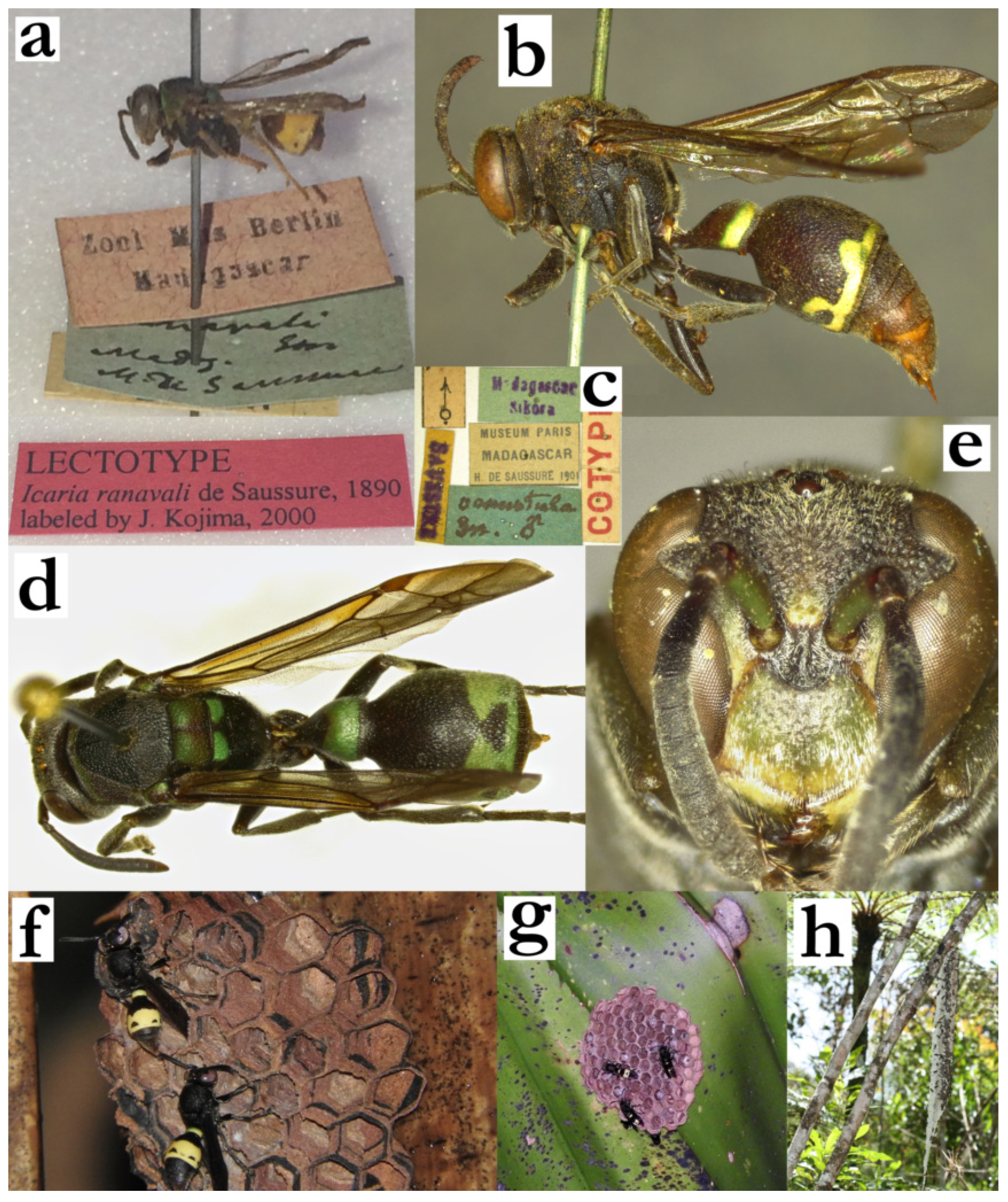
- Ropalidia regina (de Saussure), 1900Icaria regina de Saussure, 1900; “Antananarivo” (MHNG).Plazi: https://tb.plazi.org/GgServer/html/03AE106DFFD3396D25D7FE32842EF3F8 (accessed on 21 September 2026).Holotype (unpublished) photographs: http://www.vespidae.be/Polistinae/Ropalidia_regina.htm (accessed on 21 September 2026).BOLD: https://portal.boldsystems.org/bin/BOLD:ADU3591 (accessed on 21 September 2026).Number of examined specimens: 6 ♀♀, 1 ♂.
Type material. One female specimen from MHNG, with labels “Madagasc”, “12 Siv.”?, “I. regina [female sign] SSS” was considered as a holotype, and was labelled as such by Kojima (unpublished). The status is hereby contested, since the handwriting on the white label that says “I. regina” does not appear to be de Saussure’s handwriting, and the top label does not correspond to the description stating Antananarivo. In addition, there are multiple specimens mentioned in the description, thus preventing this specimen from being considered as the holotype.
Notes. This is a large and stocky, mainly green species, with very wide gena (Figure 10a), bituberculate scutellum (Figure 10b), and conspicuously wide clypeus in both females (Figure 111a) and males (Figure 11c).
Description. Male. Wing length 13.5 mm.
Head. Head frontally about 1.4× as wide as long, clypeus 1.4× as wide as long, with short and curved upes, deep juxtamandibular notches and moderately projecting apex (Figure 11c); base and centre of clypeus sparsely and minutely punctate, apex with larger and poorly defined craters (Figure 11c); frons shallowly punctate, puncture margins well defined, with 0.5–1.5× as their diameter; gena anteriorly similarly punctate, posteriorly impunctate; gena at widest as wide as eye; mandible weakly twisted, with some poorly define craters (Figure 11c); occipital carina complete and wide, eyes asetose (Figure 11c), ocelli equidistant and small; distance between posterior ocelli about 2× as long as their diameter, distance to eye about 4x as long as their diameter (Figure 111a); scape about 0.7× as long as AF1, 1.5× as wide as AF1 (Figure 11c); AF2 1.6× as long as wide (Figure 111b); tyloids dark yellowish and moderately shiny, originate at AF(4)5 (Figure 111b), terminal flagellomere elongated and apically sharp (Figure 11d).
Mesosoma. Mesosoma shallowly and densely punctate, puncture about one diameter apart, more defined on black areas; pronotum and mesoscutum similarly punctured (Figure 111b), mesepisternum somewhat less densely, but entirely punctate; metepisternum with a few well-defined scattered punctures; scutellum bituberculate (Figure 10b), with developed median carina; metanotum posteriorly flattened, with wide impunctate matte area; median depression of propodeum wide, with defined lateral markings, very developed crenulated striae in lower half; median depression of propodeum shallow and shiny, punctate; tarsal spur I developed, about 1.5× as long as the segment width.
Metasoma. T1 posteriorly wide and very developed, posteriorly globular and rounded (Figure 10b); T2 densely and superficially punctate, about one diameter apart; T2 lamella equally wide dorsally and ventrally, impunctate; remaining metasomal segments with weakly developed and shallow punctures.
Pilosity. Pilosity yellowish, in most body parts microscopic without protruding setae, except apical third of clypeus (dense silvery pubescence and some setae) and T2 surface, with yellowish longer setae that extend over T2 lamella; notably, all femur pairs only with microscopic white pubescence and no protruding setae (Figure 111c). S7 concave, coarsely punctate, punctures about 2x apart as their diameter.
Colour. Basal body colour green and yellow, except black: small area around ocelli, posterior margin of mesoscutum, median half of coxa III, area on dorsal surface of T1; T2 dorsally brownish, S2 with brownish longitudinal median line, remaining segments suffused green; coxa green (except median half of coxa III in all examined specimens; Figure 111d), femur green, tibia and tarsi olivaceous, with distal half brownish; wing nervature light brown, stigma brown and translucent, wing transparent, with modest anterior yellowing (Figure 111d); scape ventrally green, flagellum orange, dorsally infuscate (Figure 111d).
Distribution. Eastern Madagascar and central highlands (Figure 111e).
Genetics. One specimen was successfully genotyped, suggesting similarity to other mainly green species.
Figure 111.
Ropalidia regina (de Saussure), (a) head, frontal view, female, (b) head and mesosoma, dorsal view, male, (c) antenna, median view, male, (d) habitus, lateral view, male, (e) distribution map (red marker—specimen, grey marker—literature report or an observational record).
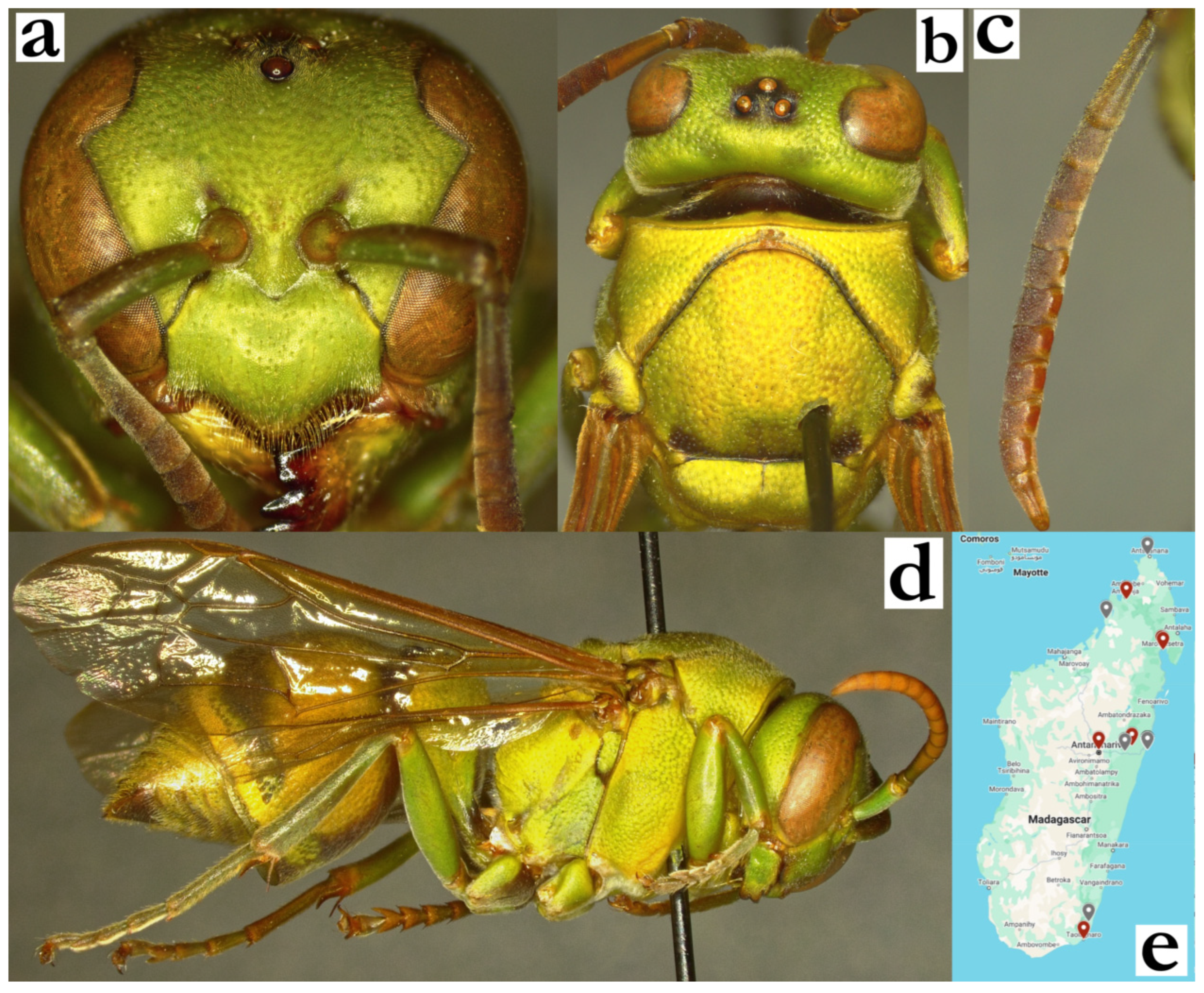
- Ropalidia rosae Blommers, 2012Ropalidia rosae n. sp., Blommers 2012: 167; ♂♀, nest, biology, Madagascar; holotype NNM.Number of examined specimens: 2 ♀♀, 1 ♂♂.
Type material. Three paratypes from nests 72.24 and 72.45 of this species were examined, corresponding to Figures 63–68 in the species description [6] (Figure 112a–e).
Notes. The most diversely colourful Malagasy Ropalidia, often with four or five colours on the body (Figure 58b and Figure 112d); the colourful mesoscutum is an important determination characteristic, alongside small body size. In terms of size, this species is comparable to R. shattenbergi sp. nov. (which has only two body colours, and a different colour pattern, Figure 58a). Several species may resemble this one in size, but they usually have darker basal colour and have much more green areas on the body.
Nest. Small colonies of fragmented and irregularly-shaped nests with paracentral petiole; the nests are sometimes comprising close to 20 cells, thus a single nest is larger than in R. favulroum Blommers [6].
Distribution. Only paratypes were examined.
Figure 112.
Ropalidia rosae Blommers, female, paratype, (a) head and mesosoma, dorsal view, (b) labels, (c) metasoma, oblique lateral view, male, paratype (d) habitus, male, paratype, (e) antenna, median view, male, paratype.
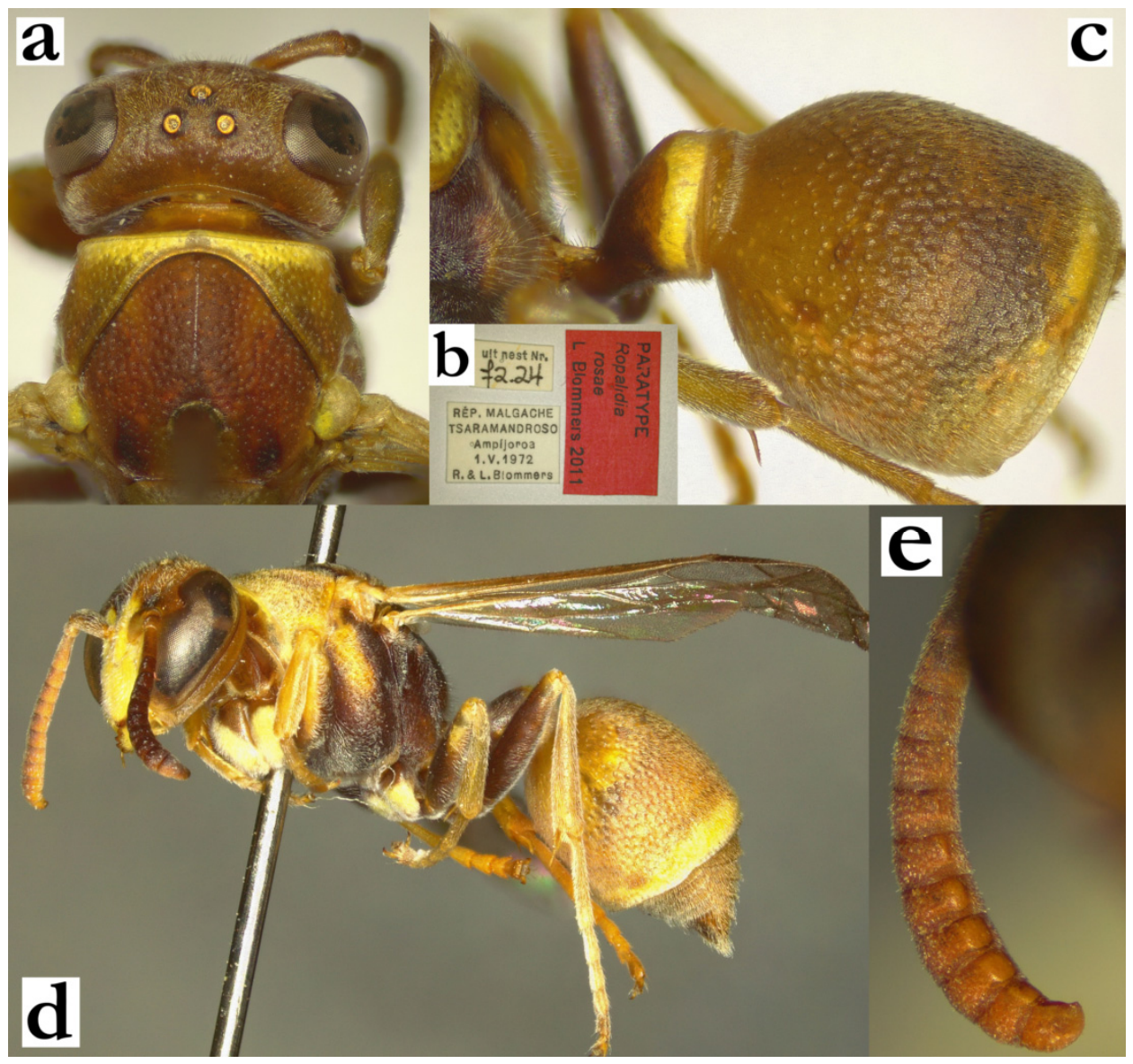
- Ropalidia sakalava (de Saussure), 1900Icaria sakalava de Saussure, 1900; “Madagascar, Antananarivo” (MHNG), Figure 113a,b.Plazi: https://tb.plazi.org/GgServer/html/03AE106DFFD3396D25D7FC07842EF52A (accessed on 21 September 2026).Number of examined specimens: 14 ♀♀, 3 ♂♂.
Type material. One female in MHNG, labelled “Madagasc/Antanariv” is confirmed as a holotype, in line with an unpublished label by J. Kojima [specimen MHNG ENTO 00089390]. The specimen is in good condition, and has a wing length of 12.4 mm.
Notes. A large and mainly black species, member of the formosa group. It can be recognized by the combination of anterior mesoscutum impunctate (Figure 4c), clypeus with large punctures clustered near base (Figure 9f for females, Figure 113e for males), scutellum with flattened surface and minimal or absent median carina; median depression of propodeum finely striate and shiny (Figure 9e), interantennal slit developed (Figure 9f,g), male tyloids originate on AF2 (Figure 113d) and terminal flagellomere elongated and thin, with a minute anterior swelling (Figure 9h).
Similar species. There is a considerable overlap between females of this species with the females or R. andalana sp. nov., which are listed in the key to species. Separation of females from other members of the formosa group is easier, based on a few features provided in the key to species; notably, there are no confirmed macroscopic features, requiring observation of the specimen under a microscope. Males have several reliable morphological features, notably tyloids that originate on AF2 in combination with setose and brownish tyloids, thin terminal flagellomere and narrow posterior ocelli.
Nest. There were no confirmed criteria for separation of nests of large and black species, with more discussion provided in the R. formosa (de Saussure) section.
Distribution. Northern and eastern Madagascar.
Figure 113.
Ropalidia sakalava (de Saussure), (a) habitus, dorsal view, holotype, (b) labels, holotype, (c) habitus, lateral view, female, (d) habitus, inferior oblique view, male, (e) head, frontal view, male, (f) antenna, median view, male.
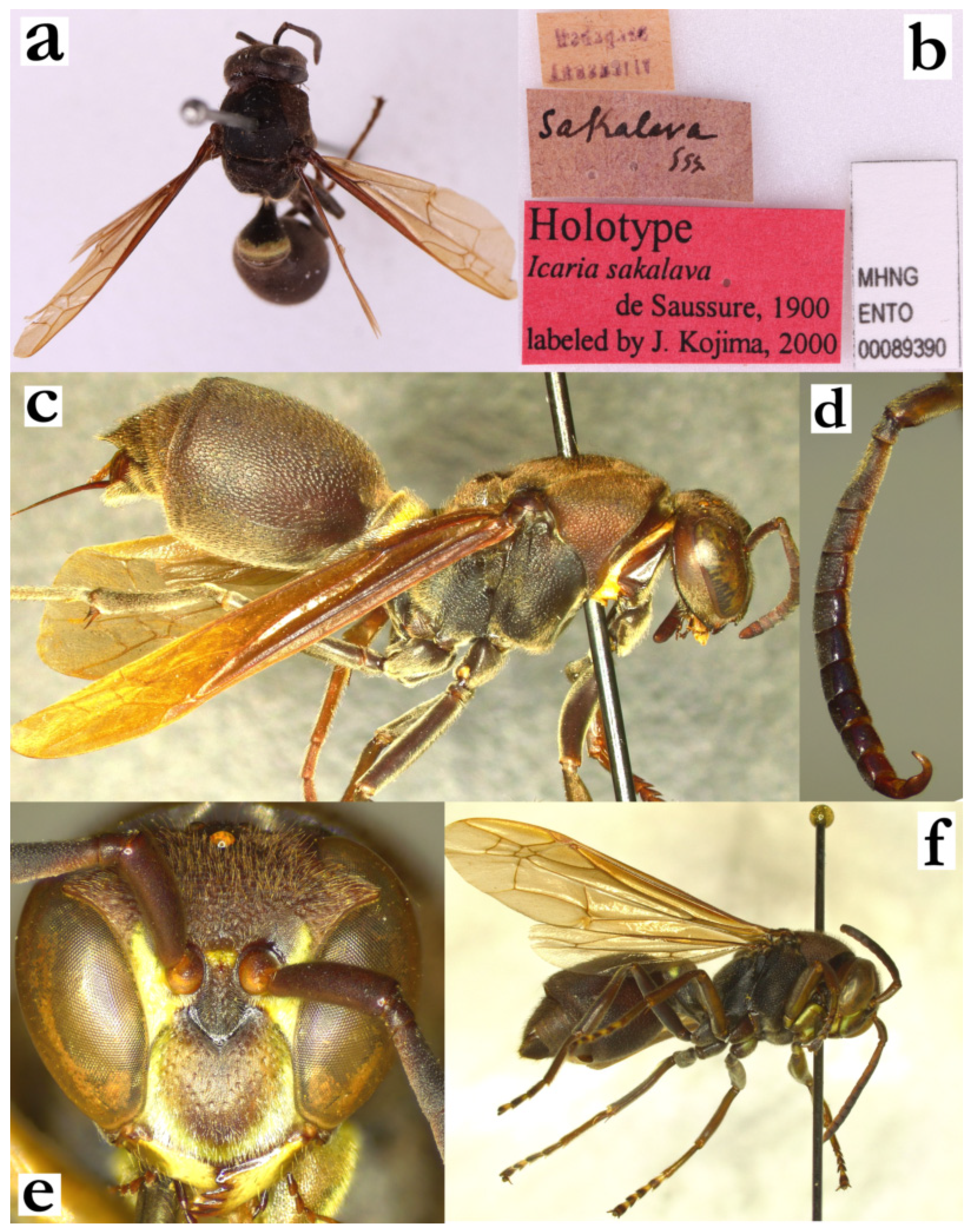
- Ropalidia saussurei Kojima, 1998Ropalidia saussurei KOJIMA, 1998; “Perinet, E. forest b, Madagascar, FitzGerald”, Museum of Comparative Zoology, Harvard University, Cambridge.Plazi: https://tb.plazi.org/GgServer/html/03AE106DFFD3396C25D7FA49842EF1B2 (accessed on 21 September 2026).Number of examined specimens: 28 ♀♀, 9 ♂♂.
Type material. One paratype from NNM was examined (Figure 114a–d).
Notes. A larger, mainly green-coloured species, easily separated from similar ones by the pilosity and colour; some confusion may be possible with larger specimens of R. perplexa Blommers due to overall longer pilosity. Notably, several previous publications misidentified this species and treated it as R. dubia (de Saussure) [4,9].
Nest. This species builds very interesting nests with primarily green cell walls. A reference to R. dubia by Vesey-FitzGerald [9] is probably R. saussurei, since the publication states that the cocoon tops were opaque and silky, “within the mouths of cells”, and that these contained green fragments, which are present in R. saussurei, and absent and elevated above cell margins in R. dubia.
Distribution. Northern and eastern Madagascar (Figure 114e).
Genetics. A single male was successfully sequenced, suggesting proximity to other mainly green-coloured species.
Figure 114.
Ropalidia saussurei Kojima, male, paratype (NNM), (a) habitus, lateral view, (b) labels, (c) head and mesosoma, dorsal view, (d) antenna, median view, (e) distribution map (red marker—specimen, grey marker—literature report or an observational record).
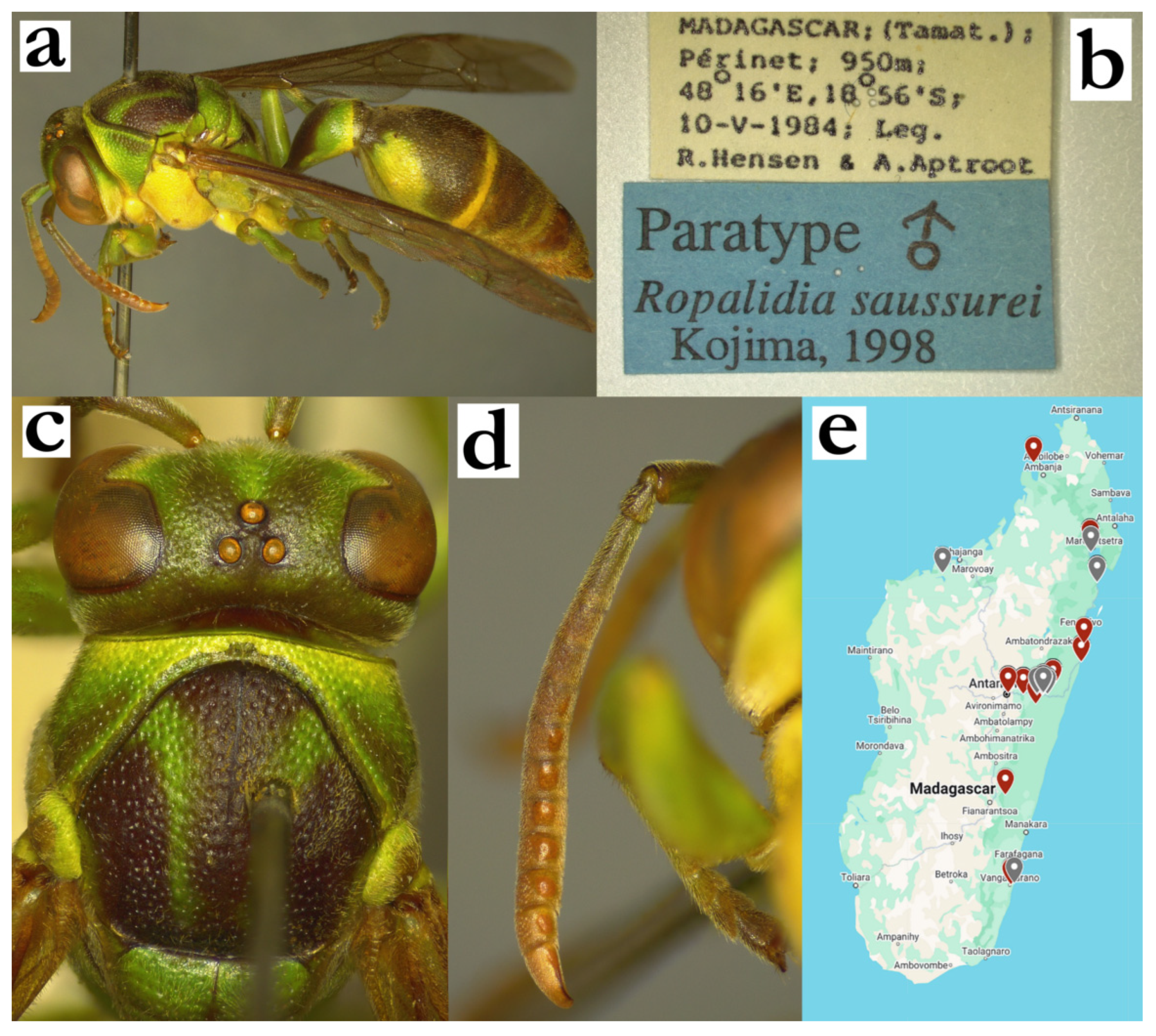
- Ropalidia schulthessi (de Saussure, 1890)Icaria schulthessi de Saussure 1890; “Madagascar … dans l’Est” (ETH); specimen not recovered.Plazi: https://tb.plazi.org/GgServer/html/03AE106DFFD2396C25D7FE29842EF3BE (accessed on 21 September 2026).Ropalidia schulthessi (de Saussure), Blommers 2012: lectotype designated (♀, ETH; Figure 115a,b)—lectotype status hereby disputed.Ropalidia nilssoni Giordani Soika 1991, syn. nov., Figure 115f.BOLD: https://portal.boldsystems.org/bin/BOLD:ADO3631 (accessed on 21 September 2026).Number of examined specimens: 24 ♀♀, 1 ♂♂.
Type material. Icaria schulthessi de Saussure. The species description mentions one specimen from “l’Est”, from Keller collection (later Schulthess), and an unknown number of specimens from Nosy Be, which are termed “var”. The specimen from the Keller collection was not recovered in ETH, while the two females from Nosy Be, labelled as co-types are there (Figure 115a,b). As per article 72.4.1 of the ICZN, these two specimens are not eligible to be a name bearing type. This means that both the unpublished label “holotype” by Kojima and a published lectotypes status by Blommers are invalid. Since there are no taxonomical uncertainties related to this taxon, in line with article 75, no additional types are hereby assigned.
Ropalidia nilssoni Giordani Soika 1991. Giordani Soika described this species based on the five specimens from the North-Eastern coast [4]. Two of these specimens were examined, alongside the photographs of the holotype and paratype, deposited in Uppsala (Figure 115c–g). The examination does not show any morphological difference from the R. schulthessi (de Saussure). This was already indicated by Kojima, who labelled the holotype specimen with a Ropalidia schulthessi label in 2000 (Figure 115f), but did not publish this.
Notes. A smallest species of the grandidieri group, characterized by a very wide T1 (Figure 115c) and deep juxtamandibular excavations in females (Figure 16b); in contrast to other species of this group, males have brown tyloids (Figure 16d). Both sexes have coarse, dense and deeper punctures of pronotum and mesoscutum, but also coarse directional punctures of T2 (Figure 115c,d).
Nest. Previous publications suggested that this species builds small combs, with four-nine cells, located on the underside of the broad leaves of coconuts, oil-palms and bananas [9].
Genetics. Genetic analysis confirmed proximity to other species from the grandidieri group.
Figure 115.
Ropalidia schulthessi (de Saussure), (a) labels, “cotype” from ETH, disputed holotype and lectotype by Blommers, (b) labels, second implied “cotype” from ETH; Ropalidia nilssoni syn. nov., (c) habitus, dorsal view, paratype, Ropalidia nilssoni syn. nov., (d) habitus, lateral, paratype, Ropalidia nilssoni syn. nov., (e) head, frontal view, holotype, (f) labels, holotype and paratype [kindly provided by Dr. Hans Mejlon]; (g) Ropalidia schulthessi (de Saussure), map (red marker—specimen, grey marker—observations and literature reports).
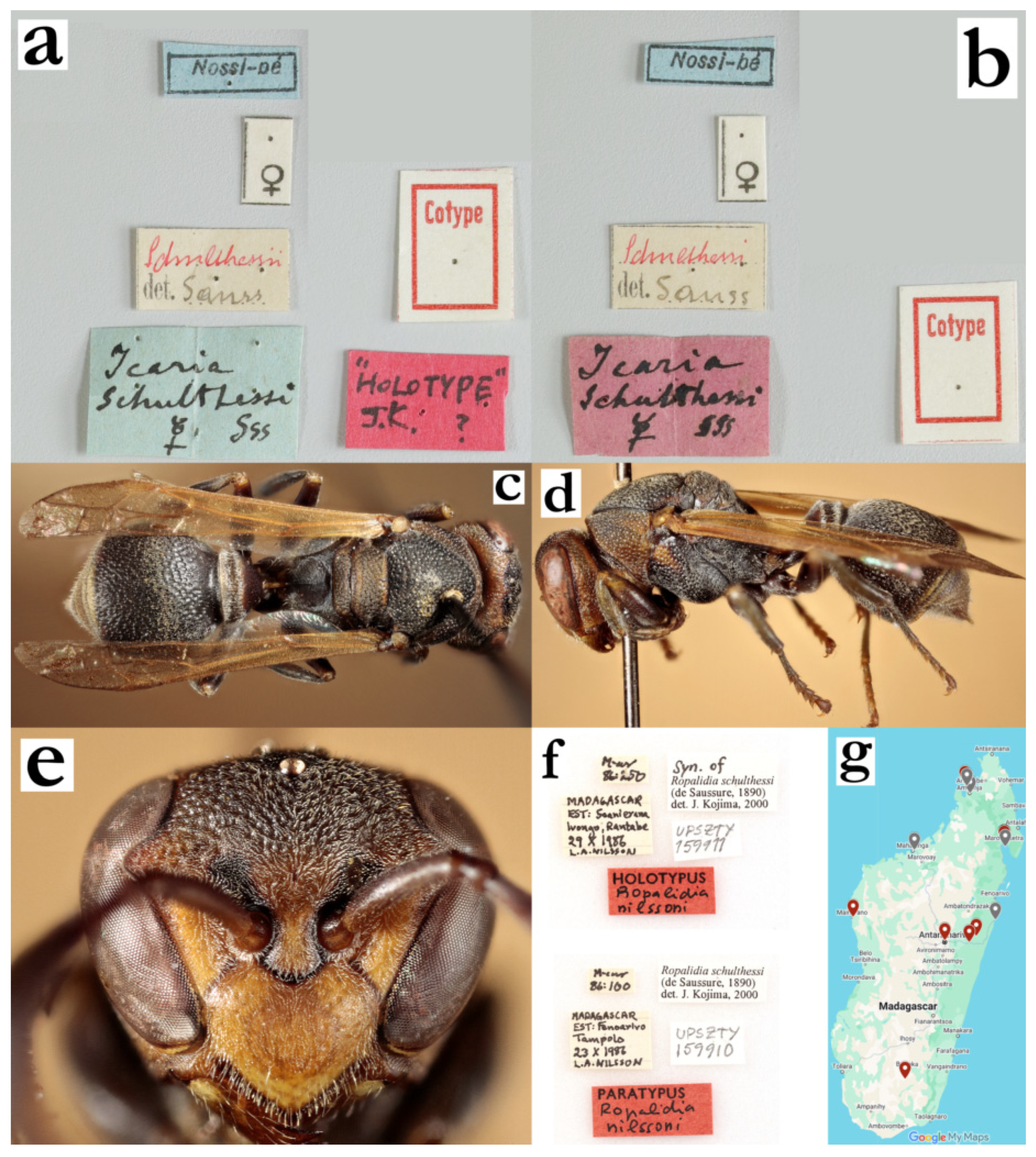
- Ropalidia scottiana (de Saussure, 1890)Icaria scottiana de Saussure 1890; “Madagascar” (MHNG); Figure 116a–c.Plazi: https://tb.plazi.org/GgServer/html/03AE106DFFD2396C25D7FC35842EF538 (accessed on 21 September 2026).Number of examined specimens: 12 ♀♀, 7 ♂♂.
Type material. A single female in Geneva, with labels “Icaria/scottiana/Madag Sss,” a green circle with “4/90” underneath, and “1323” or “1523” (ineligible; Figure 116a–c). The original description lists several more varieties, but states that all have darkened anterior part of the wing, which is present in the holotype specimen, designated here (the specimen was previously labelled by Kojima, but the designation remained unpublished). The taxonomic challenges were previously attributed to the elaborate T2 band (Figure 116d), which is similar to several different species, most notably R. ranavali (de Saussure). The reliable characteristics in that case include the shape of pronotum in dorsal view (Figure 63a) and terminal metasomal segment yellow markings (Figure 63b), which are both unique for this species. Males are described here for the first time (Figure 116e).
Description. Males. Wing length 7.5–7.9 mm
Head. Head 1.4× as wide as high, clypeus 1.5× as wide as long, with evenly convex surface, short and curved upes, weak juxtamandibular excavation and moderately projecting apex (Figure 116f); clypeus punctures weak and obscured by pubescence; mandible shiny and impunctate; frons coarsely and shallowly punctate (Figure 116f); gena with shiny cuticular surface and some poorly defined punctures; occipital carina complete and developed; ocellar tringle equidistant; oculo-malar distance very short, 0.5× as wide as AF1 base, in some specimens nearly absent (eye extending inferiorly to mandible); antenna clavate; scape 1.9× as wide as AF1 base at the widest part, 2.3× as wide as long; AF1 2.6× as long as wide, 0.8× as long as scape, AF2 1.1× as long as wide, remaining flagellomeres wider than long (Figure 116g); tyloids in all examined specimens begin as a small streak of shiny surface on distal part of AF3, continue as triangle on AF4 and remain rectangular on AF5–AF10 (Figure 63d); terminal flagellomere 2.1× as long as wide, evenly curved (with somewhat stronger median excavation), with triangular tyloid that reaches or nearly reaches the acute tip (Figure 116g).
Mesosoma. Mesosoma 1.6× as long as wide, with laterally bulging metapleuron; pronotum with large and shallow punctures, that do not exhibit conflux (may be present as a few punctures close to pronotal carina); pronotal carina dorsally twice as wide as laterally, with a translucent rim; pronotum in dorsal view with characteristic contour (Figure 63a); mesoscutum as long as wide, coarsely and densely punctate, with some puncture conflux (merged large and smaller puncture); scutellum coarsely punctate, with weak median carina that does not reach half of its length, or without it; mesepisternum coarsely punctate, with puncture interspaces equal to puncture diameter; metepisternum with less dense and less defined punctures; propodeum comparatively wider, weakly striate, impunctate, with triangular bottom that corresponds to a somewhat wider and globular T1; fore tarsal spur I close to 2x as long as wide.
Metasoma. T1 wide and posteriorly globular, with impunctate anterior and shallowly punctate posterior half; T2 punctures large and coarse, one diameter apart (Figure 116d); transition from T2 surface to lamella gradual and developed, about one puncture wide; lamella with punctate proximal half, distal half impunctate and translucent (Figure 116d); S7 flattened, with irregular surface and scattered minute, poorly defined punctures.
Pilosity. Body covered by mostly yellowish setae, which turn to silvery-whitish on clypeus, inner orbit and interantennal area (Figure 116f); setae on clypeus dense and short, barely protruding anterior margin; frons with yellowish straight setae, as long as anterior ocellus diameter or somewhat longer; mesoscutum with short protruding setae, about 0.6× as long as those on frons; metasoma with short setae that do not extend over T2/S2 lamella (Figure 116d); eyes minutely setose or asetose in some specimens (Figure 116f).
Colour. Basal colour dark brown; whitish-yellow or yellow areas include most of face, spot on mesepisternum, distal half of coxa I and almost entire coxa II and elaborate posterior band on T2 (Figure 116e); frons, pronotum under carina, two spots on scutellum, two spots on mesoscutum and posterior band on T1 variably coloured, as yellow, green or whitish, in nearly individual colour patterns (Figure 116d–f); propodeum commonly black (often with two green areas in females), anterior surface of mesepisternum exceptionally with yellow markings, most commonly in basal body colour; femora basally black, with green or yellow spot on distal half of anterior surface, tibia and tarsi green-yellow (Figure 116e); wings anteriorly somewhat infuscate, otherwise yellowish; antenna dorsally heavy infuscate to black, ventrally orange-yellowish (Figure 116e); tyloids orange and shiny (Figure 63d).
Distribution. Central highlands (Figure 116h); de Saussure reported it from the South-East [13].
Figure 116.
Ropalidia scottiana (de Saussure), (a) habitus, lateral view, female, holotype, (b) habitus, dorsal view, female, holotype, (c) labels, holotype [kindly photographed by Bernard Landry from MHNG], (d) T1 and T2, lateral view, female, (e) habitus, lateral view, male, (f) head, frontal view, male, (g) antenna, dorsal view, male, (h) distribution map (red marker—specimen, grey marker—observations and literature reports).
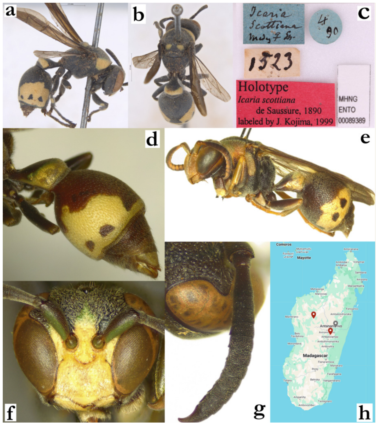
- Ropalidia seyrigi sp. nov. PolašekZooBank: 31AFA791-7400-42F9-BF07-249197838230Material examined. Holotype: “MADAGASCAR/ PERINET/ FORET COTE EST”, “Olsonfitt (?, handwritten)”, “MUSEUM PARIS/ II 34/ A. SEYRIG”, ♀ (MSNV); Figure 117a,b.Paratypes: 8♀♀, 1♂, same data as holotype (MSNV); “Ambositra/ MUSEUM PARIS/ I 34/ A. Seyrig”, 1♀ (MSNV); “Madagascar/Perinet/Foret Cote Est”, “Olsonfitt”, “MUSEUM PARIS/ III 35/ A. SEYRIG”, 8♀♀, 1♂ (MSNV); “Madagascar/Perinet/Foret Cote Est”, “Olsonfitt”, “MUSEUM PARIS/ III 36/ A. SEYRIG”, 1♀ (MSNV); “Fianarantsoa/III 38, Seyrig”, 4♀♀ (MSNV); “Andasibe/25.11.1999, leg Halada”, 1♀ (OLM); “MADAGASCAR Fia./ Abatolahy/ 14.IX.53 F. KEISER”, 1♀ (NNM).Number of examined specimens: 25 ♀♀, 2 ♂♂.
Diagnosis. This species can be separated from other small black and green species by the following characteristics: clypeus upes continues into lateral margins (no OCA), clypeus and interantennal area with fine pubescence, antennal slit absent, body without yellow markings (exceptionally with suffused minimal yellow markings), female antenna dorsally infuscate and male AF2 and AF3 longer than wide.
Description. Females. Wing length 6.6–7.4 mm.
Head. Head frontally 1.3× as wide as high, in dorsal view 2.35× as wide as long, mildly narrowing behind eyes, barely emarginate posteriorly (Figure 117c). Clypeus 1.1–1.2× as wide as long, with elongated base; upes straight, OCA absent (Figure 42a); juxtamandibular excavation shallow, apex very pointy (Figure 42a); clypeus with cuticular surface basally and some poorly defined punctures, apically less defined and obscured by the pubescence (Figure 42a). Frons sparsely and coarsely punctate, punctures 1.5–2 diameters apart, with matte interspaces (Figure 42a); gena punctate close to eye, similarly but sparsely than frons, posteriorly less defined and nearly impunctate (Figure 43a); gena at midpoint 0.6× as wide as eye; mandible punctate, occipital carina complete and very developed (Figure 43a); posterior ocelli 1.5 diameters apart, 2.3 diameters apart from compound eye and 1.5 diameters apart from occipital carina (Figure 117c). Scape 3.0–3.1× as long as wide, 1.3× as long as AF1; AF2 as long as wide, remaining flagellomeres wider than long (Figure 117e).
Mesosoma. Mesosoma slender, 0.95× as wide as head in dorsal view; 1.5× as long as wide, in lateral view 1.9× as long as wide; mesoscutum 1.15× as wide as long (Figure 117c); pronotum in dorsal view mildly sinuate anteriorly, humeri rounded or barely angled; carina of pronotum complete and thin, dorsally at most 1.4× as wide as laterally; pronotum punctures shallow, 1.5 diameters apart, with mildly swollen and matte interspaces, retained even inferiorly (Figure 43a). Mesoscutum with similar punctures (Figure 117c). Mesepisternum without carina towards mesepimeron, sparsely and coarsely punctured in lateral area, impunctate or with just a few punctures anteriorly; posterior surface with less dense and punctures of same size, like those on mesepimeron; metepisternum basally matte, with somewhat more spaced and well-defined punctures. Propodeum posteriorly narrowing; median depression of propodeum shallow and widely emarginate; surface with fine horizontal striae, with a few ill-defined punctures superiorly; lateral face of propodeum more striate (Figure 117d).
Metasoma. T1 pyriform, without posterior sulcus (Figure 117d); anterior surface impunctate, posterior with some poorly defined craters (Figure 117d). T2 1.1× as long as high, tergum and sternum similarly convex (Figure 117a); punctures shallow and coarse, directional, 1.0–1.3 diameters apart, with shiny interspace; punctures on S2 somewhat larger and sparser, especially proximally; transition between T2 surface and lamella gradual, lamella translucent and very weakly punctate proximally, distal two thirds impunctate; remaining metasomal segments shallowly punctate.
Pilosity. Body with short pubescence, silvery on head, yellowish on mesosoma and metasoma (Figure 117a); clypeus with protruding setae, 2.0× as long apically as basally; mandible with protruding setae of similar length as clypeus apically; frons with straight protruding setae, 0.9–1.4× as long as anterior ocellus diameter; gena and occiput with some short protruding setae; eyes finely setose (Figure 42a). Propodeum with some longer whitish fine setae; T1 anteriorly with fine pubescence, posteriorly with some yellowish setae that do not project over posterior margin; T2 with protruding setae that extend over lamella (Figure 117a); remaining metasomal segments with some protruding setae distally; coxa with fine silvery pubescence and some silvery protruding setae with bent tips; femora with dense silvery pubescence, femur III also with longer setae.
Colour. Basally black, except green apex of clypeus, interantennal area, inner orbits (Figure 42a), mark high on gena (Figure 43a), pronotum (except dorsal brownish area), small spot on mesepisternum (Figure 43a), two large spots on scutellum, two merged spots on metanotum, small spot on propodeum (Figure 117d), broadened posterior band on T1, thin posterior band on T2 and S2 (Figure 117a); coxa black, femora distally green, tibia green-olivaceous, tarsi olivaceous, tips ferruginous (Figure 117a). Wings yellowish, nervature brown, stigma light brown and transparent, apical spot very faint (Figure 117a). Antenna dorsally brown to infuscate, ventrally ferruginous (Figure 117e).
Males. Resemble females in general appearance (Figure 117f), except antenna much slenderer. Wing length 7.5–8.1 mm.
Head. Head in frontal view 1.25× as wide as high (Figure 43b); clypeus base with some large and shallow punctures, apex depressed and with less defined craters (Figure 43b). Scape 2.4× as long as wide, 1.4× as wide as AF1 at midpoint; AF2 1.25× as long as wide (Figure 117g). Tyloids shiny, originate at AF5, brownish and matte (Figure 117g); terminal flagellomere elongated, 1.6× as wide basally as long in straight line, with thin tyloid that reaches acute tip (Figure 43c).
Mesosoma. Fore tarsal spur I developed and long, as broad as tarsus base.
Metasoma. S7 weakly concave, with shiny and somewhat irregular surface.
Pilosity. Overall, with longer head setae than in females; clypeus with fine silvery setae in apical angles (Figure 117f); frons with setae 0.9–1.6× as long as anterior ocellus diameter (Figure 117f).
Distribution. Central Madagascar (Ambositra, Perinet, Fianarantsoa, Andasibe).
Etymology. After André Seyrig, an entomologist who collected numerous Malagasy insects, including the holotype specimen.
Similar species. Females of this species can substantially resemble females of R. favulorum Blommers, especially in cases of R. favulorum Blommers with narrower clypeal base. This situation can present a substantial challenge, but several features may assist. The first one is the forewing colour, which tends to be completely transparent in R. favulorum Blommers, while it is yellowish in R. seyrigi sp. nov. Secondly, there are several characteristics on the head: (a) gena posteriorly narrower (0.6× as eye width in R. seyrigi sp. nov., 0.7–0.8 in R. favulorum Blommers), (b) posterior margin of gena (mostly straight and parallel to posterior eye margin R. seyrigi sp. nov. (Figure 43a), weakly sinuate and with uneven width in R. favulorum Blommers), (c) mesepisternum with green spot (commonly small and sometimes even brown in R. seyrigi sp. nov., compared to commonly larger one in R. favulorum Blommers), and (d) dorsal antenna colour (olivaceous or dark green in in R. seyrigi sp. nov., blackened in R. favulorum Blommers).
Figure 117.
Ropalidia seyrigi sp. nov., (a) habitus, lateral view, female, holotype, (b) labels, holotype, (c) head and mesosoma, dorsal view, female, (d) posterior mesosoma, oblique lateral view, female, (e) antenna, dorsal view, female, (f) habitus, oblique view, male, (g) antenna, median view, male.
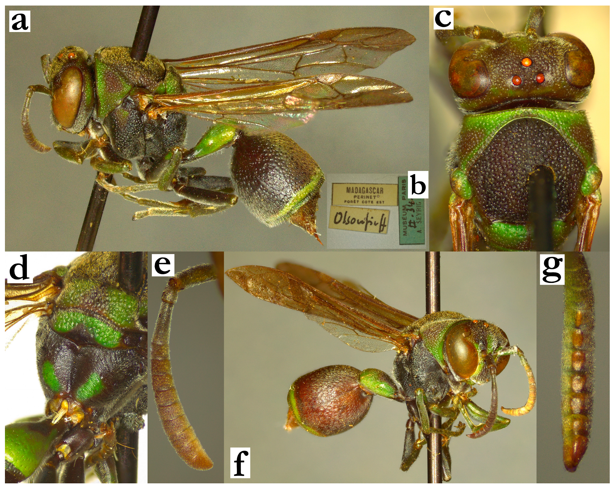
- Ropalidia shattenbergi sp. nov. Polašek and de BeerZooBank: 16C169BC-F892-4E65-8184-715F56E66FAFMaterial examined. Holotype: “Ambilobe/ IV-51 RP”, “INSTITUT/ SCIENTIFIQUE/ MADAGASCAR”, ♂ (MSNV), Figure 118a,b.Paratypes: same labels as holotype, 2♀♀ (MSNV).Number of examined specimens: 2 ♀♀, 1 ♂.
Diagnosis. The smallest Malagasy Ropalidia, with wing length that barely exceeds 5 mm, characterized by the basal brown body colour with yellow markings on mesosoma, punctures weak, pubescence short and male antenna with tyloids originating on AF(5)6 and terminal flagellomere 1.2× as long as wide.
Description. Females. Wing length 4.9–5.1 mm.
Head. Head frontally about 1.2× as wide as long, clypeus 1.25× as wide as long, with straight upes and wide and moderately developed OCA; juxtamandibular lobe moderately developed, apex pointy; base and centre of clypeus impunctate or barely punctate (Figure 59b); frons shallowly punctate (Figure 59b); gena impunctate, 0.7–0.8× as wide as eye (Figure 118c); mandible with poorly defined craters; occipital carina complete, eyes asetose (Figure 59b); ocellar triangle with a wider base, distance between posterior ocelli 1.8× their diameter; scape 1.3× as long as AF1, AF2 1.0–1.2× as long as wide.
Mesosoma. Mesosoma shallowly and finely punctate, with somewhat larger puncture diameter on pronotum and mesoscutum; mesepisternum with sparse punctures, metepisternum nearly impunctate. Median depression of propodeum barely punctate and weakly striate; mesosoma punctures in general shallower in yellow areas and coarse in brown.
Metasoma. T1 pyriform and short (Figure 59a); T2 matte, with shallow, small and dense directional punctures (Figure 59a); T2 lamella equally wide dorsally and ventrally, translucent and shiny, weakly punctate (Figure 118d); remaining metasomal segments with weakly developed and shallow punctures.
Pilosity. Pilosity yellowish, with somewhat longer setae on apical part of clypeus; frons and occiput with golden setae with bent tips, longer than anterior ocellus diameter; mesosoma with short yellowish pubescence, whitish and longer on propodeum; T2 with short and fine, yellowish pubescence that does not extend over lamella (Figure 118d).
Colour. Basal body ferruginous-brown (Figure 58a); clypeus, inner orbit and interantennal area yellow (Figure 59b); frons, vertex, gena and occiput brown (lower half of gena suffused yellow in one paratype); mandible brown or brown with antero-basal yellow spot; antenna brown and dorsally slightly infuscate, ventrally orange on distal segments; mesosoma basally brown, with numerous yellow areas; pronotum with dorsally broadened yellow line underneath carina, a patch high on mesepisternum, two spots on scutellum, a continuous line on metanotum (uninterrupted postero-medially) and two yellow patches on propodeum (Figure 58a); coxa I ventrally and laterally yellow, coxa II and III with smaller lateral yellow patch; legs uniformly light brown (Figure 58a); wings transparent, barely yellowish anteriorly, nervature brown, stigma brown, without apical darkening or apical spot; T1 and T2 with posterior thickened yellow band, S2 with triangular short yellow remnant of posterior band (Figure 58a); remaining metasomal segments brown or suffused with yellow.
Males. Resemble females in general appearance. Wing length 4.9 mm.
Head. Clypeus 1.4× as wide as long, upes straight, with stronger OCA than females, acute apex and moderate juxtamandibular excavation (Figure 59c); clypeus basally and centrally impunctate (Figure 59c); gena 0.4× as wide as eye (Figure 118e); mandible shiny, nearly impunctate; scape barely widened, 1.5× as wide as AF1 base width (Figure 118e), equally long as AF1; AF1 shorter than AF2+3+4, AF2 1.5× as long as wide (Figure 59d); tyloids yellowish and somewhat shiny, originate at AF(5)6 (Figure 118f); terminal flagellomere short, no more than 1.2× as long as wide, basally broad and narrowing towards acute tip (Figure 59d).
Mesosoma. Mesosoma dorsally shallowly punctate (Figure 118e); fore tarsal spur I developed, thin and long.
Metasoma. S7 barely concave, with shiny and irregular surface.
Pilosity. Entire clypeus with fine, equally long, silvery setae (Figure 59c).
Colour. Clypeus yellow (or yellow with basal brown spot, it is impossible to be sure due to the damage of only examined specimen; Figure 59c); mandible and lower part of gena yellow, pronotum with more yellow than females, mesepisternum with yellow patches anteriorly, superiorly and inferiorly (Figure 59c); mesoscutum with yellow patches close to tegula; all coxa yellow ventrally and laterally (Figure 118a); antenna ferruginous dorsally, slightly infuscate dorsally (Figure 118a).
Distribution. A single location is recorded in Northern Madagascar.
Etymology. The name is given to honour Jamie Shattenberg, who was stung multiple times by various Malagasy Ropalidia, during reforestation in the Mahajunga area.
Note. All three examined specimens are in poor condition, covered by a white residue, which became even more visible in one macerated specimen (due to inability t properly observe and describe morphological details). The extent of damage is the worst on a male, where it is difficult to define the real extent of yellow colour on the body due to reddish artefacts.
Figure 118.
Ropalidia shattenbergi sp. nov., (a) habitus, lateral view, female, paratype, (b) labels, paratype, (c) head, frontal view, female, paratype, (d) T1 and T2, lateral view, paratype, (e) head and anterior mesosoma, dorsal view, male, holotype, (f) antenna, median view, male, holotype.
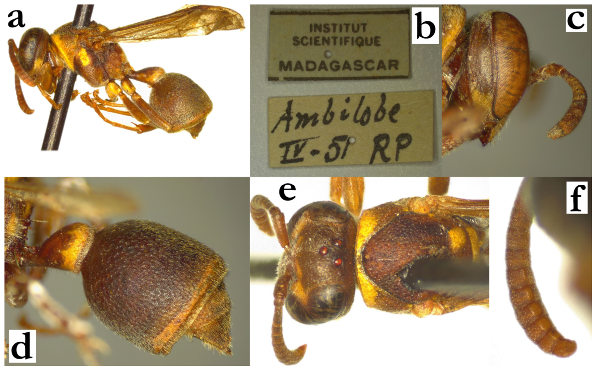
- Ropalidia shestakowi (von Schulthess, 1931)Icaria shestakowi von Schulthess, 1931; “Madagascar” (ETH).BOLD: https://portal.boldsystems.org/bin/BOLD:AED3788 (accessed on 21 September 2026).Number of examined specimens: 97 ♀♀, 26 ♂♂.
Type material. Two males from ETH were examined, one of which was previously designated as lectotype [6]. In addition, one male and female from MSNV are considered a paralectotypes, since both specimens have the same label as the lectotype (Figure 119a,b); notably, the original description does not list the number of examined specimens but mentions numerous males and females [96]. The male paralectotype has the “Cotype” label, while the female is without it. Both were labelled as paralectotypes.
Notes. Several morphological characteristics of this species listed in the key provide sufficient grounds for separation from other species. Males can unequivocally be identified based on the terminal flagellomere shape, which was the initial character used in species description, without any indication of the female of this species.
Nest. Blommers suggested that this species builds rather fragile nests, with frequent “pseudo-windows”, which he differentiates from the meconium openings and refers to as being “non-central” [6].
Distribution. This is one of the more common species of Ropalidia, distributed throughout Madagascar (Figure 119c).
Genetics. Despite a high level of morphological similarity with other members of grandidieri group, including R. tsiky sp. nov., the COI genetic structure of this species suggests a more basal position, sister to most other Malagasy species. Additional support to this claim originates from multiple specimens available in the BOLD database, with adults that morphologically resemble R. shestakowi, which have very similar sequences. Interestingly, morphological characteristics are in support of this claim, since this is the only Malagasy species with elevated male tyloids and very elaborate terminal flagellomere. These features are present in most of the mainland capensis group species, suggesting a degree of similarity between them.
Figure 119.
Ropalidia shestakowi (von Schulthess), (a) habitus, oblique lateral view, male, syntype, (b) labels, lectotype (ETH), (c) distribution map (red marker—specimen, grey marker—literature report or an observational record).
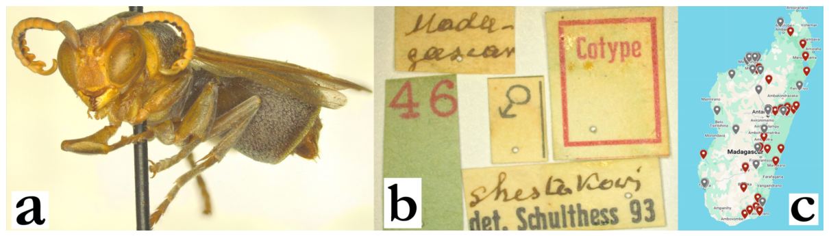
- Ropalidia sonyae sp. nov. Polašek and de BeerZooBank: 9FFDB887-B19C-4309-9C1E-9925466B82A9Material examined. Holotype: “MADAGASCAR/ Ivandro”, “MUSEUM PARIS/ XII. 38/ A. SEYRIG”, ♀ (MSNV), Figure 52a,b and Figure 120a,b.Paratypes: 3♀♀, same labels as holotype; “MARS”, “MADAGASCAR/ COLLECTION LE MOULT”, 1♀; “La Mandraka/ Madagascar/ Mus. Praha”, 3♀♀, 1♂; “MADAGASCAR/ ROGEZ/ FORET COTE EST”, “MUSEUM PARIS/ I. 37/ A. SEYRIG”, 3♂♂; “MADAGASCAR/ PERINET/ FORET COTE EST”, “MUSEUM PARIS/ II. 39/ A. SEYRIG”, 2♀♀; “MADAGASCAR/ FIANARANTSOA/ PLATEAU CENTRAL”, “MUSEUM PARIS/ III. 38/ A. SEYRIG”, 1♀ (all in MSNV); “Betsileo/ Madagascar/ 82-30.”, 1♀ (NHM); “COLL. MUS. CONGO/ Madagascar: Mandraka/ II-1944/ A. Seyrig”, 2♀♀ (RMCA); “COLL. MUS. CONGO/ Madagascar: Mandraka/ XII-1944/ A. Seyrig”, 1♀ (RMCA); “Coll. MUS. TERVUREN/ MADAGASCAR: Tam/ Morarano-Chrome/ FORET 25km W./ ii.1992, NID 18/ A. Pauly rec”, 1♀ (RMCA).Observations: Ifanadiana [97]; Anjozorobe [98]; Ankazobe [99].Number of examined specimens: 16 ♀♀, 4 ♂♂.
Diagnosis. This species can be characterized by numerous green and yellow markings on head, mesosoma and metasoma, T1 elongated and evenly rounded and shallow punctures.
Description. Females. Wing length 9.6–10.6 mm.
Head. Head frontally 1.2× as wide as long, dorsally 2.4 as wide as long, gena barely narrower than eye, posteriorly shallowly emarginate (Figure 120c). Clypeus 1.15× as wide as long, with short and straight upes, moderately developed OCA, weak juxtamandibular lobe and pointy apex (Figure 52b); clypeus shiny and shallowly punctate, apically with large and poorly defined craters; frons with larger and coarse punctures than those on clypeus, 1.8–2.1 diameters apart, with matte interspaces (Figure 52b); punctures in inner orbit and eye sinus smaller and less defined, similarly to these on gena; gena 0.4–0.6× as wide as eye, posteriorly almost entirely impunctate; mandible with poorly define craters; occipital carina complete. Ocelli large, ocellar triangle equidistant or with barely wider base; distance between posterior ocelli 0.8× as wide as their diameter, distance to eye 2.1× diameter, distance to occipital carina 1.6× diameter (Figure 120c). Eyes asetose or with minute setae; scape 1.3× as long as AF1, AF2 1.2–1.5× as long as wide (Figure 52b).
Mesosoma. Mesosoma dorsally 1.4× as long as wide, 1.05× as wide as head; disc of mesoscutum 1.05 as wide as long, with coarse and spaced punctures on matte surface; punctures about 2 diameters apart (Figure 120c); pronotum in dorsal view with straight anterior margin; pronotal carina complete (Figure 52a). Mesepisternum with sparse and shallow punctures, more than 3 diameters apart; metepisternum with even lesser puncture density and definition (Figure 120d). Disc of scutellum bituberculate, with depressed median part and developed median carina that is as long as third of scutellum length; metanotum shallowly and weakly punctate, with shiny posterior impunctate area. Propodeal median depression shiny and impunctate (some minimal striation may be present, and occasionally just barely detectable punctures); lateral surface of propodeum finely striate.
Metasoma. T1 elongate, evenly rounded, posteriorly with poorly defined punctures (Figure 32d); T2 with shallow, small and dense directional punctures (Figure 52a), somewhat larger and sparser on S2; T2 lamella equally wide dorsally and ventrally; remaining metasomal segments with weakly shallowly punctate.
Pilosity. Clypeus with whitish setae, twice as long apically (Figure 52b); frons with golden straight setae, longer than ocellus diameter (Figure 120c). Mesosoma with short yellowish pubescence and longer protruding setae; T2 with very short yellowish pubescence and longer protruding setae that barely extend over lamella (Figure 52a); remaining segments with longer pubescence than that on T2 and S2.
Colour. Basal body colour olivaceous-brown to black with numerous and large yellow areas; clypeus, inner orbit and interantennal area entirely yellow, frons transitioning from yellow, across green to basal colour (Figure 52b); mandible yellow, gena greenish or yellow; scape dorsally olivaceous, ventrally green; flagellum dorsally darkened or even blackish, ventrally green, distal segments sometimes orange (Figure 52b). Pronotum green, commonly with gradual yellowing underneath carina and dorsal parts towards mesoscutum (Figure 52a); mesoscutum dark, with two thin longitudinal medial green lines and two lateral areas next to tegula (Figure 120c); mesepisternum with large green(-yellow) spot on reddish base, metepisternum in basal colour (Figure 120d); scutellum commonly with two large green(-yellow) spots that can sometimes be completely merged, metanotum with continuous green(-yellow) line, sometimes interrupted poster-medially by darker area; propodeum with two large green(-yellow) spots (Figure 120a). Coxa I ventrally and laterally green-yellow, coxa II and III with smaller lateral yellow patch (less commonly all coxa pairs entirely green). Femora green, with irregular dorsal olivaceous patchy areas; tibia green, tarsi dark green; wings barely yellowish, nervature brown, stigma yellow and translucent, apical spot present and yellowish, barely visible (Figure 52a and Figure 120a). T1 mostly yellow, with gradual greenish area and olivaceous or blackish dorsal remaining area close to the valvula (Figure 120a); T2 with specific pattern, similar to R. ranavali (Figure 52a); S2 with fragmented posterior band, sometimes with suffused yellowish area basally; remaining metasomal segments brown or suffused with green or yellow.
Males. Substantially resemble females in general appearance. Wing length 9.6–10.1 mm.
Head. Clypeus 1.2× as wide as long, upes straight and short, with less developed OCA than females, sub-acute apex and moderate juxtamandibular excavation. Clypeus sparsely punctate, obscured by setae; gena 0.4× as wide as eye; mandible shiny, nearly impunctate. Scape mildly widened, 1.5–1.6× as wide as AF1 base, equally long as AF1; AF2 1.4–1.5× as long as wide. Tyloids specific; planar on AF3-AF7, rounded and mildly elevated on AF8-AF11; tyloids brownish and shiny, AF11 can have darkened tip; terminal flagellomere 1.9–2.2× as wide as long, with developed tyloid basally and acute apex (Figure 52d).
Mesosoma. Fore tarsal spur I developed, about 2.1× as long as wide.
Metasoma. S7 flattened, with shiny surface and scattered poorly defined craters.
Pilosity. Body with short pubescence; clypeus with dense, straight and short setae apically, of similar length basally and apically.
Colour. Similar to females, but with more yellow on the metasoma underside; anterior surface of mesepisternum and all coxa pairs green-yellowish.
Etymology. The name is given in recognition of Sonya de Beer, who discovered a nest of this species on the occasion of her farewell trip to the Ambohitantely forest, before the closure of the Eden Project activities in the area. Giordani Soika labelled one specimen with a green label “n. sp. A” and a white label “primeriana” (Figure 120b).
Nest. Nests have 2×n formula, with the larger observed nest containing about 36 cells [97]; one observation shows a partly destroyed [98], or somewhat irregular shape [99]. Nests were built on green branches, and have a very shiny pedicel. The cell wall is porous, light grey with a greenish hue. The smaller nest also has translucent enclosures of the basal cell bottoms, suggesting a very quick early colony stage, probably producing workers already in the first dozen cells.
Distribution. Central Madagascar (Figure 120e).
Figure 120.
Ropalidia sonyae sp. nov., (a) habitus, dorsal view, holotype, (b) labels, holotype, (c) head and mesosoma, dorsal view, female, (d) mesosoma, lateral view, female, (e) distribution map (red marker—specimen, grey marker—observations and literature reports.
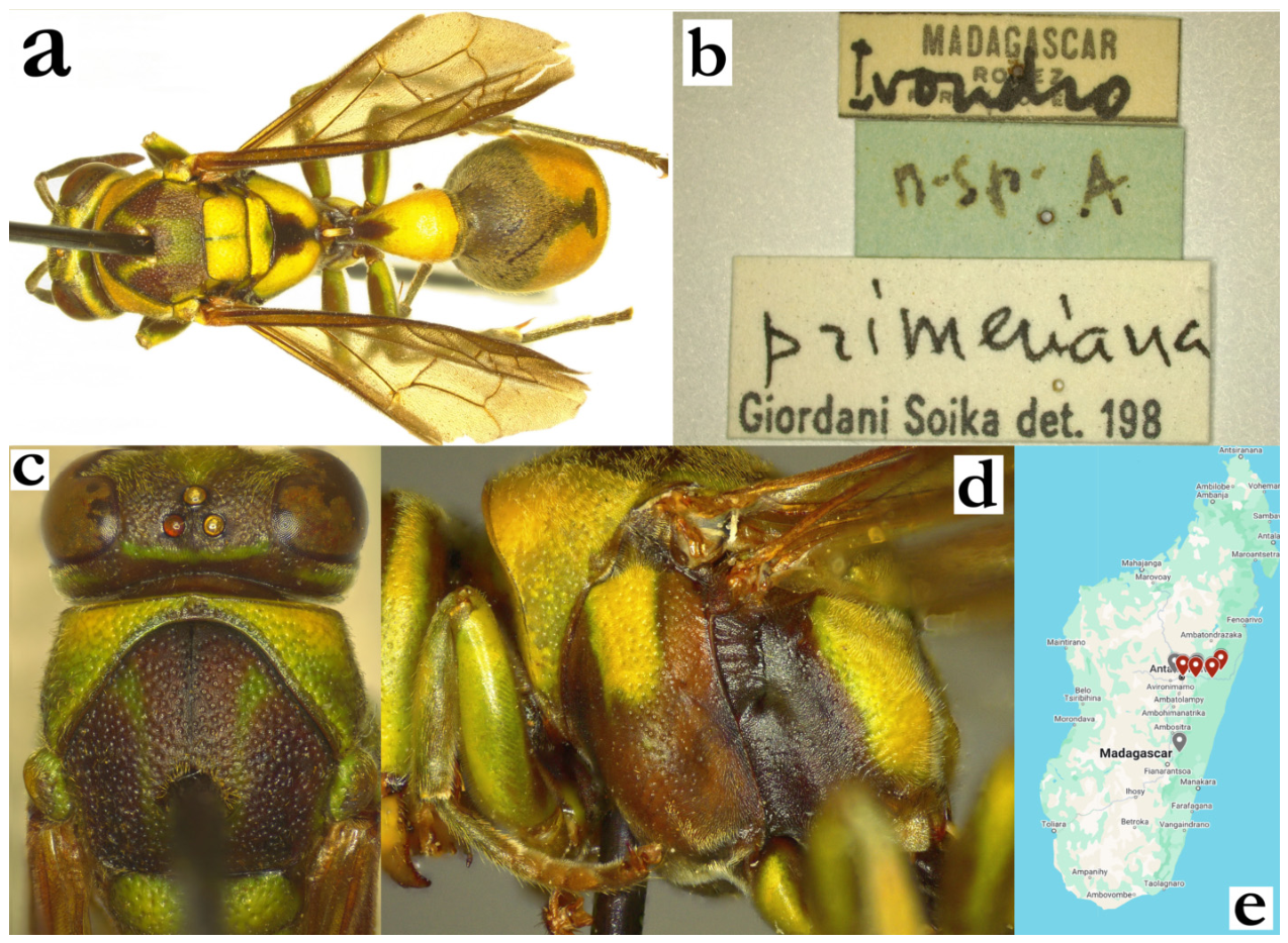
- Ropalidia subclavata (de Saussure), 1890Icaria subclavata de Saussure, 1890; “Madagascar”; designated lectotype specimen from EHT collection.Plazi: https://tb.plazi.org/GgServer/html/03AE106DFFD1396F25D7FE87842EF34C (accessed on 21 September 2026).BOLD: https://portal.boldsystems.org/bin/BOLD:ADN1866 (accessed on 21 September 2026).Number of examined specimens: 32 ♀♀, 4 ♂♂.
Type material. Two implied type specimens from MNHN [2] are not aligned with the species description. Another possible type specimen is the ETH female, with the label “Cotype”, and proposed by Kojima as a “syntype”, in a draft label on the specimen. Since all three examined specimens are conspecific, the specimen from ETH is hereby designated as the lectotype (Figure 121a–c).
Notes. A stocky, black and green coloured species, with dorsally developed T1. There are no fundamental taxonomical problems, except in smaller females, which can sometimes resemble R. phalansterica (de Saussure). Males have several unique features, reducing the risk of misidentification (Figure 121e–g).
Nest. This species has rather irregular nesting architecture, with variation in shape, colour and location of the nest. Nest shape is most commonly irregular discoid, with common early-stage formula of 10 × 15 cells (Figure 121h) and a paracentral petiole (Figure 121i). Larger nests can become less regular in shape, but also cell opening orientation, which can either be vertical [100] or horizontal [101]. Cell wall is variably coloured, with greyish(brown) basal colour (Figure 121h), sometimes with black horizontal streaks or even subtly greenish basal hue (Figure 121i). Cell wall is porous, and with frequent areas of thicker and thinner wall, even in the same cell (Figure 121h); in addition, cell margin at cell openings are always irregular (Figure 121j). Cocoon caps are also irregularly placed even in old and new cells (Figure 121h); cocoon caps are light-silvery without pulp (Figure 121h), translucent with pulp (Figure 121j), or heavy-silky yellowish or greyish (Figure 121h).
Distribution. Eastern parts and central highlands (Figure 121d).
Figure 121.
Ropalidia subclavata (de Saussure), (a) habitus, dorsal view, female, lectotype, (b) habitus, lateral view, female, lectotype, (c) labels, lectotype, (d) distribution map (red marker—specimen, grey marker—literature report or an observational record), (e) head, frontal view, male, (f) antenna, dorsal view, male, (g) antenna, median view, male, (h) nest from NHRS collection, (i) same nest, lateral, (j) nest [102].
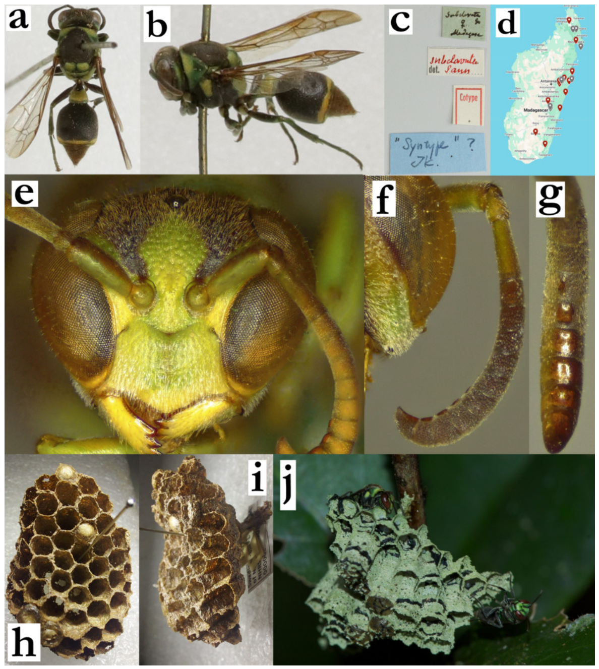
- Ropalidia takona sp. nov. PolašekZooBank: 762BC884-39B4-4835-9C47-D9F1991B311EMaterial examined. Holotype: “MADAGASCAR”, “Ivandrio”, “MUSEUM PARIS/ II 40/ A. Seyrig”, “a”, ♂ (Figure 122a,b; MSNV).Paratypes: 1♀, same labels as holotype; “Ivandrio/ SEYRIG, I 1939”, 2♀♀ (MSNV); “Mandena, 02.2004, P Bulirsh”, 4♂♂ (OLM); “MADAGASCAR/ Ankarafantsika/ (Forest reserve)/ near Marovoay/ XII-1-1959”, 5♀♀ (CAS); “Ampijoroa/VIII-56 P. Griv”, 1♂ (MSNV).Number of examined specimens: 8 ♀♀, 6 ♂♂.
Diagnosis. Member of the grandidieri group, characterized by mesoscutum protruding setae, median scutella carina absent, T2 punctures coarse, colour in females brownish, femur II in males parallel and mesosoma underside in males with yellow markings.
Description. Females. Wing length 7.3–8.2 mm.
Head. Head frontally 1.2× as wide as high, in dorsal view 2.4× as wide as long, weakly emarginate posteriorly. Clypeus 1.15–1.2× as wide as long, with straight upes and dull OCA; juxtamandibular notches shallow, lateral margins weakly concave, apex pointy (Figure 122c); base and centre of clypeus with poorly defined punctures, apical half with sparse and poorly defined craters (Figure 122c); frons with sparse and poorly defined punctures of intermediate size, up to two diameters apart; large punctures retained on anterior part of gena, posterior impunctate; gena 1.0–1.2× as wide as eye (Figure 122d); mandible punctate, occipital carina complete, posterior ocelli wide apart, their distance at least 2.4–2.7× as long as their diameter (Figure 24a); scape slightly longer than AF1, AF1 3.6× as long as wide; AF2 1.2–1.3× as long as wide (Figure 24b).
Mesosoma. Mesosoma finely punctate dorsally; punctures on mesoscutum small and 3–4 diameters apart (Figure 24a); those on pronotum somewhat denser and larger, but their interpuncture spaces more swollen (Figure 24a); mesepisternum superiorly with large punctures, inferiorly somewhat smaller (Figure 24b); scutellum dorsally rounded, shallowly punctate, with median carina absent or very short (Figure 24a); metanotum with less defined punctures than scutellum, with a posterior shiny impunctate area that reaches about half of its height; metepisternum with an occasional poorly defined puncture; median depression of propodeum very finely striate and impunctate.
Metasoma. T1 similar to other members of the grandidieri group; dorsally developed, with posterior sulcus (Figure 122d); T2 densely and shallowly punctate (Figure 122d); remaining segments shallowly punctate.
Pilosity. Body with yellow short pubescence and some short protruding setae; frons with straight yellowish setae that are 0.4–0.7× as long as anterior ocellus diameter; eyes asetose (Figure 24b); mesoscutum with short yellowish-golden pubescence, pronotum with whitish pubescence of similar length (Figure 24b; some specimens can have very short protruding setae on mesoscutum, increasing the risk of misidentification as R. shestakowi; in these cases, R. takona sp. nov. always has retained puncture density on anterior part of mesoscutum); T1 with fine yellowish pubescence, T2 with pubescence and yellow protruding setae that do not extend over lamella (Figure 122d); remaining segments with somewhat longer protruding setae.
Colour. Very variable, but generally with numerous lightly brown areas (Figure 122d); face commonly suffused ferruginous-brown (Figure 122c), mesosoma anteriorly in same colour, posterior part somewhat darker (Figure 122d); femur in basal colour, tibia olivaceous, tarsi olivaceous-brownish (Figure 122d); T1 anteriorly dark brown, posteriorly light brown; T2 in somewhat darker tone and faintly yellowish suffused posterior band (Figure 122d); remaining segments brownish; wings yellowish-transparent, nervature brown, stigma brown, opaque, with apical darkening (Figure 122d); antenna infuscate ferruginous (Figure 122c).
Males. More diversely coloured than females, with frequent reddish and yellow areas (Figure 122a). Wing length 7.1–7.7 mm.
Head. Clypeus 1.3× as wide as long, upes short, OCA developed, juxtamandibular lobes weak (Figure 122e); clypeus basally with some large and shallow punctures, apically less defined (Figure 122e); gena 0.3–0.4× as wide as eye, mandible shiny and impunctate; scape barely widened, 1.3× as wide as AF1 base, somewhat longer than AF1 (Figure 24d); AF2 1.4× as long as wide; tyloids flattened and wide, originate at AF2 as a thin line, that gets wider at AF3 and more distal flagellomeres (Figure 122f); terminal flagellomere elongated, 2.8× as long as wide, with proximally widened blackened tyloid that extends towards sub-acute tip (Figure 24d).
Mesosoma. Fore tarsal spur I developed and long, 3.0–3.5× as wide as long.
Metasoma. S7 weakly concave, with proximal half shiny and impunctate and distal coarsely punctured.
Pilosity. Similar to females, except clypeal surface with fine, equally long, silvery setae that become denser on juxtamandibular region; frons setae longer than in females.
Colour. Clypeus yellow with suffused ferruginous area, similar to interantennal area and inner orbits, mandible yellow with remaining darker postero-basal area (Figure 122a); mesosoma in combination of brown, black and yellow; mesosoma underside mostly yellow (Figure 122a); femur II anteriorly yellow; tibia olivaceous, tarsi brownish.
Distribution. Central highlands (Antananarivo to Ankarafantsika national park).
Etymology. The name originates from the Malagasy adjective takona, “hidden, not seen”, referring to difficulties in separating it from other species.
Figure 122.
Ropalidia takona sp. nov., (a) habitus, anterior oblique view, male, holotype; (b) labels, holotype, (c) head, frontal view, female, (d) habitus, lateral view, female, (e) head, frontal view, male, holotype, (f) antenna, median view, male, holotype.
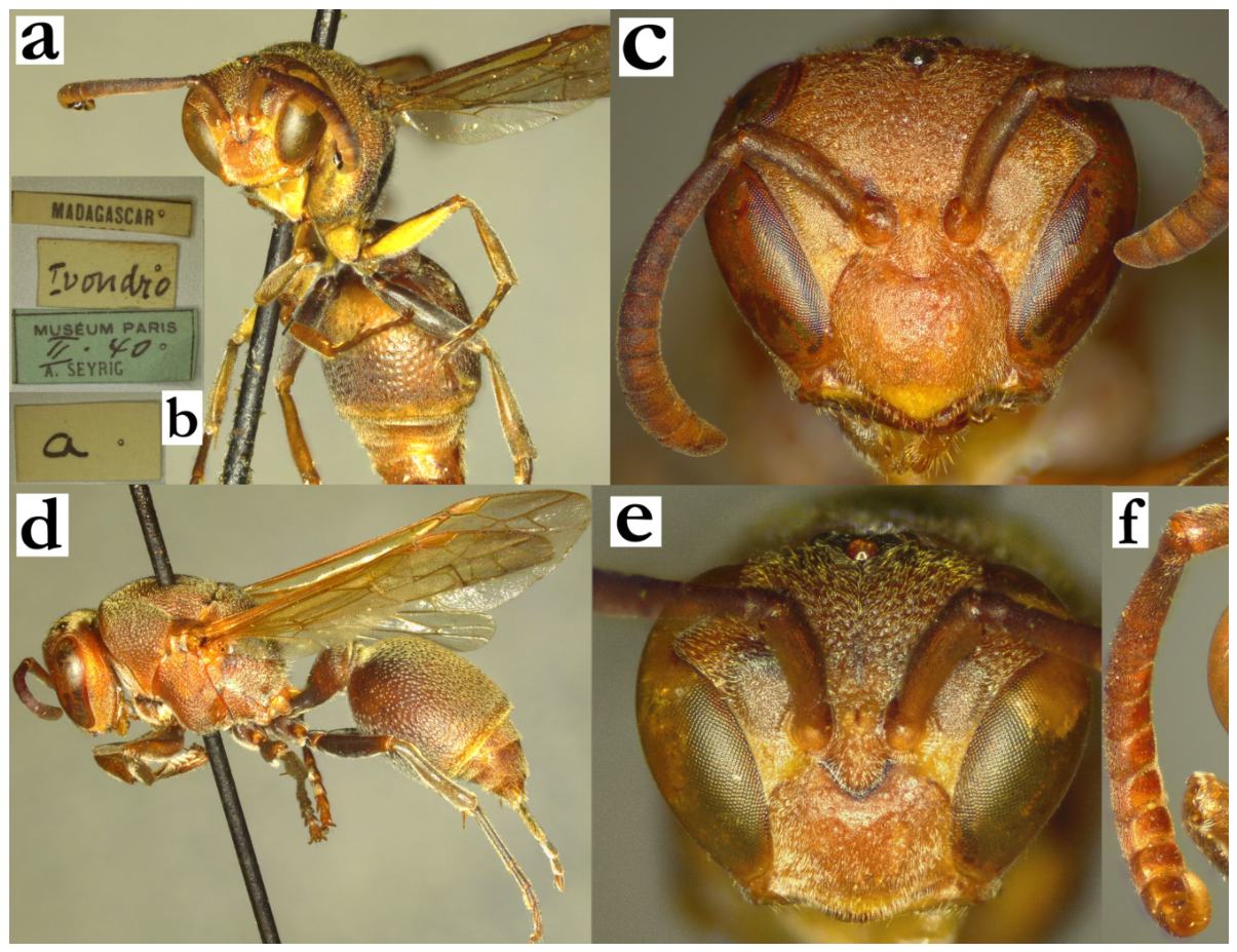
- Ropalidia tapelaka sp. nov. PolašekZooBank: D48D8EC4-B1AE-4FCB-976A-1C50E4A0211FMaterial examined. Holotype: “MADAGASCAR/ ROGEZ/ FORET COTE EST”, “MUSEUM PARIS/ IV 37/ A. SEYRIG”, ♀ (MSNV), Figure 123a–c.Paratypes: 1♀, same label as holotype; “MADAGASCAR/ ROGEZ/ FORET COTE EST”, “MUSEUM PARIS/ I 37/ A. SEYRIG”, 1♀ (MSNV); “MADAGASCAR/ ROGEZ/ FORET COTE EST”, “MUSEUM PARIS/ III 37/ A. SEYRIG”, 2♀♀, 2♂♂ (MSNV); “Environs de Rogez/ Madagascar”, 1♀ (MSNV); “Madagascar/ Env. de Rogez, Madagascar”, 1♀ (MSNV); “La Mandaraka/ Madagascar, Mus. Praha” 1♀ (MSNV); “MUSEUM PARIS/ MADAGASCAR, TAMATAVE/ R. DU BUYSSON 1909”, 1♀ (MSNV); “Andohahelo/ 1800m/ RP”, 1♀ (MSNV); “Ranomafana/23.1.58, F. KEISER”, 1♀ (NNM).Number of examined specimens: 11 ♀♀, 2 ♂♂.
Diagnosis. This species is characterized by head and mesosoma mainly green, metasoma brown or reddish and frons and dorsal side of metasoma with short pubescence; clypeus is in females flattened, with very wide base, providing easy separation from other species. Males have specific tyloids that only extend along proximal half of flagellomere.
Description. Females. Wing length 8.4–8.8 mm.
Head. Head frontally 1.2× as wide as high (Figure 51c), 2.5 as wide as long in dorsal view, equally wide behind eyes, barely posteriorly emarginate (Figure 123a). Clypeus 1.4–1.5× as wide as long, with straight upes, parallel lateral sides and sharp OCA; juxtamandibular lobe weakly developed, apex more protruding (Figure 51c). Clypeus basally impunctate and matte, apically with small and shallow punctures; inner orbit impunctate (Figure 51c). Frons with spaced and small punctures on matte interpuncture spaces (Figure 51c). Gena at mid-height as wide as eye, inferiorly widened; lowest point of gena 1.1–1.2× as wide as eye; upper half of gena with some ill-defined punctures, lower half impunctate (Figure 51b). Mandible punctate, occipital carina complete; posterior ocelli 1.6× diameter apart, distance to eye 3.1× diameter, distance to occipital carina 3.15× diameter (Figure 123a). Eyes very minutely setose (Figure 123b). Scape 1.25× as long as AF1, AF1 2.8× as long as wide at mid-length; AF2 1.1× as long as wide (Figure 51c).
Mesosoma. Mesosoma in dorsal view 1.05× as long as wide in front of tegula; disc of mesoscutum as wide as long, with large, poorly defined and shallow punctures (Figure 123a). Pronotum with similarly spaced punctures, but less defined and more swollen interpuncture surface; puncture on pronotum over one diameter apart. Mesepisternum coarsely and scarcely punctate, metepisternum with some isolated punctures. Scutellum with spaced shallow punctures and swollen interpuncture area, metanotum posteriorly rounded and punctate sparser than scutellum. Propodeum posteriorly wide, median depression finely striate and impunctate, wide and circular; lateral surface with some poorly defined punctures.
Metasoma. T1 well developed and posteriorly widened; posterior half punctate, punctures deep and poorly defined (Figure 123d). T2 densely and finely punctate, punctures about a diameter apart, less dense proximally (Figure 123d). T2 lamella posteriorly impunctate, obscured by the protruding setae (Figure 123d).
Pilosity. Very fine; head and mesosoma with very fine silvery pubescence (Figure 123b). Clypeus apically with yellow protruding setae, like those on mandible; frons setae 0.4–0.7× as long as ocellus diameter (Figure 51b). Gena with silvery pubescence and a few protruding setae (Figure 123b). Mesoscutum with golden pubescence and barely protruding setae (Figure 51b). T1 with short yellowish pubescence, T2 with yellow setae that do not extend over lamella (Figure 51a).
Colour. Head and mesosoma mainly green (Figure 51b,c). Frons and parts in some specimens olivaceous-green, small areas behind proximal half of scape black, sometimes with suffused yellow areas on clypeal apex and inner orbits (Figure 51c). Pronotum green, sometimes with a remaining dark-green postero-dorsal area (Figure 51a). Mesoscutum brownish, often with two suffused green lines on the anterior third of its length (Figure 123a). Mesepisternum basally black, with large green spot, metepisternum commonly black with a basal green spot, or entirely green (Figure 51a). Scutellum green, with a complete, thin black line underneath median carina. Metanotum with a green line and only a postero-medial remaining black marking. Propodeum basally black, with large green spot that encompasses most of its surface or completely green (Figure 51a). All coxa with green markings; femora green, sometimes with dorsal patchy darkening, tibiae uniformly green, tarsi olivaceous-green. T1 green, with only a smaller remaining diamond-shaped black area on dorsal side of petiole. T2 basally brownish(-olivaceous), with a thin green posterior band; the band extends on S2 as a short, abruptly ending mark (Figure 51a; one examined specimen has more developed and partial S2 band, but still retaining the abrupt and nearly perpendicular interruption close to the area where the T2 and S2 are merged). Remaining metasomal segments ferruginous. Wings transparent, minimally yellowish (except anterior margin, where some yellowish areas are common); apical spot not visible at all, stigma light brown or yellowish, transparent (Figure 51a). Antennal scape dorsally green, distal segments variable (green to olive-green, sometimes even brownish), underside proximally green, distally yellow (Figure 51c).
Males. Resemble females. Wing length 8.2–8.5 mm.
Head. Clypeus 1.5× as wide as long, with convex surface, short and curved upes and weakly developed juxtamandibular excavation (Figure 123e). Clypeus very shallowly punctate or completely impunctate and matte (Figure 123e). Gena 0.6–0.9× as wide as eye, occipital carina well developed. Mandible with poorly defined punctures or impunctate. Scape moderately widened, 1.4–1.5× as wide as AF1 base, shorter or just about as long as AF1; AF2 1.3–1.6× as long as wide (Figure 123f). Tyloids round, originate at AF6, brown and barely elevated above flagellomere surface, matte and small, extending only across proximal half of flagellomere (Figure 51d). Terminal flagellomere elongated, 1.8× as long as wide; tyloid widened proximally; flagellomere tip narrowed and rounded (Figure 123f).
Mesosoma. Fore tarsal spur I very small, barely developed (Figure 123g).
Metasoma. S7 flattened, with shiny and somewhat irregular surface.
Pilosity. Overall shorter than in females.
Colour. Head like females, mainly green, with somewhat darker frons; in contrast to females, males have more yellow on the head (Figure 123e). Mesosoma underside with somewhat more suffused yellow markings than females. Antenna heavily infuscate dorsally, green-orange underneath (Figure 123e).
Distribution. North-Eastern parts of the island.
Etymology. The name originates from the Malagasy word tapelaka (pelaka), translated as “something that is wide and plain”, referring to the wide and flattened clypeus in females, and is treated as indeclinable.
Similar species. Several species are similar to this one, including R. phalansterica (de Saussure; separated from this one by smaller size of females and male antenna) or R. sonyae sp. nov. (separated by the shape of T1, pilosity and general colour pattern). Some confusion is possible in relation to R. subclavata (de Saussure) females. That species has longer setae on frons (longer than the anterior ocellus diameter) and some protruding setae on mesoscutum, while R. tapelaka sp. nov. has no protruding setae on mesoscutum and the setae on frons are shorter or almost equally long as the anterior ocellus diameter. Additionally, R. subclavata (de Saussure) has developed eye setae, which are very fine or completely missing in R. tapelaka sp. nov.
Figure 123.
Ropalidia tapelaka sp. nov., (a) head and mesosoma, dorsal view, female, holotype, (b) head, oblique lateral view, female, holotype, (c) labels, holotype; (d) T2, lateral view, female, holotype, (e) head, frontal, male, (f) antenna, dorsal view, male, (g) tarsal I spur, lateral view, male.
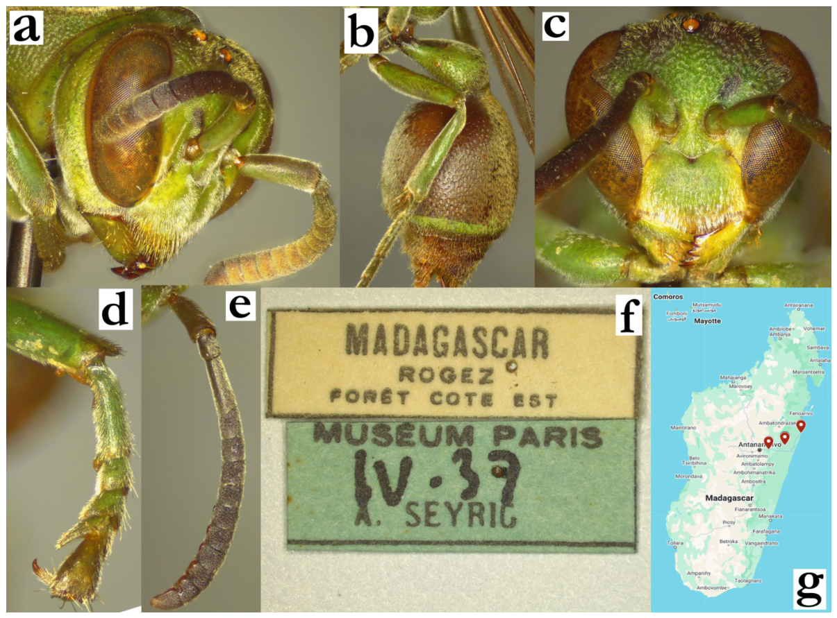
- Ropalidia tatsimo sp. nov. PolašekZooBank: FCD37EEA-CC13-4B56-801D-0FF385AF38BAMaterial examined. Holotype: “Fort Dauphin/ RP.”, “INSTITUT/ SCIENTIFIQUE/ MADAGASCAR”, ♀ (MSNV); Figure 124a,b.Paratypes: same label as holotype, 18♀♀, 4♂♂, and nest with two macerated and examined early stage adults; “Madagascar/Fort Dauphin”, “Seyrig/ V 37”, 2♀♀ (MSNV); “Region de L’Androy/ Ambovombe/ Dr J Decorse 1901”, 1♂ (MSNV).Number of examined specimens: 19 ♀♀, 4 ♂♂.
Diagnosis. This species is characterized by T1 developed dorsally, T1 lamella impunctate, widely apart posterior ocelli, mesoscutum as wide as long and specific antenna morphology in males.
Description. Females. Wing length 8.4–9.1 mm.
Head. Head frontally 1.25× as wide as long, clypeus 1.4× as wide as long, with straight upes and nearly absent OCA; juxtamandibular notches shallow, lateral margins angled apex mildly pointy (Figure 55c); base and centre of clypeus with poorly defined punctures, apical half with sparse and poorly defined craters (Figure 55c); frons with sparse punctures of intermediate size, up to two diameters apart, on velvety surface; large punctures retained on anterior part of gena, posterior impunctate; gena 1.0–1.2× as wide as eye (Figure 55b); mandible punctate, occipital carina complete, posterior ocelli wide apart, their distance at least 3x greater than their diameter (Figure 55b); scape slightly longer than AF1, AF1 shorter than AF2+3+4; AF2 1.0–1.1× as long as wide.
Mesosoma. Mesosoma punctures dorsally coarse (Figure 55a,b), ventrally smaller but retained; pronotum and mesoscutum shallowly punctate, with velvety puncture interspaces; mesepisternum superiorly with large punctures, inferiorly somewhat smaller (Figure 124c); scutellum dorsally rounded, shallowly punctate, with developed median carina; metanotum with similar or somewhat less defined punctures than scutellum, with a posterior shiny impunctate area that reaches about half of its height (Figure 124d); metepisternum with an occasional well-defined puncture; propodeum posteriorly wide, median depression of propodeum very finely striate and impunctate (Figure 124d).
Metasoma. T1 dorsally developed (Figure 124e), with poorly defined punctures; T1 posterior lamella impunctate (Figure 55a); T2 densely and shallowly punctate (Figure 124e); remaining segments shallowly punctate.
Pilosity. Body with whitish short pubescence and some protruding setae; frons with straight yellowish setae that are 0.3–0.6× as long as anterior ocellus diameter; eyes asetose (Figure 69b); mesoscutum with short yellowish-golden pubescence, pronotum with whitish pubescence of similar length (Figure 124c); T1 with fine yellowish pubescence, T2 with pubescence and yellowish protruding setae that extend over lamella (Figure 124e); remaining segments with somewhat longer protruding setae.
Colour. Basal body dark brown, with numerous dark-green suffused areas (Figure 124a); clypeus yellow with central dark-olivaceous spot attached to base; inner orbits, interantennal area, and mandible yellow (Figure 55c); gena basally brownish, with a green line superiorly and suffused olivaceous area inferiorly (Figure 124c); pronotum brownish, with a dark green line underneath carina; mesoscutum darker (Figure 124a); mesepisternum dark-brown with a brownish superior spot (Figure 124a); scutellum with faint green spot, metanotum with more defined, suffused green markings, propodeum in basal colour (Figure 124d), or with two suffused yellowish spots in more xanthic specimens; coxa I yellowish-green, coxa II and III with lateral yellow or green areas (Figure 124e); femur in basal colour, tibia olivaceous, tarsi olivaceous-brownish (Figure 124a); T1 with broader posterior green band, T2 with a thin green band (Figure 124e); remaining segments brownish; wings transparent, nervature brown, stigma ochre, semi-transparent, with minimal apical darkening (Figure 124a); antenna dorsally brownish (Figure 124a), ventrally ferruginous; scape ventrally green, dorsally dark brown (Figure 55c).
Males. More diversely coloured than females, with frequent reddish and yellow areas (Figure 124f). Wing length 8.4 mm.
Head. Clypeus 1.3× as wide as long, upes short, OCA developed, apex rounded, juxtamandibular lobes weak (Figure 124g); clypeus basally and centrally shallowly and finely punctate, with an occasional more developed puncture that is obscured by the silvery pubescence (Figure 124g); gena 0.4× as wide as eye, mandible shiny and impunctate, with basal depression; scape widened, 1.6× as wide as AF1 base, as long as AF1 (Figure 124g); AF2 1.1–1.2× as long as wide; tyloids rounded, originate at AF4, brownish and shiny, elevated above flagellomere surface as rounded beads (Figure 55d); terminal flagellomere elongated, 1.8–2.0× as long as wide, with proximally widened tyloid and beak-like, acute tip (Figure 55d).
Mesosoma. Fore tarsal spur I developed.
Metasoma. S7 weakly concave, with shiny and irregular surface.
Pilosity. Similar to females, except clypeal surface with fine, equally long, silvery setae, that become denser on juxtamandibular region (Figure 124g); frons setae comparatively longer than in females.
Colour. Clypeus yellow with faint basal greenish spot, interantennal area and inner orbits inferiorly yellow, further dorsally suffused green, mandible yellow with remaining darker postero-basal area (Figure 124g); remaining parts of the head in basal colour or suffused green and reddish; mesosoma in basal colour, pronotum with green line underneath carina or entirely reddish in more xanthic specimens (Figure 124f); mesonotum and mesepisternum in basal colour, scutellum with two (faint) green spots, metanotum basally black, with suffused olive(-green) area; the remaining colour pattern similar to females: coxa I yellow, coxa II and III with lateral yellow marks.
Nest. The type series of specimens originate from a single nest (Figure 55e), with labels similar to holotype; two cocoon caps from the nest were removed, allowing the extraction of two pre-hatching adults, which were conspecific with the holotype. The nest is discoid and has very shallow cells, that are 1.7–2.3× as wide as long (compared to 2.9–3.6× in R. grandidieri nests). The nest has 119 cells, with a maximum width of 11 × 10 cells (Figure 55e), with paracentral petiole (Figure 124h) and somewhat depressed centre (Figure 124i). The nest seems to have been built in two separate processes; the first one led to a central, darker cell wall section, while the second was based on a more lightly coloured cell wall material (Figure 124h). Cocoon tops are somewhat irregularly placed, mostly centrally (Figure 55e), with 15 of them being closed; most of them are below the cell wall margin. The most interesting part is that all the cell bottoms are enclosed by the translucent membrane (Figure 124h), and that all the cocoon caps are silky, without the cell wall nodules that are present in some species (Figure 55e). The cell wall is brittle.
Distribution. South Madagascar; Taolagnaro (previously known as Fort Dauphin) to Ambovombe.
Etymology. The name originates from the Malagasy word tatsimo, translated as “southerner”, referring to the predominantly Southern distribution of this species; the name is treated as indeclinable.
Figure 124.
Ropalidia tatsimo sp. nov., (a) habitus, oblique lateral view, female, holotype, (b) labels, holotype, (c) head and anterior mesosoma, lateral view, female, (d) posterior mesosoma, dorsal view, female, (e) T1 and T2, lateral view, female, (f) habitus, oblique lateral view, male, (g) head, frontal view, male, (h) nest, dorsal view, (i) nest, lateral view (MSNV).
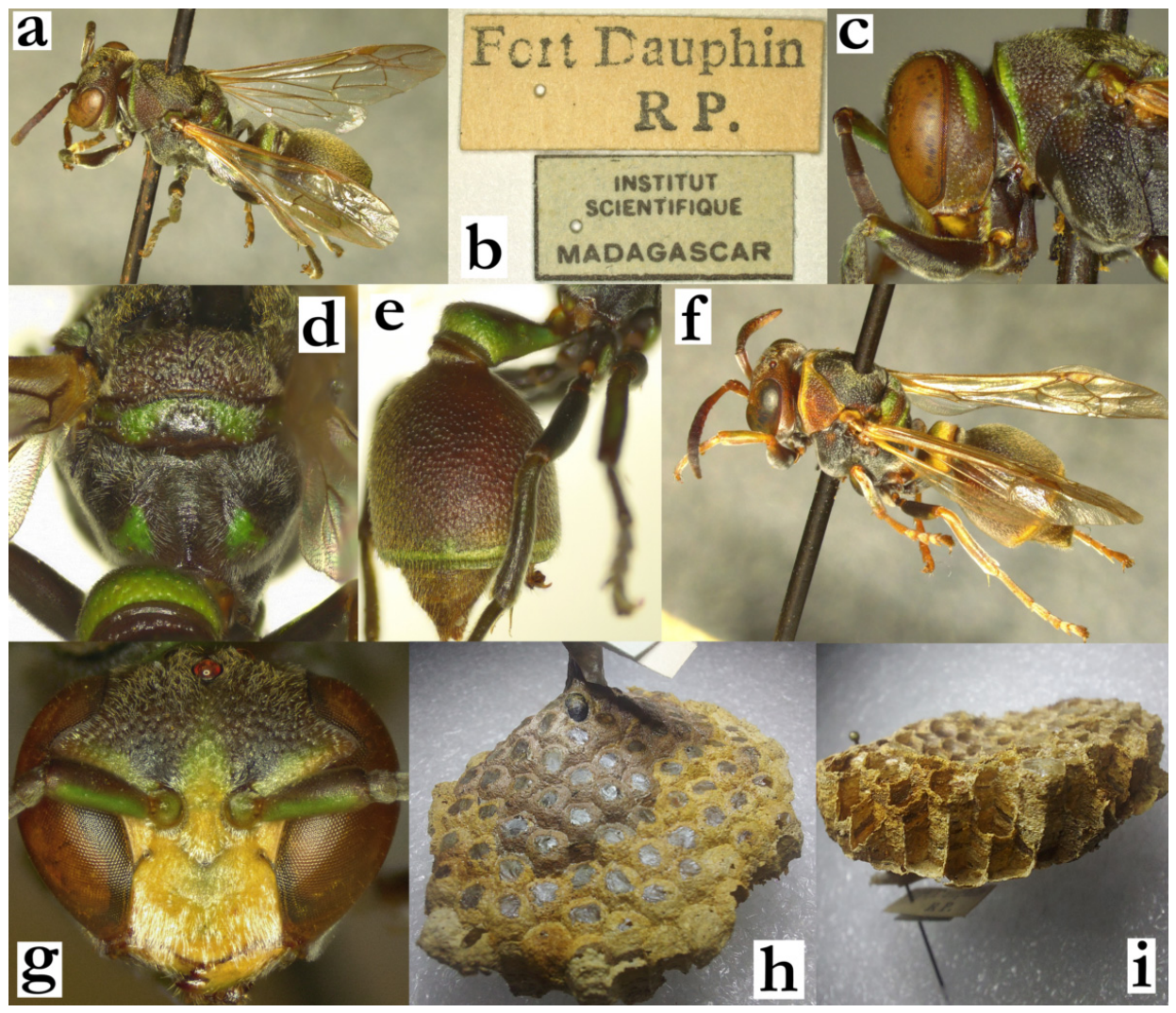
- Ropalidia telperia sp. nov., PolašekZooBank: 4B40E144-29BC-4D83-9239-9E49DB5F2BE7Material examined. Holotype: “Bekily/IV 37, Seyrig”, ♀ (MSNV), Figure 125a,b.Paratypes: 9♀♀, same label as holotype (MSNV); “Andranomandevy Didy, Ambatenazaka/X 56 E.R.”, 1♀ (MSNV); “Morafenobe/5 52 R F”, 1♀ (MSNV); “Vohipara/20.1.1992, A Pauly/Phellolophium madagascariensis”, 2♀♀ (RMCA).Number of examined specimens: 14 ♀♀.
Diagnosis. A species characterized by T1 slender and elongated, wing length under 8 mm, face yellow and short and silvery pubescence on basally green body (Figure 125a).
Description. Females. Wing length 6.4–7.2 mm.
Head. Head frontally 1.2× as wide as long, clypeus 1.2× as wide as long, pentagonal with straight and elongated upes, weak and dull OCA, moderately developed juxtamandibular excavation and a pointy apex (Figure 41b); base and centre of clypeus weakly punctate, apex with denser and less defined craters; frons with coarser and larger punctures (Figure 41b); anterior third of gena with poorly defined craters, posterior almost completely impunctate and shiny (Figure 125b); mandible punctate, occipital carina complete, ocelli equidistant, posterior ocelli at least 2 diameters apart (Figure 41a); scape longer than AF1, AF2 about as long as wide or up to 1.2× as long as wide.
Mesosoma. Entire mesosoma with well-defined punctures, obscured by the pubescence layer (Figure 41a). Mesoscutum densely and shallowly punctate (Figure 41a), pronotum similarly punctate; scutellum posteriorly rounded, median carina developed, but hardly conspicuous due to pubescence; metanotum postero-inferiorly flattened, sometimes depressed, and shiny; upper and lateral parts coarsely punctate (Figure 41c); propodeum posteriorly wide, faintly striate lower half, impunctate (Figure 41c).
Metasoma. T1 pyriform, elongated and very flattened in lateral view (Figure 40a), dorsally triangular and wide, 0.5–0.7× as wide as T2; posterior part punctate (Figure 41c); T1 lamella long and impunctate; T2 coarsely punctate, with strong pubescence, creating rugged pattern (Figure 40a).
Pilosity. Entire body with a dense silvery-yellowish layer of pubescence that is creating a halo around the body (Figure 125a); clypeus apically with twice longer setae than basally; gena with short pilosity, frons with setae that are 0.6–0.9× as wide as ocellus diameter (Figure 125b); mesosoma with short pilosity (Figure 41a); T1 with short silvery pubescence and longer protruding yellowish setae (Figure 40a); T2 with a dense layer of golden pubescence and longer golden protruding setae that barely extend over lamella (Figure 40a).
Colour. Basal body colour is dark brown, tinged with green (Figure 125a); yellow areas include clypeus (which can be completely yellow, or more commonly yellow with a suffused olivaceous-brownish spot, attached to the base), inner orbits, interantennal area and mandibles (Figure 41b); gena ferruginous or with suffused inferior yellow area (Figure 125a), frons and vertex olivaceous (Figure 41a); Pronotum basally olivaceous, most commonly with a green irregular marking underneath carina, which often becomes yellow on the inferior angles (Figure 125a); mesoscutum blackish-olivaceous, sometimes with suffused green areas adjacent to tegula; mesepisternum blackish, less commonly with a suffused green or reddish area on the upper half; metepisternum blackish; scutellum suffused green, metanotum with green transversal line, propodeum with two large rhomboid patches (Figure 41c); coxa I yellow ventrally, coxa II and III with lateral yellow line; femora basally blackish, distally greenish, often with patchy yellow longitudinal line; tibia olivaceous, tarsi lightly ferruginous (Figure 125a); T1 blackish with antero-lateral green markings and posterior yellow band (Figure 125a); T2 basally blackish, with thin posterior yellow band (Figure 125a); remaining metasomal segments ferruginous or suffused yellow; wings translucent, yellowish anteriorly, nervature light brown, stigma ochre, semi-transparent, without apical darkening or apical spot (Figure 125a); antenna infuscate, ventrally greenish-orange, scape and several flagellar segments greenish ventrally, more distal segments orange ventrally (Figure 125a).
Males. Unknown.
Distribution. Central Madagascar (Figure 125c).
Etymology. After Telperion (telpë, silver and ere, the first), the tree from J.R.R. Tolkien’s mythology, that was said to have dark green leaves, shine silvery light and gave birth to the Moon; the name refers to the green body markings and silvery pubescence, treated as noun, feminine.
Figure 125.
Ropalidia telperia sp. nov., (a) habitus, lateral view, female, holotype, (b) head and mesosoma, lateral view, female, holotype, (c) distribution map.
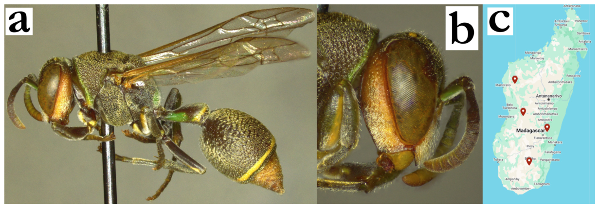
- Ropalidia tsiky sp. nov. PolašekZooBank: 59D2E7DB-A6DC-4A65-9D35-4AE97DB7B148Material examined. Holotype: “Madagascar, Andasibe/ National Park 18.935 S/ 48.414 E leg. Schmid-Egger/17.xii.2017 mad01”, ♀ (MFNB).Paratypes: “Madagascar, Ambalavao/NE town 21.819 S 46.953 E/leg. Schmid-Egger/24.xii.2017 mad04”, 5 ♀♀ (MFNB); “Inst. Scient. Madagascar/Perinet/12.x.9/J. de. D. Rakota”, 1♀ (MSNV).Number of examined specimens: 7 ♀♀.
Diagnosis. This member of the grandidieri group can be characterized by body basally dark brown with suffused greenish markings, clypeus shiny, dorsal mesosoma pilosity short and posterior mesosoma specific colour pattern (Figure 126a).
Description. Females. Wing length 7.9–8.2 mm.
Head. Head in frontal view 1.1× as wide as high, in dorsal view 2.2× as wide as long. Clypeus 1.1× as wide as long, with flattened base and more convex centre, straight upes, developed juxtamandibular excavation and strongly projecting apex (Figure 20c); clypeus basally with very scattered, large and poorly defined punctures, otherwise shiny (Figure 20c); mandible shiny, with scattered and poorly defined punctures; inner orbit impunctate, frons with scattered and weak punctures (Figure 20c). Gena as wide as eye, with some scattered punctures, more defined close to eye and mandible (Figure 126b); occipital carina complete; oculo-malar distance very short, nearly absent (eye almost extending inferiorly to mandible); posterior ocelli 2.0–2.1 diameters apart, 1.7 diameters apart from eye (Figure 126c); scape very long, 6x as long as wide, 1.5× as long as AF1; AF1 3.8× as long as wide, AF2 1.3× as long as wide, AF4 and more distal flagellomeres as long as wide or somewhat wider (Figure 20c).
Mesosoma. Mesosoma slender, 1.8× as long as wide, somewhat narrower than eye width in dorsal view; mesoscutum elongated, 1.3× as long as wide above tegula (Figure 126c); pronotum anteriorly straight, with developed carina and large and coarse punctures, less than one diameter apart; carina of pronotum weakly angulated in lateral view, 1.5× as wide dorsally as laterally (Figure 20a); mesoscutum with large and shallower punctures, which are sparser anteriorly (Figure 126c; except one paratype, with almost entirely retained anteriorly puncture density); scutellum very flattened (Figure 20a), with scattered and shallow punctures, which are somewhat less developed on metanotum; propodeum median depression wide, striate, with some poorly defined punctures.
Metasoma. T1 comparatively wider, similarly shaped as other members of the grandidieri group, posteriorly punctate (Figure 126d); T2 with dense and coarse punctures, which gradually continue to punctate lamella (Figure 126d).
Pilosity. Very short; body covered by very short yellowish setae, those on frons 0.5× as long as anterior ocellus diameter (Figure 126b), mesoscutum without protruding setae (Figure 20b), those on T2 barely visible (Figure 126d); eyes asetose (Figure 20c).
Colour. Basal colour brown(-olivaceous), with yellow spot on mandible, suffused greenish-ferruginous face (Figure 20c), green-yellow line under pronotal carina (Figure 126a), two spots on scutellum and two nearly merged spots on metanotum (Figure 20a); tibia and tarsi greenish, metasoma in basal colour (Figure 126d); antenna dorsally black, ventrally reddish; wings uniformly yellowish (Figure 126a).
Males. Unknown.
Distribution. Central Madagascar (Figure 126e).
Etymology. From the Malagasy word tsiky, “a smile”, treated as a noun, referring to the posterior mesosoma colour pattern (Figure 20a).
Figure 126.
Ropalidia tsiky sp. nov., (a) habitus, oblique lateral view, female, holotype, (b) head and anterior mesosoma, lateral view, female, (c) head and mesosoma, dorsal view, female, (d) T1 and T2, lateral view, female, (e) distribution map (red marker – examined specimen).
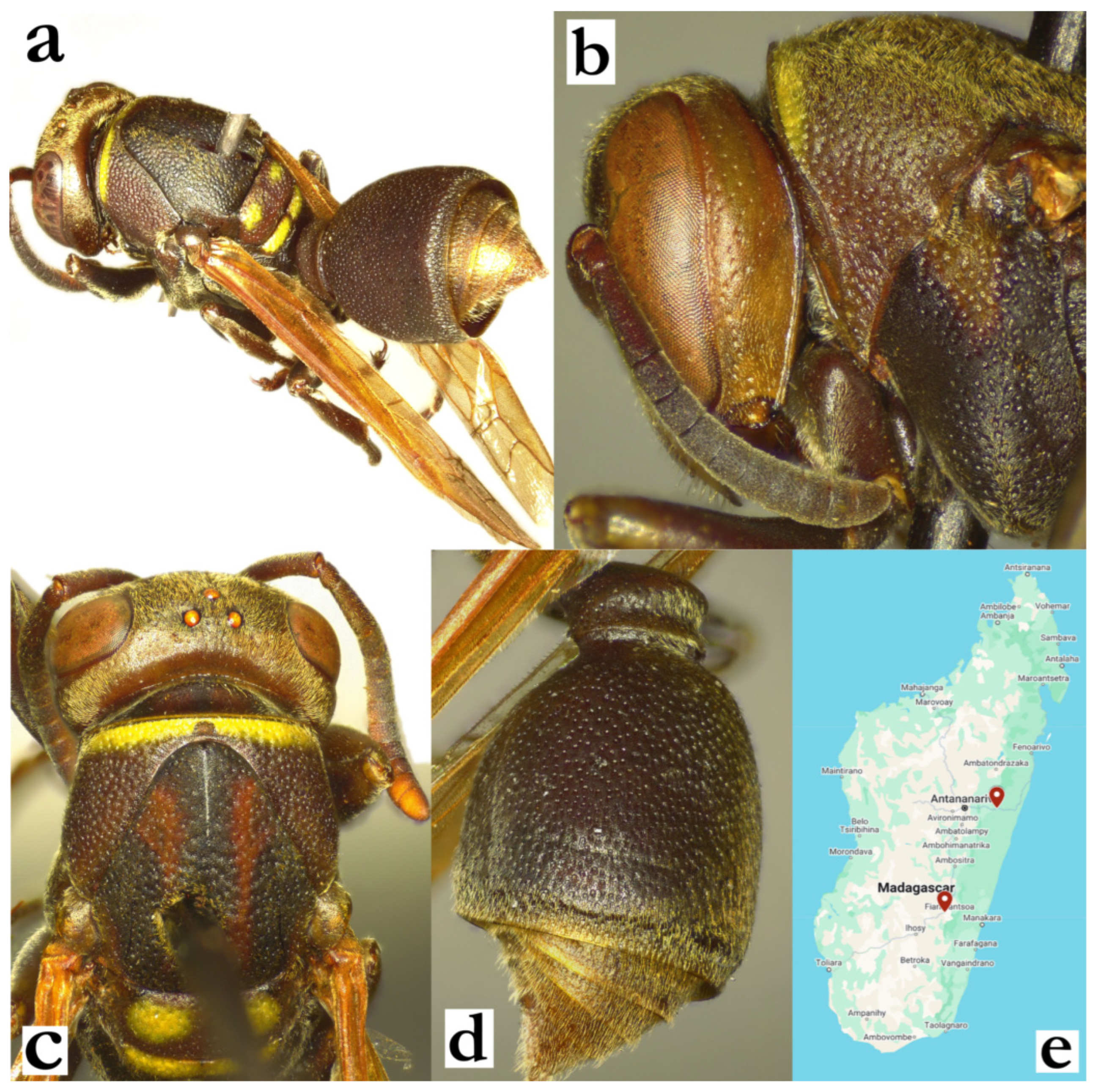
- Ropalidia vanga sp. nov. PolašekZooBank: 59F382BF-C800-4077-B1DE-9C1E005FCD37Material examined. Holotype: “Madagascar, Nosy Be, Special Reservation ‘Lokoba’, 13.-15.12.2003., Dolin & Andreeva”, ♀ (OLM; DNA.AG3H6); Figure 32a, Figure 38a–c and Figure 127a–c.Paratypes: “Madagascar, V Sombirano, A Seyrig 33”, 1♀ (MSNV); green label “Icaria phalansterica Sauss. /[female sign]”, 1♀ (MSNV).Number of examined specimens: 3 ♀♀.
Diagnosis. This species is characterized by brown and yellow body with minimal green markings dorsally on mesosoma, basal spot on T2, posterior ocelli their own diameter apart, AF2 as long as wide and mesoscutum with some short erect setae.
Description. Females. Wing length 7.4–7.6 mm.
Head. Head frontally 1.3× as wide as high, in dorsal view 2.6× as wide as long, distinctively narrowing behind eyes, shallowly emarginate posteriorly (Figure 38a). Clypeus 1.15× as wide as long, with narrow base, straight and long upes, weak OCA, shallow juxtamandibular excavations and projecting apex (Figure 127a). Clypeus weakly and shallowly punctate, punctures hardly discernible due to pubescence. Frons punctures larger and more defined than those on clypeus. Gena punctate close to eye, posteriorly punctures less defined, area near occipital carina impunctate. Mandible shiny and coarsely punctate. Occipital carina complete, placed lower than posterior head margin; posterior ocelli distance equal to posterior ocellus diameter, ocelli to eye distance 2.2× as long as ocelli diameter, distance to occipital carina 1.6× as long as ocellus diameter (Figure 38a). Scape about as long as AF1, AF1 about as long as AF2+3+4; AF2 about as long as wide.
Pilosity. Overall short silvery-whitish pubescence, with some protruding setae on clypeus, of similar length basally and apically (Figure 38c); setae on frons 0.5–0.8× as long as anterior ocellus diameter, mesoscutum with very short erect setae (Figure 38c), propodeum and metasoma with some longer setae, those on T2 barely extending over lamella. Eyes very shortly setose, nearly asetose.
Mesosoma. Mesosoma in dorsal view as long as wide, disc of mesoscutum as long as wide in front of tegula (Figure 38a). Mesoscutum shallowly and sparsely punctate, less dense than pronotum. Mesepisternum with shallow and scattered punctures, metepisternum with a few more defined and larger punctures. Scutellum dorsally rounded, shallowly punctate, with developed median carina that reaches about third of its length (Figure 38a). Metanotum with somewhat coarser punctures, especially on green areas, with shiny posterior impunctate area. Median depression of propodeum impunctate and shiny, with horizontally developed striae.
Metasoma. T1 pyriform, posteriorly globular, with visible terminal constriction. Posterior part of T1 punctate, with occasional weakly defined puncture. T2 finely and shallowly punctate with directional punctures, remaining segments with shallow punctures (Figure 127b).
Colour. Body basally brown, with numerous yellow markings, including clypeus (except central suffused brown spot), most of inner orbit and interantennal area (Figure 127a), mandible except postero-basal remaining brown area. Mesosoma basally brown, with thickened yellow line underneath pronotal carina, thin line on dorsal pronotal margin, mesoscutum with two thin yellow lines (greenish in one paratype), mesepisternum with elongated yellow spot and another on the posterior margin in holotype (Figure 127b), scutellum, metanotum and propodeum with large or merged yellow or greenish spots (Figure 127b); greenish hue present in dorsal parts of mesosoma (Figure 32a). All coxa pairs yellow ventrally, coxa II laterally yellow, medially brown, while coxa I and III dorsally completely yellow (Figure 127b). All femora with brown proximal half, distal half yellow, tibia brown with outer yellow line, tarsi ferruginous-brown (Figure 127b). Wings transparent, nervature and stigma brown, apical spot not developed. T1 with three colours; proximal dark brown to blackish, posterior part green-yellow (Figure 127b). T2 basally brown, with two basal yellow spots and a thin posterior yellow band (Figure 32a). All metasomal segments with suffused thickened posterior yellow band (Figure 127b). Antenna dorsally brown, scape ventrally yellow, flagellum ventrally orange (Figure 127a). Paratype very similar to holotype (Figure 127d), previously determined as R. phalansterica (Figure 127e).
Males. Unknown.
Similar species. Difficulties are expected in separation from R. bongomaso sp. nov., with key features listed in the key to species.
Distribution. North-Eastern parts of the island (from Nosy Be to Sambirano).
Etymology. From the Malagasy word vanga, “a mark, a trace, a line”, referring to the patchy colour pattern; the name is treated as indeclinable.
Genetics. One specimen was successfully sequenced for the COI gene, suggesting a close relationship and sister position with R. bongomaso sp. nov. BOLD barcode ID process yielded 92.26% concordance with BOLD:ACT6336 (R. formosa) and 91.63% concordance with BOLD:ADH8057 (R. zandry sp.nov.).
Figure 127.
Ropalidia vanga sp. nov., (a) head, frontal view, female, holotype, (b) habitus, lateral view, female, holotype, (c) habitus, lateral view, female, paratype, (d) labels, holotype, (e) label, paratype.
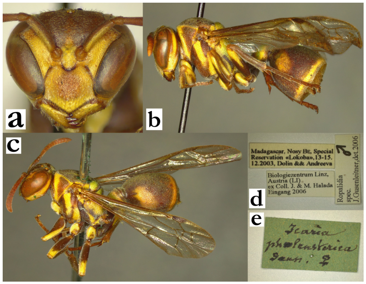
- Ropalidia variabilis (de Saussure, 1890)Icaria variabilis de Saussure, 1890; “Collection de Saussure et Musée de Berlin”, lectotype (MHNG 00089391).Plazi: https://tb.plazi.org/GgServer/html/03AE106DFFD1396F25D7FCA3842EF53A (accessed on 21 September 2026).Ropalidia variabilis (de Saussure) Blommers 2012 (male, female, nest biology; assigned lectotype status; MHNG).Number of examined specimens: 28 ♀♀, 10 ♂♂.
Type material. The species description mentions “Madagascar”, and that the depository is Geneva and Paris [13]. Blommers assigned a lectotype status to a specimen in the MHNG collection, suggesting there may be other syntypes elsewhere. The specimen has previously been labelled by Kojima, but the status remained unpublished. It bears the label “77” on an aged yellowish paper, followed by “Icaria variabilis [female sign] SSS” on a green paper, and currently bears the identification label MHNG 00089391 (Figure 128a); the lectotype is conspecific with the specimen from MSNV, which is hereby considered as a syntype (Figure 128b).
There are three specimens hosted in ETH, that were previously implied as types. One of these has label “Madagasc/Antanarivo [the label corresponds to Sikora collection]”, and the second has the label “Madagasc/Sikora”, and are therefore not a part of the type series. The third specimen has a label that corresponds to de Saussure’s style, a teal label with handwritten “Icaria variabilis Mad. SSS”; in addition, there are a female sign label, a later label of unknown handwriting that states “variabilis det. Saussure”, a white paper label “Sauss det”, and a “Cotype” label, further with the quotation mark label “Paralectotype” JK, by Junichi Kojima. This specimen is not R. variabilis, but a newly described species R. janettae sp. nov.
Notes. This is, in line with the name, a species with substantial variability. The nominal form has green markings on black(ish) body, which is not the most common colour pattern; the most common pattern includes dark head and mesosoma, with reddish metasoma (Figure 128c), while more xanthic forms include also numerous reddish or yellowish suffused markings on head and mesosoma (Figure 128d).
Distribution. Central highlands (Figure 128e).
Figure 128.
Ropalidia variabilis (de Saussure), (a) labels, lectotype, (b) habitus, lateral view, female, syntype (MSNV), (c) habitus, lateral view, female, common colour form, (d) habitus, lateral view, female, xanthic colour form, (e) distribution map (red marker—specimen, grey marker—observations and literature reports).
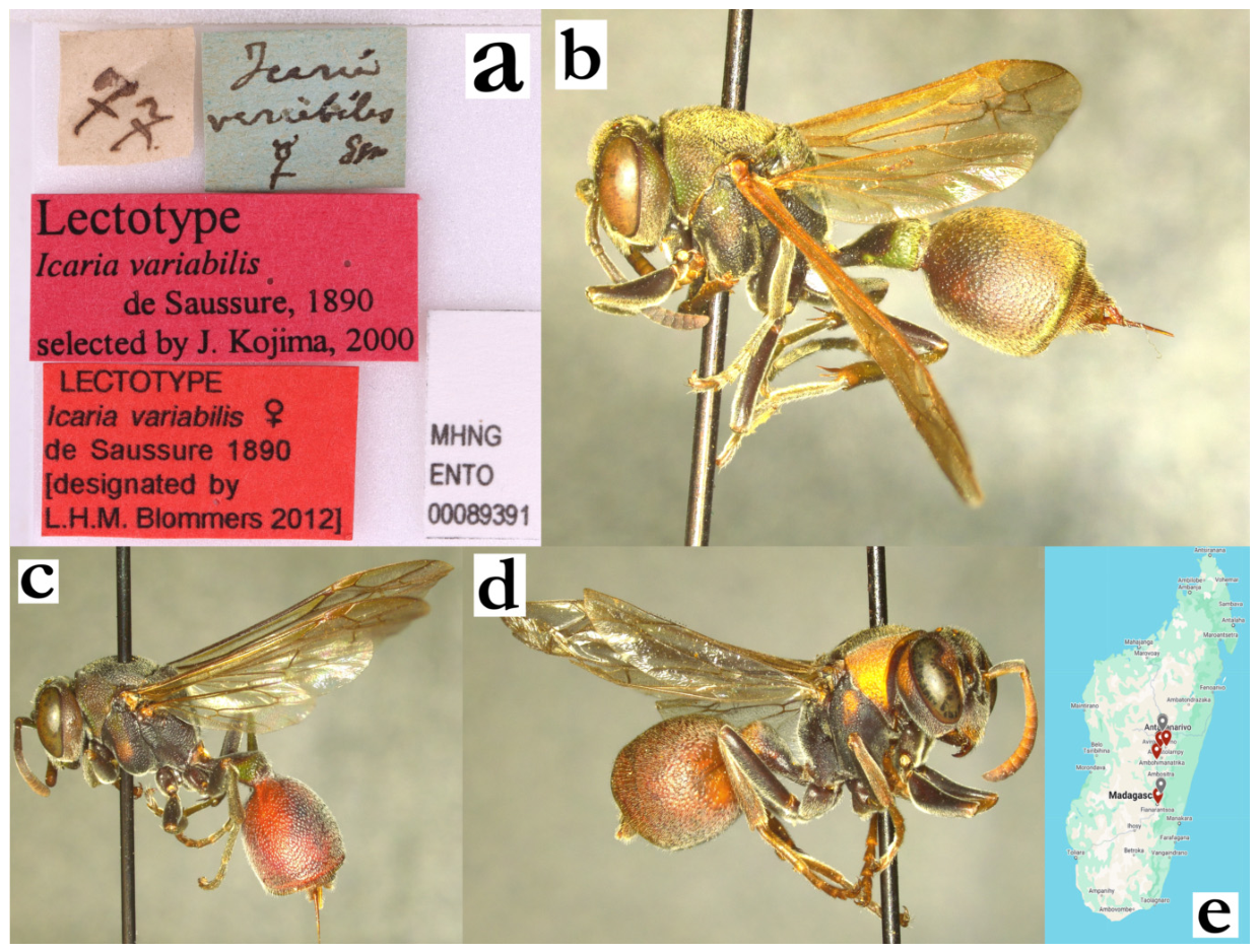
- Ropalidia velutina (de Saussure, 1890)Icaria velutina de Saussure, 1890; “Icaria/velutina/type Sss” and “Madagascar/ GRANDID” (MHNG).Plazi: https://tb.plazi.org/GgServer/html/03AE106DFFD1396E25D7FAB1842EF1B2 (accessed on 21 September 2026).BOLD: https://portal.boldsystems.org/bin/BOLD:ADN4807 (accessed on 21 September 2026).Number of examined specimens: 5 ♀♀, 2 ♂♂.
Type material. De Saussure described a single female from Grandidier’s collection, without providing more details on the specimen [13], which is considered the holotype [2].
Notes. A larger species, characterized by brown body colour and dense pubescence (Figure 129a,b). De Saussure compared it to R. atra (de Saussure) and R. grandidieri (de Saussure) [13], while Blommers suggests that it has some resemblance to R. cincinnata Blommers [6]. The overall pattern of pilosity is different in these two species; while R. velutina (de Saussure) has straight setae, setae in R. cincinnata Blommers are curly and wavy; these two species, alongside R. ducalis (de Saussure) have the longest overall body pilosity. In addition, R. velutina has comparatively much shorter second metasomal segment than other species, which is commonly not longer than high in lateral view (Figure 3f and Figure 129b). The male is described for the first time.
Description. Male.
Head. Head 1.4× wide as high (Figure 3b), in dorsal view 3x as wide as long, with gena equally wide as eyes and weak posterior head emargination (Figure 129c); head as wide as mesosoma in front of tegula (Figure 129c). Clypeus 1.4× as wide as long, with conspicuously convex surface, parallel lateral sides, slightly curved upes and weak OCA; apical clypeal margins are commonly straight, with subacute apex (Figure 3c); clypeus shallowly punctate, obscured by apically denser fine whitish setae; frons punctures larger and more defined, more than one diameter apart (Figure 3c). Gena finely punctate, posterior part close to occipital carina impunctate; occipital carina complete; mandible similarly punctate to clypeus; posterior ocelli 1.3 diameters apart, 2.5 diameters apart from eye and 2.9 diameters apart from occipital carina (Figure 129c). Scape barely broadened, about 1.3–1.4× as AF1 base width (Figure 3c and Figure 129d); AF1 abut as long as the scape, but also substantially shorter than AF2+3+4; AF1 with very thin base and wide distal part, with distal part 1.4× as wide as base (Figure 3c); AF2 1.6–1.8× as long as wide (Figure 3c); tyloids shiny, originate at AF(2)3, proximal ones brown, more distal yellow (Figure 129e); terminal flagellomere somewhat elongated, about 1.7× as long as AF10, 1.8× as long as wide, evenly curved, beak-like, with obtuse tip (Figure 3d).
Mesosoma. Mesosoma with moderately defined punctures, more than one diameter apart, somewhat coarser of pronotum, scutellum and metanotum. Median depression of propodeum striate or weakly punctate, deep, with obtuse margin. Fore tarsal spur I weakly developed, narrow and short, barely reaches half of second tarsal segment (no longer than first tarsal segment width); T1 posteriorly rounded and massive, 0.8× of T2 width.
Metasoma. S7 weakly concave, with shiny and irregular surface.
Pilosity. Entire body covered by dense layer of golden pubescence, especially developed on dorsal side of mesosoma; clypeus apex with dense layer of fine silvery setae that extend to frons, where these setae are about 1.5–1.9× as long as forwards ocellus diameter; eyes asetose (Figure 3c).
Colour. Basal body colour brown (olivaceous-brown to dark brown), except yellow areas: clypeus, interantennal area and inner orbits, mandible (with postero-basal, suffused olivaceous-green or brown area), coxa I and II entirely yellow (Figure 129d); gena, pronotum, scutellum and metanotum olivaceous or brownish; mesoscutum with two paracentral elongated reddish areas; mesepisternum with light brownish patch, metepisternum and propodeum dark brown; ventral surface of femora suffused patchy light-brown to yellow, dorsally brown; tibia and tarsi brown, terminal tarsal segment darker; T1 proximally black, posteriorly brown, with posterior light brown band; remaining metasomal segments brown-olivaceous. Wings barely yellowish, nervature brown, stigma light brown and transparent, apical spot not developed. Antenna dorsally brown, scape ventrally yellow, flagellum ventrally orange (Figure 3c).
Note. One examined female is twice stylopized, with one male and one female stylops.
Distribution. Most examined specimens have the “Madagascar” label, except for three, which originate from Nosy Be, Perinet and Marolambo, suggesting that R. velutina (de Saussure) is distributed in the northern half of the island.
Figure 129.
Ropalidia velutina (de Saussure), (a) habitus, dorsal view, female, (b) habitus, lateral view, female, (c) head and mesosoma, dorsal view, male, (d) head and mesosoma, inferior oblique view, male, (e) antenna, median view, male.
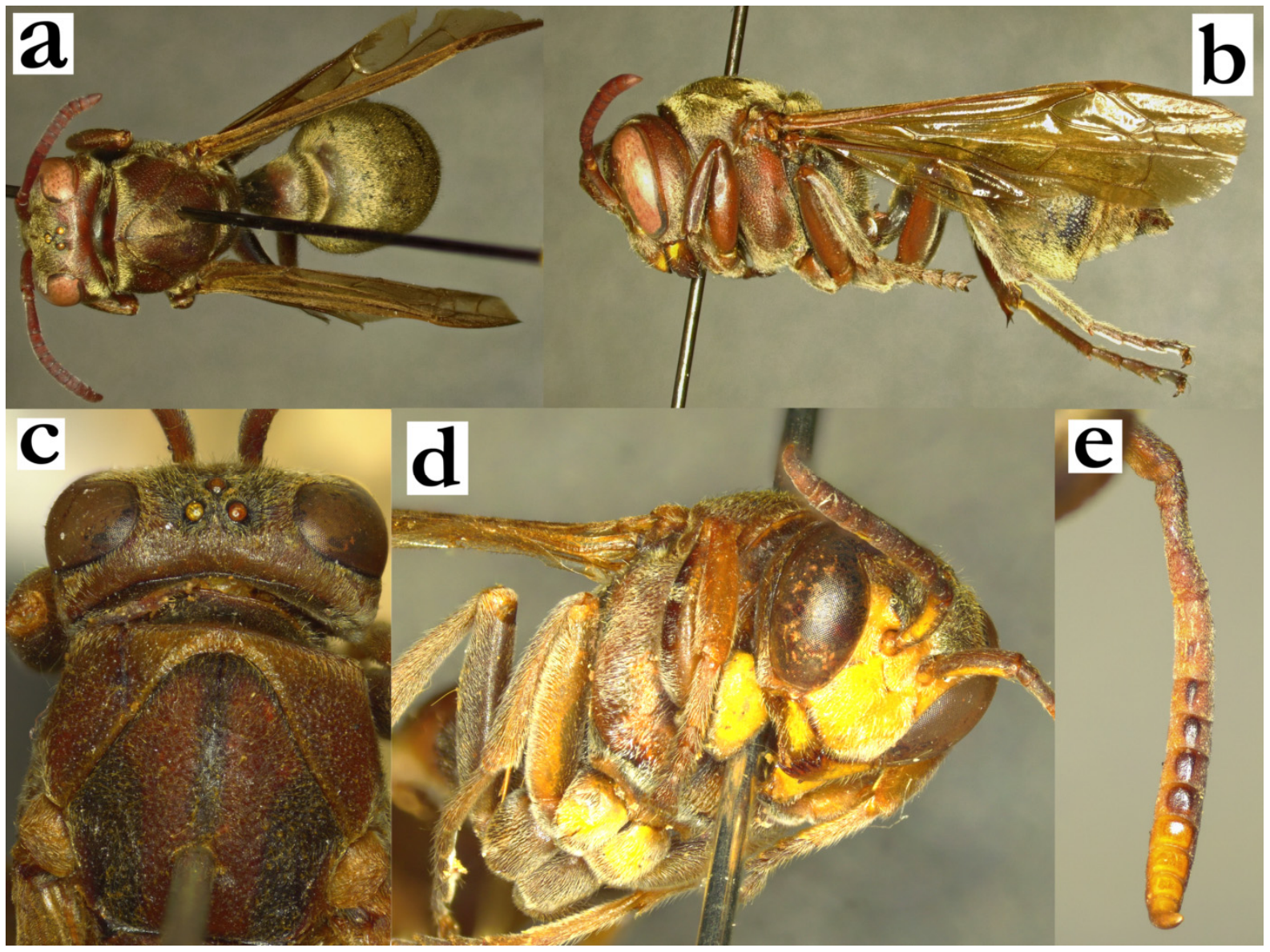
- Ropalidia vitripennis (de Saussure, 1890)Icaria vitripennis, de Saussure, 1890; “Madagascar” (NHM).Plazi: https://tb.plazi.org/GgServer/html/03AE106DFFD0396E25D7FCEC842EF50B (accessed on 21 September 2026).Number of examined specimens: 10 ♀♀.
Type material. Female from NHM, holotype, was examined (BMtype 18.877); in addition, MSNV holds another specimen which is apparently from the type series (Figure 130a,b). It bears only a handwritten label by de Saussure. A female of this species was originally described in 1890, with a vague reference of “Madagascar” [13]; Nosy Be was mentioned in the next publication [14]. Both accounts refer to ferruginous and black body colour and fine pubescence. Giordani Soika further listed broadened gena and darker pronotum in females as key characters, but did not mention the male [4]. Kojima reports male antenna (Figure 13b), but without the description [5]. Further, Kojima suggests that the larvae are similar to R. carinata (de Saussure) and R. galimatia (de Saussure), suggesting that the adult might have some resemblance to them [18]. Unfortunately, there were no males of this species in the examined specimens, therefore the male remains undescribed.
Similar species. General colour pattern may resemble R. zandry sp. nov.; useful features in these cases include globular shape of T1 in R. zandry sp. nov. and more elongated in R. vitripennis (Figure 130d).
Distribution. Northern Madagascar (Figure 130e).
Figure 130.
Ropalidia vitripennis (de Saussure), (a) habitus, lateral view, female, syntype, (b) label, syntype, (c) head and mesosoma, dorsal view, female, (d) metasoma, dorsal view, female, (e) distribution map (red marker—specimen, grey marker—literature report or an observational record).
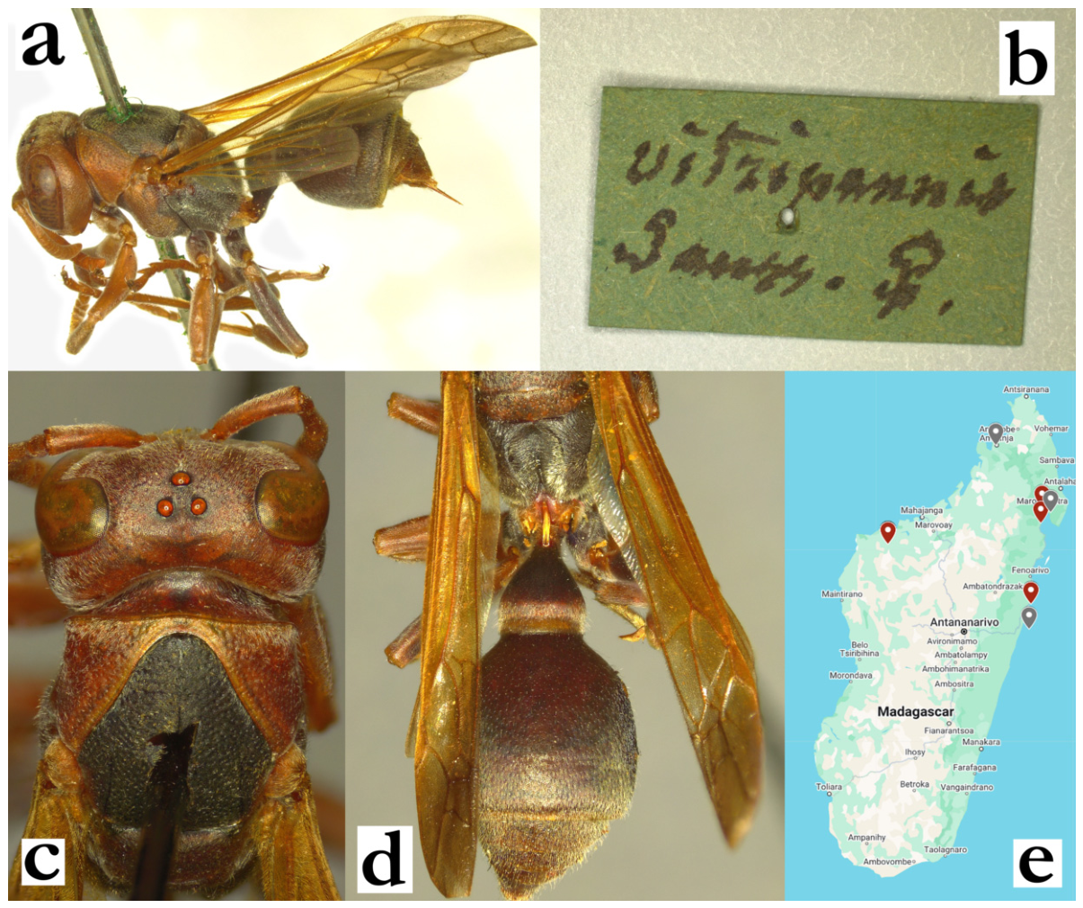
- Ropalidia volontany sp. nov. PolašekZooBank: C7B575F5-B9EF-4A9C-BEDD-DF5F5CC3DB58Material examined. Holotype: “Fampanabmo/1962, J Vadon”; ♀ (RMCA); Figure 131a,b.Paratypes: “Ambodivoangy /VIII.1961 J Vadon”; 2♀♀ (RMCA); “Fanovana/II.1936 Seyrig”, 2♀♀ (MSNV); “Rogez/Mus Praha”, 1♀ (MSNV).Number of examined specimens: 6 ♀♀.
Diagnosis. Member of the grandidieri group, with body lightly coloured, anterior margin of metanotum biphasic, metanotum with two yellow spots, T1 and T2 with posterior yellow bands.
Description. Females. Wing length 7.9–8.2 mm.
Head. Head frontally 1.15× as wide as long; clypeus elongated, 1.1× as wide as long, commonly with straight upes and wide and developed OCA; juxtamandibular lobe moderately developed, apex pointy (Figure 131a); entire clypeus covered by scattered and poorly defined small craters; frons with intermediate size and shallow punctures, those on inner orbit smaller and less defined, gena impunctate and shiny, only with some retained, well defined and scattered punctures close to eye (Figure 131c); gena thin, narrowing towards mandible (Figure 22b); mandible shiny, punctate with large and ill-defined punctures; occipital carina complete, ocelli equidistant (Figure 131d); scape longer than AF1, AF1 3.8× as long as wide; AF2 1.2–1.3× as long as wide.
Mesosoma. Mesosoma sparsely and shallowly punctate, with weak puncture margins; pronotum with large and shallow punctures, mesoscutum wide with equally dense punctures (Figure 131d); mesepisternum with equally dense punctures, similar in size to those on pronotum; metepisternum punctures less dense, but with retained puncture size; scutellum and metanotum with larger and well-defined punctures; scutellum with defined and short median carina (Figure 131d); metanotum with biphasic superior margin (Figure 22a); propodeum with rounded contour, horizontally striate and impunctate; lateral sides weakly punctate and striate.
Metasoma. T1 infundibular, short and wide, about 0.7–0.8× as wide as T2, nearly completely impunctate; posterior sulcus well developed, equally wide (somewhat shallower medially and laterally). Second metasomal segment moderately shiny, with shallow, small and dense directional punctures.
Pilosity. Overall shorter than most other species of the grandidieri group; apical third of clypeus with whitish sparse setae, longer than basally; frons with straight golden setae, as long as anterior ocellus diameter; eyes covered by short and scattered setae; mesosoma with short yellowish-golden pubescence and equally coloured protruding setae with bent tips; T2 with short and fine yellowish pubescence that does not extend over lamella.
Colour. Basal colour ferruginous-brown (Figure 131a), except yellow areas: apical line on clypeus, inner orbits and interantennal area (suffused light-brown), spot on mandible, suffused area under pronotal carina, two conspicuous spots on metanotum, two faint spots on scutellum, sometimes two suffused faint rhomboid patches on propodeum, coxa I, median half of coxa II (sometimes also coxa III), posterior line on T1 and T2; antenna brownish (infuscate) dorsally (Figure 131c), orange ventrally; wings evenly yellowish, nervature light golden-yellow, stigma yellowish, semi-transparent, tip somewhat darkened (Figure 131a).
Males. Unknown.
Distribution. Central highlands (Figure 131e).
Etymology. The name originates from the Malagasy word volontany, translated as “brownish-red”, referring to the general colouration pattern; the name is treated as indeclinable.
Similar species. Colour pattern resembles R. minor (de Saussure), but there are numerous morphological and colour differences between these two species. Some confusion may be possible in separation from R. shestakowi (which has sparser mesoscutum punctures anteriorly and no protruding setae on mesoscutum) or R. takona sp. nov. (which is smaller and has stockier eye in lateral view). Separation from other species of the grandidieri group does not present a substantial problem.
Figure 131.
Ropalidia volontany sp. nov., (a) habitus, lateral view, female, holotype, (b) labels, holotype, (c) head, frontal view, female, holotype, (d) head and mesosoma, dorsal view, female, holotype, (e) distribution map.
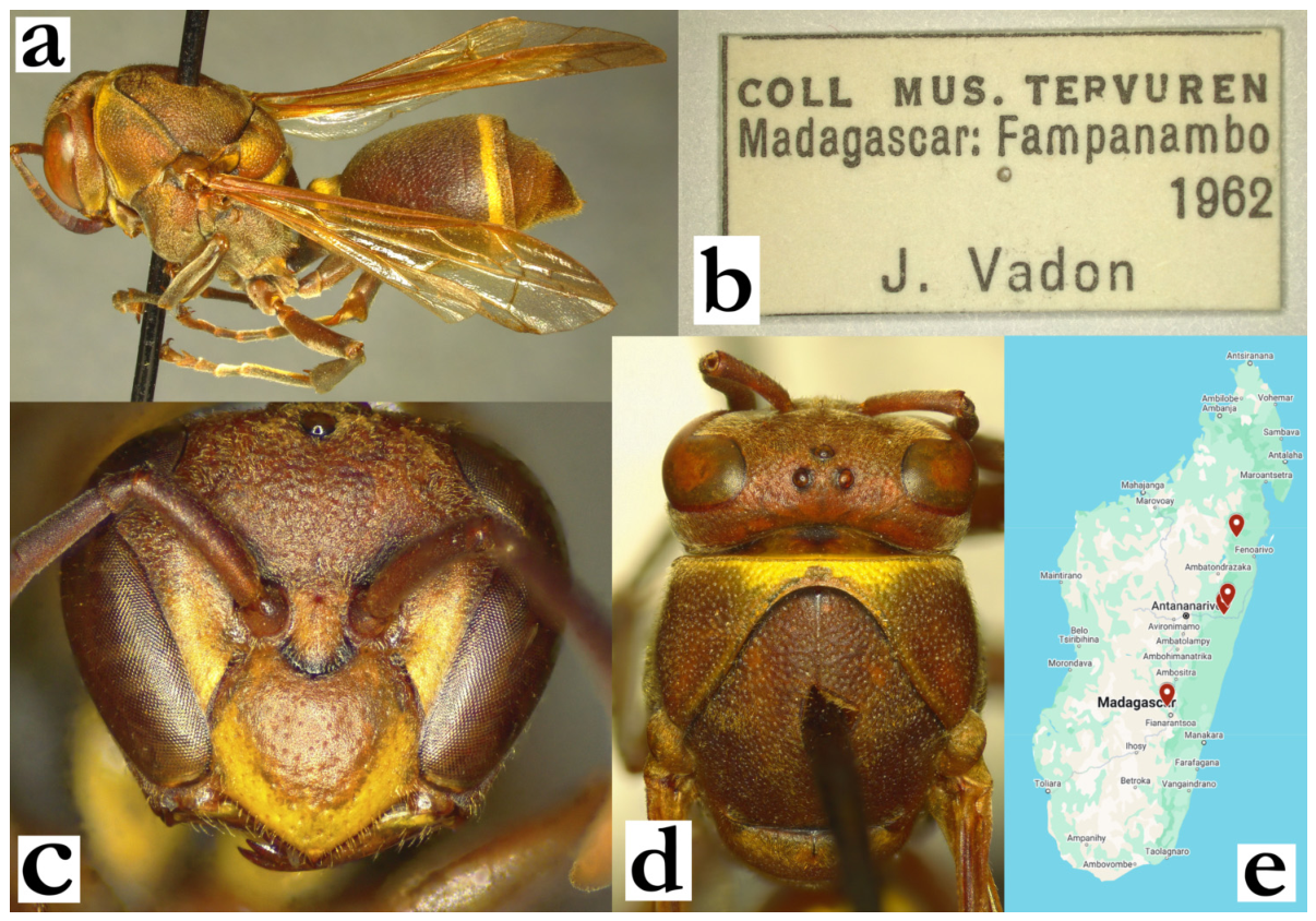
- Ropalidia zandry sp. nov. PolašekZooBank: D193DB7A-8C4F-4787-AE88-21B4DE8381E2Material examined. Holotype: “Coll. MUS. TERVUREN/ MADAGASCAR:TAM./ Didy, /16..iv.1992 NID35/A.PAULY rec.”, ♀ (RMCA), Figure 132a,b.Paratypes: 5♀♀, 5♂♂, same label as holotype (RMCA); “Coll. MUS. TERVUREN/MADAGASCAR:TAM./ Mororano-Chrome, 25kmW/11.i.1992/A.PAULY rec NID’28”, 3♀♀ (RMCA); Coll. MUS. TERVUREN/MADAGASCAR: TAN./ La Mandraka/ 10.iii.1994/ A.PAULY rec”, 1♀ (RMCA); “Rogez/ Madagascar/ Mus. Praha”, 1♀ (MSNV); “Fort Dauphin”, 1♀ (NNM); “MADAGASCAR/ Fort Dauphin 500m/ 15.IV.1968/ K. M. Guichard”, 3♀♀ (NHM).BOLD: https://portal.boldsystems.org/bin/BOLD:ADH8057 (accessed on 21 September 2026).Number of examined specimens: 15 ♀♀, 5 ♂♂.
Diagnosis. This species can be separated from other members of the fraterna group by the following characters: pronotal carina equally wide laterally and dorsally, T2 base coarsely punctate, pubescence short, occipital carina darkened and streamlined, body black and reddish, tyloids originating at AF5 in males and terminal flagellomere elongated, broad and apically acute.
Description. Females. Wing length 8.2–9.0 mm.
Head. Head frontally 1.2–1.3× as wide as high, in dorsal view 2.6× as wide as long, barely narrowing behind eyes, weakly emarginate posteriorly (Figure 30a). Clypeus 1.2× as wide as long, upes straight, OCA developed (Figure 30c); juxtamandibular excavation shallow, apex very pointy (Figure 30c); clypeus with cuticular surface and basally some larger and well-defined punctures, less defined centrally and hardly discernible apically (Figure 30c). Frons sparsely and coarsely punctate, punctures more than 2 diameters apart, with matte interspaces (Figure 30c); gena coarsely punctate close to eye, posteriorly punctures less defined but retained (Figure 30b); gena at midpoint 1.1–1.3× as wide as eye; mandible punctate, occipital carina complete and very developed, streamlined (Figure 30b); posterior ocelli 1.9–2.0x diameters apart, 3.2× diameters apart from compound eye and 2.9× diameters apart from occipital carina (Figure 30a). Scape 3.9× as long as wide, 1.3× as long as AF1; AF2 1.1× as wide as long, remaining flagellomeres wider than long (Figure 30c).
Mesosoma. Mesosoma slender, head 1.05× as wide as mesosoma in dorsal view; 1.3× as long as wide, in lateral view 1.6× as long as wide; mesoscutum 1.05× as wide as long (Figure 30a); pronotum in dorsal view mildly sinuate anteriorly, humeri rounded; carina of pronotum complete and thin, dorsally as wide as laterally; pronotum punctures shallow, 1.5× diameters apart, with mildly swollen and matte interspaces, retained even inferiorly (Figure 30b). Mesoscutum with similar punctures or somewhat more spaced apart than those on pronotum (Figure 30a). Mesepisternum without carina towards mesepimeron, sparsely and coarsely punctured in lateral area, impunctate or with just a few punctures anteriorly; posterior surface with less dense and punctures of same size, similar to those on mesepimeron; metepisternum basally matte, with somewhat more spaced and well-defined punctures. Propodeum posteriorly narrowing; median depression of propodeum shallow and widely emarginate; surface with fine horizontal striae; striae more defined in larger specimens; lateral face of propodeum more striate.
Metasoma. T1 posteriorly globular, with asymmetric sulcus, anterior surface impunctate, posterior with some poorly defined craters (Figure 132a). T2 1.2× as long as high, tergum and sternum equally convex; punctures shallow and coarse, directional, 1.0–1.4× diameters apart, with matte interspace (Figure 132a); punctures on S2 somewhat larger and sparser, especially proximally; transition between T2 surface and lamella gradual, lamella translucent and very weakly punctate proximally, distal two thirds impunctate; remaining metasomal segments shallowly punctate.
Pilosity. Body with very short yellowish pubescence (Figure 30a), except clypeus with whitish-yellow protruding setae, 2.0x as long apically as basally; mandible with protruding setae of similar length as clypeus apically; frons with straight protruding setae, 0.4–0.7× as long as anterior ocellus diameter; gena and occiput with some short protruding setae at mid-height; eyes very finely setose (Figure 30c). Mesoscutum with very short pubescence and barely longer protruding setae (Figure 30a). Propodeum with some longer whitish fine setae; T1 anteriorly with fine pubescence, posteriorly with some yellowish setae that do not project over posterior margin; T2 with protruding setae that do not extend over lamella (Figure 132a); remaining metasomal segments with some protruding setae distally; coxa with fine silvery pubescence and some silvery protruding setae with bent tips; femora with dense pubescence, femur II and III also with longer setae.
Colour. Body black and reddish-brown, without green(ish) markings (Figure 132a). Head basally ferruginous, with suffused yellow areas on clypeus apically, inner orbits and interantennal area, spot on mandible (Figure 30c); gena reddish, area underneath occipital carina dark-brown to black (Figure 30b). Mesosoma reddish with black or dark-brown pronotal collar (Figure 30b), anterior and lateral margin of mesoscutum (Figure 30a), mesepisternum, scutellum and metanotum brownish, metepisternum and propodeum black(ish); T1 basally black, distally reddish, in holotype also with a minute lateral triangular suffused yellow spot (Figure 132a); T2 basally brownish, distally with a thin yellow posterior band, remaining segments brownish (Figure 132a). Coxa and proximal part of femur black, distal part of femur, tibia and tarsi brown (Figure 132a). Wings yellowish, nervature light brown, stigma light brown and translucent, apical spot very absent (Figure 132a). Antenna dorsally infuscate, ventrally ferruginous-orange (Figure 132a).
Males. Resemble females in general appearance, except antenna much more slender and mesosoma underside more lightly coloured. Wing length 7.8–8.4 mm.
Head. Head in frontal view 1.23x as wide as high (Figure 132d); clypeus base with some large and shallow punctures, apex depressed and with less defined craters (Figure 132d). Scape 2.8× as long as wide, 1.6× as wide as AF1 at midpoint; AF2 1.35× as long as wide. Tyloids shiny, originate at AF(4)5, brownish and matte (Figure 30d); terminal flagellomere elongated, 3.8× as long as wide, with developed tyloid that reaches acute tip (Figure 30d).
Mesosoma. Fore tarsal spur I very developed and wide, as wide as tarsus base.
Metasoma. S7 weakly concave.
Pilosity. Overall, with longer head setae than in females; frons with setae 0.7–1.3× as long as anterior ocellus diameter (Figure 132d).
Nest. Several nests were observed in early colony stages. The nest shape is elongated, initially built as a type III (2 cells wide and variable length), with some low cells added to the side and thus probably converting to type IV nest at later stages of the colony cycle. Cell wall compact and grey, cocoon caps brown; cells located on branches (Figure 132e,f). The initial stage of the nest is similar to R. vitripennis, which should be considered in case of photograph-based identification as a possible risk of misidentification [32].
Distribution. Eastern Madagascar.
Etymology. The name originates from the Malagasy word zandry, translated as “a younger brother or sister; a junior”, referring to the similarity and previous misclassification with R. galimatia (de Saussure); the name is treated as indeclinable.
Similar species. Females of the fraterna group species, especially R. galimatia (de Saussure) and R. cauponae Blommers, with differences between them listed in the key to species.
Genetics. The COI sequence suggests similarity to other members of the fraterna group.
Figure 132.
Ropalidia zandry sp. nov., (a) habitus, lateral view, female, holotype, (b) labels, holotype, (c) head and mesosoma, oblique view, female, (d) head, frontal view, male, (e) nest [103], (f) nest [104].
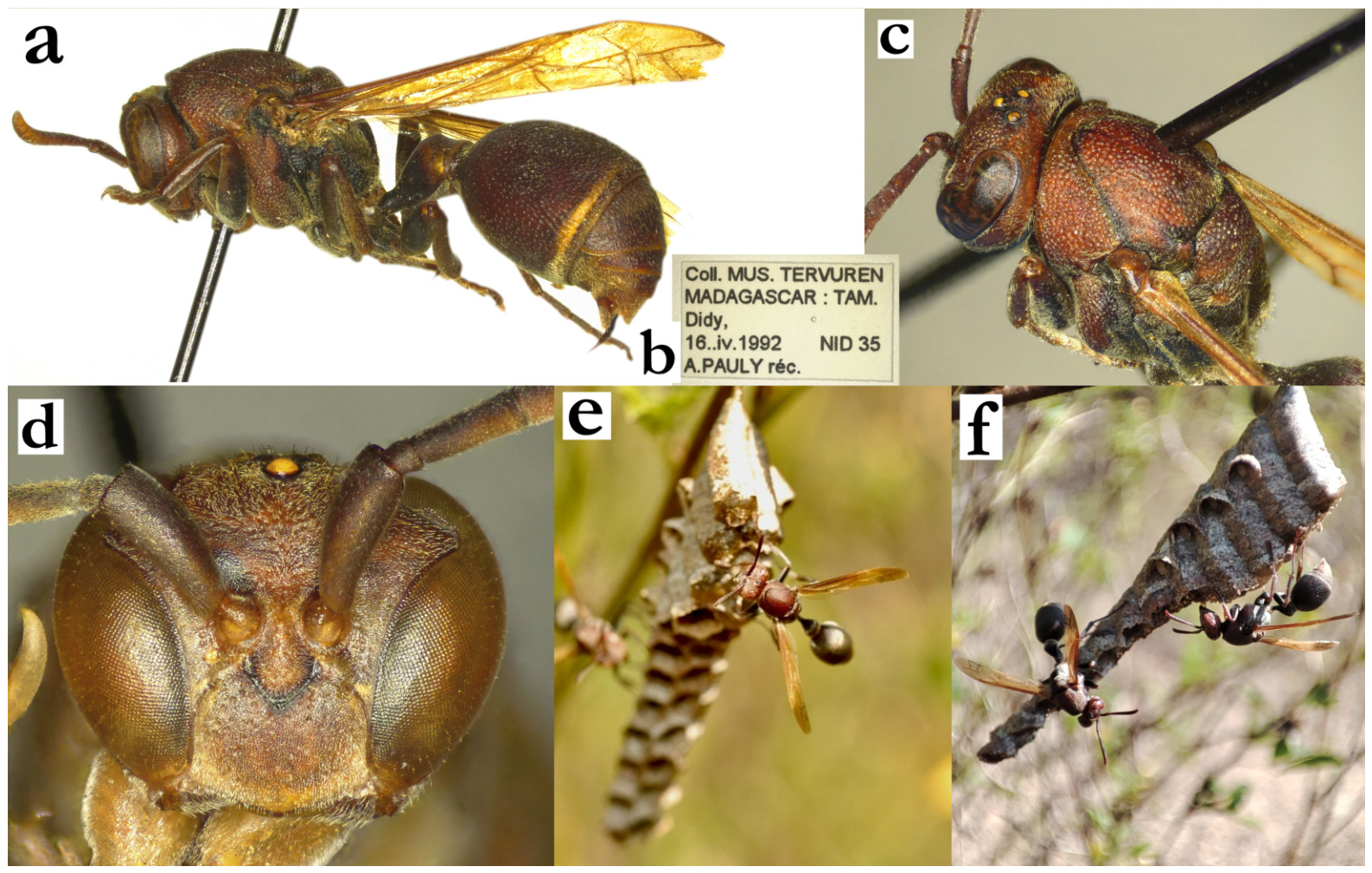
- Ropalidia democratica (de Saussure), nom. dub.
De Saussure described this taxon in 1853 [12]; the species description is already quite problematic since he put a question mark immediately next to the female description (“Fem. ?”). He suggested a large size, with green and black colour, possibly pointing to R. regina or R. prasina. Later he provided a comparison, suggesting a species that is smaller than R. formosa (de Saussure), but larger than R. dubia (de Saussure). The subsequent publication [13] stated that the specimens were lost and that this species is similar to R. grandidieri and R. variabilis. In the absence of the specimen, and due to the fact that the description is insufficiently specific, this name is considered as nomen dubium.
3.3. Phylogenetic Relationships
The use of the COI marker indicated a highly structured relationship of the available sequences (Figure 133). Two Belonogaster species were used as outgroups, and the first few splits belonged to a mixture of Asian, Australian and African mainland Ropalidia species or species groups. The most interesting split for the Malagasy species is the one separating R. acuminata and R. ophuzi, the two African mainland species of the capensis group with substantial morphological differences from other members of the group (Figure 133). The next split places all the remaining capensis group species in a sister position to all Malagasy species. All Malagasy species therefore form a single monophyletic lineage, while the mainland capensis group as currently defined is paraphyletic, since it excludes its Malagasy descendants. We therefore expand the capensis group to include the Malagasy species, which makes it monophyletic, and consider the capensis group as the origin of the Malagasy fauna, consistent with a single colonization of Madagascar.
Within the Malagasy species, the first split separates R. minor and most of the mainly green species (here termed the minor clade) from R. shestakowi and the remaining groups (Figure 133). Notably, green colour does not define this clade. R. regina, R. variabilis and R. minor belong to it despite little or no green colour, while strongly green species such as R. laureliniana sp. nov., R. bongomaso sp. nov., R. vanga sp. nov., and several members of the formosa group fall outside it. Green colour has therefore evolved several times among the Malagasy species, and we treat the mainly green species as a loose phenotypic cluster, not as a species group.
The following splits separate R. shestakowi, then R. velutina, then the fraterna group together with R. bongomaso sp. nov. and R. vanga sp. nov. from the grandidieri and formosa groups (Figure 133). The formosa and grandidieri groups are both monophyletic and are sister groups. The exception is R. shestakowi, which shares the phenotypic characteristics of the grandidieri group (notably with the very similar R. tsiky sp. nov.), but has a much deeper genetic lineage. The grandidieri group is therefore paraphyletic with respect to R. shestakowi. One more interesting finding is the position of R. bongomaso sp. nov. and R. vanga sp. nov. They resemble some of the mainly green species, but are sister species in the tree and are placed with the fraterna group, not with the minor clade (Figure 133).
Several further results fit well with the morphology. The sister relationship of the grandidieri and formosa groups fits well with the T2 lamella morphology; both groups share the same gradual decrease with continued punctures on the T2 lamella (Figure 77e and Figure 113c), virtually absent in other species (Figure 11b,f and Figure 124e). The isolated position of R. velutina is also corroborated by the morphological characteristics, which prevent a clear species group assignment for that species. Some characteristics resemble the fraterna group (modest similarity of T1 shape), but the pilosity and the overall short T2 are incomparable to any other species. Lastly, the early-diverging and isolated position of R. minor fits well with numerous morphological characteristics, most notably the short mesosoma, the lateral processus of T1 and the specific colour pattern.
Figure 133.
Results of the COI phylogenetic analysis of the available Ropalidia species. African mainland species were designated as AF, Asian as AS and Australian as AU; species without the codes are Malagasy. The tree was extended in the selected areas to better visualize the relevant nodes. The species that were not relevant for the analysis were included in the analysis, but later collapsed, to allow a better visualization of the target species relationships. An asterisk denotes an uncertain species determination, in most cases originating from the availability of the genetic sequence only, without seeing the specimen, requiring a certain degree of caution in interpretation. The green boxes attached to the Malagasy species represent the generalized amount of green cuticular colour, where one square signifies faint suffused areas, two signify a moderate amount of green colour on the body, and three signify a substantial amount with at least half of the body surface green.
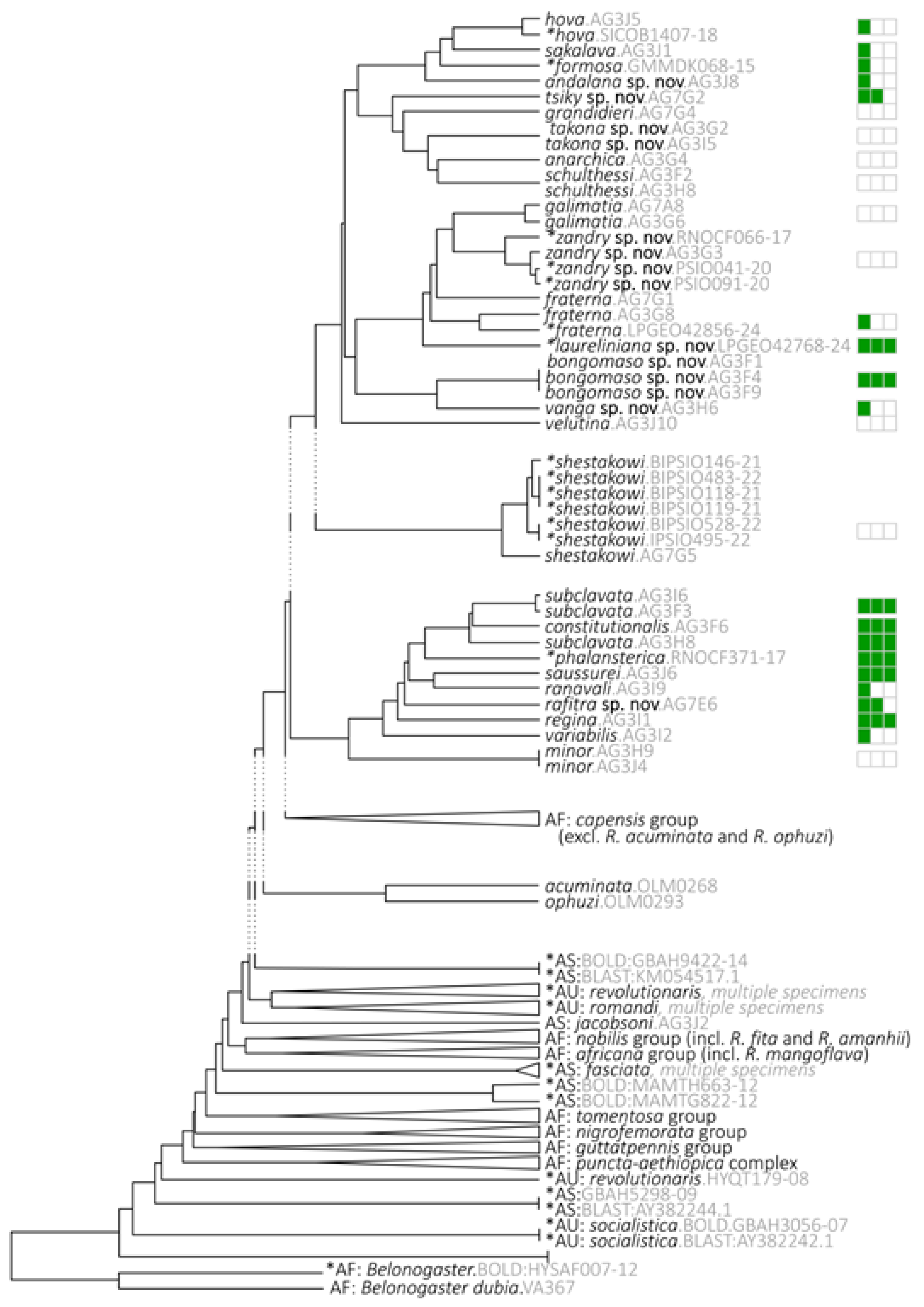
The head shape morphospace fits well with the COI tree (Figure 134). Species of the minor clade fall within the range of the mainland capensis group, the grandidieri group overlaps with the non-capensis mainland species, and the formosa group occupies the most distant part of the morphospace, in line with its derived position in the tree.
Figure 134.
Results of the 42 landmarks thin-plate spline analysis of the head morphometrics. Orange circles—members of the mainland capensis group, red circle—average mainland capensis group, light-blue circles—non-capensis mainland species, dark-blue circle—average non-capensis group, light-red area—grandidieri group, light-blue area—formosa group. Abbreviations correspond to the first three letters of the Malagasy species; species with several specimens appear more than once. Blue letters denote males of selected species. Delta emarginatum, Polistes fastidiosus and Belonogaster facialis are shown as outgroups.
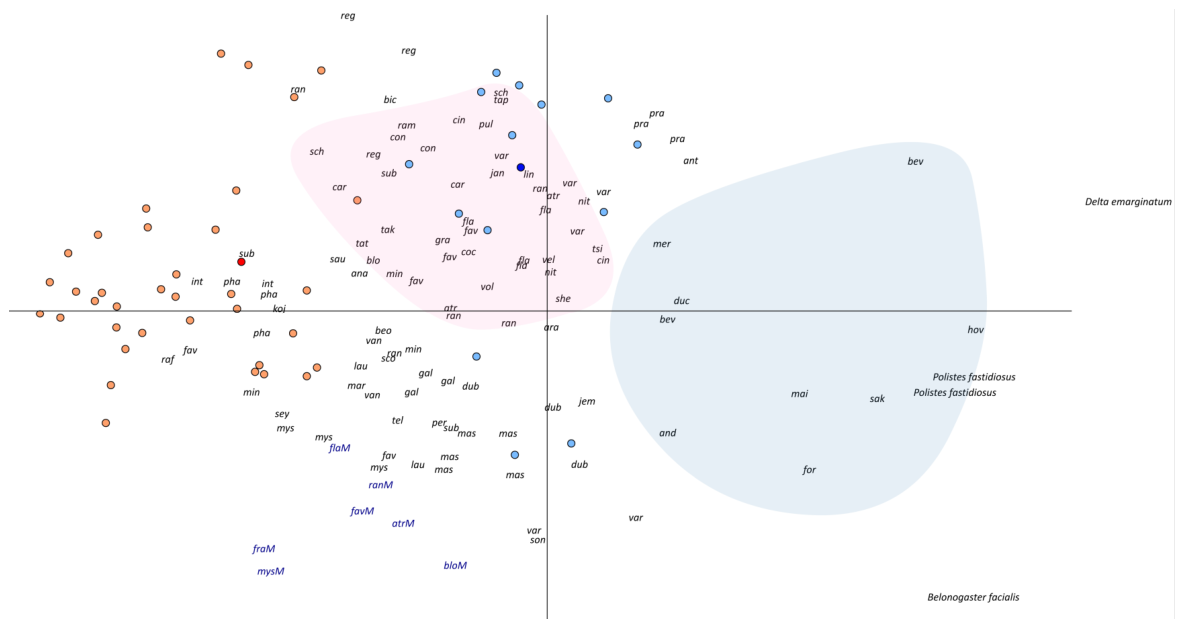
We then performed landmark-based unwarping and image averaging, in order to show the average head shape of the mainland capensis group, the non-capensis mainland species, all Malagasy species pooled, and the individual Malagasy groups (Figure 135). The average image of the mainland capensis (Figure 135c) vs. non-capensis groups (Figure 135a) indicated numerous differences, namely greater interantennal distance, longer and more curved upes, higher clypeus and longer oculo-malar area in the non-capensis group. Pooling all Malagasy species yielded an average Malagasy Ropalidia (Figure 135b), which shared short oculo-malar space, small interantennal distance, deeper juxtamandibular notches, longer lateral clypeal margins and narrower clypeus with the capensis group, again in line with the capensis origin of the Malagasy fauna.
Further sub-structuring of the Malagasy species revealed additional differences in the head morphology. The formosa group is characterized by elongated and more convex clypeus (Figure 135d), the grandidieri group by retaining the convex clypeus, more straight upes and wider clypeus (Figure 135e). Members of the fraterna group exhibited somewhat narrower and more flattened clypeus, with a longer mid-section of clypeus and deeper basal emargination (Figure 135f). The mainly green species were averaged by group, which in practice follows body size, as the phalansterica group comprises the small and the dubia group the large green species; several outliers were omitted. The phalansterica group was characterized by the straight upes, more developed OCA and wider clypeus (Figure 135h), while the dubia group had even wider and more convex clypeus, strong OCA and deep juxtamandibular notches (Figure 135g). We considered R. bongomaso sp. nov. and R. vanga sp. nov. as a special case, as they resemble the green species but are placed with the fraterna group in the COI tree, and show them separately (Figure 135i).
Lastly, we assembled the extant species into seven groups (Table 1), while the remaining species did not exhibit characteristics that would allow further grouping. Notably, these groups are informal and based on overall similarity; they are not diagnosable taxa, and the COI analysis supports only some of them (Figure 133). The fraterna, formosa and grandidieri groups are monophyletic, the last one only after excluding R. shestakowi, which makes it paraphyletic as listed in Table 1. R. velutina, previously a speculative member of the formosa group, falls outside it in the COI tree and is left unassigned. The phalansterica, dubia and ranavali groups, which contain most of the mainly green species, are not supported as separate lineages; their sequenced members all belong to the minor clade, except for R. bongomaso sp. nov. and R. vanga sp. nov., which are placed with the fraterna group. No member of the bicincta group was sequenced. The groups therefore serve as a practical aid to identification rather than as a phylogenetic classification.
Figure 135.
Head morphometric analysis, with image unwarping and averaging, showing the averaged head morphology of major species groups and clusters, (a) African mainland non-capensis group, (b) average Malagasy (all species), (c) African mainland capensis group, (d) formosa group, (e) grandidieri group, (f) fraterna group, (g) dubia group (without R. bongomaso sp. nov.+R. vanga sp. nov.), (h) phalansterica group, (i) R. bongomaso sp. nov.+R. vanga sp. nov.
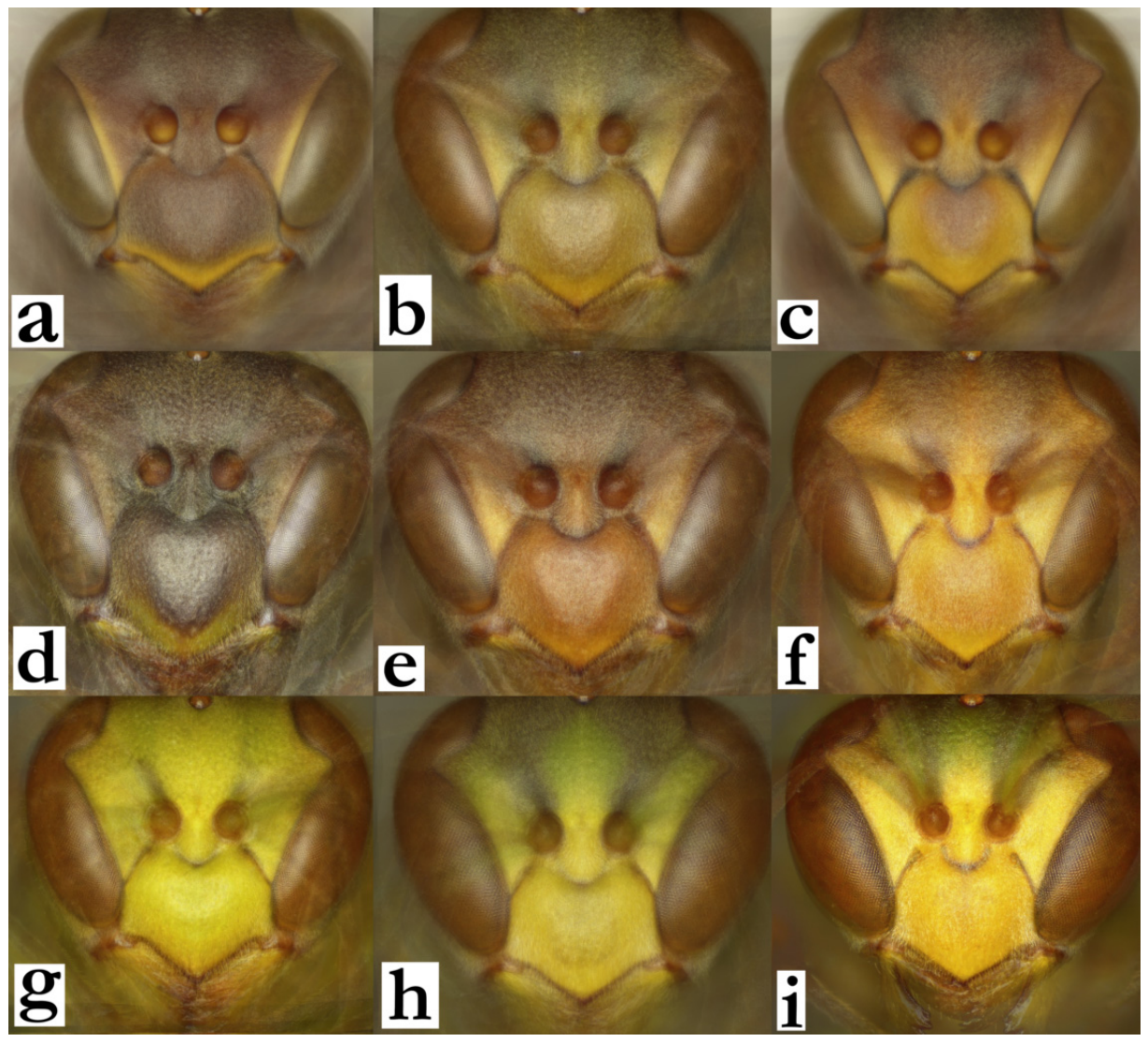
Table 1.
Species that were assigned to the species groups.
4. Discussion
The Malagasy genus Ropalidia species count is expanded by an additional 25 species, now contributing to the overall count of 66 valid species. This count is not complete, since even the citizen science platform iNaturalist hosts records of some nests and specimens that do not seem to belong to any of the known species [105,106]. This suggests that there are new species to be described in the future, which could be especially sensitive and time-restricted due to the extensive loss of habitat. One such example is the recently discovered species R. jemmae Polašek and de Beer, which is distributed in a narrow forest area and is an excellent candidate for an endangered species status. However, a recent observation of R. jemmae well outside the expected range, in an area that is devastated by deforestation [83] might provide some hope for conservation efforts, suggesting that some Ropalidia may, to a degree, survive habitat loss.
A subgeneric structure has been proposed in Ropalidia on a number of occasions, but none manage to provide a holistic solution [107]. The current scheme of Malagasy species recognizes several large groups, most commonly defined by the T1 shape, and in some cases additional characteristics (such as darkened tyloids in males of the grandidieri group or tyloids originating from AF1 in the fraterna group, in both cases with notable exceptions). However, several species do not fit this scheme. R. minor (de Saussure), R. velutina (de Saussure) and R. shestakowi (von Schulthess) represent separate lineages, supported not only by morphology but also by their early-diverging positions in the COI tree, and in the case of R. shestakowi this renders the morphologically well-defined grandidieri group paraphyletic. More generally, the COI tree supports the fraterna, formosa and grandidieri groups as lineages, but not the groups of mainly green species, whose members are intermixed; the species groups are therefore a practical aid to identification rather than a reflection of relationships. The group membership proposed in this study is in certain disagreement with adult and larval morphology [18]. Kojima identified several characteristics of larvae and linked R. flavoviridis Kojima with a few more members of the grandidieri group, but splitting away from R. carinata (de Saussure). While higher-order similarity of grandidieri and R. flavoviridis Kojima and consequently larval morphology similarities are possible, the split of R. carinata (de Saussure) from the grandidieri group is more problematic. However, as proposed by Kojima, the narrow extent and frequent apomorphic examples may contribute to this situation, which could be resolved by a wider comparative analysis of fresh material [18].
Previously implied similarities between the capensis group and Malagasy species [3] were further elaborated within this study, showing that all genotyped Malagasy species form a single monophyletic lineage nested within the capensis group of mainland species, which is thereby paraphyletic unless expanded to include them. This suggests that Malagasy Ropalidia originated from the African mainland, sometime after R. ophuzi Polašek and R. acuminata Polašek separated from the rest of the capensis group, and points to a single colonization of the island followed by substantial adaptive radiation. Such examples are common in Madagascar, sometimes occurring at an unprecedented scale and yielding speciation in a matter of only half a million years, defying previous speciation assumptions [108]. Without more extensive genetic information and fossil evidence for estimates calibration, dating the split of Malagasy Ropalidia from mainland Africa remains uncertain. Unfortunately, the extent of available material for genetic analysis was very low, thus preventing extensive analysis. Further specimens may yield more information relating to some specific relationships, most notably within the mainly green species.
Malagasy Ropalidia do not build as elaborate nests as some Asian species, which include double combs or outer paper envelopes [109]. All the Malagasy Ropalidia species build stelocyttarous and gymnodomous nests, except the astelocyttarous nest of R. ducalis (Figure 2e). Notably, only one nest of this species was examined, thus leaving the possibility for this to be an odd and isolated case, rather than a taxon-specific architecture type. Visual predation was previously implied in Ropalidia nest evolution [109], primarily focusing on foraging birds as the main colony predator [20]. Interestingly, mainland African Ropalidia build simple nests, which do not seem to be specific nor exhibit concealment properties. A notable exception to this is the South African species R. amabala, which builds elaborate and visually concealed nests directly on tree trunks [10]. The absence of morphological similarities with Malagasy species was implied as evidence that such nesting behaviour evolved independently from Malagasy Ropalidia, confirming a substantial nesting architecture plasticity in this genus [20,110].
Most of the examined nests could be further classified as type IV, which represents a single-sided comb with multiple adjacent cells [20]. The species that fall outside of this nesting architecture include species that build type I and III nests. Only one species was observed building type I nests, and that is R. bongomaso sp. nov. This nest type is built as independent cells that are attached and form a nest by using a continuous string of material that extends along the longer cell axis. This nest is an interesting example of balancing selection. It is widely accepted that nest evolution is governed by strict economic principles [109], and that type I nests exhibit poor work-to-gain ratio [20]. However, in the case of R. bongomaso sp. nov., the type I nest is a secondary feature, created by the re-use of the existing cells, a behaviour that was implied in several additional species, notably R. bongomaso sp. nov. (=R. dubia in that study), R. favulorum Blommers and R. rosae Blommers [6] and R. anarchica (de Saussure) [9]. A similar behaviour was recorded in R. linearecta, which also seems to reuse the cell wall material to reduce the used cells; notably, Blommers did not report this in his observation of R. linearecta [6], so this remains only a speculative proposal. However, both cases fit the notion that the nest build is governed by strong economic principles, reiterating a strong selective pressure against visually driven predation in nesting architecture and therefore evolution of each species. Several additional species build elongated nests, but in most cases, such nests are two, four or even more cells wide and therefore cannot be classified as type I (such as R. interjecta, R. sonyae and R. zandry, or R. prasina). Nest aggregations and nest colonies are another mechanism of colony protection, recorded in multiple Malagasy species, most notably R. merina (Figure 5d) and R. kojimai, but also small congregations of nests in R. favulorum (Figure 84f–j) and R. rafitra (Figure 108h). Multiple nests in the same locations were also reported in R. anarchica, R. cauponae, R. galimatia and R. nitidula [6,9]. In addition, there seem to be inter-species congregations, confirmed for the R. favulorum Blommers and R. rafitra sp. nov. pair, and also implied for R. merina Blommers and R. cauponae Blommers [92]. Lastly, substantial nesting concealment by building nests in hardly accessible holes was recorded in R. cocoscola Blommers, further suggesting the importance of nesting architecture.
Strong anti-visual selective pressure is further reflected in the green body colour. The COI tree shows that green colour has evolved on multiple occasions: the minor clade contains most of the strongly green species but also R. variabilis and R. minor with little or no green, while R. laureliniana sp. nov., R. bongomaso sp. nov., R. vanga sp. nov. and several formosa group species are green but belong to other lineages. Such a hypothesis does not seem impossible if additional Vespidae genera are considered. Green cuticular colour is present not only in the Malagasy Polistinae genus Belonogaster [111], but even in some Eumeninae species, such as Odynerus viridipictus Giordani Soika. Examples of Müllerian mimicry with Malagasy Ropalidia include some Eumeninae genera like Antodynerus (which have a general resemblance to R. linearecta), or similarity of the large black Delta regina (de Saussure), which has bicolorous wings. This is shared by several species from the formosa group, but also with Polistes madecassus (de Saussure). There were no other similarities between Malagasy Ropalidia with known Polistes species. Other important observations of the nesting behaviour include peaceful reactions of R. merina [6,9], which is in line with the adult green colour and concealed nests suggesting that Malagasy Ropalidia evolved in a more stealthy life cycle than their temperate, or even tropical counterparts, who often display bright warning colours. In order to fully understand their life cycle, more field observations are needed, directly competing with widespread habitat loss and a potential for irreparable biodiversity loss.
Author Contributions
Conceptualization, O.P.; methodology, O.P. and J.M.C.; formal analysis, O.P., B.C.D. and L.d.B.; investigation, L.d.B.; resources, O.P.; data curation, O.P.; writing—original draft preparation, O.P.; writing—review and editing, L.d.B., B.C.D. and J.M.C. All authors have read and agreed to the published version of the manuscript.
Funding
This research received no external funding.
Data Availability Statement
Raw genetics and morphometrics data is available upon request.
Acknowledgments
The authors would like to collectively thank all the users of iNaturalist for their work that helped substantially elevate the knowledge on Malagasy Ropalidia. We would also like to thank all the museum curators who helped with photographs, collection access or loans of material (alphabetically): Frederique Bakker (NNM), Vladimir Blagoderov (NMS), Gavin Broad and Andrew Polaszek (NHM), Leopoldo Castro (Teruel, Spain) for the donation of specimens, Brian L. Fisher, Robert L. Zuparko and Christopher Grinter (CAS), Michael Greeff and Rod Eastwood (ETH), Stéphane Hanot and Didier van den Spiegel (RMCA), Martin Husemann and Thure Dalsgaard (CENAK), Bernard Landry (MHNG), Christine Lebeau (AMNH), Michael Ohl and Lukas Kirschey (MFNB), Martin Schwarz (OLM), Agnièle Touret-Alby (MNHN), Marco Uliana (MSNV), Hege Vårdal (NHRS), Zoltan Vas (HNHM), and Dominique Simone (NMW).
Conflicts of Interest
The authors declare no conflicts of interest.
References
- Kojima, J.; Carpenter, J.M. Catalog of species in the Polistine tribe Ropalidiini (Hymenoptera: Vespidae). Am. Mus. Novit. 1997, 3199, 1–96. [Google Scholar]
- Carpenter, J.M. Taxonomic Notes on Paper Wasps (Hymenoptera: Vespidae; Polistinae). Am. Mus. Novit. 1999, 3259, 1–44. [Google Scholar]
- Polasek, O.; Onah, I.; Kehinde, T.; Rojo, V.; Noort, S.; Carpenter, J. Revision of the mainland African species of the Old World social wasp genus Ropalidia Guérin-Méneville 1831 (Hymenoptera; Vespidae). Zootaxa 2025, 5626, 1–142. [Google Scholar] [CrossRef] [Scilit] [PubMed]
- Giordani Soika, A. Vespoidei raccolti da L.A. NILSSON nel Madagascar con una tabella per determinazione delle Ropalidia del Madagascar (Hymenoptera Vespoidea). Lav. Soc. Veneziana Sci. Nat. 1991, 16, 79–90. [Google Scholar]
- Kojima, J. Male genitalia and antennae in an Old World paper wasp genus Ropalidia Guerin-Meneville, 1831 (Insecta: Hymenoptera; Vespidae, Polistinae). Nat. Hist. Bull. Ibaraki Univ. 1999, 3, 51–68. [Google Scholar]
- Blommers, L.H.M. Taxonomy and natural history of 18 Ropalidia species (Hymenoptera, Vespidae) of Madagascar. Tijdschr. Voor Entomol. 2012, 155, 133–192. [Google Scholar] [CrossRef] [Scilit]
- Polašek, O. A new species of the genus Ropalidia Guérin-Méneville from central Africa (Insecta, Hymenoptera, Vespidae). Afr. Invertebr. 2024, 65, 9–14. [Google Scholar] [CrossRef] [Scilit]
- Polašek, O. Description of the male of Ropalidia chromis Polašek, 2024 with further biological notes on the African species of the genus (Hymenoptera, Vespidae, Polistinae). Afr. Invertebr. 2026, 67, 1–18. [Google Scholar] [CrossRef] [Scilit]
- Vesey-FitzGerald, D. Notes on the genus Ropalidia (Hymenoptera: Vespidae) from Madagascar. Proc. R. Entomol. Soc. Lond. Ser. A Gen. Entomol. 1950, 25, 81–86. [Google Scholar] [CrossRef] [Scilit]
- Polašek, O.; Bellingan, T.; van Noort, S. A new species of paper wasp from the genus Ropalidia Guérin-Méneville from South Africa (Hymenoptera, Vespidae). J. Hymenopt. Res. 2022, 90, 213–222. [Google Scholar] [CrossRef] [Scilit]
- Polašek, O.; de Beer, L. A new species of social wasp from Madagascar with an inverted nest architecture (Hymenoptera, Vespidae). J. Hymenopt. Res. 2023, 96, 1031–1044. [Google Scholar] [CrossRef] [Scilit]
- De Saussure, H. Monographie des Guêpes Sociales ou de la Tribu des Vespiens; Masson: Paris, France; J. Cherbuliez: Genève, Switzerland, 1853. [Google Scholar]
- de Saussure, H. Historie Naturelle des Hymenopteres. In Historie Physique, Naturelle et Politique de Madagascar; Grandidier, A., Ed.; Imprimerie Nationale: Paris, France, 1890. [Google Scholar]
- de Saussure, H. Wissenschaftliche Ergebnisse der Reisen in Madagaskar und Ostafrika in den Jahren 1889-95 von Dr A. Voeltzkow. Hymenoptera. Vespidae. Abh. Der Senckenberg. Ges. 1900, 26, 203–240. [Google Scholar]
- Wenzel, J.W. Extreme queen-worker dimorphism in Ropalidia ignobilis, a small-colony wasp (Hymenoptera: Vespidae). Insectes Sociaux 1992, 39, 31–43. [Google Scholar] [CrossRef] [Scilit]
- Wenzel, J.W. Ropalidia formosa, a nearly solitary paper wasp from Madagascar (Hymenoptera: Vespidae). J. Kans. Entomol. Soc. 1987, 60, 549–556. [Google Scholar]
- Kojima, J.-I. The type of Icaria dubia de Saussure, 1853, and a description of a new Malagasy species of Ropalidia Guerin-Meneville, 1831 (Hymenoptera: Vespidae, Polistinae). Entomol. Sci. 1998, 1, 233–239. [Google Scholar]
- Kojima, J.I. A new species of Ropalidia from madagascar, with descriptions of mature larvae of 8 Ropalidia species (Hymenoptera, Vespidae). J. Kans. Entomol. Soc. 1988, 61, 292–302. [Google Scholar]
- Kojima, J.; Wenzel, J.W.; Carpenter, J.M. The types of Icaria galimatia de Saussure, 1853, and I. ignobilis de Saussure, 1890, with establishment of their synonymy (Hymenoptera: Vespidae, Polistinae). Entomol. Sci. 1998, 1, 393–401. [Google Scholar]
- Jeanne, R.L. The Adaptiveness of Social Wasp Nest Architecture. Q. Rev. Biol. 1975, 50, 267–287. [Google Scholar] [CrossRef] [Scilit]
- Dereeper, A.; Guignon, V.; Blanc, G.; Audic, S.; Buffet, S.; Chevenet, F.; Dufayard, J.F.; Guindon, S.; Lefort, V.; Lescot, M.; et al. Phylogeny.fr: Robust phylogenetic analysis for the non-specialist. Nucleic Acids Res. 2008, 36, W465–W469. [Google Scholar] [CrossRef] [Scilit] [PubMed]
- Huson, D.H.; Bryant, D. Application of phylogenetic networks in evolutionary studies. Mol. Biol. Evol. 2006, 23, 254–267. [Google Scholar] [CrossRef] [Scilit] [PubMed]
- Rambaut, A. Figtree ver 1.4.4. Available online: https://tree.bio.ed.ac.uk/software/figtree/ (accessed on 23 June 2026).
- de Beer, L. Ropalidia velutina. Available online: https://www.inaturalist.org/observations/191831961 (accessed on 20 June 2026).
- de Beer, L. Ropalidia merina. Available online: https://www.inaturalist.org/observations/140340663 (accessed on 20 June 2026).
- de Beer, L. Ropalidia merina. Available online: https://www.inaturalist.org/observations/198032863 (accessed on 20 June 2026).
- de Beer, L. Ropalidia hova. Available online: https://www.inaturalist.org/observations/193854565 (accessed on 20 June 2026).
- Mandák, M. Ropalidia regina. Available online: https://www.inaturalist.org/observations/63189849 (accessed on 21 June 2026).
- de Beer, L. Ropalidia prasina. Available online: https://www.inaturalist.org/observations/107464202 (accessed on 21 June 2026).
- Marjan, A. Ropalidia nitidula. Available online: https://www.inaturalist.org/observations/181042677 (accessed on 21 June 2026).
- Grellety, R. Ropalidia nitidula. Available online: https://www.inaturalist.org/observations/135089365 (accessed on 12 September 2026).
- Austin, D. Ropalidia vitripennis. Available online: https://www.inaturalist.org/observations/333883993 (accessed on 21 June 2026).
- Thehousebunting, A. Ropalidia minor. Available online: https://www.inaturalist.org/observations/190335633 (accessed on 21 June 2026).
- Negro, A. Ropalidia minor. Available online: https://www.inaturalist.org/observations/69302879 (accessed on 21 June 2026).
- de Beer, L. Ropalidia minor. Available online: https://www.inaturalist.org/observations/109563258 (accessed on 12 September 2026).
- Stefaneakame. Ropalidia schulthessi. Available online: https://www.inaturalist.org/observations/203232198 (accessed on 21 June 2026).
- Philizot, S. Ropalidia linearecta. Available online: https://www.inaturalist.org/observations/194501529 (accessed on 21 June 2026).
- Angelo, A. Ropalidia linearecta. Available online: https://www.inaturalist.org/observations/108867185 (accessed on 21 June 2026).
- de Foucault, C. Ropalidia linearecta. Available online: https://www.inaturalist.org/observations/2567075 (accessed on 21 June 2026).
- de Beer, L. Ropalidia shestakowi. Available online: https://www.inaturalist.org/observations/109849773 (accessed on 21 June 2026).
- de Beer, L. Ropalidia shestakowi. Available online: https://www.inaturalist.org/observations/107361311 (accessed on 21 June 2026).
- Falck, J. Ropalidia anarchica. Available online: https://www.inaturalist.org/observations/193883766 (accessed on 21 June 2026).
- Onjalalaina, G. Ropalidia anarchica. Available online: https://www.inaturalist.org/observations/61146749 (accessed on 21 June 2026).
- Harris, K. Small Paper Wasps Genus Ropalidia. Available online: https://www.inaturalist.org/observations/77382144 (accessed on 21 June 2026).
- de Beer, L. Ropalidia galimatia. Available online: https://www.inaturalist.org/observations/118915478 (accessed on 21 June 2026).
- de Beer, L. Ropalidia galimatia. Available online: https://www.inaturalist.org/observations/199364937 (accessed on 21 June 2026).
- Asp, J. Ropalidia fraterna. Available online: https://www.inaturalist.org/observations/151593996 (accessed on 21 June 2026).
- Sandoval, B. Ropalidia fraterna. Available online: https://www.inaturalist.org/observations/192662581 (accessed on 21 June 2026).
- Austin, D. Small Paper Wasps Genus Ropalidia. Available online: https://www.inaturalist.org/observations/178864207 (accessed on 21 June 2026).
- de Beer, L. Ropalidia interjecta. Available online: https://www.inaturalist.org/observations/116102535 (accessed on 21 June 2026).
- Lukasik, P. Ropalidia flavoviridis. Available online: https://www.inaturalist.org/observations/148652608 (accessed on 21 June 2026).
- Jarvi, J. Ropalidia flavoviridis. Available online: https://www.inaturalist.org/observations/199686514 (accessed on 21 June 2026).
- de Beer, L. Ropalidia flavoviridis. Available online: https://www.inaturalist.org/observations/224854991 (accessed on 21 June 2026).
- Oudejans, R. Ropalidia dubia. Available online: https://www.inaturalist.org/observations/36798196 (accessed on 21 June 2026).
- Falck, J. Ropalidia regina. Available online: https://www.inaturalist.org/observations/193883767 (accessed on 21 June 2026).
- Wiesner, F. Small Paper Wasps Genus Ropalidia. Available online: https://www.inaturalist.org/observations/81256863 (accessed on 21 June 2026).
- Tighe, D. Ropalidia saussurei. Available online: https://www.inaturalist.org/observations/9669640 (accessed on 21 June 2026).
- Hurni-Cranston, T. Ropalidia saussurei. Available online: https://www.inaturalist.org/observations/85477179 (accessed on 21 June 2026).
- Ruel, M. Small Paper Wasps Genus Ropalidia. Available online: https://www.inaturalist.org/observations/136450825 (accessed on 21 June 2026).
- m107791. Small Paper Wasps Genus Ropalidia. Available online: https://www.inaturalist.org/observations/196482450 (accessed on 21 June 2026).
- de Beer, L. Ropalidia subclavata. Available online: https://www.inaturalist.org/observations/136288127 (accessed on 21 June 2026).
- Cordenos, T. Ropalidia kojimai. Available online: https://www.inaturalist.org/observations/65483796 (accessed on 21 June 2026).
- Bhamla, S. Small Paper Wasps Genus Ropalidia. Available online: https://www.inaturalist.org/observations/4418333 (accessed on 21 June 2026).
- Carpenter, J.; Madl, M. A catalogue of the Vespidae of the Malagasy subregion (Insecta, Hymenoptera). Linz. Biol. Beitraege 2009, 41, 1871–1935. [Google Scholar]
- de Beer, L. Small Paper Wasps Genus Ropalidia. Available online: https://www.inaturalist.org/observations/193855484 (accessed on 21 June 2026).
- Lukasik, P. Small Paper Wasps Genus Ropalidia. Available online: https://www.inaturalist.org/observations/146235652 (accessed on 21 June 2026).
- Tahiana, T. Ropalidia cincinnata. Available online: https://www.inaturalist.org/observations/167663426 (accessed on 21 July 2026).
- Tomaszek, A. Small Paper Wasps Genus Ropalidia. Available online: https://www.inaturalist.org/observations/236771715 (accessed on 21 July 2026).
- Lee, C. Ropalidia dubia. Available online: https://www.inaturalist.org/observations/53306299 (accessed on 21 June 2026).
- Liu, Y. Small Paper Wasps Genus Ropalidia. Available online: https://www.inaturalist.org/observations/247316463 (accessed on 21 June 2026).
- Wiesner, F. Ropalidia cauponae. Available online: https://www.inaturalist.org/observations/81772733 (accessed on 21 June 2026).
- Austin, D. Ropalidia cauponae. Available online: https://www.inaturalist.org/observations/194638806 (accessed on 21 June 2026).
- Austin, D. Ropalidia cauponae. Available online: https://www.inaturalist.org/observations/194638845 (accessed on 21 July 2026).
- de Beer, L. Ropalidia cincinnata. Available online: https://www.inaturalist.org/observations/149126208 (accessed on 21 June 2026).
- de Beer, L. Ropalidia cincinnata. Available online: https://www.inaturalist.org/observations/143151581 (accessed on 21 June 2026).
- de Beer, L. Ropalidia favulorum. Available online: https://www.inaturalist.org/observations/109816287 (accessed on 21 June 2026).
- de Beer, L. Ropalidia favulorum. Available online: https://www.inaturalist.org/observations/107102450 (accessed on 21 June 2026).
- Thehousebunting, A. Ropalidia favulorum. Available online: https://www.inaturalist.org/observations/194279470 (accessed on 21 June 2026).
- Meiner, M. Small Paper Wasps Genus Ropalidia. Available online: https://www.inaturalist.org/observations/207141273 (accessed on 21 June 2026).
- Stefaneakame. Small Paper Wasps Genus Ropalidia. Available online: https://www.inaturalist.org/observations/203233734 (accessed on 21 June 2026).
- Aureglia, L. Small Paper Wasps Genus Ropalidia. Available online: https://www.inaturalist.org/observations/216000533 (accessed on 21 June 2026).
- de Beer, L. Old World Slender Ants Genus Tetraponera. Available online: https://www.inaturalist.org/observations/206798671 (accessed on 21 June 2026).
- Srinivasan, K. Ropalidia jemmae. Available online: https://www.inaturalist.org/observations/282313340 (accessed on 21 June 2026).
- Cordenos, T. Small Paper Wasps Genus Ropalidia. Available online: https://www.inaturalist.org/observations/65483794 (accessed on 21 June 2026).
- Amossé, E. Ropalidia linearecta. Available online: https://www.inaturalist.org/observations/264790106 (accessed on 21 June 2026).
- Foster, R. Ropalidia phalansterica. Available online: https://www.inaturalist.org/observations/254412274 (accessed on 21 June 2026).
- de Beer, L. Ropalidia galimatia. iNaturalist. Available online: https://www.inaturalist.org/observations/224831966 (accessed on 14 April 2025).
- Faucher, P. Ropalidia galimatia. Available online: https://www.inaturalist.org/observations/182627917 (accessed on 23 June 2026).
- Tomaszek, A. Ropalidia phalansterica. Available online: https://www.inaturalist.org/observations/236711726 (accessed on 21 June 2026).
- de Beer, L. Ropalidia phalansterica. Available online: https://www.inaturalist.org/observations/106689815 (accessed on 21 June 2026).
- Globalherping. Ropalidia phalansterica. Available online: https://www.inaturalist.org/observations/225201404 (accessed on 21 June 2026).
- Patricia. Ropalidia merina. Available online: https://www.inaturalist.org/observations/179298223 (accessed on 21 June 2026).
- de Beer, L. Ropalidia ranavali. Available online: https://www.inaturalist.org/observations/149023692 (accessed on 21 June 2026).
- de Beer, L. Ropalidia ranavali. Available online: https://www.inaturalist.org/observations/196745717 (accessed on 21 June 2026).
- Mandák, M. Ropalidia ranavali. Available online: https://www.inaturalist.org/observations/63188743 (accessed on 21 June 2026).
- Schulthess Rechberg, A. Zoologische Ergebnisse der Expedition des Herrn G. Tessmann nach Süd-Kamernn und Spanisch Guinea. Mitt. Zool. Mus. Berl. 1913, 6, 337–350. [Google Scholar] [CrossRef] [Scilit]
- Lee, C. Small Paper Wasps Genus Ropalidia. Available online: https://www.inaturalist.org/observations/70544862 (accessed on 22 June 2026).
- de Beer, L. Small Paper Wasps Genus Ropalidia. Available online: https://www.inaturalist.org/observations/141880378 (accessed on 22 June 2026).
- de Beer, L. Small Paper Wasps Genus Ropalidia. Available online: https://www.inaturalist.org/observations/255492548 (accessed on 22 June 2026).
- Langner, C. Ropalidia subclavata. Available online: https://www.inaturalist.org/observations/254731684 (accessed on 22 June 2026).
- Austin, D. Ropalidia subclavata. Available online: https://www.inaturalist.org/observations/195645929 (accessed on 22 June 2026).
- de Beer, L. Ropalidia subclavata. Available online: https://www.inaturalist.org/observations/106539646 (accessed on 22 June 2026).
- de Beer, L. Small Paper Wasps Genus Ropalidia. Available online: https://www.inaturalist.org/observations/190090712 (accessed on 23 June 2026).
- Rakotonirina, P. Small Paper Wasps Genus Ropalidia. Available online: https://www.inaturalist.org/observations/192822596 (accessed on 23 June 2026).
- de Beer, L. Small Paper Wasps Genus Ropalidia. Available online: https://www.inaturalist.org/observations/148326015 (accessed on 22 June 2026).
- oly_pos. Small Paper Wasps Genus Ropalidia. Available online: https://www.inaturalist.org/observations/261215554 (accessed on 22 June 2026).
- Kojima, J.-I. Abandonment of the subgeneric concept in the Old World polistine genus Ropalidia Guerin-Meneville, 1831 (Insecta: Hymenoptera: Vespidae). Nat. Hist. Bull. Ibaraki Univ. 1997, 1, 93–106. [Google Scholar]
- Moore, W.; Robertson, J.A. Explosive adaptive radiation and extreme phenotypic diversity within ant-nest beetles. Curr. Biol. CB 2014, 24, 2435–2439. [Google Scholar] [CrossRef] [Scilit] [PubMed]
- Kojima, J.; Jeanne, R.L. Nests of Ropalidia-(Icarielia)-nigrescens and R-(I)-extrema from the Philippines, with Reference to the Evolutionary Radiation in Nest Architecture within the Subgenus Icarielia (Hymenoptera, Vespidae). Biotropica 1986, 18, 324–336. [Google Scholar] [CrossRef] [Scilit]
- Wenzel, J.W. A generic key to the nests of hornets, yellowjackets and paper wasps worldwide (Vespidae: Vespinae, Polistinae). Am. Mus. Novit. 1998, 3224, 1–39. [Google Scholar]
- Hensen, R.V.; Blommers, L.H.M. Review of the Malagasy species of Belonogaster Saussure (Hymenoptera, Vespidae). Tijdschr. Voor Entomol. 1987, 190, 11–31. [Google Scholar]
Disclaimer/Publisher’s Note: The statements, opinions and data contained in all publications are solely those of the individual author(s) and contributor(s) and not of MDPI and/or the editor(s). MDPI and/or the editor(s) disclaim responsibility for any injury to people or property resulting from any ideas, methods, instructions or products referred to in the content. |
© 2026 by the authors. Licensee MDPI, Basel, Switzerland. This article is an open access article distributed under the terms and conditions of the Creative Commons Attribution (CC BY) license.